Total Elbow Arthroplasty in Patients Who Have Juvenile ... · TOTAL ELBOW ARTHROPLASTY IN PATIENTS...
12
The PDF of the article you requested follows this cover page. This is an enhanced PDF from The Journal of Bone and Joint Surgery 1998;80:678-88. J Bone Joint Surg Am. PATRICK M. CONNOR and BERNARD F. MORREY Arthritis Total Elbow Arthroplasty in Patients Who Have Juvenile Rheumatoid This information is current as of December 1, 2010 Reprints and Permissions Permissions] link. and click on the [Reprints and jbjs.org article, or locate the article citation on to use material from this order reprints or request permission Click here to Publisher Information www.jbjs.org 20 Pickering Street, Needham, MA 02492-3157 The Journal of Bone and Joint Surgery
-
Upload
trinhnguyet -
Category
Documents
-
view
215 -
download
0
Transcript of Total Elbow Arthroplasty in Patients Who Have Juvenile ... · TOTAL ELBOW ARTHROPLASTY IN PATIENTS...

The PDF of the article you requested follows this cover page.
This is an enhanced PDF from The Journal of Bone and Joint Surgery
1998;80:678-88. J Bone Joint Surg Am.PATRICK M. CONNOR and BERNARD F. MORREY
ArthritisTotal Elbow Arthroplasty in Patients Who Have Juvenile Rheumatoid
This information is current as of December 1, 2010
Reprints and Permissions
Permissions] link. and click on the [Reprints andjbjs.orgarticle, or locate the article citation on
to use material from thisorder reprints or request permissionClick here to
Publisher Information
www.jbjs.org20 Pickering Street, Needham, MA 02492-3157The Journal of Bone and Joint Surgery
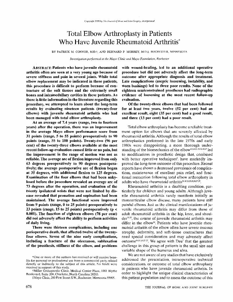
Copyright 1998 by The Journal of Bone and Joint Surgery, Incorporated
Total Elbow Arthroplasty in Patients Who Have Juvenile Rheumatoid Arthritis*
BY PATRICK M. CONNOR, M.D.t, AND BERNARD F. MORREY, M.D4, ROCHESTER, MINNESOTA
Investigation performed at the Mayo Clinic and Mayo Foundation, Rochester
ABSTRACT: Patients who have juvenile rheumatoid arthritis often are seen at a very young age because of severe stiffness and pain in several joints. While total elbow replacement may be indicated in these patients, this procedure is difficult to perform because of contracture of the soft tissues and the extremely small bones and intramedullary cavities in these patients. As there is little information in the literature regarding this procedure, we attempted to learn about the long-term results by evaluating nineteen patients (twenty-four elbows) with juvenile rheumatoid arthritis who had been managed with total elbow arthroplasty.
At an average of 7.4 years (range, two to fourteen years) after the operation, there was an improvement in the average Mayo elbow performance score from 31 points (range, 5 to 55 points) preoperatively to 90 points (range, 55 to 100 points). Twenty-two (96 per cent) of the twenty-three elbows available at the most recent follow-up evaluation caused little or no pain, but the improvement in the range of motion was not as reliable. The average arc of flexion improved from only 63 degrees preoperatively to 90 degrees postoperatively; the average postoperative arc of flexion began at 35 degrees, with additional flexion to 125 degrees. Examination of the four elbows that had been anky-losed before the procedure revealed an average arc of 73 degrees after the operation, and evaluation of the twenty ipsilateral wrists that were not limited by disease revealed that pronation and supination had been maintained. The average functional score improved from 9 points (range, 0 to 25 points) preoperatively to 23 points (range, 15 to 25 points) postoperatively (p < 0.001). The function of eighteen elbows (78 per cent) did not adversely affect the ability to perform activities of daily living.
There were thirteen complications, including one perioperative death, that affected twelve of the twenty-four elbows. Seven of the nine early complications, including a fracture of the olecranon, subluxation of the prosthesis, stiffness of the elbow, and problems
*One or more of the authors has received or will receive benefits for personal or professional use from a commercial party related directly or indirectly to the subject of this article. No funds were received in support of this study.
tMiller Orthopaedic Clinic, Medical Center Plaza, 1001 Blythe Boulevard, Suite 200, Charlotte, North Carolina 28203.
tMayo Clinic, 200 First Street S.W., Rochester, Minnesota 55905.
with wound-healing, led to an additional operative procedure but did not adversely affect the long-term outcome after appropriate diagnosis and treatment. Late complications (aseptic loosening, instability, and worn bushings) led to three poor results. None of the eighteen semiconstrained prostheses had radiographic evidence of loosening at the most recent follow-up evaluation.
Of the twenty-three elbows that had been followed for at least two years, twelve (52 per cent) had an excellent result, eight (35 per cent) had a good result, and three (13 per cent) had a poor result.
Total elbow arthroplasty has become a reliable treatment option for elbows that are severely affected by rheumatoid arthritis. Although the results of total elbow arthroplasties performed in the late 1970s and early 1980s were disappointing, a more thorough understanding of the biomechanics of the elbow4914'21-31'3245'47 led to modifications in prosthetic design that, combined with better operative techniques3, have markedly improved the long-term outcome of this procedure. Recent reports have shown a decreased prevalence of complications, maintenance of excellent pain relief, and functional restoration following total elbow arthroplasty in adults who have rheumatoid arthritis111822'23'33'34'3739.
Rheumatoid arthritis is a disabling condition, particularly for children and young adults. Although juvenile rheumatoid arthritis rarely manifests initially as monoarticular elbow disease, many patients have stiff painful elbows. Just as the clinical manifestations of juvenile rheumatoid arthritis may differ from those of adult rheumatoid arthritis in the hip, knee, and shoulder35 55, the course of juvenile rheumatoid arthritis may differ in the elbow26. Patients who have juvenile rheumatoid arthritis of the elbow often have severe osseous atrophy, deformity, and soft-tissue contractures that need special consideration and may adversely affect outcome6811162527. We agree with Dee7 that the greatest challenge in this group of patients is the small size and variable shape of the humerus and ulna.
We are not aware of any studies that have exclusively addressed the presentation, intraoperative technical considerations, or outcome of total elbow arthroplasty in patients who have juvenile rheumatoid arthritis. In order to highlight the unique clinical characteristics of this patient population and to assess the outcbme of this
678 THE JOURNAL OF BONE AND JOINT SURGERY

TOTAL ELBOW ARTHROPLASTY IN PATIENTS WHO HAVE JUVENILE RHEUMATOID ARTHRITIS 679
FIG. 1
Radiograph demonstrating a typical ankylosed elbow due to juvenile rheumatoid arthritis (grade-V involvement according to the modification of the classification system for rheumatoid arthritis37).
intervention, we reviewed our experience with total elbow arthroplasty in nineteen consecutive patients with juvenile rheumatoid arthritis who were managed over a twelve-year period.
Materials and Methods
Patient Demographics
Twenty-four consecutive total elbow arthroplasties were performed from September 1982 to October 1994 at the Mayo Clinic, Rochester, Minnesota, in nineteen patients who had juvenile rheumatoid arthritis. A diagnosis of juvenile rheumatoid arthritis had been made, in accordance with the accepted criteria of the American Rheumatology Association1, if arthritis had been present in at least one joint for six weeks to three months when the patient was less than sixteen years old. There were eighteen women and one man who had an average age of thirty-six years (range, twenty-five to fifty-six years). The average age at which the juvenile rheumatoid arthritis had been diagnosed was eleven years (range, two to fifteen years).
FIG. 2-A
Illustrations demonstrating how the collateral ligaments (Fig. 2-A) motion. The implant may be inserted slightly more proximally in the h
The severity of the disease process was assessed with the classification system of the American Rheumatology Association57. Two patients (two elbows; 8 per cent) were in class II (able to perform normal activities despite a handicap, discomfort, or limited motion), sixteen patients (twenty-one elbows; 88 per cent) were in class III (unable to perform all or most duties of the patient's usual occupation or self-care), and one patient (one elbow; 4 per cent) was in class IV (largely or completely incapacitated, with little or no ability to perform self-care and confined to a bed or wheelchair)57. Fifteen patients had had an average of five (range, two to eight) previous arthroplasties or arthrodeses of other major joints. Twenty-three elbows (96 per cent) were in patients who had a symptomatic or functionally limited contralateral elbow, and nineteen (79 per cent) were in patients who had pain or limitation of motion of the ipsilateral shoulder. Fifteen patients (79 per cent) had been managed with steroids at some point in the preoperative disease process, and six of these patients were taking steroids orally at the time of the elbow arthroplasty.
The indication for the total elbow arthroplasty was refractory pain in the elbow or limited motion that interfered with function, or both, in association with concomitant destruction of the ulnohumeral joint. Pain was the primary indication for the procedure in twenty elbows (83 per cent), and ankylosis or less than 30 degrees of motion was the primary indication in four. Seven patients had had a previous synovectomy and excision of the radial head on the side of the affected elbow; two of these patients also had had a failed interposition arthroplasty.
A semiconstrained Coonrad-Morrey total elbow prosthesis (Zimmer, Warsaw, Indiana) was used in eighteen elbows (75 per cent); the design of the implant and subsequent modifications have been described previously44. An unconstrained, resurfacing prosthesis was used in six elbows (Capitellocondylar911 [Johnson and
FIG. 2-B
and anterior aspect of the capsule (Fig. 2-B) are released to maximize iumerus by removal of the olecranon fossa to gain additional extension.
VOL. 80-A, NO. 5, MAY 1998

680 P. M. CONNOR AND B. F. MORREY
FIG. 3-A
Anteroposterior radiograph of an elbow in which little condylar bone remains. The stem fills most of the intramedullary canal.
Johnson Products, Orthopaedic Division, New Brunswick, New Jersey] in four and Pritchard ERS4849 [DePuy] in two) early in the study period. A resurfacing prosthesis was not used in elbows that had gross instability, severe contractures, osseous ankylosis, or severe osseous deformity. In addition, no individualized so-called custom prostheses were used.
Two systems were used for evaluation. The Mayo elbow performance score3744 was employed to document subjective, objective, and functional characteristics before and after the total elbow arthroplasty. This system places the greatest emphasis on pain relief (45 points) and the ability of the patient to perform functional activities (25 points); assessments of motion (20 points) and stability (10 points) are also included. The results are defined as excellent (90 to 100 points), good (75 to 89 points), fair (60 to 74 points), or poor (less than 60 points). Satisfactory data were available to allow the calculation of a preoperative score for all twenty-four elbows.
The second system was used to evaluate the extent of preoperative radiographic and pathological changes in the elbow. Grade I indicates no radiographic changes
with the exception of osteoporosis; grade II, narrowing of the joint space with the architecture intact; grade III, alteration of the architecture of the joint; and grade IV, gross destruction of the joint37. A grade of V was established for the present study to indicate elbows that had radiographic ankylosis, as defined by the absence of an identifiable ulnohumeral joint on the anteroposterior and lateral radiographs or by the presence of mature osseous trabeculation crossing the ulnohumeral joint (Fig. 1). Three elbows (13 per cent) had grade-II involvement, fifteen (63 per cent) had grade-Ill involvement, two (8 per cent) had grade-IV involvement, and four (17 per cent) had grade-V involvement.
Operative Technique
The technique of total elbow arthroplasty for the rheumatoid elbow has been well described39. In order to avoid potential complications, it is essential that the surgeon spend time planning preoperatively for the anatomical deformities and technical challenges that are common with elbows affected by juvenile rheumatoid arthritis. Emphasis is placed on ensuring that an implant is available to accommodate the small intramedullary canal.
A posterior incision is used, and the ulnar nerve is routinely transferred anteriorly to a subcutaneous position. The triceps is then released in a subperiosteal manner from the olecranon, in continuity with the ulnar periosteum and the fascia of the forearm along with the anconeus3. Elbows affected by juvenile rheumatoid arthritis are characterized by very small, fragile bones; thus, care should be taken during the exposure to avoid excessive forces about the elbow that could cause a fracture. If there is osseous ankylosis, a microsagittal saw or a small osteotome is used to reestablish the joint line after osseous landmarks have been identified. Care
FIG. 3-B
Lateral radiograph of the elbow, showing the ulnar and humeral components filling most of the intramedullary canals.
THE JOURNAL OF BONE AND JOINT SURGERY

TOTAL ELBOW ARTHROPLASTY IN PATIENTS WHO HAVE JUVENILE RHEUMATOID ARTHRITIS 6 8 1
FIG. 4 Photograph of a specially designed small-circumference ulnar component (arrow), which is cut to length to fit into the very small ulnar canal
that is sometimes encountered.
is taken to create the osteotomy at the proper center of rotation of the ulnohumeral joint to maximize the biomechanical function of the prosthetic elbow1416-31.
In elbows with severe soft-tissue contractures or osseous ankylosis, or both, circumferential capsular and collateral ligament releases are necessary to maximize postoperative motion and function. As the stability of unconstrained, resurfacing prostheses depends on functional collateral ligaments and proper soft-tissue balancing, these prostheses are not typically indicated in this clinical situation. Rather, a semiconstrained prosthesis is used, the collateral ligaments are released, and the anterior aspect of the capsule is completely excised (Figs. 2-A and 2-B) and further reflected from the distal aspect of the humerus with an osteotome or a blunt periosteal elevator. These maneuvers and this prosthesis allow the greatest possible motion and stability for patients who have severe preoperative stiffness.
When a resurfacing prosthesis is to be implanted, the treatment of the radial head depends on the particular prosthesis being used. For the semiconstrained Coonrad-Morrey prosthesis, the periphery of the radial head is excised with a small rongeur to create a resection arthroplasty of the proximal radioulnar joint in patients who have perioperative pain with rotation of the forearm. The radius is not routinely shortened.
The intramedullary canals of the humerus and the ulna characteristically are very narrow or are completely obliterated in patients who have severe juvenile rheumatoid arthritis (Figs. 3-A and 3-B). Thus, care must be taken in the identification and preparation of these canals. Occasionally, if the canal has been obliterated by cortical bone, it is necessary to use a small burr to create a new canal, especially in the ulna. If this is done, it is important to stay centered on the ulna and to remain within the confines of the cortical bone. A small (four-millimeter) cannulated flexible reamer that employs a guide-pin is used for this purpose. The Coonrad-Morrey implant system includes an extra-small ulnar compo
nent with an intramedullary circumference of only 1.5 centimeters (Fig. 4). This component is often used in elbows affected by juvenile rheumatoid arthritis that have tiny bones. Although the use of individualized custom prostheses was not necessary in the elbows in our study, it is important to have an appropriate inventory of prosthetic sizes available to address the small intramedullary canal. In four of the eighteen elbows treated with a Coonrad-Morrey prosthesis, the stem was slightly modified further with a cam-lever bending device because an angular change was needed.
The cementing technique used in patients who have juvenile rheumatoid arthritis differs little from standard techniques. Tobramycin-impregnated cement is routinely used in an effort to lessen the likelihood of infection, and the cement for both the humeral and the ulnar component is inserted with an intramedullary injection system. As many patients who have juvenile rheumatoid arthritis have involvement of the ipsilateral shoulder, which may eventually necessitate prosthetic replacement, care is taken to limit the level of the cement injected in the humerus by placing bone graft down the canal; also, a short, ten-centimeter humeral component is typically used.
After repair of the extensor mechanism, as described by one of us (B. F. M.) and Bryan3, and routine closure, the elbow is placed in a compressive dressing and elevated in a vertical elbow sling for twenty-four hours. The patient is then allowed to use the extremity as tolerated for activities of daily living. Although patients who have juvenile rheumatoid arthritis are encouraged to use the elbow in the extremes of flexion and extension to enhance postoperative motion, as is the case for all patients who have an elbow replacement, no formal physical therapy is provided.
Follow-up Evaluation
One patient, a forty-three-year-old woman who had a thirty-one-year history of juvenile rheumatoid arthri-
VOL. 80-A, NO. 5, MAY 1998

682 P. M. CONNOR AND B. F. MORREY
FIG. 5-A Anteroposterior and lateral radiographs showing destruction of the elbow and a small ulnar canal.
tis and a twenty-year history of steroid use, died on the first postoperative day of an acute cardiac embolism. This left eighteen patients (twenty-three elbows) who were followed for a minimum of two years. Six elbows (26 per cent) were most recently assessed at our institution and seventeen (74 per cent) were most recently assessed by a local orthopaedic surgeon (nine elbows) or on the basis of a questionnaire and radiographs (eight elbows). The mean duration of follow-up was 7.4 years (range, two to fourteen years).
All elbows were evaluated with anteroposterior and lateral radiographs, at an average of 6.1 years (minimum, two years) postoperatively. The radiographs were assessed for progressive radiolucent lines as well as for the presence and incorporation of bone graft that had been placed between the anterior flange of the Coonrad-Morrey prosthesis and the distal aspect of the humerus.
Statistical A nalysis
A one-tailed t test was used to determine the significance of both discrete and continuous variables. Significance was assigned when the probability that the difference was due to chance was less than 0.05.
Results
Clinical Results
The result at the latest follow-up evaluation was excellent for twelve elbows (52 per cent), good for eight (35 per cent), and poor for three (13 per cent), according to the Mayo elbow performance score. The average preoperative score was 31 points (range, 5 to 55 points) and the average postoperative score was 90 points (range, 55 to 100 points) (p < 0.001). All three elbows that had a poor result had a late complication that necessitated a major revision procedure. The
marked improvement in the score was primarily due to the relief of pain.
Pain
Preoperatively, eighteen (78 per cent) of the twenty-three elbows caused severe pain, four (17 per cent) caused moderate pain, and one (4 per cent) caused mild pain. At the most recent follow-up evaluation, seventeen elbows (74 per cent) caused no pain, five (22 per cent) caused mild pain, and one (4 per cent) caused moderate pain. None of the elbows caused severe pain postoperatively. The average score for the pain component of the elbow performance score (maximum, 45 points) improved from 4 points preoperatively to 41 points at the most recent follow-up evaluation (p < 0.001).
Range of Motion
The average preoperative arc of flexion was 63 degrees (range, 0 to 125 degrees) and began at an average of 44 degrees (range, 0 to 90 degrees), with additional flexion to an average of 107 degrees (range, 75 to 150 degrees). Postoperatively, the arc of flexion was 90 degrees (range, 45 to 150 degrees), beginning at 35 degrees (range, 5 to 70 degrees) with additional flexion to 125 degrees (range, 90 to 150 degrees). The average increase in the arc of flexion was 27 degrees (an average increase of 9 degrees of extension and 18 degrees of flexion). With the numbers available for study, we could not detect a significant improvement in postoperative motion.
The average postoperative arc of flexion of the four elbows that had been ankylosed preoperatively was 73 degrees (range, 45 to 95 degrees). For the seventeen elbows that had had an arc of flexion of less than 100 degrees preoperatively (average, 45 degrees; range, 0 to 85 degrees), the arc improved 32 degrees, to 77 degrees (range, 45 to 110 degrees), postoperatively. Only ten
THE JOURNAL OF BONE AND JOINT SURGERY

TOTAL ELBOW ARTHROPLASTY IN PATIENTS WHO HAVE JUVENILE RHEUMATOID ARTHRITIS 6 8 3
FIG. 5-B Anteroposterior and lateral radiographs, made six years postoperatively, showing a well seated implant. The arc of flexion was 30 to 125
degrees.
elbows (43 per cent), however, had a postoperative arc of 100 degrees or more. As a result of the operation, seventeen elbows (74 per cent) had an improvement in motion, one elbow had the same motion, and five (22 per cent) lost motion.
The elbows that were treated with a semiconstrained prosthesis had had an average preoperative arc of flexion of 57 degrees (range, 0 to 120 degrees), which began at an average of 48 degrees with additional flexion to 105 degrees. Postoperatively, the average arc of flexion improved to 85 degrees (range,45 to 120 degrees), beginning at an average of 36 degrees with additional flexion to 121 degrees. The average preoperative arc of flexion of the elbows that were treated with a resurfacing arthroplasty had been 88 degrees (range, 50 to 125 degrees), beginning at an average of 27 degrees with additional flexion to 115 degrees. Postoperatively, the average arc improved to 107 degrees (range, 70 to 150 degrees), beginning at an average of 30 degrees with additional flexion to 137 degrees. Thus, the average arc of flexion improved 28 degrees for the elbows that had been treated with a semiconstrained prosthesis and 19 degrees for those that had been treated with a resurfacing prosthesis (p < 0.02). The fourteen elbows that had had an arc of flexion of less than 100 degrees before
treatment with a semiconstrained implant had an average improvement of 36 degrees (range, 0 to 95 degrees). The three elbows that had had an arc of less than 100 degrees before treatment with a resurfacing implant had an average improvement of 15 degrees (range, 0 to 35 degrees).
Three extremities had severe concomitant disease in the ipsilateral wrist that precluded improvement in pronation and supination after the elbow arthroplasty. When these three elbows were excluded, the average preoperative arc was 52 degrees (range, 0 to 80 degrees) of pronation to 36 degrees (range, 0 to 70 degrees) of supination. Postoperatively, the average arc was 61 degrees (range, 30 to 80 degrees) of pronation to 53 degrees (range, 10 to 80 degrees) of supination.
Stability
Preoperatively, two elbows (9 per cent) were grossly unstable and eight (35 per cent) were moderately unstable, as defined by 5 to 10 degrees of varus-valgus laxity. Four of the five elbows that had been treated with a resurfacing prosthesis were stable at the most recent follow-up evaluation; the fifth elbow, which had been moderately unstable preoperatively, was found to be grossly unstable after the resurfacing arthroplasty. No
VOL. 80-A, NO. 5, MAY 1998
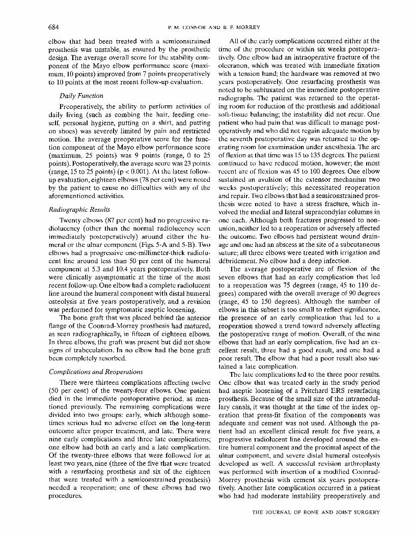
684 P. M. CONNOR AND B. F. MORREY
elbow that had been treated with a semiconstrained prosthesis was unstable, as ensured by the prosthetic design. The average overall score for the stability component of the Mayo elbow performance score (maximum, 10 points) improved from 7 points preoperatively to 10 points at the most recent follow-up evaluation.
Daily Function
Preoperatively, the ability to perform activities of daily living (such as combing the hair, feeding oneself, personal hygiene, putting on a shirt, and putting on shoes) was severely limited by pain and restricted motion. The average preoperative score for the function component of the Mayo elbow performance score (maximum, 25 points) was 9 points (range, 0 to 25 points). Postoperatively, the average score was 23 points (range, 15 to 25 points) (p < 0.001). At the latest follow-up evaluation, eighteen elbows (78 per cent) were noted by the patient to cause no difficulties with any of the aforementioned activities.
Radiographic Results
Twenty elbows (87 per cent) had no progressive ra-diolucency (other than the normal radiolucency seen immediately postoperatively) around either the humeral or the ulnar component (Figs. 5-A and 5-B). Two elbows had a progressive one-millimeter-thick radiolu-cent line around less than 50 per cent of the humeral component at 5.3 and 10.4 years postoperatively. Both were clinically asymptomatic at the time of the most recent follow-up. One elbow had a complete radiolucent line around the humeral component with distal humeral osteolysis at five years postoperatively, and a revision was performed for symptomatic aseptic loosening.
The bone graft that was placed behind the anterior flange of the Coonrad-Morrey prosthesis had matured, as seen radiographically, in fifteen of eighteen elbows. In three elbows, the graft was present but did not show signs of trabeculation. In no elbow had the bone graft been completely resorbed.
Complications and Reoperations
There were thirteen complications affecting twelve (50 per cent) of the twenty-four elbows. One patient died in the immediate postoperative period, as mentioned previously. The remaining complications were divided into two groups: early, which although sometimes serious had no adverse effect on the long-term outcome after proper treatment, and late. There were nine early complications and three late complications; one elbow had both an early and a late complication. Of the twenty-three elbows that were followed for at least two years, nine (three of the five that were treated with a resurfacing prosthesis and six of the eighteen that were treated with a semiconstrained prosthesis) needed a reoperation; one of these elbows had two procedures.
All of the early complications occurred either at the time of the procedure or within six weeks postoperatively. One elbow had an intraoperative fracture of the olecranon, which was treated with immediate fixation with a tension band; the hardware was removed at two years postoperatively. One resurfacing prosthesis was noted to be subluxated on the immediate postoperative radiographs. The patient was returned to the operating room for reduction of the prosthesis and additional soft-tissue balancing; the instability did not recur. One patient who had pain that was difficult to manage postoperatively and who did not regain adequate motion by the seventh postoperative day was returned to the operating room for examination under anesthesia. The arc of flexion at that time was 15 to 135 degrees. The patient continued to have reduced motion, however; the most recent arc of flexion was 45 to 100 degrees. One elbow sustained an avulsion of the extensor mechanism two weeks postoperatively; this necessitated reoperation and repair. Two elbows that had a semiconstrained prosthesis were noted to have a stress fracture, which involved the medial and lateral supracondylar columns in one each. Although both fractures progressed to nonunion, neither led to a reoperation or adversely affected the outcome. Two elbows had persistent wound drainage and one had an abscess at the site of a subcutaneous suture; all three elbows were treated with irrigation and debridement. No elbow had a deep infection.
The average postoperative arc of flexion of the seven elbows that had an early complication that led to a reoperation was 75 degrees (range, 45 to 110 degrees) compared with the overall average of 90 degrees (range, 45 to 150 degrees). Although the number of elbows in this subset is too small to reflect significance, the presence of an early complication that led to a reoperation showed a trend toward adversely affecting the postoperative range of motion. Overall, of the nine elbows that had an early complication, five had an excellent result, three had a good result, and one had a poor result. The elbow that had a poor result also sustained a late complication.
The late complications led to the three poor results. One elbow that was treated early in the study period had aseptic loosening of a Pritchard ERS resurfacing prosthesis. Because of the small size of the intramedullary canals, it was thought at the time of the index operation that press-fit fixation of the components was adequate and cement was not used. Although the patient had an excellent clinical result for five years, a progressive radiolucent line developed around the entire humeral component and the proximal aspect of the ulnar component, and severe distal humeral osteolysis developed as well. A successful revision arthroplasty was performed with insertion of a modified Coonrad-Morrey prosthesis with cement six years postoperatively. Another late complication occurred in a patient who had had moderate instability preoperatively and
THE JOURNAL OF BONE AND JOINT SURGERY

TOTAL ELBOW ARTHROPLASTY IN PATIENTS WHO HAVE JUVENILE RHEUMATOID ARTHRITIS 685
who had been managed with a Capitellocondylar resurfacing prosthesis. Six months postoperatively, the elbow was symptomaticaHy unstable. A revision procedure was performed in an attempt to balance the soft-tissues; however, instability persisted. At the time of writing, the patient was considering revision to a semiconstrained prosthesis. The third late complication was in a patient who had an excellent result for three years and then was seen because of swelling of the elbow and a gradually decreasing range of motion. Radiographs revealed asymmetry of the ulnohumeral articulation suggestive of wear of the polyethylene bushings but no evidence of loosening of the components. At the time of the revision, the bushings were worn and there was polyethylene-induced synovitis and wear of the articulating titanium pin; the humeral and ulnar components, however, remained well fixed. The polyethylene bushings were replaced and the titanium pin was exchanged for a stainless-steel one. Although the patient was doing well at the time of the most recent follow-up, the result was considered poor because of the need for a late revision procedure.
Discussion
Juvenile rheumatoid arthritis is a debilitating condition that affects nearly a quarter of a million children in the United States1-55. The severe nature of this disease is evidenced by the fact that fifteen patients in the present study had had an average of five previous arthroplasties or arthrodeses involving a major joint by an average age of thirty-six years. The hip and knee are the joints most frequently affected by juvenile rheumatoid arthritis; however, the elbow and shoulder often become involved in young adulthood35. At the time of presentation, 96 per cent of the elbows in the present study were in patients who had a symptomatic or functionally limited contralateral elbow and 79 per cent were in patients who had a symptomatic or functionally limited ipsilateral shoulder.
The arthritic elbows of patients who have juvenile rheumatoid arthritis differ in several important aspects from those of patients who have adult rheumatoid arthritis. Although pain is the most common symptom in both groups, elbows affected by juvenile rheumatoid arthritis have a distinct predilection for stiffness. In two recent large studies of total elbow arthroplasty in patients who had adult rheumatoid arthritis"'37, the average preoperative arc of flexion was 35 to 118 degrees (an 83-degree arc). In the current study, the average preoperative arc was only 44 to 107 degrees (a 63-degree arc). In addition, seventeen elbows (71 per cent) in the current study had a preoperative arc of flexion of less than 100 degrees and four had complete ankylosis. For this reason, we added a grade-V radiographic classification, indicating ankylosis, to the system described previously37.
In elbows affected by juvenile rheumatoid arthritis, the underdeveloped bones, which have an extremely
narrow or completely obliterated intramedullary canal, present one of several obvious technical difficulties involved in the performance of a total elbow arthroplasty in these patients. Forcing a typically sized implant into these bones either causes an intraoperative fracture or prevents the component from being completely seated; if the component is not completely seated, the anatomical center of rotation cannot be re-created and the opportunity for improved motion is decreased'416'31. Although individualized custom prostheses have been advocated for elbows with similar anatomical deformities"16'2327, they were not found to be necessary in the present series; thus, issues involving very high cost were avoided. The modified Coonrad-Morrey prosthesis provides a small humeral component and an extra-small ulnar component that can easily be shortened during the operation, thus enabling adequate sizing in these patients (Fig. 4). Regardless of the prosthesis that is used, however, the importance of thorough preoperative planning for elbow arthroplasty in patients who have juvenile rheumatoid arthritis cannot be overemphasized.
Satisfactory pain relief was nearly universal in the present study. Twenty-two (96 per cent) of the twenty-three elbows had no or mild pain at the time of the most recent follow-up. Two of the three patients who eventually needed a revision arthroplasty had had a painless elbow for three and five years. These results with regard to pain relief compare favorably with or are better than those reported in published series of elbow arthroplasty for adults who had rheumatoid ar-thritis210'"20'3033'3437'4651-53'56 or another diagnosis1517'24'38-42. The reliable achievement of satisfactory pain relief should be considered when other potential operative options are reviewed for an elbow that is severely affected by juvenile rheumatoid arthritis12'192936'54.
However, marked improvement in the range of motion after elbow arthroplasty in patients who have juvenile rheumatoid arthritis is not as reliable. Overall, seventeen elbows (74 per cent) had an improvement in the range of motion and five (22 per cent) had less motion postoperatively than they had had preoperatively. The average 90-degree postoperative arc of flexion represents an improvement of only 27 degrees compared with the preoperative average. These results are not as favorable as the expected improvements in motion following arthroplasty of an elbow affected by adult rheumatoid arthritis1137. Although some authors have not noted less improvement in postoperative motion after elbow arthroplasty in patients who have juvenile rheumatoid arthritis", others816-2627, including us, have reported such a finding.
Although the gains in postoperative motion were modest, functional ability was consistently enhanced. It has been shown that most daily activities require approximately 100 degrees of flexion of the elbow (from 30 to 130 degrees) and 100 degrees of rotation of the forearm (from 50 degrees of pronation to 50 degrees
VOL. 80-A, NO. 5, MAY 1998

686 P. M. CONNOR AND B. F. MORREY
of supination)43. Despite maintenance of adequate rotation of the forearm, less than half of the elbows in the present series had a postoperative arc of flexion of 100 degrees or more. Eighteen elbows (78 per cent), however, did not limit the performance of any activities of daily living. This somewhat unexpected result may be attributable to the excellent postoperative relief of pain, which allowed uninhibited use of the extremity.
A relatively high overall prevalence of complications and reoperations has been associated with total elbow arthroplasty5'10'1'''18'23'29-3'''40-'", with the prevalence depending on the severity of the condition being treated and the experience of the surgeon40. In recent years, however, rates of complications have decreased coincident with modifications in prosthetic design and improvements in operative technique3739. Most of the complications reported in the present study were diagnosed and treated in the early postoperative period and had no adverse effect on the long-term outcome. An error in judgment involving resurfacing implants contributed to two of the three late complications. One of these complications involved aseptic loosening, after five years, of a humeral component that had been inserted without cement and the other involved an elbow with moderate preoperative instability that, in retrospect, did not have the ligamentous support necessary for an unconstrained implant. To avoid these complications, the humeral and ulnar components should routinely be inserted with cement and a semiconstrained prosthesis should be used when there is preoperative instability or when extensive soft-tissue releases are needed to gain motion.
No definitive comparison of resurfacing and semiconstrained elbow arthroplasties can or should be made on the basis of this study. Resurfacing prostheses depend on adequate bone stock and ligamentous integrity for stability and function"'28'313239. In elbows that are severely affected by juvenile rheumatoid arthritis, with fibrous or osseous ankylosis, the need for extensive, circumferential capsular and ligamentous releases is tempered by concern about instability. However, if there are adequate bone stock, ligamentous support, and preop
erative motion, a resurfacing prosthesis can provide satisfactory results.
As stability is an inherent aspect of the semiconstrained prosthetic design, the capsule and ligaments can be released extensively in a stiff or ankylosed elbow without concern regarding postoperative stability16. In fact, use of the semiconstrained device resulted in a significantly greater improvement in motion (p < 0.02) in this study. In addition, the semiconstrained elbow arthroplasty reliably restores both stability and function if there is gross osseous destruction and a flail extremity1350. O'Driscoll et al.41 showed that the functional laxity of the Coonrad-Morrey prosthesis is consistently less than its inherent structural laxity, thus explaining the low rate of aseptic loosening of this implant when it is used to treat rheumatoid arthritis37, traumatic conditions38, and other diagnoses39. This observation is supported by our finding of no clinical or radiographic evidence of loosening of the semiconstrained prostheses at the most recent follow-up evaluation. Furthermore, as the present study has, to our knowledge, the longest duration of follow-up (average, 7.4 years) of any series of total elbow arthroplasties in the current literature, the implications are particularly relevant for patients with juvenile rheumatoid arthritis who have an elbow arthroplasty when they are young.
Total elbow arthroplasty in patients who have juvenile rheumatoid arthritis provides rewarding pain relief and improvements in function. However, the favorable restoration of motion that is seen after elbow arthroplasty in patients who have adult rheumatoid arthritis cannot be expected. As many elbows affected by juvenile rheumatoid arthritis have preexisting anatomical deformities, there are complex and challenging technical considerations and thorough preoperative planning must be emphasized. This is particularly relevant with regard to determining the size and type of the prosthetic implant. Finally, experience with operations on the elbow, and with prosthetic replacement in particular, will increase the likelihood of a satisfactory outcome.
References 1. Brewer, E. J., Jr.; Bass, J. C; Cassidy, J. T.; Duran, B. S.; Fink, C. W.; Jacobs, J. C; Markowitz, M.; Reynolds, W. E.; Schaller, J.; Stillman,
J. S.; and Wallace, S. L.: Criteria for the classification of juvenile rheumatoid arthritis. Bull. Rheumat. Dis., 23: 712-719,1972. 2. Brumfield, R. H., Jr.; Kuschner, S. H.; Gellman, H.; Redix, L.; and Stevenson, D. V.: Total elbow arthroplasty. J. Arthroplasty, 5:
359-363,1990. 3. Bryan, R. S., and Morrey, B. F.: Extensive posterior exposure of the elbow. A triceps-sparing approach. Clin. Orthop., 166:188-192,1982. 4. Coonrad, R. W.: Comments on the historical milestones in the development of elbow arthroplasty: indications and complications. In
Instructional Course Lectures, American Academy of Orthopaedic Surgeons. Vol. 40, pp. 51-55. Park Ridge, Illinois, American Academy of Orthopaedic Surgeons, 1991.
5. Davis, R. F;; Weiland, A. J.; Hungerford, D. S.; Moore, J. R.; and Volenec-Dowling, S.: Nonconstrairted total elbow arthroplasty. Clin. Orthop., Ill: 156-160,1982.
6. Dee, R.: Total replacement arthroplasty of the elbow for rheumatoid arthritis. J. Bone and Joint Surg., 54-B(l): 88-95,1972. 7. Dee, R.: Total replacement of the elbow joint. Orthop. Clin. North America, 4:415-433,1973. 8. Dennis, D. A.; Clayton, M. L.; Ferlic, D. C; Stringer, E. A.; and Bramlett, K. W.: Capitello-condylar total elbow arthroplasty for
rheumatoid arthritis. / Arthroplasty, 5 (Supplement): S83-S88,1990. 9. Ewald, F. C: Total elbow replacement. Orthop. Clin. North America, 6: 685-696,1975.
10. Ewald, F. C; Scheinberg, R. D.; Poss, R.; Thomas, W. H.; Scott, R. D.; and Sledge, C. B.: Capitellocondylar total elbow arthroplasty. Two to five-year follow-up in rheumatoid arthritis. / Bone and Joint Surg., 62-A: 1259-1263, Dec. 1980.
THE JOURNAL OF BONE AND JOINT SURGERY
TOTAL ELBOW ARTHROPLASTY IN PATIENTS WHO HAVE JUVENILE RHEUMATOID ARTHRITIS 687
11. Ewald, F. C; Simmons, E. D.; Sullivan, J. A.; Thomas, W. H.; Scott, R. D.; Poss, R.; Thornhill, T. S.; and Sledge, C. B.: Capitellocondylar total elbow replacement in rheumatoid arthritis. Long-term results. /. Bone and Joint Surg., IS-A: 498-507, April 1993.
12. Ferlic, D. C; Patched, C. E.; Clayton, M. L.; and Freeman, A. C: Elbow synovectomy in rheumatoid arthritis. Long-term results. Clin. Orthop., 220:119-125,1987.
13. Figgie, H. E., Ill; Inglis, A. E.; and Mow, C: Total elbow arthroplasty in the face of significant bone stock or soft tissue losses. Preliminary results of custom-fit arthroplasty. J. Arthroplasty, 1:71-81,1986.
14. Figgie, H. E., Ill; Inglis, A. E.; and Mow, C : A critical analysis of alignment factors affecting functional outcome in total elbow arthroplasty. J. Arthroplasty, 1:169-173,1986.
15. Figgie, H. E., Ill; Inglis, A. E.; Ranawat, C. S.; and Rosenberg, G. M.: Results of total elbow arthroplasty as a salvage procedure for failed elbow reconstructive operations. Clin. Orthop., 219:185-193,1987.
16. Figgie, M. P.; Inglis, A. E.; Mow, C. S.; and Figgie, H. E., Ill: Total elbow arthroplasty for complete ankylosis of the elbow. /. Bone and Joint Surg., 71-A: 513-520, April 1989.
17. Figgie, M. P.; Inglis, A. E.; Mow, C. S.; and Figgie, H. E., Ill: Salvage of non-union of supracondylar fractures of the humerus by total elbow arthroplasty./ Bone and Joint Surg., 71-A: 1058-1065, Aug. 1989.
1.8. Friedman, R. J.; Lee, D. E.; and Ewald, F. C: Nonconstrained total elbow arthroplasty. Development and results in patients with functional class IV rheumatoid arthritis. J. Arthroplasty, 4:31-37,1989.
19. Froimson, A. L.; Silva, J. E.; and Richey, D. G.: Cutis arthroplasty of the elbow joint. /. Bone and Joint Surg., 58-A: 863-865, Sept. 1976. 20. Garrett, J. C; Ewald, F. C; Thomas, W. H.; and Sledge, C. B.: Loosening associated with G.S.B. hinge total elbow replacement in patients
with rheumatoid arthritis. Clin. Orthop., 127:170-174,1977. 21. Goel, V. K.; Lee, I. K.; and Blair, W. F.: Stress distribution in the ulna following a hinged elbow arthroplasty. A finite element analysis.
/. Arthroplasty, 4:163-171,1989. 22. Goldberg, V. M.; Figgie, H. E., Ill; Inglis, A. E.; and Figgie, M. P.: Current concepts review. Total elbow arthroplasty. J. Bone and Joint
Surg., 70-A: 778-783, June 1988. 23. Gschwend, N.; Simmen, B. R.; and Matejovsky, Z.: Late complications in elbow arthroplasty. /. Shoulder and Elbow Surg., 5:86-96,1996. 24. Inglis, A. E.: Revision surgery following a failed total elbow arthroplasty. Clin. Orthop., 170:213-218,1982. 25. Inglis, A. E.: Total elbow arthroplasty advances and techniques. Tech. Orthop., 6:19-26,1991. 26. Inglis, A. E., and Figgie, M. P.: Rheumatoid arthritis. In The Elbow and Its Disorders, edited by B. F. Morrey. Ed. 2, p. 753. Philadelphia,
W. B. Saunders, 1993. 27. Inglis, A. E., and Pellicci, P. M.: Total elbow replacement. J. Bone and Joint Surg., 62-A: 1252-1258, Dec. 1980. 28. Itoi, E.; King, G. J. W.; Niebur, G. L.; Morrey, B. F.; and An, K.-N.: Malrotation of the humeral component of the capitellocondylar total
elbow replacement is not the sole cause of dislocation. J. Orthop. Res., 12:665-671,1994. 29. Jonsson, B., and Larsson, S. E.: Elbow arthroplasty in rheumatoid arthritis. Function after 1-2 years in 20 cases. Acta Orthop. .
Scandinavica, 61: 344-347,1990. 30. Kasten, M. D., and Skinner, H. B.: Total elbow arthroplasty. An 18-year experience. Clin. Orthop., 290:177-188,1993. 31. King, G. J.; Glauser, S. J.; Westreich, A.; Morrey, B. F.; and An, K. N.: In vitro stability of an unconstrained total elbow prosthesis.
Influence of axial loading and joint flexion angle. J. Arthroplasty, 8:291-298,1993. 32. King, G. J. W.; Itoi, E.; Niebur, G. L.; Morrey, B. E; and An, K. N.: Motion and laxity of the capitellocondylar total elbow prosthesis.
J. Bone and Joint Surg, 76-A: 1000-1008, July 1994. 33. Kraay, M. J.; Figgie, M. P.; Inglis, A. E.; Wolfe, S. W.; and Ranawat, C. S.: Primary semiconstrained total elbow arthroplasty. Survival
analysis of 113 consecutive cases../. Bone and Joint Surg., 76-B(4): 636-640,1994. 34. Kudo, H.; Iwano, K.; and Prefecture, K.: Total elbow arthroplasty with a non-constrained surface-replacement prosthesis in patients who
have rheumatoid arthritis. A long-term follow-up study. J. Bone and Joint Surg., 12-A: 355-362, March 1990. 35. Levine, W. N.; Scott, R. D.; Sledge, C. B.; Thomas, W.; and Thornhill, T. S.: Shoulder arthroplasty in juvenile rheumatoid arthritis: mean
12-year follow-up. Unpublished data. 36. Ljung, P.; Jonsson, K.; Larsson, K.; and Rydholm, U.: Interposition arthroplasty of the elbow with rheumatoid arthritis. / Shoulder and
Elbow Surg, 5: 81-85,1996. 37. Morrey, B. E, and Adams, R. A.: Semiconstrained arthroplasty for the treatment of rheumatoid arthritis of the elbow. J. Bone and Joint
Surg., 74-A: 479-490, April 1992. 38. Morrey, B. F., and Adams, R. A.: Semiconstrained elbow replacement for distal humeral nonunion. /. Bone and Joint Surg., 77-B(l):
67-72,1995. 39. Morrey, B. F., and Adams, R. A.: Semiconstrained devices. In Reconstructive Surgery of the Joints, edited by B. F. Morrey. Ed. 2, vol. 1, pp.
515-537. New York, Churchill Livingstone, 1996. 40. Morrey, B. F., and Bryan, R. S.: Complications of total elbow arthroplasty. Clin. Orthop., 170: 204-212,1982. 41. Morrey, B. F., and Bryan, R. S.: Infection after total elbow arthroplasty. J. Bone and Joint Surg., 65-A: 330-338, March 1983. 42. Morrey, B. F., and Bryan, R. S.: Revision total elbow arthroplasty. J. Bone and Joint Surg., 69-A: 523-532, April 1987. 43. Morrey, B. F., and Chao, E. Y. S.: Passive motion of the elbow joint. A biomechanical analysis. /. Bone and Joint Surg., 58-A: 501-508,
June 1976. 44. Morrey, B. F.; Adams, R. A.; and Bryan, R. S.: Total replacement for post-traumatic arthritis of the elbow. J. Bone and Joint Surg.,
73-B(4): 607-612,1991. 45. Morrey, B. E; Askew, L. J.; An, K. N.; and Chao, E. Y.: A biomechanical study of normal functional elbow motion. / Bone and Joint
Surg, 63-A: 872-877, July 1981. 46. Morrey, B. F.; Bryan, R. S.; Dobyns, J. H.; and Linscheid, R. L.: Total elbow arthroplasty. A five-year experience at the Mayo Clinic.
/. Bone and Joint Surg., 63-A: 1050-1063, Sept. 1981. 47. O'Driscoll, S. W.; An, K.-N.; Korinek, S.; and Morrey, B. F.: Kinematics of semiconstrained total elbow arthroplasty. J. Bone and Joint
Surg., 74-B(2): 297-299,1992. 48. Pritchard, R. W.: Long-term follow-up study: semi-constrained elbow prosthesis. Orthopedics, 4:151-155,1981. 49. Pritchard, R. W.: Total elbow joint arthroplasty in patients with rheumatoid arthritis. Sem. Arthrit. and Rheumat., 21:24-29,1991. 50. Ramsey, M. L., and Morrey, B. E: Total elbow arthroplasty for dysfunctional instability of the elbow. Unpublished data.
VOL. 80-A, NO. 5, MAY 1998

6 8 8 P. M. CONNOR AND B. F. MORREY
51. Rosenberg, G. M., and Turner, R. H.: Nonconstrained total elbow arthroplasty. Clin. Orthop., 187:154-162,1984. 52. Ruth, J. T., and Wilde, A. H.: Capitellocondylar total elbow replacement. A long-term follow-up study. J. Bone and Joint Surg., 74-A:
95-100, Jan. 1992. 53. Rydholm, U.; Tjornstrand, B.; Pettersson, H.; and Lidgren, L.: Surface replacement of the elbow in rheumatoid arthritis. Early results
with the Wadsworth prosthesis../. Bone and Joint Surg., 66-B(5): 737-741,1984. 54. Rymaszewski, L. A.; MacKay, L; Amis, A. A.; and Miller, J. H.: Long-term effects of excision of the radial head in rheumatoid arthritis.
J. Bone and Joint Surg., 66-B(l): 109-113,1984. 55. Scott, R. D., and Sledge, C. B.: The surgery of juvenile rheumatoid arthritis. In Textbook of Rheumatology, edited by W. N. Kelley, E. D.
Harris, Jr., S. Ruddy, and C. B. Sledge. Ed. 2, vol. 2, pp. 1910-1916. Philadelphia, W. B. Saunders, 1985. 56. Souter, W. A.: Arthroplasty of the elbow with particular reference to metallic hinge arthroplasty in rheumatoid arthritis. Orthop. Clin.
North America, 4: 395-413,1973. 57. Steinbrocker, O.; Traeger, C. H.; and Batterman, R. C: Therapeutic criteria in rheumatoid arthritis./ Am. Med. Assn., 140:659-662,1949.
THE JOURNAL OF BONE AND JOINT SURGERY