total body photography - Canfield Scientific · total body photography. for tracking pigmented...
4
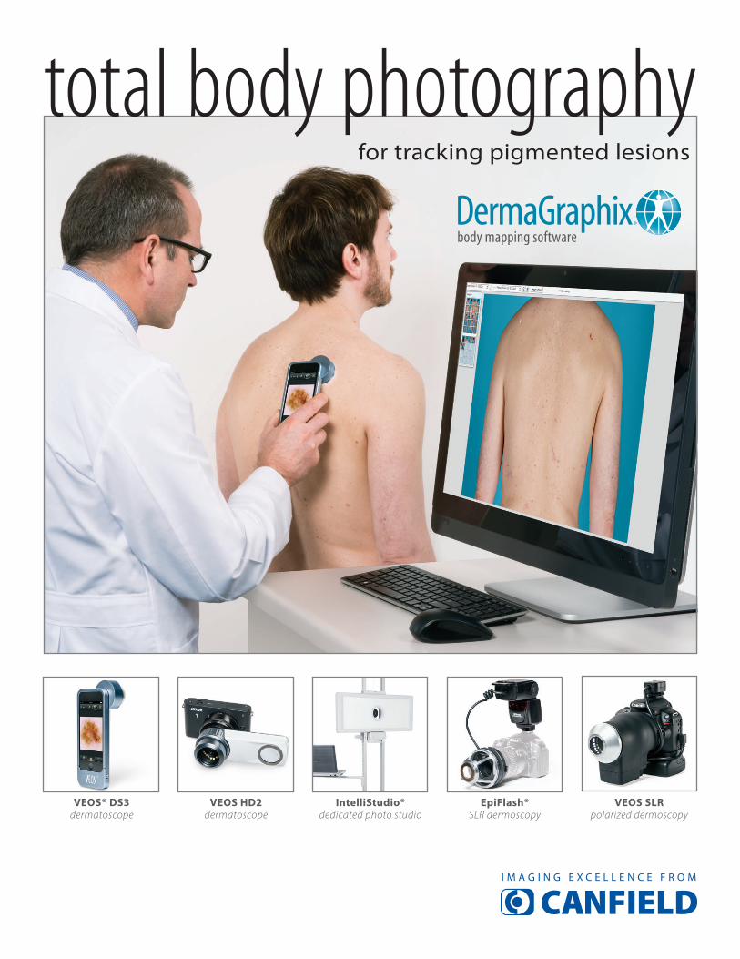
® total body photography for tracking pigmented lesions VEOS® DS3 dermatoscope VEOS HD2 dermatoscope IntelliStudio® dedicated photo studio EpiFlash® SLR dermoscopy VEOS SLR polarized dermoscopy
Transcript of total body photography - Canfield Scientific · total body photography. for tracking pigmented...
®
total body photographyfor tracking pigmented lesions
VEOS® DS3dermatoscope
VEOS HD2dermatoscope
IntelliStudio®dedicated photo studio
EpiFlash®SLR dermoscopy
VEOS SLRpolarized dermoscopy

Whole Body Photography is a well established tool for managing patients at risk for melanoma. A photographic baseline is invaluable when attempting to detect new lesions or suspicious change in existing lesions in patients with numerous nevi. Many of the leading cancer centers and over 60 percent of dermatology academic centers in the United States employ this technique to aid in the early detection of suspicious lesions.
The Mirror® Body Mapping Module enables linking of close-up images to specific points on a patient’s overview photo. Ideal for tracking pigmented lesions (i.e., mole mapping), body mapping is also useful for following psoriasis, eczema and other conditions. The Body Mapping Module integrates with Mirror PhotoFile® medical image management software to create a seamless digital photography and lesion tracking solution, including camera tethering and measurements. Body mapping provides the easiest, most reliable means of accurately comparing and tracking lesions, and conducting total body skin exams.
®
Suspicious lesions may be more closely monitored through the use of dermoscopy and Mirror Body Mapping image linking.
Mirror Body Mapping comparisons to baseline photos assist in the detection of new lesions or changes in existing lesions.
Calibrated measurement tools in Mirror Body Mapping: a. perimeter and surface area b. length and width
Mirror Body Mapping facilitates total body photography and subsequent patient exams.
CPT Code 96904 for Whole Body Integumentary Photography enables providers to be reimbursed for offering this standard of care for their patients at risk for melanoma.
www.canfieldsci.com / [email protected] / phone +1.973.276.0336 / (USA) 800.815.4330
The Mirror Body Mapping Module can create stand-alone
viewer CDs for your patients’ self-examinations.
a.
b.
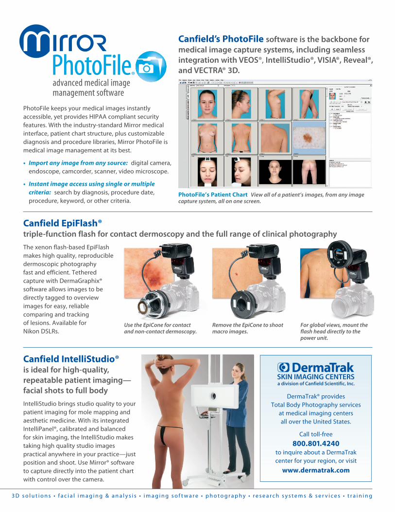
For global views, mount the flash head directly to the power unit.
Remove the EpiCone to shoot macro images.
Use the EpiCone for contact and non-contact dermoscopy.
Canfield EpiFlash® triple-function flash for contact dermoscopy and the full range of clinical photography
The xenon flash-based EpiFlash makes high quality, reproducible dermoscopic photography fast and efficient. Tethered capture with DermaGraphix® software allows images to be directly tagged to overview images for easy, reliable comparing and tracking of lesions. Available for Nikon DSLRs.
Canfield IntelliStudio® is ideal for high-quality, repeatable patient imaging—facial shots to full body
IntelliStudio brings studio quality to your patient imaging for mole mapping and aesthetic medicine. With its integrated IntelliPanel®, calibrated and balanced for skin imaging, the IntelliStudio makes taking high quality studio images practical anywhere in your practice—just position and shoot. Use Mirror® software to capture directly into the patient chart with control over the camera.
3 D s o l u t i o n s • f a c i a l i m a g i n g & a n a l y s i s • i m a g i n g s o f t w a r e • p h o t o g r a p h y • r e s e a r c h s y s t e m s & s e r v i c e s • t r a i n i n g
®
PhotoFile’s Patient Chart View all of a patient’s images, from any image capture system, all on one screen.
Canfield’s PhotoFile software is the backbone for medical image capture systems, including seamless integration with VEOS®, IntelliStudio®, VISIA®, Reveal®, and VECTRA® 3D.
PhotoFile keeps your medical images instantly accessible, yet provides HIPAA compliant security features. With the industry-standard Mirror medical interface, patient chart structure, plus customizable diagnosis and procedure libraries, Mirror PhotoFile is medical image management at its best.
• Import any image from any source: digital camera, endoscope, camcorder, scanner, video microscope.
• Instant image access using single or multiple criteria: search by diagnosis, procedure date, procedure, keyword, or other criteria.
DermaTrak® provides Total Body Photography services
at medical imaging centers all over the United States.
Call toll-free
800.801.4240 to inquire about a DermaTrak center for your region, or visit
www.dermatrak.com

www.canfieldsci.com / [email protected] / phone +1.973.276.0336 / (USA) 800.815.4330 3 D S O L U T I O N S • F A C I A L I M A G I N G & A N A L Y S I S • I M A G I N G S O F T W A R E • P H O T O G R A P H Y • R E S E A R C H S Y S T E M S & S E R V I C E S • T R A I N I N G
Mirror, DermaGraphix, PhotoFile, VEOS, IntelliStudio, EpiFlash, and DermaTrak are registered trademarks of Canfield Scientific, Inc. 1401-15iPhone and iPod are trademarks of Apple Inc., registered in the U.S. and other countries
VEOS SLR polarized dermoscopy
The VEOS SLR is the first LED-flash-based skin imaging solution for dermoscopic and macro imaging of lesions and hair. Fast, compact and easy to use, VEOS SLR captures consistent, reproducible, dermoscopic and clinical images. Sequentially capture cross-polarized and non-polarized images in either contact or non-contact modes.
36 LEDs provide a bright video preview and then are pulsed to flash intensely for the capture. The 22mm field of view and 18 megapixel resolution ensure sharp details and excellent clinical information. Zoom in and review images on the camera’s 1 million dot 3-inch LCD display. VEOS SLR is only available as a complete calibrated solution including Canon SLR.
Optional iPhone® integration turns the VEOS HD into a flexible image capture system.
Magnetic rings firmly attach VEOS HD to Nikon 1 camera.
VEOS HD1 with cross-polarized lighting is the perfect solution for viewing subdermal pigmentation and vascularity.
VEOS HD2 with dual mode lighting is ideal for viewing either subdermal pigmentation and vascularity or surface structure.
The trim design of either scope fits easily in a pocket and offers a comfortable ergonomic feel in the hand. Precision optics and lighting deliver exceptionally bright and clear views of the finest detail. With the HD2 you can quickly switch from cross-polarized to non-polarized lighting with the touch of a button.
dermatoscopes
With wireless Mirror DermaGraphix
integration, tag lesions directly to body map or capture to the Mirror patient chart.
Pivotable lens for quick switch between overview and close-up.
On-screen lesion tagging.
VEOS DS3 with integrated iPod® touch
Quickly switch from cross-polarized to non-polarized lighting with a tap on the VEOS app screen. Capture
overview, close-up and tag lesion to body map in one continuous workflow.