Topology Regulation during Replication of the Kinetoplast DNA
14
Topology Regulation during Replication of the Kinetoplast DNA Davide Michieletto, 1 Davide Marenduzzo, 2 and Matthew S. Turner 1 1 Department of Physics and Centre for Complexity Science, University of Warwick, Coventry CV4 7AL, United Kingdom. 2 School of Physics and Astronomy, University of Edinburgh, Mayfield Road, Edinburgh EH9 3JZ, Scotland, United Kingdom. Abstract. We study theoretically the replication of Kinetoplast DNA consisting of several thousands separate mini-circles found in organisms of the class Kinetoplastida. When the cell is not actively dividing these are topologically connected in a marginally linked network of rings with only one connected component. During cell division each mini-circle is removed from the network, duplicated and then re-attached, along with its progeny. We study this process under the hypothesis that there is a coupling between the topological state of the mini-circles and the expression of genetic information encoded on them, leading to the production of Topoisomerase. This model describes a self-regulating system capable of full replication that reproduces several previous experimental findings. We find that the fixed point of the system depends on a primary free parameter of the model: the ratio between the rate of removal of mini-circles from the network (R) and their (re)attachment rate (A). The final topological state is found to be that of a marginally linked network structure in which the fraction of mini- circles linked to the largest connected component approaches unity as R/A decreases. Finally we discuss how this may suggest an evolutionary trade-off between the speed of replication and the accuracy with which a fully topologically linked state is produced. arXiv:1408.4237v2 [cond-mat.soft] 20 Aug 2014
Transcript of Topology Regulation during Replication of the Kinetoplast DNA
Topology Regulation during Replication of the
Kinetoplast DNA
Davide Michieletto,1 Davide Marenduzzo,2 and Matthew S.
Turner1
1 Department of Physics and Centre for Complexity Science, University of Warwick,
Coventry CV4 7AL, United Kingdom. 2 School of Physics and Astronomy, University
of Edinburgh, Mayfield Road, Edinburgh EH9 3JZ, Scotland, United Kingdom.
Abstract. We study theoretically the replication of Kinetoplast DNA consisting of
several thousands separate mini-circles found in organisms of the class Kinetoplastida.
When the cell is not actively dividing these are topologically connected in a marginally
linked network of rings with only one connected component. During cell division each
mini-circle is removed from the network, duplicated and then re-attached, along with its
progeny. We study this process under the hypothesis that there is a coupling between
the topological state of the mini-circles and the expression of genetic information
encoded on them, leading to the production of Topoisomerase. This model describes
a self-regulating system capable of full replication that reproduces several previous
experimental findings. We find that the fixed point of the system depends on a primary
free parameter of the model: the ratio between the rate of removal of mini-circles
from the network (R) and their (re)attachment rate (A). The final topological state is
found to be that of a marginally linked network structure in which the fraction of mini-
circles linked to the largest connected component approaches unity as R/A decreases.
Finally we discuss how this may suggest an evolutionary trade-off between the speed of
replication and the accuracy with which a fully topologically linked state is produced.
arX
iv:1
408.
4237
v2 [
cond
-mat
.sof
t] 2
0 A
ug 2
014
Topology Regulation during Replication of the Kinetoplast DNA 2
1. Introduction
The Kinetoplast DNA (or kDNA) [1] is a network of linked DNA loops commonly
found in a group of unicellular eukaryotic organisms of the class Kinetoplastida. Some
of these organisms are responsible for human diseases such as sleeping sickness and
leishmaniasis [2–4]. The Kinetoplast DNA (kDNA) is known for its unique structure,
which resembles a percolating network of linked mini-circles at its critical point [5–8].
During replication, catenation between the loops introduces a non-trivial
topological problem, which is solved as follows. First Topoisomerase II disentangles
one loop at a time from the network [9], the loop then diffuses through a region called
the Kinetoflagellar Zone (KFZ), undergoes duplication via θ structure, and reaches the
anti-podal sites, where, together with the progeny mini-circle, it links to the periphery
of the network [10]. Both mini-circles are left with some nicks along their contour,
so that they are no longer suitable for duplication [10]. This mechanism ensures that
mini-circles are duplicated once and only once [11]. Finally, when the cell divides,
two copies of the network are produced by slicing the network in two, through the
middle [10, 12]. This process has the unique feature that it must undergo complex
topological changes in a precise and consistent order. Previous studies have shown that
the action of Topoisomerases is crucial for this machinery to work correctly, not only for
decatenation of mini-circles from the network, but also for the re-attachment at the end
of duplication [13]. RNAi experiments showed that in absence of Topoisomerase, the
Kinetoplast is unable to form and most of the mini-circles remain in a free (unlinked)
state [14]. Type II Topoisomerase is well-known to play a crucial role in simplifying
knots and catenane which occur in DNA [15]. On the other hand, it has been shown
that the same enzyme is also capable of creating complex, linked structures rather than
just simplifying them [16–18]. It has been conjectured that its role in this respect
is related to DNA concentration and is mediated by the presence of polyamines, e.g.
spermidine [19].
In this work, we focus on the role of Topoisomerase in both adding and removing
mini-circles to and from the network. We suggest that this process is intimately
related to the local concentration of linked or free mini-circles together with the local
concentration of Topoisomerase. The central idea of our work is that we model the latter
as itself depending on the linkedness of the mini-circles via the local concentration of
free mini-circles. This means that our model can describe the Kinetoplast replication as
a self-regulating mechanism that, when switched on, can drive the removal, duplication
and attachment of mini-circles without further intervention. Our model correctly
reproduces key features observed during the Kinetoplast replication, as the centripetal
movement of newly linked mini-circles toward the centre of the network and the
asymmetric distribution of Topoisomerase which localises the edge of the network.
Finally, we investigate the relationship between the efficiency with which the Kinetoplast
re-attaches the mini-circles and the speed at which the whole network duplicates. We
argue that the balance between these two factors can shed some light on why the
Topology Regulation during Replication of the Kinetoplast DNA 3
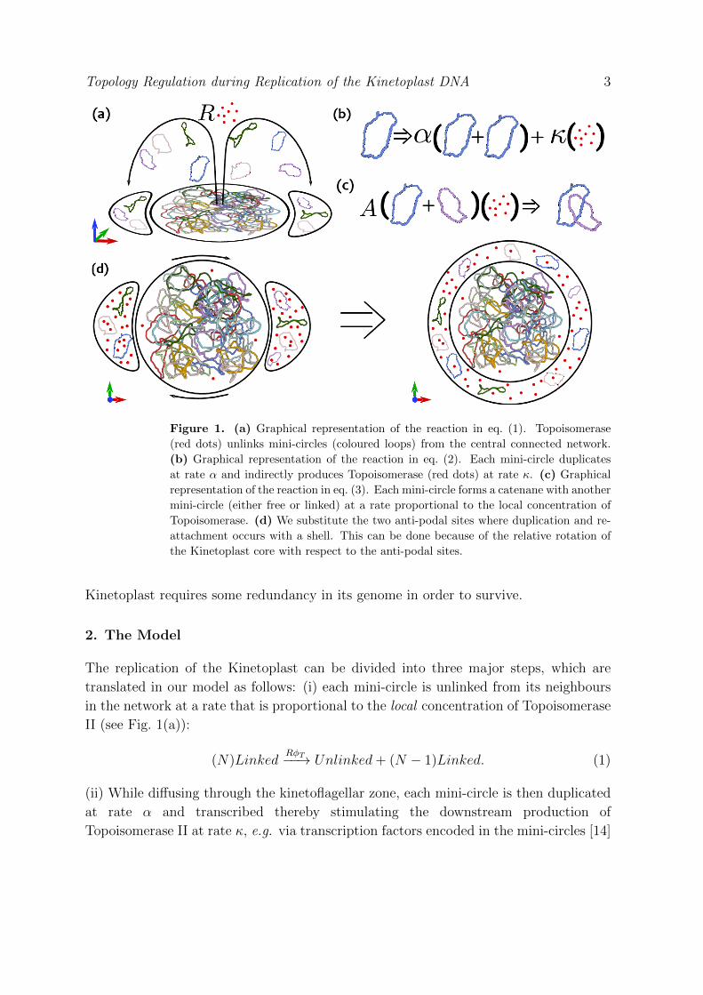
Figure 1. (a) Graphical representation of the reaction in eq. (1). Topoisomerase
(red dots) unlinks mini-circles (coloured loops) from the central connected network.
(b) Graphical representation of the reaction in eq. (2). Each mini-circle duplicates
at rate α and indirectly produces Topoisomerase (red dots) at rate κ. (c) Graphical
representation of the reaction in eq. (3). Each mini-circle forms a catenane with another
mini-circle (either free or linked) at a rate proportional to the local concentration of
Topoisomerase. (d) We substitute the two anti-podal sites where duplication and re-
attachment occurs with a shell. This can be done because of the relative rotation of
the Kinetoplast core with respect to the anti-podal sites.
Kinetoplast requires some redundancy in its genome in order to survive.
2. The Model
The replication of the Kinetoplast can be divided into three major steps, which are
translated in our model as follows: (i) each mini-circle is unlinked from its neighbours
in the network at a rate that is proportional to the local concentration of Topoisomerase
II (see Fig. 1(a)):
(N)LinkedRφT−−→ Unlinked+ (N − 1)Linked. (1)
(ii) While diffusing through the kinetoflagellar zone, each mini-circle is then duplicated
at rate α and transcribed thereby stimulating the downstream production of
Topoisomerase II at rate κ, e.g. via transcription factors encoded in the mini-circles [14]
Topology Regulation during Replication of the Kinetoplast DNA 4
(see Fig. 1(b)):
Unlinkedα−→ Unlinked+ Unlinked
and
Unlinkedκ−→ Topoisomerase. (2)
(iii) Once a mini-circle reaches the anti-podal sites, it is then re-ligated and re-attached
at the periphery of the network or linked together with another mini-circle at a rate
again proportional to the local concentration of linker enzyme, Topoisomerase II, (see
Fig. 1(c)):
Unlinked+ UnlinkedAφT−−→ Linked
or
Unlinked+ LinkedAφT−−→ Linked. (3)
The rates R and A are key parameters in our model and describe the rates of removal
(R) of mini-circles from the network and their re-attachment (A). They will be further
discussed in the following sections. Previous studies showed that during the replicating
phase, the Kinetoplast rotates relatively to the anti-podal sites where re-attachment oc-
curs. This has been thought to facilitate an even redistribution of progeny mini-circles
around the periphery of the network [9, 10]. In our model, this feature is modelled by
substituting the two anti-podal complexes with a shell which surrounds the Kinetoplast
core (see Fig. 1(d)). In fact, this is equivalent to performing a time-average of the
relative position of the complexes with respect to the core over the whole replicating
phase. In light of this, we can visualise the entire system as a rotationally symmetric
2-dimensional disk divided into a core r ≤ Rk filled by the Kinetoplast at the beginning
of the replication (t = 0) and a shell Rk < r < Rmax (initially empty) which plays the
role of region where re-attachment occurs.
We describe the system in terms of the relative density of linked and unlinked
mini-circles, i.e. at the beginning of replication, the density of linked mini-circles is
1 (ρl = 1) within the Kinetoplast, and there are no unlinked mini-circles (ρu = 0).
During the replicating phase, the number of mini-circles grows and we assume that, at
the end of the replication, ρl + ρu = 2, which corresponds to a concentration of about
50 mg/ml [7]. The equations describing our model in terms of these relative densities
and in terms of the concentration of Topoisomerase φT are the following:
dρldt
=−RρlφT + AφTρu (ρl + ρu) +Dl∇2ρl (4)
dρudt
= +RρlφT − AφTρu (ρl + ρu) +Du∇2ρu+
+ αρu [2− (ρu + ρl)] (5)
dφTdt
= + κρu −φTτ
+DT∇2φT . (6)
Topology Regulation during Replication of the Kinetoplast DNA 5
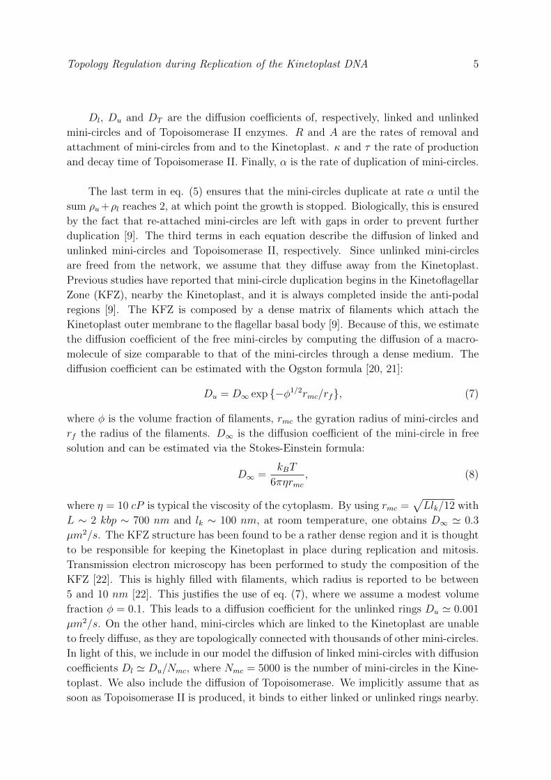
Dl, Du and DT are the diffusion coefficients of, respectively, linked and unlinked
mini-circles and of Topoisomerase II enzymes. R and A are the rates of removal and
attachment of mini-circles from and to the Kinetoplast. κ and τ the rate of production
and decay time of Topoisomerase II. Finally, α is the rate of duplication of mini-circles.
The last term in eq. (5) ensures that the mini-circles duplicate at rate α until the
sum ρu+ρl reaches 2, at which point the growth is stopped. Biologically, this is ensured
by the fact that re-attached mini-circles are left with gaps in order to prevent further
duplication [9]. The third terms in each equation describe the diffusion of linked and
unlinked mini-circles and Topoisomerase II, respectively. Since unlinked mini-circles
are freed from the network, we assume that they diffuse away from the Kinetoplast.
Previous studies have reported that mini-circle duplication begins in the Kinetoflagellar
Zone (KFZ), nearby the Kinetoplast, and it is always completed inside the anti-podal
regions [9]. The KFZ is composed by a dense matrix of filaments which attach the
Kinetoplast outer membrane to the flagellar basal body [9]. Because of this, we estimate
the diffusion coefficient of the free mini-circles by computing the diffusion of a macro-
molecule of size comparable to that of the mini-circles through a dense medium. The
diffusion coefficient can be estimated with the Ogston formula [20, 21]:
Du = D∞ exp {−φ1/2rmc/rf}, (7)
where φ is the volume fraction of filaments, rmc the gyration radius of mini-circles and
rf the radius of the filaments. D∞ is the diffusion coefficient of the mini-circle in free
solution and can be estimated via the Stokes-Einstein formula:
D∞ =kBT
6πηrmc, (8)
where η = 10 cP is typical the viscosity of the cytoplasm. By using rmc =√Llk/12 with
L ∼ 2 kbp ∼ 700 nm and lk ∼ 100 nm, at room temperature, one obtains D∞ ' 0.3
µm2/s. The KFZ structure has been found to be a rather dense region and it is thought
to be responsible for keeping the Kinetoplast in place during replication and mitosis.
Transmission electron microscopy has been performed to study the composition of the
KFZ [22]. This is highly filled with filaments, which radius is reported to be between
5 and 10 nm [22]. This justifies the use of eq. (7), where we assume a modest volume
fraction φ = 0.1. This leads to a diffusion coefficient for the unlinked rings Du ' 0.001
µm2/s. On the other hand, mini-circles which are linked to the Kinetoplast are unable
to freely diffuse, as they are topologically connected with thousands of other mini-circles.
In light of this, we include in our model the diffusion of linked mini-circles with diffusion
coefficients Dl ' Du/Nmc, where Nmc = 5000 is the number of mini-circles in the Kine-
toplast. We also include the diffusion of Topoisomerase. We implicitly assume that as
soon as Topoisomerase II is produced, it binds to either linked or unlinked rings nearby.
Topology Regulation during Replication of the Kinetoplast DNA 6
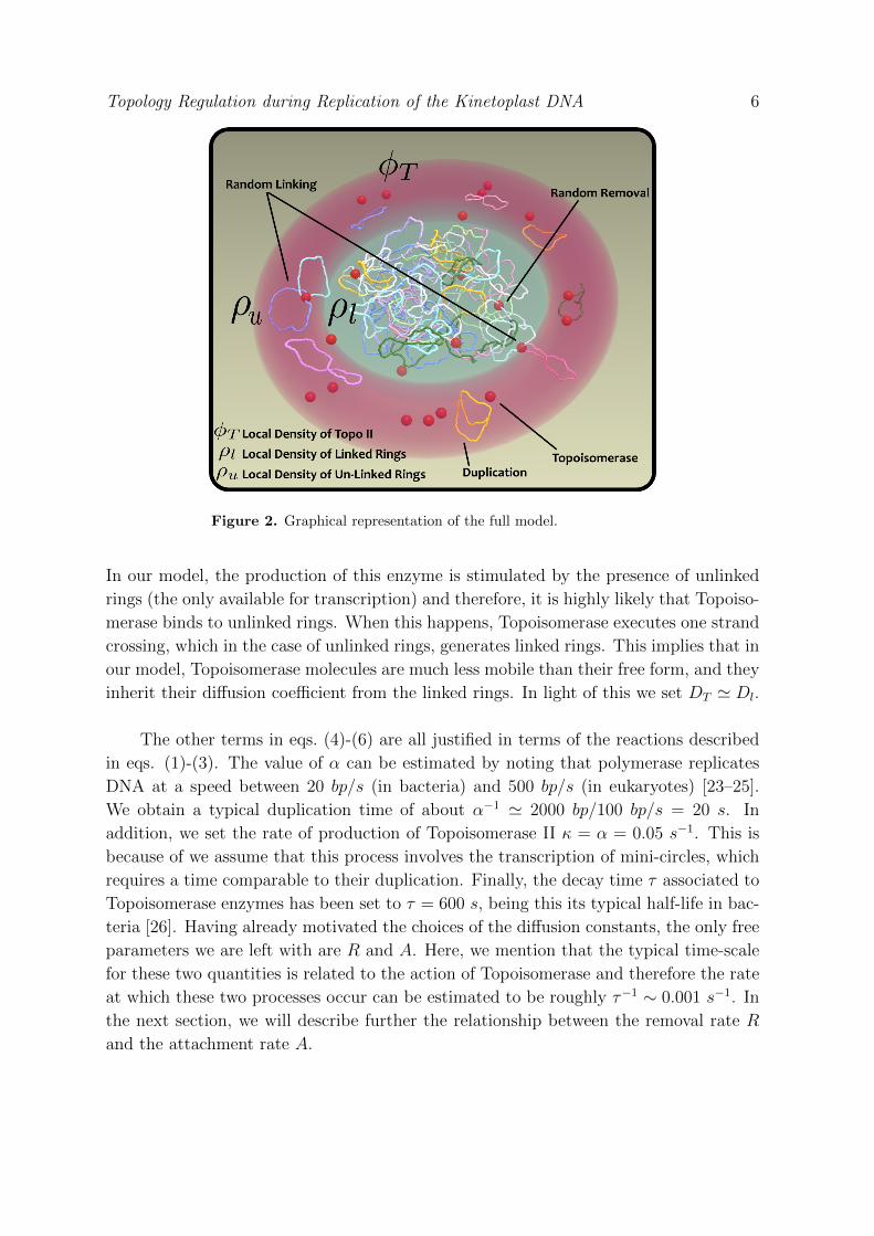
Figure 2. Graphical representation of the full model.
In our model, the production of this enzyme is stimulated by the presence of unlinked
rings (the only available for transcription) and therefore, it is highly likely that Topoiso-
merase binds to unlinked rings. When this happens, Topoisomerase executes one strand
crossing, which in the case of unlinked rings, generates linked rings. This implies that in
our model, Topoisomerase molecules are much less mobile than their free form, and they
inherit their diffusion coefficient from the linked rings. In light of this we set DT ' Dl.
The other terms in eqs. (4)-(6) are all justified in terms of the reactions described
in eqs. (1)-(3). The value of α can be estimated by noting that polymerase replicates
DNA at a speed between 20 bp/s (in bacteria) and 500 bp/s (in eukaryotes) [23–25].
We obtain a typical duplication time of about α−1 ' 2000 bp/100 bp/s = 20 s. In
addition, we set the rate of production of Topoisomerase II κ = α = 0.05 s−1. This is
because of we assume that this process involves the transcription of mini-circles, which
requires a time comparable to their duplication. Finally, the decay time τ associated to
Topoisomerase enzymes has been set to τ = 600 s, being this its typical half-life in bac-
teria [26]. Having already motivated the choices of the diffusion constants, the only free
parameters we are left with are R and A. Here, we mention that the typical time-scale
for these two quantities is related to the action of Topoisomerase and therefore the rate
at which these two processes occur can be estimated to be roughly τ−1 ∼ 0.001 s−1. In
the next section, we will describe further the relationship between the removal rate R
and the attachment rate A.
Topology Regulation during Replication of the Kinetoplast DNA 7
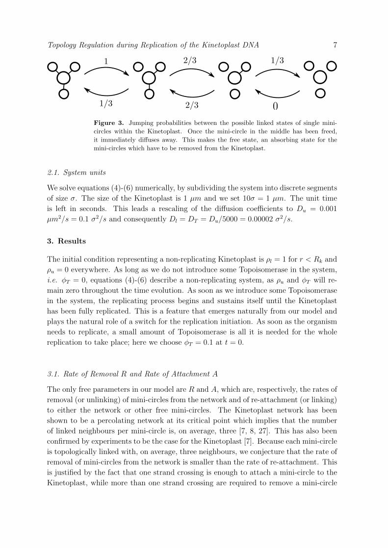
Figure 3. Jumping probabilities between the possible linked states of single mini-
circles within the Kinetoplast. Once the mini-circle in the middle has been freed,
it immediately diffuses away. This makes the free state, an absorbing state for the
mini-circles which have to be removed from the Kinetoplast.
2.1. System units
We solve equations (4)-(6) numerically, by subdividing the system into discrete segments
of size σ. The size of the Kinetoplast is 1 µm and we set 10σ = 1 µm. The unit time
is left in seconds. This leads a rescaling of the diffusion coefficients to Du = 0.001
µm2/s = 0.1 σ2/s and consequently Dl = DT = Du/5000 = 0.00002 σ2/s.
3. Results
The initial condition representing a non-replicating Kinetoplast is ρl = 1 for r < Rk and
ρu = 0 everywhere. As long as we do not introduce some Topoisomerase in the system,
i.e. φT = 0, equations (4)-(6) describe a non-replicating system, as ρu and φT will re-
main zero throughout the time evolution. As soon as we introduce some Topoisomerase
in the system, the replicating process begins and sustains itself until the Kinetoplast
has been fully replicated. This is a feature that emerges naturally from our model and
plays the natural role of a switch for the replication initiation. As soon as the organism
needs to replicate, a small amount of Topoisomerase is all it is needed for the whole
replication to take place; here we choose φT = 0.1 at t = 0.
3.1. Rate of Removal R and Rate of Attachment A
The only free parameters in our model are R and A, which are, respectively, the rates of
removal (or unlinking) of mini-circles from the network and of re-attachment (or linking)
to either the network or other free mini-circles. The Kinetoplast network has been
shown to be a percolating network at its critical point which implies that the number
of linked neighbours per mini-circle is, on average, three [7, 8, 27]. This has also been
confirmed by experiments to be the case for the Kinetoplast [7]. Because each mini-circle
is topologically linked with, on average, three neighbours, we conjecture that the rate of
removal of mini-circles from the network is smaller than the rate of re-attachment. This
is justified by the fact that one strand crossing is enough to attach a mini-circle to the
Kinetoplast, while more than one strand crossing are required to remove a mini-circle
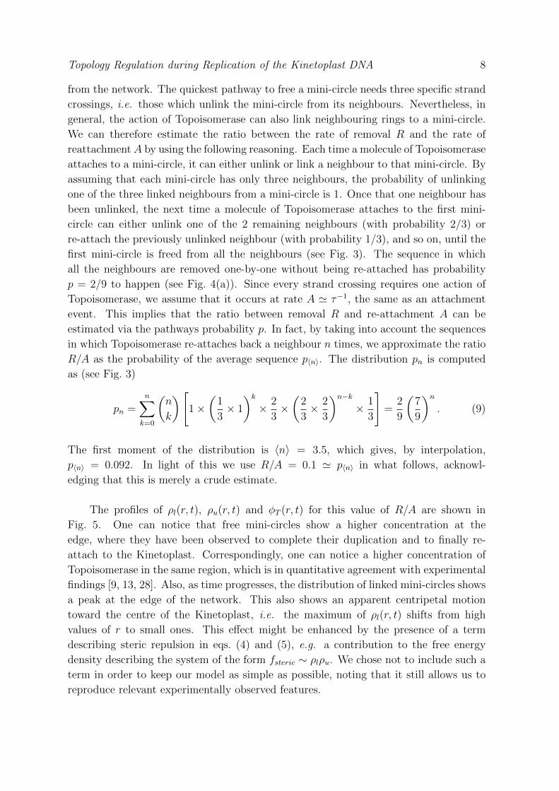
Topology Regulation during Replication of the Kinetoplast DNA 8
from the network. The quickest pathway to free a mini-circle needs three specific strand
crossings, i.e. those which unlink the mini-circle from its neighbours. Nevertheless, in
general, the action of Topoisomerase can also link neighbouring rings to a mini-circle.
We can therefore estimate the ratio between the rate of removal R and the rate of
reattachmentA by using the following reasoning. Each time a molecule of Topoisomerase
attaches to a mini-circle, it can either unlink or link a neighbour to that mini-circle. By
assuming that each mini-circle has only three neighbours, the probability of unlinking
one of the three linked neighbours from a mini-circle is 1. Once that one neighbour has
been unlinked, the next time a molecule of Topoisomerase attaches to the first mini-
circle can either unlink one of the 2 remaining neighbours (with probability 2/3) or
re-attach the previously unlinked neighbour (with probability 1/3), and so on, until the
first mini-circle is freed from all the neighbours (see Fig. 3). The sequence in which
all the neighbours are removed one-by-one without being re-attached has probability
p = 2/9 to happen (see Fig. 4(a)). Since every strand crossing requires one action of
Topoisomerase, we assume that it occurs at rate A ' τ−1, the same as an attachment
event. This implies that the ratio between removal R and re-attachment A can be
estimated via the pathways probability p. In fact, by taking into account the sequences
in which Topoisomerase re-attaches back a neighbour n times, we approximate the ratio
R/A as the probability of the average sequence p〈n〉. The distribution pn is computed
as (see Fig. 3)
pn =n∑k=0
(n
k
)[1×
(1
3× 1
)k× 2
3×(
2
3× 2
3
)n−k× 1
3
]=
2
9
(7
9
)n. (9)
The first moment of the distribution is 〈n〉 = 3.5, which gives, by interpolation,
p〈n〉 = 0.092. In light of this we use R/A = 0.1 ' p〈n〉 in what follows, acknowl-
edging that this is merely a crude estimate.
The profiles of ρl(r, t), ρu(r, t) and φT (r, t) for this value of R/A are shown in
Fig. 5. One can notice that free mini-circles show a higher concentration at the
edge, where they have been observed to complete their duplication and to finally re-
attach to the Kinetoplast. Correspondingly, one can notice a higher concentration of
Topoisomerase in the same region, which is in quantitative agreement with experimental
findings [9, 13, 28]. Also, as time progresses, the distribution of linked mini-circles shows
a peak at the edge of the network. This also shows an apparent centripetal motion
toward the centre of the Kinetoplast, i.e. the maximum of ρl(r, t) shifts from high
values of r to small ones. This effect might be enhanced by the presence of a term
describing steric repulsion in eqs. (4) and (5), e.g. a contribution to the free energy
density describing the system of the form fsteric ∼ ρlρu. We chose not to include such a
term in order to keep our model as simple as possible, noting that it still allows us to
reproduce relevant experimentally observed features.
Topology Regulation during Replication of the Kinetoplast DNA 9
(a)
(b)
(c)
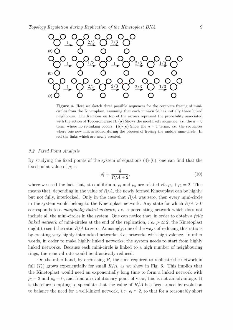
Figure 4. Here we sketch three possible sequences for the complete freeing of mini-
circles from the Kinetoplast, assuming that each mini-circle has initially three linked
neighbours. The fractions on top of the arrows represent the probability associated
with the action of Topoisomerase II. (a) Shows the most likely sequence, i.e. the n = 0
term, where no re-linking occurs. (b)-(c) Show the n = 1 terms, i.e. the sequences
where one new link is added during the process of freeing the middle mini-circle. In
red the links which are newly created.
3.2. Fixed Point Analysis
By studying the fixed points of the system of equations (4)-(6), one can find that the
fixed point value of ρl is
ρ∗l =4
R/A+ 2, (10)
where we used the fact that, at equilibrium, ρl and ρu are related via ρu + ρl = 2. This
means that, depending in the value of R/A, the newly formed Kinetoplast can be highly,
but not fully, interlocked. Only in the case that R/A was zero, then every mini-circle
in the system would belong to the Kinetoplast network. Any state for which R/A > 0
corresponds to a marginally linked network, i.e. a percolating network which does not
include all the mini-circles in the system. One can notice that, in order to obtain a fully
linked network of mini-circles at the end of the replication, i.e. ρl ' 2, the Kinetoplast
ought to send the ratio R/A to zero. Amusingly, one of the ways of reducing this ratio is
by creating very highly interlocked networks, i.e. networks with high valence. In other
words, in order to make highly linked networks, the system needs to start from highly
linked networks. Because each mini-circle is linked to a high number of neighbouring
rings, the removal rate would be drastically reduced.
On the other hand, by decreasing R, the time required to replicate the network in
full (Tr) grows exponentially for small R/A, as we show in Fig. 6. This implies that
the Kinetoplast would need an exponentially long time to form a linked network with
ρl = 2 and ρu = 0, and from an evolutionary point of view, this is not an advantage. It
is therefore tempting to speculate that the value of R/A has been tuned by evolution
to balance the need for a well-linked network, i.e. ρl ' 2, to that for a reasonably short
Topology Regulation during Replication of the Kinetoplast DNA 10
0.00
0.05
0.10
0.15
0.20
0 1 2 3 4 5
ρu
r [σ]
t=0 st=100 s
t=250 st=500 s
t=1000 st=2000 s
t=5000 st=10000 s
(b)
0.0
0.5
1.0
1.5
2.0
2.5
3.0
0 1 2 3 4 5
ρl
r [σ]
t=0 st=100 s
t=250 st=500 s
t=1000 st=2000 s
t=5000 st=10000 s
0.0
1.0
2.0
3.0
4.0
5.0
6.0
7.0
8.0
9.0
0 1 2 3 4 5
ΦT
r [σ]
t=0 st=100 s
t=250 st=500 s
t=1000 st=2000 s
t=5000 st=1000 s
(c)
(a)
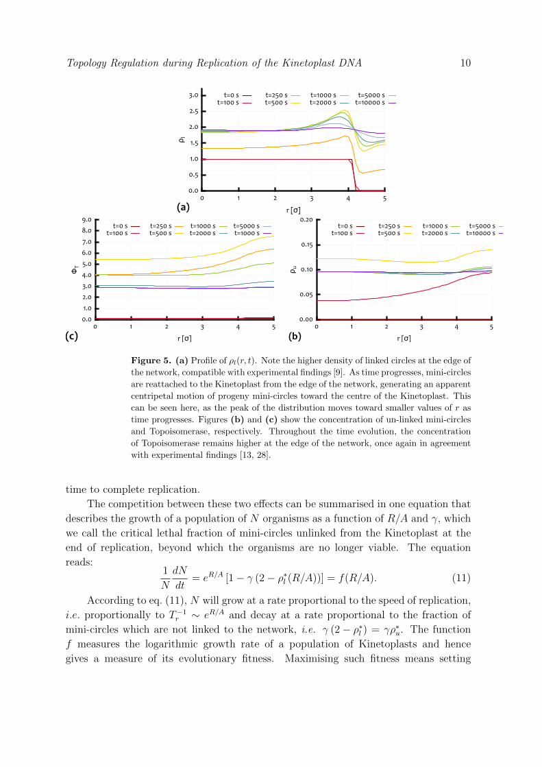
Figure 5. (a) Profile of ρl(r, t). Note the higher density of linked circles at the edge of
the network, compatible with experimental findings [9]. As time progresses, mini-circles
are reattached to the Kinetoplast from the edge of the network, generating an apparent
centripetal motion of progeny mini-circles toward the centre of the Kinetoplast. This
can be seen here, as the peak of the distribution moves toward smaller values of r as
time progresses. Figures (b) and (c) show the concentration of un-linked mini-circles
and Topoisomerase, respectively. Throughout the time evolution, the concentration
of Topoisomerase remains higher at the edge of the network, once again in agreement
with experimental findings [13, 28].
time to complete replication.
The competition between these two effects can be summarised in one equation that
describes the growth of a population of N organisms as a function of R/A and γ, which
we call the critical lethal fraction of mini-circles unlinked from the Kinetoplast at the
end of replication, beyond which the organisms are no longer viable. The equation
reads:1
N
dN
dt= eR/A [1− γ (2− ρ∗l (R/A))] = f(R/A). (11)
According to eq. (11), N will grow at a rate proportional to the speed of replication,
i.e. proportionally to T−1r ∼ eR/A and decay at a rate proportional to the fraction of
mini-circles which are not linked to the network, i.e. γ (2− ρ∗l ) = γρ∗u. The function
f measures the logarithmic growth rate of a population of Kinetoplasts and hence
gives a measure of its evolutionary fitness. Maximising such fitness means setting
Topology Regulation during Replication of the Kinetoplast DNA 11
102
103
104
105
0.00 0.20 0.40 0.60 0.80 1.00
Tr(R/A)[s]
R/A
e‐R/A
(R/A)‐1.5
data
fit
0 0.2 0.4 0.6 0.8 1R/A
0.900.910.920.930.940.950.960.970.980.991.00
γ
(a) (b)
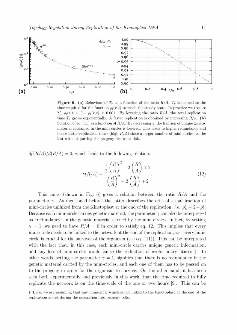
Figure 6. (a) Behaviour of Tr as a function of the ratio R/A. Tr is defined as the
time required for the function ρl(r, t) to reach the steady state. In practice we require∑r ρl(r, t + 1) − ρl(r, t) < 0.001. By lowering the ratio R/A, the total replication
time Tr grows exponentially. A faster replication is obtained by increasing R/A. (b)
Solution of eq. (11) as a function of R/A. By decreasing γ, the fraction of unique genetic
material contained in the mini-circles is lowered. This leads to higher redundancy and
hence faster replication times (high R/A) since a larger number of mini-circles can be
lost without putting the progeny fitness at risk.
df(R/A)/d(R/A) = 0, which leads to the following relation:
γ(R/A) =
1
2
(R
A
)2
+ 2
(R
A
)+ 2(
R
A
)2
+ 2
(R
A
)+ 2
. (12)
This curve (shown in Fig. 6) gives a relation between the ratio R/A and the
parameter γ. As mentioned before, the latter describes the critical lethal fraction of
mini-circles unlinked from the Kinetoplast at the end of the replication, i.e. ρ∗u = 2−ρ∗l .Because each mini-circle carries genetic material, the parameter γ can also be interpreted
as “redundancy” in the genetic material carried by the mini-circles. In fact, by setting
γ = 1, we need to have R/A = 0 in order to satisfy eq. 12. This implies that every
mini-circle needs to be linked to the network at the end of the replication, i.e. every mini-
circle is crucial for the survival of the organism (see eq. (11)). This can be interpreted
with the fact that, in this case, each mini-circle carries unique genetic information,
and any loss of mini-circles would cause the reduction of evolutionary fitness ‡. In
other words, setting the parameter γ = 1, signifies that there is no redundancy in the
genetic material carried by the mini-circles, and each one of them has to be passed on
to the progeny in order for the organism to survive. On the other hand, it has been
seen both experimentally and previously in this work, that the time required to fully
replicate the network is on the time-scale of the one or two hours [9]. This can be
‡ Here, we are assuming that any mini-circle which is not linked to the Kinetoplast at the end of the
replication is lost during the separation into progeny cells.
Topology Regulation during Replication of the Kinetoplast DNA 12
achieved by setting 0 < R/A . 0.3 (from Fig. 6(a)), which implies that the parameter
γ has to be strictly smaller than unity (from eq. 12 and Fig. 6(b)). In other words,
the mini-circles carry heterogeneous, but non unique, genetic material. This is once
again in agreement with experimental findings, which have observed that although the
mini-circles are heterogeneous in the genetic material, they also show, to some extent,
redundancy of the genetic information [9]. It is also worth noting that for a broad
range of values for R/A, the value of γ stays close to 1, meaning that the system does
not need a large redundancy in the genetic material to ensure survival, which is again
qualitatively consistent with experimental observations, which showed that most of the
mini-circles are genetically heterogeneous.
4. Conclusions
Our model naturally connects the topological state of the Kinetoplast DNA with the
genetic information encoded in the mini-circles. We propose an analytical model for
the replication of the Kinetoplast DNA. This is treated as a two dimensional disk,
surrounded by a shell which plays the role of the anti-podal regions where the enzymes
necessary for the duplication and transcription of the mini-circles are enriched. We
assume that the genetic material of the mini-cirles contains transcription factors which
promote the production of the Topoisomerase enzymes. We find that the replication
mechanism can be described as a self-regulated process, requiring only the input of a
small initial level of Topoisomerase to fully replicate the Kinetoplast. We find that the
ratio between rate of removal R and rate of attachment A of mini-circles from and to
the Kinetoplast controls how the linked network is contracted. Our model qualitatively
reproduces the experimentally observed higher concentration of Topoisomerase enzyme
at the edge of the network and the apparent centripetal motion of newly linked mini-
circles from the edge of the network toward its centre. It also reproduces the typical
duplication time-scale, which has been found to be of the order of hours. The final
topological state of the network is found to be controlled by the ratio R/A with the
degree of linkage decreasing with increasing R/A. On the other hand, we find that
lowering this quantity leads to exponentially longer replication times, which decrease
the evolutionary fitness. By considering these two effects, we propose an explicit formula
for the growth of a population of these organisms, which gives a relationship between
accuracy and speed of the replication mechanism as a function of the “redundancy
factor” γ. This factor can be interpreted either as the lethal fraction of mini-circles
unlinked from the network at the end of replication or as a measure of the degree of
uniqueness (absence of redundancy) of crucial genetic material carried by the mini-
circles.
REFERENCES 13
ACKNOWLEDGEMENTS
DMi acknowledges the support from the Complexity Science Doctoral Training Centre at
the University of Warwick with funding provided by the EPSRC (EP/E501311). We also
acknowledge the support of EPSRC to DMa, EP/I034661/1, and MST, EP/1005439/1,
the latter funding a Leadership Fellowship.
References
[1] Fairlamb A H, Weislogel P O, Hoeijmakers J H and Borst P 1978 J. Cell Biol. 76
293–309
[2] Young D and Morales A 1987 J. Med. Entomol. 24 587–589
[3] Jacobson R L 2003 Folia parasitol. 50 241–250
[4] MacLean L, Chisi J and Odiit M 2004 Infect. Immun. 72 7040–7044
[5] Shapiro T and Englund P 1995 Annual Reviews in Microbiology 49 117–143
[6] Kellenberger E, Carlemalm E, Sechaud J, Ryter A and Haller G 1986 Bacterial
Chromatin ed. CO Gualerzi and CL Pon 11–25
[7] Chen J, Rauch C A, White J H, Englund P T and Cozzarelli N R 1995 Cell 80
61–69
[8] Michieletto D, Marenduzzo D and Orlandini E 2014 arXiv preprint arXiv:1406.0141
1–6 (Preprint arXiv:1406.0141v1)
[9] Jensen R E and Englund P T 2012 Annu. Rev. Microbiol. 66 473–491
[10] Perez-Morga D L and Englund P T 1993 Cell 74 703–711
[11] Kitchin P, Klein V, Fein B and Englund P 1984 J. Biol. Chem. 259 15532–15539
[12] Liu B, Liu Y, Motyka S a, Agbo E E C and Englund P T 2005 Trends Parasitol.
21 363–369
[13] Shlomai J and Zadok A 1983 Nucleic Acids Res. 11 4019–4034
[14] Wang Z, Morris J C, Drew M E and Englund P T 2000 J. Biol. Chem. 275 40174–9
[15] White J H, Millett K C and Cozzarelli N 1987 J. Mol. Biol. 197 585–603
[16] Hsieh T and Brutlag D 1980 Cell 21 115–125
[17] Kreuzer K and Cozzarelli N 1980 Cell 20 245–254
[18] Brown P O and Cozzarelli N R 1981 Proc. Natl. Acad. Sci. USA 78 843–7
[19] Krasnow M and Cozzarelli N 1982 J. Biol. Chem. 257 2687–2693
[20] A G Ogston Preston B N J D W 1973 Proc. R. Soc. Lond. A 333 297
[21] Johnson E M, Berk D a, Jain R K and Deen W M 1996 Biophys. J. 70 1017–23
[22] Ogbadoyi E, Robinson D and Gull K 2003 Mol. Biol. Cell 14 1769–1779
[23] Dignam J, Lebovitz R and Roeder R 1983 Nucleic Acids Res. 1 1475–1489
[24] Wickiser J K, Winkler W C, Breaker R R and Crothers D M 2005 Mol. Cell 18
49–60
REFERENCES 14
[25] Schwartz J J and Quake S R 2009 Proc. Natl. Acad. Sci. USA 107 1254–1254
[26] Taniguchi Y, Choi P J, Li G W, Chen H, Babu M, Hearn J, Emili A and Xie X S
2010 Science 329 533–538 ISSN 1095-9203
[27] Diao Y, Hinson K and Arsuaga J 2012 J. Phys. A:Math. Gen. 45 035004 ISSN
1751-8113
[28] Shlomai J 1994 Parasitology today 10 341–6
[29] Perez-Morga D and Englund P 1993 J. Cell. Biol. 123