Topics today Normal puerperium Diseases of puerperium Gestational trophoblastic diseases,GTD.
46
Topics today Topics today Normal puerperium Diseases of puerperium Gestational trophoblastic diseases,GTD
-
Upload
william-little -
Category
Documents
-
view
236 -
download
0
Transcript of Topics today Normal puerperium Diseases of puerperium Gestational trophoblastic diseases,GTD.

Topics todayTopics today
Normal puerperium Diseases of puerperium Gestational trophoblastic
diseases,GTD
Normal puerperiumNormal puerperium(Postpartum care)(Postpartum care)
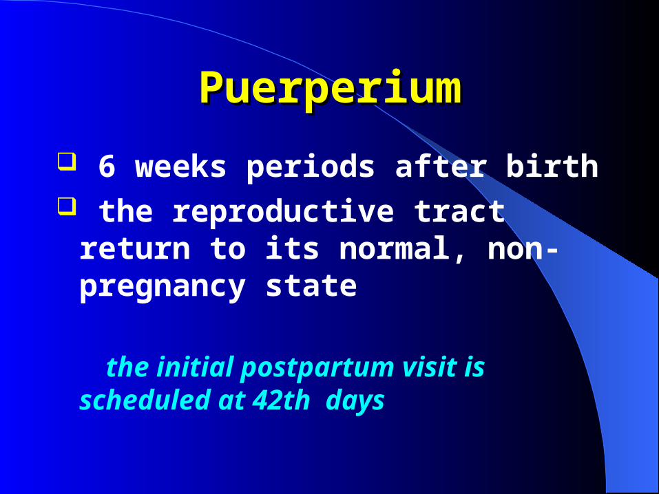
PuerperiumPuerperium
6 weeks periods after birth the reproductive tract return to its
normal, non-pregnancy state
the initial postpartum visit is scheduled at 42th days
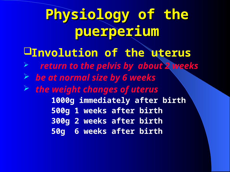
Physiology of the puerperiumPhysiology of the puerperium
Involution of the uterus return to the pelvis by about 2 weeks be at normal size by 6 weeks the weight changes of uterus 1000g immediately after birth 500g 1 weeks after birth 300g 2 weeks after birth 50g 6 weeks after birth
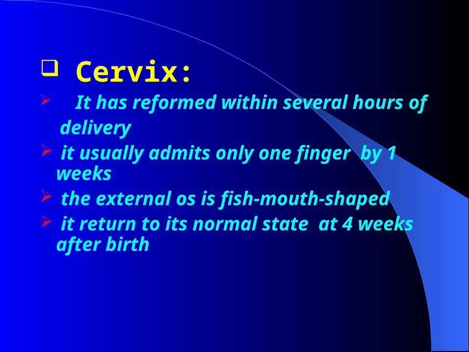
Cervix: It has reformed within several hours of delivery it usually admits only one finger by 1
weeks the external os is fish-mouth-shaped it return to its normal state at 4 weeks
after birth
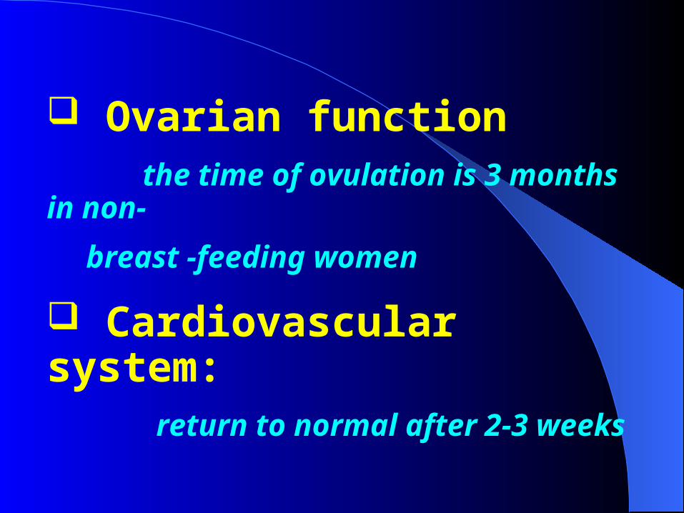
Ovarian function the time of ovulation is 3 months in non-
breast -feeding women
Cardiovascular system: return to normal after 2-3 weeks
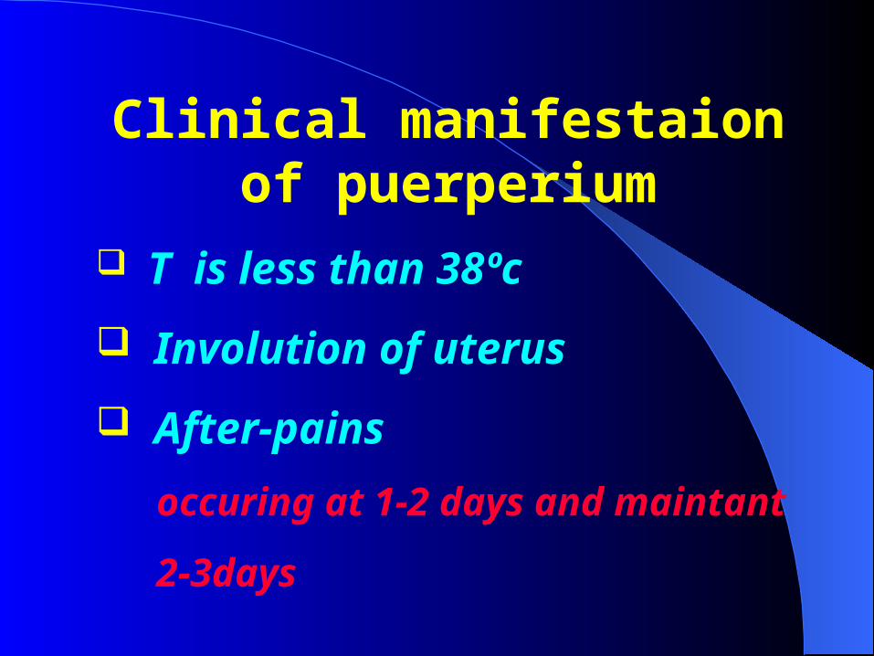
Clinical manifestaion of puerperium
T is less than 38ºc
Involution of uterus
After-pains
occuring at 1-2 days and maintant
2-3days
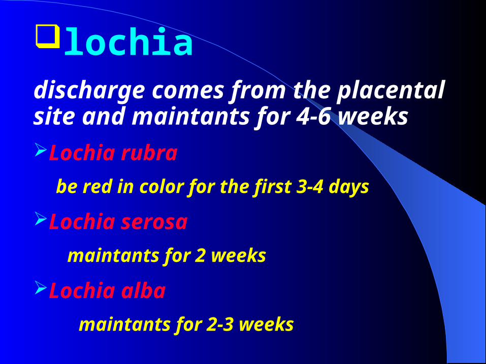
lochiadischarge comes from the placental site and maintants for 4-6 weeksLochia rubra
be red in color for the first 3-4 days
Lochia serosa
maintants for 2 weeks
Lochia alba
maintants for 2-3 weeks
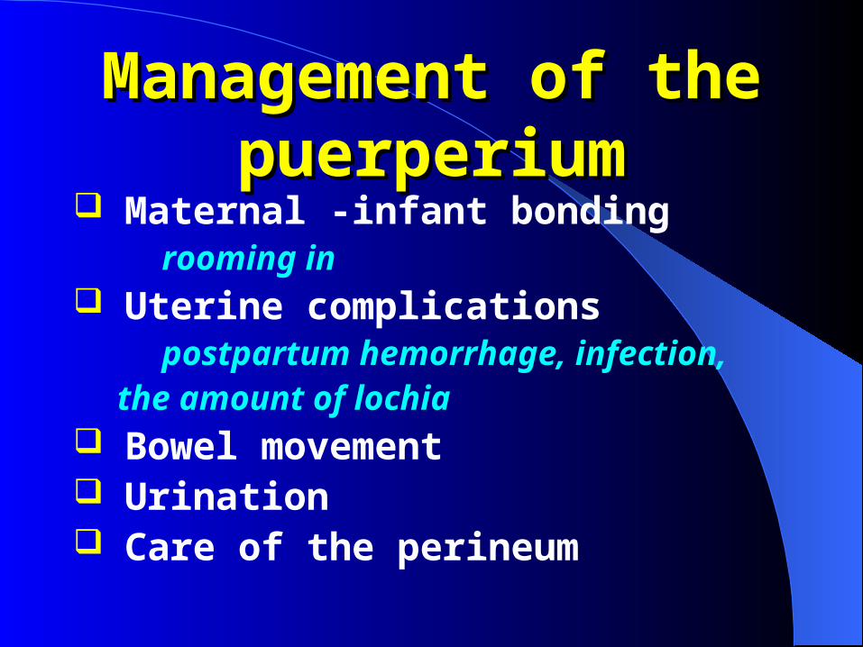
Management of the Management of the puerperiumpuerperium
Maternal -infant bonding
rooming in Uterine complications postpartum hemorrhage, infection, the amount of lochia Bowel movement Urination Care of the perineum

Management of breastBreast-feedingthe benefits of breast-feeding increase the conversation decrease the cost improve infant nutrition and protect
against infection and allergic reaction uterus contraction

Finding Engorgement Mastitis Plugged duct
Onset Gradual Sudden Gradual
Location Bilateral Unilateral Unilateral
Swelling Generalized Localized Localized
Pain Generalized Intense,
localizedLocalized
Systemic symptoms
Feels well Feels ill Feels well
Fever No Yes No
Differential diagnosis of engorgement, mastitis and plugged duct

Diseases of puerperium
Puerperal infection Late puerperal hemorrhage Postpartum depression puerperal heat stroke
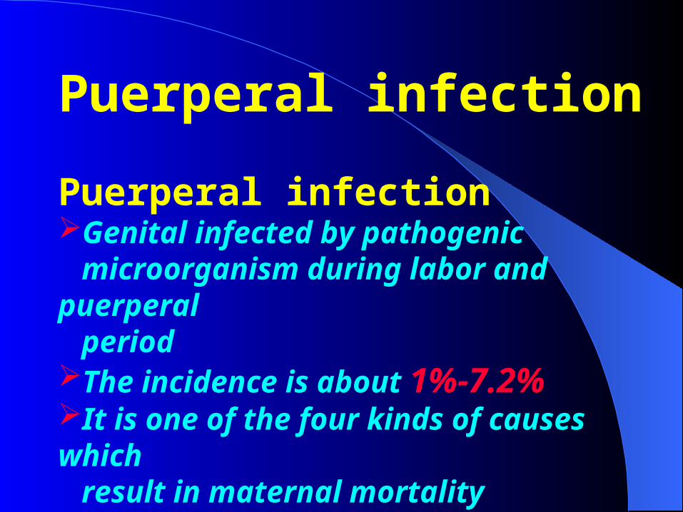
Puerperal infection
Puerperal infectionGenital infected by pathogenic microorganism during labor and puerperal periodThe incidence is about 1%-7.2%It is one of the four kinds of causes which result in maternal mortality
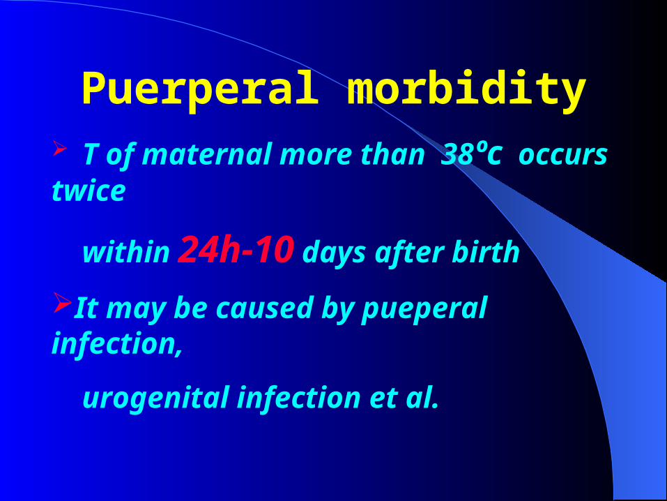
Puerperal morbidity T of maternal more than 38ºc occurs twice
within 24h-10 days after birth
It may be caused by pueperal infection,
urogenital infection et al.
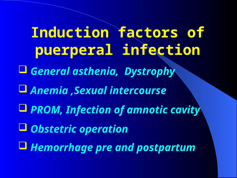
Induction factors of puerperal infection
General asthenia, Dystrophy
Anemia ,Sexual intercourse
PROM, Infection of amnotic cavity
Obstetric operation
Hemorrhage pre and postpartum

The kinds of pathogen Bata-hemolytic streptococcus
Anaerobic streptococcus
Anaerobic bacillus
Staphylococcus
Bacillus coli
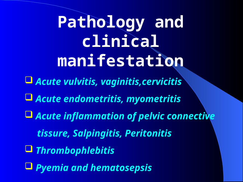
Pathology and clinical manifestation
Acute vulvitis, vaginitis,cervicitis
Acute endometritis, myometritis
Acute inflammation of pelvic connective
tissure, Salpingitis, Peritonitis
Thrombophlebitis
Pyemia and hematosepsis

Diagnosis and treatment supporting treatment
Delete the induction factors
Broad-spectrun antibiotic
Expectant treatment
Late puerperal hemorrhage Excessive bleeding in puerperal period after 24h delivery It can occur sudden and profuse It can occur slowly but prolonged and
persistent
Etiology and clinical manifestation
Retained placenta and membrane Lochia rubra prolonged
Blood loss repeated or bleeding excessive suddendly
Sabinvolution of urerus
Relax of cervix
Placenta tissure can be palpable
Retained decidua
Infection of the placenta attachment
area
Sabinvolution of uterus
Fissuration of uterine insision
postcesarean
Trophoblastic tumor postpartum
Submucus myoma

Diagnosis and treatment supporting treatment
Delete the etiologic factors
Broad-spectrun antibiotic
Expectant treatment
Gestational trophoblastic diseases(GTD)
Molar pregnancy(hydatidiform mole) Invisave mole Choriocarcinoma Placentalsite trophoblastic tumor(PSTT)
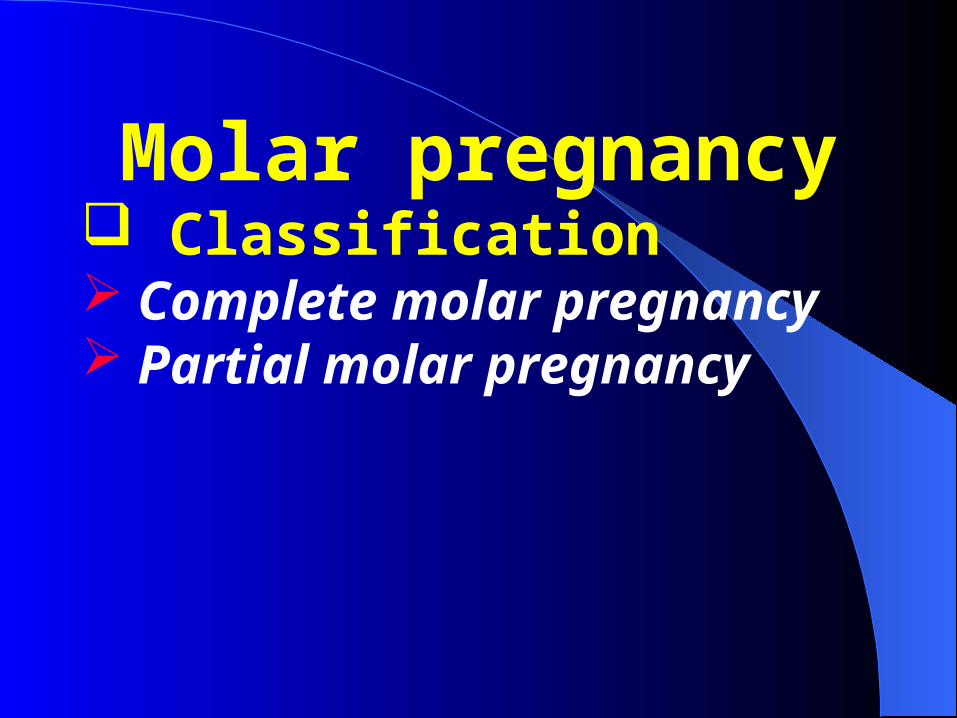
Molar pregnancy Classification Complete molar pregnancy Partial molar pregnancy

EpidemiologyThe incidence varies among different national
and ethnic groups
The highest occurring among Asian women(up
to 1 in 500-600)
The lowest incidence occurring in white
women of western European and U.S ( 1 in
1500-2000)

Etiology
Unknown?
Associated with
age
Dietary deficiencies
Economic status, et al
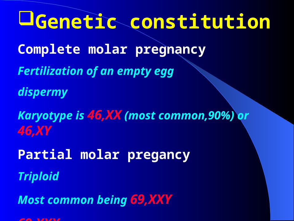
Genetic constitutionComplete molar pregnancy
Fertilization of an empty egg
dispermy
Karyotype is 46,XX (most common,90%) or 46,XY
Partial molar pregancy
Triploid
Most common being 69,XXY
69,XXX
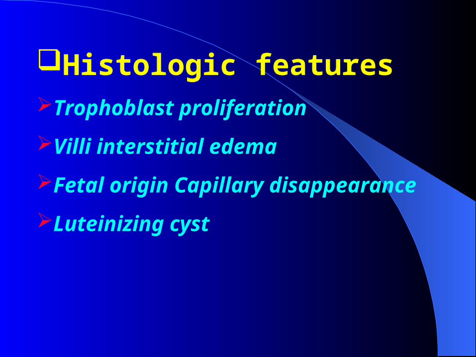
Histologic featuresTrophoblast proliferation
Villi interstitial edema
Fetal origin Capillary disappearance
Luteinizing cyst
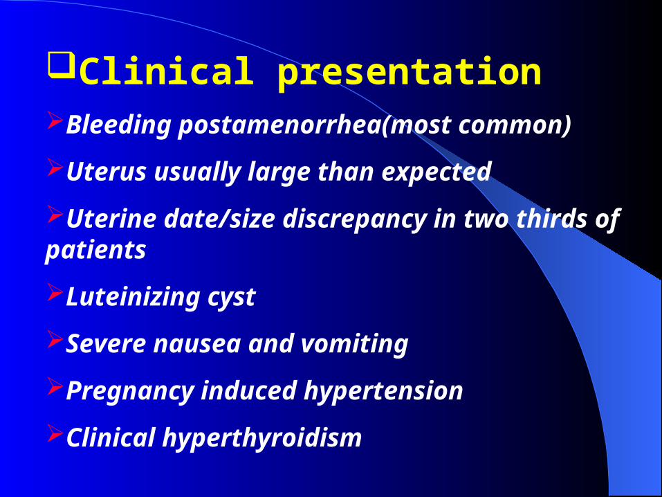
Clinical presentationBleeding postamenorrhea(most common)
Uterus usually large than expected
Uterine date/size discrepancy in two thirds of patients
Luteinizing cyst
Severe nausea and vomiting
Pregnancy induced hypertension
Clinical hyperthyroidism

DiagnosisClinical presentation
Ascertain the level of HCG
Ultrasound:snowstorm appearance
Histology
TreatmentRemove the intrauterine contents promply
Hysterectomy
in the older reproductive group who have no interest in further childbearing
Management of luteinizing cyst
Preventive chemotherapyAge more than 40
Level of serum HCG increased significantaly(more than
100KIU/L)
Titer of HCG has not returned to normal after 12 weeks
postevacuation
Re-elevated HCG level
Uterus larger than expected
Diameter of luteinizing cyst more than 6cm
Trophoblast hyperproliferation still after second curettage
Has no condition to follow-up
Follow-upPelvic examination, ultrasound examination
Assessment of HCG
Serum quantitative HCG level every 1 week until normal
Every 1 week(three month)
Every 2 weeks(three month)
Every 1 month( half year)
Every half year(one year)
Contraception for 1-2 years
Invasive moleIs a complete mole invading the myometrium or vascular
Most common occuring within 6 months after curretage of a complete mole following evaluation for HCG levels that do not fall appropriately
Histology
Type I
amount of mole
Invading myometrium or vascular
Hemorrhage or necrosis rarely
Type IIModerate of mole
Trophoblast proliferation moderate
partial trophoblast undifferentiated
Hemorrhage and necrosis
Type IIIAmount of Hemorrhage or necrosis tissue
Trophoblast hyperproliferation and
undifferentiated
The histology is very same as choriocarcinoma
Clinical presentationPresentation of primary diseaseVaginal bleeding irregular
Involution of uterus prolonged
If the uterus perforation occuring
Abdominal pain
Presentation of intraperitoneal hemorrhage
Presentation of metastasisLung is the most common metastatic
location
The second is vagina, side of uterus and
brain

DiagnosisHistory and presentation
presentation occuring within 6 months of mole curretage
Assessmant of HCG
Persistant high level 8 weeks after curretage
Or the titer of HCG evaluated fast after it returned
to normal
Deplete retained mole, luteinizing cyst and
pregnancy again
Ultrasound examination
Histologic diagnosis
Treatment and follow-up
Same as to choriocarconoma
Choriocarcinoma Hyper-malignant tumor
50% of patients follow molar pregnancy
25% of patients follow abortion
25% of patients follow term pregnancy
few of patient follow ectopic pregnancy
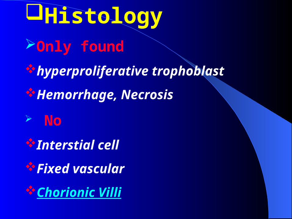
HistologyOnly found
hyperproliferative trophoblast
Hemorrhage, Necrosis
No
Interstial cell
Fixed vascular
Chorionic Villi
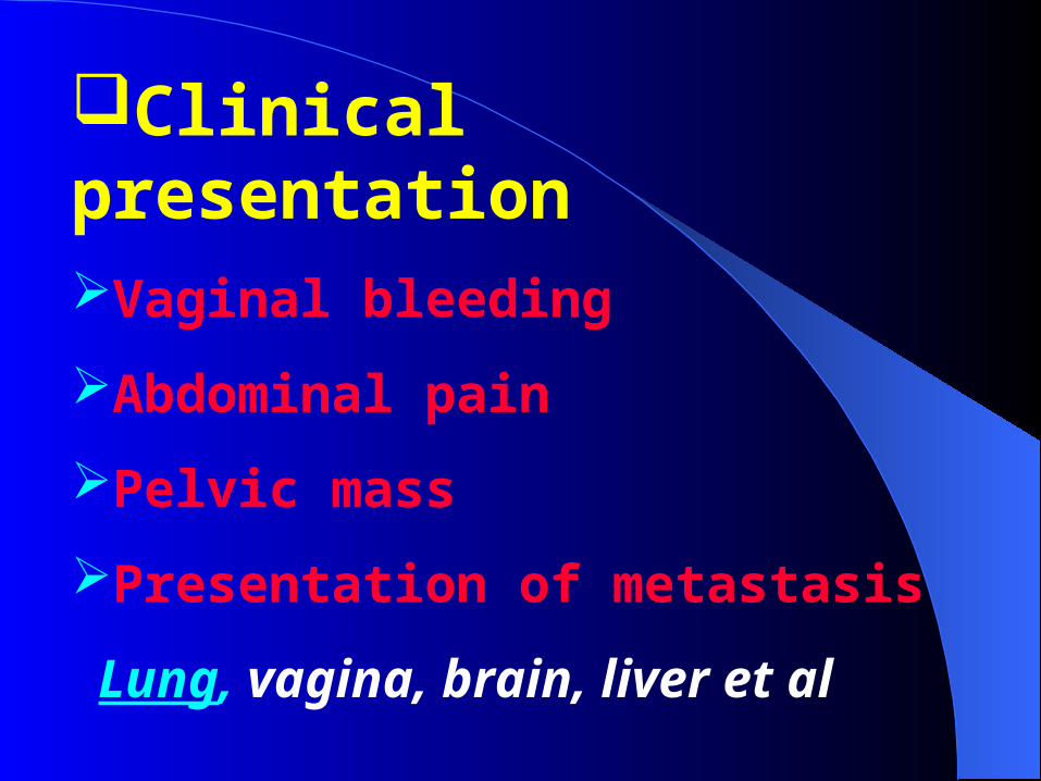
Clinical presentation
Vaginal bleeding
Abdominal pain
Pelvic mass
Presentation of metastasis
Lung, vagina, brain, liver et al
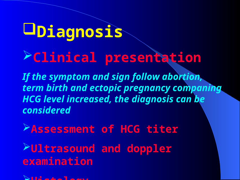
Diagnosis
Clinical presentation If the symptom and sign follow abortion, term birth and ectopic pregnancy companing HCG level increased, the diagnosis can be considered
Assessment of HCG titer
Ultrasound and doppler examination
Histology
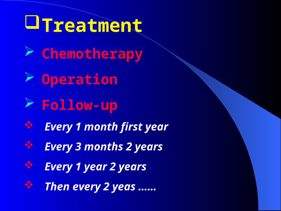
Treatment Chemotherapy
Operation
Follow-up Every 1 month first year
Every 3 months 2 years
Every 1 year 2 years
Then every 2 yeas ……