
TOPICAL REVIEW Labelling of cells with quantum...
17
INSTITUTE OF PHYSICS PUBLISHING NANOTECHNOLOGY Nanotechnology 16 (2005) R9–R25 doi:10.1088/0957-4484/16/2/R01 TOPICAL REVIEW Labelling of cells with quantum dots Wolfgang J Parak 1,3 , Teresa Pellegrino 1 and Christian Plank 2 1 Center for Nanoscience, Ludwig Maximilians Universit¨ at M¨ unchen, Amalienstrasse 54, 80799 M¨ unchen, Germany 2 Institute of Experimental Oncology, Technische Universit¨ atM¨ unchen, Ismaninger Strasse 22, 81675 M¨ unchen, Germany E-mail: [email protected] Received 2 September 2004, in final form 15 December 2004 Published 25 January 2005 Online at stacks.iop.org/Nano/16/R9 Abstract Colloidal quantum dots are semiconductor nanocrystals well dispersed in a solvent. The optical properties of quantum dots, in particular the wavelength of their fluorescence, depend strongly on their size. Because of their reduced tendency to photobleach, colloidal quantum dots are interesting fluorescence probes for all types of labelling studies. In this review we will give an overview on how quantum dots have been used so far in cell biology. In particular we will discuss the biologically relevant properties of quantum dots and focus on four topics: labelling of cellular structures and receptors with quantum dots, incorporation of quantum dots by living cells, tracking the path and the fate of individual cells using quantum dot labels, and quantum dots as contrast agents. 1. Introduction To investigate cells and cellular processes it is very important to visualize structures and compartments within cells and molecules that are involved in the cell’s biochemistry. Since cells are almost transparent to visible light and individual molecules are very small, typically with microscopy no direct observation of structural compartments or molecules is possible. Therefore the structures or molecules of interest have to be labelled with a marker that can be directly observed. One of the most common techniques used in cell biology is certainly fluorescence labelling [1–3]. Either fluorophores can be directly attached to the target to be visualized, or they can be attached to a molecule (e.g. an antibody) [4] that binds to the target via molecular recognition. Such fluorescence labelling techniques are well established, and are used for most cellular labelling experiments. However, traditional fluorophores have their limitations. The conformation of fluorophores is very sensitive to their local environment, e.g. to thermal fluctuation of the solvent [5]. As a consequence of conformational fluctuations, fluorophores can be reversibly transferred to states in which they cannot fluoresce anymore 3 Author to whom any correspondence should be addressed. and the fluorescence of single molecules statistically goes ‘on’ and ‘off’, which is called blinking [6]. Upon optical excitation, organic fluorophores can undergo irreversible light- induced reactions such as photo-oxidation. These molecules are then no longer fluorescent, a phenomenon known as photobleaching [7]. Photobleaching limits the time for which such labels can be observed under a fluorescence microscope. This limitation is of special importance for all studies in which the label is to be observed over extended periods of time, as is the case in any experiment involving temporally resolved imaging. Another restriction of organic fluorophores is the inherent resolution limit of optical microscopy. Although the fluorescence of individual fluorophores of a few nanometre sizes can be observed, such single-molecule fluorescence techniques are based on diluted distributions of labels, i.e. the distance between individual fluorophores has to be bigger than the resolution limit. Therefore, with classical wide field optical microscopy, no structures smaller than a few hundred nanometres can be resolved. To overcome some of the above described limitations there is an ongoing development of alternative techniques. One established technique is the use of colloidal metal particles, in particular small gold particles. These labels can be almost as small as organic fluorophores, but due to their metallic 0957-4484/05/020009+17$30.00 © 2005 IOP Publishing Ltd Printed in the UK R9
Transcript of TOPICAL REVIEW Labelling of cells with quantum...
INSTITUTE OF PHYSICS PUBLISHING NANOTECHNOLOGY
Nanotechnology 16 (2005) R9–R25 doi:10.1088/0957-4484/16/2/R01
TOPICAL REVIEW
Labelling of cells with quantum dotsWolfgang J Parak1,3, Teresa Pellegrino1 and Christian Plank2
1 Center for Nanoscience, Ludwig Maximilians Universitat Munchen, Amalienstrasse 54,80799 Munchen, Germany2 Institute of Experimental Oncology, Technische Universitat Munchen, Ismaninger Strasse22, 81675 Munchen, Germany
E-mail: [email protected]
Received 2 September 2004, in final form 15 December 2004Published 25 January 2005Online at stacks.iop.org/Nano/16/R9
AbstractColloidal quantum dots are semiconductor nanocrystals well dispersed in asolvent. The optical properties of quantum dots, in particular the wavelengthof their fluorescence, depend strongly on their size. Because of theirreduced tendency to photobleach, colloidal quantum dots are interestingfluorescence probes for all types of labelling studies. In this review we willgive an overview on how quantum dots have been used so far in cell biology.In particular we will discuss the biologically relevant properties of quantumdots and focus on four topics: labelling of cellular structures and receptorswith quantum dots, incorporation of quantum dots by living cells, trackingthe path and the fate of individual cells using quantum dot labels, andquantum dots as contrast agents.
1. Introduction
To investigate cells and cellular processes it is very importantto visualize structures and compartments within cells andmolecules that are involved in the cell’s biochemistry. Sincecells are almost transparent to visible light and individualmolecules are very small, typically with microscopy nodirect observation of structural compartments or molecules ispossible. Therefore the structures or molecules of interest haveto be labelled with a marker that can be directly observed.One of the most common techniques used in cell biology iscertainly fluorescence labelling [1–3]. Either fluorophores canbe directly attached to the target to be visualized, or they canbe attached to a molecule (e.g. an antibody) [4] that bindsto the target via molecular recognition. Such fluorescencelabelling techniques are well established, and are used formost cellular labelling experiments. However, traditionalfluorophores have their limitations. The conformation offluorophores is very sensitive to their local environment, e.g. tothermal fluctuation of the solvent [5]. As a consequence ofconformational fluctuations, fluorophores can be reversiblytransferred to states in which they cannot fluoresce anymore
3 Author to whom any correspondence should be addressed.
and the fluorescence of single molecules statistically goes‘on’ and ‘off’, which is called blinking [6]. Upon opticalexcitation, organic fluorophores can undergo irreversible light-induced reactions such as photo-oxidation. These moleculesare then no longer fluorescent, a phenomenon known asphotobleaching [7]. Photobleaching limits the time for whichsuch labels can be observed under a fluorescence microscope.This limitation is of special importance for all studies in whichthe label is to be observed over extended periods of time, asis the case in any experiment involving temporally resolvedimaging. Another restriction of organic fluorophores is theinherent resolution limit of optical microscopy. Although thefluorescence of individual fluorophores of a few nanometresizes can be observed, such single-molecule fluorescencetechniques are based on diluted distributions of labels, i.e. thedistance between individual fluorophores has to be biggerthan the resolution limit. Therefore, with classical wide fieldoptical microscopy, no structures smaller than a few hundrednanometres can be resolved.
To overcome some of the above described limitations thereis an ongoing development of alternative techniques. Oneestablished technique is the use of colloidal metal particles,in particular small gold particles. These labels can be almostas small as organic fluorophores, but due to their metallic
0957-4484/05/020009+17$30.00 © 2005 IOP Publishing Ltd Printed in the UK R9
Topical Review
nature they offer enough contrast to be imaged with electronmicroscopy. Since the resolution for electron microscopyis much better than for optical microscopy, biologicalstructures can be visualized with nanometre resolution [8–10], for example the protein distribution in membranes.Unfortunately, TEM is typically only possible with fixedand thus dead samples. However, many interesting labellingexperiments have also been reported in which gold colloidshave been imaged with optical microscopy [11]. Using goldparticles instead of fluorophores circumvents the drawbacks ofphotobleaching and blinking.
In the last decade semiconductor nanocrystals (so-calledquantum dots) have been introduced as a new type of colloidsfor biolabelling. The fluorescence wavelengths of thesenanoparticles are size dependent, as will be discussed later.First biological applications have been discussed in manynews and views/feature articles [12–18] and reviews [19–28].Since these fluorescent nanoparticles are inorganic solids, theycan be expected to be more robust than organic fluorophores(e.g. towards photobleaching) and in addition they can also beobserved with high resolution by electron microscopy.
The use of nanoparticles in biology and medicalapplications is a multi-disciplinary field of science whereaspects of synthetic chemistry, physics and biology are equallyimportant. It is difficult to find common language and thinkingamong these scientific disciplines. Chemists and physicistshave produced and characterized in detail a multitude of highquality colloidal particles with tuneable properties. However,their application in life sciences has often been restricted todemonstration experiments despite their potential usefulnessin a wide variety of applications. Here we want to reviewthe use of semiconductor nanocrystals as fluorescence labelsemphasizing chemical and physical aspects of these materialsas the basis of their applicability in biological sciences. Wewill discuss how these particles have been used in experimentswith cells and living tissue and we will highlight some of theirpotential future applications in the life sciences.
2. Chemical and physical properties of quantumdots relevant for their use as fluorescence labels incell biology
2.1. Solubility in aqueous buffers
High quality nanocrystals (in terms of crystallinity and sizedistribution) of various semiconductor materials such asCdS, CdSe, CdTe, or CdSe/ZnS can be synthesized by avariety of approaches [29–32]. Most commonly the synthesisis carried out in organic solvents at high temperatures inthe presence of surfactants, these reaction conditions beingrequired to yield monodisperse and stable particles. Thissynthetic approach produces surfactant-coated particles, thepolar surfactant head group attached to the inorganic surface,the hydrophobic chain protruding into the organic solvent,mediating colloidal stability. At this stage, the particles willbe well dispersed in solvents such as toluene or chloroform,but because of their hydrophobic surface layer they are notsoluble in aqueous media. However, almost all experimentsinvolving cells require water-soluble materials. This problemhas been solved by replacing the surfactant layer or by
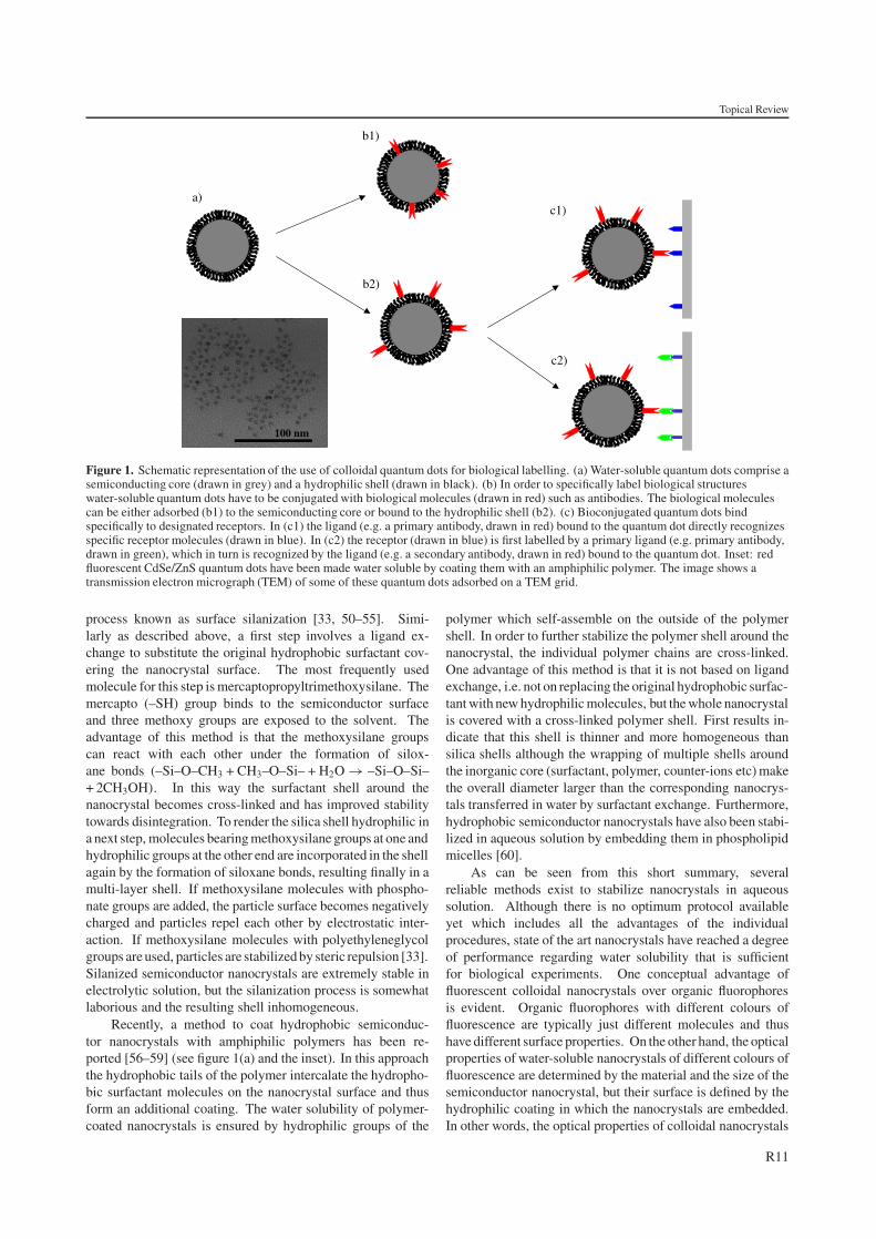
coating with an additional layer introducing either electriccharge or hydrophilic polymers for mediating solubilityin water. Coulomb repulsion between nanocrystals withsurface charge of the same polarity prevents aggregationin water. However, in salt-containing solutions such ascell culture media charged particles tend to aggregate, aphenomenon well known in colloid chemistry (‘salt-inducedaggregation’). Qualitatively, this phenomenon is explained bythe reduction of the thickness of the so-called diffuse layerof a counter-ion cloud surrounding a charged particle withincreasing ionic strength (the thickness of the diffuse layer isinversely proportional to the Debye–Huckel parameter whichis proportional to the square root of the ionic strength). As aconsequence, in salt-containing solutions charged particles canapproach each other closely enough during thermal collisionssuch that the repulsive electrostatic forces are overcome bythe short range attractive van der Waals forces. The systemflocculates. The second principle, using hydrophilic polymers,involves steric stabilization. The considerable space occupiedby a hydrophilic or amphipathic coating polymer such aspolyethyleneglycol [33] or dextrane [34] cannot be occupiedby another polymer chain from the coating layer of anotherparticle. The particles then cannot approach each other suchthat van der Waals forces lead to flocculation. In any case, bothdiscussed principles involve the conversion of hydrophobic tohydrophilic surfaces [23] (see figure 1(a)).
In practice, hydrophobic nanocrystals can be madehydrophilic by several methods. Most of them rely on theexchange of the hydrophobic surfactant coatings with ligandmolecules that carry on one end functional groups that arereactive towards the nanocrystal surface, and hydrophilicgroups on the other end, which ensure water solubility.The most frequently used anchoring groups reactive tothe surface of semiconductor nanocrystals are thiol (–SH)functionalities, and carboxyl (–COOH) functionalities aremost often used as hydrophilic head groups. For pH values>5 the carboxyl groups are deprotonated and the nanocrystalsrepel each other by the negative charge of the carboxylateions (–COO−). Examples for such mercaptohydrocarbonicacids (SH– · · ·–COOH) are mercaptoacetic acid [35–38],mercaptopropionic acid [39], mercaptoundecanoic acid [40],mercaptobenzoic acid [41], dihydrolipoic acid [42], andcysteine [43, 44]. Pinaud et al have used synthetic peptideswith multiple cysteines as the anchor [45]. Also non-chargedmolecules like dithiothreitol [46], organic dendrons [47],and pyridine-functionalized polyethylene glycol (PEG) [48]have been used. In the case of pyridine-functionalized PEGthe pyridine functionality is used to bind to the nanocrystalsurface, whereas the hydrophilic PEG chain stabilizes thenanocrystals in aqueous solution by steric repulsion. Althoughligand exchange from hydrophobic to hydrophilic surfactantsis straightforward, there are drawbacks connected with thismethod. Unfortunately, the bond between thiol (–SH) groupsand semiconductor surfaces is not very strong and thereforefrom the viewpoint of long term stability the hydrophilic ligandshell around the nanocrystal is prone to disintegration, whichis tantamount to particle aggregation [49, 50].
An alternative approach is based on the growth of ahydrophilic silica shell around the nanocrystals, through a
R10
Topical Review
100 nm
a)
b1)
b2)
c1)
c2)
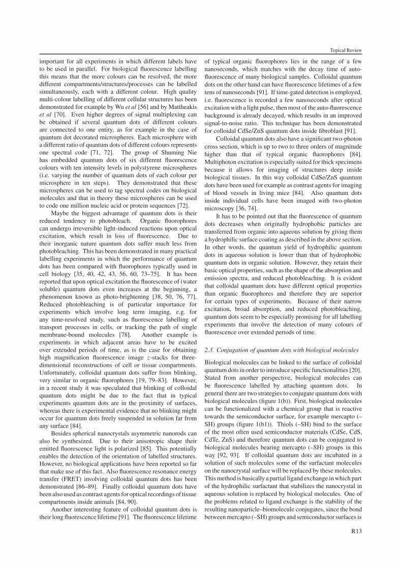
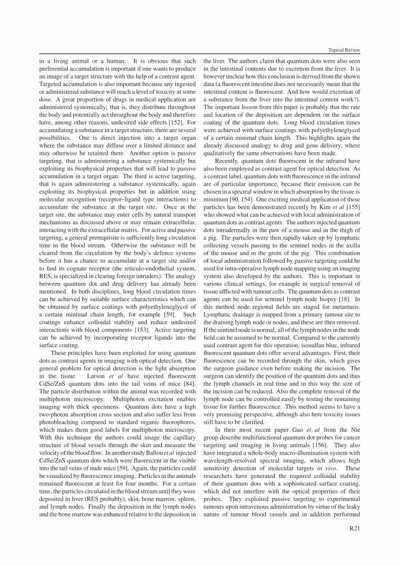
Figure 1. Schematic representation of the use of colloidal quantum dots for biological labelling. (a) Water-soluble quantum dots comprise asemiconducting core (drawn in grey) and a hydrophilic shell (drawn in black). (b) In order to specifically label biological structureswater-soluble quantum dots have to be conjugated with biological molecules (drawn in red) such as antibodies. The biological moleculescan be either adsorbed (b1) to the semiconducting core or bound to the hydrophilic shell (b2). (c) Bioconjugated quantum dots bindspecifically to designated receptors. In (c1) the ligand (e.g. a primary antibody, drawn in red) bound to the quantum dot directly recognizesspecific receptor molecules (drawn in blue). In (c2) the receptor (drawn in blue) is first labelled by a primary ligand (e.g. primary antibody,drawn in green), which in turn is recognized by the ligand (e.g. a secondary antibody, drawn in red) bound to the quantum dot. Inset: redfluorescent CdSe/ZnS quantum dots have been made water soluble by coating them with an amphiphilic polymer. The image shows atransmission electron micrograph (TEM) of some of these quantum dots adsorbed on a TEM grid.
process known as surface silanization [33, 50–55]. Simi-larly as described above, a first step involves a ligand ex-change to substitute the original hydrophobic surfactant cov-ering the nanocrystal surface. The most frequently usedmolecule for this step is mercaptopropyltrimethoxysilane. Themercapto (–SH) group binds to the semiconductor surfaceand three methoxy groups are exposed to the solvent. Theadvantage of this method is that the methoxysilane groupscan react with each other under the formation of silox-ane bonds (–Si–O–CH3 + CH3–O–Si– + H2O → –Si–O–Si–+ 2CH3OH). In this way the surfactant shell around thenanocrystal becomes cross-linked and has improved stabilitytowards disintegration. To render the silica shell hydrophilic ina next step, molecules bearing methoxysilane groups at one andhydrophilic groups at the other end are incorporated in the shellagain by the formation of siloxane bonds, resulting finally in amulti-layer shell. If methoxysilane molecules with phospho-nate groups are added, the particle surface becomes negativelycharged and particles repel each other by electrostatic inter-action. If methoxysilane molecules with polyethyleneglycolgroups are used, particles are stabilized by steric repulsion [33].Silanized semiconductor nanocrystals are extremely stable inelectrolytic solution, but the silanization process is somewhatlaborious and the resulting shell inhomogeneous.
Recently, a method to coat hydrophobic semiconduc-tor nanocrystals with amphiphilic polymers has been re-ported [56–59] (see figure 1(a) and the inset). In this approachthe hydrophobic tails of the polymer intercalate the hydropho-bic surfactant molecules on the nanocrystal surface and thusform an additional coating. The water solubility of polymer-coated nanocrystals is ensured by hydrophilic groups of the
polymer which self-assemble on the outside of the polymershell. In order to further stabilize the polymer shell around thenanocrystal, the individual polymer chains are cross-linked.One advantage of this method is that it is not based on ligandexchange, i.e. not on replacing the original hydrophobic surfac-tant with new hydrophilic molecules, but the whole nanocrystalis covered with a cross-linked polymer shell. First results in-dicate that this shell is thinner and more homogeneous thansilica shells although the wrapping of multiple shells aroundthe inorganic core (surfactant, polymer, counter-ions etc) makethe overall diameter larger than the corresponding nanocrys-tals transferred in water by surfactant exchange. Furthermore,hydrophobic semiconductor nanocrystals have also been stabi-lized in aqueous solution by embedding them in phospholipidmicelles [60].
As can be seen from this short summary, severalreliable methods exist to stabilize nanocrystals in aqueoussolution. Although there is no optimum protocol availableyet which includes all the advantages of the individualprocedures, state of the art nanocrystals have reached a degreeof performance regarding water solubility that is sufficientfor biological experiments. One conceptual advantage offluorescent colloidal nanocrystals over organic fluorophoresis evident. Organic fluorophores with different colours offluorescence are typically just different molecules and thushave different surface properties. On the other hand, the opticalproperties of water-soluble nanocrystals of different colours offluorescence are determined by the material and the size of thesemiconductor nanocrystal, but their surface is defined by thehydrophilic coating in which the nanocrystals are embedded.In other words, the optical properties of colloidal nanocrystals
R11
Topical Review
400 500 600 700 8000.0
0.5
1.0
1.5
Abs
orpt
ion
/ Flu
ores
cenc
e
wavelength (nm)
Polymer coated CdSe/ZnS
Absorption: green orange red
Fluorescence: green orange red
400 500 600 700 8000.0
0.2
0.4
0.6
0.8
1.0Absorption:
fluorescein TAMRA Cy5
Fluorescence: fluorescein TAMRA Cy5
a) b)
Abs
orpt
ion/
Flu
ores
cenc
e
wavelength (nm)
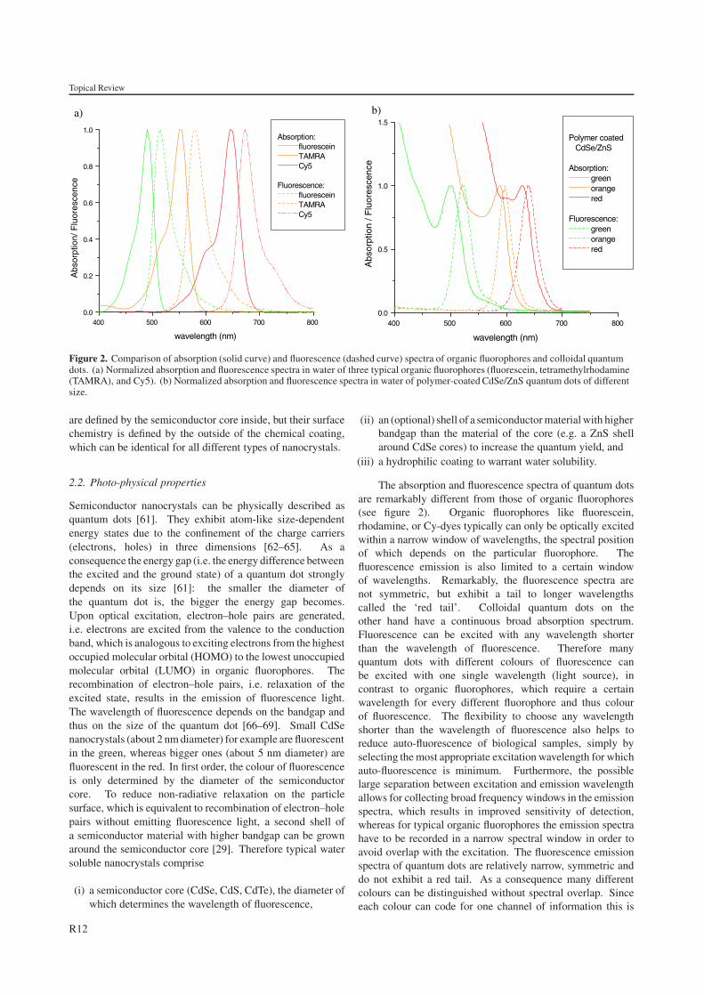
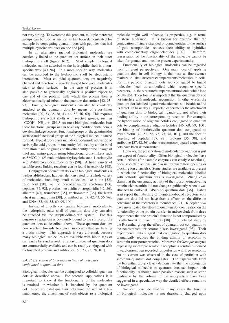
Figure 2. Comparison of absorption (solid curve) and fluorescence (dashed curve) spectra of organic fluorophores and colloidal quantumdots. (a) Normalized absorption and fluorescence spectra in water of three typical organic fluorophores (fluorescein, tetramethylrhodamine(TAMRA), and Cy5). (b) Normalized absorption and fluorescence spectra in water of polymer-coated CdSe/ZnS quantum dots of differentsize.
are defined by the semiconductor core inside, but their surfacechemistry is defined by the outside of the chemical coating,which can be identical for all different types of nanocrystals.
2.2. Photo-physical properties
Semiconductor nanocrystals can be physically described asquantum dots [61]. They exhibit atom-like size-dependentenergy states due to the confinement of the charge carriers(electrons, holes) in three dimensions [62–65]. As aconsequence the energy gap (i.e. the energy difference betweenthe excited and the ground state) of a quantum dot stronglydepends on its size [61]: the smaller the diameter ofthe quantum dot is, the bigger the energy gap becomes.Upon optical excitation, electron–hole pairs are generated,i.e. electrons are excited from the valence to the conductionband, which is analogous to exciting electrons from the highestoccupied molecular orbital (HOMO) to the lowest unoccupiedmolecular orbital (LUMO) in organic fluorophores. Therecombination of electron–hole pairs, i.e. relaxation of theexcited state, results in the emission of fluorescence light.The wavelength of fluorescence depends on the bandgap andthus on the size of the quantum dot [66–69]. Small CdSenanocrystals (about 2 nm diameter) for example are fluorescentin the green, whereas bigger ones (about 5 nm diameter) arefluorescent in the red. In first order, the colour of fluorescenceis only determined by the diameter of the semiconductorcore. To reduce non-radiative relaxation on the particlesurface, which is equivalent to recombination of electron–holepairs without emitting fluorescence light, a second shell ofa semiconductor material with higher bandgap can be grownaround the semiconductor core [29]. Therefore typical watersoluble nanocrystals comprise
(i) a semiconductor core (CdSe, CdS, CdTe), the diameter ofwhich determines the wavelength of fluorescence,
(ii) an (optional) shell of a semiconductor material with higherbandgap than the material of the core (e.g. a ZnS shellaround CdSe cores) to increase the quantum yield, and
(iii) a hydrophilic coating to warrant water solubility.
The absorption and fluorescence spectra of quantum dotsare remarkably different from those of organic fluorophores(see figure 2). Organic fluorophores like fluorescein,rhodamine, or Cy-dyes typically can only be optically excitedwithin a narrow window of wavelengths, the spectral positionof which depends on the particular fluorophore. Thefluorescence emission is also limited to a certain windowof wavelengths. Remarkably, the fluorescence spectra arenot symmetric, but exhibit a tail to longer wavelengthscalled the ‘red tail’. Colloidal quantum dots on theother hand have a continuous broad absorption spectrum.Fluorescence can be excited with any wavelength shorterthan the wavelength of fluorescence. Therefore manyquantum dots with different colours of fluorescence canbe excited with one single wavelength (light source), incontrast to organic fluorophores, which require a certainwavelength for every different fluorophore and thus colourof fluorescence. The flexibility to choose any wavelengthshorter than the wavelength of fluorescence also helps toreduce auto-fluorescence of biological samples, simply byselecting the most appropriate excitation wavelength for whichauto-fluorescence is minimum. Furthermore, the possiblelarge separation between excitation and emission wavelengthallows for collecting broad frequency windows in the emissionspectra, which results in improved sensitivity of detection,whereas for typical organic fluorophores the emission spectrahave to be recorded in a narrow spectral window in order toavoid overlap with the excitation. The fluorescence emissionspectra of quantum dots are relatively narrow, symmetric anddo not exhibit a red tail. As a consequence many differentcolours can be distinguished without spectral overlap. Sinceeach colour can code for one channel of information this is
R12
Topical Review
important for all experiments in which different labels haveto be used in parallel. For biological fluorescence labellingthis means that the more colours can be resolved, the moredifferent compartments/structures/processes can be labelledsimultaneously, each with a different colour. High qualitymulti-colour labelling of different cellular structures has beendemonstrated for example by Wu et al [56] and by Mattheakiset al [70]. Even higher degrees of signal multiplexing canbe obtained if several quantum dots of different coloursare connected to one entity, as for example in the case ofquantum dot decorated microspheres. Each microsphere witha different ratio of quantum dots of different colours representsone spectral code [71, 72]. The group of Shuming Niehas embedded quantum dots of six different fluorescencecolours with ten intensity levels in polystyrene microspheres(i.e. varying the number of quantum dots of each colour permicrosphere in ten steps). They demonstrated that thesemicrospheres can be used to tag spectral codes on biologicalmolecules and that in theory these microspheres can be usedto code one million nucleic acid or protein sequences [72].
Maybe the biggest advantage of quantum dots is theirreduced tendency to photobleach. Organic fluorophorescan undergo irreversible light-induced reactions upon opticalexcitation, which result in loss of fluorescence. Due totheir inorganic nature quantum dots suffer much less fromphotobleaching. This has been demonstrated in many practicallabelling experiments in which the performance of quantumdots has been compared with fluorophores typically used incell biology [35, 40, 42, 43, 56, 60, 73–75]. It has beenreported that upon optical excitation the fluorescence of (watersoluble) quantum dots even increases at the beginning, aphenomenon known as photo-brightening [38, 50, 76, 77].Reduced photobleaching is of particular importance forexperiments which involve long term imaging, e.g. forany time-resolved study, such as fluorescence labelling oftransport processes in cells, or tracking the path of singlemembrane-bound molecules [78]. Another example isexperiments in which adjacent areas have to be excitedover extended periods of time, as is the case for obtaininghigh magnification fluorescence image z-stacks for three-dimensional reconstructions of cell or tissue compartments.Unfortunately, colloidal quantum dots suffer from blinking,very similar to organic fluorophores [19, 79–83]. However,in a recent study it was speculated that blinking of colloidalquantum dots might be due to the fact that in typicalexperiments quantum dots are in the proximity of surfaces,whereas there is experimental evidence that no blinking mightoccur for quantum dots freely suspended in solution far fromany surface [84].
Besides spherical nanocrystals asymmetric nanorods canalso be synthesized. Due to their anisotropic shape theiremitted fluorescence light is polarized [85]. This potentiallyenables the detection of the orientation of labelled structures.However, no biological applications have been reported so farthat make use of this fact. Also fluorescence resonance energytransfer (FRET) involving colloidal quantum dots has beendemonstrated [86–89]. Finally colloidal quantum dots havebeen also used as contrast agents for optical recordings of tissuecompartments inside animals [84, 90].
Another interesting feature of colloidal quantum dots istheir long fluorescence lifetime [91]. The fluorescence lifetime
of typical organic fluorophores lies in the range of a fewnanoseconds, which matches with the decay time of auto-fluorescence of many biological samples. Colloidal quantumdots on the other hand can have fluorescence lifetimes of a fewtens of nanoseconds [91]. If time-gated detection is employed,i.e. fluorescence is recorded a few nanoseconds after opticalexcitation with a light pulse, then most of the auto-fluorescencebackground is already decayed, which results in an improvedsignal-to-noise ratio. This technique has been demonstratedfor colloidal CdSe/ZnS quantum dots inside fibroblast [91].
Colloidal quantum dots also have a significant two-photoncross section, which is up to two to three orders of magnitudehigher than that of typical organic fluorophores [84].Multiphoton excitation is especially suited for thick specimensbecause it allows for imaging of structures deep insidebiological tissues. In this way colloidal CdSe/ZnS quantumdots have been used for example as contrast agents for imagingof blood vessels in living mice [84]. Also quantum dotsinside individual cells have been imaged with two-photonmicroscopy [36, 74].
It has to be pointed out that the fluorescence of quantumdots decreases when originally hydrophobic particles aretransferred from organic into aqueous solution by giving thema hydrophilic surface coating as described in the above section.In other words, the quantum yield of hydrophilic quantumdots in aqueous solution is lower than that of hydrophobicquantum dots in organic solution. However, they retain theirbasic optical properties, such as the shape of the absorption andemission spectra, and reduced photobleaching. It is evidentthat colloidal quantum dots have different optical propertiesthan organic fluorophores and therefore they are superiorfor certain types of experiments. Because of their narrowexcitation, broad absorption, and reduced photobleaching,quantum dots seem to be especially promising for all labellingexperiments that involve the detection of many colours offluorescence over extended periods of time.
2.3. Conjugation of quantum dots with biological molecules
Biological molecules can be linked to the surface of colloidalquantum dots in order to introduce specific functionalities [20].Stated from another perspective, biological molecules canbe fluorescence labelled by attaching quantum dots. Ingeneral there are two strategies to conjugate quantum dots withbiological molecules (figure 1(b)). First, biological moleculescan be functionalized with a chemical group that is reactivetowards the semiconductor surface, for example mercapto (–SH) groups (figure 1(b1)). Thiols (–SH) bind to the surfaceof the most often used semiconductor materials (CdSe, CdS,CdTe, ZnS) and therefore quantum dots can be conjugated tobiological molecules bearing mercapto (–SH) groups in thisway [92, 93]. If colloidal quantum dots are incubated in asolution of such molecules some of the surfactant moleculeson the nanocrystal surface will be replaced by these molecules.This method is basically a partial ligand exchange in which partof the hydrophilic surfactant that stabilizes the nanocrystal inaqueous solution is replaced by biological molecules. One ofthe problems related to ligand exchange is the stability of theresulting nanoparticle–biomolecule conjugates, since the bondbetween mercapto (–SH) groups and semiconductor surfaces is
R13
Topical Review
not very strong. To overcome this problem, multiple mercaptogroups can be used as anchor, as has been demonstrated forexample by conjugating quantum dots with peptides that hadmultiple cysteine residues on one end [45].
In an alternative method biological molecules arecovalently linked to the quantum dot surface on their outerhydrophilic shell (figure 1(b2)). Most simply, biologicalmolecules can be adsorbed to the hydrophilic shell in a non-specific way [40, 94]. In a more specific way, moleculescan be adsorbed to the hydrophilic shell by electrostaticinteraction. Most colloidal quantum dots are negativelycharged and therefore positively charged biological moleculesstick to their surface. In the case of proteins it isalso possible to genetically engineer a positive zipper toone end of the protein, with which the protein then iselectrostatically adsorbed to the quantum dot surface [42, 95–97]. Finally, biological molecules can also be covalentlyattached to the quantum dots by means of cross-linkermolecules [20, 33, 35–38, 43, 46, 52, 56, 60]. This requireshydrophilic surfactant shells with reactive groups, such as–COOH, –NH2, or –SH. Since most biological molecules bearthese functional groups or can be easily modified with them, acovalent linkage between functional groups on the quantum dotsurface and functional groups of the biological molecule can beformed. Typical procedures include carbodiimide activation ofcarboxylic acid groups on one entity followed by amide bondformation to amino groups on the other entity or the linkage ofthiol and amino groups using bifunctional cross-linkers suchas SMCC (4-(N -maleimidomethyl)cyclohexane-1-carboxylicacid N -hydroxysuccinimide ester) [98]. A huge variety ofsuitable cross-linking reactions can be found in textbooks [98].
Conjugation of quantum dots with biological molecules iswell established and has been demonstrated for a whole varietyof molecules, including small molecules like biotin [52],folic acid [20], or the neurotransmitter serotonin [93],peptides [37, 92], proteins like avidin or streptavidin [42, 56],albumin [40], transferrin [35], trichosanthin [36], the lectinwheat germ agglutinin [38], or antibodies [37, 42, 43, 56, 96],and DNA [33, 46, 55, 60, 99, 100].
Instead of directly conjugating biological molecules tothe hydrophilic outer shell of quantum dots they can alsobe attached via the streptavidin–biotin system. For thispurpose streptavidin is covalently bound to the surface of thequantum dots as described above. These quantum dots arenow reactive towards biological molecules that are bearinga biotin moiety. This approach is very universal, becausemany biological molecules are available with biotin tags orcan easily be synthesized. Streptavidin-coated quantum dotsare commercially available and can be readily conjugated withbiotinylated proteins and antibodies [56, 73, 75, 78, 101].
2.4. Preservation of biological activity of moleculesconjugated to quantum dots
Biological molecules can be conjugated to colloidal quantumdots as described above. For potential applications it isimportant to know if the functionality of the moleculesis retained or whether it is impaired by the quantumdot. Since colloidal quantum dots have the size of a fewnanometres, the attachment of such objects to a biological
molecule might well influence its properties, e.g. in termsof steric hindrance. It is known for example that theconjugation of single-stranded oligonucleotides to the surfaceof gold nanoparticles reduces their ability to hybridizewith complementary oligonucleotides [102]. Therefore,preservation of the functionality of the molecule cannot betaken for granted and must be proven experimentally.
Functionality of biological molecules can be regardedfrom different perspectives. One main idea of applyingquantum dots in cell biology is their use as fluorescencemarkers to label structures/compartments/molecules in cells.For this purpose quantum dots are conjugated to ligandmolecules (such as antibodies) which recognize specificreceptors, i.e. the structure/compartment/molecule which is tobe labelled. Therefore, it is important that the quantum dots donot interfere with molecular recognition. In other words, thequantum-dot-labelled ligand molecule must still be able to findits target. In basically all reported experiments the attachmentof quantum dots to biological ligands did not affect theirbinding ability to the corresponding receptor. For example,the hybridization of oligonucleotides conjugated to quantumdots to complementary oligonucleotides [46, 60, 99, 100],the binding of biotin/avidin quantum dots conjugated toavidin/biotin [42, 52, 56, 73, 75, 78, 101], and the specifictargeting of peptides [37, 92], proteins [35, 38], andantibodies [37, 42, 56] to their receptors conjugated to quantumdots have been demonstrated.
However, the preservation of molecular recognition is justone aspect of functionality. Often biological molecules havecertain effects (for example enzymes can catalyse reactions),or cause certain actions (such as neurotransmitters opening orblocking ion channels). Some studies are available at presentin which the functionality of biological molecules labelledwith colloidal quantum dots is investigated. Zhang et alclaim that the enzymatic activity of the ribosome-inactivatingprotein trichosanthin did not change significantly when it wasattached to colloidal CdSe/ZnS quantum dots [36]. Dahanet al report that labelling of membrane-bound receptors withquantum dots did not have drastic effects on the diffusionbehaviour of the receptors in membranes [91]. Kloepfer et alhave investigated the effect of quantum dot conjugation on thefunctionality of the protein transferrin and conclude from threeexperiments that the protein’s function is not compromised byits attachment to quantum dots [38]. In a detailed study bythe Rosenthal group the effect of quantum dot conjugation tothe neurotransmitter serotonin was investigated [93]. Theirexperimental data suggest that conjugation to quantum dotsdramatically reduces the binding affinity of serotonin toserotonin-transporter proteins. Moreover, for Xenopus oocytesexpressing ionotropic serotonin receptors a serotonin-inducedinward current was recorded for perfusion with free serotonin,but no current was observed in the case of perfusion withserotonin–quantum dot conjugates. The experiments fromthe Rosenthal group clearly demonstrate that the conjugationof biological molecules to quantum dots can impair theirfunctionality. Although some possible reasons such as sterichindrance by the volume of the nanoparticle have beensuggested in a speculative way the detailed effects remain tobe investigated.
We can conclude that in many cases the functionof biological molecules is not drastically changed upon
R14
Topical Review
conjugation to colloidal quantum dots. In particular, the abilityof molecular recognition, i.e. the affinity of ligands to theirreceptor, seems at least to be partially preserved. On the otherhand it has been reported that sophisticated molecules thattrigger an effect, like the opening of an ion channel, can beimpaired upon conjugation to quantum dots. However, to ourknowledge there are no detailed studies available at present inwhich the functionality of molecules conjugated to quantumdots is analysed in a quantitative way, e.g. in terms of bindingconstants or catalytic activity.
2.5. Toxicity/biocompatibility
Since constituents of colloidal quantum dots such ascadmium [103] or selenium [104] are toxic to manycells, harmful effects can be expected, especially when thehydrophilic shell around the quantum dots is not stable andthey might dissolve under the release of toxic ions. Onthe other hand, some cells have developed mechanisms tocope with such ions, such as assembling them to particles ina biomineralization process, which efficiently removes ionsfrom solution. In this way, fluorescent colloidal CdS [105, 106]or PbS [107] quantum dots are grown in some cells. In terms ofbiocompatibility or toxicity we have to differentiate betweenseveral modes how quantum dots are introduced into cells.Certainly the results will also strongly depend on the type ofcells and on the hydrophilic shell used to stabilize the quantumdots in aqueous solution.
Quantum dots injected into the tail veins of mice arereported to cause no visible toxic effect after 24 h [92] andeven after days [59, 84] (20 pmol quantum dots injected pergram animal weight [59]). Dubertret et al report the injectionof quantum dots in individual cells of early stage Xenopusembryos [60]. Such embryonic systems are very sensitive toany perturbation and are therefore ideal for investigating toxiceffects, which typically result in phenotype abnormalities.Whereas typical injections of 2 × 10⁹ quantum dots per celldid not statistically change the health of the embryos, forinjections of >5 × 10⁹ quantum dots per cell abnormalitieslike changes in cell size, cell movement, axis elongation, andposterior truncations became apparent. Dubtret et al speculatethat these abnormalities might be caused by changes in theosmotic equilibrium of the cells due to the injected quantumdots.
Colloid quantum dots dissolved in the culture mediumare ingested by many cells. This will be discussed in detailbelow. Concerning toxicity, some groups report as a generalstatement (e.g. without details about the concentration ofquantum dots) that there are no detectable differences fromunlabelled cells up to 2 weeks after incubation [45, 70, 74, 91].Derfus et al measured the liver specific function of hepatocytecells incubated with quantum dots in terms of the albuminsecretion and could not determine any difference fromuntreated cells [108]. In a more quantitative way Hanakiet al demonstrated that the viability of cells was notaffected for quantum dot concentrations of 0.4 g l−1 inthe medium [40]. Jaiswal et al report that 400–600 nMquantum dots in the medium had no detectable effects on cellmorphology or physiology [42]. Furthermore quantum dotsin these concentrations did not interfere with the initiation
of development of Dictyostelium discoideum cells and theirresponse to cAMP [42]. Finally, Winter et al report thatthe addition of 0.03 nM colloidal quantum dots (which werenot ingested by the cells) did not alter the proliferation andattachment of cells within 5 days [37].
All studies described above can at best be called semi-quantitative and most of them are based on judging the viabilityof the cells by optical light microscopy. In a quantitativeway, cell viability can be measured with colorimetric assays,such as the MTT reagent (3-(4,5-dimethylthiazol-2-yl)-2,5-diphenyl tetrazolium bromide). Such quantitative assays allowfor measuring absolute numbers for the survival rate of cellsupon incubation with toxins. With the MTT assay Mattheakiset al demonstrated that no toxicity of highly purified polymer-coated CdSe/ZnS quantum dots could be observed for typicallabelling concentrations of 10 nM [70]. Furthermore, thesame authors demonstrated in a quantitative way for severalcell lines that under the above mentioned conditions there isno interference of the quantum dot labels with the followingcellular functions: ligand binding to cellular compartments,protein trafficking, and signal transduction. The same MTTassay has been used for the so far most comprehensivecytotoxicity study on quantum dots by Derfus et al [108].For enhanced sensitivity this study was performed on primaryhepatocyte cells from rats instead of using cell lines, sinceit is known that even low levels of Cd2+ reduce the viabilityof hepatocytes in vitro [109]. The authors have performedtests with different types of quantum dots. As a ‘worst case’,plain CdSe quantum dots coated with mercaptoacetic acidwere used. In particular after UV exposure or exposure to airsignificant levels of free Cd2+ ions were found in the quantumdot solutions. The viability of the cells incubated with thesesamples correlated with the concentration of free Cd2+ ions insolution [108]. Based on these results the authors concludethat cytotoxicity of CdSe quantum dots correlates with theliberation of free Cd2+ ions from the CdSe lattice. Exposureto air induces oxidation of the quantum dot surface [29] andexposure to UV radiation catalyses oxidation of the quantumdot surface [49], which finally results in the release of Cd2+
ions. Free Cd2+ ions are known to interfere with the functionof the mitochondria [110]. Derfus et al could demonstratethat release of Cd2+ ions und thus cytotoxicity can be reducedby coating CdSe quantum dots with appropriate shells [108].Already the overcoating of CdSe quantum dots with a ZnS shelldramatically reduced Cd2+ release and cytotoxicity. Adsorbedbovine serum albumin (BSA) or coating the particles witha polymer shell further suppressed Cd2+ release. For theseprotected particles no cytotoxic effects were found for particleconcentrations up to 1 mg ml−1, which corresponds to µMconcentrations.
In summary, these results suggest that in the case ofmoderate concentrations of quantum dots toxic effects on cellscan be neglected in first order, especially when Cd2+ releasefrom the quantum dots is reduced by appropriate encapsulationof the CdSe cores. However, the field would still benefit fromadditional systematic studies on the concentration limits and onthe dependence of the surface chemistry of the quantum dots.We also want to mention the following dilemma for labellingstudies with animals or humans: cytotoxicity has certainlyto be considered from a different perspective for labelling
R15
Topical Review
cell cultures compared to labelling animals or humans. Forcell cultures, inert quantum dots that do not release any toxicions are perfectly suited. On the other hand, as will bedescribed in detail later in this article, living cells ingestquantum dots: quantum dots can be used as contrast agentsin animals and even usage in humans is thinkable. In theshort range quantum dots as inert as possible are also desirablein order to prevent acute damage of the tissue. However,many of these ingested dots remain in the living tissue formonths and presumably even for years [59]. No encapsulationcan be as inert as to withstand degradation forever. Thismeans that even quantum dots that have been encapsulatedin several protection shells might slowly release toxic ions inthe time course of years. Therefore, for studies on humansalternative quantum dot materials would be desirable, whichwould be based on biodegradable constituents. For magneticresonance imaging on humans iron oxide nanoparticles havefor example been used as contrast agents. These particles arealso ingested by living cells [111], but they are deliberatelynot inert. Biodegradation of the particles finally results in freeiron that is incorporated into haemoglobin [112] and after somemonths no residues of the particles remain in the body.
3. Labelling of cellular structures and receptorswith quantum dots
The most widespread method to visualize structures or certainmolecules in cells is fluorescence microscopy. Due to theirinteresting optical properties colloidal quantum dots havebeen introduced as fluorescence labels for biological stainingexperiments since some years. Their application is in principlevery similar to that of organic fluorophores. After attachinga ligand, for example an antibody, to the label [4], thisconjugate binds with high specificity to its target receptor,which in turn can now be visualized by the fluorescenceof the label (see figure 1(c)). Since the size of typicalligands is too big to allow their transport through naturalpores in the cell membrane, fluorescence-labelled ligands haveto be artificially introduced inside cells if structures in thecell’s inside are to be labelled (this is true both for organicfluorophores and for colloidal quantum dots). If only a fewcells are to be labelled, fluorescence-labelled ligands can beinjected into living cells with micropipettes [113, 114]. Alsoelectroporation is used [115, 116]. In order to stain manycells in parallel, cells are typically fixed, i.e. stabilized bycross-linking their sugar skeleton on the cell surface (e.g. byglutaraldehyde) and their membrane is permeabilized withappropriate reagents (e.g. by triton). Fixed cells are deadand can be stored at 4 ◦C. Fluorescence-labelled antibodiesnow can enter the cells by the created artificial pores. Theselabelling techniques are well established and protocols can befound in almost any textbook about cell biology [117, 118].
Using quantum dot labels is very similar to using theirorganic counterparts. Cells are fixed and permeabilized,incubated in buffer with ligand-conjugated quantum dots,for example oligonucleotide-conjugated quantum dots [46],rinsed, and fluorescence is recorded with an opticalmicroscope. The first labelling experiment was performed in1998 by the Alivisatos group [52]. Nowadays high qualityimages like the dual-colour labelling shown in figure 3(a) can
be obtained with standardized procedures. If antibodies areused as ligands, typically a two-step reaction is used. First,a primary antibody is reacted with the target. Then quantumdots conjugated with a secondary antibody, which targets theprimary antibody, are added [43, 44, 56]. Since more thanone secondary antibody can bind to each primary antibody,such cascade reactions amplify the fluorescence signal.However, the direct conjugation of antibodies to quantumdots is laborious; often biotinylated secondary antibodiesare used, which are finally labelled by streptavidin–quantumdot conjugates via biotin–avidin interaction [73, 75, 119].This strategy is advantageous, because it involves only oneuniversal conjugation of quantum dots with streptavidin, andmost antibodies are commercially available with an attachedbiotin group. Further signal amplification can be achieved withthe so-called TSA enzyme amplification technique. Here thesecondary antibody is conjugated with horseradish peroxidase(HRP) and quantum dots are conjugated to tyramide (e.g. viabiotin–avidin interaction). Horseradish peroxidase convertstyramide to a very reactive oxidized intermediate, whichrapidly binds to all proteins in close proximity. In this way thewhole local environment of the secondary antibody is labelledwith fluorescent quantum dots [73]. Quantum dot labelling ofinner and outer structures of fixed cells has been used for avariety of receptors.
If structures on the surface of cells are to be labelled, thequantum dot labels do not need to traverse the cell membrane.Therefore, for outside labelling no fixation and poration isrequired and labelling can also be performed with livingcells [45, 56, 78, 93, 101]. In this way even the dynamicarrangement of single proteins within the cell membranecould be traced [78, 101]. Two examples for quantum dotlabelling of membrane bound proteins, one from the Rosenthalgroup, the other from Quantum Dot Corporation, are shown infigure 3. Quantum dot labelling of cells is at present usedby more and more groups and many new studies are to beexpected in the future. We think that quantum dots willbe used in the future in particular for time-resolved studiesof single molecules, as demonstrated by Dahan et al andLidke et al [78, 101]. The use of colloidal quantum dotsin these experiments offered significant advantages comparedto other methods: whereas single-molecule spectroscopyusing conventional organic fluorophores as labels sufferedfrom photobleaching which resulted in restricted observationtimes [120], the use of latex beads as labels suffered fromthe extended size of the beads [121]. It is likely thatcolloidal quantum dots never will completely replace organicfluorophores as fluorescence labels, but there are niches aslong term imaging for which their advantages are obvious andtherefore widely spread use is to be expected.
4. Incorporation of quantum dots by living cells
In addition to the use of colloidal quantum dots as fluorescencemarkers to label cellular structures [52] the Nie group reportedthat quantum dots are incorporated by living cells [35]. Sincethen many groups have reported the uptake of quantum dotsby individual cells or by animal tissue. The uptake of colloidalparticles into cells is an important area of biological researchin itself because it also relates to the cell’s communication with
R16
Topical Review
d)c)
a) b)
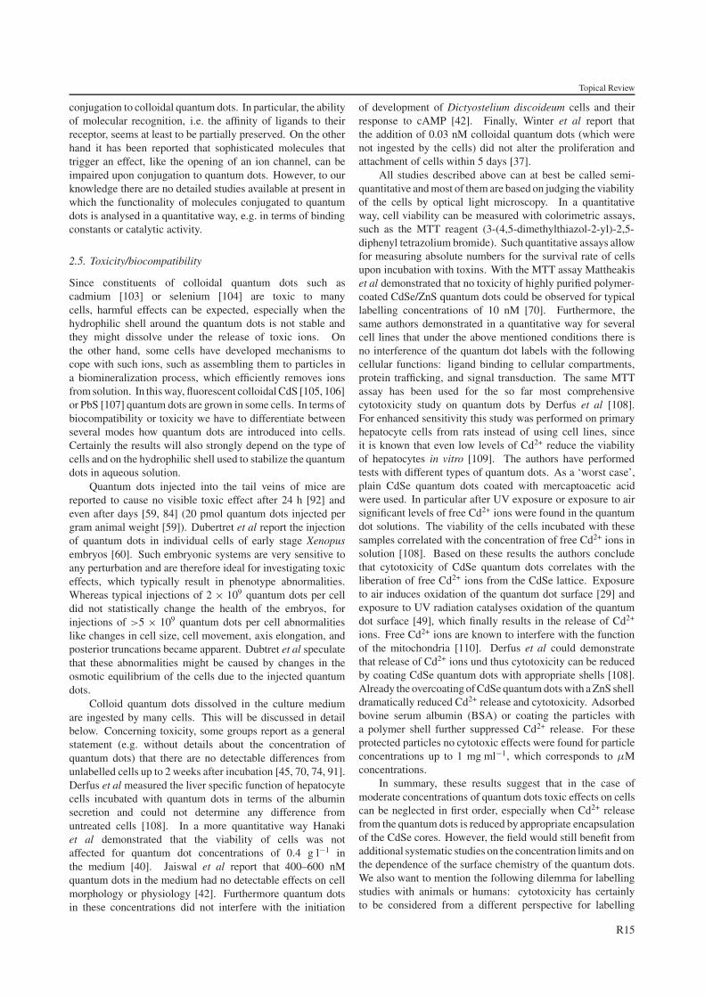
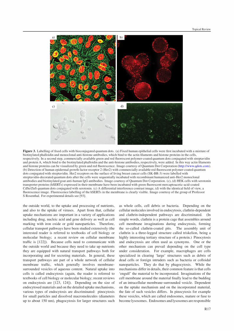
Figure 3. Labelling of fixed cells with bioconjugated quantum dots. (a) Fixed human epithelial cells were first incubated with a mixture ofbiotinylated phalloidin and monoclonal anti-histone antibodies, which bind to the actin filaments and histone proteins in the cells,respectively. In a second step, commercially available green and red fluorescent polymer-coated quantum dots conjugated with streptavidinand protein A, which bind to the biotinylated phalloidin and the anti-histone antibodies, respectively, were added. In this way actin filamentsand histone proteins can be visualized by green and red fluorescence. Image courtesy of Quantum Dot Corporation (http://www.qdots.com).(b) Detection of human epidermal growth factor receptor 2 (Her2) with commercially available red fluorescent polymer-coated quantumdots conjugated with streptavidin. Her2 receptors on the surface of living breast cancer cells (SK-BR-3) were labelled withstreptavidin-decorated quantum dots after the cells were sequentially incubated with recombinant humanized anti-Her2 monoclonalantibodies and biotinylated goat anti-human IgG antibodies. Image courtesy of Quantum Dot Corporation. (c), (d) HEK cells with serotonintransporter proteins (hSERTs) expressed in their membrane have been incubated with green fluorescent mercaptoacetic-acid-coatedCdSe/ZnS quantum dots conjugated with serotonin. (c) A differential interference contrast image, (d) with the identical field of view, afluorescence image. Fluorescence labelling of the hSERTs in the membrane is clearly visible. Image courtesy of the group of ProfessorS Rosenthal. For experimental details see [93].
the outside world, to the uptake and processing of nutrients,and also to the uptake of viruses. Apart from that, cellularuptake mechanisms are important in a variety of applicationsincluding drug, nucleic acid and gene delivery as well as cellmarking with iron oxide or gold nanoparticles. Therefore,cellular transport pathways have been studied extensively (theinterested reader is referred to textbooks of cell biology ormolecular biology; a recent review on cellular membranetraffic is [122]). Because cells need to communicate withthe outside world and because they need to take up nutrientsthey are equipped with natural transport pathways both forincorporating and for secreting materials. In general, thesetransport pathways are part of a whole network of cellularmembrane traffic, which generally involves membrane-surrounded vesicles of aqueous content. Natural uptake intocells is called endocytosis (again, the reader is referred totextbooks of cell biology or molecular biology; recent reviewson endocytosis are [123, 124]). Depending on the size ofendocytosed materials and on the detailed uptake mechanisms,various types of endocytosis are discriminated: pinocytosisfor small particles and dissolved macromolecules (diametersup to about 150 nm), phagocytosis for larger structures such
as whole cells, cell debris or bacteria. Depending on thecellular molecules involved in endocytosis, clathrin-dependentand clathrin-independent pathways are discriminated. (Insimple words, clathrin is a protein cage that assembles aroundcell membrane invaginations during endocytosis, formingthe so-called clathrin-coated pits. The assembly unit ofclathrin is a three-legged structure called triskelion, being ahighly interesting tertiary structure of a protein.) Pinocytosisand endocytosis are often used as synonyms. One or theother mechanism can prevail depending on the cell typeunder consideration. For example, macrophages are cellsspecialized in clearing ‘large’ structures such as debris ofdead cells or foreign intruders such as bacteria or colloidalnanoparticles. They do that by phagocytosis. While themechanisms differ in details, their common feature is that cells‘engulf’ the material to be incorporated. Invaginations of thecell membrane around the material finally lead to the buddingof an intracellular membrane-surrounded vesicle. Dependenton the uptake mechanism and on the incorporated material,the fate of such vesicles differs. In pinocytosis for examplethese vesicles, which are called endosomes, mature or fuse tobecome lysosomes. Endosomes and lysosomes are responsible
R17
Topical Review
for the breakdown of ingested materials. These vesiclesare equipped with specialized enzymes that would degradeproteins and nucleic acids. Among the uptake mechanisms,fluid phase uptake and uptake of materials bound to thecell surface are discriminated. Fluid phase uptake is thecontinuous incorporation of solutes from the media cells livein. Uptake of bound material can be specific or unspecificin terms of the type of binding to cell surfaces. Receptor-mediated endocytosis is called specific because it involvesand is triggered by a specific key/keyhole type receptor–ligand binding interaction. Among the many examples, atypical one is the uptake of transferrin via the transferrinreceptor, which provides cells with their iron ‘diet’. In thiscase, receptor and ligand are recycled to the cell surfaceafter intracellular release of iron. The transferrin/transferrinreceptor system has been widely used in drug and genedelivery [125, 126]. Receptor-mediated endocytosis is alsoinvolved in cellular communication, called signalling. Oneexample of medical relevance in tumour growth is theendocytosis of the urokinase-type plasminogen activator (uPA)receptor (uPA-R) upon binding of uPA. Without going intomolecular details, this event gives the cell a signal to divideand/or to migrate [127]. Triggering uptake into cells uponbinding does not necessarily require specific receptor–ligandtype binding events. In most cases unspecific binding issufficient and is easily accomplished with cationic materialsor particles. These bind by simple electrostatic interaction tothe outside of cells, which are generally negatively charged(negatively charged glycosaminoglycans of the extracellularmatrix are responsible). However, it is well known thatnegatively charged particles are also taken up into cells, and infact most cell labelling experiments with quantum dots havebeen carried out with anionic surface-coated particles.
Aiming at exploiting cellular transport mechanisms suchas endocytosis for cell labelling, it is highly instructive toconsider the vast amount of literature available from the fieldsof drug and gene delivery. Just making cells fluorescentby making them ingest quantum dots is a simple task.Usually, these particles, as they cannot be degraded, will bestored in membrane-surrounded compartments that will not beaccessible to solutes of the cytoplasm. Using quantum dots forlabelling specific intracellular structures outside endocytosedvesicles or to even image cellular reactions in the cytoplasmor the nucleus will require more sophisticated tools. Thesame problem prevails in drug and particularly in nucleicacid/gene delivery [128]. To exert their desired effect, thesemacromolecules need to be transported to the cell nucleus inthe majority of current applications (genes are transcribed intomessenger RNA, mRNA, in the nucleus; mRNA then carriesthe sequence information into the cytoplasm which is theretranslated into protein sequences). For transport across cellularmembranes nucleic acids are self-assembled (compacted) withpolycations or cationic lipids to form charged nanoparticles(also called ‘complexes’) [128]. Such particles can bind tocell surfaces non-specifically via electrostatic interaction orspecifically if equipped with receptor ligands. This can easilybe accomplished by chemical coupling of ligand moleculesto pre-assembled particles or to the cationic moiety beforeassembly of the particle [129]. Uptake into cells proceeds viaendocytosis. Escape from internal vesicles can be mediated
by also incorporating fusogenic components into the complexsuch as membrane-disrupting peptides that unfold their activityduring the internalization process [130–132]. Otherwise,the cationic moiety used to compact the polyanionic nucleicacid macromolecules can be chosen to comprise an inherentchemical structure that would interfere with the ‘standard’degradation of internalized material. For example, thepolycation polyethylenimine comprises secondary and tertiaryamino groups that can buffer the natural acidification processwithin endosomes (the so-called ‘proton sponge effect’, [133]).This in turn leads to an osmotic destabilization of thesevesicles, which together with the swelling of polyethylenimineat acidic pH leads to endosome disruption [134]. Thechemical structures of selected lipids also promote membranereorganizations that can be exploited to release internalizedmaterials such as nucleic acids into the cytoplasm [135].Exploiting cellular transport processes for ‘invasion’ has beencopied from viruses, which evolved mechanisms of trickingcells for their own purposes to perfection.
This excursion to nucleic acid and drug delivery andexisting quantum dot literature leads us to conclude thatnatural transport processes can and should be exploited forquantum dot delivery to cells in an analogous manner. Theinvolved surface chemistries are similar (see above); receptorligands have been coupled to quantum dots and endocytoticuptake, receptor mediated [20, 35, 36, 38, 45, 101] aswell as unspecific/adsorptive, has been described for theseparticles [23, 40, 42, 74, 91, 136]. The efficacy of uptakeas well as the exact uptake mechanisms (receptor specificversus adsorptive/unspecific) certainly depend on a varietyof parameters including particle size, surface chemistry,and, importantly, also the cell type under consideration.Experimental evidence for endocytotic uptake of quantumdots has been obtained by colocalization experiments withendosome markers. Hanaki et al used fluorescein-labelleddextrane as marker for endosomes/lysosomes [40] whichwere found to colocalize with ingested quantum dots.Jaiswal et al found colocalization of quantum dots withthe endosome specific marker pECFP [42]. This groupfurthermore demonstrated that quantum dot uptake can beblocked by cooling the cells to 4 ◦C, a technique knownto block endocytosis. Coating the quantum dot surfacewith polyethylene glycol (PEG) reduces adsorption to themembrane and uptake of these particles by cells [45, 59].Ligand-modified quantum dots that bind to membrane-bound receptors are internalized together with the receptormolecules [45, 101]. Lidke et al demonstrated that epidermal-growth-factor- (EGF-) conjugated quantum dots bind to erbBXtransmembrane receptors which are responsible for mediatingcellular responses to EGF [101]. EGF-conjugated quantumdots bound to the erbBX receptors were internalized by thecells. The authors demonstrated this to be via clathrin-coated pits by simultaneously adding fluorescence-labelledtransferrin, which was found to colocalize with the quantumdots as well on the cell surface as after internalization in earlyendosomes.
Most groups that studied cellular quantum dot uptakereport that in cell cultures or animal models, at later stagesquantum dots are stored in granular compartments around thenucleus [36, 40, 42, 70, 74], without penetrating the nucleus,
R18
Topical Review
b)
c)
a)
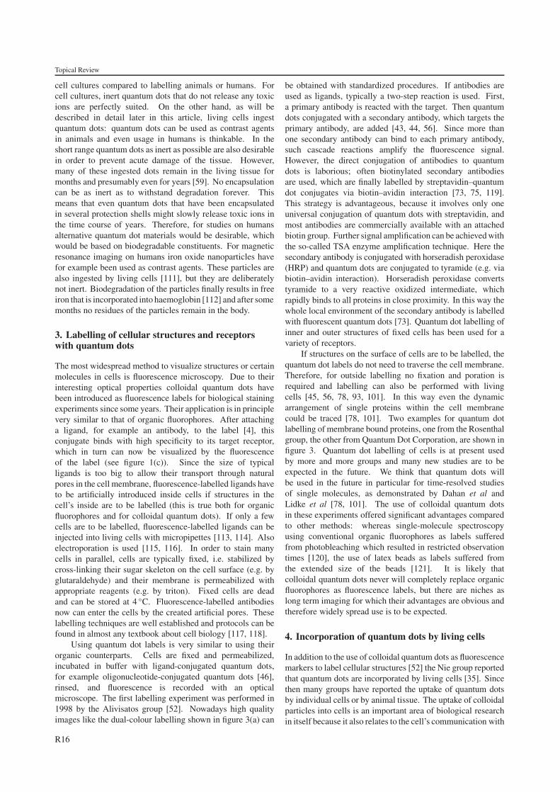
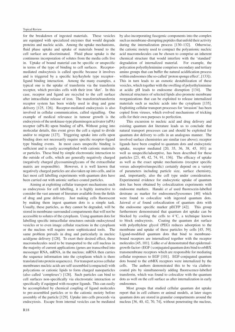
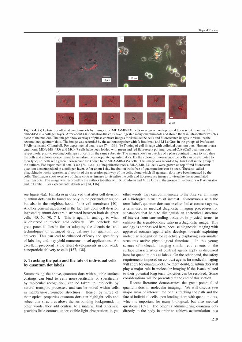
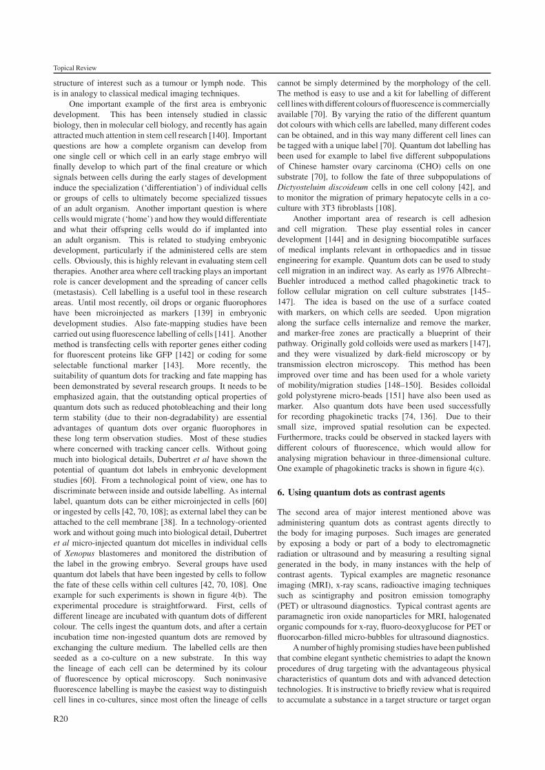
Figure 4. (a) Uptake of colloidal quantum dots by living cells. MDA-MB-231 cells were grown on top of red fluorescent quantum dotsembedded in a collagen layer. After about 4 h incubation the cells have ingested many quantum dots and stored them in intracellular vesiclesclose to the nucleus. The images show overlays of phase contrast images to visualize the cells and fluorescence images to visualize theaccumulated quantum dots. The image was recorded by the authors together with R Boudreau and M Le Gros in the groups of ProfessorsP Alivisatos and C Larabell. For experimental details see [74, 136]. (b) Tracing of cell lineage with colloidal quantum dots. Human breastcarcinoma MDA-MB-435s and MCF-7 cells have been loaded with green and red fluorescent polymer-coated CdSe/ZnS quantum dots,respectively, prior to seeding both types of cells on the same substrate. The image shows an overlay of a phase contrast image to visualizethe cells and a fluorescence image to visualize the incorporated quantum dots. By the colour of fluorescence the cells can be attributed totheir type, i.e. cells with green fluorescence are known to be MDA-MB-435s cells. This image was recorded by Tim Liedl in the group ofthe authors. For experimental details see [74, 136]. (c) Phagokinetic tracks. MDA-MB-231 cells were grown on top of red fluorescentquantum dots embedded in a collagen layer. After about 1 day incubation trails free of quantum dots can be seen. These so-calledphagokinetic tracks represent a blueprint of the migration pathway of the cells, along which all quantum dots have been ingested by thecells. The images show overlays of phase contrast images to visualize the cells and fluorescence images to visualize the accumulatedquantum dots. The image was recorded by the authors together with R Boudreau and M Le Gros in the groups of Professors A P Alivisatosand C Larabell. For experimental details see [74, 136].
see figure 4(a). Hanaki et al observed that after cell divisionquantum dots can be found not only in the perinuclear regionbut also in the neighbourhood of the cell membrane [40].Another general agreement is the fact that upon cell divisioningested quantum dots are distributed between both daughtercells [40, 60, 70, 74]. This is again in analogy to whatis observed in nucleic acid delivery. We conclude thatgreat potential lies in further adopting the chemistries andtechnologies of advanced drug delivery for quantum dotdelivery. This can lead to enhanced efficacy and specificityof labelling and may yield numerous novel applications. Anexcellent precedent is the latest developments in iron oxidenanoparticle delivery to cells [137, 138].
5. Tracking the path and the fate of individual cellsby quantum dot labels
Summarizing the above, quantum dots with suitable surfacecoatings can bind to cells non-specifically or specificallyby molecular recognition, can be taken up into cells bynatural transport processes, and can be stored within cellsin membrane-surrounded structures. Hence, by virtue oftheir optical properties quantum dots can highlight cells andsubcellular structures above the surrounding background; inother words, they add contrast to a material that otherwiseprovides little contrast under visible light observation; in yet
other words, they can communicate to the observer an imageof a biological structure of interest. Synonymous with theterm ‘label’, quantum dots can be classified as contrast agents,a term used in medical diagnostic imaging procedures forsubstances that help to distinguish an anatomical structureof interest from surrounding tissue or, in physical terms, toenhance the signal-to-noise ratio in a diagnostic image. Thisanalogy is emphasized here, because diagnostic imaging withapproved contrast agents also develops towards exploitingmolecular recognition for selectively displaying ever-smallerstructures and/or physiological functions. In this youngscience of molecular imaging similar requirements on thesurface characteristics of contrast agents apply as discussedhere for quantum dots as labels. On the other hand, the safetyrequirements imposed on contrast agents for medical imagingwill apply for quantum dots. Without doubt, quantum dots willplay a major role in molecular imaging if the issues relatedto their potential long term toxicities can be resolved. Someconsiderations will be presented at the end of this section.
Recent literature demonstrates the great potential ofquantum dots in molecular imaging. We will discuss twomajor areas of interest: the one is tracking the path and thefate of individual cells upon loading them with quantum dots,which is important for many biological, but also medicalquestions [139]. The other is administering quantum dotsdirectly to the body in order to achieve accumulation in a
R19
Topical Review
structure of interest such as a tumour or lymph node. Thisis in analogy to classical medical imaging techniques.
One important example of the first area is embryonicdevelopment. This has been intensely studied in classicbiology, then in molecular cell biology, and recently has againattracted much attention in stem cell research [140]. Importantquestions are how a complete organism can develop fromone single cell or which cell in an early stage embryo willfinally develop to which part of the final creature or whichsignals between cells during the early stages of developmentinduce the specialization (‘differentiation’) of individual cellsor groups of cells to ultimately become specialized tissuesof an adult organism. Another important question is wherecells would migrate (‘home’) and how they would differentiateand what their offspring cells would do if implanted intoan adult organism. This is related to studying embryonicdevelopment, particularly if the administered cells are stemcells. Obviously, this is highly relevant in evaluating stem celltherapies. Another area where cell tracking plays an importantrole is cancer development and the spreading of cancer cells(metastasis). Cell labelling is a useful tool in these researchareas. Until most recently, oil drops or organic fluorophoreshave been microinjected as markers [139] in embryonicdevelopment studies. Also fate-mapping studies have beencarried out using fluorescence labelling of cells [141]. Anothermethod is transfecting cells with reporter genes either codingfor fluorescent proteins like GFP [142] or coding for someselectable functional marker [143]. More recently, thesuitability of quantum dots for tracking and fate mapping hasbeen demonstrated by several research groups. It needs to beemphasized again, that the outstanding optical properties ofquantum dots such as reduced photobleaching and their longterm stability (due to their non-degradability) are essentialadvantages of quantum dots over organic fluorophores inthese long term observation studies. Most of these studieswhere concerned with tracking cancer cells. Without goingmuch into biological details, Dubertret et al have shown thepotential of quantum dot labels in embryonic developmentstudies [60]. From a technological point of view, one has todiscriminate between inside and outside labelling. As internallabel, quantum dots can be either microinjected in cells [60]or ingested by cells [42, 70, 108]; as external label they can beattached to the cell membrane [38]. In a technology-orientedwork and without going much into biological detail, Dubertretet al micro-injected quantum dot micelles in individual cellsof Xenopus blastomeres and monitored the distribution ofthe label in the growing embryo. Several groups have usedquantum dot labels that have been ingested by cells to followthe fate of these cells within cell cultures [42, 70, 108]. Oneexample for such experiments is shown in figure 4(b). Theexperimental procedure is straightforward. First, cells ofdifferent lineage are incubated with quantum dots of differentcolour. The cells ingest the quantum dots, and after a certainincubation time non-ingested quantum dots are removed byexchanging the culture medium. The labelled cells are thenseeded as a co-culture on a new substrate. In this waythe lineage of each cell can be determined by its colourof fluorescence by optical microscopy. Such noninvasivefluorescence labelling is maybe the easiest way to distinguishcell lines in co-cultures, since most often the lineage of cells
cannot be simply determined by the morphology of the cell.The method is easy to use and a kit for labelling of differentcell lines with different colours of fluorescence is commerciallyavailable [70]. By varying the ratio of the different quantumdot colours with which cells are labelled, many different codescan be obtained, and in this way many different cell lines canbe tagged with a unique label [70]. Quantum dot labelling hasbeen used for example to label five different subpopulationsof Chinese hamster ovary carcinoma (CHO) cells on onesubstrate [70], to follow the fate of three subpopulations ofDictyosteluim discoideum cells in one cell colony [42], andto monitor the migration of primary hepatocyte cells in a co-culture with 3T3 fibroblasts [108].
Another important area of research is cell adhesionand cell migration. These play essential roles in cancerdevelopment [144] and in designing biocompatible surfacesof medical implants relevant in orthopaedics and in tissueengineering for example. Quantum dots can be used to studycell migration in an indirect way. As early as 1976 Albrecht–Buehler introduced a method called phagokinetic track tofollow cellular migration on cell culture substrates [145–147]. The idea is based on the use of a surface coatedwith markers, on which cells are seeded. Upon migrationalong the surface cells internalize and remove the marker,and marker-free zones are practically a blueprint of theirpathway. Originally gold colloids were used as markers [147],and they were visualized by dark-field microscopy or bytransmission electron microscopy. This method has beenimproved over time and has been used for a whole varietyof mobility/migration studies [148–150]. Besides colloidalgold polystyrene micro-beads [151] have also been used asmarker. Also quantum dots have been used successfullyfor recording phagokinetic tracks [74, 136]. Due to theirsmall size, improved spatial resolution can be expected.Furthermore, tracks could be observed in stacked layers withdifferent colours of fluorescence, which would allow foranalysing migration behaviour in three-dimensional culture.One example of phagokinetic tracks is shown in figure 4(c).
6. Using quantum dots as contrast agents
The second area of major interest mentioned above wasadministering quantum dots as contrast agents directly tothe body for imaging purposes. Such images are generatedby exposing a body or part of a body to electromagneticradiation or ultrasound and by measuring a resulting signalgenerated in the body, in many instances with the help ofcontrast agents. Typical examples are magnetic resonanceimaging (MRI), x-ray scans, radioactive imaging techniquessuch as scintigraphy and positron emission tomography(PET) or ultrasound diagnostics. Typical contrast agents areparamagnetic iron oxide nanoparticles for MRI, halogenatedorganic compounds for x-ray, fluoro-deoxyglucose for PET orfluorocarbon-filled micro-bubbles for ultrasound diagnostics.
A number of highly promising studies have been publishedthat combine elegant synthetic chemistries to adapt the knownprocedures of drug targeting with the advantageous physicalcharacteristics of quantum dots and with advanced detectiontechnologies. It is instructive to briefly review what is requiredto accumulate a substance in a target structure or target organ
R20
Topical Review
in a living animal or a human. It is obvious that suchpreferential accumulation is important if one wants to producean image of a target structure with the help of a contrast agent.Targeted accumulation is also important because any ingestedor administered substance will reach a level of toxicity at somedose. A great proportion of drugs in medical application areadministered systemically; that is, they distribute throughoutthe body and potentially act throughout the body and thereforehave, among other reasons, undesired side effects [152]. Foraccumulating a substance in a target structure, there are severalpossibilities. One is direct injection into a target organwhere the substance may diffuse over a limited distance andmay otherwise be retained there. Another option is passivetargeting, that is administering a substance systemically butexploiting its biophysical properties that will lead to passiveaccumulation in a target organ. The third is active targeting,that is again administering a substance systemically, againexploiting its biophysical properties but in addition usingmolecular recognition (receptor–ligand type interactions) toaccumulate the substance at the target site. Once at thetarget site, the substance may enter cells by natural transportmechanisms as discussed above or may remain extracellular,interacting with the extracellular matrix. For active and passivetargeting, a general prerequisite is sufficiently long circulationtime in the blood stream. Otherwise the substance will becleared from the circulation by the body’s defence systemsbefore it has a chance to accumulate at a target site and/orto find its cognate receptor (the reticulo-endothelial system,RES, is specialized in clearing foreign intruders). The analogybetween quantum dot and drug delivery has already beenmentioned. In both disciplines, long blood circulation timescan be achieved by suitable surface characteristics which canbe obtained by surface coatings with polyethyleneglycol ofa certain minimal chain length, for example [59]. Suchcoatings enhance colloidal stability and reduce undesiredinteractions with blood components [153]. Active targetingcan be achieved by incorporating receptor ligands into thesurface coating.
These principles have been exploited for using quantumdots as contrast agents in imaging with optical detection. Onegeneral problem for optical detection is the light absorptionin the tissue. Larson et al have injected fluorescentCdSe/ZnS quantum dots into the tail veins of mice [84].The particle distribution within the animal was recorded withmultiphoton microscopy. Multiphoton excitation enablesimaging with thick specimens. Quantum dots have a hightwo-photon absorption cross section and also suffer less fromphotobleaching compared to standard organic fluorophores,which makes them good labels for multiphoton microscopy.With this technique the authors could image the capillarystructure of blood vessels through the skin and measure thevelocity of the blood flow. In another study Ballou et al injectedCdSe/ZnS quantum dots which were fluorescent in the visibleinto the tail veins of nude mice [59]. Again, the particles couldbe visualized by fluorescence imaging. Particles in the animalsremained fluorescent at least for four months. For a certaintime, the particles circulated in the blood stream until they weredeposited in liver (RES probably), skin, bone marrow, spleen,and lymph nodes. Finally the deposition in the lymph nodesand the bone marrow was enhanced relative to the deposition in
the liver. The authors claim that quantum dots were also seenin the intestinal contents due to excretion from the liver. It ishowever unclear how this conclusion is derived from the showndata (a fluorescent intestine does not necessarily mean that theintestinal content is fluorescent. And how would excretion ofa substance from the liver into the intestinal content work?).The important lesson from this paper is probably that the rateand location of the deposition are dependent on the surfacecoating of the quantum dots. Long blood circulation timeswere achieved with surface coatings with polyethyleneglycolof a certain minimal chain length. This highlights again thealready discussed analogy to drug and gene delivery, wherequalitatively the same observations have been made.
Recently, quantum dots fluorescent in the infrared havealso been employed as contrast agent for optical detection. Asa contrast label, quantum dots with fluorescence in the infraredare of particular importance, because their emission can bechosen in a spectral window in which absorption by the tissue isminimum [90, 154]. One exciting medical application of theseparticles has been demonstrated recently by Kim et al [155]who showed what can be achieved with local administration ofquantum dots as contrast agents. The authors injected quantumdots intradermally in the paw of a mouse and in the thigh ofa pig. The particles were then rapidly taken up by lymphaticcollecting vessels passing to the sentinel nodes in the axillaof the mouse and in the groin of the pig. This combinationof local administration followed by passive targeting could beused for intra-operative lymph node mapping using an imagingsystem also developed by the authors. This is important invarious clinical settings, for example in surgical removal oftissue afflicted with tumour cells. The quantum dots as contrastagents can be used for sentinel lymph node biopsy [18]. Inthis method node regional fields are staged for metastasis.Lymphatic drainage is mapped from a primary tumour site tothe draining lymph node or nodes, and these are then removed.If the sentinel node is normal, all of the lymph nodes in the nodefield can be assumed to be normal. Compared to the currentlyused contrast agent for this operation, isosulfan blue, infraredfluorescent quantum dots offer several advantages. First, theirfluorescence can be recorded through the skin, which givesthe surgeon guidance even before making the incision. Thesurgeon can identify the position of the quantum dots and thusthe lymph channels in real time and in this way the size ofthe incision can be reduced. Also the complete removal of thelymph node can be controlled easily by testing the remainingtissue for further fluorescence. This method seems to have avery promising perspective, although also here toxicity issuesstill have to be clarified.
In their most recent paper Gao et al from the Niegroup describe multifunctional quantum dot probes for cancertargeting and imaging in living animals [156]. They alsohave integrated a whole-body macro-illumination system withwavelength-resolved spectral imaging, which allows highsensitivity detection of molecular targets in vivo. Theseresearchers have generated the required colloidal stabilityof their quantum dots with a sophisticated surface coating,which did not interfere with the optical properties of theirprobes. They exploited passive targeting to experimentaltumours upon intravenous administration by virtue of the leakynature of tumour blood vessels and in addition performed
R21
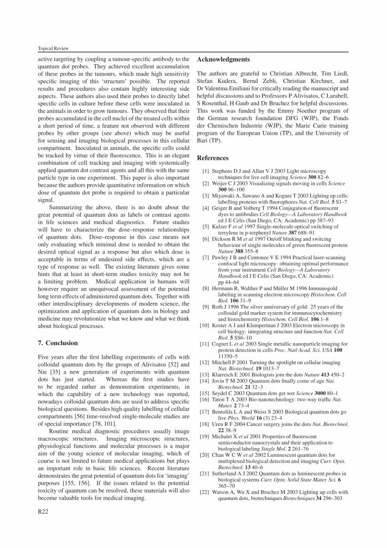
Topical Review
active targeting by coupling a tumour-specific antibody to thequantum dot probes. They achieved excellent accumulationof these probes in the tumours, which made high sensitivityspecific imaging of this ‘structure’ possible. The reportedresults and procedures also contain highly interesting sideaspects. These authors also used their probes to directly labelspecific cells in culture before these cells were inoculated inthe animals in order to grow tumours. They observed that theirprobes accumulated in the cell nuclei of the treated cells withina short period of time, a feature not observed with differentprobes by other groups (see above) which may be usefulfor sensing and imaging biological processes in this cellularcompartment. Inoculated in animals, the specific cells couldbe tracked by virtue of their fluorescence. This is an elegantcombination of cell tracking and imaging with systemicallyapplied quantum dot contrast agents and all this with the sameparticle type in one experiment. This paper is also importantbecause the authors provide quantitative information on whichdose of quantum dot probe is required to obtain a particularsignal.
Summarizing the above, there is no doubt about thegreat potential of quantum dots as labels or contrast agentsin life sciences and medical diagnostics. Future studieswill have to characterize the dose–response relationshipsof quantum dots. Dose–response in this case means notonly evaluating which minimal dose is needed to obtain thedesired optical signal as a response but also which dose isacceptable in terms of undesired side effects, which are atype of response as well. The existing literature gives somehints that at least in short-term studies toxicity may not bea limiting problem. Medical application in humans willhowever require an unequivocal assessment of the potentiallong term effects of administered quantum dots. Together withother interdisciplinary developments of modern science, theoptimization and application of quantum dots in biology andmedicine may revolutionize what we know and what we thinkabout biological processes.
7. Conclusion
Five years after the first labelling experiments of cells withcolloidal quantum dots by the groups of Alivisatos [52] andNie [35] a new generation of experiments with quantumdots has just started. Whereas the first studies haveto be regarded rather as demonstration experiments, inwhich the capability of a new technology was reported,nowadays colloidal quantum dots are used to address specificbiological questions. Besides high quality labelling of cellularcompartments [56] time-resolved single-molecule studies areof special importance [78, 101].
Routine medical diagnostic procedures usually imagemacroscopic structures. Imaging microscopic structures,physiological functions and molecular processes is a majoraim of the young science of molecular imaging, which ofcourse is not limited to future medical applications but playsan important role in basic life sciences. Recent literaturedemonstrates the great potential of quantum dots for ‘imaging’purposes [155, 156]. If the issues related to the potentialtoxicity of quantum can be resolved, these materials will alsobecome valuable tools for medical imaging.
Acknowledgments
The authors are grateful to Christian Albrecht, Tim Liedl,Stefan Kudera, Bernd Zebli, Christian Kirchner, andDr Valentina Emiliani for critically reading the manuscript andhelpful discussions and to Professors P Alivisatos, C Larabell,S Rosenthal, H Gaub and Dr Bruchez for helpful discussions.This work was funded by the Emmy Noether program ofthe German research foundation DFG (WJP), the Fondsder Chemischen Industrie (WJP), the Marie Curie trainingprogram of the European Union (TP), and the University ofBari (TP).
References
[1] Stephens D J and Allan V J 2003 Light microscopytechniques for live cell imaging Science 300 82–6
[2] Weijer C J 2003 Visualizing signals moving in cells Science300 96–100
[3] Miyawaki A, Sawano A and Kogure T 2003 Lighting up cells:labelling proteins with fluorophores Nat. Cell Biol. 5 S1–7
[4] Geiger B and Volberg T 1994 Conjugation of fluorescentdyes to antibodies Cell Biology—A Laboratory Handbooked J E Celis (San Diego, CA: Academic) pp 387–93
[5] Kulzer F et al 1997 Single-molecule optical switching ofterrylene in p-terphenyl Nature 387 688–91
[6] Dickson R M et al 1997 On/off blinking and switcingbehaviour of single molecules of green fluorescent proteinNature 388 355–8
[7] Pawley J B and Centonze V E 1994 Practical laser-scanningconfocal light microscopy: obtaining optimal performancefrom your instrument Cell Biology—A LaboratoryHandbook ed J E Celis (San Diego, CA: Academic)pp 44–64
[8] Hermann R, Walther P and Muller M 1996 Immunogoldlabeling in scanning electron microscopy Histochem. CellBiol. 106 31–9
[9] Roth J 1996 The silver anniversary of gold: 25 years of thecolloidal gold marker system for immunocytochemistryand histochemistry Histochem. Cell Biol. 106 1–8
[10] Koster A J and Klumperman J 2003 Electron microscopy incell biology: integrating structure and function Nat. CellBiol. 5 SS6–10
[11] Cognet L et al 2003 Single metallic nanoparticle imaging forprotein detection in cells Proc. Natl Acad. Sci. USA 10011350–5
[12] Mitchell P 2001 Turning the spotlight on cellular imagingNat. Biotechnol. 19 1013–7
[13] Klarreich E 2001 Biologists join the dots Nature 413 450–2[14] Jovin T M 2003 Quantum dots finally come of age Nat.
Biotechnol. 21 32–3[15] Seydel C 2003 Quantum dots get wet Science 3000 80–1[16] Taton T A 2003 Bio-nanotechnology: two-way traffic Nat.
Mater. 2 73–4[17] Bentolila L A and Weiss S 2003 Biological quantum dots go
live Phys. World 16 (3) 23–4[18] Uren R F 2004 Cancer surgery joins the dots Nat. Biotechnol.
22 38–9[19] Michalet X et al 2001 Properties of fluorescent
semiconductor nanocrystals and their application tobiological labeling Single Mol. 2 261–76
[20] Chan W C W et al 2002 Luminescent quantum dots formultiplexed biological detection and imaging Curr. Opin.Biotechnol. 13 40–6
[21] Sutherland A J 2002 Quantum dots as luminescent probes inbiological systems Curr. Opin. Solid State Mater. Sci. 6365–70
[22] Watson A, Wu X and Bruchez M 2003 Lighting up cells withquantum dots, biotechniques Biotechniques 34 296–303
R22
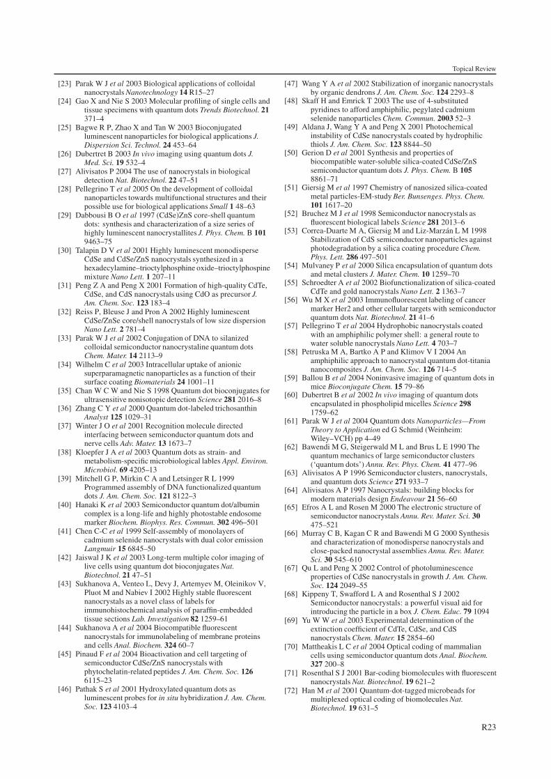
Topical Review
[23] Parak W J et al 2003 Biological applications of colloidalnanocrystals Nanotechnology 14 R15–27
[24] Gao X and Nie S 2003 Molecular profiling of single cells andtissue specimens with quantum dots Trends Biotechnol. 21371–4
[25] Bagwe R P, Zhao X and Tan W 2003 Bioconjugatedluminescent nanoparticles for biological applications J.Dispersion Sci. Technol. 24 453–64
[26] Dubertret B 2003 In vivo imaging using quantum dots J.Med. Sci. 19 532–4
[27] Alivisatos P 2004 The use of nanocrystals in biologicaldetection Nat. Biotechnol. 22 47–51
[28] Pellegrino T et al 2005 On the development of colloidalnanoparticles towards multifunctional structures and theirpossible use for biological applications Small 1 48–63
[29] Dabbousi B O et al 1997 (CdSe)ZnS core-shell quantumdots: synthesis and characterization of a size series ofhighly luminescent nanocrystallites J. Phys. Chem. B 1019463–75
[30] Talapin D V et al 2001 Highly luminescent monodisperseCdSe and CdSe/ZnS nanocrystals synthesized in ahexadecylamine–trioctylphosphine oxide–trioctylphospinemixture Nano Lett. 1 207–11
[31] Peng Z A and Peng X 2001 Formation of high-quality CdTe,CdSe, and CdS nanocrystals using CdO as precursor J.Am. Chem. Soc. 123 183–4
[32] Reiss P, Bleuse J and Pron A 2002 Highly luminescentCdSe/ZnSe core/shell nanocrystals of low size dispersionNano Lett. 2 781–4
[33] Parak W J et al 2002 Conjugation of DNA to silanizedcolloidal semiconductor nanocrystaline quantum dotsChem. Mater. 14 2113–9
[34] Wilhelm C et al 2003 Intracellular uptake of anionicsuperparamagnetic nanoparticles as a function of theirsurface coating Biomaterials 24 1001–11
[35] Chan W C W and Nie S 1998 Quantum dot bioconjugates forultrasensitive nonisotopic detection Science 281 2016–8
[36] Zhang C Y et al 2000 Quantum dot-labeled trichosanthinAnalyst 125 1029–31
[37] Winter J O et al 2001 Recognition molecule directedinterfacing between semiconductor quantum dots andnerve cells Adv. Mater. 13 1673–7
[38] Kloepfer J A et al 2003 Quantum dots as strain- andmetabolism-specific microbiological lables Appl. Environ.Microbiol. 69 4205–13
[39] Mitchell G P, Mirkin C A and Letsinger R L 1999Programmed assembly of DNA functionalized quantumdots J. Am. Chem. Soc. 121 8122–3
[40] Hanaki K et al 2003 Semiconductor quantum dot/albumincomplex is a long-life and highly photostable endosomemarker Biochem. Biophys. Res. Commun. 302 496–501
[41] Chen C-C et al 1999 Self-assembly of monolayers ofcadmium selenide nanocrystals with dual color emissionLangmuir 15 6845–50
[42] Jaiswal J K et al 2003 Long-term multiple color imaging oflive cells using quantum dot bioconjugates Nat.Biotechnol. 21 47–51
[43] Sukhanova A, Venteo L, Devy J, Artemyev M, Oleinikov V,Pluot M and Nabiev I 2002 Highly stable fluorescentnanocrystals as a novel class of labels forimmunohistochemical analysis of paraffin-embeddedtissue sections Lab. Investigation 82 1259–61
[44] Sukhanova A et al 2004 Biocompatible fluorescentnanocrystals for immunolabeling of membrane proteinsand cells Anal. Biochem. 324 60–7
[45] Pinaud F et al 2004 Bioactivation and cell targeting ofsemiconductor CdSe/ZnS nanocrystals withphytochelatin-related peptides J. Am. Chem. Soc. 1266115–23
[46] Pathak S et al 2001 Hydroxylated quantum dots asluminescent probes for in situ hybridization J. Am. Chem.Soc. 123 4103–4
[47] Wang Y A et al 2002 Stabilization of inorganic nanocrystalsby organic dendrons J. Am. Chem. Soc. 124 2293–8
[48] Skaff H and Emrick T 2003 The use of 4-substitutedpyridines to afford amphiphilic, pegylated cadmiumselenide nanoparticles Chem. Commun. 2003 52–3
[49] Aldana J, Wang Y A and Peng X 2001 Photochemicalinstability of CdSe nanocrystals coated by hydrophilicthiols J. Am. Chem. Soc. 123 8844–50
[50] Gerion D et al 2001 Synthesis and properties ofbiocompatible water-soluble silica-coated CdSe/ZnSsemiconductor quantum dots J. Phys. Chem. B 1058861–71
[51] Giersig M et al 1997 Chemistry of nanosized silica-coatedmetal particles-EM-study Ber. Bunsenges. Phys. Chem.101 1617–20
[52] Bruchez M J et al 1998 Semiconductor nanocrystals asfluorescent biological labels Science 281 2013–6
[53] Correa-Duarte M A, Giersig M and Liz-Marzan L M 1998Stabilization of CdS semiconductor nanoparticles againstphotodegradation by a silica coating procedure Chem.Phys. Lett. 286 497–501
[54] Mulvaney P et al 2000 Silica encapsulation of quantum dotsand metal clusters J. Mater. Chem. 10 1259–70
[55] Schroedter A et al 2002 Biofunctionalization of silica-coatedCdTe and gold nanocrystals Nano Lett. 2 1363–7
[56] Wu M X et al 2003 Immunofluorescent labeling of cancermarker Her2 and other cellular targets with semiconductorquantum dots Nat. Biotechnol. 21 41–6
[57] Pellegrino T et al 2004 Hydrophobic nanocrystals coatedwith an amphiphilic polymer shell: a general route towater soluble nanocrystals Nano Lett. 4 703–7
[58] Petruska M A, Bartko A P and Klimov V I 2004 Anamphiphilic approach to nanocrystal quantum dot-titaniananocomposites J. Am. Chem. Soc. 126 714–5
[59] Ballou B et al 2004 Noninvasive imaging of quantum dots inmice Bioconjugate Chem. 15 79–86
[60] Dubertret B et al 2002 In vivo imaging of quantum dotsencapsulated in phospholipid micelles Science 2981759–62
[61] Parak W J et al 2004 Quantum dots Nanoparticles—FromTheory to Application ed G Schmid (Weinheim:Wiley–VCH) pp 4–49
[62] Bawendi M G, Steigerwald M L and Brus L E 1990 Thequantum mechanics of large semiconductor clusters(‘quantum dots’) Annu. Rev. Phys. Chem. 41 477–96
[63] Alivisatos A P 1996 Semiconductor clusters, nanocrystals,and quantum dots Science 271 933–7
[64] Alivisatos A P 1997 Nanocrystals: building blocks formodern materials design Endeavour 21 56–60
[65] Efros A L and Rosen M 2000 The electronic structure ofsemiconductor nanocrystals Annu. Rev. Mater. Sci. 30475–521
[66] Murray C B, Kagan C R and Bawendi M G 2000 Synthesisand characterization of monodisperse nanocrystals andclose-packed nanocrystal assemblies Annu. Rev. Mater.Sci. 30 545–610
[67] Qu L and Peng X 2002 Control of photoluminescenceproperties of CdSe nanocrystals in growth J. Am. Chem.Soc. 124 2049–55
[68] Kippeny T, Swafford L A and Rosenthal S J 2002Semiconductor nanocrystals: a powerful visual aid forintroducing the particle in a box J. Chem. Educ. 79 1094
[69] Yu W W et al 2003 Experimental determination of theextinction coefficient of CdTe, CdSe, and CdSnanocrystals Chem. Mater. 15 2854–60
[70] Mattheakis L C et al 2004 Optical coding of mammaliancells using semiconductor quantum dots Anal. Biochem.327 200–8
[71] Rosenthal S J 2001 Bar-coding biomolecules with fluorescentnanocrystals Nat. Biotechnol. 19 621–2
[72] Han M et al 2001 Quantum-dot-tagged microbeads formultiplexed optical coding of biomolecules Nat.Biotechnol. 19 631–5
R23
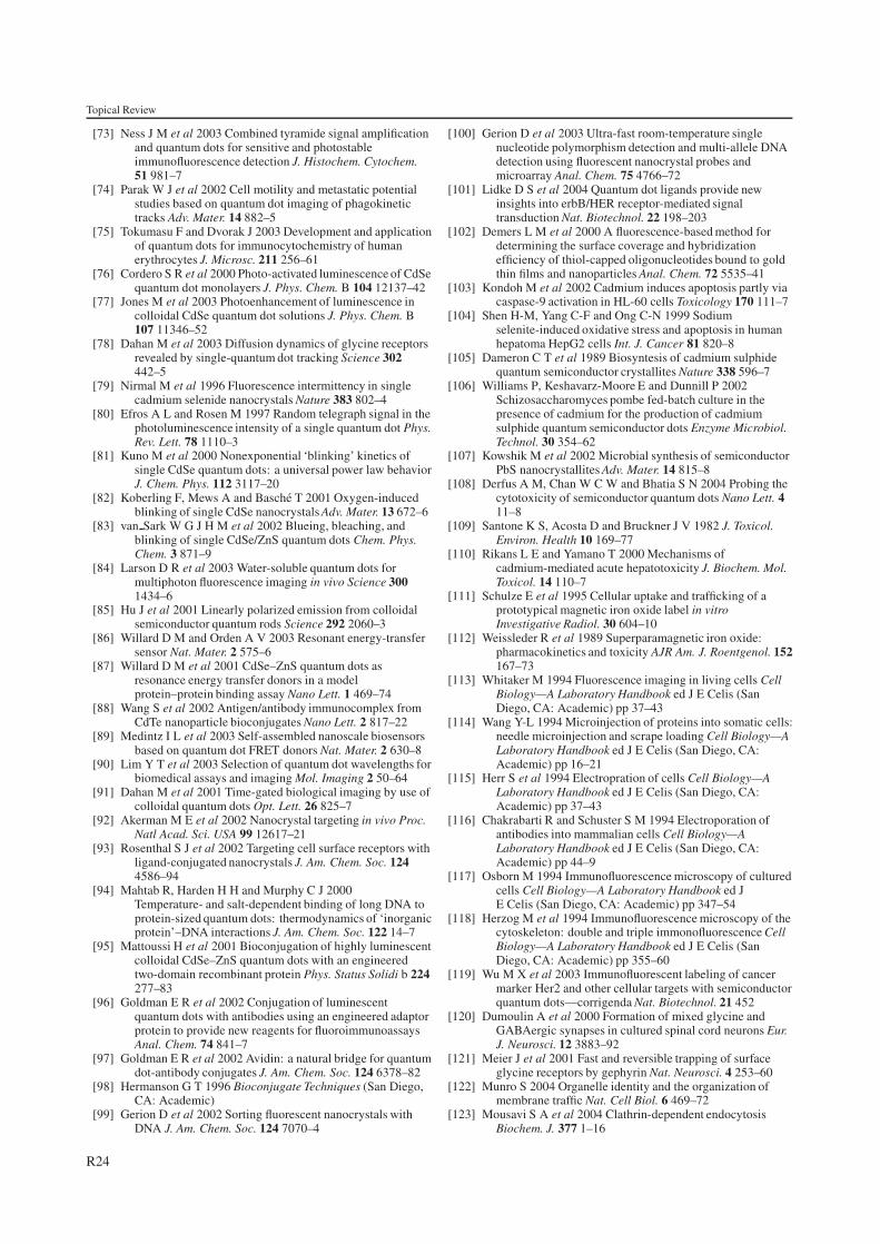
Topical Review
[73] Ness J M et al 2003 Combined tyramide signal amplificationand quantum dots for sensitive and photostableimmunofluorescence detection J. Histochem. Cytochem.51 981–7
[74] Parak W J et al 2002 Cell motility and metastatic potentialstudies based on quantum dot imaging of phagokinetictracks Adv. Mater. 14 882–5
[75] Tokumasu F and Dvorak J 2003 Development and applicationof quantum dots for immunocytochemistry of humanerythrocytes J. Microsc. 211 256–61
[76] Cordero S R et al 2000 Photo-activated luminescence of CdSequantum dot monolayers J. Phys. Chem. B 104 12137–42
[77] Jones M et al 2003 Photoenhancement of luminescence incolloidal CdSe quantum dot solutions J. Phys. Chem. B107 11346–52
[78] Dahan M et al 2003 Diffusion dynamics of glycine receptorsrevealed by single-quantum dot tracking Science 302442–5
[79] Nirmal M et al 1996 Fluorescence intermittency in singlecadmium selenide nanocrystals Nature 383 802–4
[80] Efros A L and Rosen M 1997 Random telegraph signal in thephotoluminescence intensity of a single quantum dot Phys.Rev. Lett. 78 1110–3
[81] Kuno M et al 2000 Nonexponential ‘blinking’ kinetics ofsingle CdSe quantum dots: a universal power law behaviorJ. Chem. Phys. 112 3117–20
[82] Koberling F, Mews A and Basche T 2001 Oxygen-inducedblinking of single CdSe nanocrystals Adv. Mater. 13 672–6
[83] van Sark W G J H M et al 2002 Blueing, bleaching, andblinking of single CdSe/ZnS quantum dots Chem. Phys.Chem. 3 871–9
[84] Larson D R et al 2003 Water-soluble quantum dots formultiphoton fluorescence imaging in vivo Science 3001434–6
[85] Hu J et al 2001 Linearly polarized emission from colloidalsemiconductor quantum rods Science 292 2060–3
[86] Willard D M and Orden A V 2003 Resonant energy-transfersensor Nat. Mater. 2 575–6
[87] Willard D M et al 2001 CdSe–ZnS quantum dots asresonance energy transfer donors in a modelprotein–protein binding assay Nano Lett. 1 469–74
[88] Wang S et al 2002 Antigen/antibody immunocomplex fromCdTe nanoparticle bioconjugates Nano Lett. 2 817–22
[89] Medintz I L et al 2003 Self-assembled nanoscale biosensorsbased on quantum dot FRET donors Nat. Mater. 2 630–8
[90] Lim Y T et al 2003 Selection of quantum dot wavelengths forbiomedical assays and imaging Mol. Imaging 2 50–64
[91] Dahan M et al 2001 Time-gated biological imaging by use ofcolloidal quantum dots Opt. Lett. 26 825–7
[92] Akerman M E et al 2002 Nanocrystal targeting in vivo Proc.Natl Acad. Sci. USA 99 12617–21
[93] Rosenthal S J et al 2002 Targeting cell surface receptors withligand-conjugated nanocrystals J. Am. Chem. Soc. 1244586–94
[94] Mahtab R, Harden H H and Murphy C J 2000Temperature- and salt-dependent binding of long DNA toprotein-sized quantum dots: thermodynamics of ‘inorganicprotein’–DNA interactions J. Am. Chem. Soc. 122 14–7
[95] Mattoussi H et al 2001 Bioconjugation of highly luminescentcolloidal CdSe–ZnS quantum dots with an engineeredtwo-domain recombinant protein Phys. Status Solidi b 224277–83
[96] Goldman E R et al 2002 Conjugation of luminescentquantum dots with antibodies using an engineered adaptorprotein to provide new reagents for fluoroimmunoassaysAnal. Chem. 74 841–7
[97] Goldman E R et al 2002 Avidin: a natural bridge for quantumdot-antibody conjugates J. Am. Chem. Soc. 124 6378–82
[98] Hermanson G T 1996 Bioconjugate Techniques (San Diego,CA: Academic)
[99] Gerion D et al 2002 Sorting fluorescent nanocrystals withDNA J. Am. Chem. Soc. 124 7070–4
[100] Gerion D et al 2003 Ultra-fast room-temperature singlenucleotide polymorphism detection and multi-allele DNAdetection using fluorescent nanocrystal probes andmicroarray Anal. Chem. 75 4766–72
[101] Lidke D S et al 2004 Quantum dot ligands provide newinsights into erbB/HER receptor-mediated signaltransduction Nat. Biotechnol. 22 198–203
[102] Demers L M et al 2000 A fluorescence-based method fordetermining the surface coverage and hybridizationefficiency of thiol-capped oligonucleotides bound to goldthin films and nanoparticles Anal. Chem. 72 5535–41
[103] Kondoh M et al 2002 Cadmium induces apoptosis partly viacaspase-9 activation in HL-60 cells Toxicology 170 111–7
[104] Shen H-M, Yang C-F and Ong C-N 1999 Sodiumselenite-induced oxidative stress and apoptosis in humanhepatoma HepG2 cells Int. J. Cancer 81 820–8
[105] Dameron C T et al 1989 Biosyntesis of cadmium sulphidequantum semiconductor crystallites Nature 338 596–7
[106] Williams P, Keshavarz-Moore E and Dunnill P 2002Schizosaccharomyces pombe fed-batch culture in thepresence of cadmium for the production of cadmiumsulphide quantum semiconductor dots Enzyme Microbiol.Technol. 30 354–62
[107] Kowshik M et al 2002 Microbial synthesis of semiconductorPbS nanocrystallites Adv. Mater. 14 815–8
[108] Derfus A M, Chan W C W and Bhatia S N 2004 Probing thecytotoxicity of semiconductor quantum dots Nano Lett. 411–8
[109] Santone K S, Acosta D and Bruckner J V 1982 J. Toxicol.Environ. Health 10 169–77
[110] Rikans L E and Yamano T 2000 Mechanisms ofcadmium-mediated acute hepatotoxicity J. Biochem. Mol.Toxicol. 14 110–7
[111] Schulze E et al 1995 Cellular uptake and trafficking of aprototypical magnetic iron oxide label in vitroInvestigative Radiol. 30 604–10
[112] Weissleder R et al 1989 Superparamagnetic iron oxide:pharmacokinetics and toxicity AJR Am. J. Roentgenol. 152167–73
[113] Whitaker M 1994 Fluorescence imaging in living cells CellBiology—A Laboratory Handbook ed J E Celis (SanDiego, CA: Academic) pp 37–43
[114] Wang Y-L 1994 Microinjection of proteins into somatic cells:needle microinjection and scrape loading Cell Biology—ALaboratory Handbook ed J E Celis (San Diego, CA:Academic) pp 16–21
[115] Herr S et al 1994 Electropration of cells Cell Biology—ALaboratory Handbook ed J E Celis (San Diego, CA:Academic) pp 37–43
[116] Chakrabarti R and Schuster S M 1994 Electroporation ofantibodies into mammalian cells Cell Biology—ALaboratory Handbook ed J E Celis (San Diego, CA:Academic) pp 44–9
[117] Osborn M 1994 Immunofluorescence microscopy of culturedcells Cell Biology—A Laboratory Handbook ed JE Celis (San Diego, CA: Academic) pp 347–54
[118] Herzog M et al 1994 Immunofluorescence microscopy of thecytoskeleton: double and triple immonofluorescence CellBiology—A Laboratory Handbook ed J E Celis (SanDiego, CA: Academic) pp 355–60
[119] Wu M X et al 2003 Immunofluorescent labeling of cancermarker Her2 and other cellular targets with semiconductorquantum dots—corrigenda Nat. Biotechnol. 21 452
[120] Dumoulin A et al 2000 Formation of mixed glycine andGABAergic synapses in cultured spinal cord neurons Eur.J. Neurosci. 12 3883–92
[121] Meier J et al 2001 Fast and reversible trapping of surfaceglycine receptors by gephyrin Nat. Neurosci. 4 253–60
[122] Munro S 2004 Organelle identity and the organization ofmembrane traffic Nat. Cell Biol. 6 469–72
[123] Mousavi S A et al 2004 Clathrin-dependent endocytosisBiochem. J. 377 1–16
R24
Topical Review
[124] Pelkmans L and Helenius A 2003 Insider information: whatviruses tell us about endocytosis Curr. Opin. Cell Biol. 15414–22
[125] Wagner E, Curiel D and Cotten M 1994 Delivery of drugs,proteins and genes into cells using transferrin as a ligandfor receptor-mediated endocytosis Adv. Drug Deliv. Rev.14 113–35
[126] Widera A, Norouziyan F and Shen W C 2003 Mechanisms ofTfR-mediated transcytosis and sorting in epithelial cellsand applications toward drug delivery Adv. Drug Deliv.Rev. 55 1439–66
[127] Reuning U et al 2003 Urokinase-type plasminogen activator(uPA) and its receptor (uPAR): development of antagonistsof uPA/uPAR interaction and their effects in vitro and invivo Curr. Pharm. Des. 9 1529–43
[128] Niidome T and Huang L 2002 Gene therapy progress andprospects: nonviral vectors Gene Ther. 9 1647–52
[129] Cotten M and Wagner E 1993 Non-viral approaches to genetherapy Curr. Opin. Biotechnol. 4 705–10
[130] Wagner E et al 1992 Influenza virus hemagglutinin HA-2N-terminal fusogenic peptides augment gene transfer bytransferrin-polylysine-DNA complexes: toward a syntheticvirus-like gene-transfer vehicle Proc. Natl Acad. Sci. USA89 7934–8
[131] Plank C et al 1994 The influence of endosome-disruptivepeptides on gene transfer using synthetic virus-like genetransfer systems J. Biol. Chem. 269 12918–24
[132] Plank C, Zauner W and Wagner E 1998 Application ofmembrane-active peptides for drug and gene deliveryacross cellular membranes Adv. Drug Deliv. Rev. 34 21–35
[133] Boussif O et al 1995 A versatile vector for gene andoligonucleotide transfer into cells in culture and in vivo:polyethylenimine Proc. Natl Acad. Sci. USA 92 7297–301
[134] Sonawane N D, Szoka F C Jr and Verkman A S 2003Chloride accumulation and swelling in endosomesenhances DNA transfer by polyamine-DNA polyplexes J.Biol. Chem. 278 44826–31
[135] Xu Y and Szoka F C Jr 1996 Mechanism of DNA releasefrom cationic liposome/DNA complexes used in celltransfection Biochemistry 35 5616–23
[136] Pellegrino T et al 2003 Quantum dot-based cell motilityassay Differentiation 71 542–8
[137] Lewin M et al 2000 Tat peptide-derivatized magneticnanoparticles allow in vivo tracking and recovery ofprogenitor cells Nat. Biotechnol. 18 410–4
[138] Weissleder R et al 2000 In vivo magnetic resonance imagingof transgene expression Nat. Med. 6 351–5
[139] Pearson H 2002 Developmental biology: your destiny, fromday one Nature 417 14–5
[140] Kubo A et al 2004 Development of definitive endoderm fromembryonic stem cells in culture Development 131 1651–62
[141] Ferrari A et al 2001 In vivo tracking of bone marrowfibroblasts with fluorescent carbocyanine dye J. Biomed.Mater. Res. 56 361–7
[142] Tombolini R and Jansson J K 1998 Monitoring of GFP-taggedbacterial cells Bioluminescence Methods and Protocols edR A LaRossa (Totowa, NJ: Humana Press) pp 285–98
[143] Brenner M K et al 1993 Gene-marking to trace origin ofrelapse after autologous bone-marrow transplantationLancet 341 85–6
[144] Fuchs M et al 2004 Dynamics of cell adhesion and motilityin living cells is altered by a single amino acid change inE-cadherin fused to enhanced green fluorescent proteinCell Motil. Cytoskeleton 59 50–61
[145] Albrecht-Buehler G 1977 The phagokinetic tracks of 3T3cells Cell 11 395–404
[146] Albrecht-Buehler G 1977 Phagokinetic tracks of 3T3 cells:parallels between the orientation of track segments and ofcellular structures which contain actin or tubulin Cell 12333–9
[147] Albrecht-Buehler G and Lancaster R M 1976 A quantitativedescription of the extenion and retraction of surfaceprotrusions in spreading 3T3 mouse fibroblasts J. CellBiol. 71 370–82
[148] Chen J D et al 1994 Human keratincytes make uniquelylinear phagokinetic tracks Dermatology 188 6–12
[149] Stokes C L, Lauffenburger D A and Williams S K 1991Migration of individual microvessel endothelial cells:stochastic model and parameter measurement J. Cell Sci.99 41–430
[150] Scott W N, McCool K and Nelson J 2000 Improved methodfor the production of gold colloid monolayers for use inthe phagokinetic track assay for cell motility Anal.Biochem. 287 343–4
[151] Obeso J L and Auerbach R 1984 A new microtechnique forquantitating cell movement in vitro using polystyrene beadmonolayers J. Immunol. Methods 70 141–52
[152] Plank C et al 2003 Enhancing and targeting nucleic aciddelivery by magnetic force Expert Opin. Biol. Ther. 3745–58
[153] Finsinger D et al 2000 Protective copolymers for nonviralgene vectors: synthesis, vector characterization andapplication in gene delivery Gene Ther. 7 1183–92
[154] Kim S et al 2003 Type-II quantum dots:CdTe/CdSe(Core/Shell) and CdSe/ZnTe(Core/Shell)heterostructures J. Am. Chem. Soc. 125 11466–7
[155] Kim S et al 2004 Near-infrared fluorescent type II quantumdots for sentinel lymph node mapping Nat. Biotechnol. 2293–7
[156] Gao X et al 2004 In vivo cancer targeting and imagingwith semiconductor quantum dots Nat. Biotechnol. 22969–76
R25