Topic of the month: Radiological pathology of meningiomas
46
INDEX INTRODUCTION CT SCAN IMAGING OF MENINGIOMAS MR IMAGING OF MENINGIOMAS SPINAL MENINGIOMA RADIOLOGICAL PATHOLOGY OF MENINGIOMAS Meningioma is the most common nonglial primary intracranial tumor, with a female preponderance, occurring most commonly in the 40- to 60-year-old age range. 7 Most arise from arachnoid cap cells in arachnoid granulations, and 90% are supratentorial. They are commonly located along meningeal surfaces in the parasagittal region, lateral convexity, falx, sphenoid ridge, olfactory groove, cerebellopontine angle, petrous ridge, and tentorium in descending order of frequency. In approximately 8% of cases meningiomas are multiple,
-
Upload
professor-yasser-metwally -
Category
Health & Medicine
-
view
2.835 -
download
2
description
Topic of the month: Radiological pathology of meningiomas
Transcript of Topic of the month: Radiological pathology of meningiomas

INDEX
INTRODUCTION
CT SCAN IMAGINGOF MENINGIOMAS
MR IMAGING OFMENINGIOMAS
SPINALMENINGIOMA
RADIOLOGICAL PATHOLOGY OF MENINGIOMAS
Meningioma is the most common nonglial primary intracranial tumor, with a femalepreponderance, occurring most commonly in the 40- to 60-year-old age range. 7 Most arisefrom arachnoid cap cells in arachnoid granulations, and 90% are supratentorial. They arecommonly located along meningeal surfaces in the parasagittal region, lateral convexity,falx, sphenoid ridge, olfactory groove, cerebellopontine angle, petrous ridge, and tentoriumin descending order of frequency. In approximately 8% of cases meningiomas are multiple,

and the multiplicity is usually sporadic but may be familial or associated withneurofibromatosis type II. Other causes of meningiomas include prior cranial irradiationand previous head trauma. 10 There is an increased incidence of meningioma with breastcarcinoma and pregnancy, 11,12 suggesting a hormonal influence.
Figure 1. Common sites for meningiomas (A) and (B) The 10 most common locations inwhich meningiomas are found, in order of frequency, are: parasagittal (1), cerebralconvexity (2), sphenoid ridge (3), olfactory groove (4), suprasellar (5), cerebellopontineangle (6), spinal (7), floor of middle fossa (8), torcular (9), and intraventricular (10).

Figure 2. A, parasagittal meningioma, B, olfactory groove meningioma
Table 1. Common sites for meningiomas
Location SymptomsParasagittal Monoparesis of the contralateral legSubfrontal Change in mentation, apathy or disinhibited behavior, urinary incontinence
Olfactory groove Anosmia with possible ipsilateral optic atrophy and contralateralpapilledema. This triad is termed Kennedy-Foster syndrome.
Cavernous sinus Multiple cranial deficits (II, III, IV, V and VI), leading to decreased visionand diplopia with associated to facial numbness
Occipital lobe Contralateral hemianopsiaCerebellopontineangle Decreased hearing with possible facial weakness and facial numbness
Spinal cord Localized spinal pain, Brown-Sequard (hemi-spinal cord) syndrome
Optic nervemeningiomas
Exophthalmos, Monocular loss of vision or blindness; ipsilateral dilatedpupil that does not react to direct light stimulation but might contract onconsensual light stimulation. Often monocular optic nerve swelling withopto-ciliary shunt vessels.
Sphenoid wingmeningiomas
Seizures; multiple cranial nerve palsies if the superior orbital fissure isinvolved.
Tentorialmeningiomas
Tentorial meningiomas may protrude within the supratentorial andinfratentorial compartments. Meningiomas in this location producesymptoms by compressing specific structures within these twocompartments.
Foramen magnummeningiomas
Paraparesis; sphincteric troubles; tongue atrophy associated tofasciculation.

Meningiomas can be divided into three histological groups: (1) classic, (2) angioblastic, and(3) malignant. There are histological subtypes for each of these groupings as well. Theclassic type of meningioma includes syncytial, transitional, and fibroblastic subtypes. Mostintracranial meningiomas are of the syncytial or; transitional subtype. The angioblasticgroup includes hemangioblastic and hemangiopericytic subtypes. The angioblasticmeningioma is a rapidly growing aggressive variant with extensive thin-walled vascularspaces. Although meningiomas tend to invade venous sinuses, distant metastasis is rare,with an incidence of 0.1%. 13 The angioblastic type is the most frequent type tometastasize.7
Figure 3. A, Meningioma. Whirls of cells and elongated cells. No psammoma bodies. B,Meningioma. Note whirling pattern of tumor cells and psammoma bodies (round densepurple structures). C, Multiple meningiomas in a case with neurofibromatosis type 2
Meningiomas are generally well-circumscribed, expansive tumors. They produce symptomsby external compression of the brain. 2 Consequently, they usually are amenable tocomplete resection. The major exceptions are meningiomas of the skull base, particularly ofthe cavernous sinus, where the tumors disseminate around multiple vital structures,usually precluding extirpation. Meningiomas tend to be smooth and round or lobular.Their cut surfaces range from firm, white, and fibrous to soft and myxoid. Brain invasion israre, but infiltration into and, if untreated, through the skull is not unusual. 1,2 Mostmeningiomas are benign, WHO grade I tumors, but a spectrum of aggressive tumorsoccurs, including essentially sarcomatous grade IV tumors.

Figure 4. A, Meningioma. Whirls of cells and elongated cells. No psammoma bodies. B,Meningioma. Note whirling pattern of tumor cells and psammoma bodies (round densepurple structures).
Meningiomas have myriad microscopic appearances. Their defined subtypes are toonumerous to list here. Befitting their heritage as tumors of cells with both structural andepithelial functions, the most common histologic types are fibrous, meningothelial, andtransitional, which combines the features of the first two. The psammoma body, a lamellarcalcospherite, is a pathognomonic feature that can dominate some tumors. Cytologicatypia, a high mitotic rate, and necrosis are all positively correlated with increasedaggressiveness. 1 For individual tumors, however, prognosis is determined primarily by theextent of resection. Although brain invasion is uncommon and usually associated with theother high-grade features, it also occurs in otherwise typically benign slow-growing tumors.

Figure 5. A, Meningioma. Note common parasagittal location. Note compression but notinvasion of the brain. B, convexity meningioma.
The relationship between neurofibromatosis type 2 and meningioma development has beenwell established. The most common genetic abnormality associated with meningioma is thedeletion of chromosome 22 and an associated tumor-suppressor gene specific tomeningioma formation. Aggressive or invasive variants of this lesion have been associatedwith additional chromosomal aberrations involving chromosomes 1 and 14. 3

Table 2. Histological subtype of meningiomas
Histologicalsubtype
Comment
Fibroblasticmeningiomas
Composed of large, narrow spindle cells. The distinct feature is thepresence of abundant reticulum and collagen fibers between individualcells. 21 On MR imaging, fibroblastic meningiomas with cellsembedded in a dense collagenous matrix appear as low signal intensityin TI-weighted and T2-weighted pulse sequences. 10
Transitionalmeningiomas(psammomatous)
Characterized by whorl formations in which the cells are wrappedtogether resembling onion skins. 10 The whorls may degenerate andcalcify, becoming psammoma bodies. Marked calcifications can be seenin this histologic type. MR imaging of transitional meningiomas thusalso demonstrates low signal intensity on Tl- weighted and T2-weightedimages, with the calcifications contributing to the low signal intensity. 5
Syncytial(meningothelial,endotheliomatous)meningiomas
Contain polygonal cells, poorly defined and arranged in lobules. 28Syncytial meningiomas composed of sheets of contiguous cells withsparse interstitium might account for higher signal intensity in T2-weighted images. Microcystic changes and nuclear vesicles can alsocontribute to increased signal intensity. 5
Angioblasticmeningiomas
Highly cellular and vascular tumors with a spongy appearance.Increased signal in T2-weighted pulse sequence of these tumors is dueto high cellularity with increase in water content of tumor. Thus basedon the correlation between histology and MR imaging appearance ofmeningiomas. 5
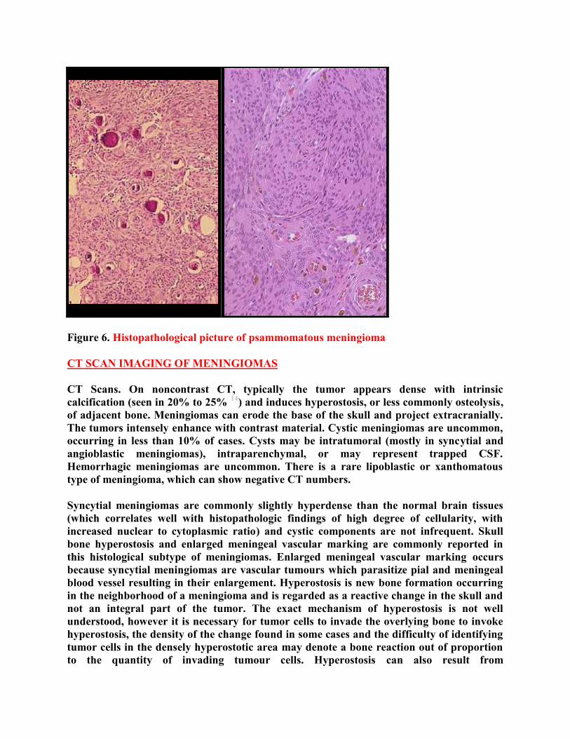
Figure 6. Histopathological picture of psammomatous meningioma
CT SCAN IMAGING OF MENINGIOMAS
CT Scans. On noncontrast CT, typically the tumor appears dense with intrinsiccalcification (seen in 20% to 25% 14) and induces hyperostosis, or less commonly osteolysis,of adjacent bone. Meningiomas can erode the base of the skull and project extracranially.The tumors intensely enhance with contrast material. Cystic meningiomas are uncommon,occurring in less than 10% of cases. Cysts may be intratumoral (mostly in syncytial andangioblastic meningiomas), intraparenchymal, or may represent trapped CSF.Hemorrhagic meningiomas are uncommon. There is a rare lipoblastic or xanthomatoustype of meningioma, which can show negative CT numbers.
Syncytial meningiomas are commonly slightly hyperdense than the normal brain tissues(which correlates well with histopathologic findings of high degree of cellularity, withincreased nuclear to cytoplasmic ratio) and cystic components are not infrequent. Skullbone hyperostosis and enlarged meningeal vascular marking are commonly reported inthis histological subtype of meningiomas. Enlarged meningeal vascular marking occursbecause syncytial meningiomas are vascular tumours which parasitize pial and meningealblood vessel resulting in their enlargement. Hyperostosis is new bone formation occurringin the neighborhood of a meningioma and is regarded as a reactive change in the skull andnot an integral part of the tumor. The exact mechanism of hyperostosis is not wellunderstood, however it is necessary for tumor cells to invade the overlying bone to invokehyperostosis, the density of the change found in some cases and the difficulty of identifyingtumor cells in the densely hyperostotic area may denote a bone reaction out of proportionto the quantity of invading tumour cells. Hyperostosis can also result from

hypervascularity of the periosteum overlying the meningiomas. Involvement of the outertable -by hyperostosis- makes tumor invasion more likely.
Edema and mass effect are common in syncytial and angioblastic meningiomas andcontrast enhancement is intense and uniform. The existence of prominent edema and masseffect in syncytial and angioblastic meningiomas probably indicates the higher rate ofgrowth of this neoplasm compared with that of the calcified transitional (psammomatous)meningiomas. Prominent clinical signs and symptoms on presentation definitely denotesthe more aggressive biological behavior of syncytial meningiomas.
Vasogenic edema is characterized by increased permeability of brain capillary endothelialcells to macromolecules, such as the plasma proteins and various other molecules, whoseentry is limited by the capillary endothelial cells (blood brain barrier). The high vascularity(with defective endothelial lining of the newly formed blood vessels) of the syncytial andangioblastic meningiomas probably accounts for the edema observed in these subtypes ofmeningiomas.
Figure 7. A, Plain x ray showing enlarged vascular markings ending in hyperostotic bone.B, Gross specimen showing bone hyperostosis, Meningiomas often evoke reactive changesin the adjacent bone to produce hyperstosis. This figure shows the inner aspect of the boneadjacent to a meningioma. The tumor cells have infiltrated the bone marrow spaces andinduced the deposits of new bone.
Bone hyperostosis and enlarged meningeal vascular marking are almost invariably coupled inevery patient, they occur almost exclusively in syncytial meningiomas

Figure 8. Enlarged meningeal vascular marking. The enlarged channels are seen ending ina hyperostotic bony region
Figure 9. Left frontal syncytial meningioma causing hyperostosis, notice the perilesionaledema.
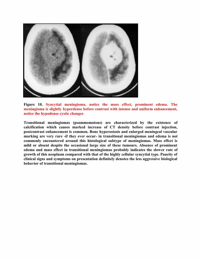
Figure 10. Syncytial meningioma, notice the mass effect, prominent edema. Themeningioma is slightly hyperdense before contrast with intense and uniform enhancement,notice the hypodense cystic changes
Transitional meningiomas (psammomatous) are characterized by the existence ofcalcification which causes marked increase of CT density before contrast injection,postcontrast enhancement is common. Bone hyperostosis and enlarged meningeal vascularmarking are very rare -if they ever occur- in transitional meningiomas and edema is notcommonly encountered around this histological subtype of meningiomas. Mass effect ismild or absent despite the occasional large size of these tumours. Absence of prominentedema and mass effect in transitional meningiomas probably indicates the slower rate ofgrowth of this neoplasm compared with that of the highly cellular syncytial type. Paucity ofclinical signs and symptoms on presentation definitely denotes the less aggressive biologicalbehavior of transitional meningiomas.

Figure 11. Bifrontal heavily calcified psammomatous meningioma with intense postcontrastenhancement, notice absence of edema
Table 3. Plain X ray and CT scan differences between Syncytial meningiomas andTransitional meningiomas (psammomatous)
Finding Syncytialmeningiomas
Transitional meningiomas(psammomatous)
Skull bone hyperostosisand enlarged vascularmarking
Present Absent
Perilesional edema Present* AbsentTumour calcification Absent PresentCystic changes Present AbsentPrecontrast CT density + ++++Contrast enhancement Intense and uniform Intense and uniformMass effect Prominent Mild or absentRate of growth ++++ +Biological behavior More aggressive Less aggressiveVascularity More vascular Less vascular
*Syncytial meningiomas -compared with transitional meningiomas- are vascular tumours with defective endotheliallining of blood vessels resulting in increased permeability of endothelial cells to macromolecules, such as the plasma

proteins and various other molecules, whose entry is limited by the capillary endothelial cells (blood brain barrier).Increased permeability of the endothelial cells of the newly formed blood vessels results in vasogenic edema.
Figure 12. Angioblastic meningioma. The lesion is markedly vascular and surrounded byprominent edema.
MR IMAGING OF MENINGIOMAS
Precontrast and postcontrast MR imaging studies can easily diagnose meningioma as wellas CT. MR imaging can also predict histologic subtypes of meningioma.
Diagnosis of meningiomas using MR imaging is made by demonstrating the extra-axialnature of the mass. Several key MR imaging signs aid in this distinction including: (1) theCSF cleft sign (a cleft of CSF between the lesion and the brain); (2) direct visualization ofdisplaced or involved dura; (3) demonstration of displaced pial vessels, which lie betweenthe brain and the extra-axial mass; and (4) buckling of the gray-white matter junction. 8,9
Meningiomas are thus characterized by the existence of a hypointense cleft between thetumour and the brain that probably represents blood vessels or a CSF interface.

Figure 13. MRI T1 pre and postcontrast showing a convexity syncytial meningioma. Thetumour is hypointense on the precontrast scan (A), with an apparent CSF cleft, denseenhancement and a meningeal tail on the postcontrast scan.
Anther characteristic feature is the existence meningeal tail on the enhanced T1 images.The tail extends to a variable degree away from the meningioma site. This tail does notrepresent neoplastic infiltration and may instead reflect fibrovascular proliferation inreaction to the tumour.
The dural tail or "dural flair"
The dural tail is a curvilinear region of dural enhancement adjacent to the bulkyhemispheric tumor. The finding was originally thought to represent dural infiltration bytumor, and resection of all enhancing dura mater was thought to be appropriate. However,later studies helped confirm that most of the linear dural enhancement, especially when itwas more than a centimeter away from the tumor bulk, was probably caused by a reactiveprocess. This reactive process includes both vasocongestion and accumulation of interstitialedema, both of which increase the thickness of the dura mater. Because the duralcapillaries are "nonneural," they do not form a blood-brain barrier, and, withaccumulation of water within the dura mater, contrast material enhancement occurs.

Figure 14. Dural tail enhancement with meningioma. (a) Diagram illustrates the thin,relatively curvilinear enhancement that extends from the edge of a meningioma. Most ofthis enhancement is caused by vasocongestion and edema, rather than neoplasticinfiltration. The bulk of the neoplastic tissue is in the hemispheric extraaxial mass;nonetheless, the dural tail must be carefully evaluated at surgery to avoid leavingneoplastic tissue behind. (b) Photograph of a resected meningioma shows the dense,"meaty," well-vascularized neoplastic tissue. At the margin of the lesion, there is a "claw"of neoplastic tissue (arrowhead) overlying the dura mater (arrows) that is not directlyinvolved with tumor.

Figure 15. Dural tailenhancement withmeningioma. Sagittalgadolinium-enhanced T1-weighted MR imagereveals a large extraaxialenhancing mass. Thedural tail (arrows)extends severalcentimeters from thesmooth edge of thedensely enhancinghemispheric mass. Mostof this dural tailenhancement is caused byreactive changes in thedura mater
Figure 16. Dural tail tissue adjacent to meningioma. Lower portion of the photomicrograph(original magnification, x250; hematoxylin-eosin [H-E] stain) shows normal dura materthat is largely collagen. The upper region shows reactive changes characterized by vascular

congestion and loosening of the connective tissue. Slow flow within these vessels andaccumulation of edema in the dura mater allow enhancement to be visualized ongadolinium-enhanced T1-weighted MR images.
Grossly meningiomas are characterized, by the existence of a vascular rim that surroundsthe meningioma and appears signal void on both T1,T2 MRI images, this finding isconsistent with the overall blood supply of meningiomas (the peripheries of meningiomasare supplied by branches from the anterior or middle cerebral arteries that encircle thetumour and form the characteristic vascular rim). Meningiomas encase, narrow andparasitize pial and meningeal vessels. Vascular rim is common in syncytial and angioblastictypes and much less commonly seen in transitional meningiomas.
Heterogeneous appearance of meningiomas in T2-weighted pulse sequence can be due totumor vascularity, calcifications, and cystic foci. MR imaging has also been found to besuperior to CT in evaluating meningiomas for venous sinus invasion or internal carotidartery encasement. Brain edema is observed in about 50% of meningiomas, with severeedema occurring with syncytial and angioblastic types. 5
Elster et al 5 reported a strong correlation between tumor histology and tumor intensity onT2-weighted images compared with those of the cortex. Meningiomas are classified intofour basic subtypes: fibroblastic, transitional, syncytial, and angioblastic. 4,6 Elster et al 5
have stated that meningiomas significantly hyperintense to cortex tend to be primarily ofsyncytial or angioblastic type, whereas meningiomas hypointense to cortex tend to beprimarily of fibrous or transitional type.
Table 4. MRI appearance of the various types of meningiomas
Type CommentFibroblasticmeningiomas
Fibroblastic meningiomas are composed of large, narrow spindle cells. The distinct feature isthe presence of abundant reticulum and collagen fibers between individual cells. On MRimaging, fibroblastic meningiomas with cells embedded in a dense collagenous matrix appearas low signal intensity in Tl-weighted and T2-weighted pulse sequences.
Transitionalmeningiomas
Transitional meningiomas are characterized by whorl formations in which the cells arewrapped together resembling onion skins. The whorls may degenerate and calcify, becomingpsammoma bodies. Marked calcifications can be seen in this histologic type. MR imaging oftransitional meningiomas thus also demonstrates low signal intensity on Tl- weighted and T2-weighted images, with the calcifications contributing to the low signal intensity.
Syncytialmeningiomas
Syncytial (meningothelial, endotheliomatous) meningiomas contain polygonal cells, poorlydefined and arranged in lobules. Syncytial meningiomas composed of sheets of contiguous cellswith sparse interstitium might account for higher signal intensity in T2-weighted images.Microcystic changes and nuclear vesicles can also contribute to increased signal intensity.
Angioblasticmeningiomas
Angioblastic meningiomas are highly cellular and vascular tumors with a spongy appearance.Increased signal in T2-weighted pulse sequence of these tumors is due to high cellularity withincrease in water content of tumor.

Figure 17. MRI T1 precontrast A,Band postcontrast C,D,E, and MRI T2image F, showing two syncytialmeningiomas in the same patient,notice the CSF cleft A,F, the cysticchanges (both intratumoural andintraparenchymal) A,D, the intensepostcontrast enhancement, D,E,F, themeningeal tail D,E. Also notice thatthe tumour is slightly hyperintense onthe MRI T2 image F. There is alsocompression and displacement of the4th ventricle.
Figure 18. Thepsammomatousmeningioma ishypointense on theT2 images

Figure 19. Convexity syncytial meningioma, A, MRI T2 image, MRI proton density imageB.
Thus based on the correlation between histology and MR imaging appearance ofmeningiomas, it has been concluded that meningiomas significantly hyperintense to cortextend to be primarily of syncytial or angioblastic type, whereas meningiomas hypointense tocortex tend to be primarily of fibrous or transitional type. Heterogeneous appearance ofmeningiomas in T2-weighted pulse sequence can be due to tumor vascularity, calcifications,and cystic foci.
Table 5. MRI characteristics of meningiomas
Pathologicaltype
T2 MRI appearance
Fibroblastic Hypointense on the T2 images because of the existence of dense collagen andfibrous tissue
Transitional Hypointense on the T2 images because of the existence of densely calcifiedpsammoma bodies
Syncytial Hyperintense on the T2 images because of the existence of high cell count,microcysts or significant tissue oedema
Angioblastic Same as the syncytial type. Blood vessels appear as signal void convolutedstructures
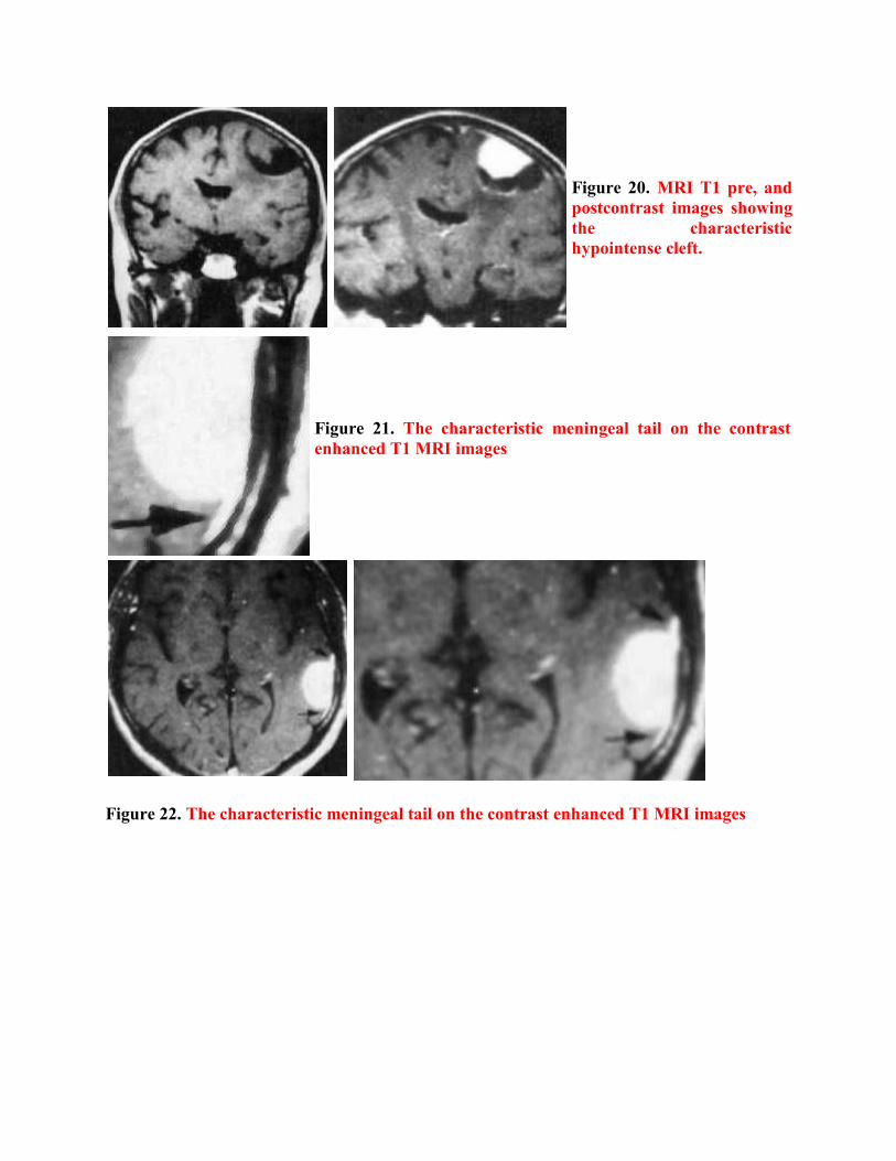
Figure 20. MRI T1 pre, andpostcontrast images showingthe characteristichypointense cleft.
Figure 21. The characteristic meningeal tail on the contrastenhanced T1 MRI images
Figure 22. The characteristic meningeal tail on the contrast enhanced T1 MRI images

Figure 23. A, Postmortem specimen, B,CMRI T1 postcontrast studies showingconvexity meningioma with thecharacteristic meningeal tail

Table 6. MRI CHARACTERISTICS OF MENINGIOMAS
MRI feature DescriptionVascularrim
The peripheries of meningiomas are supplied by branches from the anterioror middle cerebral arteries that encircle the tumour and form thecharacteristic vascular rim
Meningealtail
The tail extends to a variable degree away from the meningioma site andprobably represents a meningeal reaction to the tumour
Hypointensecleft
Hypointense cleft between the tumour and the brain that probablyrepresents blood vessels or a CSF interface
Figure 24. MRI T1 postcontrast studies showing parasagittal meningioma (left two images)and retroclivus meningioma (right image), notice the characteristic meningeal tail, and thedense contrast enhancement.
Figure 25. MRI precontrast T1, proton density and T2, proton density images showingmedial sphenoidal ridge syncytial meningioma, notice the vascular rim demonstrated assignal void linear structures surrounding the tumour (arrows). Also notice the surroundingedema.

Figure 26. Meningioma in a 27-year-old woman whopresented with new-onset seizure. A, Axial unenhanced CTimage demonstrates a large hyperdense extra-axial mass inthe left temporal region with associated central calcification(black arrow) and surrounding edema. B, Axial enhanced CTdemonstrates intense homogeneous enhancement. Distinctionof intra- versus extra-axial mass by CT can be difficult. C,Axial T2-weighted MR image clearly demonstrates a CSFcleft around the circumference of the tumor (arrowhead)indicating this to be an extra-axial mass. D, Sagittalpostcontrast Tl -weighted image demonstrates a dural tailanteriorly and posteriorly along the tentorium (white arrows).
Figure 27. Cystic meningioma. A,Axial postcontrast Tl -weightedimage reveals a cystic mass lesioninvolving the left frontal lobewith peripheral enhancement, aswell as enhancement around atrapped CSF intensity collectionlaterally (white arrow). B, Axialpostcontrast Tl -weighted imagenear vertex of the headdemonstrates the extra-axialnature of the mass withassociated dural attachment(white arrow).

UNUSUAL LOCATIONS OF MENINGIOMA
Cerebellopontine Angle Meningioma
The meningioma is the second most common mass lesion of the cerebellopontine angle, with13%-18% of all neoplastic lesions in this location being meningioma 42,43. Less than 5% ofall intracranial meningiomas occur in the cerebellopontine angle (8,9). The acousticschwannoma, from which meningiomas must be distinguished, is by far the most commontumor in this region. Meningiomas, however, tend to be larger, more hemispheric in shaperather than spherical, and more homogeneously enhancing. Meningiomas may beassociated with hyperostosis. They do not have a propensity to involve the internal auditorycanal (which is a fairly constant feature of schwannomas) (10).
Orbital Meningioma (optic sheath meningioma)
Orbital meningiomas account for less than 2% of cranial meningiomas but constitute 10%of all intraorbital neoplasms 39. Most of these tumors arise from the optic nerve sheathbetween the globe and the optic canal 39. They may produce diffuse thickening of the opticnerve, a well-defined and rounded mass, or even an eccentric lesion with an irregularborder. Calcification along the optic nerve sheath is highly suggestive of meningioma.
Multiple Meningiomas
In one series 45, CT demonstrated multiple tumors in about 9% of patients withintracranial meningioma. This approaches the 16% frequency of multiplicity found in anautopsy series 36. As with solitary examples, multiple meningiomas are more commonlyseen in women. Although multiple meningiomas are associated with neurofibromatosis type2 (“central” neurofibromatosis), the majority of patients do not have other characteristicfeatures such as multiple schwannomas. Further research with genetic testing is requiredto determine whether multiple meningiomas are inherited without neurofibromatosis type2. Secondary spread of tumor via the subarachnoid space is a less well-acceptedexplanation for multiple meningiomas 35.
En Plaque Meningioma
En plaque meningiomas cloak the inner table of the skull, where they may infiltrate boththe dura mater and underlying bone. On CT scans, especially those obtained withoutcontrast material, it may be difficult to distinguish the tumor itself from the associatedhyperostosis. The extent of radiographic hyperostosis has little relation to the degree orpresence of bone invasion and may occur secondary to local hypervascularity 35,39.Peritumoral edema is less common with en plaque tumors. MR images obtained withgadolinium enhancement enable this type of meningioma to be easily distinguished fromthe associated bone changes 39,46.
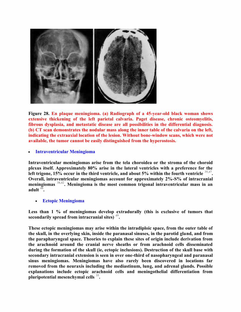
Figure 28. En plaque meningioma. (a) Radiograph of a 45-year-old black woman showsextensive thickening of the left parietal calvaria. Paget disease, chronic osteomyelitis,fibrous dysplasia, and metastatic disease are all possibilities in the differential diagnosis.(b) CT scan demonstrates the nodular mass along the inner table of the calvaria on the left,indicating the extraaxial location of the lesion. Without bone-window scans, which were notavailable, the tumor cannot be easily distinguished from the hyperostosis.
Intraventricular Meningioma
Intraventricular meningiomas arise from the tela choroidea or the stroma of the choroidplexus itself. Approximately 80% arise in the lateral ventricles with a preference for theleft trigone, 15% occur in the third ventricle, and about 5% within the fourth ventricle 35,47.Overall, intraventricular meningiomas account for approximately 2%-S% of intracranialmeningiomas 38,42. Meningioma is the most common trigonal intraventricular mass in anadult 48.
Ectopic Meningioma
Less than 1 % of meningiomas develop extradurally (this is exclusive of tumors thatsecondarily spread from intracranial sites) 49.
These ectopic meningiomas may arise within the intradiploic space, from the outer table ofthe skull, in the overlying skin, inside the paranasal sinuses, in the parotid gland, and fromthe parapharyngeal space. Theories to explain these sites of origin include derivation fromthe arachnoid around the cranial nerve sheaths or from arachnoid cells disseminatedduring the formation of the skull (ie, ectopic inclusions). Destruction of the skull base withsecondary intracranial extension is seen in over one-third of nasopharyngeal and paranasalsinus meningiomas. Meningiomas have also rarely been discovered in locations farremoved from the neuraxis including the mediastinum, lung, and adrenal glands. Possibleexplanations include ectopic arachnoid cells and meningothelial differentiation frompluripotential mesenchymal cells 35.

Figure 29. Intradiploic meningioma. (A) Radiograph of a 34-year-old black man, whocomplained of a bump on his head and orbital pressure, reveals a central radiolucent lesionwith partial loss of the outer table of the skull (arrows) and with extension into the frontalsinus. The tumor arose within bone but had extended through the dura mater and involvedthe frontal sinus. (B) Lateral image from an external carotid arteriogram of a 20-year oldman with mild frontal headaches reveals marked hyperostosis of the frontal bone andanterior aspect of the parietal bone. There is marked widening of the diploic space withperpendicular spiculation (arrowhead). Radiolucent areas proved at microscopicexamination to be medullary spaces of lamellar bone, filled with tumor cells, fibrous tissue,and a few osteoclasts. (C) CT scan of a 69-year old white man, who complained of a bumpon his head for the past 10 years, demonstrates an osteoblastic area within the rightparietal bone with mild expansion of the diploic space. A completely intraosseousmeningioma with marked hyperostotic reaction was confirmed pathologically.

Figure 30. (A) Ethmoid meningioma. Contrast-enhanced CT scan of a 20-year-old blackman with a 1-year history of decreased visual acuity and proptosis of the right eyedemonstrates an enhancing paranasal sinus mass with infiltration and destruction of theethmoid air cells. There is extension through the right medial orbital wall. The radiologicfindings are nonspecific, and other neoplastic or inflammatory conditions might have asimilar appearance. (B) Sphenoid and nasopharyngeal meningioma. Unenhanced CT scanobtained with bone windows of a 77-year-old white man with spontaneous epistaxisdemonstrates a smooth lobulated and partially calcified mass within both sphenoid sinuscompartments. No bone destruction and no intracranial component were found.Parapharyngeal meningioma. Axial (C) and coronal (D) contrast-enhanced CT scans of ayoung girl with a hearing loss in the left ear reveal a large tumor that involves the leftnasopharyngeal space, infratemporal fossa, and pterygoid fossa. The tumor also extendsintracranially through the sphenoid bone. Note the bone remodeling and hyperostosis ofthe maxillary sinus wall (arrows in C). At surgery, tumor was discovered in the leftmaxillary sinus, ethmoid air cells, and orbit.
ATYPICAL IMAGING FEATURES OF MENINGIOMA
In general, the various imaging features of meningiomas may not accurately reflect thespecific histologic subtypes of this common neoplasm, and the biologic and clinicalbehavior of meningiomas does not always correlate with the different histologic variants33,37,49. Therefore, from an imaging standpoint, it is important to recognize the variable andpleomorphic features exhibited by these neoplasms, so that an unusual appearingmeningioma is not confused with other intracranial masses.
Cystic Meningioma
The term cystic meningioma has been used to describe two different morphologies:intratumoral cavities and extratumoral or arachnoid cysts. Therefore, the cysts can belocated within the tumor mass, either centrally or eccentrically; outside and adjacent to theedge of the tumor; and, occasionally, inside the adjacent brain parenchyma. Trueintratumoral cystic meningiomas, with large dominant fluid-filled cysts, are an uncommonvariant. Benign meningiomas with heterogeneous enhancement that contain smallnonenhancing areas of cystic change or necrosis occur much more frequently (up to 8%-23% of cases) 37,38,39. A large cystic meningioma may have an atypical clinical presentation,in that they are more common in male and pediatric patients; these unusual clinicalfeatures often contribute to a misdiagnosis of a cystic or necrotic glioma 51.
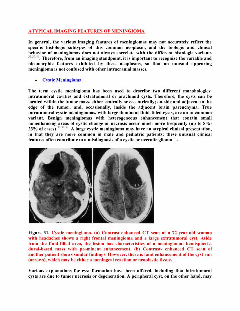
Figure 31. Cystic meningioma. (a) Contrast-enhanced CT scan of a 72-year-old womanwith headaches shows a right frontal meningioma and a large extratumoral cyst. Asidefrom the fluid-filled area, the lesion has characteristics of a meningioma: hemispheric,dural-based mass with prominent enhancement. (b) Contrast- enhanced CT scan ofanother patient shows similar findings. However, there is faint enhancement of the cyst rim(arrows), which may be either a meningeal reaction or neoplastic tissue.
Various explanations for cyst formation have been offered, including that intratumoralcysts are due to tumor necrosis or degeneration. A peripheral cyst, on the other hand, may
represent either peripheral degeneration or an arachnoid cyst. Although the imagingdifferentiation between a peripheral (neoplastic) intratumoral cyst and an extratumoral(reactive) arachnoid cyst may be suggested when ring enhancement is seen surrounding thefluid collection, histologic analysis, demonstrating neoplastic cells in the cyst wall, may berequired for confirmation. In addition, cysts may result from direct secretion of fluid bytumor cells, from absorption of internal hemorrhage, or from loculated cerebrospinal fluidin tissues within or adjacent to the meningioma 51.
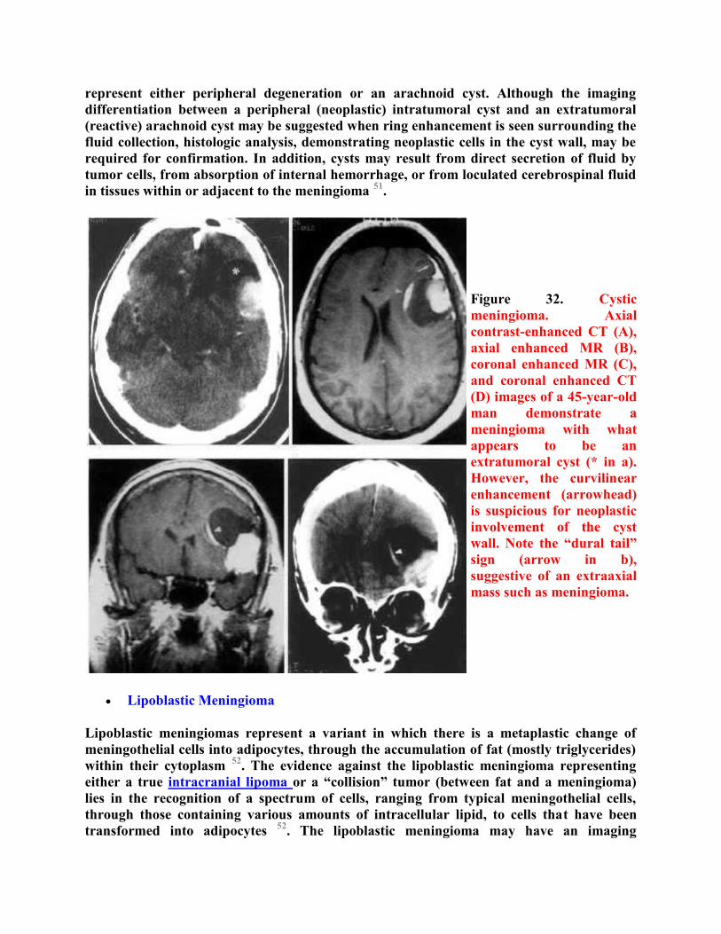
Figure 32. Cysticmeningioma. Axialcontrast-enhanced CT (A),axial enhanced MR (B),coronal enhanced MR (C),and coronal enhanced CT(D) images of a 45-year-oldman demonstrate ameningioma with whatappears to be anextratumoral cyst (* in a).However, the curvilinearenhancement (arrowhead)is suspicious for neoplasticinvolvement of the cystwall. Note the “dural tail”sign (arrow in b),suggestive of an extraaxialmass such as meningioma.
Lipoblastic Meningioma
Lipoblastic meningiomas represent a variant in which there is a metaplastic change ofmeningothelial cells into adipocytes, through the accumulation of fat (mostly triglycerides)within their cytoplasm 52. The evidence against the lipoblastic meningioma representingeither a true intracranial lipoma or a “collision” tumor (between fat and a meningioma)lies in the recognition of a spectrum of cells, ranging from typical meningothelial cells,through those containing various amounts of intracellular lipid, to cells that have beentransformed into adipocytes 52. The lipoblastic meningioma may have an imaging

appearance of a fatty tumor, with low negative attenuation on CT scans and a short TIrelaxation time with high signal intensity on T1-weighted MR images 53,54. Xanthomatouschange in meningioma can be differentiated histologically from the lipoblastic variant;however, since both contain excess lipid, the nadiologic distinction may be difficult.However, the lipoblastic meningioma may be suggested when the fatty regions are larger,are more confluent, and do not have prominent enhancement 53.
Figure 33. Lipoblasticmeningioma. (A) Contrast-enhanced CT scan of a 60-year-old white woman witha 2-week history of seeingflashing lights anddifficulty in reading showsa well-circumscribed low-attenuation lesion. The rimof the lesion is enhanced,and faint intratumoralstrands of enhancing tissueare seen. Sagittal TI-weighted (B) and axial T2-weighted (C) MR imagesdemonstrate a signalintensity within the lesionthat is similar to that ofsubcutaneous fat. (D)Gross specimen shows awell-circumscribed massand the yellowish color offatty metaplasia.

Figure 34. Lipoblastic meningioma. (A) Contrast-enhanced CT scan of a 36-year-old whitewoman with progressive gait difficulty demonstrates a left frontoparietal mass with anextremely low-attenuation (compatible with fat) center and a thick enhancing rind. Notethe small mound of hyperostosis (*) underlying the central enhancing nodule ofmeningioma (arrow). (B) Right external carotid arteriogram shows enlargement of themiddle meningeal artery that supplies the tumor. The spoke-wheel pattern of the fineradial arterioles is characteristic of meningioma. The ‘ ‘dimple’ ‘ in the center of theneovascularity (arrow) corresponds to the mound of bone seen in a. (C) Photograph of thecut specimen shows the attachment of the tumor to the dura mater (arrows) and theyellow-white color typical of lipoblastic meningioma.
Meningeal Hemangiopericytoma
Hemangiopericytoma of the meninges is an aggressive, highly vascular neoplasm that iscommonly grouped with “angioblastic” or “malignant” meningiomas 55,56. However,hemangiopericytoma of the meninges is a distinct nosologic entity arising from the vascularpericytes rather than from meningothelial cells; thus, it is not a true meningioma at all 57 .These tumors generally recur more frequently and earlier than meningiomas, and theyhave a greater propensity to develop distant metastases 57,58. The following features aresuggestive (but not pathognomonic) of a meningeal hemangiopericytoma: a multilobulatedcontour, a narrow dural base or ‘ ‘mushroom’ ‘ shape, large intratumoral vascular signalvoids on MR images, multiple irregular feeding vessels on angiograms, and bone erosionrather than hyperostosis 56,59. It has also been reported that prominent peritumoral edemaand increased signal on T2- weighted MR images are more common in the syncytial andthe angioblastic meningiomas (a category that includes hemangiopericytoma) than in othertypes 50,60.

Peritumoral Edema
Vasogenic edema within the white matter of the brain is a common feature of intraaxialmasses like glioma, metastatic disease, and abscess. However, mild to moderate intraaxialvasogenic edema is also seen around meningiomas (which are extraaxial masses) in up to75% of cases 41,61. The finding of edema can be problematic, since its presence may beincorrectly suggestive of an intraaxial lesion (eg, glioma). This problem is compoundedwhen the meningioma is small and the surrounding edema is extensive.
The cause of intraaxial peritumoral vasogenic edema associated with meningiomas iscontroversial. Some theories implicate active fluid production (secretion or excretion) bythe tumor, with “flow” through the thinned contiguous cortex 62. Others have suggestedthat the tumor injures the brain mechanically (by means of direct compression) orischemically (from parasitization of the cortical arteries, compression of the cortical veins,or frank involvement of the dural sinuses). Most likely, the edema is caused by acombination of different mechanisms. Reports about the importance of these factors havebeen conflicting 61,63,64. However, recent studies have found poor correlation betweenperitumoral edema and either the vascular supply of a meningioma or the presence ofdural sinus invasion 61. Whatever the mechanisms, the degree of peritumoral edema inmeningiomas has little correlation with tumor size 39,61.
Ring Enhancement
As mentioned, meningiomas are usually fairly homogeneous masses, with homogeneousenhancement. However, they may have an atypical ringed appearance 52,59 rather thanoccur as a solid mass. This unusual feature can be seen in both histologically typicalmeningiomas and in some malignant on aggressive histologic variants that may have cystformation, hemorrhage, or necrosis. The peripheral enhancement represents the normalpattern for viable meningeal neoplasms, and the center is an avascular or necrotic region.The causes for the central nonenhancing zone vary and include bland tumor infarction,necrosis in aggressive histologic variants, and true cyst formation from benign fluidaccumulation (see above) 51,65. A convexity meningioma with ring enhancement may easilybe confused with a necrotic on cystic glioma, a metastasis, or even an abscess. If such ameningioma arises from the falx cerebri, bilateral growth can even mimic a “butterfly”glioma, which is usually a glioblastoma multiforme (grade 4 astrocytoma).
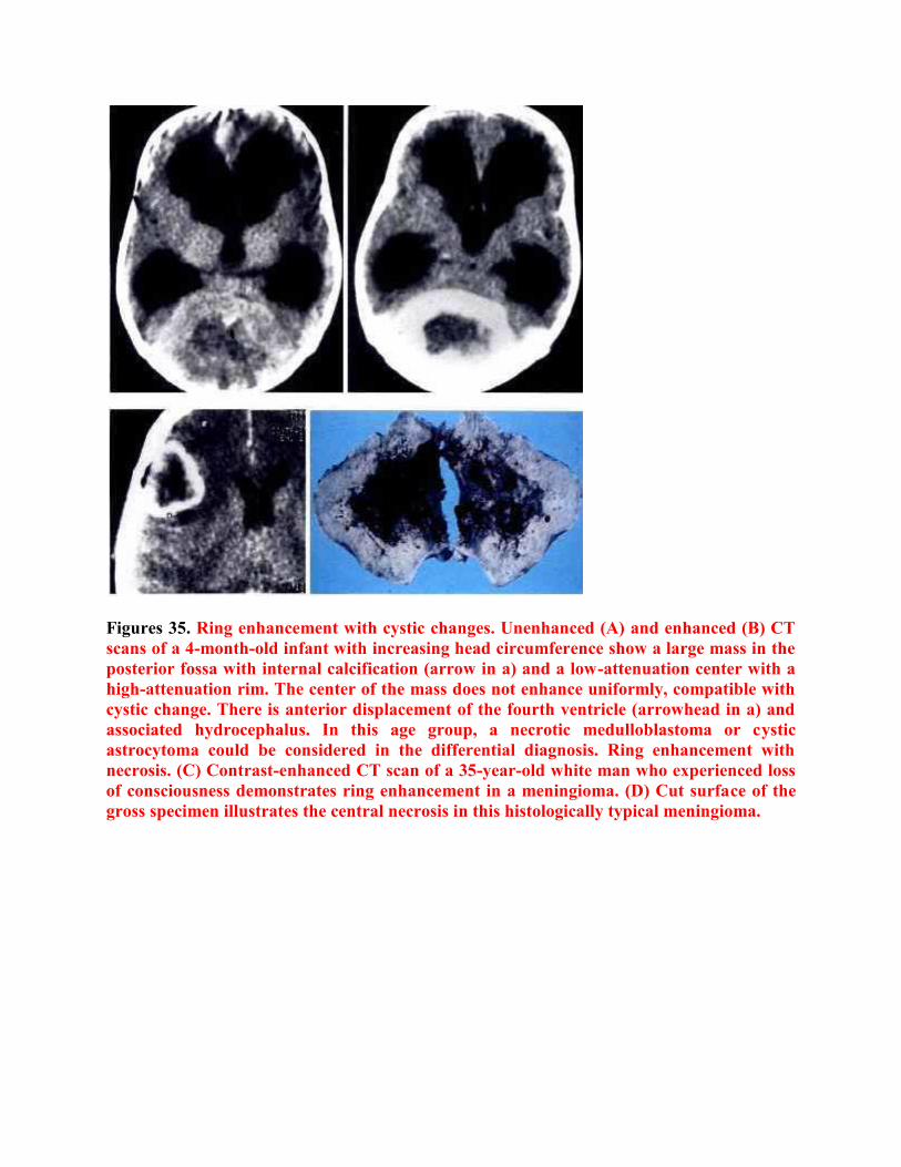
Figures 35. Ring enhancement with cystic changes. Unenhanced (A) and enhanced (B) CTscans of a 4-month-old infant with increasing head circumference show a large mass in theposterior fossa with internal calcification (arrow in a) and a low-attenuation center with ahigh-attenuation rim. The center of the mass does not enhance uniformly, compatible withcystic change. There is anterior displacement of the fourth ventricle (arrowhead in a) andassociated hydrocephalus. In this age group, a necrotic medulloblastoma or cysticastrocytoma could be considered in the differential diagnosis. Ring enhancement withnecrosis. (C) Contrast-enhanced CT scan of a 35-year-old white man who experienced lossof consciousness demonstrates ring enhancement in a meningioma. (D) Cut surface of thegross specimen illustrates the central necrosis in this histologically typical meningioma.

Figure 36. Hemangiopericytoma of the meninges in a 73-year-old man. (A) Contrast-enhanced CT scan shows homogeneously enhancing, markedly lobulated tumor indentingthe parietal lobes. (B) Gross specimen from a different patient exhibits the characteristiclobulated tumor surface.
Figure 37. “Butterfly” meningioma. Contrast-enhanced CT scan (A) and enhanced MRimage (B) demonstrate a falx meningioma with bilateral extension and central cavitationfrom necrosis in a 73-year-old white woman. This appearance is similar to that of a ‘‘butterfly’ ‘ glioblastoma multiforme. An unusual feature that can be seen in bothhistologically typical meningiomas and in some malignant and aggressive histologicvariants that may have cyst formation, hemorrhage, or necrosis. The peripheralenhancement represents the normal pattern for viable meningeal neoplasms, and the centeris an avascular or necrotic region. The causes for the central non-enhancing zone vary andinclude bland tumor infarction, necrosis in aggressive histologic variants, and true cystformation from benign fluid accumulation. A convexity meningioma with ringenhancement may easily be confused with a necrotic or cystic glioma, a metastasis, or evenan abscess. If such a meningioma arises from the falx cerebri, bilateral growth can evenmimic a “butterfly” glioma, which is usually a glioblastoma multiforme (grade 4astrocytoma).

MIMICS OF MENINGIOMA
Many atypical gross and imaging features of meningiomas have been presented here. Itshould also be recognized that other extraaxial soft-tissue lesions as well as some superficialintraaxial tumors may also exhibit a broad contact with the dural surface andhomogeneous contrast enhancement thereby mimicking meningioma. For example,hematologic neoplasms such as leukemia or secondary involvement of the central nervoussystem by Hodgkin lymphoma, which is a late manifestation of the disease, will typicallyinvolve the extraaxial spaces rather than the brain parenchyma 66. Such cases may bedifficult to differentiate from meningiomas. Other dural-based masses that may imitatemeningioma include dural and calvarial metastases from breast cancer and metastaticneuroblastoma.
Figure 38.Parasagittalmeningioma atoperation
SPINAL MENINGIOMA
Spinal meningiomas are unique in that there is a 4:1 female-to-male predominance, andmost patients are older than 40 years of age. Eighty percent of the lesions can be found inthe thoracic spine, although some are located at the upper cervical or lumbar regions. Theyoften are located anterolaterally or posterolaterally in the canal, and they are the mostcommon tumor of the foramen magnum, where they are frequently located anteriorly or

laterally. Meningiomas are rarely both intradural and extradural (6%), or purelyextradural (7%). 16
Meningiomas are the second most common tumor in the intradural extramedullarylocation, second only to tumors of the nerve sheath. Meningiomas account forapproximately 25% of all spinal tumors. Approximately 80% of spinal meningiomas arelocated in the thoracic spine, followed by cervical spine (15%), lumbar spine (3%), and theforamen magnum (2%). Most intradural spinal tumors are benign and potentiallyresectable. The prognosis after surgical resection is excellent.
Spinal meningiomas are often located laterally or dorsolaterally in the thoracic spine.Meningiomas of the cervical and foramen magnum tend to be located ventral to the spinalcord. They are believed to arise from the arachnoid cluster cells located at the entry zone ofthe nerve roots or at the junction of dentate ligaments and dura mater, where the spinalarteries penetrate. For this reason, lateral tumors are more common than dorsal andventral lesions. Most meningiomas are intradural and extramedullary. Occasionally, theycan be purely extradural (7%) or intradural and extradural (6%).
Compression of the cord by the meningioma can cause deterioration of neurologic function.Improvement of neurologic findings can be expected after resection of the tumor. Spinalmeningiomas differ from intracranial meningiomas by their slightly greater proclivity forpsammomatous change. In general, histopathologic features of spinal meningiomas aresimilar to their intracranial counterparts. Meningotheliomatous and transitional featuresare most common in spinal lesions. Spinal meningiomas are typically globoid, and theyvary in consistency depending on the extent of calcification. Multiple meningiomas are rare(2%) and most often associated with neurofibromatosis type II.
Frequency
In the US: Intradural spinal tumors can be classified as intramedullary or extramedullary.The incidence of intradural spinal tumors is approximately 3-10 cases per 100,000population. In children, 50% of intradural lesions are extramedullary, and 50% areintramedullary, whereas in adults, 70% are extramedullary, and 30% are intramedullary.
Mortality/Morbidity
Meningiomas and schwannomas and/or neurofibromas are the most common intraduralextramedullary spinal tumors. These benign lesions usually produce an insidious onset ofclinical symptoms, which are characterized by myelopathy and radiculopathy, respectively.As tumors grow, the symptom complex may merge, and significant neurologic deficits,including paraplegia, may develop.
Resection of spinal meningiomas can result in excellent recovery, even in patients withnotable preoperative deficits. The surgical morbidity rate is low because surgical resectionof a meningioma can easily be accomplished by means of simple laminectomy. Therecurrence rate is substantially lower than that seen in an intracranial lesion. This

observation may be secondary to the greater resectability of spinal meningiomas comparedwith intracranial lesions. Factors associated with poor outcome include calcified tumors,ventrally located lesions, age (ie, elderly patients), duration and severity of symptoms,subtotal resection, and an extradural component to the tumor.
Sex
Meningiomas most frequently affect women, with a 4:1 female-to-male ratio. Spinalmeningiomas are typically seen in women older than 40 years. Most spinal meningiomas inwomen occur in the thoracic spine. Although meningiomas of the spine occur in males, theydo so throughout the spinal canal without a predilection for the thoracic spine.
Age
Meningiomas are typically seen in women in the fifth and sixth decades. Approximately 3-6% of spinal meningiomas occur in children. Spinal meningiomas in children usually areassociated with neurofibromatosis.
Anatomy
Spinal meningiomas often are located laterally or dorsolaterally in the spinal canal. Theyare believed to arise from the arachnoid cluster cells, and therefore, they are located at theentry zone of the nerve roots or the junction of the dentate ligaments and dura mater. Mostmeningiomas are intradural and extramedullary in location. The spinal cord is typicallycompressed and displaced away from the lesion. The subarachnoid space above and belowthe mass lesion is widened, with cerebrospinal fluid capping the lesion from above andbelow. On occasion, they can be purely extradural (7%) or intradural and extradural (6%).
Clinical Details
Symptoms produced by meningiomas are secondary to their broad dural attachment andthe gradual growth of the tumor with compression of the cord. The clinical course may beinsidious, and symptoms are often confused with symptoms of other lesions of the spine,peripheral nervous system, and thorax. The duration of symptoms may span 6-23 months.Because meningiomas do not arise from nerve root sheaths, as do schwannomas, theytypically result in myelopathic rather than radiculopathic findings.
On physical examination, sensory and motor deficits are seen almost equally. A highincidence of Brown-Sequard syndrome is seen, with ipsilateral paralysis, decreased tactileand deep sensation, and a contralateral deficit in pain and temperature sensation. Thisfinding is most likely secondary to the high incidence of laterally positioned meningiomas.With substantial growth of the tumors, clinical findings may merge. Patients mostfrequently complain of regional back pain, especially at night, followed by sensorimotorchanges and, eventually, bowel and bladder dysfunction.

Pathological details
Macroscopically, most meningiomas are globose and expand centripetally inside the duralsac. A few have an en plaque configuration, and a small fraction assume a dumbbell-shaped profile, growing centrifugally into the epidural space; multiple spinal meningiomasalso have been reported. The histology is similar to their cranial counterparts in that theyhave a wide range of histopathologic appearances. Of the various subtypes, cyncytial,fibrous, and transitional meningiomas are the most common; however the psammomatoustype seems to be the most frequent histologic variety of spinal meningiomas. 15
Figure 39. A, spinal meningioma, B, Intraoperative photograph obtained using theoperative microscope demonstrating the intradural extamedullary meningioma attached tothe lateral dura surface and severely compressing the spinal cord.
Neuroimaging of spinal meningioma

o CT scan inaging
CT scans obtained without the intravenous injection of contrast material occasionallydemonstrate a hyperattenuating lesion resulting from psammomatous calcification ordense tumor tissue. CT scans obtained with the intravenous injection of contrast materialmay show a homogeneous enhancing tumor.
Myelography or CT myelography is required to demonstrate the intraduralextramedullary location of the mass.
The spinal cord is displaced away from the lesion and usually compressed. A sharpmeniscus is seen where the contrast agent caps the lesion from above and below. Thesubarachnoid space on the side of the lesion is widened. On CT, the degree of confidence ismoderate.
o MR imaging
MRI demonstrates the intradural extramedullary location of meningiomas. Lesions areusually isointense to spinal cord on both T1-weighted and T2-weighted images. Lesions aresometimes hypointense on T1-weighted images and hyperintense on T2-weighted images.Homogeneous intense enhancement of the lesion is seen after an intravenous injection ofgadolinium-based contrast agent.
Most spinal meningiomas demonstrate broad-based dural attachment. On occasion, adensely calcified meningioma may demonstrate hypointensity on both T1-weighted and T2-weighted images. The spinal cord is displaced away from the lesion and usuallycompressed. The subarachnoid space above and below the lesion is widened, and ameniscus capping the lesion may be seen. On MRI, the degree of confidence is high.
False Positives/Negatives
A meningioma with intradural and extradural components occasionally mimic a nervesheath tumor, or a nerve sheath tumor with a predominant intradural component maymimic a meningioma. However, nerve sheath tumors usually have hyperintensity on T2-weighted images, whereas meningiomas usually are isointense to the spinal cord on T2-weighted images. Most meningiomas are lateral or dorsal, whereas most nerve sheathtumors are ventral. Furthermore, a mass lesion with both intradural and extraduralcomponents is most likely to be a nerve sheath tumor.

Figure 40. Sagittal Tl -weighted (A) and T2-weighted (B) MR images of the dorsal spineshowing an isodense intradural extramedullary transitional meningioma compressing thespinal cord. A hemangioma in the adjacent vertebra also can be observed in B.
Figure 41. MRI T1 images precontrast (A) and postcontrast (B,C) showing a dorsalsyncytial meningioma, notice the T1 hypointensity (A), the dense contrast enhancementand the dural tail (B,C)

Figure 42. MRI T1 images (A, precontrast and B, postcontrast) and T2 image (c) showing ahigh cervical syncytial meningioma, notice the precontrast T1 slight hypointensity, thedense contrast enhancement, the cavity caudal to the tumour (A) and the T2 hyperintensity(C). Also notice the CSF cleft that separate the tumour from the spinal cord (A)
Figure 43. A, Sagittal contrast-enhanced T1-weighted MR image of the cervical spine.Multiple extramedullary enhancing dural-based tumors (meningiomas) are seen at the C2and C7-T1 levels (black solid arrows). The tumor at the C7-T1 level results in cordcompression. In addition, an enhancing intramedullary tumor (white solid arrows) at the

T3-T4 level causes focal cord engorgement. An associated syrinx (open arrow) is seen in asmall segment of the cord proximal to this tumor. The patient had neurofibromatosis type2. B,C Lumbar meningioma
CONCLUSION
Meningioma is the most common nonglial primary neoplasm of the central nervous system.The diagnosis of meningioma is relatively uncomplicated when the tumor is in a typicallocation and has characteristic radiologic findings. However, it must be remembered thatmeningiomas may occur in unusual locations and with misleading or atypical imagingfeatures.
References
1. Burger PC, Scheithauer BW, Vogel FS: Surgical Pathology of the Nervous System andIts Coverings, ed 3. New York, Churchill-Livingstone, 1991
2. Kepes Jj: Meningiomas: Biology, Pathology and Differential Diagnosis. New York,Masson Publishing, 1982
3. Shapiro JR, Coons SW: Genetics of adult malignant gliomas. BNI Quarterly 14:27- 38,1998
4. Courville CB: Pathology of the Central Nervous System, ed 3. Mountain View, CA,Pacific, 1950, p 383
5. Elster AD, Challa VR, Gilbert TH, et al: Meningiomas: MR and histopathologicfeatures. Radiology 170:857, 1989
6. Russell DS, Rubinstein Lj: Pathology of Tumors of the Nervous System, ed 4. Baltimore,Williams & Wilkins, 1977, p 48
7. Russel D, Rubenstein L (ed): Pathology of Tumors of the Nervous System. Baltimore,Williams and Wilkins, 1989
8. Atlas SW: Adult supratentorial tumors. Semin Roentgenol 25:130-154,1990
9. Osborne A (ed): Diagnostic Neuroradiology St. Louis, Mosby-Year Book, 1994
10. Zulch K (ed): Brain Tumors: Their Biology and Pathology, ed 3. New York, Spxinger-Verlag, 1986
11. Smith F, Slavik M, McDonald L: Association of breast cancer with meningioma.Cancer 42:1992-1994, 1978

12. Roelvink N, Kamphorst W, Alphen HV: Pregnancy related primary brain and spinaltumors. Arch Neurol 44:209-215,1987
13. Som P, Sacher M, Strenger S, et al: 'Benign" metastasizing meningiomas. AJNR Am jNeuroradiol 8:127-130, 1987
14. Claveria L, Sutton D, Tress B: The radiological diagnosis of meningiomas: The impactof EMI scanning. Br j Radiol 50:15-22, 1977
15. Louis DN, Scheithauer BW, Budka H, et al: Meningiomas. In Kleihues P, Cavenee WC(eds): Pathology and Genetics-Tumours of the Nervous System. Lyon, World HealthOrganization and International Agency for Research on Cancer, 2000, p 176
16. Masaryk Tj: Neoplastic diseases of the spine. Radiol Clin North Am 29:829,1991
17. Chamberlain MC, Sandy AD, Press GA: Spinal cord tumors: gadolinium-DTPA-enhanced MR imaging. Neuroradiology 1991; 33(6): 469-74.
18. Derenda M, Bayassi S: [Thoracic spine meningioma mimicking intramedullary tumor].Neurol Neurochir Pol 2000 Mar-Apr; 34(2): 357-65.
19. Dillon WP, Norman D, Newton TH, et al: Intradural spinal cord lesions: Gd-DTPA-enhanced MR imaging. Radiology 1989 Jan; 170(1 Pt 1): 229-37.
20. Doita M, Harada T, Nishida K, et al: Recurrent calcified spinal meningioma detectedby plain radiograph. Spine 2001 Jun 1; 26(11): E249-52.
21. Egelhoff JC, Bates DJ, Ross JS, et al: Spinal MR findings in neurofibromatosis types 1and 2. AJNR Am J Neuroradiol 1992 Jul-Aug; 13(4): 1071-7.
22. Gamache FW Jr, Wang JC, Deck M, Heise C: Unusual appearance of an en plaquemeningioma of the cervical spinal canal. A case report and literature review. Spine 2001Mar 1; 26(5): E87-9.
23. Levy WJ Jr, Bay J, Dohn D: Spinal cord meningioma. J Neurosurg 1982 Dec; 57(6):804-12.
24. Li MH, Holtas S, Larsson EM: MR imaging of intradural extramedullary tumors. ActaRadiol 1992 May; 33(3): 207-12.
25. Masaryk TJ: Neoplastic disease of the spine. Radiol Clin North Am 1991 Jul; 29(4):829-45.
26. Matsumoto S, Hasuo K, Uchino A, et al: MRI of intradural-extramedullary spinalneurinomas and meningiomas. Clin Imaging 1993 Jan-Mar; 17(1): 46-52.

27. McCormick PC, Post KD, Stein BM: Intradural extramedullary tumors in adults.Neurosurg Clin N Am 1990 Jul; 1(3): 591-608.
28. Onofrio BM: Intradural extramedullary spinal cord tumors. Clin Neurosurg 1978; 25:540-55.
29. Schroth G, Thron A, Guhl L, et al: Magnetic resonance imaging of spinal meningiomasand neurinomas. Improvement of imaging by paramagnetic contrast enhancement. JNeurosurg 1987 May; 66(5): 695-700.
30. Solero CL, Fornari M, Giombini S, et al: Spinal meningiomas: review of 174 operatedcases. Neurosurgery 1989 Aug; 25(2): 153-60.
31. Souweidane MM, Benjamin V: Spinal cord meningiomas. Neurosurg Clin N Am 1994Apr; 5(2): 283-91.
32. Weck M, Pause M, Pinzer T: [Spinal meningioma as differential diagnosis of diabeticpolyneuropathy]. Dtsch Med Wochenschr 2001 May 18; 126(20): 590-2.
33. Weil SM, Gewirtz RJ, Tew JM Jr: Concurrent intradural and extradural meningiomasof the cervical spine. Neurosurgery 1990 Oct; 27(4): 629-31.
34. Zee CS, Henderson R, Huprich J: Imaging and relevant anatomy. In: Banzel E,Stillerman C, eds. The Thoracic Spine. Quality Medical Publishing; 1999: 80-122.
35 . Russell DS, Rubinstein U. Pathology of tumors of the nervous system. 5th ed.Baltimore: Williams & Wilkins, 1989; 449-483.
36. Wood MW, White R, KernohanJ. One hundred meningiomas found incidentally atnecropsy. J Neunopathol Exp Neurol 1957; 16:337-340.
37. Russell EJ, George AE, Knicheffll, Budzilovich G. Atypical computed tomographicfeatures of intracranial meningioma: radiological-pathological correlation in a series of 131consecutive cases. Radiology 1980; 135:673- 682. Rohninger M, Sutherland GR, Louw DF,Sima AAF. Incidence and clinicopathological features of meningioma. J Neurosurg 1989;71: 665-672.
38. Bradac GB, Ferszt R, Kendall BE. Cranial meningiomas. Berlin: Springer-Verlag,1990; 1-128.
39. Zimmerman RD, Fleming CA, Saint-Louis LA, Lee BCP, ManningJJ, Deck MDF.Magnetic resonance imaging of meningiomas. AJNR 1985; 6: 149-157.
40. New PFJ, Anonow 5, HesselinkJR. National Cancer Institute study: evaluation ofcomputed tomography in the diagnosis of intra- cranial neoplasms. IV. Meningiomas.Radiology 1980; 136:665-675.

41. Kendall B, Symon L. Investigation of patients presenting with cerebellopontine anglesyndromes. Neuroradiology 1977; 13:65-84.
42. Mikhael MA, Ciric IS, WoIifAP. Differentiation of cerebellopontine angle neuromasand meningiomas with MR imaging. J Comput Assist Tomog. 1985; 9:852-856.
43. Wu E, Tang Y, Zhang Y, Bai R. CT in diagnosis of acoustic neuromas. AJNR 1986;7:645- 650.
44 . Lusin JO, Nakagawa H. Multiple meningiomas evaluated by computed tomography.Neurosurgery 198i; 9:137-141.
45. Bydder GM, Kingsley DPE, BrownJ, Niendorf HP, Young IR. MR imaging ofmeningiomas including studies with and without gadolinium- DTPA. J Comput AssistTomogr 1985; 9:690-697.
46. Mani RL, Hedgcock MW, Mass SI, Gilmor RI, Enzmann DR, Eisenberg RI.Radiographic diagnosis of meningiomas of the lateral ventriculo: review of 22 cases. JNeurosurg 1978; 49:249-255.
47. JelinekJ, Smirniotopoulos JG, Panisi JE, Kanzen M. Lateral ventricular neoplasms ofthe brain: differential diagnosis based on clinical CT and MR findings. AJNR 1990;11:567-574.
48. Geoffray A, Lee YY, Jing BS, Wallace S. Extracranial meningiomas of the head andneck. AJNR 1984; 5:599-604.
49. Elster AD, Calla VR, Gilbert TH, Richardson DN, ContentoJC. Meningiomas: MR andhistopathologic features. Radiology 1989; 170:857-862.
50. Worthington C, CaronJ, Melanson D, Leblanc R. Meningioma cysts. Neurology 1985;35: 1720-1724.
51. KepesJJ. Meningiomas: biology, pathology, and differential diagnosis. New York:Masson, 1982; 75-109.
52. LeRoux P, Hope A, Lofton S, Harris AB. Lipomatous meningioma: an uncommontumor with distinct radiographic findings. Sung Neurol 1989; 32:360-365.
53. Salibi 55, Nauta HJW, Brem H, Epstein JI, Cho KR. Lipomeningioma: report of threecases and review of the literature. Neurosurgery 1989; 25:122-126.
54. Horten BC, Unich H, Rubinstein U, Montague SR. The angioblastic meningioma: areappraisal of a nosological problem. J Neurol Sci 1977; 31:387-410.
55. MarcJA, Takei Y, Schecter MM. Intracranial hemangiopericytomas: angiography,pathology, and differential diagnosis. AJR 1975; 125: 823-832.
56. Mena H, RibasJL, Pezeshkpour GH, Cowan DN, Panisi JE. Hemangiopericytoma of thecentral nervous system: a review of 94 cases. Hum Pathol 1991; 22:84-91.
57. JaaskelainenJ, Serve A, Haltia M, Wahlstrom T, Valtonen S. IntracranialHemangiopericytoma: radiology, surgery, radiotherapy, and outcome in 2 1 patients. SungNeurol 1985; 23: 227-236.
58. Osborne DE, Dubois P, Drayer B, Sage M, Burger P, Heinz ER. Primary intracranialmeningeal and spinal hemangiopericytoma: radiologic manifestations. AJNR 1981; 2:69-74.
59. Smith HP, Challa VR, Moody DM, Kelly DL. Biological features of meningiomas thatdetermine the production of cerebral edema. Neurosurgery 1981; 8:429-433.
60. Bnadac GB, Ferszt R, Bender A, Schorner W. Peritumoral edema in meningioma: aradiological and histological study. Neuroradiology 1986; 28:304-312.
61. Philippon J, Foncin JF, Gnov R, Snoun A, Poisson M, Pentuiset BF. Cerebral edemaassociated with meningiomas: possible role of a secretory-excretory phenomenon.Neurosurgery 1984; 14:295-301.
62. StevensJM, Rui.zJS, Kendall BE. Observations on peritumoral oedema in meningioma.I. Distribution, spread, and resolution of vasogenic oedema seen on computed tomography.Neuroradiology 1983; 25:71-80.
63. StevensJM, RuizJS, Kendall BE. Observations on peritumoral oedema in meningioma.II. Mechanisms of oedema production. Neuroradiology 1983; 25: 125-131.
64 . Pullicin P, Wilbur DC, Levy RJ, Eskin TA, Kido DK. Infarction in a meningioma aftercardiac arrest: computed tomographic and pathologic findings. Arch Neurol 1983; 40: 456-457.
65. Zimmerman RA. Central nervous system lymphoma. Radiol Clin North Am 1990; 28:697-721.
The author: Professor Yasser Metwally
Professor of neurology, Ain Shams university, Cairo, Egypt
www.yassermetwally.com