Topic 3 Biochemistry Two Nucleic Acids & Proteins
114
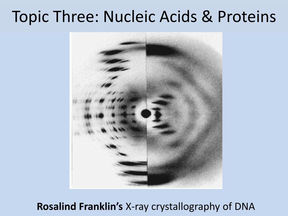
Topic Three: Nucleic Acids & Proteins Rosalind Franklin’s X-ray crystallography of DNA
-
Upload
bob-smullen -
Category
Education
-
view
152 -
download
0
Transcript of Topic 3 Biochemistry Two Nucleic Acids & Proteins
Topic Three: Nucleic Acids & Proteins
Rosalind Franklin’s X-ray crystallography of DNA

2.6 Structure of DNA and RNA
Essential idea: The structure of DNA allows efficient storage of genetic information.

Understandings, Applications and Skills
Statement Guidance
2.6 U.1 The nucleic acids DNA and RNA are polymers of nucleotides.
2.6 U.2 DNA differs from RNA in the number of strands present, the base composition and the type of pentose.
2.6 U.3 DNA is a double helix made of two antiparallel strands of nucleotides linked by hydrogen bonding between complementary base pairs.
2.6 A.1 Crick and Watson’s elucidation of the structure of DNA using model making.
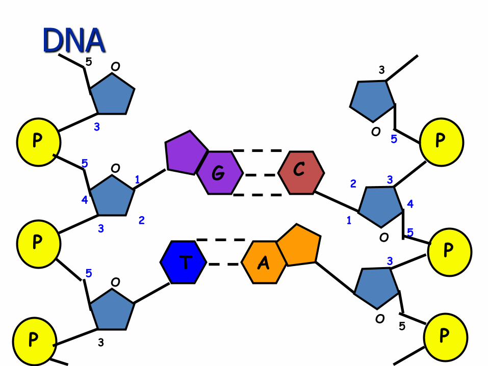
2.6 S.1 Drawing simple diagrams of the structure of single nucleotides of DNA and RNA, using circles, pentagons and rectangles to represent phosphates, pentose and bases.
In diagrams of DNA structure, the helical shape does not need to be shown, but the two strands should be shown antiparallel. Adenine should be shown paired with thymine and guanine with cytosine, but the relative lengths of the purine and pyrimidine bases do not need to be recalled, nor the numbers of hydrogen bonds between the base pairs.

Hershey and Chase experiment providing evidence that DNA is the genetic material.
Hershey and Chase Experiments (1952): Definitive proof that DNA rather than Protein carries the hereditary information of life
E. Coli bacteriophage: A virus that infects bacteria.
Bacteriophages only contain a protein coat (capsid) and DNA.
They wanted to find out whether the protein or DNA carried the genetic instructions to make more viruses.
They labeled either the viral proteins or DNA:– Protein capsid: Labeled with radioactive sulfur (35S)
– DNA: Labeled with radioactive phosphorus (32P)
Radioactive labeled viruses were used to infect cells.
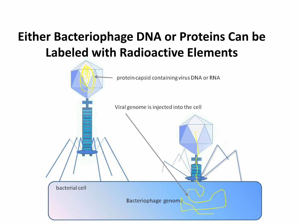
Either Bacteriophage DNA or Proteins Can be Labeled with Radioactive Elements

Hershey and Chase Experiments (1952):
Bacterial cells that were infected with the two types of bacteriophage, were then spun down into a pellet (centrifuged), and examined.Results:
1. Labeled viral proteins did not enterinfected bacteria (found in supernatant).
2. Labeled viral DNA did enter bacteria during viral infection (found in cell pellet).
Conclusion:
Protein is not necessary to make new viruses.
DNA is the molecule that carries the genetic information to make new viruses!!!!
Hershey and Chase experiment providing evidence that DNA is the genetic material.
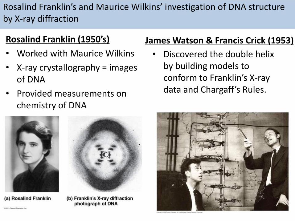
Rosalind Franklin (1950’s)
• Worked with Maurice Wilkins
• X-ray crystallography = images of DNA
• Provided measurements on chemistry of DNA
Rosalind Franklin’s and Maurice Wilkins’ investigation of DNA structure by X-ray diffraction
James Watson & Francis Crick (1953)
• Discovered the double helix by building models to conform to Franklin’s X-ray data and Chargaff’s Rules.

DNA
• Stands for Deoxyribonucleic acid
• Made up of subunits called nucleotides
• Nucleotide made of:
1. Phosphate group
2. 5-carbon sugar
3. Nitrogenous base

2.6 U.1 The nucleic acids DNA and RNA are polymers of nucleotides.
A nucleotide: a single unit of a nucleic acid
There are two types of nucleic acid: DNA and RNA.
Nucleic acids are very large molecules that are constructed by linking together nucleotides to form a polymer.

covalent bond
covalent bond
A nucleotide: a single unit of a nucleic acid
• five carbon atoms = a pentose sugar
• If the sugar is Deoxyribose the polymer is Deoxyribose Nucleic Acid (DNA)
• If the sugar Ribose the polymer is Ribose Nucleic Acid (RNA)
• acidic• negatively
charged• contains nitrogen• has one or two rings
in it’s structure

2.6 U.1 The nucleic acids DNA and RNA are polymers of nucleotides.
• Nucleotides a linked into a single by condensation reaction
• Bonds are formed between the phosphate of one nucleotide and the pentose sugar of the next.
• The phosphate group (attached to the 5'-C of the sugar) joins with the hydroxyl (OH) group attached to the 3'-C of the sugar
• Successive condensation reactions between nucleotides results in the formation of a long single strand

2.6 U.3 DNA is a double helix made of two antiparallel strands of nucleotides linked by hydrogen bonding between complementary base pairs.
• DNA is double stranded and shaped like a ladder, with the sides of the ladder made out of repeating phosphate and deoxyribose sugar molecules covalently bonded together. The two strands are antiparallel to each other due to base pairing.
• The rungs of the ladder contain two nitrogenous bases (one from each strand) that are bonded together by hydrogen bonds.
• The nitrogenous bases match up according the Chargaff’s Rules in which adenine always bonds to thymine, and guanine always bonds with cytosine. These bonds are hydrogen bonds.

2.6 U.1 The nucleic acids DNA and RNA are polymers of nucleotides.
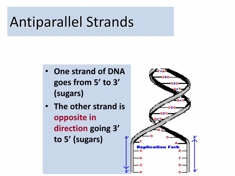
Antiparallel Strands
• One strand of DNA goes from 5’ to 3’ (sugars)
• The other strand is opposite in direction going 3’ to 5’ (sugars)
15
DNA
P
P
P
O
O
O
1
23
4
5
5
3
3
5
P
P
PO
O
O
1
2 3
4
5
5
3
5
3
G C
T A
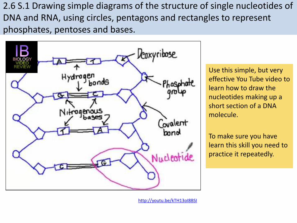
2.6 S.1 Drawing simple diagrams of the structure of single nucleotides of DNA and RNA, using circles, pentagons and rectangles to represent phosphates, pentoses and bases.
Use this simple, but very effective You Tube video to learn how to draw the nucleotides making up a short section of a DNA molecule.
To make sure you have learn this skill you need to practice it repeatedly.
http://youtu.be/kTH13oI8BSI

RNA DNA
Bases
Adenine (A)Guanine (G)
Uracil (U)Cytosine (C)
Adenine (A)Guanine (G)Thymine (T)Cytosine (C)
Sugar
Ribose Deoxyribose
Number of strands
Single stranded, and often,but not always, linear in
shape
Two anti-parallel, complementary strands
form a double helix
2.6 U.2 DNA differs from RNA in the number of strands present, the base composition and the type of pentose.
http://commons.wikimedia.org/wiki/File:RiboseAndDeoxy.gif

2.6 A.1 Crick and Watson’s elucidation of the structure of DNA using model making.
http://scarc.library.oregonstate.edu/coll/nonspcoll/catalogue/picture-dnamodel-900w.jpg
While others worked using an experimental basis Watson and Crick used stick-and-ball models to test their ideas on the possible structure of DNA. Building models allowed them to visualize the molecule and to quickly see how well it fitted the available evidence.
It was not all easy going however. Their first model, a triple helix, was rejected for several reasons:• The ratio of Adenine to Thymine was not 1:1 (as discovered
by Chargaff)• It required too much magnesium (identified by Franklin)
From their setbacks they realized:• DNA must be a double helix.• The relationship between the bases and base pairing• The strands must be anti-parallel to allow base pairing to
happen
Because of the visual nature of their work the second and the correct model quickly suggested:• Possible mechanisms for replication• Information was encoded in triplets of bases

http://www.nobelprize.org/educational/medicine/dna_double_helix/readmore.html
http://youtu.be/sf0YXnAFBs8
Find out more about the discovery of DNA:
Watson and Crick gained Nobel prizes for their discovery. It should be remembered that their success was based on the evidence they gained from the work of others. In particular the work of Rosalind Franklin and Maurice Wilkins, who were using X-ray diffraction was critical to their success.
2.6 A.1 Crick and Watson’s elucidation of the structure of DNA using model making.
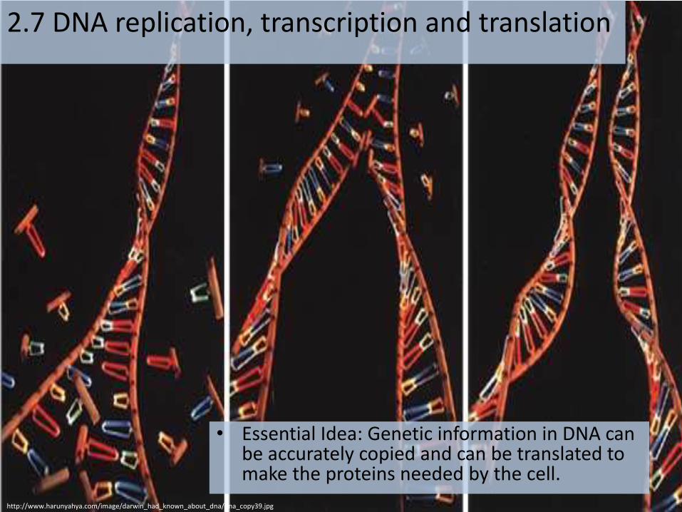
2.7 DNA replication, transcription and translation
• Essential Idea: Genetic information in DNA can be accurately copied and can be translated to make the proteins needed by the cell.
http://www.harunyahya.com/image/darwin_had_known_about_dna/dna_copy39.jpg

UnderstandingsStatement Guidance
2.7 U.1 The replication of DNA is semi-conservative and
depends on complementary base pairing.
2.7 U.2 Helicase unwinds the double helix and separates
the two strands by breaking hydrogen bonds.
2.7 U.3 DNA polymerase links nucleotides together to form
a new strand, using the pre-existing strand as a
template.
The different types of DNA polymerase do not
need to be distinguished.
2.7 U.4 Transcription is the synthesis of mRNA copied from
the DNA base sequences by RNA polymerase.
2.7 U.5 Translation is the synthesis of polypeptides on
ribosomes.
2.7 U.6 The amino acid sequence of polypeptides is
determined by mRNA according to the genetic
code.
2.7 U.7 Codons of three bases on mRNA correspond to
one amino acid in a polypeptide.
2.7 U.8 Translation depends on complementary base
pairing between codons on mRNA and anticodons
on tRNA.
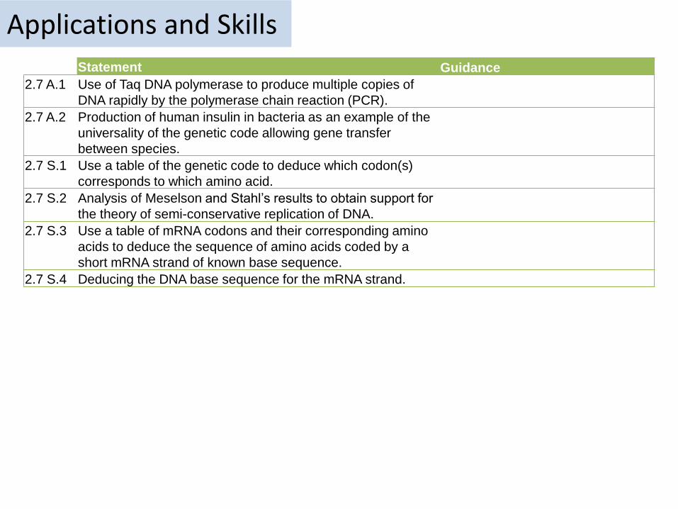
Applications and SkillsStatement Guidance
2.7 A.1 Use of Taq DNA polymerase to produce multiple copies of
DNA rapidly by the polymerase chain reaction (PCR).
2.7 A.2 Production of human insulin in bacteria as an example of the
universality of the genetic code allowing gene transfer
between species.
2.7 S.1 Use a table of the genetic code to deduce which codon(s)
corresponds to which amino acid.
2.7 S.2 Analysis of Meselson and Stahl’s results to obtain support for
the theory of semi-conservative replication of DNA.
2.7 S.3 Use a table of mRNA codons and their corresponding amino
acids to deduce the sequence of amino acids coded by a
short mRNA strand of known base sequence.
2.7 S.4 Deducing the DNA base sequence for the mRNA strand.
DNA
• Two jobs
1. Creating DNA for two cell (DNA Replication)
2. Creating Proteins using RNA

• DNA replication is very specific to the arrangements of base pairs
• In DNA replication, the strands separate
– Enzymes use each strand as a template to assemble the new strands
DNA REPLICATION
Parental moleculeof DNA
Both parental strands serveas templates
Two identical daughtermolecules of DNA
Nucleotides
A

Synthesis Phase (S phase)• S phase during interphase of the cell
cycle
• Nucleus of eukaryotes
Mitosis-prophase-metaphase-anaphase-telophase
G1 G2
Sphase
interphase
DNA replication takesplace in the S phase.
Mitosis-prophase-metaphase-anaphase-telophase
G1 G2
Sphase
interphase
DNA replication takesplace in the S phase.
Super coiling begin in prophase, making chromosomes visible for the first time

DNA Replication

2.7 U.1 The replication of DNA is semi-conservative and depends on complementary base pairing.
https://upload.wikimedia.org/wikipedia/commons/3/33/DNA_replication_split_horizontal.svg
1. Each of the nitrogenous bases can only pair with its partner (A=T and G=C) this is called complementary base pairing.
2. The two new strands formed will be identical to the original strand.
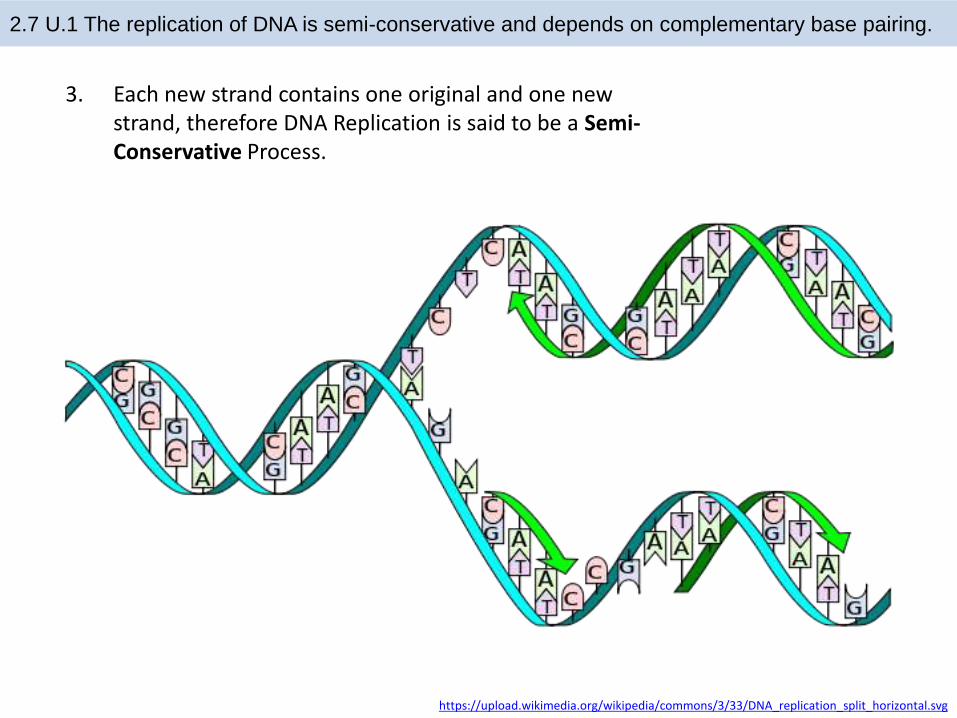
2.7 U.1 The replication of DNA is semi-conservative and depends on complementary base pairing.
https://upload.wikimedia.org/wikipedia/commons/3/33/DNA_replication_split_horizontal.svg
3. Each new strand contains one original and one new strand, therefore DNA Replication is said to be a Semi-Conservative Process.
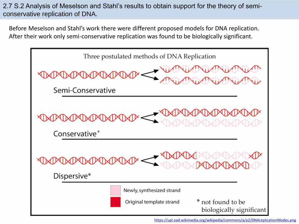
2.7 S.2 Analysis of Meselson and Stahl’s results to obtain support for the theory of semi-
conservative replication of DNA.
https://upl oad.wikimedia.org/wikipedia/commons/a/a2/DNAreplicationModes.png
Before Meselson and Stahl’s work there were different proposed models for DNA replication. After their work only semi-conservative replication was found to be biologically significant.
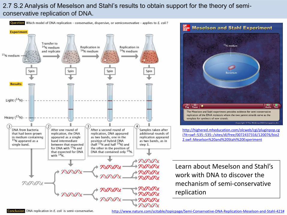
2.7 S.2 Analysis of Meselson and Stahl’s results to obtain support for the theory of semi-
conservative replication of DNA.
Learn about Meselson and Stahl’s work with DNA to discover the mechanism of semi-conservative replication
http://highered.mheducation.com/olcweb/cgi/pluginpop.cgi?it=swf::535::535::/sites/dl/free/0072437316/120076/bio22.swf::Meselson%20and%20Stahl%20Experiment
http://www.nature.com/scitable/topicpage/Semi-Conservative-DNA-Replication-Meselson-and-Stahl-421#

2.7 U.2 Helicase unwinds the double helix and separates the two strands by breaking hydrogen
bonds.
http://en.wikipedia.org/wiki/File:Helicase.png
Helicase• The ‘ase’ ending indicates it is an
enzyme• This family of proteins varies, but
are often formed from multiple polypeptides and doughnut in shape
• This enzyme is responsible for “unzipping” the DNA. This exposes the nucleotides which act as a template of the new strands.
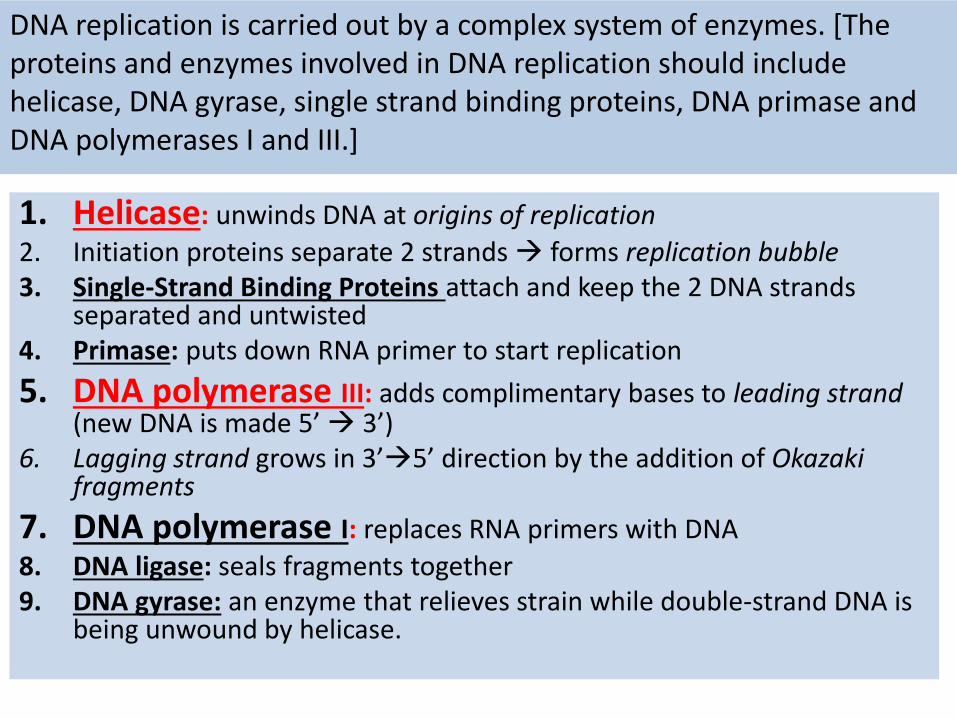
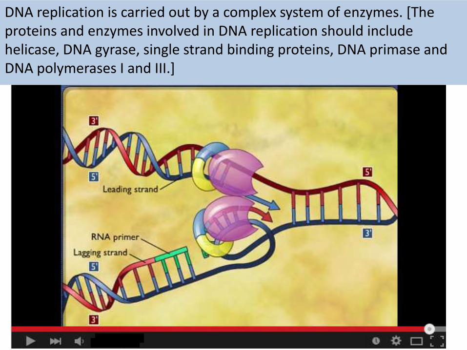
1. Helicase: unwinds DNA at origins of replication
2. Initiation proteins separate 2 strands forms replication bubble3. Single-Strand Binding Proteins attach and keep the 2 DNA strands
separated and untwisted4. Primase: puts down RNA primer to start replication
5. DNA polymerase III: adds complimentary bases to leading strand (new DNA is made 5’ 3’)
6. Lagging strand grows in 3’5’ direction by the addition of Okazaki fragments
7. DNA polymerase I: replaces RNA primers with DNA
8. DNA ligase: seals fragments together 9. DNA gyrase: an enzyme that relieves strain while double-strand DNA is
being unwound by helicase.
DNA replication is carried out by a complex system of enzymes. [The proteins and enzymes involved in DNA replication should include helicase, DNA gyrase, single strand binding proteins, DNA primase and DNA polymerases I and III.]

2.7 U.3 DNA polymerase links nucleotides together to form a new strand, using the pre-existing
strand as a template.
• DNA polymerase always moves in a 5’ to 3’ direction
• DNA polymerase catalyzes the covalent phosphodiester bonds between sugars and phosphate groups

2.7 U.3 DNA polymerase links nucleotides together to form a new strand, using the pre-existing
strand as a template.
• Free nucleotides are deoxynucleosidetriphosphates
• The extra phosphate groups carry energy which is used for formation of covalent bonds
• DNA polymerase always moves in a 5’ to 3’ direction
• DNA polymerase catalyzes the covalent phosphodiester bonds between sugars and phosphate groups
• DNA Polymerase proof reads the complementary base pairing. Consequently mistakes are very infrequent occurring approx. once in every billion bases pairs
1. HelicaseUnzips the DNA
DNA replication is carried out by a complex system of enzymes. [The proteins and enzymes involved in DNA replication should include helicase, DNA gyrase, single strand binding proteins, DNA primase and DNA polymerases I and III.]

2.7 A.1 Use of Taq DNA polymerase to produce multiple copies of DNA rapidly by the polymerase
chain reaction (PCR).
http://www.dnai.org/b/index.html
After clicking on the myDNAlink choose techniques and then amplifying to access the tutorials on the polymerase chain reaction (PCR).
Alternatively watch the McGraw-Hill tutorial
http://highered.mcgraw-hill.com/olc/dl/120078/micro15.swf
2.7 A.1 Use of Taq DNA polymerase to produce multiple copies of DNA rapidly by the polymerase
chain reaction (PCR).
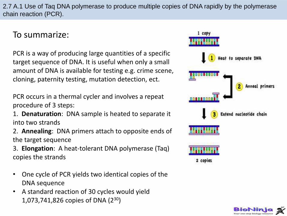
To summarize:
PCR is a way of producing large quantities of a specific target sequence of DNA. It is useful when only a small amount of DNA is available for testing e.g. crime scene, cloning, paternity testing, mutation detection, ect.
PCR occurs in a thermal cycler and involves a repeat procedure of 3 steps:1. Denaturation: DNA sample is heated to separate it into two strands2. Annealing: DNA primers attach to opposite ends of the target sequence3. Elongation: A heat-tolerant DNA polymerase (Taq) copies the strands
• One cycle of PCR yields two identical copies of the DNA sequence
• A standard reaction of 30 cycles would yield 1,073,741,826 copies of DNA (230)

Job 2 Protein Synthesis

2.7 U.4 Transcription is the synthesis of mRNA copied from the DNA base sequences by
RNA polymerase.
2.7 U.5 Translation is the synthesis of polypeptides on ribosomes.
http://learn.genetics.utah.edu/content/molecules/transcribe/
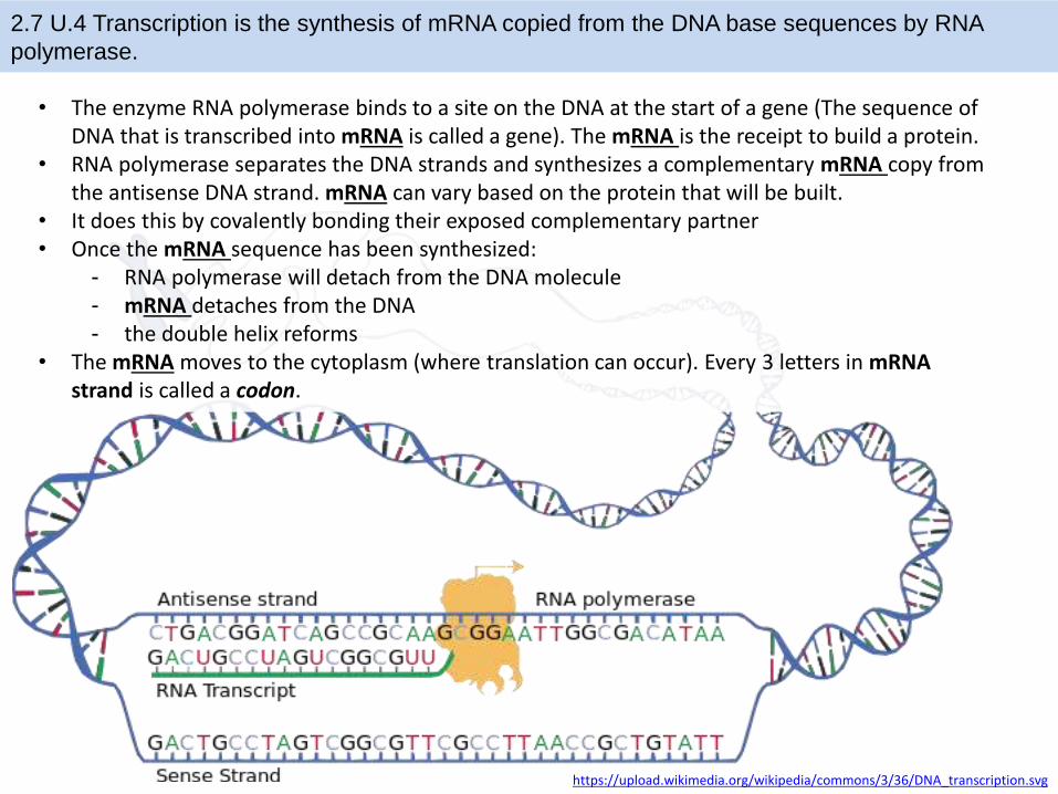
• The enzyme RNA polymerase binds to a site on the DNA at the start of a gene (The sequence of DNA that is transcribed into mRNA is called a gene). The mRNA is the receipt to build a protein.
• RNA polymerase separates the DNA strands and synthesizes a complementary mRNA copy from the antisense DNA strand. mRNA can vary based on the protein that will be built.
• It does this by covalently bonding their exposed complementary partner • Once the mRNA sequence has been synthesized:
- RNA polymerase will detach from the DNA molecule- mRNA detaches from the DNA- the double helix reforms
• The mRNA moves to the cytoplasm (where translation can occur). Every 3 letters in mRNA strand is called a codon.
2.7 U.4 Transcription is the synthesis of mRNA copied from the DNA base sequences by RNA
polymerase.
https://upload.wikimedia.org/wikipedia/commons/3/36/DNA_transcription.svg
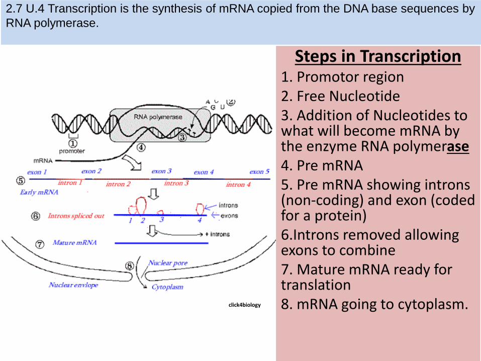
Steps in Transcription 1. Promotor region2. Free Nucleotide3. Addition of Nucleotides to what will become mRNA by the enzyme RNA polymerase4. Pre mRNA5. Pre mRNA showing introns (non-coding) and exon (coded for a protein)6.Introns removed allowing exons to combine7. Mature mRNA ready for translation8. mRNA going to cytoplasm.click4biology
2.7 U.4 Transcription is the synthesis of mRNA copied from the DNA base sequences by
RNA polymerase.

Post Transcriptional Modification
2.7 U.5 Translation is the synthesis of polypeptides on ribosomes.
http://www.nature.com/scitable/topicpage/ribosomes-transcription-and-translation-14120660
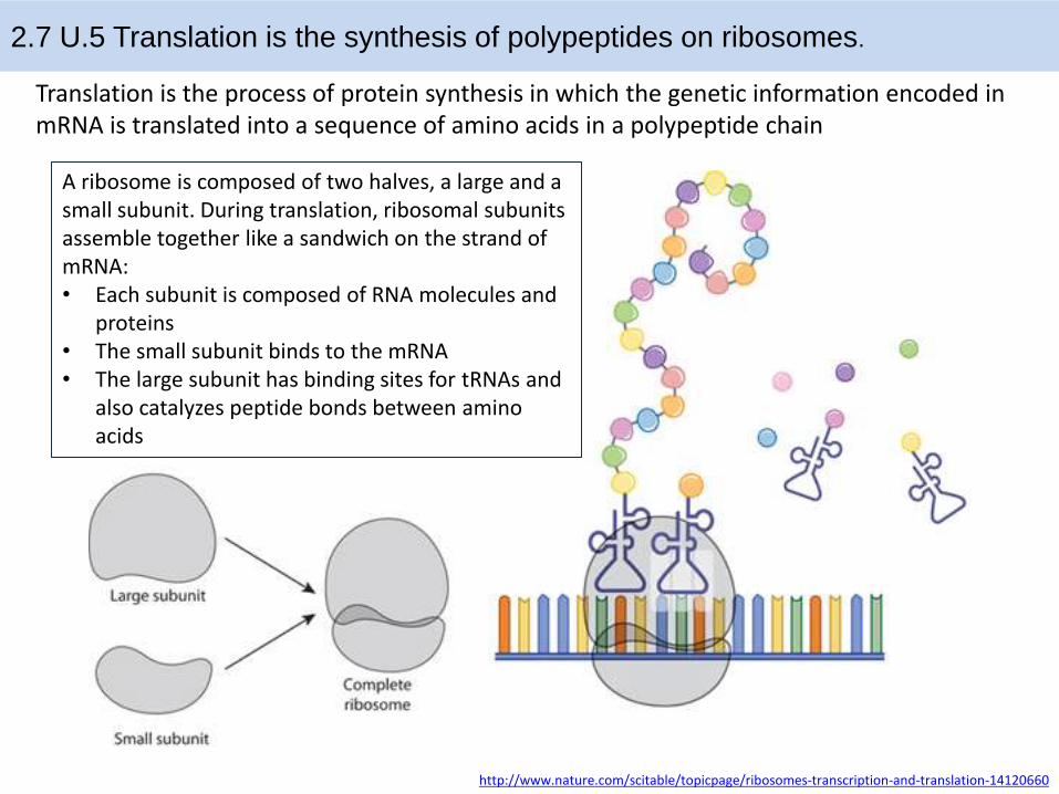
Translation is the process of protein synthesis in which the genetic information encoded in mRNA is translated into a sequence of amino acids in a polypeptide chain
A ribosome is composed of two halves, a large and a small subunit. During translation, ribosomal subunits assemble together like a sandwich on the strand of mRNA:• Each subunit is composed of RNA molecules and
proteins • The small subunit binds to the mRNA• The large subunit has binding sites for tRNAs and
also catalyzes peptide bonds between amino acids
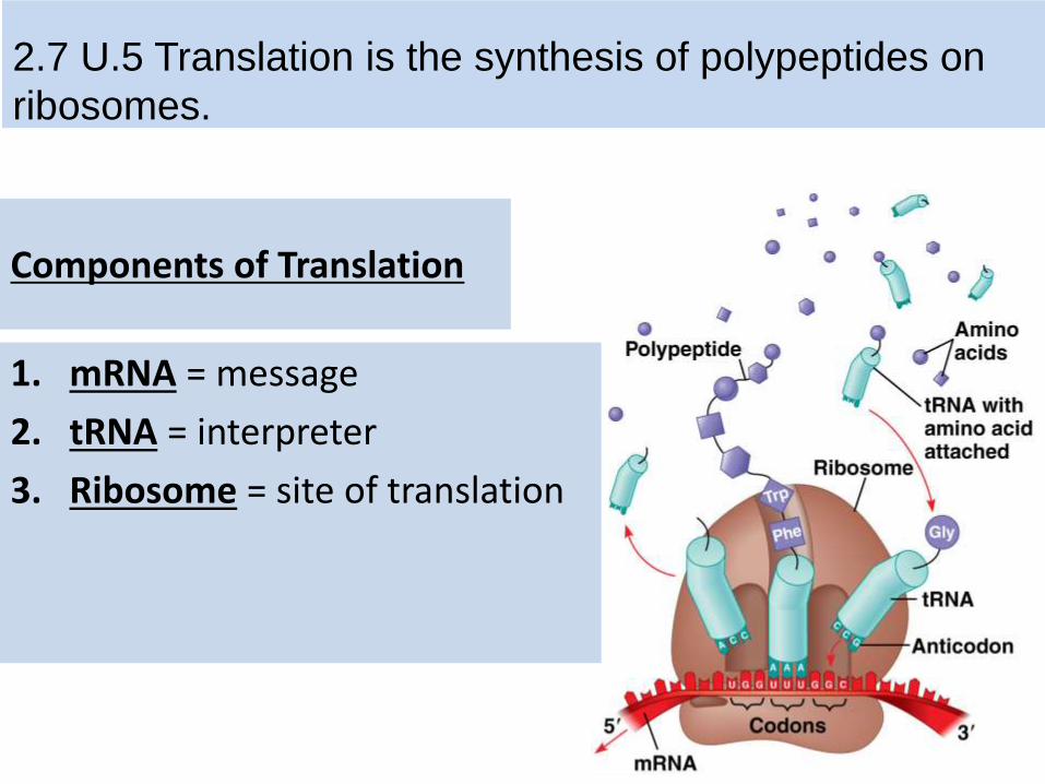
Components of Translation
1. mRNA = message
2. tRNA = interpreter
3. Ribosome = site of translation
2.7 U.5 Translation is the synthesis of polypeptides on
ribosomes.

tRNA
• Transcribed in nucleus
• Specific to each amino acid
• Transfer Amino Acids to ribosomes
• Anticodon: pairs with complementary mRNA codon
• Base-pairing rules between 3rd
base of codon & anticodon are not as strict.
7.3 U.1 Initiation of translation involves assembly of the components that carry out the process.
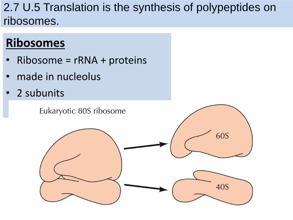
Ribosomes
• Ribosome = rRNA + proteins
• made in nucleolus
• 2 subunits
2.7 U.5 Translation is the synthesis of polypeptides on
ribosomes.

Ribosomes
Active sites:
• A site: holds AA to be added
• P site: holds growing polypeptide chain
• E site: exit site for tRNA
2.7 U.5 Translation is the synthesis of polypeptides on
ribosomes.
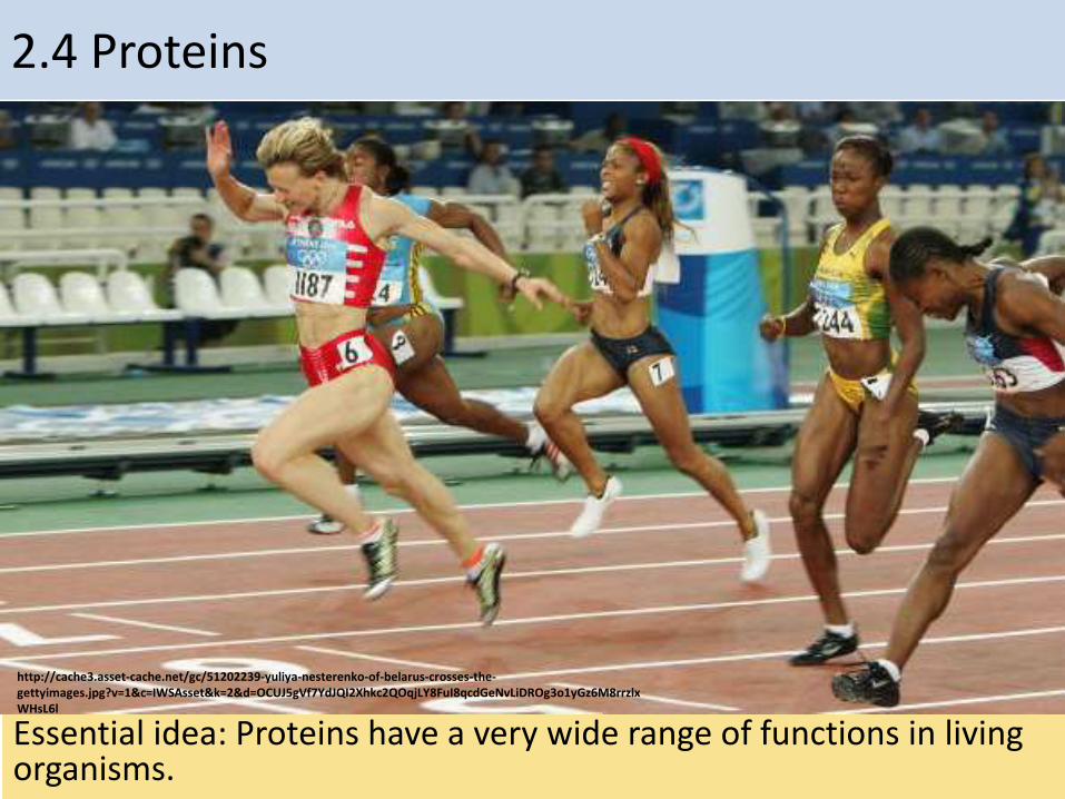
2.4 Proteins
Essential idea: Proteins have a very wide range of functions in living organisms.
http://cache3.asset-cache.net/gc/51202239-yuliya-nesterenko-of-belarus-crosses-the-gettyimages.jpg?v=1&c=IWSAsset&k=2&d=OCUJ5gVf7YdJQI2Xhkc2QOqjLY8FuI8qcdGeNvLiDROg3o1yGz6M8rrzlxWHsL6l
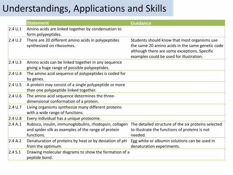
Understandings, Applications and SkillsStatement Guidance
2.4 U.1 Amino acids are linked together by condensation to form polypeptides.
2.4 U.2 There are 20 different amino acids in polypeptides synthesized on ribosomes.
Students should know that most organisms use the same 20 amino acids in the same genetic code although there are some exceptions. Specific examples could be used for illustration.
2.4 U.3 Amino acids can be linked together in any sequence giving a huge range of possible polypeptides.
2.4 U.4 The amino acid sequence of polypeptides is coded for by genes.
2.4 U.5 A protein may consist of a single polypeptide or more than one polypeptide linked together.
2.4 U.6 The amino acid sequence determines the three-dimensional conformation of a protein.
2.4 U.7 Living organisms synthesize many different proteins with a wide range of functions.
2.4 U.8 Every individual has a unique proteome.
2.4 A.1 Rubisco, insulin, immunoglobulins, rhodopsin, collagen and spider silk as examples of the range of protein functions.
The detailed structure of the six proteins selected to illustrate the functions of proteins is not needed.
2.4 A.2 Denaturation of proteins by heat or by deviation of pH from the optimum.
Egg white or albumin solutions can be used in denaturation experiments.
2.4 S.1 Drawing molecular diagrams to show the formation of a peptide bond.
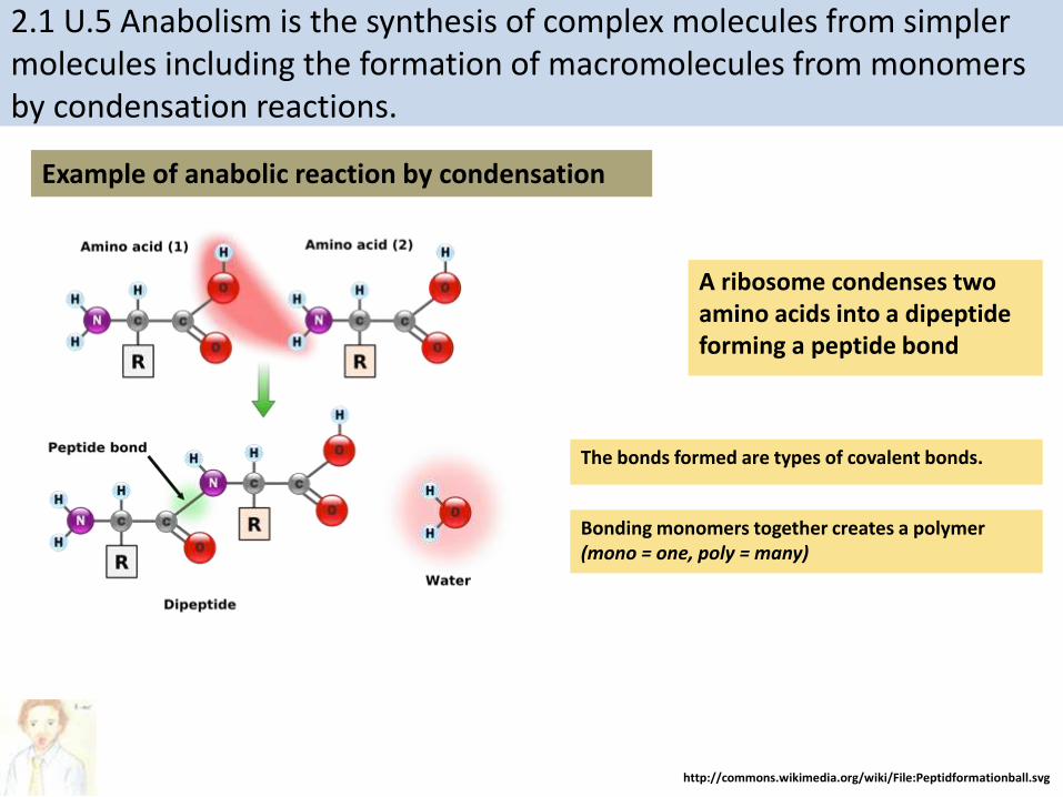
2.1 U.5 Anabolism is the synthesis of complex molecules from simpler molecules including the formation of macromolecules from monomers by condensation reactions.
A ribosome condenses two amino acids into a dipeptide forming a peptide bond
Example of anabolic reaction by condensation
http://commons.wikimedia.org/wiki/File:Peptidformationball.svg
The bonds formed are types of covalent bonds.
Bonding monomers together creates a polymer (mono = one, poly = many)

• Twenty different amino acids are used by the ribosomes to create polypeptides in our body
• They all contain an amine (NH2) group, a carboxyl (-COOH) group which combine to form the peptide bond and a “R” group
• The different “R” groups are what makes the amino acids different and allow the proteins to form a wide array of structures and functions
• Some are charged or polar, hence they are hydrophilic
• Some are not charged and are non-polar, hence they are hydrophobic
2.4 U.2 There are 20 different amino acids in polypeptides synthesized on ribosomes.
http://study.com/cimages/multimages/16/amino_acid_structure.png

https://en.wikipedia.org/wiki/File:Peptide_syn.png
http://www.rcsb.org/pdb/education_discussion/molecule_of_the_month/images/2wdk_2wdl_front.jpg
Ribosomes are the molecules within cells that facilitate the formation of peptide bonds and hence where polypeptides are synthesized
peptide bond
2.4 U.2 There are 20 different amino acids in polypeptides synthesized on ribosomes.

http://commons.wikimedia.org/wiki/File:Amino_Acids.svg
n.b. there are 22 amino acids, but only 20 amino acids are encoded by the universal genetic code.
2.4 U.2 There are 20 different amino acids in polypeptides synthesized on ribosomes.
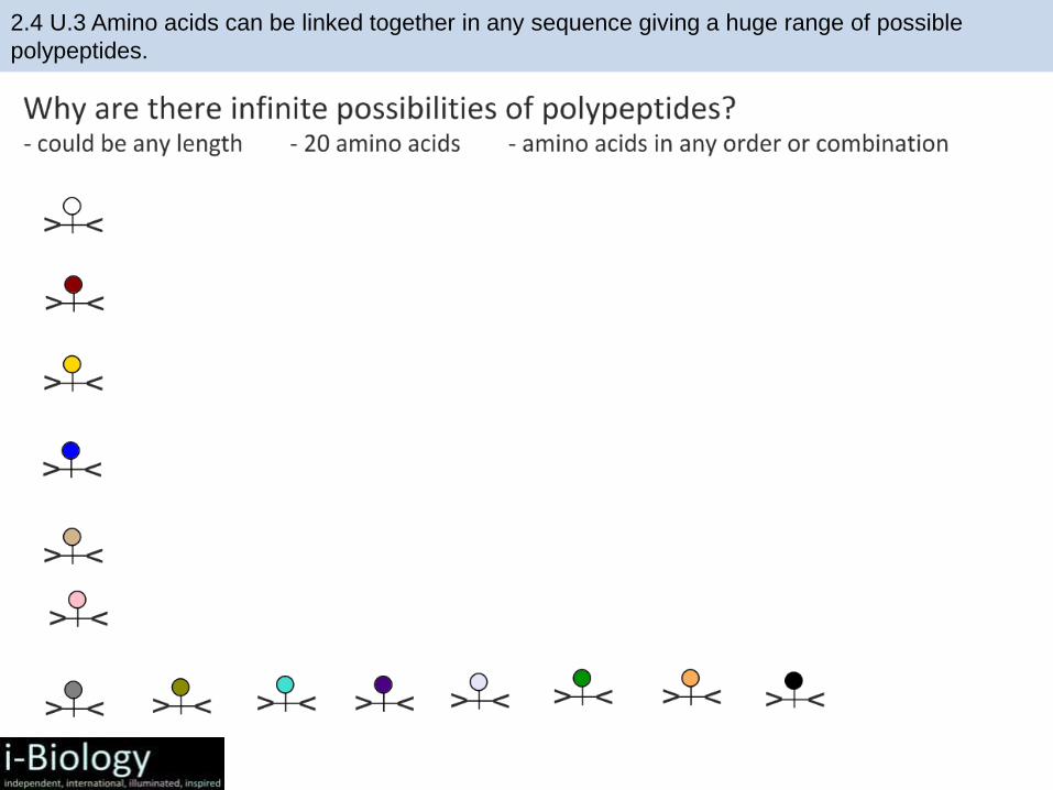
2.4 U.3 Amino acids can be linked together in any sequence giving a huge range of possible
polypeptides.

2.4 U.3 Amino acids can be linked together in any sequence giving a huge range of possible
polypeptides.

2.4 U.3 Amino acids can be linked together in any sequence giving a huge range of possible
polypeptides.
If a polypeptide contains just 7 amino acids there can be 207
= 1,280,000,000 possible polypeptides generated.
Given that polypeptides can contain up to 30,000 amino acids (e.g. Titin) the different possible combinations of polypeptides are effectively infinite.

2.4 U.4 The amino acid sequence of polypeptides is coded for by genes.
https://en.wikipedia.org/wiki/File:Peptide_syn.png
Ribosomes are the site of polypeptide synthesis, but ribosomes need a template – the messenger RNA, which, in turn, is translated by transfer RNA molecules which, in turn, carry specific amino acids.
peptide bond
Q – Where does the messenger RNA come from?

2.4 U.4 The amino acid sequence of polypeptides is coded for by genes.

2.4 U.6 The amino acid sequence determines the three-dimensional conformation of a protein.2.4 U.5 A protein may consist of a single polypeptide or more than one polypeptide linked together.
1. Primary structure:•The order/ number of amino acids in a polypeptide chain.•Linear shape (no internal bonding)
Folding in the primary structure is caused by charged groups on the amino acid chain.
Changes in the R group can result in interactions within the strands:• Hydrogen bonds • Ionic bonds• Covalent bonds. (disulphide
bridge• Hydrophobic regions• Interactions between multiple
amino acids strands

2. Secondary Structure:
Add a second type of bond (hydrogen bonding) in addition to the
covalent bond in primary structure. This causes the structure of
the polypeptide to fold and coil in two ways:
• Alpha Helix
• Beta pleated sheets
2.4 U.6 The amino acid sequence determines the three-dimensional conformation of a protein.2.4 U.5 A protein may consist of a single polypeptide or more than one polypeptide linked together.

Alpha helix proteins
• Alpha-helix:
• Formed from Hydrogen Bonds
• Notice the regular helix shape.

Beta-pleated sheets:
• Flat, zig-zag structure
• A number of chains which are hydrogen bonded together
• Forms a sheet
Example: Fibers in in silk

3. Tertiary Structures• Tertiary structure is the three-
dimensional conformation of a polypeptide.
• In other words there are folds in a polypeptide chain.
• The polypeptide folds just after it is formed in translation.
• The shape is maintained by intermolecular bonds

4. Quaternary structure• Not all proteins have
quaternary structures• Those that do have
two or more polypeptides strands that aggregate together
• Example: Hemoglobin
https://aberdeenc.files.wordpress.com/2013/02/hemoglobin.jpg

2.4 U.6 The amino acid sequence determines the three-dimensional conformation of a protein.2.4 U.5 A protein may consist of a single polypeptide or more than one polypeptide linked together.
There are four levels of protein structure. Which level a protein conforms to is determined by it’s amino acid sequence.
(Polypeptide)• The order / sequence of the
amino acids of which the protein is composed
• Formed by covalent peptide bonds between adjacent amino acids
• Controls all subsequent levels of structure
• The chains of amino acids fold or turn upon themselves
• Held together by hydrogen bonds between (non-adjacent) amine (N-H) and carboxylic (C-O) groups
• H-bonds provide a level of structural stability
• Fibrous proteins
• The polypeptide folds and coils to form a complex 3D shape
• Caused by interactions between R groups (H-bonds, disulphidebridges, ionic bonds and hydrophilic / hydrophobic interactions)
• Tertiary structure may be important for the function (e.g. specificity of active site in enzymes)
• Globular proteins
• The interaction between multiple polypeptides or prosthetic groups
• A prosthetic group is an inorganic compound involved in a protein (e.g. the heme group in haemoglobin)
• Fibrous and Globular proteins
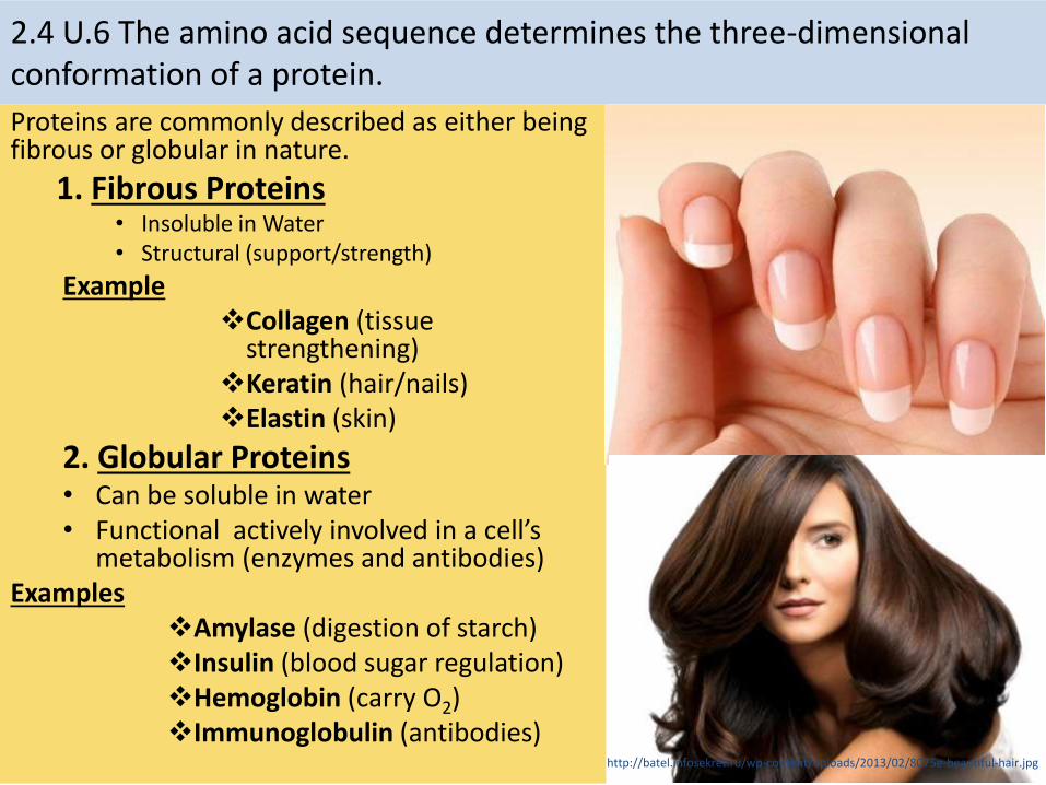
Proteins are commonly described as either being fibrous or globular in nature.
1. Fibrous Proteins• Insoluble in Water• Structural (support/strength)
ExampleCollagen (tissue
strengthening)Keratin (hair/nails)Elastin (skin)
2. Globular Proteins• Can be soluble in water• Functional actively involved in a cell’s
metabolism (enzymes and antibodies)Examples
Amylase (digestion of starch)Insulin (blood sugar regulation)Hemoglobin (carry O2)Immunoglobulin (antibodies)
2.4 U.6 The amino acid sequence determines the three-dimensional conformation of a protein.
http://batel.infosekret.ru/wp-content/uploads/2013/02/8075e-beautiful-hair.jpg
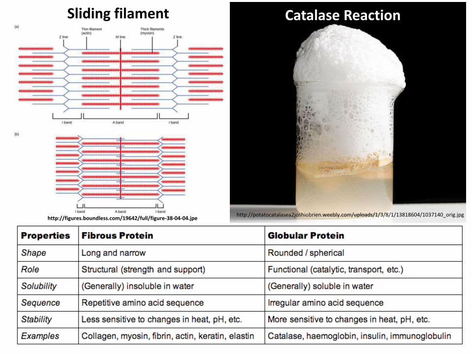
Catalase Reaction
http://potatocatalasea2joshiobrien.weebly.com/uploads/1/3/8/1/13818604/1037140_orig.jpg
Sliding filament
http://figures.boundless.com/19642/full/figure-38-04-04.jpe

2.4 U.7 Living organisms synthesize many different proteins with a wide range of functions.
Function Description Key examples
Catalysis There are thousands of different enzymes to catalyze specific chemical reactions within the cell or outside it.
Rubisco
Muscle contraction Actin and myosin together cause the muscle contractions used in locomotion and transport around the body.
Actin and myosin
Cytoskeletons Tubulin is the subunit of microtubules that give animals cells their shape and pull on chromosomes during mitosis.
Tubulin
Tensile strengthening
collagen
Blood clotting Plasma proteins act as clotting factors that cause blood to turn from a liquid to a gel in wounds.
Fibrin
Transport of nutrients and gases
Proteins in blood help transport oxygen, carbon dioxide, iron and lipids.
Serum albumin
Nothing can compare with the versatility of proteins. Their functionality and usage in organisms is unrivalled.
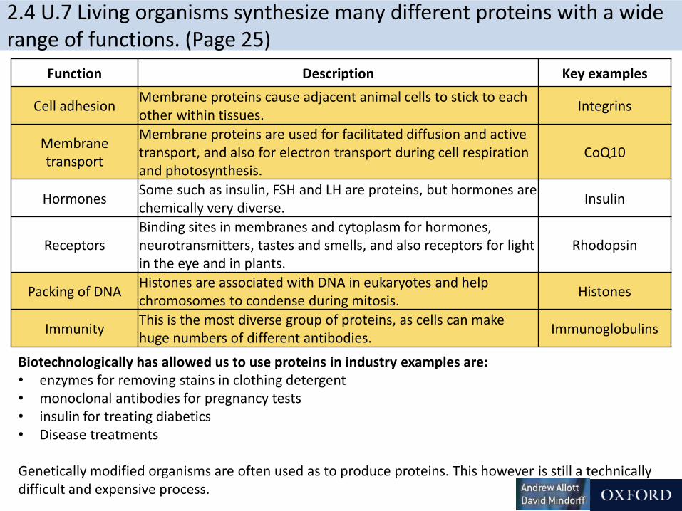
2.4 U.7 Living organisms synthesize many different proteins with a wide range of functions. (Page 25)
Function Description Key examples
Cell adhesion Membrane proteins cause adjacent animal cells to stick to each other within tissues.
Integrins
Membrane transport
Membrane proteins are used for facilitated diffusion and active transport, and also for electron transport during cell respiration and photosynthesis.
CoQ10
Hormones Some such as insulin, FSH and LH are proteins, but hormones are chemically very diverse.
Insulin
Receptors Binding sites in membranes and cytoplasm for hormones, neurotransmitters, tastes and smells, and also receptors for light in the eye and in plants.
Rhodopsin
Packing of DNA Histones are associated with DNA in eukaryotes and help chromosomes to condense during mitosis.
Histones
Immunity This is the most diverse group of proteins, as cells can make huge numbers of different antibodies.
Immunoglobulins
Biotechnologically has allowed us to use proteins in industry examples are:• enzymes for removing stains in clothing detergent• monoclonal antibodies for pregnancy tests• insulin for treating diabetics• Disease treatments
Genetically modified organisms are often used as to produce proteins. This however is still a technically difficult and expensive process.
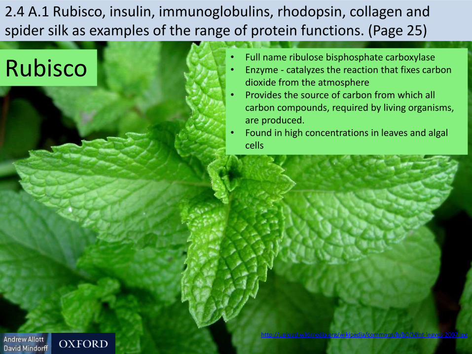
Rubisco• Full name ribulose bisphosphate carboxylase• Enzyme - catalyzes the reaction that fixes carbon
dioxide from the atmosphere• Provides the source of carbon from which all
carbon compounds, required by living organisms, are produced.
• Found in high concentrations in leaves and algal cells
http://upload.wikimedia.org/wikipedia/commons/b/b0/Mint-leaves-2007.jpg
2.4 A.1 Rubisco, insulin, immunoglobulins, rhodopsin, collagen and spider silk as examples of the range of protein functions. (Page 25)

2.4 A.1 Rubisco, insulin, immunoglobulins, rhodopsin, collagen and spider silk as examples of the range of protein functions.
• A hormone – signals many cells (e.g. liver cells) to absorb glucose and help reduce the glucose concentration of the blood.
• Affected cells have receptor (proteins) on their surface to which insulin can (reversibly) bind to.
• Secreted by β cells in the pancreas and transported by the blood.
http://www.biotopics.co.uk/as/insulinproteinstructure.html
The pancreas of type I diabetics don’t produce sufficient insulin therefore they must periodically inject synthetically produced insulin to correct their blood sugar concentration.
Insulin
https://en.wikipedia.org/wiki/File:Inzul%C3%ADn.jpg

Immunoglobulins
https://upload.wikimedia.org/wikipedia/commons/thumb/a/a9/Antibody_IgG2.png/320px-Antibody_IgG2.png
• Also known as antibodies.• Two antigen (a molecule on the pathogen which provokes an immune
response) binding sites - one on each ‘arm’• Binding sites vary greatly between immunoglobulins (hypervariable) to
enable them to respond a huge range of pathogens.• Other parts of the immunoglobulin molecule cause a response, e.g.
acting as a marker to phagocytes (which engulf the pathogen)
2.4 A.1 Rubisco, insulin, immunoglobulins, rhodopsin, collagen and spider silk as examples of the range of protein functions.

Rhodopsin
• A pigment that absorbs light• Membrane protein of rod cells of the retina (light sensitive region at the
back of the eye)• Rhodopsin consists of the opsin polypeptide surrounding a retinal
prosthetic group• retinal molecule absorbs a single photon of light -> changes shape ->
change to the opsin -> the rod cell sends a nerve impulse to the brain • Even very low light intensities can be detected.
https://en.wikipedia.org/wiki/Retina#mediaviewer/File:Fundus_photograph_of_normal_left_eye.jpg
http://commons.wikimedia.org/wiki/File:Rhodopsin.jpg
2.4 A.1 Rubisco, insulin, immunoglobulins, rhodopsin, collagen and spider silk as examples of the range of protein functions.

Collagen
• A number of different forms• All are rope-like proteins made of three polypeptides wound
together.• About a quarter of all protein in the human body is collagen• Forms a mesh of fibers in skin and in blood vessel walls that resists
tearing.• Gives strength to tendons, ligaments, skin and blood vessel walls.• Forms part of teeth and bones, helps to prevent cracks and fractures
to bones and teeth https://en.wikipedia.org/wiki/Tooth_(human)#mediaviewer/File:Teeth_by_David_Shankbone.jpg
2.4 A.1 Rubisco, insulin, immunoglobulins, rhodopsin, collagen and spider silk as examples of the range of protein functions.

spider silk• Different types of silk with
different functions• Dragline silk is stronger
than steel and tougher than Kevlar
• When first made it contains regions where the polypeptide forms parallel arrays (bottom)
• Some regions seem like a disordered tangle (middle)
• When the stretched the polypeptide gradually extends, making the silk extensible and very resistant to breaking.
https://en.wikipedia.org/wiki/Spider_silk#mediaviewer/File:Structure_of_spider_silk_thread_Modified.svg
2.4 A.1 Rubisco, insulin, immunoglobulins, rhodopsin, collagen and spider silk as examples of the range of protein functions.
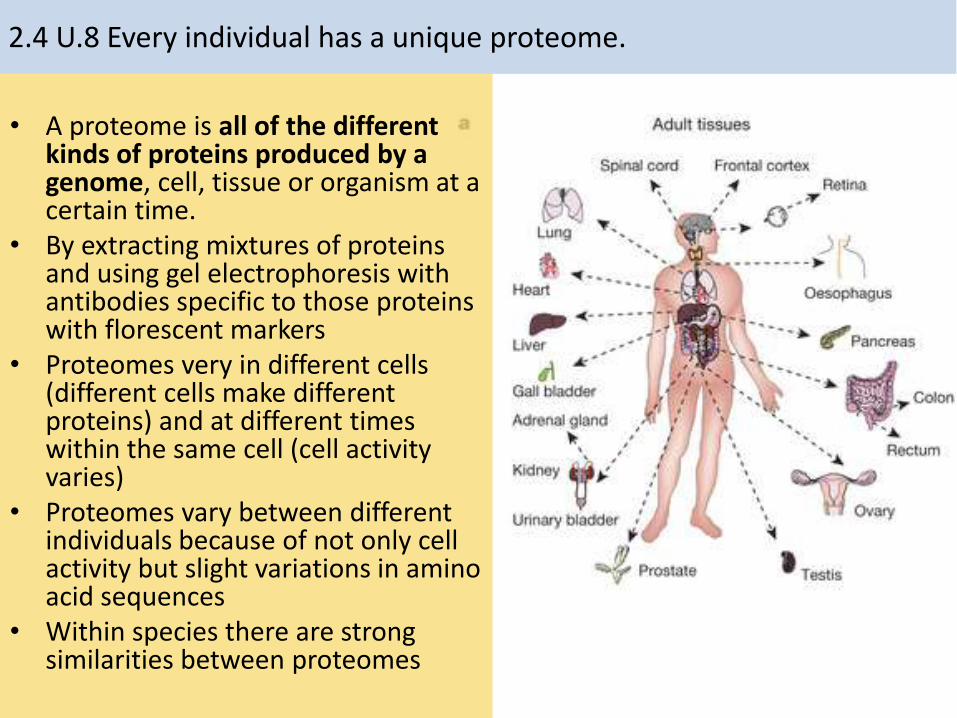
• A proteome is all of the different kinds of proteins produced by a genome, cell, tissue or organism at a certain time.
• By extracting mixtures of proteins and using gel electrophoresis with antibodies specific to those proteins with florescent markers
• Proteomes very in different cells (different cells make different proteins) and at different times within the same cell (cell activity varies)
• Proteomes vary between different individuals because of not only cell activity but slight variations in amino acid sequences
• Within species there are strong similarities between proteomes
2.4 U.8 Every individual has a unique proteome.

2.4 U.8 Every individual has a unique proteome.
To analyze a proteome mixtures of proteins are extracted from a sample and are then separated by gel electrophoresis. The background shows a stained example of gel electrophoresis.
Genome: all of the genes of a cell, a tissue or an organism
The genome determines what proteins an organism can possibly produce. A genome is unique to most individuals (identical twins and clones share a genome)
Proteome: all of the proteins produced by a cell, a tissue or an organism.
• Being a function of both the genome and the environment to which the organism is exposed the proteome is both variable (over time) and unique to every individual (including identical twins and clones).
• It reveals what is happening in an organism at a particular time
Environmental factors
The environment influences what proteins an organism needs to produce and in what quantity. Example factors would be nutrition, temperature, activity levels and anything else that affects a cell’s activities.
http://proteomics.arizona.edu/sites/proteomics.arizona.edu/files/1D_Gel_CD_4.png
Q – Genome or proteome, which is larger? Explain the reasons for your answer.

To analyze a proteome mixtures of proteins are extracted from a sample and are then separated by gel electrophoresis. The background shows a stained example of gel electrophoresis.
http://proteomics.arizona.edu/sites/proteomics.arizona.edu/files/1D_Gel_CD_4.png
Q – Genome or proteome, which is larger? Explain the reasons for your answer.
A – Proteome:
• Not all genes produce polypeptides
• Multiple polypeptides and prosthetic groups can interact
• Amino acids can be modified (e.g. Collagen)
• A polypeptide can fold into different levels of structure (e.g. insulin)
2.4 U.8 Every individual has a unique proteome.
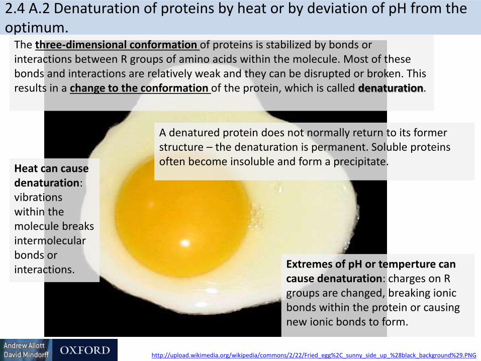
2.4 A.2 Denaturation of proteins by heat or by deviation of pH from the optimum.
Extremes of pH or temperture can cause denaturation: charges on R groups are changed, breaking ionic bonds within the protein or causing new ionic bonds to form.
A denatured protein does not normally return to its former structure – the denaturation is permanent. Soluble proteins often become insoluble and form a precipitate.
The three-dimensional conformation of proteins is stabilized by bonds or interactions between R groups of amino acids within the molecule. Most of these bonds and interactions are relatively weak and they can be disrupted or broken. This results in a change to the conformation of the protein, which is called denaturation.
Heat can cause denaturation: vibrations within the molecule breaks intermolecular bonds or interactions.
http://upload.wikimedia.org/wikipedia/commons/2/22/Fried_egg%2C_sunny_side_up_%28black_background%29.PNG
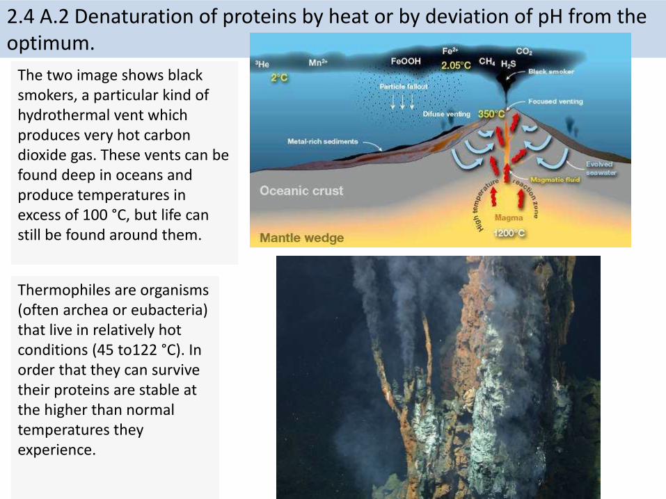
2.4 A.2 Denaturation of proteins by heat or by deviation of pH from the optimum.
Thermophiles are organisms (often archea or eubacteria) that live in relatively hot conditions (45 to122 °C). In order that they can survive their proteins are stable at the higher than normal temperatures they experience.
The two image shows black smokers, a particular kind of hydrothermal vent which produces very hot carbon dioxide gas. These vents can be found deep in oceans and produce temperatures in excess of 100 °C, but life can still be found around them.
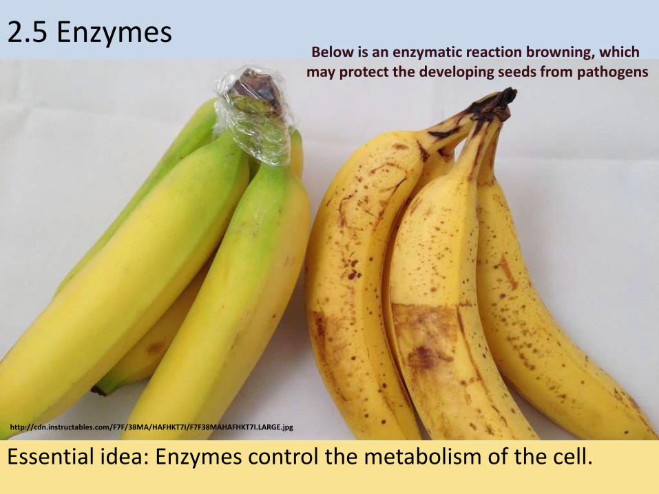
2.5 Enzymes
Essential idea: Enzymes control the metabolism of the cell.
http://cdn.instructables.com/F7F/38MA/HAFHKT7I/F7F38MAHAFHKT7I.LARGE.jpg
Below is an enzymatic reaction browning, which may protect the developing seeds from pathogens

Understandings, Applications and Skills
Statement Guidance
2.5 U.1 Enzymes have an active site to which specific substrates bind.
2.5 U.2 Enzyme catalysis involves molecular motion and the collision of substrates with the active site.
2.5 U.3 Temperature, pH and substrate concentration affect the rate of activity of enzymes.
Students should be able to sketch graphs to show the expected effects of temperature, pH and substrate concentration on the activity of enzymes. They should be able to explain the patterns or trends apparent in these graphs.
2.5 U.4 Enzymes can be denatured.
2.5 U.5 Immobilized enzymes are widely used in industry.
2.5 A.1 Methods of production of lactose-free milk and its advantages.
Lactase can be immobilized in alginate beads and experiments can then be carried out in which the lactose in milk is hydrolyzed.
2.5 S.1 Design of experiments to test the effect of temperature, pH and substrate concentration on the activity of enzymes.
2.5 S.2 Experimental investigation of a factor affecting enzyme activity. (Practical 3)

2.5 U.1 Enzymes have an active site to which specific substrates bind.2.5 U.2 Enzyme catalysis involves molecular motion and the collision of substrates with the active site.
Enzyme: A globular protein that increases the rate of a biochemical reaction by lowering the activation energy threshold (i.e. a biologicalcatalyst)
http://www.northland.cc.mn.us/biology/biology1111/animations/enzyme.swf
Use the animation to find out more about enzymes and how they work.
A good alternative is How Enzymes Work from McGraw and Hill
http://highered.mheducation.com/sites/0072495855/student_view0/chapter2/animation__how_enzymes_work.html

• Enzymes are protein catalysts that enormously speed up reactions. They often have an “-ase” ending to their name.
– e.g., hexokinase, catalase, peptidase, mutase
• They are not themselves changed (except for a brief period of time) and are the same before and after a reaction.
• Enzymes:1. Lower the activation energy: this is the MOST important
characteristic
2. Do not add or remove energy from a reaction
3. Do not change the equilibrium for a reaction
4. Are reused over and over
2.5 U.1 Enzymes have an active site to which specific substrates bind.
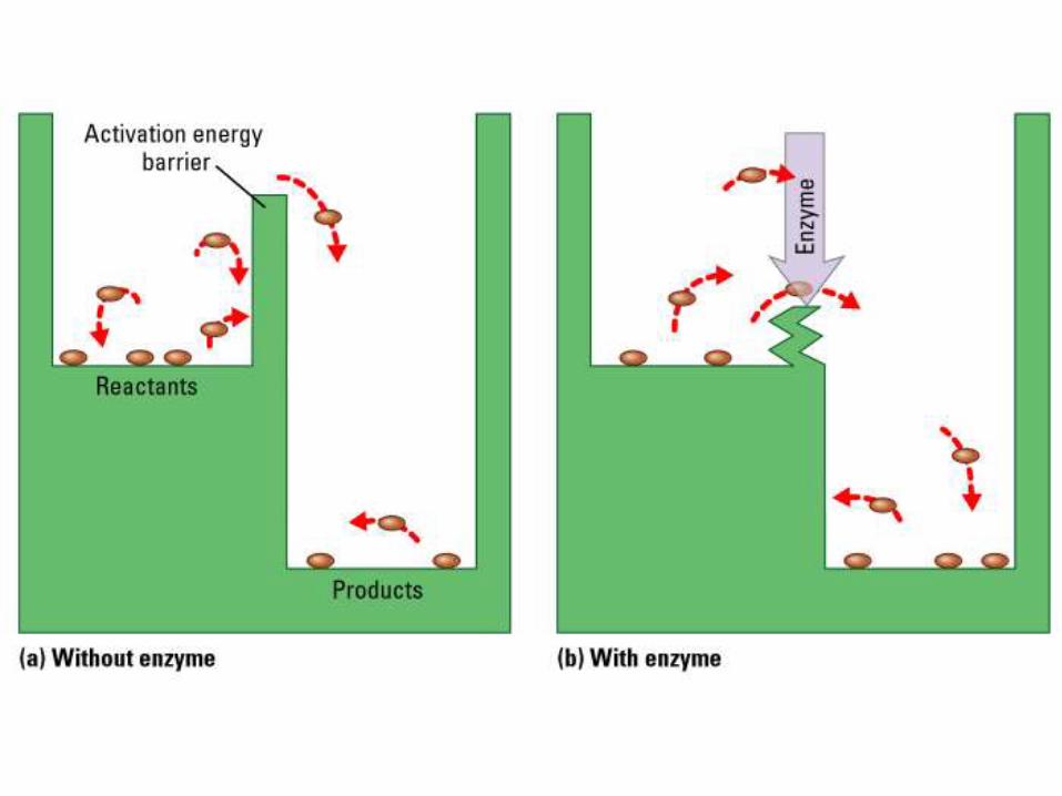
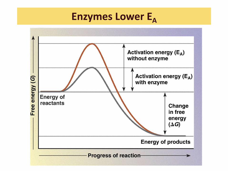
Enzymes Lower EA

2.5 U.2 Enzyme catalysis involves molecular motion and the collision of substrates with the active site.
http://www.kscience.co.uk/animations/model.swf
The simulation from KScienceallows you to both see enzyme kinetics happening and secondly how it is affected by different factors
• Two substances must have the proper alignment and energy(in the form of motion) to create a chemical reaction
• The direction and movement is constantly changing and is random
• Collisions occur at random between the substrate and enzyme
• Successful reactions only occur if the substrate and the active site of the enzyme are correctly aligned and the collide with sufficient KE

Lock & Key Hypothesisa) Large globular protein enzyme
b) Active Site where the substrate combines to the enzyme
c)Substrate which fits the active site
d) Activated complex. The substrate is weakened to allow the reaction.
e)Unchanged enzyme/ re-used at low concentrations
f) Product of the reaction

Induced Fit Hypothesis• Better explains enzyme activity if
the lock and key model were true one enzyme would only catalyze one reaction, while we some enzymes capable of catalyzing several reactions.
• As the substrate approaches the enzyme a conformational change takes place in the activation site, it changes shape to induce a fit.
• This stress reduces the activation energy or the reaction
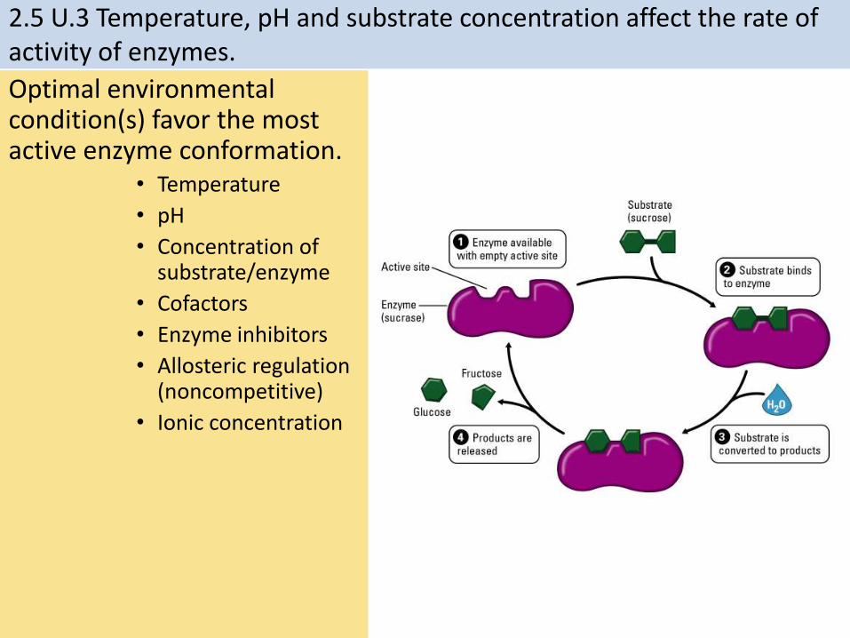
Optimal environmental condition(s) favor the most active enzyme conformation.
• Temperature
• pH
• Concentration of substrate/enzyme
• Cofactors
• Enzyme inhibitors
• Allosteric regulation (noncompetitive)
• Ionic concentration
2.5 U.3 Temperature, pH and substrate concentration affect the rate of activity of enzymes.
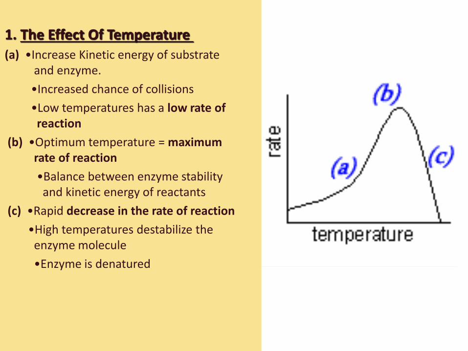
1. The Effect Of Temperature(a) •Increase Kinetic energy of substrate
and enzyme.
•Increased chance of collisions
•Low temperatures has a low rate of reaction
(b) •Optimum temperature = maximum rate of reaction
•Balance between enzyme stability and kinetic energy of reactants
(c) •Rapid decrease in the rate of reaction
•High temperatures destabilize the enzyme molecule
•Enzyme is denatured
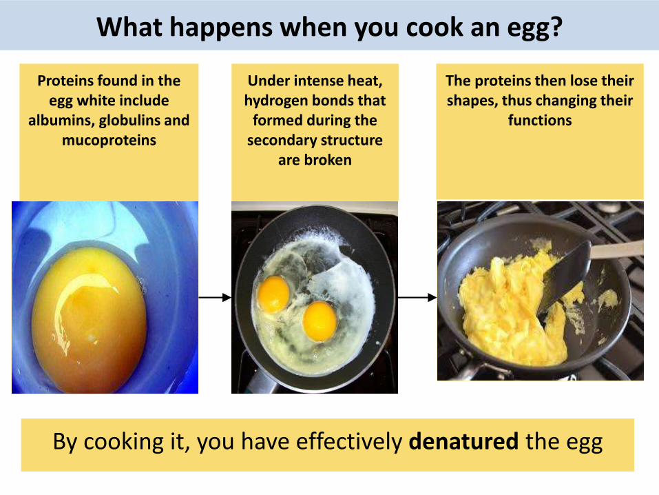
Proteins found in the egg white include
albumins, globulins and mucoproteins
Under intense heat, hydrogen bonds that
formed during the secondary structure
are broken
The proteins then lose their shapes, thus changing their
functions
By cooking it, you have effectively denatured the egg
What happens when you cook an egg?

2.4 A.2 Denaturation of proteins by heat or by deviation of pH from the optimum.
Available equipment:• Waterbaths• Albumen otherwise
known as egg white• Thermometers• Colorimeters (optional)
Your task: determine the temperature stability of albumen
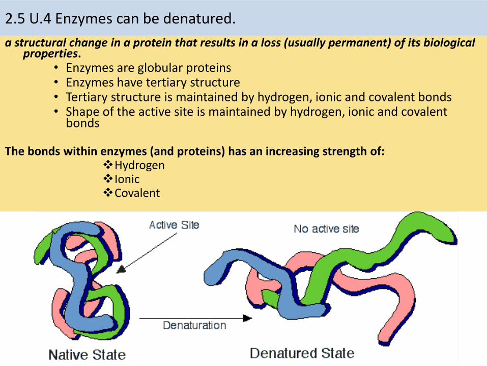
a structural change in a protein that results in a loss (usually permanent) of its biological properties.
• Enzymes are globular proteins• Enzymes have tertiary structure• Tertiary structure is maintained by hydrogen, ionic and covalent bonds• Shape of the active site is maintained by hydrogen, ionic and covalent
bonds
The bonds within enzymes (and proteins) has an increasing strength of:Hydrogen Ionic Covalent
2.5 U.4 Enzymes can be denatured.
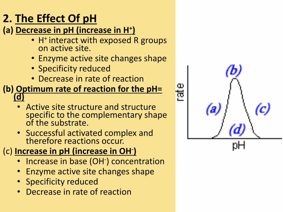
2. The Effect Of pH(a) Decrease in pH (increase in H+)
• H+ interact with exposed R groups on active site.
• Enzyme active site changes shape• Specificity reduced• Decrease in rate of reaction
(b) Optimum rate of reaction for the pH= (d)• Active site structure and structure
specific to the complementary shape of the substrate.
• Successful activated complex and therefore reactions occur.
(c) Increase in pH (increase in OH-)• Increase in base (OH-) concentration• Enzyme active site changes shape• Specificity reduced• Decrease in rate of reaction
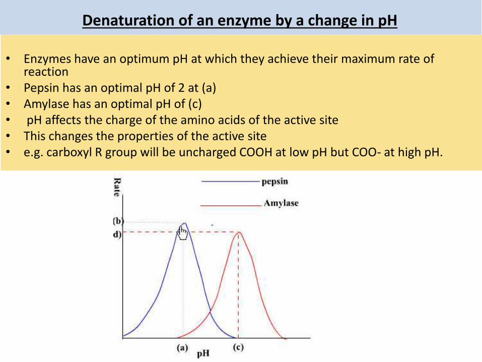
Denaturation of an enzyme by a change in pH
• Enzymes have an optimum pH at which they achieve their maximum rate of reaction
• Pepsin has an optimal pH of 2 at (a) • Amylase has an optimal pH of (c)• pH affects the charge of the amino acids of the active site • This changes the properties of the active site • e.g. carboxyl R group will be uncharged COOH at low pH but COO- at high pH.

3. The effect of
substrate concentration(a) •Increase conc. of substrate molecules
•Increased chance of collision with enzyme
•Greater chance of forming activated complex
•Increase in rate of reaction
(b) •Rate begins to level
•Active sites beginning to become saturated with substrate (fully occupied)
•New substrate must wait for previous reaction to complete and the product to exit the active site
(c) •Full saturation of the active sites by substrate
•Rate becomes constant for further
increases in substrate concentration.

Denaturation of an enzyme by a change in pH
• If there is a deviation from the optimal pH the hydrogen sulfide bridges break and the enzyme loses shape.
• Loss of the activation site shape leads to loss of function

2.5 U.5 Immobilized enzymes are widely used in industry.
http://pubs.rsc.org/services/images/RSCpubs.ePlatform.Service.FreeContent.ImageService.svc/ImageService/Articleimage/2013/CS/c3cs35506c/c3cs35506c-f1.gif
Advantages of enzyme immobilization: (CRSS)• Concentration of substrate can be increased as the enzyme is not dissolved – this
increases the rate of reaction
• Recycled enzymes can be used many times, immobilized enzymes are easy to separate
from the reaction mixture, resulting in a cost saving.
• Separation of the products is straight forward (this also means that the the reaction can
stopped at the correct time).
• Stability of the enzyme to changes in temperature and pH is increased reducing the rate
of degradation, again resulting in a cost saving.
Enzymes used in industry are usually immobilized. They are attached to a material so that their movement is restricted. Common ways of doing this are:• Aggregations of enzymes bonded together• Attached to surfaces, e.g. glass• Entrapped in gels, e.g. alginate gel beads

2.5 U.5 Immobilized enzymes are widely used in industry.
Common uses of enzymes in industry include:
http://pubs.rsc.org/services/images/RSCpubs.ePlatform.Service.FreeContent.ImageService.svc/ImageService/Articleimage/2013/CS/c3cs35506c/c3cs35506c-f1.gif
1.Detergents2.Biofuels3.Textiles4.Brewing 5.Medicine &
Biotechnology6.Juice yield 7.Paper production

1. Detergents contain proteases and lipases to help breakdown protein and fat stains https://i1.ytimg.com/vi/lQ6fCZgYc8g/hqdefault.jpg

2. Enzymes are used to breakdown the starch in grains into biofuelsthat can be combusted
http://chromblog.thermoscientific.com/Portals/49739/images/biofuel9.jpg
http://greenodin.com/wp-content/uploads/2014/12/GreenOdin-GO-Biodiesel-Van-1200.png
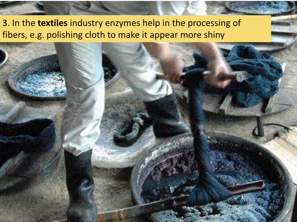
3. In the textiles industry enzymes help in the processing of fibers, e.g. polishing cloth to make it appear more shiny

4. In the brewing industryenzymes help a number of processes including the clarification of the beerhttp://www.brewreviewcrew.com/cans-vs-bottles-fight/
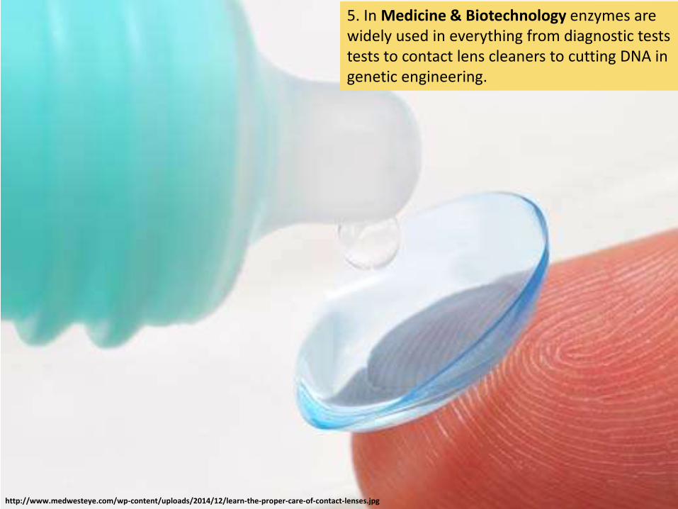
5. In Medicine & Biotechnology enzymes are widely used in everything from diagnostic tests tests to contact lens cleaners to cutting DNA in genetic engineering.
http://www.medwesteye.com/wp-content/uploads/2014/12/learn-the-proper-care-of-contact-lenses.jpg

6. Enzymes are widely used in the food industry, e.g.• fruit juice, pectin to increase the juice yield from
fruit• Fructose is used as a sweetener, it is converted from
glucose by isomerase• Rennin is used to help in cheese productionhttps://theramblingreed.files.wordpress.com/2014/08/img_6338.jpg

7. Paper production uses enzymes to helping in the pulping of wood
http://i00.i.aliimg.com/img/pb/479/389/262/1281752366918_hz-cnmyalibaba-web2_15708.jpg

Lactose Intolerance
• Lactose (milk sugar) can cause allergies in some people.
• This is often because they are unable to produce the enzyme lactase in sufficient quantities.
• Most people produce less lactase as they get older. After all, we don’t live off milk once we have been weaned.
• In some regions such as Europe, a mutation has allowed lactose production to continue into adulthood. This mutation is not present in people who are lactose intolerant
2.5 A.1 Methods of production of lactose-free milk and its advantages.

Green High Tolerance Red Low Tolerance
Global estimates of lactose intolerance.

How can we cope with lactose intolerance?
• Take a lactase supplement. These are produced industrially using the Aspergillus nigerfungus
• Drink lactose free milk. Milk treated with lactase (produced by A. niger) and essentially ‘pre-digested’ before being packaged.

2.5 A.1 Methods of production of lactose-free milk and its advantages.
Other uses of lactose free milk:• As a means to increase the sweetness of milk (glucose and
galactose are sweeter in flavor), thus negating the need for artificial sweeteners
• As a way of reducing the crystallization of ice-creams (glucose and galactose are more soluble than lactose)
• As a means of shortening the production time for yogurts or cheese (bacteria ferment glucose and galactose more readily than lactose)
Production of Lactose-free milk• Lactase obtained from commonly from yeast
(bacteria is an alternative)• Lactase is bound to the surface of alginate beads• Milk is passed (repeatedly) over the beads• The lactose is broken down into glucose and
galactose• The immobilized enzyme remains to be used
again and does not affect the quality of the lactose free milk

2.5 S.1 Design of experiments to test the effect of temperature, pH and substrate concentration on the activity of enzymes.2.5 S.2 Experimental investigation of a factor affecting enzyme activity. (Practical 3)
Possible research questions, what are you going to investigate (independent variable)?• What is the effect of substrate concentration?• What is the effect of temperature?• What is the effect of pH?• Which type of yeast has a higher concentration of catalase?
Important things to consider:• How are you going to vary the mass/volume/concentration of your variable?• What units will you be measuring your variable in?• Have you chosen an effect range or values to answer your question?• Are the concentrations/chemicals you are using safe to handle?
Catalase is one of the most widespread enzymes. It catalyzes the conversion of hydrogen peroxide, a toxic by-product of metabolism, into water and oxygen.

How are you going to measure your results (dependent variable)?• Are you measuring the increase of a product or the disappearance of a substrate?• Are you measuring directly (e.g. testing for the concentration of the product) or
indirectly (change in pH)?• What equipment will you be using to measure your results?• What are the units and uncertainty given both the equipment and how you choose
to use it?• What time period do you need to run the experiment for? How fast is the enzyme
action likely to be?• How many repeats will you need to make sure your results are reliable?
2.5 S.1 Design of experiments to test the effect of temperature, pH and substrate concentration on the activity of enzymes.2.5 S.2 Experimental investigation of a factor affecting enzyme activity. (Practical 3)
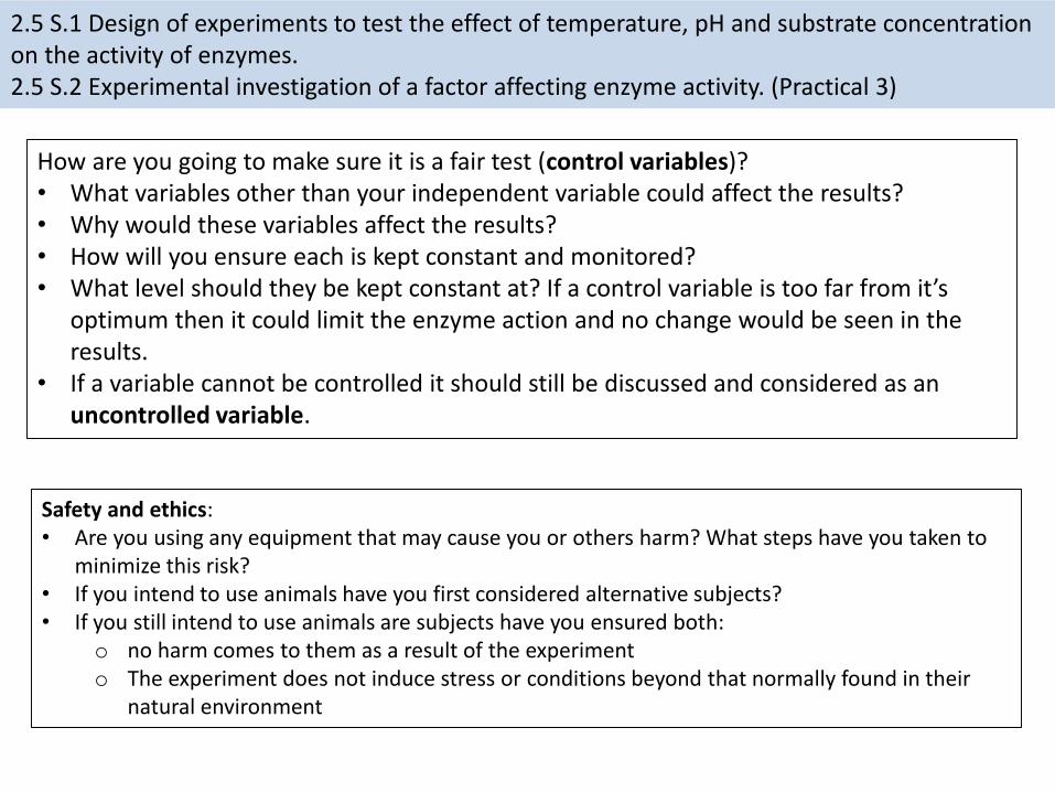
How are you going to make sure it is a fair test (control variables)?• What variables other than your independent variable could affect the results?• Why would these variables affect the results?• How will you ensure each is kept constant and monitored?• What level should they be kept constant at? If a control variable is too far from it’s
optimum then it could limit the enzyme action and no change would be seen in the results.
• If a variable cannot be controlled it should still be discussed and considered as an uncontrolled variable.
Safety and ethics:• Are you using any equipment that may cause you or others harm? What steps have you taken to
minimize this risk?• If you intend to use animals have you first considered alternative subjects?• If you still intend to use animals are subjects have you ensured both:
o no harm comes to them as a result of the experimento The experiment does not induce stress or conditions beyond that normally found in their
natural environment
2.5 S.1 Design of experiments to test the effect of temperature, pH and substrate concentration on the activity of enzymes.2.5 S.2 Experimental investigation of a factor affecting enzyme activity. (Practical 3)