Top 5(ish) -Foot & AnkleThe Top 5 (ish) 1. Ankle Sprains 2. Heel Pain - Plantar Fasciitis 3. Ankle...
65
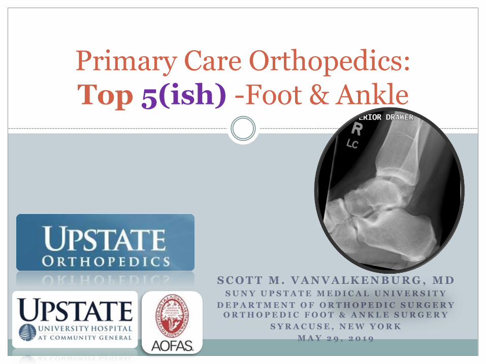
SCOTT M. VANVALKENBURG, MD SUNY UPSTATE MEDICAL UNIVERSITY DEPARTMENT OF ORTHOPEDIC SURGERY ORTHOPEDIC FOOT & ANKLE SURGERY SYRACUSE, NEW YORK MAY 29, 2019 Primary Care Orthopedics: Top 5(ish) -Foot & Ankle
Transcript of Top 5(ish) -Foot & AnkleThe Top 5 (ish) 1. Ankle Sprains 2. Heel Pain - Plantar Fasciitis 3. Ankle...
S C O T T M . V A N V A L K E N B U R G , M DS U N Y U P S T A T E M E D I C A L U N I V E R S I T Y
D E P A R T M E N T O F O R T H O P E D I C S U R G E R YO R T H O P E D I C F O O T & A N K L E S U R G E R Y
S Y R A C U S E , N E W Y O R K
M A Y 2 9 , 2 0 1 9
Primary Care Orthopedics:Top 5(ish) -Foot & Ankle
DISCLOSURES
No consultant arrangements
No patents
Nothing related to this talk
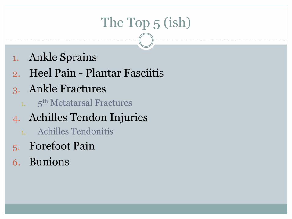
The Top 5 (ish)
1. Ankle Sprains
2. Heel Pain - Plantar Fasciitis
3. Ankle Fractures
1. 5th Metatarsal Fractures
4. Achilles Tendon Injuries
1. Achilles Tendonitis
5. Forefoot Pain
6. Bunions
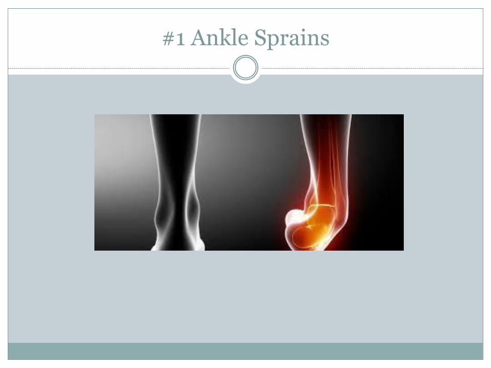
#1 Ankle Sprains
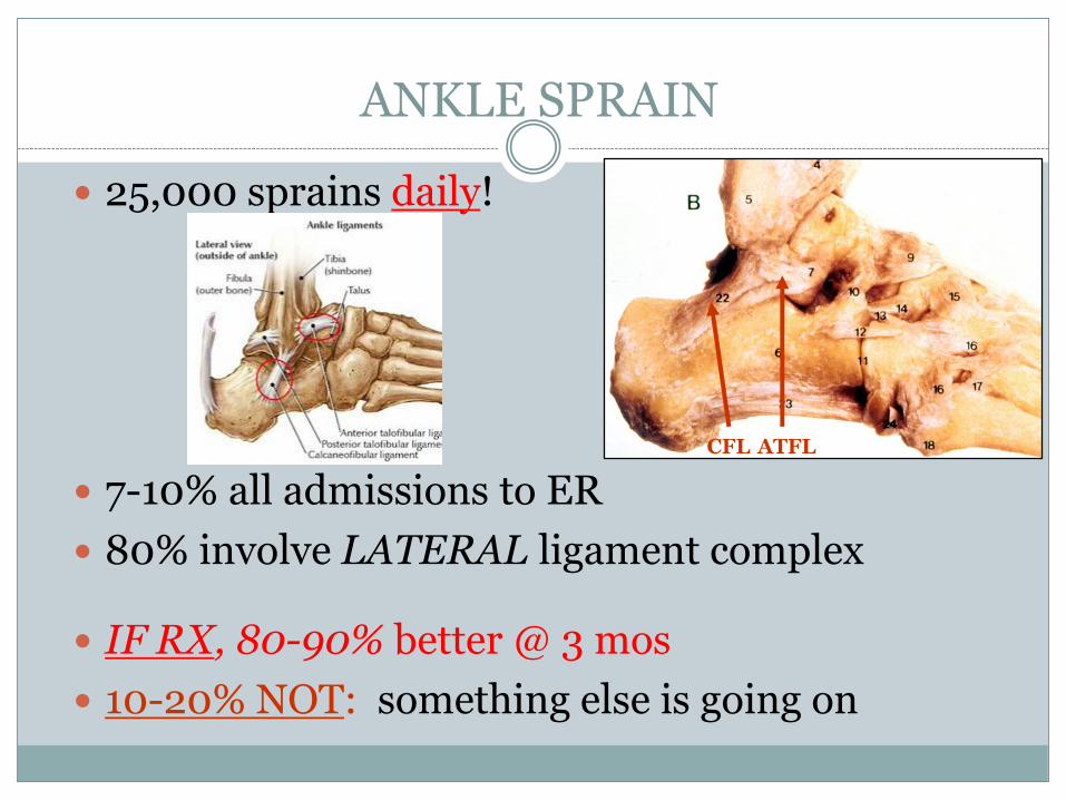
ANKLE SPRAIN
25,000 sprains daily!
7-10% all admissions to ER
80% involve LATERAL ligament complex
IF RX, 80-90% better @ 3 mos
10-20% NOT: something else is going on
CFL ATFL
ANKLE SPRAIN
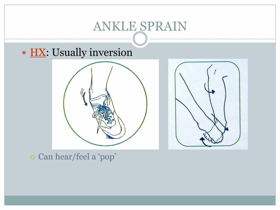
HX: Usually inversion
Can hear/feel a ‘pop’
ANKLE SPRAIN
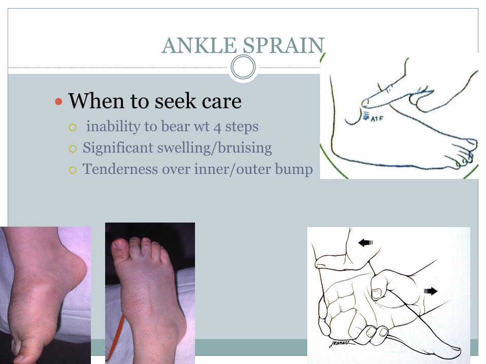
When to seek care inability to bear wt 4 steps
Significant swelling/bruising
Tenderness over inner/outer bump
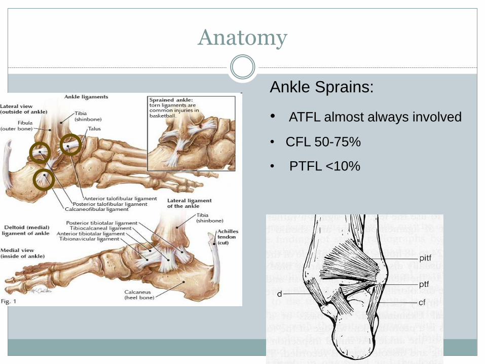
Anatomy
Ankle Sprains:
• ATFL almost always involved
• CFL 50-75%
• PTFL <10%
ANKLE SPRAIN
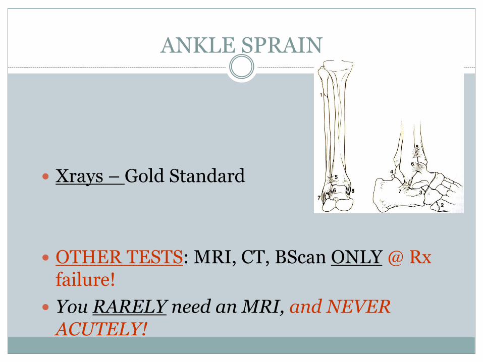
Xrays – Gold Standard
OTHER TESTS: MRI, CT, BScan ONLY @ Rx failure!
You RARELY need an MRI, and NEVER ACUTELY!
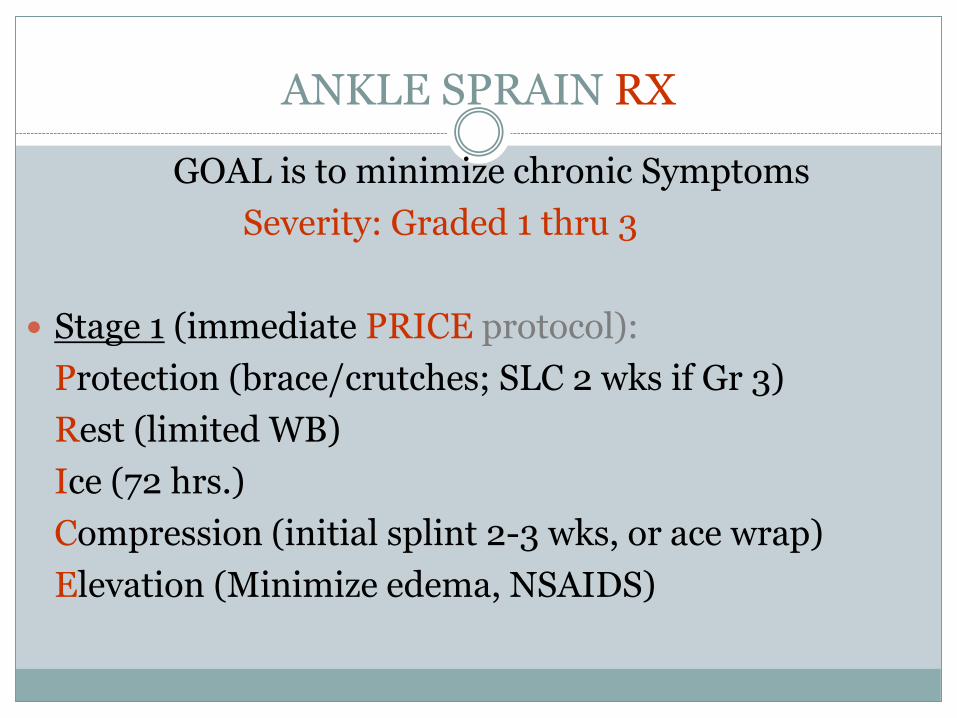
ANKLE SPRAIN RX
GOAL is to minimize chronic Symptoms
Severity: Graded 1 thru 3
Stage 1 (immediate PRICE protocol):
Protection (brace/crutches; SLC 2 wks if Gr 3)
Rest (limited WB)
Ice (72 hrs.)
Compression (initial splint 2-3 wks, or ace wrap)
Elevation (Minimize edema, NSAIDS)
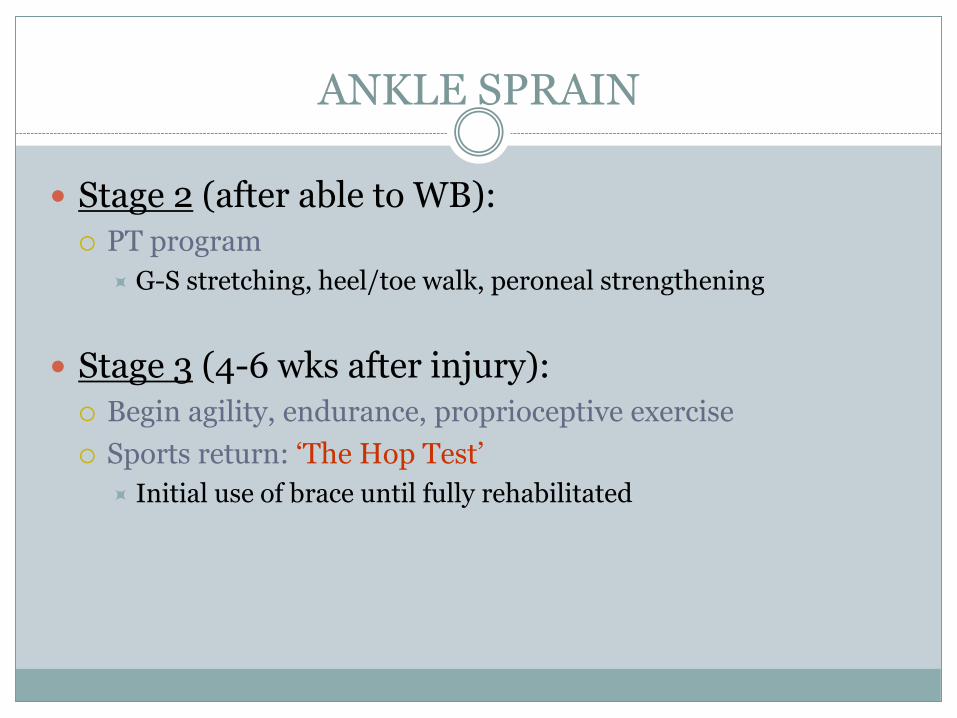
ANKLE SPRAIN
Stage 2 (after able to WB):
PT program
G-S stretching, heel/toe walk, peroneal strengthening
Stage 3 (4-6 wks after injury):
Begin agility, endurance, proprioceptive exercise
Sports return: ‘The Hop Test’
Initial use of brace until fully rehabilitated
Treatment
Delayed repair as efficacious as early repair
Early mobilization
Positive effect on local metabolic activity
? Speeds healing process
Cost
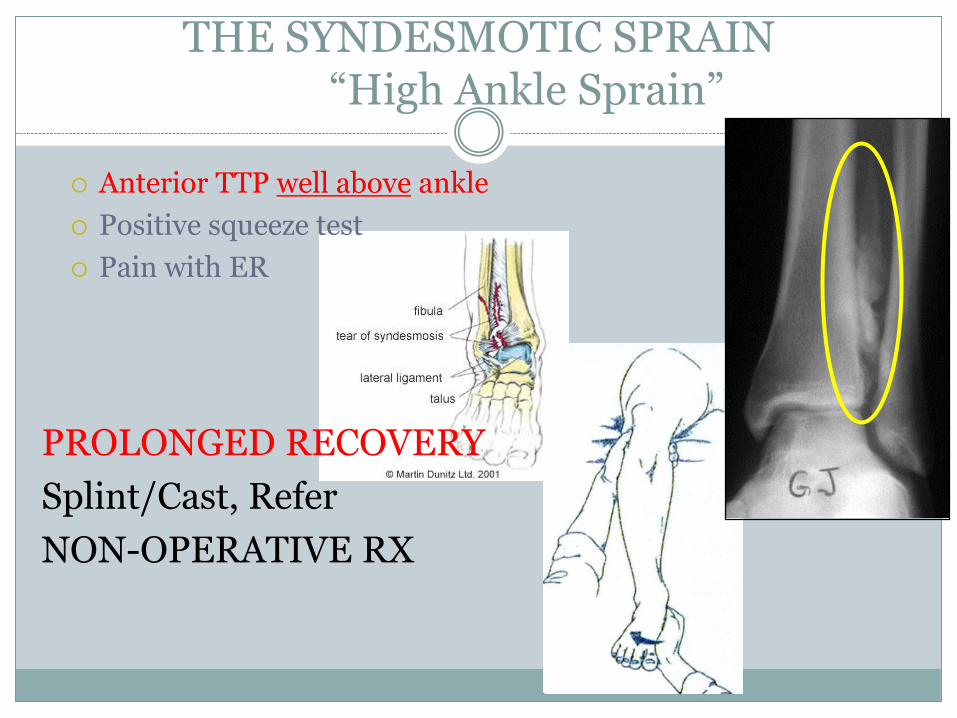
THE SYNDESMOTIC SPRAIN“High Ankle Sprain”
Anterior TTP well above ankle
Positive squeeze test
Pain with ER
PROLONGED RECOVERY
Splint/Cast, Refer
NON-OPERATIVE RX
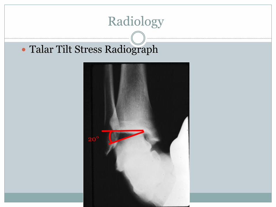
Radiology
Talar Tilt Stress Radiograph
20°
Heel Pain
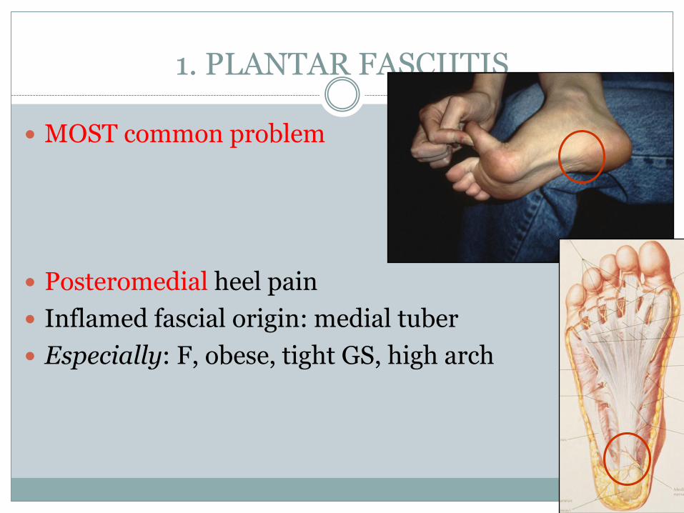
1. PLANTAR FASCIITIS
MOST common problem
Posteromedial heel pain
Inflamed fascial origin: medial tuber
Especially: F, obese, tight GS, high arch
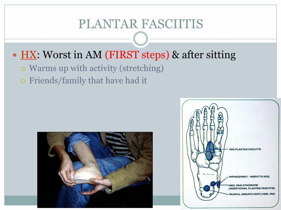
PLANTAR FASCIITIS
HX: Worst in AM (FIRST steps) & after sitting
Warms up with activity (stretching)
Friends/family that have had it
PLANTAR FASCIITIS
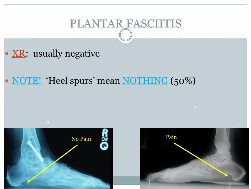
XR: usually negative
NOTE! ‘Heel spurs’ mean NOTHING (50%)
No Pain Pain
PLANTAR FASCIITIS
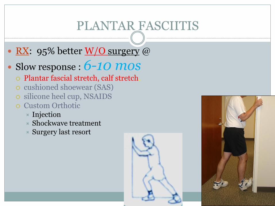
RX: 95% better W/O surgery @
Slow response : 6-10 mos Plantar fascial stretch, calf stretch. cushioned shoewear (SAS) silicone heel cup, NSAIDS Custom Orthotic
Injection Shockwave treatment Surgery last resort

HEEL Pad Syndrome
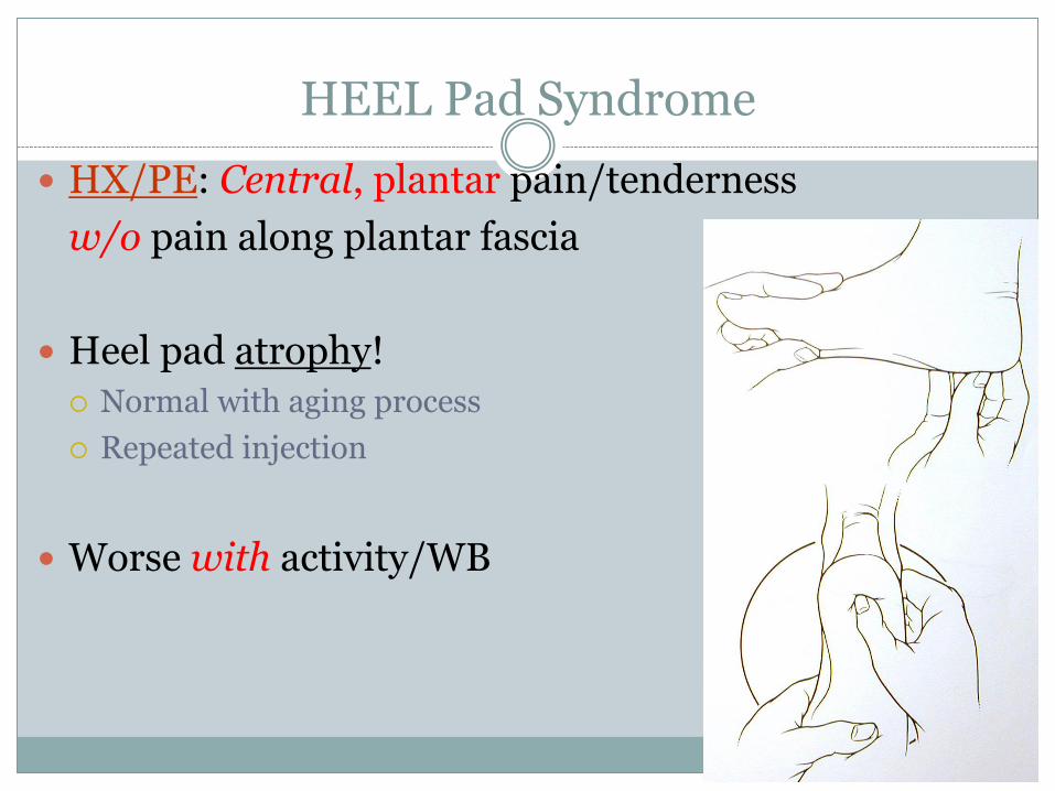
HX/PE: Central, plantar pain/tenderness
w/o pain along plantar fascia
Heel pad atrophy!
Normal with aging process
Repeated injection
Worse with activity/WB
HEEL PAIN
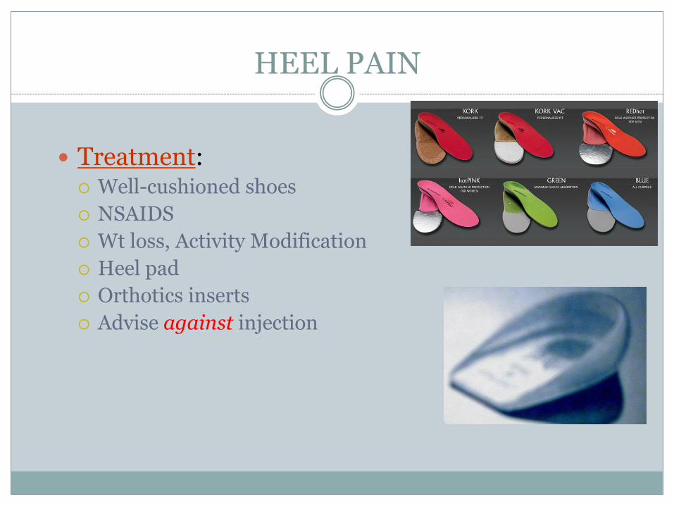
Treatment: Well-cushioned shoes
NSAIDS
Wt loss, Activity Modification
Heel pad
Orthotics inserts
Advise against injection
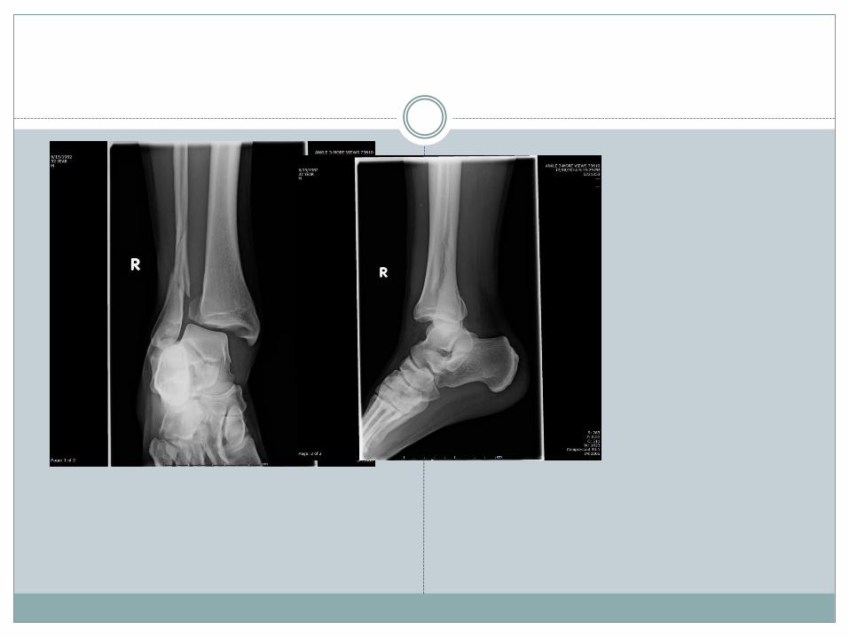
Ankle Fractures
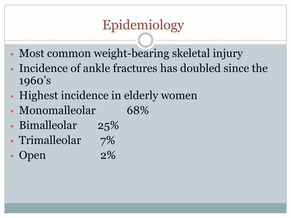
Epidemiology
• Most common weight-bearing skeletal injury
• Incidence of ankle fractures has doubled since the 1960’s
• Highest incidence in elderly women
• Monomalleolar 68%
• Bimalleolar 25%
• Trimalleolar 7%
• Open 2%
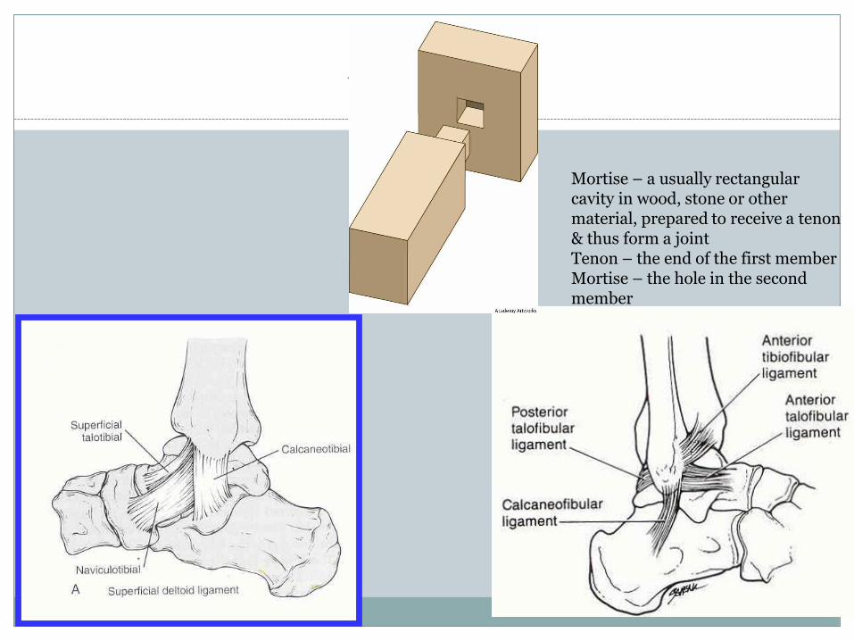
Anatomy
Mortise – a usually rectangular cavity in wood, stone or other material, prepared to receive a tenon& thus form a jointTenon – the end of the first memberMortise – the hole in the second member
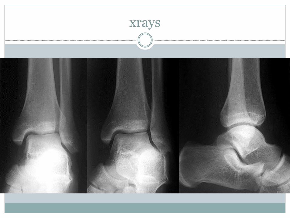
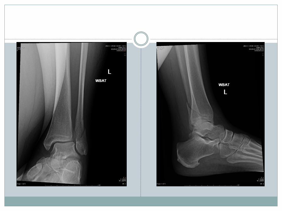
xrays
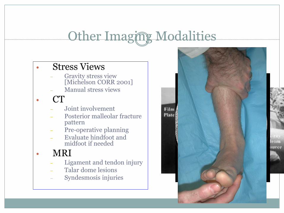
Other Imaging Modalities
• Stress Views– Gravity stress view
[Michelson CORR 2001]– Manual stress views
• CT– Joint involvement– Posterior malleolar fracture
pattern– Pre-operative planning– Evaluate hindfoot and
midfoot if needed
• MRI– Ligament and tendon injury – Talar dome lesions– Syndesmosis injuries
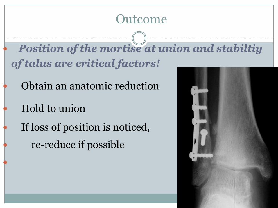
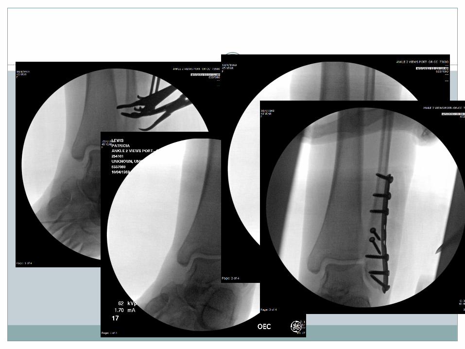
Outcome
Position of the mortise at union and stabiltiy
of talus are critical factors!
Obtain an anatomic reduction
Hold to union
If loss of position is noticed,
re-reduce if possible
Outcome
• Stable ankle fractures without lateral talar shift treated conservatively have 90% good to excellent results
• Operative fixation of unstable ankle fractures have 85-90% good to excellent results
• 2 year follow up 80-90% have unlimited ability to work, walk and participate
in leisure activities
20-30% report swelling or stiffness
41% have reduced dorsiflexion ( Lindsjo, Clin Orthop, 1985)
Outcome
• At one year following surgery, patients are generally doing well
• Most have few restrictions and little pain
• There is a significant improvement at one year compared to six months Recovery may take up to one year, let patients know this
• Younger age, male sex, absence of diabetes, and lower ASA class are predictive of functional recovery at one year
• By nine weeks, the total braking time of patients who have undergone fixation returns to the normal baseline value
Egol JBJS 2006
Egol JBJS 2003

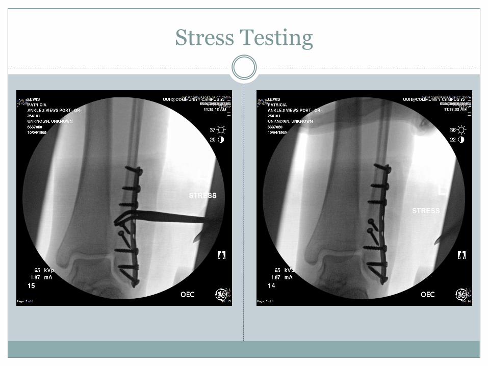
Stress Testing
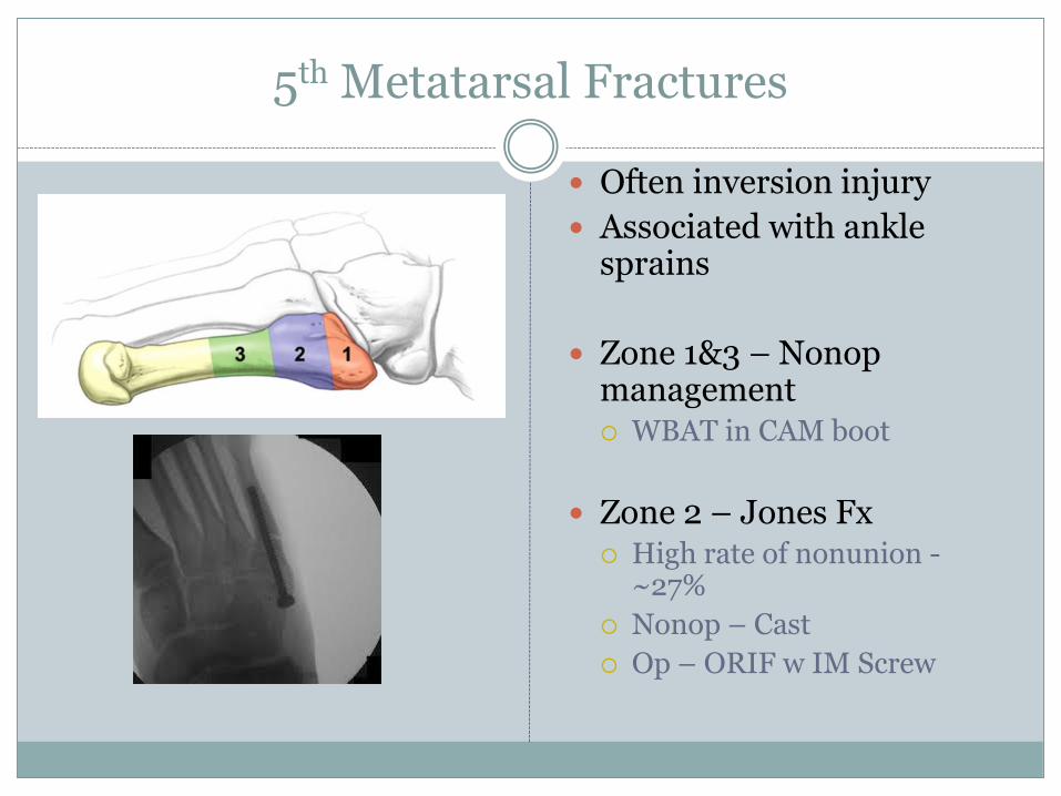
5th Metatarsal Fractures
Often inversion injury
Associated with ankle sprains
Zone 1&3 – Nonopmanagement WBAT in CAM boot
Zone 2 – Jones Fx High rate of nonunion -
~27%
Nonop – Cast
Op – ORIF w IM Screw
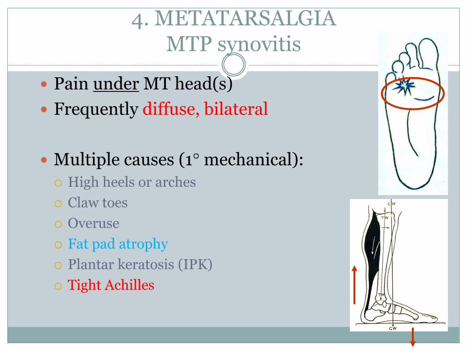
4. METATARSALGIAMTP synovitis
Pain under MT head(s)
Frequently diffuse, bilateral
Multiple causes (1° mechanical):
High heels or arches
Claw toes
Overuse
Fat pad atrophy
Plantar keratosis (IPK)
Tight Achilles
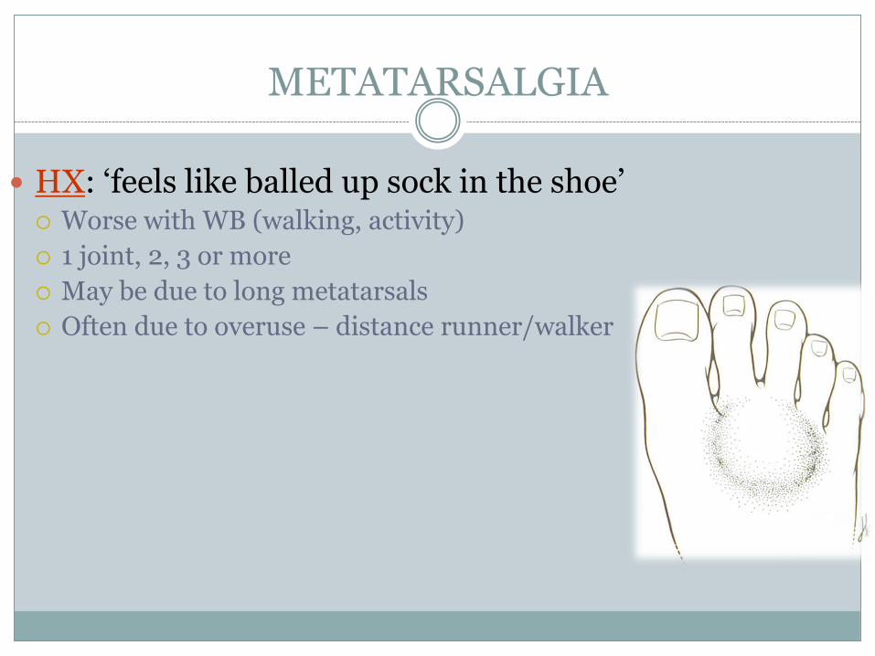
METATARSALGIA
HX: ‘feels like balled up sock in the shoe’ Worse with WB (walking, activity)
1 joint, 2, 3 or more
May be due to long metatarsals
Often due to overuse – distance runner/walker
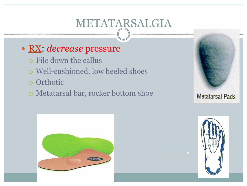
METATARSALGIA
RX: decrease pressure
File down the callus
Well-cushioned, low heeled shoes
Orthotic
Metatarsal bar, rocker bottom shoe
METATARSALGIA
Treatment: rarely required
Only when focal and recalcitrant after 6-8 mos
Surgery rare…generally not much else that can be done beyond judicious activity/shoewear
EDUCATE pts to avoid their frustration
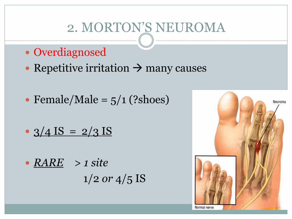
2. MORTON’S NEUROMA
Overdiagnosed
Repetitive irritation many causes
Female/Male = 5/1 (?shoes)
3/4 IS = 2/3 IS
RARE > 1 site
1/2 or 4/5 IS
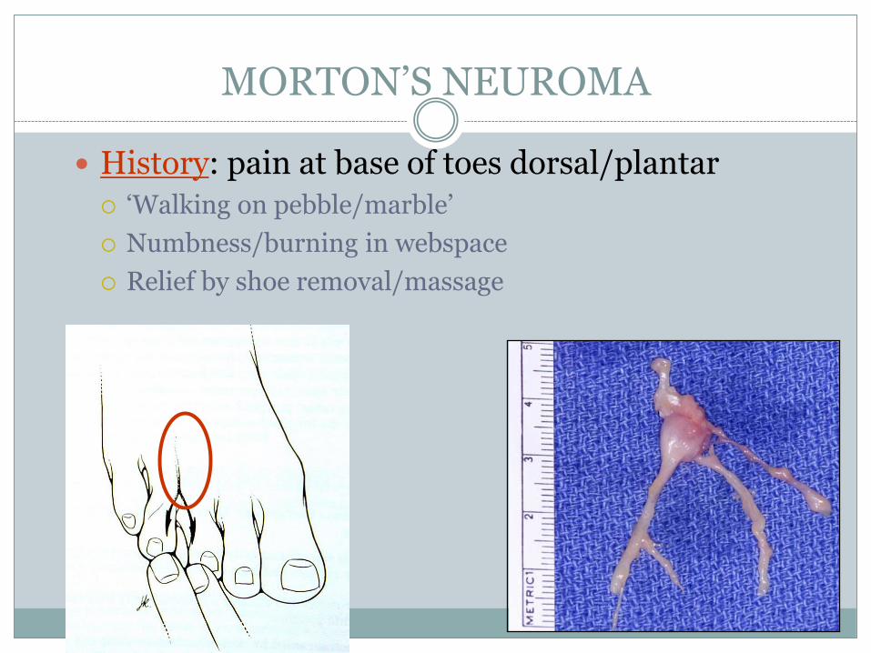
MORTON’S NEUROMA
History: pain at base of toes dorsal/plantar
‘Walking on pebble/marble’
Numbness/burning in webspace
Relief by shoe removal/massage
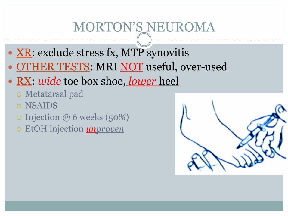
MORTON’S NEUROMA
XR: exclude stress fx, MTP synovitis
OTHER TESTS: MRI NOT useful, over-used
RX: wide toe box shoe, lower heel Metatarsal pad
NSAIDS
Injection @ 6 weeks (50%)
EtOH injection unproven
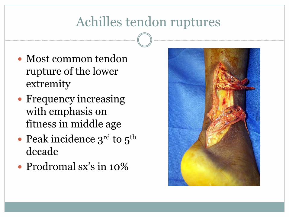
Achilles tendon ruptures
Most common tendon rupture of the lower extremity
Frequency increasing with emphasis on fitness in middle age
Peak incidence 3rd to 5th
decade
Prodromal sx’s in 10%
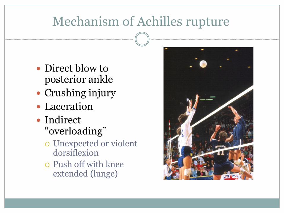
Mechanism of Achilles rupture
Direct blow to posterior ankle
Crushing injury
Laceration
Indirect “overloading” Unexpected or violent
dorsiflexion
Push off with knee extended (lunge)
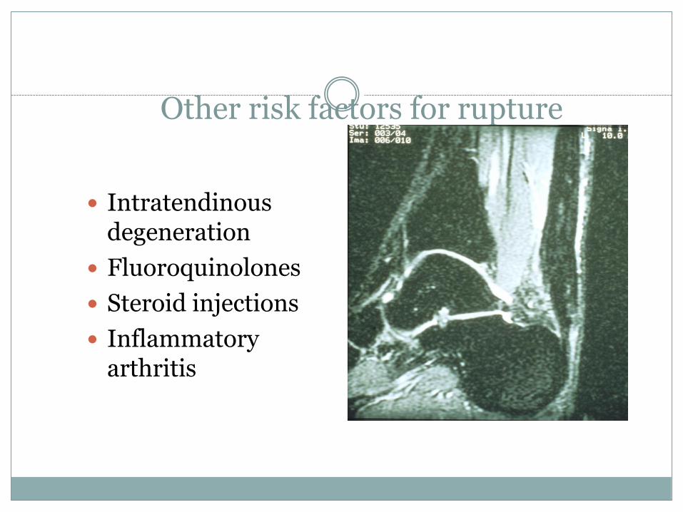
Other risk factors for rupture
Intratendinous degeneration
Fluoroquinolones
Steroid injections
Inflammatory arthritis
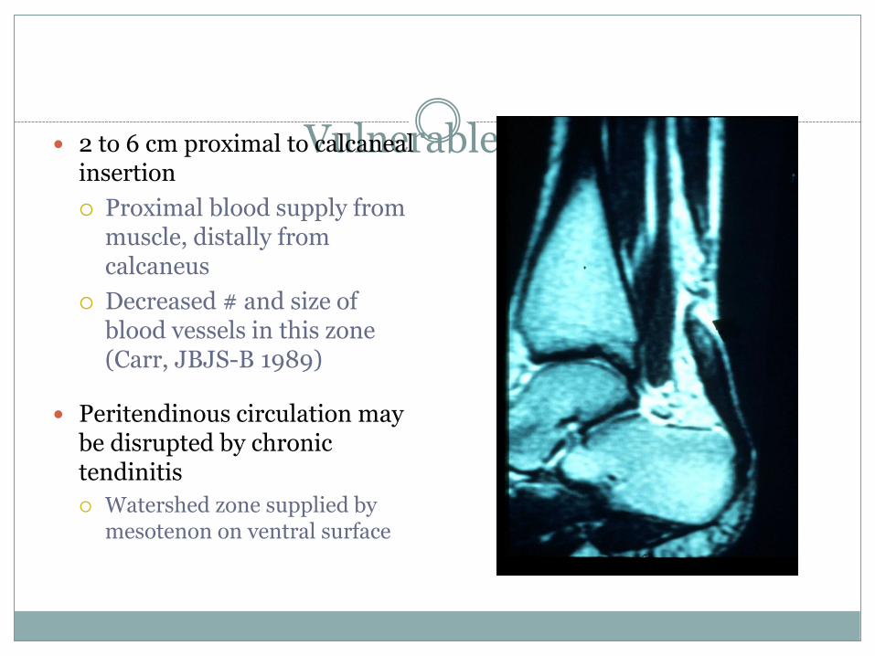
Vulnerable region 2 to 6 cm proximal to calcaneal insertion
Proximal blood supply from muscle, distally from calcaneus
Decreased # and size of blood vessels in this zone (Carr, JBJS-B 1989)
Peritendinous circulation may be disrupted by chronic tendinitis
Watershed zone supplied by mesotenon on ventral surface
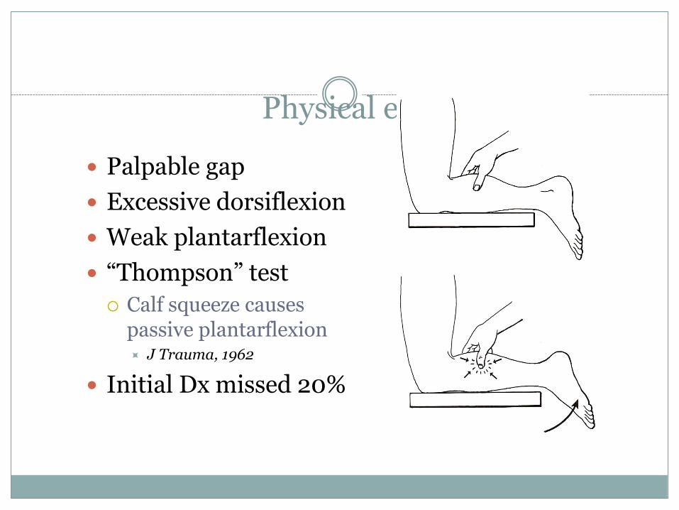
Physical exam
Palpable gap
Excessive dorsiflexion
Weak plantarflexion
“Thompson” test
Calf squeeze causes passive plantarflexion J Trauma, 1962
Initial Dx missed 20%
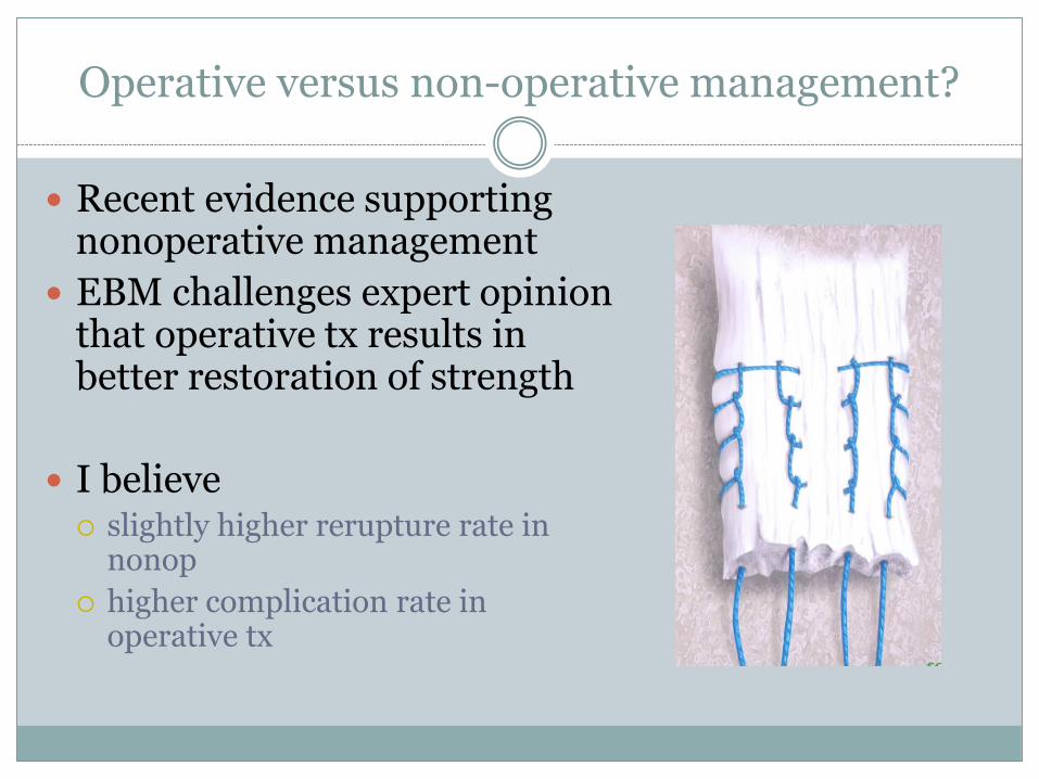
Operative versus non-operative management?
Recent evidence supporting nonoperative management
EBM challenges expert opinion that operative tx results in better restoration of strength
I believe slightly higher rerupture rate in
nonop
higher complication rate in operative tx
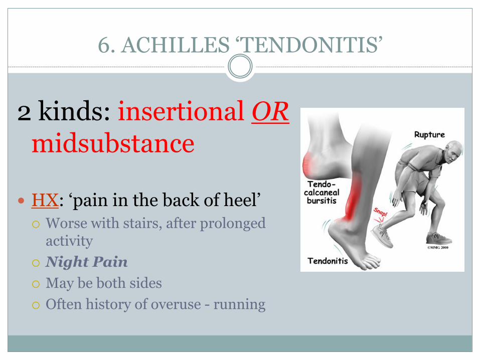
6. ACHILLES ‘TENDONITIS’
2 kinds: insertional ORmidsubstance
HX: ‘pain in the back of heel’
Worse with stairs, after prolonged activity
Night Pain
May be both sides
Often history of overuse - running
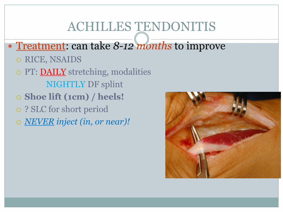
ACHILLES TENDONITIS
Treatment: can take 8-12 months to improve
RICE, NSAIDS
PT: DAILY stretching, modalities
NIGHTLY DF splint
Shoe lift (1cm) / heels!
? SLC for short period
NEVER inject (in, or near)!
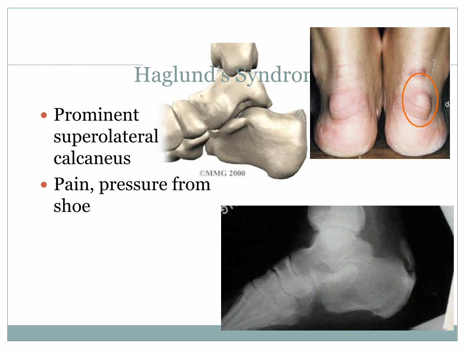
Haglund’s Syndrome
Prominent superolateralcalcaneus
Pain, pressure from shoe
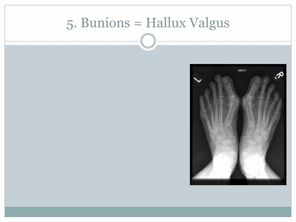
5. Bunions = Hallux Valgus
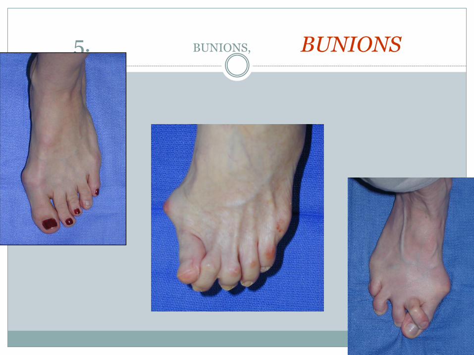
5. There are BUNIONS, and BUNIONS
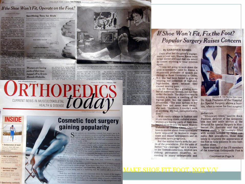
MAKE SHOE FIT FOOT, NOT V/V
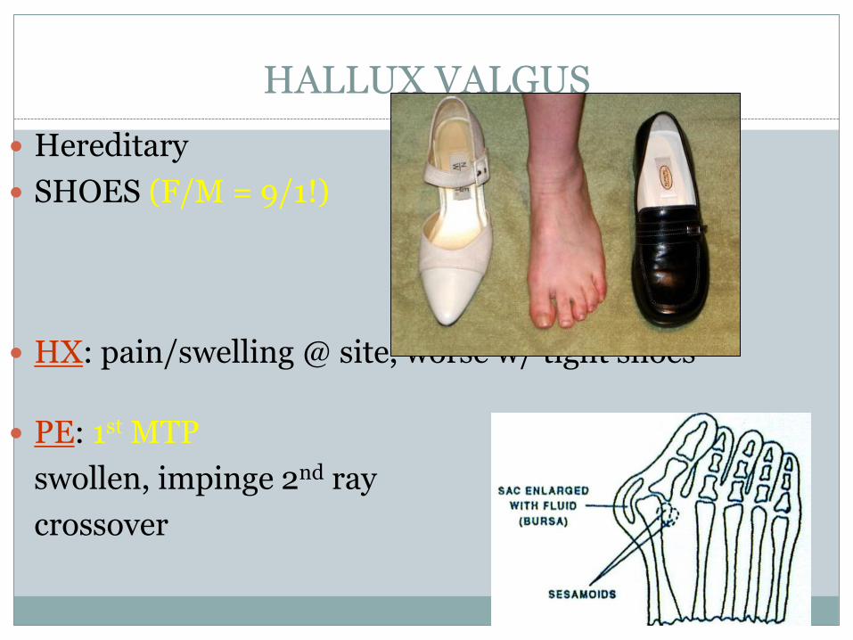
HALLUX VALGUS
Hereditary
SHOES (F/M = 9/1!)
HX: pain/swelling @ site, worse w/ tight shoes
PE: 1st MTP
swollen, impinge 2nd ray
crossover
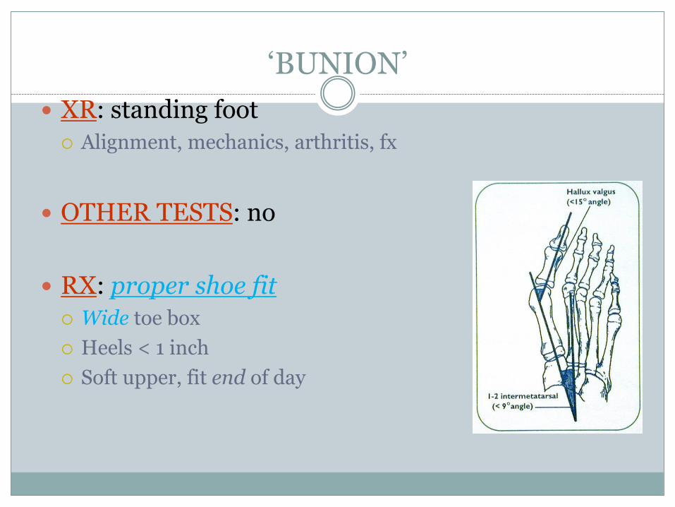
‘BUNION’
XR: standing foot
Alignment, mechanics, arthritis, fx
OTHER TESTS: no
RX: proper shoe fit
Wide toe box
Heels < 1 inch
Soft upper, fit end of day
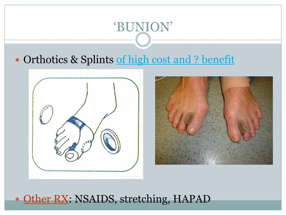
‘BUNION’
Orthotics & Splints of high cost and ? benefit
Other RX: NSAIDS, stretching, HAPAD
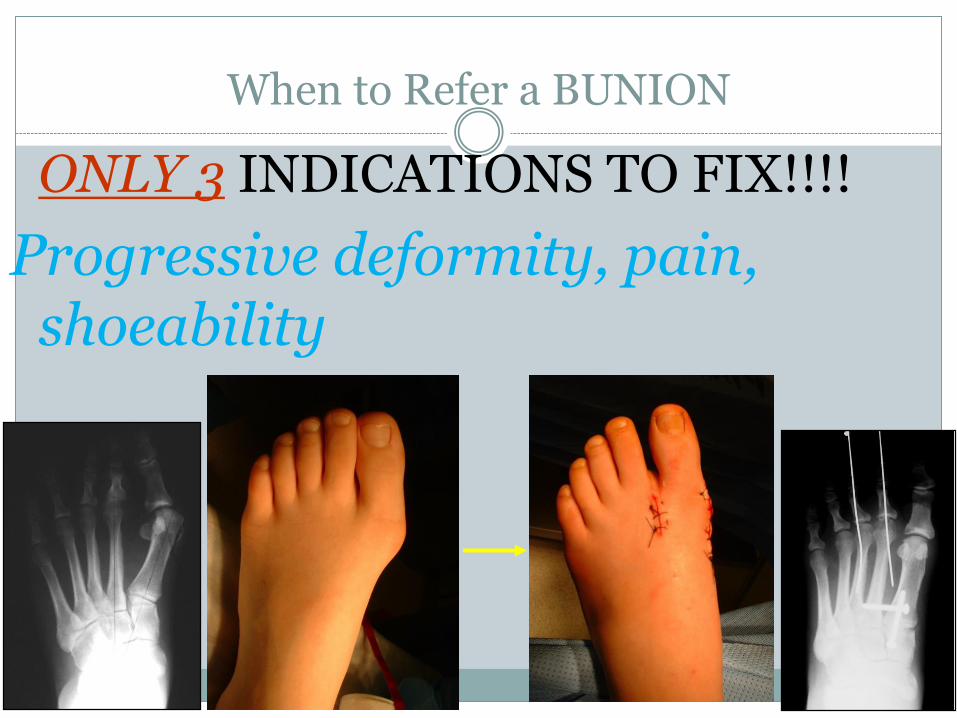
When to Refer a BUNION
ONLY 3 INDICATIONS TO FIX!!!!
Progressive deformity, pain, shoeability
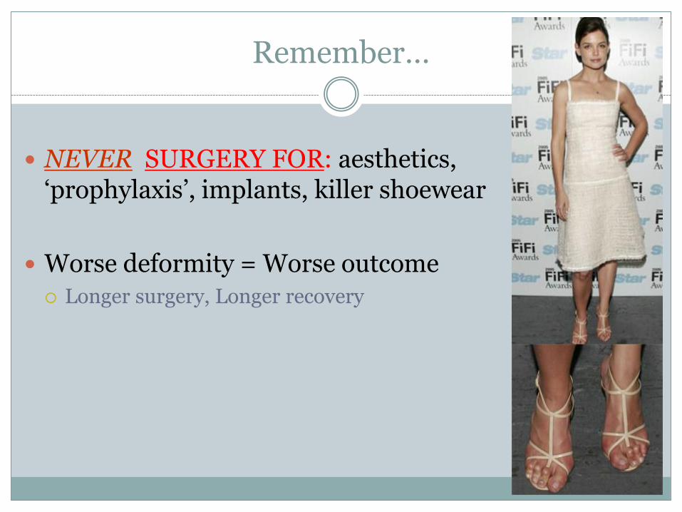
Remember…
NEVER SURGERY FOR: aesthetics, ‘prophylaxis’, implants, killer shoewear
Worse deformity = Worse outcome
Longer surgery, Longer recovery
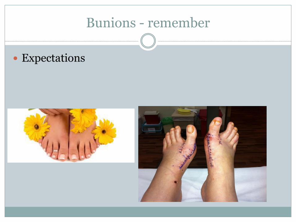
Bunions - remember
Expectations
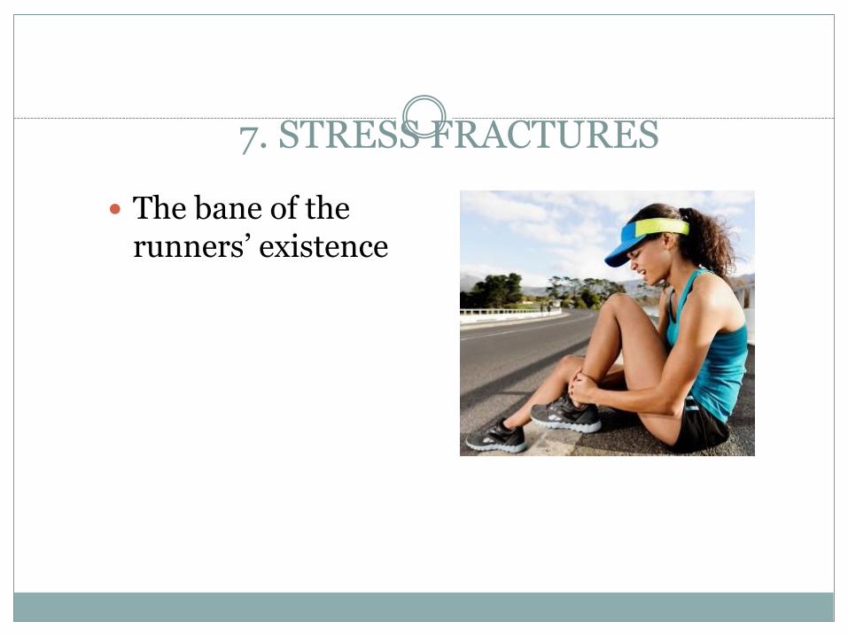
7. STRESS FRACTURES
The bane of the runners’ existence
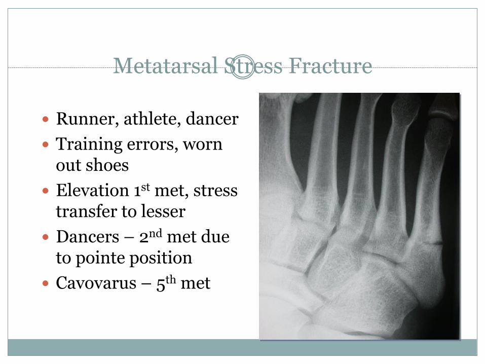
Metatarsal Stress Fracture
Runner, athlete, dancer
Training errors, worn out shoes
Elevation 1st met, stress transfer to lesser
Dancers – 2nd met due to pointe position
Cavovarus – 5th met
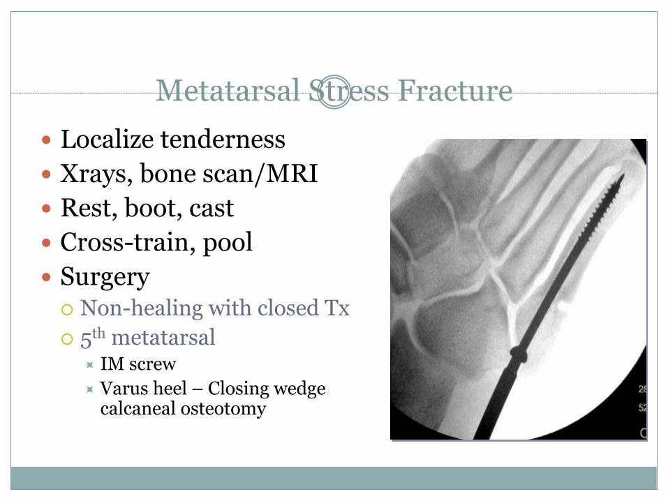
Metatarsal Stress Fracture
Localize tenderness
Xrays, bone scan/MRI
Rest, boot, cast
Cross-train, pool
Surgery Non-healing with closed Tx
5th metatarsal IM screw
Varus heel – Closing wedge calcaneal osteotomy
THANK YOU
“The human foot is a masterpiece of engineering…and a work of art.”
- Leonardo da Vinci, The Notebooks (c. 1508-1518)