TonEBP/OREBPIsaRegulatorofNucleusPulposusCell ... ... TauT, BGT-1, and SMIT; above 450 mosmol/kg,...
10
TonEBP/OREBP Is a Regulator of Nucleus Pulposus Cell Function and Survival in the Intervertebral Disc * □ S Received for publication, March 1, 2006, and in revised form, April 26, 2006 Published, JBC Papers in Press, June 13, 2006, DOI 10.1074/jbc.M601969200 Tsung-Ting Tsai ‡§ , Keith G. Danielson ‡ , Asha Guttapalli ‡ , Erbil Oguz ‡¶ , Todd J. Albert ‡ , Irving M. Shapiro ‡ , and Makarand V. Risbud ‡1 From the ‡ Department of Orthopaedic Surgery, Thomas Jefferson University, Philadelphia, Pennsylvania 19107, the § Department of Orthopaedic Surgery, Chang Gung Memorial Hospital, Chang Gung University, Taoyuan 33305, Taiwan, and the ¶ Gulhane Military Medical Academy, Department of Orthopaedics and Traumatology, Etlik, 06018 Ankara, Turkey The nucleus pulposus is an aggrecan-rich hydrated tissue that permits the intervertebral disc to resist compressive loads. Adaptation to loading is achieved through an elevation in disc osmolarity mediated by the numerous charged glycosoamin- oglycan side chains of the aggrecan molecule. The goal of this investigation was to determine the functional role of the osmo- regulatory protein, TonEBP, in cells of the nucleus pulposus. We found that TonEBP and its downstream target genes were robustly expressed in the tissues of the disc. Above 330 mosmol/ kg, cultured nucleus pulposus cells up-regulated target genes TauT, BGT-1, and SMIT; above 450 mosmol/kg, there was raised expression of HSP-70. In hypertonic media there was activation of TauT and heat shock protein-70 (HSP-70) reporter activity and increased binding of TonEBP to the TonE motif. When cells were transfected with the dominant-negative form of TonEBP (DN-TonEBP) there was suppression of TauT and HSP-70 reporter gene expression; pTonEBP enhanced reporter gene expression. Moreover, in hypertonic media, forced expres- sion of DN-TonEBP induced apoptosis. We suppressed TonEBP using small interfering RNA technique and noted a decrease in TauT reporter activity in isotonic as well as hyperosmolar media. Finally, we report that the aggrecan promoter contains two conserved TonE motifs. To evaluate the importance of these motifs, we overexpressed DN-TonEBP and partially silenced TonEBP using small interfering RNA. Both ap- proaches resulted in suppression of aggrecan promoter activ- ity. It is concluded that TonEBP permits the disc cells to adapt to the hyperosmotic milieu while autoregulating the expression of molecules that generate the unique extracellular environment. The intervertebral disc is a specialized biomechanical struc- ture that permits movement between vertebrae and protects the vertebral bone from mechanical forces. It consists of an outer ligament, the annulus fibrosus that encloses a gel-like tissue, the nucleus pulposus. While sparse, cells in the nucleus pulposus secrete a complex extracellular matrix that contains fibrillar collagens and the proteoglycan, aggrecan. The numer- ous glycosaminoglycan side chains of the aggrecan molecule bind cations thereby raising the osmolarity of the disc tissues (1– 4). While the hydration properties of the nucleus pulposus permits dynamic loading and unloading, the mechanism by which these cells adapt to elevated osmotic forces is poorly understood. In a number of tissues, cellular adaptation to hyperosmotic stress is mediated by the tonicity enhancer-binding protein (TonEBP), 2 also called OREBP (5) or NFAT5, (5, 6). Upon acti- vation, TonEBP binds to the tonicity-responsive enhancer ele- ment (TonE) of genes required for osmotolerance and cell sur- vival. These genes include the betaine/-aminobutyric acid transporter, sodium myo-inositol co-transporter (7–9), taurine transporter (10, 11), and aldose reductase (6). By regulating levels of betaine, myo-inositol, taurine, and sorbitol, these genes control the osmotic properties of the cytosol. HSP-70, a molec- ular chaperone that maintains cellular function under hyper- tonic stress is also induced by TonEBP (12, 13). Most homozygous TonEBP knock-out mice evidence mid- gestational lethality. Of the few that survive, all exhibit severe growth retardation and kidney dysfunction (14). A transgenic mouse expressing a dominant-negative form of TonEBP (DN- TonEBP) in collecting duct epithelial cells demonstrates an absolute requirement of TonEBP for expression of the urea transporter gene and aquaporin-2 (15). Aside from osmoregu- lation, TonEBP is required for T cell proliferation and function (16, 17), and it is implicated in cancer cell migration and metas- tasis (18). A recent study by Wang et al. (19) showed that expression of DN-TonEBP in lens fiber cells promotes cataract formation by causing defects in their elongation. Since TonEBP is expressed by a number of cell types, it is reasonable to assume that it serves a variety of physiologic functions, especially those that impact on tissue hydration and the osmotic environment (20). The goal of the present study is to examine the role of TonEBP in cells of the postnatal intervertebral disc. We show that TonEBP, and its downstream target genes, are * This work was supported by National Institutes of Health Grant AR050087. The costs of publication of this article were defrayed in part by the pay- ment of page charges. This article must therefore be hereby marked “advertisement” in accordance with 18 U.S.C. Section 1734 solely to indi- cate this fact. □ S The on-line version of this article (available at http://www.jbc.org) contains supplemental Table 1. 1 To whom correspondence should be addressed: Dept. of Orthopaedic Sur- gery, Thomas Jefferson University, 1015 Walnut St., Suite 501 Curtis Bldg., Philadelphia, PA 19107. Tel.: 215-955-1063; Fax: 215-955-9159; E-mail: [email protected]. 2 The abbreviations used are: TonEBP, tonicity enhancer-binding protein; DN- TonEBP, dominant-negative form of TonEBP; RT, reverse transcription; PBS, phosphate-buffered saline; Wt, wild type; Mt, mutant; MTT, 3-(4,5-dimeth- ylthiazol-2-yl)-2,5-diphenyltetrazolium bromide; siRNA, small interfering RNA. THE JOURNAL OF BIOLOGICAL CHEMISTRY VOL. 281, NO. 35, pp. 25416 –25424, September 1, 2006 © 2006 by The American Society for Biochemistry and Molecular Biology, Inc. Printed in the U.S.A. 25416 JOURNAL OF BIOLOGICAL CHEMISTRY VOLUME 281 • NUMBER 35 • SEPTEMBER 1, 2006 by guest on June 29, 2018 http://www.jbc.org/ Downloaded from
Transcript of TonEBP/OREBPIsaRegulatorofNucleusPulposusCell ... ... TauT, BGT-1, and SMIT; above 450 mosmol/kg,...

TonEBP/OREBP Is a Regulator of Nucleus Pulposus CellFunction and Survival in the Intervertebral Disc*□S
Received for publication, March 1, 2006, and in revised form, April 26, 2006 Published, JBC Papers in Press, June 13, 2006, DOI 10.1074/jbc.M601969200
Tsung-Ting Tsai‡§, Keith G. Danielson‡, Asha Guttapalli‡, Erbil Oguz‡¶, Todd J. Albert‡, Irving M. Shapiro‡,and Makarand V. Risbud‡1
From the ‡Department of Orthopaedic Surgery, Thomas Jefferson University, Philadelphia, Pennsylvania 19107, the §Departmentof Orthopaedic Surgery, Chang Gung Memorial Hospital, Chang Gung University, Taoyuan 33305, Taiwan, and the ¶GulhaneMilitary Medical Academy, Department of Orthopaedics and Traumatology, Etlik, 06018 Ankara, Turkey
The nucleus pulposus is an aggrecan-rich hydrated tissue thatpermits the intervertebral disc to resist compressive loads.Adaptation to loading is achieved through an elevation in discosmolarity mediated by the numerous charged glycosoamin-oglycan side chains of the aggrecan molecule. The goal of thisinvestigation was to determine the functional role of the osmo-regulatory protein, TonEBP, in cells of the nucleus pulposus.We found that TonEBP and its downstream target genes wererobustly expressed in the tissues of the disc. Above 330mosmol/kg, cultured nucleus pulposus cells up-regulated target genesTauT, BGT-1, and SMIT; above 450 mosmol/kg, there wasraised expression of HSP-70. In hypertonic media there wasactivation of TauT andheat shock protein-70 (HSP-70) reporteractivity and increased binding of TonEBP to the TonE motif.When cells were transfected with the dominant-negative formof TonEBP (DN-TonEBP) there was suppression of TauT andHSP-70 reporter gene expression; pTonEBP enhanced reportergene expression.Moreover, in hypertonicmedia, forced expres-sion ofDN-TonEBP induced apoptosis.We suppressedTonEBPusing small interfering RNA technique and noted a decrease inTauT reporter activity in isotonic as well as hyperosmolarmedia. Finally, we report that the aggrecan promoter containstwo conserved TonE motifs. To evaluate the importance ofthese motifs, we overexpressed DN-TonEBP and partiallysilenced TonEBP using small interfering RNA. Both ap-proaches resulted in suppression of aggrecan promoter activ-ity. It is concluded thatTonEBPpermits thedisc cells to adapt tothe hyperosmotic milieu while autoregulating the expression ofmolecules that generate the unique extracellular environment.
The intervertebral disc is a specialized biomechanical struc-ture that permits movement between vertebrae and protectsthe vertebral bone from mechanical forces. It consists of anouter ligament, the annulus fibrosus that encloses a gel-like
tissue, the nucleus pulposus. While sparse, cells in the nucleuspulposus secrete a complex extracellular matrix that containsfibrillar collagens and the proteoglycan, aggrecan. The numer-ous glycosaminoglycan side chains of the aggrecan moleculebind cations thereby raising the osmolarity of the disc tissues(1–4). While the hydration properties of the nucleus pulposuspermits dynamic loading and unloading, the mechanism bywhich these cells adapt to elevated osmotic forces is poorlyunderstood.In a number of tissues, cellular adaptation to hyperosmotic
stress is mediated by the tonicity enhancer-binding protein(TonEBP),2 also called OREBP (5) or NFAT5, (5, 6). Upon acti-vation, TonEBP binds to the tonicity-responsive enhancer ele-ment (TonE) of genes required for osmotolerance and cell sur-vival. These genes include the betaine/�-aminobutyric acidtransporter, sodiummyo-inositol co-transporter (7–9), taurinetransporter (10, 11), and aldose reductase (6). By regulatinglevels of betaine,myo-inositol, taurine, and sorbitol, these genescontrol the osmotic properties of the cytosol. HSP-70, a molec-ular chaperone that maintains cellular function under hyper-tonic stress is also induced by TonEBP (12, 13).Most homozygous TonEBP knock-out mice evidence mid-
gestational lethality. Of the few that survive, all exhibit severegrowth retardation and kidney dysfunction (14). A transgenicmouse expressing a dominant-negative form of TonEBP (DN-TonEBP) in collecting duct epithelial cells demonstrates anabsolute requirement of TonEBP for expression of the ureatransporter gene and aquaporin-2 (15). Aside from osmoregu-lation, TonEBP is required for T cell proliferation and function(16, 17), and it is implicated in cancer cell migration andmetas-tasis (18). A recent study by Wang et al. (19) showed thatexpression of DN-TonEBP in lens fiber cells promotes cataractformation by causing defects in their elongation. Since TonEBPis expressed by a number of cell types, it is reasonable to assumethat it serves a variety of physiologic functions, especially thosethat impact on tissue hydration and the osmotic environment(20).The goal of the present study is to examine the role of
TonEBP in cells of the postnatal intervertebral disc. Weshow that TonEBP, and its downstream target genes, are
* This work was supported by National Institutes of Health Grant AR050087.The costs of publication of this article were defrayed in part by the pay-ment of page charges. This article must therefore be hereby marked“advertisement” in accordance with 18 U.S.C. Section 1734 solely to indi-cate this fact.
□S The on-line version of this article (available at http://www.jbc.org) containssupplemental Table 1.
1 To whom correspondence should be addressed: Dept. of Orthopaedic Sur-gery, Thomas Jefferson University, 1015 Walnut St., Suite 501 Curtis Bldg.,Philadelphia, PA 19107. Tel.: 215-955-1063; Fax: 215-955-9159; E-mail:[email protected].
2 The abbreviations used are: TonEBP, tonicity enhancer-binding protein; DN-TonEBP, dominant-negative form of TonEBP; RT, reverse transcription; PBS,phosphate-buffered saline; Wt, wild type; Mt, mutant; MTT, 3-(4,5-dimeth-ylthiazol-2-yl)-2,5-diphenyltetrazolium bromide; siRNA, small interferingRNA.
THE JOURNAL OF BIOLOGICAL CHEMISTRY VOL. 281, NO. 35, pp. 25416 –25424, September 1, 2006© 2006 by The American Society for Biochemistry and Molecular Biology, Inc. Printed in the U.S.A.
25416 JOURNAL OF BIOLOGICAL CHEMISTRY VOLUME 281 • NUMBER 35 • SEPTEMBER 1, 2006
by guest on June 29, 2018http://w
ww
.jbc.org/D
ownloaded from

expressed in the nucleus pulposus and the annulus fibrosus.Importantly, our data indicate that TonEBP binds to a TonEmotif in the aggrecan promoter and regulates its transcriptionalactivity. This finding lends credence to the view that TonEBPregulates the hydration status of the disc thereby enhancing cellfunction in the hyperosmotic environment.
EXPERIMENTAL PROCEDURES
Isolation of Nucleus Pulposus Cells—Rat nucleus pulposuscells were isolated using method reported earlier by Risbudet al. (21). Briefly, male Wistar rats (250 g) were euthanizedwith CO2, and lumbar intervertebral discs were removed fromthe spinal column. The gel-like nucleus pulposus was separatedfrom the annulus fibrosus, using a dissecting microscope, andtreated with 0.1% collagenase and 10 units/ml hyaluronidasefor 4–6 h. The partially digested tissue was maintained as anexplant in Dulbeccos modified Eagle’s medium and 10% fetalbovine serum supplementedwith antibiotics.Nucleus pulposuscells migrated out of the explant after 1 week. When confluent,the cells were lifted using a trypsin (0.25%) EDTA (1 mM) solu-tion and subcultured in 10-cm dishes.RT-PCR Analysis—RNA was isolated from cells using Trizol
reagent (Invitrogen) following the manufacturer’s instructions.Briefly, 2 �g of total RNA was reverse-transcribed into cDNAusing Superscript II RT enzyme (Invitrogen) and oligo(dT)primers. PCR reactions were performed using cDNA samples(1 �l) with Superscript DNA polymerase (Invitrogen). Primersfor rat genes were custom designed and synthesized by Inte-grated DNA Technologies (Coralville, IA). The PCR productwas run on a 1.2% agarose gel and the amplicon visualized on aKodak 440 imaging station.Immunohistological Studies—Freshly isolated discs were
immediately fixed in 4% paraformaldehyde in PBS and thenembedded in paraffin. Transverse and coronal sections, 6–8�m in thickness, were deparaffinized in xylene, rehydratedthrough graded ethanol, and stained with Alcian blue, andwith eosin and propidium iodide. For localizing TonEBP,sections were incubated with the anti-TonEBP antibody(Abcam, Cambridge, MA) in 2% bovine serum albumin in PBSat a dilution of 1:100 at 4 °C overnight. After thoroughly wash-ing the sections, the bound primary antibody was incubatedwith biotinylated universal secondary antibody, at a dilution of1:20 (Vector Laboratories, Burlingame, CA) for 10min at roomtemperature. Sections were incubated with a streptavidin/per-oxidase complex for 5min and washed with PBS, and color wasdeveloped using 3�-3-diaminobenzidine (Vecta Stain UniversalQuick Kit, Vector Laboratories).Immunofluorescence Microscopy—Cells were plated in flat
bottom 96-well plates and maintained in isotonic or hypertonicmedium for 24 h. After incubation, cells were fixed with 4%paraformaldehyde, permeabilized with 0.2% Triton X-100 in PBSfor 10 min, blocked with PBS containing 5% fetal bovine serum,and incubatedwithanti-TonEBPantibody (1:200) (Abcam)at 4 °Covernight. As a negative control, cells were reacted with mouseisotype IgGunder similar conditions.Afterwashing, the cellswereincubated with Alexa Fluor-488-conjugated anti-mouse second-aryantibody (MolecularProbes, St.Louis,MO)atadilutionof1:50and 10 �M propidium iodide for 1 h at room temperature. Quan-
titative image analysis was performed using nine random fields ofcells per experimental group. The 36-bit color images were re-corded by confocal microscopy using the green and red channelfor TonEBP and propidium iodide (nuclear), respectively. ImagePro-plus software (Media Cybernetics, Silver Spring, MD) wasused to calculate the threshold for all cells in the field. The meandensity of TonEBP was then plotted as a histogram. Caspase-3activation was evaluated by visualizing cleavage of the PhiPhiLux-G1D2 substrate (OncoImmune Inc., Gaithersburg,MD) followingthemanufacturer’s protocol. Cells were imaged using a laser scan-ning confocalmicroscope (Olympus Fluoview,Tokyo, Japan), andimages were analyzed as described above.Nuclear Extracts and Western Blotting—Nuclear extracts
were prepared according to the method of Dignam et al. (22)using the CellLytic NuCLEAR extraction kit (Sigma). After cul-ture in isotonic or hypertonic medium, cells were immediatelyplaced on ice. Cells were treatedwith a hypotonic buffer (10mMHEPES, pH 7.9, 1.5 mM MgCl2, 10 mM KCl, and 0.5 mM dithio-threitol) and incubated on ice for 15 min. Igeapal CA-630 wasadded to a final concentration of 0.6% and the mixture wasvortexed vigorously for 10 s. Nuclei were recovered by centrif-ugation at 3,300 � g for 30 s at 4 °C and extracted by gentleshaking in buffer containing 20 mM HEPES, pH 7.9, 0.42 MNaCl, 25% glycerol, 1.5 mMMgC12, 0.2 mM EDTA, and 0.5 mMdithiothreitol for 30 min at 4 °C. The extract was then centri-fuged for 15 min at 25,000 � g, and the supernatant was snap-frozen at �70 °C. All buffers contained a protease inhibitormixture.Nuclear extracts were resolved on 10% SDS-polyacrylamide
gels. Proteins were transferred by electroblotting to nitrocellu-lose membranes (Bio-Rad). Themembranes were blocked with5% nonfat drymilk in TBST (50mMTris, pH 7.6, 150mMNaCl,0.1% Tween 20) and incubated overnight at 4 °C in 3% nonfatdry milk in TBST with the anti-TonEBP antibody (Abcam) at adilution of 1:500. Immunolabeling was detected using the ECL(Amersham Biosciences).Reporter Plasmids—PCR amplification of a 2.5 kb fragment
of the upstream mouse aggrecan promoter spanning �2204to �290 was performed using LA Taq polymerase (TakaraMirus Bio) using the following primers: forward, 5�-GAG-GAGCTCCAGCACAATATCGATGCTCCAGCTAGTGTG-CGAAAT-3�, SacI site underlined; reverse, 5�-GGTAGATCT-GCAGGGTCGATGAAGGGAAGGGACACAGAAAGGTG-3�,BglII site underlined. The resulting DNA fragment was sub-cloned into pCR2.1 (Invitrogen), isolated by restriction diges-tion with SacI and BglII, and ligated into the luciferase basicexpression vector, pGL3 (Promega). As an internal transfectioncontrol, vector pRL-TK (Promega) containing Renilla renifor-mis luciferase genes was used. The amount of transfected plas-mid, the pretransfection period after seeding, and the post-transfection period before harvesting were optimized for ratnucleus pulposus cells using pSV�-galactosidase plasmid (Pro-mega). Reporter plasmids were kindly provided by Dr. TakashiIto,OsakaUniversity,Osaka, Japan (taurine transporter (TauT)(Wt), TauT (Mt); Ref. 11) and Dr. H. M. Kwon, University ofMaryland (HSP-70 (Wt), HSP-70 (Mt); Ref. 12); pTauT (Wt)and pHSP-70 (Wt) contains active TonE, whereas the basemutations in pTauT (Mt) and pHSP-70 (Mt) disrupt TonEBP
Role of TonEBP in the Intervertebral Disc
SEPTEMBER 1, 2006 • VOLUME 281 • NUMBER 35 JOURNAL OF BIOLOGICAL CHEMISTRY 25417
by guest on June 29, 2018http://w
ww
.jbc.org/D
ownloaded from

binding to the TonE site. Dr. Ben C. Ko, University of HongKong, Hong Kong, China, kindly provided FLAG-DN-TonEBP(dominant-negative expression vector), FLAG-TonEBP, andFLAG-CMV2 expression plasmids (15).Transfections and Dual Luciferase Assay—Nucleus pulposus
cells were transferred to 24-well plates at a density of 7.5 � 104cells/well 1 day before transfection. Lipofectamine 2000 (In-vitrogen) was used as a transfection reagent. For each transfec-tion, 330–500 ng of reporter gene plasmid, and 330–500 ng ofcontrol plasmid pRL-TK, andwhere applicable, 330 ng of eitherDN-TonEBP or FLAG-TonEBP or FLAG-CMV2 (empty vec-tor) expression plasmid, were premixed with the transfectionreagent. Twenty-four hours after transfection, the osmolarity
of the medium was increased to 400or 500 mosmol/kg by addition ofNaCl. The next day, the cells wereharvested and a Dual-LuciferaseTMreporter assay system (Promega)was used for sequential measure-ments of firefly and Renilla lucifer-ase activities. Quantification of lu-ciferase activities and calculation ofrelative ratioswere carried out usinga luminometer (TD-20/20, TurnerDesigns, Sunnyvale, CA). At leastthree independent transfectionswere performed, and all analyseswere carried out in triplicate.Electrophoretic Mobility Shift
Assay—Electromobility shift assayswere performed as previously de-scribed with minor modifications(21). Briefly, the binding reactionwas carried out in 12.5 mM HEPES,pH 7.9, 50–100 mM NaCl, 5% glyc-
erol, 0.5 mg/ml BSA, 1–2 �g poly(dI-dC), 0.1 mM EDTA, 0.1mM dithiothreitol, using 50 fmol of biotin end-labeled double-stranded oligonucleotide (top strand sequence 5�-CAAGCTG-GTATTTTTCCACCCAGCA-3� for TauT and 5�-TGAGAG-ATTCGGGAGATTTCCACTACACTGCCTGA-3� for aggre-can) and 10–15�g of nuclear protein. Following incubation for45 min at room temperature, extracts were loaded onto 5%acrylamide, 0.5� Tris borate-EDTA gels, electrophoresed at130 V for 1 h, and transferred onto a positively charged nylonmembrane (HybondTM-N�, Pierce) in 0.5� Tris borate-EDTAat 100V for 45min. TransferredDNAswere cross-linked to themembrane at 120 mJ/cm2 and detected using horseradishperoxidase-conjugated streptavidin according to the manufac-
FIGURE 1. A, expression level of TonEBP and other osmotically active genes in the intervertebral disc and other rat tissues. mRNA was extracted from disctissue, costochondral cartilage, heart, kidney, and liver of adult rats and subjected to RT-PCR analysis. Note, there is robust expression of TonEBP and itstarget genes: HSP-70, BGT-1), and SMIT mRNA. Cartilage, another aggrecan-rich skeletal tissue contains lower levels of TonEBP mRNA than the disc.Kidney maximally expresses TonEBP as well as the target genes. B–G, saggital and longitudinal sections of disc tissue from neonatal (B) and mature rat(D and F ) spines that were treated with anti-TonEBP antibody, or counter-stained with Alcian blue, eosin, and propidium iodide (C, E, G). Note thatnucleus pulposus cells in the neonatal (B) as well as skeletally mature disc cells (D) express TonEBP protein; much of the staining is localized to thenucleus (B, arrows). Some staining is also evident in the cytosol of the nucleus pulposus cells of mature discs (D, arrowhead ). Furthermore, annulusfibrosus cells localized in a narrow zone of Alcian blue-positive matrix (G, arrowhead ) express TonEBP protein (F, arrow). Isotype and secondary antibodycontrols were negative (data not shown). Magnification: �20.
FIGURE 2. A, influence of osmolarity on expression of TonEBP and its target genes by rat nucleus pulposuscells. Following isolation from the disc, nucleus pulposus cells were maintained in culture in hyperosmolarmedia (400 –550 mosmol/kg). The expression of TonEBP and target genes was evaluated by RT-PCR. Anincrease in medium osmolarity from isotonic to 400 mosmol/kg results in an elevation in TonEBP, TauT,SMIT, BGT-1, and HSP-70 mRNA levels. Except for HSP-70, further increases in medium osmolarity did notelevate any of the expressed genes. B, Western blot analysis of the expression of TonEBP protein bynucleus pulposus cells at 330 –500 mosmol/kg. Most of the TonEBP protein is present in the nuclearfraction of the nucleus pulposus cells. An increase in medium osmolarity from isotonic to 400 and 500mosmol/kg results in a robust increase in TonEBP protein in the nuclear fraction. Under hypertonic con-ditions (400 mosmol/kg), there is also a small increase in the cytosolic level of TonEBP, while at 500mosmol/kg all the protein is localized to the nucleus.
Role of TonEBP in the Intervertebral Disc
25418 JOURNAL OF BIOLOGICAL CHEMISTRY VOLUME 281 • NUMBER 35 • SEPTEMBER 1, 2006
by guest on June 29, 2018http://w
ww
.jbc.org/D
ownloaded from

turer’s instructions (LightShiftTM Chemiluminescent EMSAkit, Pierce).Silencing of TonEBP by siRNA—The motif chosen corre-
sponded to the second codon initiating site for the rat TonEBP/NFAT5 gene (23). As a negative control, the inverse cDNAsequence of the second motif was utilized (23). The annealedoligonucleotides were ligated into an expression vector (pSup-pressorNeo, Imgenex, San Diego, CA). The plasmid DNA wasthen linearized with ScaI at a site in the 8-bp loop region andsequenced in both directions as suggested by Ducat et al. (24).Primary rat nucleus pulposus cells were transfected with circu-lar plasmidDNA containing siRNA sequences using the Trans-Fast transfection reagent (Promega), and clones were selectedby culturing in medium containing G418 (400 �g/ml) for 7days. Following expansion of the clones, total RNAwas isolatedusing RNAeasy columns (Qiagen, Valencia, CA). RT-PCR wasused to determine which clones were partially silenced.Measurement of Cell Viability—Tomeasure cell viability, the
MTT (3-(4,5-dimethylthiazol-2-yl)-2,5-diphenyltetrazoliumbromide) assay was carried out as described previously (25).Briefly, after treatment, MTT diluted in PBS was added to theculture medium to a final concentration of 0.5 mg/ml. At theend of the incubation period (2–4 h at 37 °C), the medium wasremoved, and the precipitated formazan crystals were solubi-lized in dimethyl sulfoxide. Product formationwasmeasured byreading the absorbance at 560 nm using a microplate reader(Spectra Flour Plus, Tecan, Durham, NC).Statistical Analysis—All measurements were performed in
triplicate, data are presented as mean � S.D. Differencesbetween groups were analyzed by the Student’s t test; *, p �0.05.
RESULTS
Fig. 1A shows that intervertebral disc tissue of the adult ratrobustly express TonEBP mRNA. Cartilage, another aggrecan-rich skeletal tissue, also expresses TonEBP mRNA; however,the level of expression is lower than the disc. As might beexpected, TonEBPmRNA expression in the kidney is the high-est of all the tissues analyzed. The expression of theTauT,HSP-70, and betaine-�-aminobutyric acid (GABA) transporter-1(BGT-1) is elevated in the disc tissue. However, the level ofexpression of sodiummyo-inositol transporter (SMIT) is lowerthan that of heart, kidney, or liver. The level of expression ofTauT,HSP-70, SMIT, and BGT-1 in the disc is higher than thatof cartilage. Immunohistological analysis of neonatal as well asadult rat disc reveals that TonEBP is expressed in neonatal andmature nucleus pulposus cells (Fig. 1, B and D). In both cases,much of the staining is nuclear. However, some staining is seenin the cytosol of the nucleus pulposus cells of the mature discs(Fig. 1D). There is also a prominent expression of TonEBP pro-tein in the annulus fibrosus cells of themature rat disc (Fig. 1F),which are localized in a narrow zone of Alcian blue-positivematrix (Fig. 1G).Fig. 2A shows that when rat nucleus pulposus cells are cul-
tured in hyperosmotic media (400 mosmol/kg) there is induc-tion of TonEBP mRNA. However, no further increases inmRNA levels are evident when themedium osmolarity is raisedto 550 mosmol/kg. A similar pattern of expression is observed
FIGURE 3. A and B, evaluation of TonEBP responsive promoter activity innucleus pulposus cells cultured in hypertonic medium. TauT (A) or HSP-70(B) reporter plasmids containing a Wt or a Mt TonE, driving firefly lucifer-ase, were transfected into rat nucleus pulposus cells along with pRL-TKvector. Cells were cultured in isotonic (330 mosmol/kg) or hypertonic (400or 500 mosmol/kg) media for 24 h, and luciferase reporter activity wasmeasured. When the medium osmolarity is raised to 400 mosmol/kg thereis a 5-fold increase in TauT reporter activity. At 500 mosmol/kg there is afurther doubling of activity. HSP-70 reporter activity does not change untilthe osmolarity of the medium is increased to 500 mosmol/kg, it thenincreases 5– 6-fold. Cells transfected with mutant TauT (A) or HSP-70 plas-mids (Mt) (B) display a very low luciferase activity. Values shown aremean � S.D. of three independent experiments; *, p � 0.05. C, electromo-bility shift assay to examine the functional binding of TonEBP protein toTonE. Nuclear protein was isolated from rat nucleus pulposus cells cul-tured under isotonic or hypertonic conditions. 10 �g of nuclear proteinwas incubated with 50 fmol of biotin end-labeled double-stranded oligo-nucleotide probes containing the TonE motif in the TauT promoter. Bind-ing was resolved on a polyacrylamide gel, transferred to a Nylon mem-brane, and detected using chemiluminescence. Under hypertonicconditions, there is more pronounced binding of TonEBP protein to theDNA probe. When a probe containing the mutant TonE site was used inthe binding reaction, there is no or very little binding. Furthermore, themutant competitor probe did not compete with the binding of TonEBP tolabeled wild type TonE probe, confirming the specificity of the interaction.
Role of TonEBP in the Intervertebral Disc
SEPTEMBER 1, 2006 • VOLUME 281 • NUMBER 35 JOURNAL OF BIOLOGICAL CHEMISTRY 25419
by guest on June 29, 2018http://w
ww
.jbc.org/D
ownloaded from
forTauT, SMIT, andBGT-1. In contrast, an increase inHSP-70mRNA expression is seen when the osmolarity is raised to 450mosmol/kg. Fig. 2B shows that TonEBP protein is robustlyexpressed under hyperosmotic culture conditions. In accordwith the immunohistochemical observations (see Fig. 1), mostof the protein is localized to the cell nucleus.Fig. 3, A and B, shows TauT and HSP-70 reporter activities
under hypertonic conditions. When the medium osmolarity israised, a significant induction in TauT reporter activity is seen.Thus, at 400 mosmol/kg there is 5–6-fold increase in reporteractivity. A further increase in medium osmolarity to 500mosmol/kg causes an additional doubling of the activity.HSP-70 reporter activity is marginally induced at 400 mosmol/kg; however, when the medium osmolarity is raised to 500mosmol/kg there is a 5–6-fold increase in activity. To furtherexamine the functional interaction of TonEBP protein with theTauT promoter, a gel mobility shift assay was performed. Fig.3C shows thatwhen themedium is isotonic (330mosmol/kg) orhypertonic (400–500 mosmol/kg), there is binding of TonEBP
to the oligonuclotide probe containing the TonE site of theTauT promoter. The gel shift assay shows that binding of theTonEBP to the TonE probe is increased with raised mediumosmolarity. To examine the specificity of this interaction, weused a probe containing mutation in the TonE site; in additiona competition analysis was performed. As expected, there wasno/or extremely low binding to the mutant probe. Likewise,excessmutant probe could not compete the binding of TonEBPto wild type TonE.To confirm that TonEBP is required for the induction of
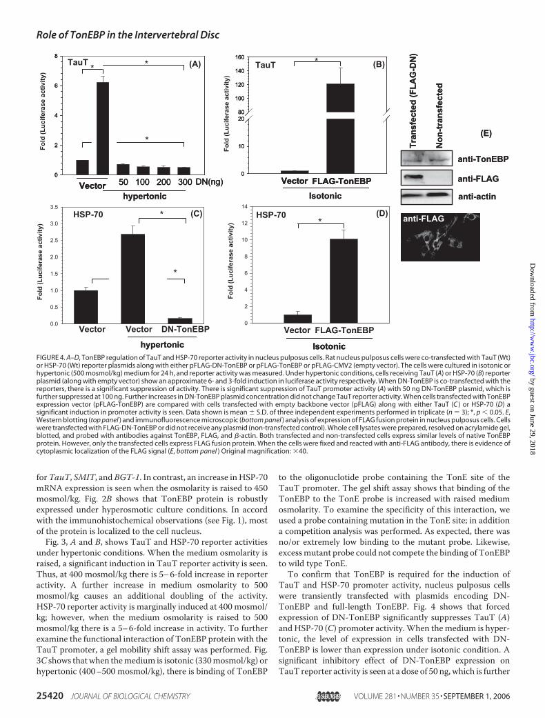
TauT and HSP-70 promoter activity, nucleus pulposus cellswere transiently transfected with plasmids encoding DN-TonEBP and full-length TonEBP. Fig. 4 shows that forcedexpression of DN-TonEBP significantly suppresses TauT (A)andHSP-70 (C) promoter activity.When themedium is hyper-tonic, the level of expression in cells transfected with DN-TonEBP is lower than expression under isotonic condition. Asignificant inhibitory effect of DN-TonEBP expression onTauT reporter activity is seen at a dose of 50 ng, which is further
FIGURE 4. A–D, TonEBP regulation of TauT and HSP-70 reporter activity in nucleus pulposus cells. Rat nucleus pulposus cells were co-transfected with TauT (Wt)or HSP-70 (Wt) reporter plasmids along with either pFLAG-DN-TonEBP or pFLAG-TonEBP or pFLAG-CMV2 (empty vector). The cells were cultured in isotonic orhypertonic (500 mosmol/kg) medium for 24 h, and reporter activity was measured. Under hypertonic conditions, cells receiving TauT (A) or HSP-70 (B) reporterplasmid (along with empty vector) show an approximate 6- and 3-fold induction in luciferase activity respectively. When DN-TonEBP is co-transfected with thereporters, there is a significant suppression of activity. There is significant suppression of TauT promoter activity (A) with 50 ng DN-TonEBP plasmid, which isfurther suppressed at 100 ng. Further increases in DN-TonEBP plasmid concentration did not change TauT reporter activity. When cells transfected with TonEBPexpression vector (pFLAG-TonEBP) are compared with cells transfected with empty backbone vector (pFLAG) along with either TauT (C ) or HSP-70 (D) asignificant induction in promoter activity is seen. Data shown is mean � S.D. of three independent experiments performed in triplicate (n � 3); *, p � 0.05. E,Western blotting (top panel ) and immunofluorescence microscopic (bottom panel ) analysis of expression of FLAG fusion protein in nucleus pulposus cells. Cellswere transfected with FLAG-DN-TonEBP or did not receive any plasmid (non-transfected control). Whole cell lysates were prepared, resolved on acrylamide gel,blotted, and probed with antibodies against TonEBP, FLAG, and �-actin. Both transfected and non-transfected cells express similar levels of native TonEBPprotein. However, only the transfected cells express FLAG fusion protein. When the cells were fixed and reacted with anti-FLAG antibody, there is evidence ofcytoplasmic localization of the FLAG signal (E, bottom panel ) Original magnification: �40.
Role of TonEBP in the Intervertebral Disc
25420 JOURNAL OF BIOLOGICAL CHEMISTRY VOLUME 281 • NUMBER 35 • SEPTEMBER 1, 2006
by guest on June 29, 2018http://w
ww
.jbc.org/D
ownloaded from

enhanced when the concentration of the DN plasmid isincreased to 100 ng. Despite further increases in the DN plas-mid dose, the reporter activity is not decreased. On the otherhand, overexpression of TonEBP under isotonic conditions,using pFLAG-TonEBPvector, results in a significant increase inboth TauT (Fig. 4B) and HSP-70 (Fig. 4D) promoter activities.To ensure that the FLAG fusion protein is expressed in nucleuspulposus cells, Western blot and immunofluorescnce analysiswas performed (Fig. 4E). As expected, using both the tech-niques, anti-FLAG antibody detects FLAG-DN-TonEBP intransfected cells only. Moreover, DN-TonEBP does not alterthe level of native TonEBP expression in transfected cells.To further explore the importance of TonEBP in regulation
of the osmotic response, we stably transfected rat nucleus pul-posus cells with TonEBP siRNA. Among different clonesscreened, one clone displayed a partial suppression of TonEBP
mRNA (Fig. 5A). Thus, there wasabout a 2-fold decrease in TonEBPmRNA expression. We confirmedthat TonEBP protein levels are lowby immunofluorescence analysis(Fig. 5, B and C). Accordingly, thefluorescence yield of the siRNA-treated cells is minimal under iso-tonic as well as hypertonic condi-tions (Fig. 5C). The silenced siRNAcells are smaller in size and exhibit aslow growth rate when comparedwith the wild type cells (data notshown). Similarly, under both iso-tonic and hypertonic conditions,the silenced cells display a lowerTauT reporter activity than controlcells (Fig. 5D).Forced expression of DN-TonEBP
in nucleus pulposus cells results in adecrease in cell viability under hyper-tonic culture conditions. Thus, whenevaluated by theMTT assay there isa 50% decrease in the number of via-ble cells (Fig. 6A). To ascertain themode of cell death, caspase-3 activ-ity was measured in the silencedcells using the fluorescent substratePhiPhiLux-G1D2. Transient expres-sion of DN-TonEBP causes a signif-icant increase in caspase-3 activa-tion under hypertonic conditions(Fig. 6, B and C).Analysis of mouse and rat aggre-
can promoter showed two con-served TonE binding sites at �912bases and�390 bases from the tran-scription start site (Fig. 7, A and B).To examine the functional interac-tion of the TonE site with TonEBPprotein, a electromobility gel shiftassay was performed using an oligo-
nucleostide probe that contained the wild type and mutantTonE site located at �912 bases in the rat aggrecan promoter.As seen in Fig. 7C, there is functional binding of TonEBP pro-tein to the TonE-containing probe. Moreover, this binding isenhancedwhen themediumosmolarity is raised to 400 and 500mosmol/kg. To examine the specificity of this binding reaction,a wild type competitor probe was included in the binding reac-tion. In the presence of excess unlabeled competitor probe,binding of TonEBP protein to TonE site is significantly reduced(Fig. 7C). Likewise, when mutant TonE probe was included inthe binding reaction, no binding to labeled probe was evident.To further explore the role of TonEBP in the regulation of the
aggrecan promoter, we analyzed promoter activity in nucleuspulposus cells transiently transfected with DN-TonEBP. Fig.7D shows that forced expression of DN-TonEBP results inapproximately a 50% reduction in aggrecan promoter activity,
FIGURE 5. Phenotype and TauT reporter activity of TonEBP silenced rat nucleus pulposus cells. A, ratnucleus pulposus cells were transfected with plasmid DNA containing siRNA sequences and G418 resistantcells were selected. RT-PCR analysis of TonEBP mRNA levels in siRNA transfected nucleus pulposus cells show a2-fold reduction in TonEBP mRNA abundance; C, control cells; M, marker. B, siRNA and control cells werecultured under isotonic and hypertonic conditions for 24 h, and TonEBP protein expression was evaluated byimmunofluorescence; nuclei were stained with propidium iodide (red signal). Note that siRNA cells (top panel )exhibit a reduction in TonEBP protein level (green fluorescence) in both isotonic (left) and hypertonic (right)medium when compared with control cells (bottom panel ) under isotonic (left) or hypertonic (right) conditionsrespectively. Note that TonEBP is mostly localized to cell nuclei. C, quantitative image analysis of TonEBPfluorescence in control (C ) and siRNA cells in isotonic and hypertonic media. Under both isotonic and hyper-tonic conditions siRNA cells exhibit significantly lower levels of fluorescence when compared with control cells.*, p � 0.05. D, relative TauT reporter activity of siRNA cells cultured in isotonic and hypertonic media. siRNA cellswere transfected with either Wt or Mt TauT reporter plasmid and luciferase activity assayed after 24 h in eitherisotonic or hypertonic (400 mosmol/kg) media. In isotonic media, basal TauT reporter activity of the siRNA cellsis reduced by approximately half. Similarly, under hypertonic conditions, when compared with control cells (C )the silenced cells exhibit a significantly lower level of luciferase activity. Data represents mean � S.D. of fourindependent experiments performed in triplicate (n � 4); *, p � 0.05.
Role of TonEBP in the Intervertebral Disc
SEPTEMBER 1, 2006 • VOLUME 281 • NUMBER 35 JOURNAL OF BIOLOGICAL CHEMISTRY 25421
by guest on June 29, 2018http://w
ww
.jbc.org/D
ownloaded from

compared with cells that receive the empty backbone plasmid(pFLAG-CMV). Similarly, nucleus pulposus cells expressingTonEBP siRNA display a marked decrease in aggrecan pro-moter activity compared with control cells under isotonicconditions.
DISCUSSION
The current study is an extension of our earlier work that wasdirected at identifying key phenotypic markers of nucleus pul-posus cells (26). Building on the observation that the cells existin a unique microenvironment, we evaluated TonEBP and itstarget gene expression in the discal tissues. At the mRNA level,therewas a robust expression of TonEBP. Although costochon-dral cartilage also expressed TonEBP mRNA, the level ofexpression was low when compared with disc or kidney. Weused immunohistochemistry to probe TonEBP proteinexpression in the disc. Not surprisingly, there was expressionof TonEBP protein in nucleus pulposus cells of both the neo-natal and adult rat discs; the protein was also expressed bycells of the annulus fibrosus. This observation was not unex-pected as the annulus, like the nucleus, is rich in proteogly-cans and hydrodynamically stressed (4). We also showedthat TonEBP positively regulates aggrecan gene expression.This latter finding is of some importance as aggrecan, amajor constituent of the nucleus pulposus binds water mol-ecules and provides resistance to applied mechanical forces.Accordingly, the expression of TonEBP permits nucleus pul-posus cells to autoregulate the osmotic environment, whileat the same time accommodating to the hyperosmotic pres-sure of the intervertebral disc.To learn if there was transcriptional activation of TonEBP,
TauT and HSP-70 reporter activity and protein-DNA bindingwas studied in nucleus pulposus cells maintained in hyperos-molarmedia. The results of this study showed that there was anactivation of both the reporters and increased binding ofTonEBP to the TonEmotif indicating that in hypertonicmedia,TonEBP was transcriptionally active in the nucleus pulposuscells. We examined the hyperosmotic reactivity of the TonEBPby transfecting cells using pDN-TonEBP or pTonEBP. Predict-ably, pDN-TonEBP suppressedTauT (11) andHSP-70 reportergene activity (12), while pTonEBP enhanced reporter geneexpression (11, 12). In a related study, we silenced TonEBP innucleus pulposus cells and evaluatedTauT reporter expression.We noted that the basal level of TauT reporter activity in thestably transfected silenced cells was reduced. Moreover, whenthe cells were maintained in hyperosmotic media, there was amarked decrease in reporter activity. It should also be notedthat the TonEBP activation is marked by translocation of theprotein to the nucleus (5, 27). In nucleus pulposus cells, therewas nuclear localization of TonEBP and induction of targetgene expression under hypertonic conditions. These observa-tions provide a basis for considering that the intervertebral disccells possess a functionally active osmolyte system that serves toadapt the nucleus pulposus, and possibly the annulus fibrosuscells, to the high osmotic pressure and possibly the gradient inosmolarity that has been reported to exist within the interver-tebral disc (28).Although a considerable number of water bindingmolecules
FIGURE 6. Osmoprotective role of TonEBP in nucleus pulposus cells. A, ratnucleus pulposus cells were transfected with DN-TonEBP and cultured underhypertonic media (500 mosmol/kg) for 24 h. Control cells received emptybackbone vector. Viability of cells was evaluated by the MTT assay. Note thatexposure to the hyperosmolar medium results in decreased viability of thecells expressing DN-TonEBP. B, to assess the mode of cell death, cells trans-fected with empty vector (FLAG-CMV) and DN-TonEBP expression vectorwere treated with the caspase-3 substrate PhiPhiLux-G1D2 in hypertonicmedium and evaluated by phase contrast (top panels) and fluorescent (bot-tom panels) microscopy. Note, a high number of DN-TonEBP-expressing cellsexhibit green fluorescence demonstrating increased activation of caspase-3,a pro-apoptotic molecule (arrow). Magnification: �20. C, quantitative imageanalysis of caspase-3 activity in nuclus pulposus cells. The figure shows thatthere is significant increase in PhiPhiLux-G1D2 fluorescence yield in cellsexpressing DN-TonEBP under hypertonic conditions. *, p � 0.05.
Role of TonEBP in the Intervertebral Disc
25422 JOURNAL OF BIOLOGICAL CHEMISTRY VOLUME 281 • NUMBER 35 • SEPTEMBER 1, 2006
by guest on June 29, 2018http://w
ww
.jbc.org/D
ownloaded from

contribute to the regulation of the osmotic pressure, we focusedon aggrecan as it is the major polyelectrolyte constituent. Thecharged COO� and SO4
2� groups of N-acetylgalactosamine,glucuronic acid, and other substituted sugars bind hydratedNa� ions thereby regulating the osmotic properties of the disc.While it is known that aggrecan transcription and osmoticpressure are linked (29, 30), details of the relationship areobscure. Promoter analysis of TonEBP provided a new insightinto this relationship. We found two TonE sites at �390 and�912 bp in the aggrecan gene promoter sequence. A similarsequence was noted in the human aggrecan promotersequence. The observation that the human sequence was at�890 bp probably reflects differences in species specific orga-nization of the aggrecan promoter sequence. The presence ofthese conserved motifs provide a direct link between aggrecanexpression and tissue osmolarity.That the aggrecan promoter was responsive to TonEBP was
evident from experiments performed using the DN-TonEBP
transfected cells. These studies revealed that forced expressionof DN-TonEBP in isotonic media resulted in partial loss ofaggrecan promoter activity. When TonEBP expression innucleus pulposus cells was partially silenced by siRNA, againthere was suppression of aggrecan promoter activity. In anongoing study, we have slowly adapted disc cells to an increasein medium osmolarity. In this case, there is continued induc-tion of aggrecan mRNA expression suggesting that there is adirect relationship between aggrecan expression and the tonic-ity of the medium. Together, these findings indicate that asidefrom Sox-9 (31) and other transcriptionally active proteins,TonEBP serves as a regulator of aggrecan expression that con-stitutes a major functionally active component of the discmatrix.The nucleus pulposus cells were probably able to survive in a
hyperosmotic medium through activation of TonEBP. In addi-tion, the expression of almost all of TonEBP target genes waselevated. BGT-1 and SMIT appeared to bemost sensitive to the
FIGURE 7. Regulation of aggrecan gene promoter activity by TonEBP. A, DNA sequence of the promoter region of rat and mouse aggrecan gene. TonEconsensus sequence is marked in bold and underlined. B, promoter organization of the rat aggrecan gene. The transcription start site is marked as �1. TonE sitesare shown as ovals on either side of a conserved Sox-9 binding site. C, electromobility shift assay to examine functional binding of TonEBP to TonE motif in therat aggrecan gene promoter. An oligonucleotide probe containing the TonE motif (�912 b) in the rat aggrecan promoter was incubated with nuclear extractsfrom rat nucleus pulposus cells cultured under isotonic and hypertonic (400 and 500 mosmol/kg) conditions, and binding was detected using chemilumines-cence. Specificity was confirmed by inclusion of excess unlabeled wild type probe or a probe containing mutation in the TonE site (Mt probe) in the bindingreaction. The binding signal is significantly diminished when either a wild type competitor probe or a mutant probe is used. D, nucleus pulposus cells wereco-transfected with DN-TonEBP and aggrecan reporter plasmids. Twenty-four hours after transfection, cells were cultured in isotonic medium for 24 h andluciferase activity measured. Expression of DN-TonEBP results in decreased aggrecan promoter activity compared with control cells that receive emptybackbone vector. E, aggrecan promoter construct was transiently transfected into siRNA expressing and control cells (C ) and reporter activity measured inisotonic media. Compared with control cells, the silenced nucleus pulposus cells elicit a marked reduction in aggrecan reporter activity. Data represent mean �S.D. from three independent experiments, performed in triplicate (n � 3); *, p � 0.05.
Role of TonEBP in the Intervertebral Disc
SEPTEMBER 1, 2006 • VOLUME 281 • NUMBER 35 JOURNAL OF BIOLOGICAL CHEMISTRY 25423
by guest on June 29, 2018http://w
ww
.jbc.org/D
ownloaded from

increased osmolarity, while at 450 mosmol/kg,HSP-70 expres-sion appeared to be further increased. This result suggests thatTonEBP can respond to hyperosmolarity by activating the tran-scription of a series of gene products that adapt the nucleuspulposus cell to changing environmental conditions. WhenTonEBP activity was inhibited using DN-TonEBP, we noted amarked decrease in cell survival. At 500 mosmol/kg, the num-ber of viable cells was reduced by 50%. Since there was activa-tion of caspase-3, it is probable that the osmotic stress stimu-lated the intrinsic apoptotic pathway. The observed increase inapoptosis is in agreement with studies of TonEBP nullmice andtransgenic animals expressing DN-TonEBP in the thymus andin the lens (16, 17, 19). In both these conditions, there was anacceleration of cell death through apoptosis. The relationshipbetween TonEBP and cell death lends strength to the ideas pre-sented here on the critical importance of this transcription fac-tor in the life history of cells of the nucleus pulposus. Aside fromits overt role in adapting cells to the microenvironment withinthe disc, it appears to play a survival role in protecting cells ofthe nucleus pulposus from the induction of apoptosis as well asregulating the expression of one and possibly more compo-nents of the extracellular matrix. Experiments are now in pro-gress to learn if fibrillar proteins are regulated by this transcrip-tion factor and whether the osmotic environment promotesinduction of water transporting proteins in the disc.
Acknowledgments—We thank Dr. Takashi Ito, Osaka University,Dr. H. M. Kwon, University of Maryland, and Dr. Ben C. Ko, Uni-versity of Hong Kong, for kindly providing reagents. We thank Dr.Theresa Freeman for technical help with quantitative imageanalysis.
REFERENCES1. Maroudas, A., Muir, H., and Wingham, J. (1969) Biochim. Biophys. Acta
177, 492–5002. Maroudas, A. (1970) Biophys. J. 10, 365–3793. Urban, J. P., Holm, S., and Maroudas, A. (1978) Biorheology 15, 203–2214. Kraemer, J., Kolditz, D., and Gowin, R. (1985) Spine 10, 69–715. Miyakawa, H., Woo, S. K., Dahl, S. C., Handler, J. S., and Kwon, H. M.
(1999) Proc. Natl. Acad. Sci. U. S. A. 96, 2538–25426. Lopez-Rodriguez, C., Aramburu, J., Rakeman, A. S., and Rao, A. (1999)
Proc. Natl. Acad. Sci. U. S. A. 96, 7214–72197. Ko, B. C., Ruepp, B., Bohren, K. M., Gabbay, K. H., and Chung, S. S. (1997)
J. Biol. Chem. 272, 16431–16437
8. Miyakawa,H.,Woo, S. K., Chen, C. P., Dahl, S. C., Handler, J. S., andKwon,H. M. (1998) Am. J. Physiol. 274, F753–F761
9. Rim, J. S., Atta, M. G., Dahl, S. C., Berry, G. T., Handler, J. S., and Kwon,H. M. (1998) J. Biol. Chem. 273, 20615–20621
10. Zhang, Z., Ferraris, J. D., Brooks, H. L., Brisc, I., and Burg, M. B. (2003)Am. J. Physiol. 285, F688–F693
11. Ito, T., Fujio, Y., Hirata, M., Takatani, T., Matsuda, T., Muraoka, S., Taka-hashi, K., and Azuma, J. (2004) Biochem. J. 382, 177–182
12. Woo, S. K., Lee, S. D., Na, K. Y., Park, W. K., and Kwon, H. M. (2002)Mol.Cell. Biol. 22, 5753–5760
13. Shim, E. H., Kim, J. I., Bang, E. S., Heo, J. S., Lee, J. S., Kim, E. Y., Lee, J. E.,Park,W. Y., Kim, S. H., Kim,H. S., Smithies, O., Jang, J. J., Jin, D. I., and Seo,J. S. (2002) EMBO Rep. 3, 857–861
14. Lopez-Rodriguez, C., Antos, C. L., Shelton, J. M., Richardson, J. A., Lin, F.,Novobrantseva, T. I., Bronson, R. T., Igarashi, P., Rao, A., and Olson, E. N.(2004) Proc. Natl. Acad. Sci. U. S. A. 101, 2392–2397
15. Lam, A. K., Ko, B. C., Tam, S., Morris, R., Yang, J. Y., Chung, S. K., andChung, S. S. (2004) J. Biol. Chem. 279, 48048–48054
16. Trama, J., Go, W. Y., and Ho, S. N. (2002) J. Immunol. 169, 5477–548817. Go,W.Y., Liu, X., Roti,M.A., Liu, F., andHo, S.N. (2004)Proc. Natl. Acad.
Sci. U. S. A. 101, 10673–1067818. Jauliac, S., Lopez-Rodriguez, C., Shaw, L. M., Brown, L. F., Rao, A., and
Toker, A. (2002) Nat. Cell Biol. 4, 540–54419. Wang, Y., Ko, B. C., Yang, J. Y., Lam, T. T., Jiang, Z., Zhang, J., Chung, S. K.,
and Chung, S. S. (2005) J. Biol. Chem. 280, 19986–1999120. Maouyo, D., Kim, J. Y., Lee, S. D., Wu, Y., Woo, S. K., and Kwon, H. M.
(2002) Am. J. Physiol. 282, F802–F80921. Risbud, M. V., Guttapalli, A., Stokes, D. G., Hawkins, D., Danielson, K. G.,
Schaer, T. P., Albert, T. J., and Shapiro, I. M. (2006) J. Cell. Biochem. 98,152–159
22. Dignam, J. D., Lebovitz, R. M., and Roeder, R. G. (1983)Nucleic Acids Res.11, 1475–1489
23. Na, K. Y., Woo, S. K., Lee, S. D., and Kwon, H. M. (2003) J. Am. Soc.Nephrol. 14, 283–288
24. Ducat, D. C., Herrera, F. J., and Triezenberg, S. J. (2003) BioTechniques 34,2, 1140–1144
25. Risbud, M. V., Fertala, J., Vresilovic, E. J., Albert, T. J., and Shapiro, I. M.(2005) Spine 30, 882–889
26. Rajpurohit, R., Risbud, M. V., Ducheyne, P., Vresilovic, E. J., and Shapiro,I. M. (2002) Cell Tissue Res. 308, 401–407
27. Ko, B. C., Turck, C. W., Lee, K. W., Yang, Y., and Chung, S. S. (2000)Biochem. Biophys. Res. Commun. 270, 52–61
28. Urban, J. P. (2002) Biochem. Soc. Trans. 30, 858–86429. Ishihara, H., Warensjo, K., Roberts, S., and Urban, J. P. (1997) Am. J.
Physiol. 272, C1499–C150630. Risbud, M. V., Di Martino, A., Guttapalli, A., Seghatoleslami, R., Denaro,
V., Vaccaro, A. R., Albert, T. J., and Shapiro, I. M. (2006) Spine 31,884–890
31. Sekiya, I., Tsuji, K., Koopman, P., Watanabe, H., Yamada, Y., Shinomiya,K., Nifuji, A., and Noda, M. (2000) J. Biol. Chem. 275, 10738–10744
Role of TonEBP in the Intervertebral Disc
25424 JOURNAL OF BIOLOGICAL CHEMISTRY VOLUME 281 • NUMBER 35 • SEPTEMBER 1, 2006
by guest on June 29, 2018http://w
ww
.jbc.org/D
ownloaded from
Irving M. Shapiro and Makarand V. RisbudTsung-Ting Tsai, Keith G. Danielson, Asha Guttapalli, Erbil Oguz, Todd J. Albert,
the Intervertebral DiscTonEBP/OREBP Is a Regulator of Nucleus Pulposus Cell Function and Survival in
doi: 10.1074/jbc.M601969200 originally published online June 13, 20062006, 281:25416-25424.J. Biol. Chem.
10.1074/jbc.M601969200Access the most updated version of this article at doi:
Alerts:
When a correction for this article is posted•
When this article is cited•
to choose from all of JBC's e-mail alertsClick here
Supplemental material:
http://www.jbc.org/content/suppl/2006/07/25/M601969200.DC1
http://www.jbc.org/content/281/35/25416.full.html#ref-list-1
This article cites 31 references, 13 of which can be accessed free at
by guest on June 29, 2018http://w
ww
.jbc.org/D
ownloaded from