Tomographic Imaging Software CCP - ccpi.ac.uk · PDF fileTomographic Imaging Software CCP ......
8
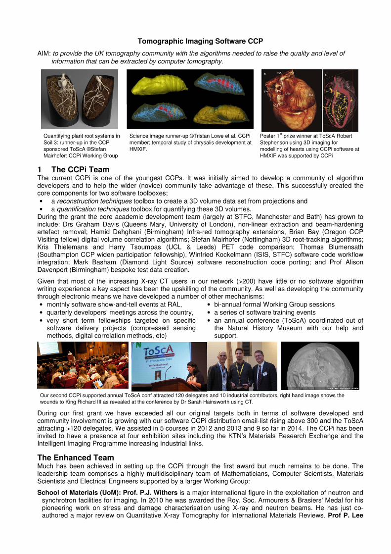
Tomographic Imaging Software CCP AIM: to provide the UK tomography community with the algorithms needed to raise the quality and level of information that can be extracted by computer tomography. Quantifying plant root systems in Soil 3: runner-up in the CCPi sponsored ToScA ©Stefan Mairhofer: CCPi Working Group Science image runner-up ©Tristan Lowe et al. CCPi member; temporal study of chrysalis development at HMXIF. Poster 1 st prize winner at ToScA Robert Stephenson using 3D imaging for modelling of hearts using CCPi software at HMXIF was supported by CCPi 1 The CCPi Team The current CCPi is one of the youngest CCPs. It was initially aimed to develop a community of algorithm developers and to help the wider (novice) community take advantage of these. This successfully created the core components for two software toolboxes; • a reconstruction techniques toolbox to create a 3D volume data set from projections and • a quantification techniques toolbox for quantifying these 3D volumes. During the grant the core academic development team (largely at STFC, Manchester and Bath) has grown to include: Drs Graham Davis (Queens Mary, University of London), non-linear extraction and beam-hardening artefact removal; Hamid Dehghani (Birmingham) Infra-red tomography extensions, Brian Bay (Oregon CCP Visiting fellow) digital volume correlation algorithms; Stefan Mairhofer (Nottingham) 3D root-tracking algorithms; Kris Thielemans and Harry Tsoumpas (UCL & Leeds) PET code comparison; Thomas Blumensath (Southampton CCP widen participation fellowship), Winfried Kockelmann (ISIS, STFC) software code workflow integration; Mark Basham (Diamond Light Source) software reconstruction code porting; and Prof Alison Davenport (Birmingham) bespoke test data creation. Given that most of the increasing X-ray CT users in our network (>200) have little or no software algorithm writing experience a key aspect has been the upskilling of the community. As well as developing the community through electronic means we have developed a number of other mechanisms: • monthly software show-and-tell events at RAL, • quarterly developers’ meetings across the country, • very short term fellowships targeted on specific software delivery projects (compressed sensing methods, digital correlation methods, etc) • bi-annual formal Working Group sessions • a series of software training events • an annual conference (ToScA) coordinated out of the Natural History Museum with our help and support. Our second CCPi supported annual ToScA conf attracted 120 delegates and 10 industrial contributors, right hand image shows the wounds to King Richard III as revealed at the conference by Dr Sarah Hainsworth using CT. During our first grant we have exceeded all our original targets both in terms of software developed and community involvement is growing with our software CCPi distribution email-list rising above 300 and the ToScA attracting >120 delegates. We assisted in 5 courses in 2012 and 2013 and 9 so far in 2014. The CCPi has been invited to have a presence at four exhibition sites including the KTN’s Materials Research Exchange and the Intelligent Imaging Programme increasing industrial links. The Enhanced Team Much has been achieved in setting up the CCPi through the first award but much remains to be done. The leadership team comprises a highly multidisciplinary team of Mathematicians, Computer Scientists, Materials Scientists and Electrical Engineers supported by a larger Working Group: School of Materials (UoM): Prof. P.J. Withers is a major international figure in the exploitation of neutron and synchrotron facilities for imaging. In 2010 he was awarded the Roy. Soc. Armourers & Brasiers' Medal for his pioneering work on stress and damage characterisation using X-ray and neutron beams. He has just co- authored a major review on Quantitative X-ray Tomography for International Materials Reviews. Prof P. Lee
Transcript of Tomographic Imaging Software CCP - ccpi.ac.uk · PDF fileTomographic Imaging Software CCP ......
Tomographic Imaging Software CCP
AIM: to provide the UK tomography community with the algorithms needed to raise the quality and level of
information that can be extracted by computer tomography.
Quantifying plant root systems in
Soil 3: runner-up in the CCPi
sponsored ToScA ©Stefan
Mairhofer: CCPi Working Group
Science image runner-up ©Tristan Lowe et al. CCPi
member; temporal study of chrysalis development at
HMXIF.
Poster 1st prize winner at ToScA Robert
Stephenson using 3D imaging for
modelling of hearts using CCPi software at
HMXIF was supported by CCPi
1 The CCPi Team The current CCPi is one of the youngest CCPs. It was initially aimed to develop a community of algorithm developers and to help the wider (novice) community take advantage of these. This successfully created the core components for two software toolboxes; • a reconstruction techniques toolbox to create a 3D volume data set from projections and • a quantification techniques toolbox for quantifying these 3D volumes.
During the grant the core academic development team (largely at STFC, Manchester and Bath) has grown to include: Drs Graham Davis (Queens Mary, University of London), non-linear extraction and beam-hardening artefact removal; Hamid Dehghani (Birmingham) Infra-red tomography extensions, Brian Bay (Oregon CCP Visiting fellow) digital volume correlation algorithms; Stefan Mairhofer (Nottingham) 3D root-tracking algorithms; Kris Thielemans and Harry Tsoumpas (UCL & Leeds) PET code comparison; Thomas Blumensath (Southampton CCP widen participation fellowship), Winfried Kockelmann (ISIS, STFC) software code workflow integration; Mark Basham (Diamond Light Source) software reconstruction code porting; and Prof Alison Davenport (Birmingham) bespoke test data creation.
Given that most of the increasing X-ray CT users in our network (>200) have little or no software algorithm writing experience a key aspect has been the upskilling of the community. As well as developing the community through electronic means we have developed a number of other mechanisms: • monthly software show-and-tell events at RAL, • quarterly developers’ meetings across the country, • very short term fellowships targeted on specific
software delivery projects (compressed sensing methods, digital correlation methods, etc)
• bi-annual formal Working Group sessions • a series of software training events • an annual conference (ToScA) coordinated out of
the Natural History Museum with our help and support.
Our second CCPi supported annual ToScA conf attracted 120 delegates and 10 industrial contributors, right hand image shows the
wounds to King Richard III as revealed at the conference by Dr Sarah Hainsworth using CT.
During our first grant we have exceeded all our original targets both in terms of software developed and community involvement is growing with our software CCPi distribution email-list rising above 300 and the ToScA attracting >120 delegates. We assisted in 5 courses in 2012 and 2013 and 9 so far in 2014. The CCPi has been invited to have a presence at four exhibition sites including the KTN’s Materials Research Exchange and the Intelligent Imaging Programme increasing industrial links.
The Enhanced Team Much has been achieved in setting up the CCPi through the first award but much remains to be done. The leadership team comprises a highly multidisciplinary team of Mathematicians, Computer Scientists, Materials Scientists and Electrical Engineers supported by a larger Working Group:
School of Materials (UoM): Prof. P.J. Withers is a major international figure in the exploitation of neutron and synchrotron facilities for imaging. In 2010 he was awarded the Roy. Soc. Armourers & Brasiers' Medal for his pioneering work on stress and damage characterisation using X-ray and neutron beams. He has just co-authored a major review on Quantitative X-ray Tomography for International Materials Reviews. Prof P. Lee

directs the UoM activity at RAL, based in the Research Complex at Harwell (RCaH) with 5 academics, 25 researchers, and 8 students (most joint appointment). Lee has just been appointed Assistant Director of the RCaH to lead EPS interaction with the other large facilities, which will help facilitate integration of the CCPi into DLS, ISIS and CLF. Taken together across Manchester and Harwell we have one of the most extensive suites of lab. x-ray scanners in Europe. The excellence, innovation and impact of our research has been recognised by the award of the most prestigious prize in the UK HE sector, the Queen's Anniversary Prize for Higher and Further Education (2012-2014) for “New Techniques in X-ray Imaging of Materials Critical for Power, Transport and Other Key Industries”, awarded for our work with over 35 Universities and 90 Industries.
School of Mathematics (UoM): Prof. W. Lionheart leads the Inverse Problems Group comprising 2 academics, 3 postdocs and 9 PhD students working on industrial, biomedical and geophysical applications of inverse problems. He received The University of Manchester's Researcher of the Year Award in 2011 in recognition of the quality of his interdisciplinary and industrially relevant research and in 2013 is the Otto Mösted Visiting Professor at the Technical University of Denmark. Projects include EP/L019108/1 Robust Repeatable Respiratory Monitoring with EIT, EP/F028695/1 Advanced Reconstruction Algorithms for PET Imaging, EP/K039865/1 Generalised Polarisation Tensors for Maxwell's Equations and industrial projects include tensor tomography for photoelasticity, biomedical impedance imaging, a scanning metal detector and an inverse problem for liquid crystals.
Electronic and Electrical Engineering (UoB): Dr M Soleimani who leads the Engineering Tomography Lab (ETL) at the University of Bath that was set up in 2011. There are also 15 PhD students and PDRAs studying inverse problems and tomography over a range of scientific fields, including medical imaging, industrial process tomography and tomography for material characterisation. Despite this wide range, many of the problems to be solved have strong mathematical similarities. Of particular value to this project, the team have wide experience in developing software for cone beam CT image reconstruction and multi-modality tomography. ETL works intensively with a wide range of UK industrial end-users and leading international groups including a multi-modality tomography project with CERN
Industry involvement is important providing guidance, advice and commercial exploitation. Key to the project is their reconstruction expertise, input through workshops and individual interactions. The following industrial companies and national organisations have all agreed to become members of the network and to support the project with advice, information about data formats and instrument configurations. Nikon Metrology is a manufacturer of industrial tomography systems; FEI is a supplier of the major commercial tomography visualisation packages Avizo and Amira; LaVision is a key supplier in quantitative image correlation software used within the community and running a KTN through the University of Southampton; Zeiss is a manufacturer of industrial tomography systems; Bruker is a manufacturer of high resolution tomography lab. and synchrotron X-ray systems; Deben is a manufacturer of in scanning electron microscope X-ray tomography systems; Simpleware is a spin out company from Exeter University. They are a world leader in writing software constructing finite element modelling meshes and models directly from tomography data. The development of discrete tomography is particularly of interest to this company; DLS Diamond Light Source are now using CCPi code within some of their beamlines. Tessella (integrated with ISIS part of STFC) an international analytics, technology and consultancy firm are working on a new instrument independent data analysis framework for use in Neutron and Muon experiments (Mantid); BSI Group is pioneering an effort with CCPi partners to create the first ISO standard for tomography measurements; Natural History Museum is coordinating a science user groups for lab based x-ray tomography applications and main organisation behind Team ToScA. The current Working group has expanded to 16 members, from 9 organisations; this has provided a strong link to the various different parts of the UK network. Extra members proposed who have agreed to support include; STFC Outreach Fellow Prof Phil Manning; BSI Group who are now developing the first UK standards for Tomography, Natural History Museum who are spearheading the management for new user communities and the Royal Microscopic Society.
The existing and a wider tomography community 20 years ago there were only a handful of experts developing x-ray tomography in the UK and the only industrial suppliers of tomography systems were medical imaging companies supplying low resolution systems. The landscape has changed dramatically with most research active Universities having at least one tomography system and scans touching the 100GB of data per tomograph. Over 300 researchers have been involved in consultations to create and maintain this CCP with momentum increasing. The initial outcomes were directed at the x-ray imaging community, but there are complementary tomographic imaging modalities (Neutron, electro-magnetic, electron microscopy, PET, etc.). Discussions with leading academics in the UK Neutron and the PET/SPECT communities have agreed to formal structures for collaboration and to allow code exchange with the ISIS Mantid project and the STIR project (http://stir.sourceforge.net/). There are now strengthening links to software from the EU COST network, (http://www.visielab.ua.ac.be/software) Enhanced X-ray Tomographic Reconstruction: Experiment, Modeling,
and Algorithms and US ANL, Advanced Photon Source; (http://sourceforge.net/projects/pytomo/)
1. Current State of the Art and Case for Support: Scientific Remit
Non-destructive 3D X-ray imaging has become an essential process in many areas of science including application to Energy, Healthcare and Security. For example it is having a dramatic impact on fields as diverse as security (e.g. baggage and body scanning at airports and screening of vehicles at ports), engineering (e.g. visualising stress corrosion cracking in nuclear plant and the degradation of fuel cells) and medicine (e.g. cancer treatment and artificial tissue engineering). RC funded synchrotron sources are rapidly increasing the numbers of x-ray imaging instruments available (the European Synchrotron Radiation Facility (ESRF) now has 10 beamlines and the Diamond Light Source (DLS) has 3 imaging beamlines and is building 2 more). Laboratory x-ray imaging facilities are becoming increasingly widespread; and spatial and temporal resolutions are increasing dramatically. This expansion is mirrored elsewhere with the global non-medical CT market now worth $150M (+$5.9B in medical CT by 2019) both expanding at 7%pa; with papers expending at an even faster rate. The UK is building its first neutron tomography beamline capable of element sensitive imaging.
In phase I we have established reconstruction and quantification toolkits. In phase II this work will continue but we will also develop a framework to such that even the novice can select different tools and undertake high level image reconstruction. At the moment only the simplest reconstruction tools are available to such users. Core support will then focus on continuing to write wrappers for the tools and then to integrate them within the framework. A series of visualisation needs surveys were carried out within the community and from this there we plan to continue or reconstruction and quantification algorithm support while adding a third aimed at the correction and calibration of raw image captured data prior to reconstruction.
1.1 Image capture: correction and calibration algorithms Pre-processing is possibly the most important stage of data-flow. Consequently, researchers and developers in the CCPi community have requested and received toolkit functionality for this stage. This will expand to include integration with the NeXus data format (HDF5 – EU project defining metadata standards for neutron, x-ray and muon science; http://www.nexusformat.org/), low noise quantitative tomography pre-processing, beam-hardening artefact reduction (key work started in QMUL), specialist light and dark field experiments including Fractal analysis of median noise artefact removal for electrical interference and ring removal artefacts from the raw input data.
For next generation of analysis these are critical; for example in the correction for beam-hardening when using a polychromatic source and integrating detectors is a vital area for development. It is necessary to use some form of a priori information in order to estimate a true reconstruction and this is often in the form of an assumption that the specimen is homogeneous in terms of both density and composition (but not structure) throughout.
1.2 Image Reconstruction algorithms We will expand our image reconstruction tools both in terms of functionality but also in their capacity to reconstruct large datasets in feasible timescale. The new software will incorporate not just time but, colour X-ray tomography by using multi-energy X-ray image reconstruction and through multimodality tomography with X-ray CT as the core imaging modality.
Multimodality: The problem of fusing images can arise in many applications where data is acquired from different imaging systems or modalities. Recent advances in medical hybrid scanners have posed new challenges in data fusion between data sets representing different characteristics of the biological materials. The measured data from hybrid scanners can be reconstructed separately and fused or partial volume corrected, or alternatively, the additional information can be embedded directly into a reconstruction process by means of a priori information. Our flagship fellow has started to develop reconstruction images that can use hybrid data, for example complementing the spatial resolution of X-ray with the sensitivity to water of neutron tomography [12]; this work is showing that by adding the supplementary information from different modality to reconstruction algorithm we can remove the need of fusing images and simplify quantification challenges. The main goal however is to transfer information from all data sets into a single domain which represents all the available data in the most complete way. Although multimodal reconstruction is currently more oriented to medical applications it can also be applied to material science problems (e.g. neutron and x-ray tomography). Multi-energy CT: The conventional reconstructed image is represented in the grayscale intensity format, however with the fast development of CT it becomes possible to perform multi-energy scans. This should eventually lead to colour imaging or high dynamic range (HDR) tomography. Similar to multi-modal reconstruction problem there is a need for novel iterative reconstruction algorithms which can process multi energy data. In material science the problem of multi energy reconstruction is even more prominent than in medical applications. The dosage limitation is not the biggest issue, however the benefit of exploring physical properties under different exposure frequencies can lead to a better material characterisation.
This project complements the phase 1 CCPi activity and extends the scope considerably further by going ‘beyond 3D’ – that is multi-modality and interfacing with computer models to reveal dynamic and chemical
behaviour. The project will expand the CCPi to cover a wider range of applications and it is anticipated that this community-based software will then stimulate extensive unexpected innovations.
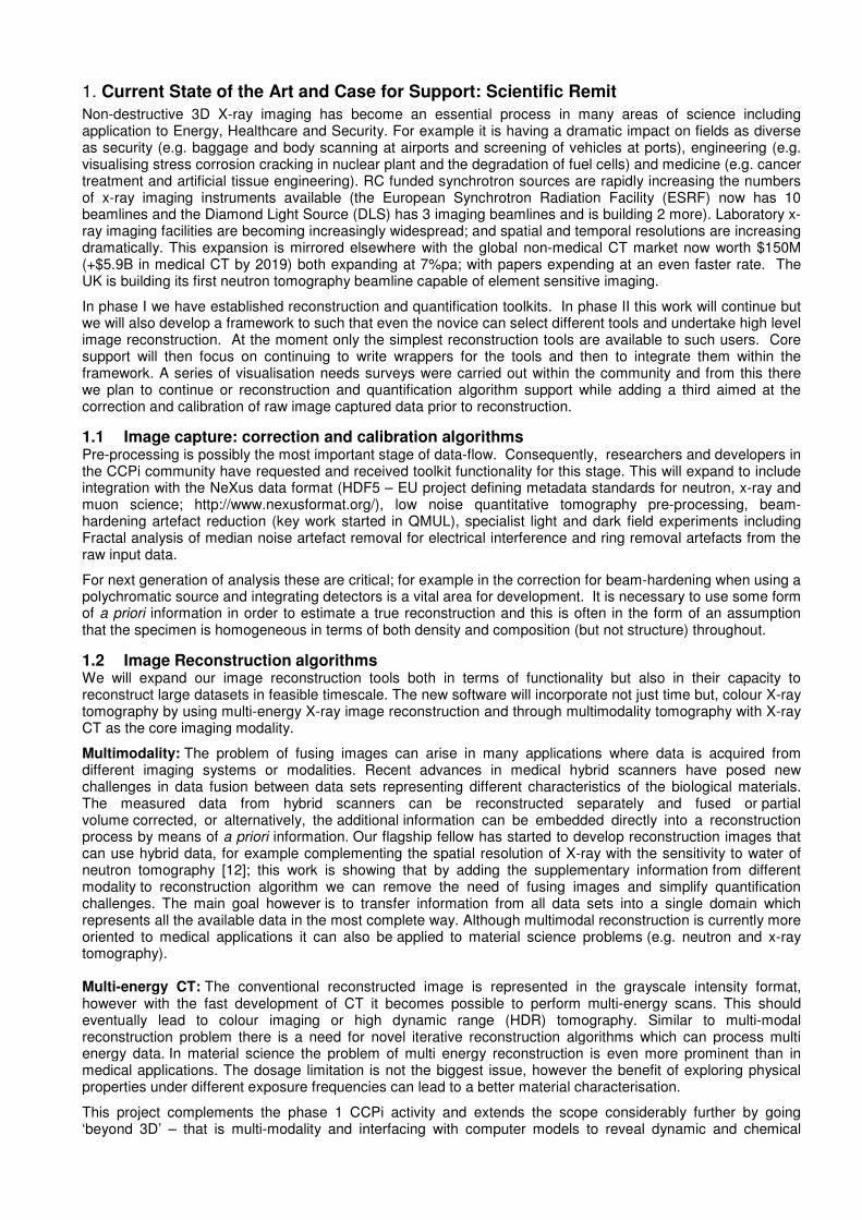
Current CCPi reconstruction software has been modified to run on high-end GPU cluster machines for Diamond, large memory versions on the supercomputing facilities at Hartee Centre in STFC (above, which can run for many hours but create higher quality results) and new releases are being made available to laboratory based workstations.
1.3 Image Quantification algorithms Quantification of images is by bespoke post-processing routines written on a case by case basis by individual researchers using various packages. Consequently, the information content is often massively under-exploited and much time is wasted as inexpert users keep reinventing the wheel. While some 3D analysis routines exist for example as part of the ITK (Image Visualisation ToolKit) and VTK (Visualisation ToolKit) projects in the US, most users are too inexpert to exploit them. Given the multi-disciplinary and diverse nature of the UK 3D imaging community (from medics to palaeontologists (biomaterials); from nanotechnologists to civil engineers (structures), from food technologists to geologists (pores)), what is needed is a supportive network providing training and a toolbox to enable greater and more accurate information to be obtained from 3D images. In summary, we need to take analysis of these complex 3D and 4D datasets beyond the current medical trend of qualitative analysis into a quantifiable form. Consultation showed this to be a major community need.
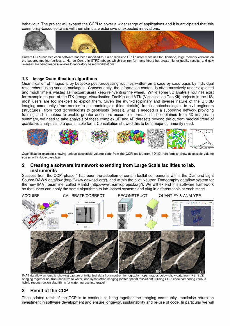
Quantification example showing unique accessible volume code from the CCPi toolkit, from 3D/4D transform to show accessible volume scales within bioactive glass.
2 Creating a software framework extending from Large Scale facilities to lab. instruments
Success from the CCPi phase 1 has been the adoption of certain toolkit components within the Diamond Light Source DAWN dataflow (http://www.dawnsci.org/), and within the pilot Neutron Tomography dataflow system for the new IMAT beamline, called Mantid (http://www.mantidproject.org/). We will extend this software framework so that users can apply the same algorithms to lab.-based systems and plug in different tools at each stage.
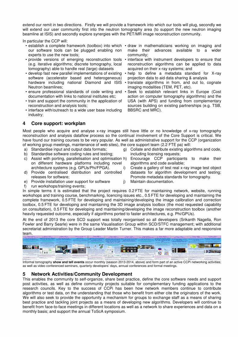
ACQUIRE CALIBRATE/CORRECT RECONSTRUCT QUANTIFY & ANALYSE
IMAT dataflow schematic showing capture of initial test data from neutron tomography (top). Images below show data from (PSI SLS) bringing together neutron (sensitive to water) and synchrotron imaging (better spatial resolution) utilising CCPi code comparing various hybrid reconstruction algorithms for water ingress into gravel.
3 Remit of the CCP
The updated remit of the CCP is to continue to bring together the imaging community, maximise return on investment in software development and ensure longevity, sustainability and re-use of code. In particular we will
extend our remit in two directions. Firstly we will provide a framework into which our tools will plug, secondly we will extend our user community first into the neutron tomography area (to support the new neutron imaging beamline at ISIS) and secondly explore synergies with the PET/MR image reconstruction community.
In particular the CCP will: • establish a complete framework (toolbox) into which
our software tools can be plugged enabling non experts to use the new tools;
• provide versions of emerging reconstruction tools (e.g. iterative algorithms; discrete tomography, local tomography) able to handle real (large) datasets
• develop fast new parallel implementations of existing software (accelerator based and heterogeneous) hardware including national Diamond and ISIS Neutron beamlines;
• ensure professional standards of code writing and documentation with links to national institutes etc;
• train and support the community in the application of reconstruction and analysis tools;
• interface with/outreach to a wide user base including industry;
• draw in mathematicians working on imaging and make their advances available to a wider community;
• interface with instrument developers to ensure that reconstruction algorithms can be applied to data acquired on their x-ray systems; and
• help to define a metadata standard for X-ray projection data to aid data sharing & analysis
• translate algorithms in from, and out to, cognate imaging modalities (TEM, PET, etc).
• Seek to establish relevant links in Europe (Cost action on computer tomography algorithms) and the USA (with APS) and funding from complementary sources building on existing partnerships (e.g. TSB, BBSRC and MRC).
4 Core support: workplan
Most people who acquire and analyse x-ray images still have little or no knowledge of x-ray tomography reconstruction and analysis dataflow process so the continual involvement of the Core Support is critical. We have found our training courses to be very popular. As well as administrative support for the CCP (organization of working group meetings, maintenance of web sites), the core support team (2.2 FTE pa) will: a) Standardise input and output data formats; b) Standardise software coding rules and testing; c) Assist with porting, parallelisation and optimisation
on different hardware platforms including novel architecture systems (e.g. GPUs/Phi/FPGA).
d) Provide centralised distribution and controlled releases for software;
e) Provide installation user support for software f) run workshops/training events;
g) Collate and distribute existing algorithms and code, including licensing requests;
h) Encourage CCP participants to make their algorithms and code available;
i) Create a gallery of test real x-ray image test object datasets for algorithm development and testing; Promote metadata standards for tomography.
j) Maintain documentation.
In simple terms it is estimated that the project requires 0.2 FTE for maintaining network, website, running workshops and training course, benchmarking, licencing issues etc., 0.5 FTE for developing and maintaining the complete framework, 0.5 FTE for developing and maintaining/developing the image calibration and correction toolbox, 0.5 FTE for developing and maintaining the 3D image analysis toolbox (the most requested capability on consultation), 0.5 FTE for developing and maintaining/developing the image reconstruction toolbox (another heavily requested outcome, especially if algorithms ported to faster architectures, e.g. Phi/GPUs).
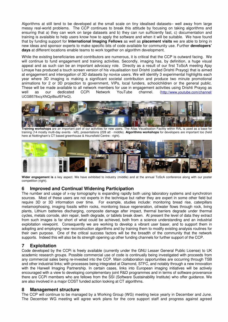
At the end of 2013 the core SCD support was totally reorganised so all developers (Srikanth Nagella, Ron Fowler and Barry Searle) were in the same Visualisation Group within SCD/STFC management; with additional secretariat administration by the Group Leader Martin Turner. This makes a far more adaptable and responsive team.
Informal tomography show and tell events occur monthly (season 2013-2014, above) and form part of an active CCPi networking activities; as well as video conferenced seminars, quarterly developers’ days, annual conferences and formal meetings.
5 Network Activities/Community Development This enables the community to self-organize, share best practice, define the core software needs and support post activities, as well as define community projects suitable for complementary funding applications to the research councils. Key to the success of CCPi has been how network members continue to contribute algorithms or test data, on the understanding that those who benefit from either cite the originators of the work. We will also seek to provide the opportunity a mechanism for groups to exchange staff as a means of sharing best practice and tackling joint projects as a means of developing new algorithms. Developers will continue to benefit from face-to-face meetings in different locations as well as a network to share experiences and data on a monthly basis; and support the annual ToScA symposium.
Algorithms at still tend to be developed at the small scale on tiny idealised datasets– well away from large messy real-world problems. The CCP continues to break this attitude by focusing on taking algorithms and ensuring that a) they can work on large datasets and b) they can run sufficiently fast, c) documentation and training is avalaible to help users know how to apply the software and when it will be suitable. We have found that by funding support for International Imaging Fellows as well as placement visits we are able to bring in new ideas and sponsor experts to make specific bits of code available for community use. Further developers’ days at different locations enable teams to work together on algorithm development.
While the existing beneficiaries and contributors are numerous, it is critical that the CCP is outward facing. We will continue to fund engagement and training activities. Secondly, imaging has, by definition, a huge visual appeal and as such can be an important advocacy role. Directly as a result of our first ToScA meeting Ajay Limaye has produced a touch screen version of his visualisation tool Drishti (called Drishti Prayog) that is aimed at engagement and interogation of 3D datasets by novice users. We will identify 3 experimental highlights each year where 3D imaging is making a significant societal contribution and produce two minute promotional animations for 2 or 3D projection to government, VIPs, local funders, schoolchildren or the general public. These will be made available to all network members for use in engagement activities using Drishti Prayog as well as our dedicated CCPi Network YouTube channel. (http://www.youtube.com/channel/
UCGB578xcyXNQyiBsufEFIeQ).
Training workshops are an important part of our activities for new users. The Atlas Visualisation Facility within RAL is used as a base for training (14 mostly multi-day events - left), presentations (228 att - middle). Algorithms workshops for developers are important too (held here at Nottingham’s CT based greenhouse & Hounsfield Centre - right).
Wider engagement is a key aspect. We have exhibited to industry (middle) and at the annual ToScA conference along with our poster competition (right).
6 Improved and Continual Widening Participation The number and usage of x-ray tomography is expanding rapidly both using laboratory systems and synchrotron sources. Most of these users are not experts in the technique but rather they are expert in some other field but require 3D or 3D information over time. For example, studies include: monitoring bread rise, caterpillars metamorphosing, imaging fossils within rocks, monitoring tissue regeneration, oil/water flows through rock, living plants, Lithium batteries discharging, composite damage after impact, thermal barriers degrade under thermal cycles, metals corrode, skin repair, teeth degrade, or tablets break down. At present the level of data they extract from such images is far short of what could be achieved, both from a science understanding and an industrial exploitation viewpoint. Consequently we are working to develop a vibrant user baser, and to support them in adopting and employing new reconstruction algorithms and by training them to modify existing analysis routines for their own purpose. One of the critical success factors will be the breadth of the community that the network supports. Indeed this will also be its strength opening up other funding channels for further support of the CCP.
7 Exploitation Code developed by the CCPi is freely available (currently under the GNU Lesser General Public License) to UK academic research groups. Possible commercial use of code is continually being investigated with proceeds from any commercial sales being re-invested into the CCP. Main collaboration opportunities are occurring through TSB and other industrial links; with processes being integrated at Diamond, STFC, and notably through a new innovation with the Harwell Imaging Partnership. In certain cases, links into European imaging initiatives will be actively encouraged with a view to developing complementary joint R&D programmes and in terms of software provenance there are CCPi members who are fellows from the SSI (Software Sustainability Institute) who offer guidance. We are also involved in a major COST funded action looking at CT algorithms.
8 Management structure The CCP will continue to be managed by a Working Group (WG) meeting twice yearly in December and June. The December WG meeting will agree work plans for the core support staff and progress against agreed

milestones that will be reviewed in the June meeting. In addition, the WG will monitor progress of any external grants as required. The CCP will continue to involve both experimental users and theoretical/computational developers on the WG to ensure that the project remains focussed on the solution of real world issues faced by end users. Membership of the WG will be open and aims to have a representative from each active imaging algorithm research group in the UK as well as the major tomographic imaging end users. Strong Industrial involvement on the working group will also continue to ensure that the CCP remains focussed on providing real impact and value for money. If more than 20 wish to join the WG, elections will be held whilst trying to maintain a spread between algorithm developers, users and industry. The Network will also seek to attract representatives from all end user communities, with the goal of leveraging the EPSRC support by developing funding applications to other bodies for exploitation including the BBSRC and MRC. The chair of the CCP will be elected by the WG and will represent the CCP on the CCP Steering Panel. The term of office of the chair will be four years. Network participants would be able to propose candidates for 1 month international fellowships, staff knowledge transfer Placements, as well as to identify Benchmark datasets and topics for the Highlights videos and touchtable interfaces development. The WG would continue to allocate these according to the likely level of benefit to the network.
9 Lower funding scenario (See justification of resources)
10 Critical success factors All the success factors from phase one have been exceeded with 11 months still to run:
• A special edition in the Royal Society Phil Trans journal looking solely at our reconstruction algorithms is in preparations and,
• 8 other direct papers. • increased network to over 85 active members
with 300+ member on email lists
• 117 attendees at day training sessions excluding week long maths mini conferences;
• Engagement with industry, were we have been attending their events as well as ours
The following factors will be critical to the next stage of the development of the CCPi: Mid-term – 2.5yrs End of grant – 5yrs
Networking User base • 300 members of network
• Expand to include neutron tomography
• 300 members of network • Devlop links with the electromagnetic and
PET imaging community membership International links • 3 International Fellows visits • 6 International Fellows Visits
• 3 International contributors to algorithms Links with industry • 5 companies attend our annual
conference/year • 5 Industrialists attend training
courses/workshops
• 5 companies attend our annual conference/year
• 10 Industrialists attend training courses/workshops
Feedback from workshops/training
• 100 different people have attended workshops/training
• 200 different people have attended training/workshops
Outreach • 2 how-to videos/webinars • 5 outreach videos for schools
available on youtube • 5 touch-screen 3D models
• 4 how-to videos/webinars • 15 outreach videos for schools available on
youtube • 10 touch-screen 3D models
Software development New scientific functionality added
• Beta version of our image acquisition and analysis workflow capable of running our tools running on synchrotron data
• 3 pre-processing analysis routines added
• 4 post-processing analysis routines added
• 2 reconstruction tools added for multi-modality
• Version 1.0 of our image acquisition and analysis workflow capable of running on synchrotron, neutron and lab CT systems.
• 5 analysis routines in toolbox • 5 post-processing analysis routines added • 4 reconstruction tools added
Ability to run on new platforms
o 2 GPU/Phi/FPGA version of codes piloted
o 4 GPU/Phi/FPGA version of codes
New algorithms � Beyond 3D – multimodal reconstruction
� Fewer projection methods
� Correlative and Diffraction Contrast Tomography
� Discrete Tomography tool with interface to CFD and finite element meshing code.
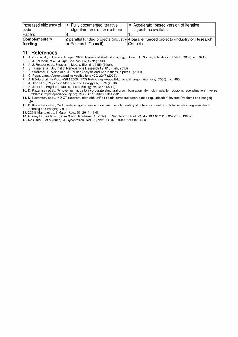
Increased efficiency of code
� Fully documented iterative algorithm for cluster systems
� Accelerator based version of iterative algorithms available
Papers 8 16 Complementary funding
2 parallel funded projects (industry or Research Council)
4 parallel funded projects (industry or Research Council)
11 References 1. J. Zhou et al., in Medical Imaging 2008: Physics of Medical Imaging, J. Hsieh, E. Samei, Eds. (Proc. of SPIE, 2008), vol. 6913. 2. S. J. LaRoque et al., J. Opt. Soc. Am. 25, 1772 (2008). 3. A. J. Reader et al., Physics in Med. & Biol. 51, 5455 (2006). 4. S. Turner et al., Journal of Nanoparticle Research 12, 615 (Feb, 2010). 5. T. Strohmer, R. Vershynin, J. Fourier Analysis and Applications In press, (2011). 6. C. Popa, Linear Algebra and its Applications 429, 2247 (2008). 7. A. Băutu et al., in Proc. ASIM 2005. (SCS Publishing House Erlangen, Erlangen, Germany, 2005), pp. 650. 8. J. Bian et al., Physics in Medicine and Biology 55, 6575 (2010). 9. X. Jia et al., Physics in Medicine and Biology 56, 3787 (2011) 10. D. Kazantsev et al., “A novel technique to incorporate structural prior information into multi-modal tomographic reconstruction” Inverse
Problems: http://iopscience.iop.org/0266-5611/30/6/065004 (2013) 11. D. Kazantsev et al., “4D-CT reconstruction with unified spatial-temporal patch-based regularization” inverse Problems and Imaging
(2014) 12. D. Kazantsev et al., “Multimodal image reconstruction using supplementary structural information in total variation regularization”
Sensing and Imaging (2014) 13. 225 E Maire, et al., I. Mater. Rev., 59 (2014), 1-43. 14. Gursoy D, De Carlo F, Xiao X and Jacobsen, C. (2014). J. Synchrotron Rad. 21, doi:10.1107/S1600577514013939 15. De Carlo F, et al.(2014). J. Synchrotron Rad. 21, doi:10.1107/S1600577514013939