Toll-like receptor 4 signaling in trigeminal ganglion neurons
10
RESEARCH Open Access Toll-like receptor 4 signaling in trigeminal ganglion neurons contributes tongue-referred pain associated with tooth pulp inflammation Kinuyo Ohara 1 , Kohei Shimizu 1,2* , Shingo Matsuura 1 , Bunnai Ogiso 1,2 , Daisuke Omagari 3 , Masatake Asano 3,4 , Yoshiyuki Tsuboi 5,6 , Masamichi Shinoda 5,6 and Koichi Iwata 5,6,7 Abstract Background: The purpose of the present study is to evaluate the mechanisms underlying tongue-referred pain associated with tooth pulp inflammation. Method: Using mechanical and temperature stimulation following dental surgery, we have demonstrated that dental inflammation and hyperalgesia correlates with increased immunohistochemical staining of neurons for TLR4 and HSP70. Results: Mechanical or heat hyperalgesia significantly enhanced in the ipsilateral tongue at 1 to 9 days after complete Freund’s adjuvant (CFA) application to the left lower molar tooth pulp compared with that of sham-treated or vehicle-applied rats. The number of fluorogold (FG)-labeled TLR4-immunoreactive (IR) cells was significantly larger in CFA-applied rats compared with sham-treated or vehicle-applied rats to the molar tooth. The number of heat shock protein (Hsp) 70-IR neurons in trigeminal ganglion (TG) was significantly increased on day 3 after CFA application compared with sham-treated or vehicle-applied rats to the molar tooth. About 9.2% of TG neurons were labeled with DiI applied to the molar tooth and FG injected into the tongue, and 15.4% of TG neurons were labeled with FG injected into the tongue and Alexa-labeled Hsp70-IR applied to the tooth. Three days after Hsp70 or lipopolysaccharide (LPS) application to the tooth in naive rats, mechanical or heat hyperalgesia was significantly enhanced compared with that of saline-applied rats. Following successive LPS-RS, an antagonist of TLR4, administration to the TG for 3 days, the enhanced mechanical or heat hyperalgesia was significantly reversed compared with that of saline-injected rats. Noxious mechanical responses of TG neurons innervating the tongue were significantly higher in CFA-applied rats compare with sham rats to the tooth. Hsp70 mRNA levels of the tooth pulp and TG were not different between CFA-applied rats and sham rats. Conclusions: The present findings indicate that Hsp70 transported from the tooth pulp to TG neurons or expressed in TG neurons is released from TG neurons innervating inflamed tooth pulp, and is taken by TG neurons innervating the tongue, suggesting that the Hsp70-TLR4 signaling in TG plays a pivotal role in tongue-referred pain associated with tooth pulp inflammation. Keywords: Tooth-pulp inflammation, Tongue-referred pain, Trigeminal ganglion, Heat shock protein * Correspondence: [email protected] 1 Department of Endodontics, Nihon University School of Dentistry, 1-8-13 Kandasurugadai, Chiyoda-ku, Tokyo 101-8310, Japan 2 Divisions of Advanced Dental Treatment, Dental Research Center, Nihon University School of Dentistry, Tokyo 101-8310, Japan Full list of author information is available at the end of the article JOURNAL OF NEUROINFLAMMATION © 2013 Ohara et al.; licensee BioMed Central Ltd. This is an open access article distributed under the terms of the Creative Commons Attribution License (http://creativecommons.org/licenses/by/2.0), which permits unrestricted use, distribution, and reproduction in any medium, provided the original work is properly cited. Ohara et al. Journal of Neuroinflammation 2013, 10:139 http://www.jneuroinflammation.com/content/10/1/139
Transcript of Toll-like receptor 4 signaling in trigeminal ganglion neurons
JOURNAL OF NEUROINFLAMMATION
Ohara et al. Journal of Neuroinflammation 2013, 10:139http://www.jneuroinflammation.com/content/10/1/139
RESEARCH Open Access
Toll-like receptor 4 signaling in trigeminalganglion neurons contributes tongue-referredpain associated with tooth pulp inflammationKinuyo Ohara1, Kohei Shimizu1,2*, Shingo Matsuura1, Bunnai Ogiso1,2, Daisuke Omagari3, Masatake Asano3,4,Yoshiyuki Tsuboi5,6, Masamichi Shinoda5,6 and Koichi Iwata5,6,7
Abstract
Background: The purpose of the present study is to evaluate the mechanisms underlying tongue-referred painassociated with tooth pulp inflammation.
Method: Using mechanical and temperature stimulation following dental surgery, we have demonstrated thatdental inflammation and hyperalgesia correlates with increased immunohistochemical staining of neurons for TLR4and HSP70.
Results: Mechanical or heat hyperalgesia significantly enhanced in the ipsilateral tongue at 1 to 9 days after completeFreund’s adjuvant (CFA) application to the left lower molar tooth pulp compared with that of sham-treated orvehicle-applied rats. The number of fluorogold (FG)-labeled TLR4-immunoreactive (IR) cells was significantly larger inCFA-applied rats compared with sham-treated or vehicle-applied rats to the molar tooth. The number of heat shockprotein (Hsp) 70-IR neurons in trigeminal ganglion (TG) was significantly increased on day 3 after CFA applicationcompared with sham-treated or vehicle-applied rats to the molar tooth. About 9.2% of TG neurons were labeled withDiI applied to the molar tooth and FG injected into the tongue, and 15.4% of TG neurons were labeled with FGinjected into the tongue and Alexa-labeled Hsp70-IR applied to the tooth. Three days after Hsp70 or lipopolysaccharide(LPS) application to the tooth in naive rats, mechanical or heat hyperalgesia was significantly enhanced compared withthat of saline-applied rats. Following successive LPS-RS, an antagonist of TLR4, administration to the TG for 3 days, theenhanced mechanical or heat hyperalgesia was significantly reversed compared with that of saline-injected rats.Noxious mechanical responses of TG neurons innervating the tongue were significantly higher in CFA-applied ratscompare with sham rats to the tooth. Hsp70 mRNA levels of the tooth pulp and TG were not different betweenCFA-applied rats and sham rats.
Conclusions: The present findings indicate that Hsp70 transported from the tooth pulp to TG neurons or expressed inTG neurons is released from TG neurons innervating inflamed tooth pulp, and is taken by TG neurons innervating thetongue, suggesting that the Hsp70-TLR4 signaling in TG plays a pivotal role in tongue-referred pain associated withtooth pulp inflammation.
Keywords: Tooth-pulp inflammation, Tongue-referred pain, Trigeminal ganglion, Heat shock protein
* Correspondence: [email protected] of Endodontics, Nihon University School of Dentistry, 1-8-13Kandasurugadai, Chiyoda-ku, Tokyo 101-8310, Japan2Divisions of Advanced Dental Treatment, Dental Research Center, NihonUniversity School of Dentistry, Tokyo 101-8310, JapanFull list of author information is available at the end of the article
© 2013 Ohara et al.; licensee BioMed Central Ltd. This is an open access article distributed under the terms of the CreativeCommons Attribution License (http://creativecommons.org/licenses/by/2.0), which permits unrestricted use, distribution, andreproduction in any medium, provided the original work is properly cited.
Ohara et al. Journal of Neuroinflammation 2013, 10:139 Page 2 of 10http://www.jneuroinflammation.com/content/10/1/139
BackgroundIt is well known that orofacial dysesthesia or referred painsometimes occurs as a secondary hyperalgesia associatedwith tooth pulpal inflammation [1]. A number of previousstudies have reported that the orofacial persistent pain fol-lowing trigeminal nerve injury or orofacial inflammation isknown to cause various motor as well as sensory disordersin the orofacial regions such as masticatory dysfunctionand/or swallowing disorder [2]. It has also been reportedthat persistent pain is sometimes developed in oralstructures following tooth pulp (TP) inflammation inhuman subjects [3]. Although sensitization of the per-ipheral nervous systems (PNS) following pulpal inflam-mation is thought to be involved in pathogenesis of thereferred pain in oral structures, the peripheral mecha-nisms underlying orofacial-referred pain associated withTP inflammation remain unclear. Since the presence oforofacial dysesthesia and referred pain often lead to ser-ious problems such as misdiagnosis and inappropriatetreatment [4], it is necessary to clarify the mechanismsunderlying orofacial dysesthesia and referred pain asso-ciated with TP inflammation.Bacterial byproducts and various chemical mediators
induced by peripheral infection or inflammation activatenociceptors in primary afferent neurons, and a barrage ofaction potentials are generated in primary afferent neuronswhich are sent to the central nervous system (CNS), result-ing in the sensitization of nociceptive neurons in the PNS[5]. Substance P or calcitonin gene-related peptide is knownto be released from trigeminal ganglion (TG) neurons fol-lowing temporomandibular joint inflammation and theseneuropeptides affect the excitability of adjacent TG neuronsinnervating the non-inflamed facial skin [6]. These findingsstrongly suggest that neuron-neuron interaction in the TGis involved in modulation of TG neurons innervating non-inflamed oral structures following TP inflammation.Toll-like receptors (TLRs) are known as transmembrane
pattern-recognition receptors that initiate signals in re-sponse to diverse pathogen-associated molecular patterns(PAMPs). After tissue injury or cellular stress, TLRs detectendogenous ligands known as danger-associated molecularpatterns (DAMPs) [7]. It is well known that TLRs in pri-mary sensory neurons, such as dorsal root ganglion (DRG)and trigeminal ganglion (TG) neurons, are involved in themodulation of neuronal excitation, in particular, primarysensory neurons expressing TLR4 and TLR7 to sense ex-ogenous PAMPs and endogenous DAMPs released aftertissue injury or cellular stress [8]. These neuronal TLRsare thought to be involved in the development of patho-logical pain following peripheral inflammation.Heat shock protein 70 (Hsp70) is well known as a spe-
cific ligand for TLRs and has been reported to be expressedin the brain [9] and heart [10], and is involved in patho-logical pain associated with tissue injury or inflammation
[11]. Previous studies have also reported that Hsp70 isexpressed in the dental pulp following pulpal trauma or in-flammation [12], therefore it is highly possible that Hsp70is involved in the development of pathological pulpal painand orofacial referred pain.Together, we hypothesized that Hsp70 was expressed in
the pulpal tissue or TG neurons after the TP inflammationand affected the excitability of TG neurons through TLR4signaling, and played a crucial role in the development oftongue-referred pain. To test this hypothesis, we analyzedmechanical- and heat-evoked nocifensive reflex, TLR4 ex-pression in TG after pulpal inflammation, primary afferenttracing using fluorogold (FG) and DiI applied into thetongue and lower first molar tooth (M1) to study if thesingle TG neuron innervates both pulp and tongue, Hsp70expression in the TG after pulpal inflammation, fluores-cent labeled-Hsp70 expression in TG neurons via axonalflow following labeled Hsp70 injection into the left M1TP,TG neuronal activity, RT-PCR analysis of Hsp70 in the in-flamed tooth pulp and TG, and the effect of TLR4 agonistand antagonist on the nocifensive reflex.
MethodsAnimalsThis study was approved by the Animal ExperimentationCommittee at Nihon University. All surgery and animalcare were conducted in accordance with the National In-stitutes of Health Guide for the Care and Use of Labora-tory Animals and the guidelines for Institutional AnimalCare, and the guidelines of the International Associationfor the Study of Pain [13]. Male Sprague–Dawley rats (n =177, Japan SLC, Shizuoka, Japan) weighing 250 to 350 gwere used in this study. The animals were maintained in atemperature-controlled room (23°C) with a 12/12-h light/dark cycle. Food and water were freely available.
CFA application to the TPRats were lightly anesthetized with 2% isoflurane (Mylan,Canonsburg, PA, USA) and then deeply anesthetized withan intraperitoneal (i.p.) application of sodium pentobar-bital (50 mg/kg; Schering Plough, Whitehouse Station, NJ,USA). Then the rats were placed on a warm mat (37°C) inthe supine position to allow for the application of CFA(Sigma-Aldrich, St. Louis, MI, USA). A total of 50% CFA(diluted in saline) or vehicle (isotonic saline) was appliedto the M1 unilaterally. The rat’s mouth was gently openedand the left M1TP was exposed by means of a low-speeddental drill with a round tungsten carbide bur under watercooling. A small piece of dental paper point (diameter,0.15 mm; length, 1.5 mm) soaked with CFA or vehicle wasapplied to the exposed M1TP. Then the exposed pulp cav-ity was sealed with dental cement.

Ohara et al. Journal of Neuroinflammation 2013, 10:139 Page 3 of 10http://www.jneuroinflammation.com/content/10/1/139
Head-withdrawal reflex threshold measurementThe head-withdrawal reflex threshold (HWT) to mechan-ical and heat stimulation of the lateral edge of tongue(3 mm posterior from tip of tongue) was measured on day3 after saline or CFA application to the tooth pulp, underlight anesthesia with 2% isoflurane (Mylan, Canonsburg,PA, USA) in oxygen. Bipolar enamel-coated stainless steelwire electrodes (Narishige, Tokyo, Japan) were placed inthe splenius capitis muscle for electromyogram (EMG) re-cording of the reflex response (inter-electrode distance, 5to 6 mm).Lower jaw was gently pulled with plastic strings and
rat’s mouth was kept open, and then mechanical stimu-lation (0 to 130 g; 10 g/s; cutoff, 130 g) was applied tothe lateral edge of the tongue ipsilateral to the CFA orvehicle application by using forceps with flat tips (4 mm2;Panlab s.l., Barcelona, Spain) in lightly anesthetized rats(n = 7 in each group). The stimulus velocity was manuallycontrolled consecutively from 0 g to threshold values at aspeed of approximately 10 g/s. The threshold intensity forevoking EMG activity by mechanical stimulation of thetongue was defined as the mechanical HWT.Heat stimulation (35-60°C; 1°C/s; cutoff, 60°C) was
also applied to the lateral edge of the tongue ipsilateralto the CFA or vehicle application by using a contact heatprobe (9 mm2; Intercross, Tokyo, Japan) in lightly anes-thetized rats (n = 7 in each group). The threshold tem-perature for evoking EMG activity by heat stimulationto the tongue was defined as the heat HWT. The mech-anical or heat stimulation was applied three times with5-min intervals, and the mean value of the HWTs wascalculated. The splenius capitis EMG was recorded fol-lowing mechanical and heat stimulation of the tongueon days 1, 3, 5, 7, 9, 11, 14, 21, 28 (days 21, 28 data notshown.). Time-course change in mean HWT value tomechanical or heat stimulation of the ipsilateral tonguewas measured in CFA, vehicle, or sham rats. The base-line HWT value to mechanical or heat stimulation wasmeasured before CFA or vehicle application, or shamtreatment of the tooth.
TLR4 immunohistochemistry in combination withFG tracerFor TLR4 immunohistochemistry in combination with FGtracer into tongue, 5.0 μL of 10% FG (Wako) dissolved insaline was applied into the lateral edge of the tongue of therats anesthetized with 2% isoflurane. On day 3 after FG ap-plication, rats were lightly anesthetized with 2% isofluraneand then deeply anesthetized with sodium pentobarbital(50 mg/kg, i.p.) for the application of CFA or vehicle intothe M1TP. On day 3 after CFA or vehicle application, ratswere transcardially perfused with saline, followed by a fixa-tive containing 4% paraformaldehyde in 0.1 M phosphatebuffer (pH 7.4) under same anesthesia used for CFA or
vehicle application (n = 5 in each group). TGs in the ipsilat-eral side to CFA, vehicle, or sham operation were dissectedout after perfusion and post-fixed in 4% PFA for 1 dayat 4°C. The specimens were then transferred to 20%sucrose (w/v) in distilled water for several days forcryopreservation, were then embedded in Tissue Tek(Sakura Finetek, Torrance, CA, USA), and stored untilcryosectioning at −20°C. TG sections of 10 μm were cut inthe horizontal plane along the longitudinal axis. Everyeighth section was thaw-mounted on MAS-GP microslideglass (Matunami, Osaka, Japan) and dried overnight atroom temperature. Four sections were chosen from eachTG in each rat and these were processed for TLR4 immu-nohistochemistry. Sections were incubated with rabbitanti-TLR4 polyclonal antibody (1:200; abcam) after dilu-tion at a concentration of 1:800 in 0.01 M PBS containing4% normal goat serum (NGS) and 0.3% Triton X-100(Sigma-Aldrich) on day 3 at 4°C. After rinsing with 0.01 MPBS, sections were incubated in Alexa Fluor 488 goat anti-rabbit IgG (1:200 in 0.01 M PBS; Invitrogen, Paisley, UK)for 2 h at room temperature. After rinsing with 0.01 MPBS, sections were cover-slipped in mounting medium(Thermo Fisher Scientific, Fremont, CA, USA) and exam-ined under a fluorescence microscope and analyzed usinga BZ-9000 system (Keyence, Osaka, Japan). No specific la-beling was observed in the absence of primary antibody.The number of TLR4-IR cells in TG in the V3 branch re-gion were analyzed and counted in each rat (SensiveMea-sure; Mitani, Fukui, Japan; n = 5 in each group). In addition,the relative numbers of them were calculated by the follow-ing formula: 100 × number of neurons for TLR4- or FG-IRcells/FG-IR cells.
Hsp70 immunohistochemistryFor Hsp70 immunohistochemistry, sections were incu-bated with rabbit anti-Hsp70 polyclonal antibody (1:200;abcam) after dilution at a concentration of 1:800 in 0.01 MPBS containing 4% NGS and 0.3% Triton X-100 (Sigma-Aldrich) on day 3 at 4°C. After rinsing with 0.01 M PBS,sections were incubated in Alexa Fluor 488 goat anti-rabbit IgG (1:200 in 0.01 M PBS; Invitrogen, Paisley, UK)for 2 h at room temperature. After rinsing with 0.01 MPBS, sections were cover-slipped in mounting medium(Thermo Fisher Scientific, Fremont, CA, USA) and exam-ined under a fluorescence microscope and analyzed usinga BZ-9000 system (Keyence, Osaka, Japan). No specific la-beling was observed in the absence of primary antibody.The number of Hsp70-IR cells in TG in the V3 branchregion were analyzed and counted in each rat (Sensive-Measure; Mitani, Fukui, Japan; n = 5 in each group). Inaddition, the relative numbers of them were calculatedby the following formula: 100 × number of Hsp70-IRcells/all TG neurons.

Ohara et al. Journal of Neuroinflammation 2013, 10:139 Page 4 of 10http://www.jneuroinflammation.com/content/10/1/139
Labeling of recombinant Hsp70The Alexa Fluor 594 labeling of Hsp70 (R&D Systems)was performed with Alexa Fluor 594 microscale proteinlabeling kit (Molecular Probes). Briefly, 50 μg of Hsp70was mixed with 5.0 μL of 1 M sodium bicarbonate andpipetted thoroughly. The sample was further mixed with2.6 μL of Alexa Fluor 594 solution and incubated for15 min at room temperature. The reaction mixture wasapplied to the spin column and unreacted dye was elimi-nated. The flowthrough was collected and the proteinconcentration was measured. The labeled protein wasaliquoted and stored at −80°C until use.
FG, DiI, and recombinant Hsp70 application to the pulpFor FG, DiI, and labeled recombinant Hsp70 applicationto the pulp, rats were deeply anesthetized with sodiumpentobarbital (50 mg/kg, i.p.) and 5.0 μL of 10% FG dis-solved in saline was applied into the lateral edge of thetongue. On day 4 after FG application, DiI (Invitrogen) sat-urated in 100% ethanol or labeled recombinant Hsp70 wasalso applied into M1 with paper point. Subsequently onday 3, rats were deeply anesthetized and perfused, and thenTG was removed and sectioned. FG- and/or recombinantHsp70-labeled cells, or FG- and/or DiI-labeled cells werestudied in TG under fluorescent microscopy, respectively.
Hsp70 or LPS application to the M1TPRats were anesthetized with sodium pentobarbital (50 mg/kg, i.p.), the recombinant Hsp70 or LPS was applied to theleft M1TP with the same procedures as CFA applicationto the M1TP. On day 3 after drug application HWT tomechanical and heat stimulation of the tongue was mea-sured. Saline was also administered as vehicle control.
LPS-RS administration into TGRats were anesthetized, and a small hole (diameter, 1 mm)was drilled in the skull above V1/V2 and V3 branch ofTG. The guide cannula was extended into the hole 9 mmbelow the skull surface into TG and was fixed to the skullwith three stainless-steel screws and dental resin, accord-ing to the method by Katagiri et al. [14]. CFA- rats wereadministered saline (0.5 μL) or TLR4 antagonist LPS-RS(0.1 mM 0.5 μL/day; Invivo Gen) dissolved in saline (n = 5in each group), and naive rats were administrated saline(0.5 μL) or LPS-RS (0.5 μL/day; Invivo Gen) dissolved insaline (n = 5 in each group) once a day into TG for 3 suc-cessive days (day 0 through day 2) (n = 5 in each group).HWTs were then measured on day 3 day under lightanesthesia with 2% isoflurane in oxygen.
Single neuron recording from TG neuronsThree days after CFA application to the M1TP or shamtreatment of the M1 teeth, rats were anesthetized withpentobarbital-Na (50 mg/kg, i.p.). Trachea and femoral
vein were cannulated for artificial respiration and intra-venous administration of drugs, and then rats were fixedin the stereotaxic flame. Brain tissue over the TG was re-moved and TG surface was exposed, and the enamel-coated tungsten electrodes were inserted into the TGand single neuronal activities were recorded. During therecording session, rats were immobilized with pancuroniumbromide (0.6 mg/kg/h, i.v. Schering-Plough, Darmstadt,Germany) and artificially ventilated, and end-tidal CO2 con-centration and body temperature were maintained at 3.5 to4.5% and at 37°C by a feedback-controlled heating blanket(Nihon koden, Tokyo, Japan). We used gentle brush andpressure stimuli as the search stimuli. When neuronal ac-tivity was obtained, brush, pressure, or noxious pinch (50 g)was applied to the receptive fields for 5 s with camel brush,brunt forceps, or small arterial clip (50 g), respectively.Spontaneous activity was recorded for 1 min before appli-cation of brush or pressure stimulus.
Real-time PCRTotal RNA was purified using an RNeasy mini kit (QIA-GEN, Tokyo, Japan). One μg of total RNA was subjectedto first-strand cDNA synthesis with Superscript III reversetranscriptase (Life Technologies, Carlsbad, CA, USA), aspreviously described [15]. Real-time PCR was performedusing LightCycler ® Nano (Roche, Tokyo, Japan) with SYBRgreen (TaKaRa, Tokyo, Japan). The Hsp70 and GAPDHprimers were purchased from TaKaRa.
Statistical analysisData were expressed as means ± SEM. Statistical analyseswere performed by student’s t-test, or one-way ANOVAor two-way repeated-measures ANOVA followed byBonferroni’s multiple comparison tests where appropri-ate. A value of P <0.05 was considered as significant.
ResultsNocifensive reflex to mechanical or heat stimulation ofthe tongueThe HWT to mechanical or heat stimulation of the ipsilat-eral tongue significantly decreased at 1 to 9 days after CFAapplication to the M1TP compared with that of sham-treated or vehicle-applied rats (mechanical, P < 0.001; heat,P < 0.001; n = 7 in each group) (Figure 1A: mechanical, B:heat). We also observed significant decrease in the head-withdrawal threshold to mechanical stimulation of thetongue in vehicle-applied rats at 1 to 9 days after vehicleapplication compared with sham rats (P < 0.001), and ondays 3 and 7 heat head-withdrawal threshold was signifi-cantly lower in vehicle-applied rats compared to sham rats(P < 0.001). Sham-treated rats did not show any changes inHWT to mechanical or heat stimulation of the tongue.
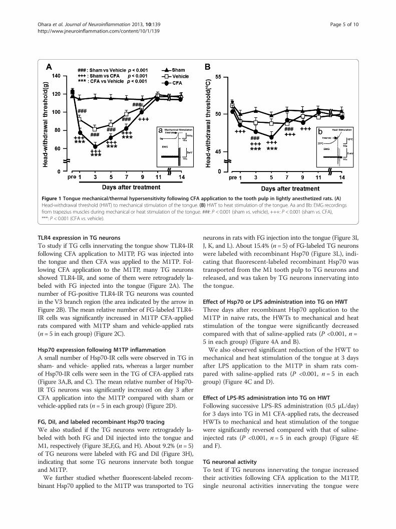
Figure 1 Tongue mechanical/thermal hypersensitivity following CFA application to the tooth pulp in lightly anesthetized rats. (A)Head-withdrawal threshold (HWT) to mechanical stimulation of the tongue. (B) HWT to heat stimulation of the tongue. Aa and Bb: EMG recordingsfrom trapezius muscles during mechanical or heat stimulation of the tongue. ###: P < 0.001 (sham vs. vehicle), +++: P < 0.001 (sham vs. CFA),***: P < 0.001 (CFA vs. vehicle).
Ohara et al. Journal of Neuroinflammation 2013, 10:139 Page 5 of 10http://www.jneuroinflammation.com/content/10/1/139
TLR4 expression in TG neuronsTo study if TG cells innervating the tongue show TLR4-IRfollowing CFA application to M1TP, FG was injected intothe tongue and then CFA was applied to the M1TP. Fol-lowing CFA application to the M1TP, many TG neuronsshowed TLR4-IR, and some of them were retrogradely la-beled with FG injected into the tongue (Figure 2A). Thenumber of FG-positive TLR4-IR TG neurons was countedin the V3 branch region (the area indicated by the arrow inFigure 2B). The mean relative number of FG-labeled TLR4-IR cells was significantly increased in M1TP CFA-appliedrats compared with M1TP sham and vehicle-applied rats(n = 5 in each group) (Figure 2C).
Hsp70 expression following M1TP inflammationA small number of Hsp70-IR cells were observed in TG insham- and vehicle- applied rats, whereas a larger numberof Hsp70-IR cells were seen in the TG of CFA-applied rats(Figure 3A,B, and C). The mean relative number of Hsp70-IR TG neurons was significantly increased on day 3 afterCFA application into the M1TP compared with sham orvehicle-applied rats (n = 5 in each group) (Figure 2D).
FG, DiI, and labeled recombinant Hsp70 tracingWe also studied if the TG neurons were retrogradely la-beled with both FG and DiI injected into the tongue andM1, respectively (Figure 3E,F,G, and H). About 9.2% (n = 5)of TG neurons were labeled with FG and DiI (Figure 3H),indicating that some TG neurons innervate both tongueand M1TP.We further studied whether fluorescent-labeled recom-
binant Hsp70 applied to the M1TP was transported to TG
neurons in rats with FG injection into the tongue (Figure 3I,J, K, and L). About 15.4% (n = 5) of FG-labeled TG neuronswere labeled with recombinant Hsp70 (Figure 3L), indi-cating that fluorescent-labeled recombinant Hsp70 wastransported from the M1 tooth pulp to TG neurons andreleased, and was taken by TG neurons innervating intothe tongue.
Effect of Hsp70 or LPS administration into TG on HWTThree days after recombinant Hsp70 application to theM1TP in naive rats, the HWTs to mechanical and heatstimulation of the tongue were significantly decreasedcompared with that of saline-applied rats (P <0.001, n =5 in each group) (Figure 4A and B).We also observed significant reduction of the HWT to
mechanical and heat stimulation of the tongue at 3 daysafter LPS application to the M1TP in sham rats com-pared with saline-applied rats (P <0.001, n = 5 in eachgroup) (Figure 4C and D).
Effect of LPS-RS administration into TG on HWTFollowing successive LPS-RS administration (0.5 μL/day)for 3 days into TG in M1 CFA-applied rats, the decreasedHWTs to mechanical and heat stimulation of the tonguewere significantly reversed compared with that of saline-injected rats (P <0.001, n = 5 in each group) (Figure 4Eand F).
TG neuronal activityTo test if TG neurons innervating the tongue increasedtheir activities following CFA application to the M1TP,single neuronal activities innervating the tongue were
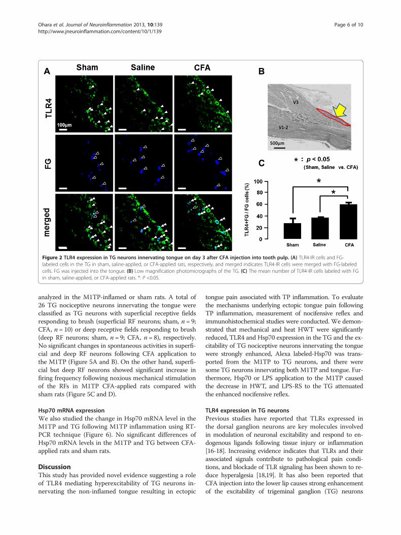
Figure 2 TLR4 expression in TG neurons innervating tongue on day 3 after CFA injection into tooth pulp. (A) TLR4-IR cells and FG-labeled cells in the TG in sham, saline-applied, or CFA-applied rats, respectively, and merged indicates TLR4-IR cells were merged with FG-labeledcells. FG was injected into the tongue. (B) Low magnification photomicrographs of the TG. (C) The mean number of TLR4-IR cells labeled with FGin sham, saline-applied, or CFA-applied rats. *: P <0.05.
Ohara et al. Journal of Neuroinflammation 2013, 10:139 Page 6 of 10http://www.jneuroinflammation.com/content/10/1/139
analyzed in the M1TP-inflamed or sham rats. A total of26 TG nociceptive neurons innervating the tongue wereclassified as TG neurons with superficial receptive fieldsresponding to brush (superficial RF neurons; sham, n = 9;CFA, n = 10) or deep receptive fields responding to brush(deep RF neurons; sham, n = 9; CFA, n = 8), respectively.No significant changes in spontaneous activities in superfi-cial and deep RF neurons following CFA application tothe M1TP (Figure 5A and B). On the other hand, superfi-cial but deep RF neurons showed significant increase infiring frequency following noxious mechanical stimulationof the RFs in M1TP CFA-applied rats compared withsham rats (Figure 5C and D).
Hsp70 mRNA expressionWe also studied the change in Hsp70 mRNA level in theM1TP and TG following M1TP inflammation using RT-PCR technique (Figure 6). No significant differences ofHsp70 mRNA levels in the M1TP and TG between CFA-applied rats and sham rats.
DiscussionThis study has provided novel evidence suggesting a roleof TLR4 mediating hyperexcitability of TG neurons in-nervating the non-inflamed tongue resulting in ectopic
tongue pain associated with TP inflammation. To evaluatethe mechanisms underlying ectopic tongue pain followingTP inflammation, measurement of nocifensive reflex andimmunohistochemical studies were conducted. We demon-strated that mechanical and heat HWT were significantlyreduced, TLR4 and Hsp70 expression in the TG and the ex-citability of TG nociceptive neurons innervating the tonguewere strongly enhanced, Alexa labeled-Hsp70 was trans-ported from the M1TP to TG neurons, and there weresome TG neurons innervating both M1TP and tongue. Fur-thermore, Hsp70 or LPS application to the M1TP causedthe decrease in HWT, and LPS-RS to the TG attenuatedthe enhanced nocifensive reflex.
TLR4 expression in TG neuronsPrevious studies have reported that TLRs expressed inthe dorsal ganglion neurons are key molecules involvedin modulation of neuronal excitability and respond to en-dogenous ligands following tissue injury or inflammation[16-18]. Increasing evidence indicates that TLRs and theirassociated signals contribute to pathological pain condi-tions, and blockade of TLR signaling has been shown to re-duce hyperalgesia [18,19]. It has also been reported thatCFA injection into the lower lip causes strong enhancementof the excitability of trigeminal ganglion (TG) neurons
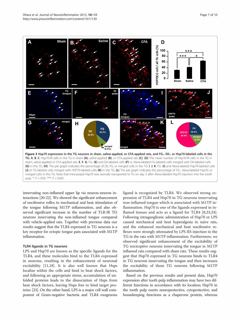
Figure 3 Hsp70 expression in the TG neurons in sham, saline-applied, or CFA-applied rats, and FG-, DiI-, or Hsp70-labeled cells in theTG. A, B, C: Hsp70-IR cells in the TG in sham (A), saline-applied (B), or CFA-applied rats (C). (D) The mean number of Hsp70-IR cells in the TG insham, saline-applied or CFA-applied rats. E, F, G: FG- (E) and DiI-labeled cells (F) or Alexa-labeled FG-labeled cells merged with DiI-labeled cells(G) in the TG. (H): The pie graph indicates the percentage of DiI, FG, or merged cells in the TG. I, J, K: FG- (I) and Alexa-labeled Hsp70-labeled cells(J) or FG-labeled cells merged with HSP70-labeled cells (K) in the TG. (L) The pie graph indicates the percentage of FG-, Alexa-labeled Hsp70, ormerged cells in the TG. Note that intra-pulpal Hsp70 was axonally transported to TG on day 3 after Alexa-labeled Hsp70 injection into the toothpulp. *: P < 0.05, ***: P < 0.001.
Ohara et al. Journal of Neuroinflammation 2013, 10:139 Page 7 of 10http://www.jneuroinflammation.com/content/10/1/139
innervating non-inflamed upper lip via neuron-neuron in-teractions [20-22]. We showed the significant enhancementof nocifensive reflex to mechanical and heat stimulation ofthe tongue following M1TP inflammation, and also ob-served significant increase in the number of TLR-IR TGneurons innervating the non-inflamed tongue comparedwith vehicle-applied rats. Together with previous data ourresults suggest that the TLR4 expressed in TG neurons is akey receptor for ectopic tongue pain associated with M1TPinflammation.
TLR4 ligands in TG neuronsLPS and Hsp70 are known as the specific ligands for theTLR4, and these molecules bind to the TLR4 expressedin neurons, resulting in the enhancement of neuronalexcitability [11,18]. It is also well known that Hspslocalize within the cells and bind to heat shock factors,and following an appropriate stress, accumulation of un-folded proteins leads to the dissociation of Hsps fromheat shock factors, leaving Hsps free to bind target pro-teins [23]. On the other hand, LPS is a major cell wall com-ponent of Gram-negative bacteria and TLR4 exogenous
ligand is recognized by TLR4. We observed strong ex-pression of TLR4 and Hsp70 in TG neurons innervatingnon-inflamed tongue which is associated with M1TP in-flammation. Hsp70 is one of the ligands expressed in in-flamed tissues and acts as a ligand for TLR4 [8,23,24].Following intraganglionic administration of Hsp70 or LPScaused mechanical and heat hyperalgesia in naive rats,and the enhanced mechanical and heat nocifensive re-flexes were strongly attenuated by LPS-RS injection to theTG in the rats with M1TP inflammation. Furthermore, weobserved significant enhancement of the excitability ofTG nociceptive neurons innervating the tongue in M1TPinflamed rats compared with sham rats. These results sug-gest that Hsp70 expressed in TG neurons binds to TLR4in TG neurons innervating the tongue and then increasesthe excitability of those TG neurons following M1TPinflammation.Based on the previous results and present data, Hsp70
expression after tooth pulp inflammation may have two dif-ferent functions in accordance with its location; Hsp70 inthe tooth pulp exerts neuroprotective, cytoprotective, andhousekeeping functions as a chaperone protein, whereas
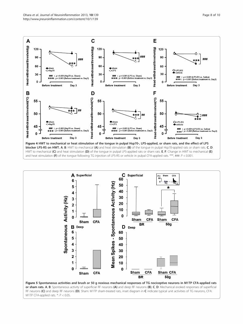
Figure 4 HWT to mechanical or heat stimulation of the tongue in pulpal Hsp70-, LPS-applied, or sham rats, and the effect of LPSblocker LPS-RS on HWT. A, B: HWT to mechanical (A) and heat stimulation (B) of the tongue in pulpal Hsp70-applied rats or sham rats. C, D:HWT to mechanical (C) and heat stimulation (D) of the tongue in pulpal LPS-applied rats or sham rats. E, F: Change in HWT to mechanical (E)and heat stimulation (F) of the tongue following TG injection of LPS-RS or vehicle in pulpal CFA-applied rats. ***, ###: P < 0.001.
Figure 5 Spontaneous activities and brush or 50 g noxious mechanical responses of TG nociceptive neurons in M1TP CFA-applied ratsor sham rats. A, B: Spontaneous activity of superficial RF neurons (A) and deep RF neurons (B), C, D: Mechanical evoked responses of superficialRF neurons (C) and deep RF neurons (D). Sham: M1TP sham-treated rats, inset diagram in C indicate typical unit activities of TG neurons, CFA:M1TP CFA-applied rats. *: P < 0.05.
Ohara et al. Journal of Neuroinflammation 2013, 10:139 Page 8 of 10http://www.jneuroinflammation.com/content/10/1/139
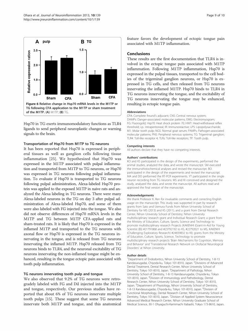
Figure 6 Relative change in Hsp70 mRNA levels in the M1TP orTG following CFA application to the M1TP or sham treatmentof the M1TP. (A) M1TP; (B) TG.
Ohara et al. Journal of Neuroinflammation 2013, 10:139 Page 9 of 10http://www.jneuroinflammation.com/content/10/1/139
Hsp70 in TG exerts immunomodulatory functions as TLR4ligands to send peripheral neuroplastic changes or warningsignals to the brain.
Transportation of Hsp70 from M1TP to TG neuronsIt has been reported that Hsp70 is expressed in periph-eral tissues as well as ganglion cells following tissueinflammation [25]. We hypothesized that Hsp70 wasexpressed in the M1TP associated with pulpal inflamma-tion and transported from M1TP to TG neurons, or Hsp70was expressed in TG neurons following pulpal inflamma-tion. To evaluate if Hsp70 is transported to TG neuronsfollowing pulpal administration, Alexa-labeled Hsp70 pro-tein was applied to the exposed M1TP in naive rats and an-alyzed the Alexa labeling in TG neurons. There were manyAlexa-labeled neurons in the TG on day 3 after pulpal ad-ministration of Alexa-labeled Hsp70, and some of themwere also labeled with FG injected into the tongue. We alsodid not observe differences of Hsp70 mRNA levels in theM1TP and TG between M1TP CFA-applied rats andsham-treated rats. It is likely that Hsp70 is expressed in theinflamed M1TP and transported to the TG neurons withaxonal flow or Hsp70 is expressed in the TG neurons in-nervating in the tongue, and is released from TG neuronsinnervating the inflamed M1TP. Hsp70 released from TGneurons binds to TLR4, and the neuronal excitability of TGneurons innervating the non-inflamed tongue might be en-hanced, resulting in the tongue ectopic pain associated withtooth pulp inflammation.
TG neurons innervating tooth pulp and tongueWe also observed that 9.2% of TG neurons were retro-gradely labeled with FG and DiI injected into the M1TPand tongue, respectively. Our previous studies have re-ported that about 6% of TG neurons innervate multipletooth pulps [15]. These suggest that some TG neuronsinnervate both M1TP and tongue, and this anatomical
feature favors the development of ectopic tongue painassociated with M1TP inflammation.
ConclusionsThese results are the first documentation that TLR4 is in-volved in the ectopic tongue pain associated with M1TPinflammation. Following M1TP inflammation, Hsp70 isexpressed in the pulpal tissues, transported to the cell bod-ies of the trigeminal ganglion neurons, or Hsp70 is ex-pressed in TG cells, and then released from TG neuronsinnervating the inflamed M1TP. Hsp70 binds to TLR4 inTG neurons innervating the tongue, and the excitability ofTG neurons innervating the tongue may be enhanced,resulting in ectopic tongue pain.
AbbreviationsCFA: Complete freund’s adjuvant; CNS: Central nervous system;DAMPs: Danger-associated molecular patterns; EMG: Electromyogram;FG: Fluorogold; Hsp70: Heat shock protein 70; HWT: Head-withdrawal reflexthreshold; i.p.: Intraperitoneal; IR: Immunoreactive; LPS: Lipopolysaccharide;M1: Molar tooth pulp; NGS: Normal goat serum; PAMPs: Pathogen-associatedmolecular patterns; PNS: Peripheral nervous systems; TG: Trigeminal ganglion;TLR4: Toll-like receptor 4; TLRs: Toll-like receptors; TP: Tooth pulp.
Competing interestsAll authors declare that they have no competing interests.
Authors’ contributionsKO and KS participated in the design of the experiments, performed theanimal studies, analyzed the data, and wrote the manuscript. SM executedthe immunohistochemical analyses and revised the manuscript. BOparticipated in the design of the experiments and revised the manuscript.MA and DO performed the RT-PCR experiments. YT participated in the singleneuron recording from TG neurons. MS and KI conceived and designed thestudy, analyzed the data, and wrote the manuscript. All authors read andapproved the final version of the manuscript.
AcknowledgementsWe thank Professor K. Ren for invaluable comments and correcting Englishusage on the manuscript. This study was supported in part by researchgrants from Sato and Uemura Funds from Nihon University School ofDentistry for MS (2013) and KS (2011), and a grant from Dental ResearchCenter, Nihon University School of Dentistry; Nihon Universitymultidisciplinary research grant and Individual Research Grant; a grant fromthe Ministry of Education, Culture, Sports, Science and Technology topromote multidisciplinary research Projects (KAKENHI (Grant-in-Aid for YoungScientist (B)) #21791868 and #23792192 to KS, #22792021 to MS, KAKENHI(Challenging Exploratory Research) #24659832 to KI); grants from the Ministryof Education, Culture, Sports, Science, Technology to promotemultidisciplinary research projects ‘Brain Mechanisms for Cognition, Memoryand Behavior’ and ‘Translational Research Network on Orofacial NeurologicalDisorders’ at Nihon University.
Author details1Department of Endodontics, Nihon University School of Dentistry, 1-8-13Kandasurugadai, Chiyoda-ku, Tokyo 101-8310, Japan. 2Divisions of AdvancedDental Treatment, Dental Research Center, Nihon University School ofDentistry, Tokyo 101-8310, Japan. 3Department of Pathology, NihonUniversity School of Dentistry, 1-8-13 Kandasurugadai, Chiyoda-ku, Tokyo101-8310, Japan. 4Division of Immunology and Pathobiology, DentalResearch Center, Nihon University School of Dentistry, Tokyo 101-8310,Japan. 5Department of Physiology, Nihon University School of Dentistry,1-8-13 Kandasurugadai, Chiyoda-ku, Tokyo 101-8310, Japan. 6Division ofFunctional Morphology, Dental Research Center, Nihon University School ofDentistry, Tokyo 101-8310, Japan. 7Division of Applied System NeuroscienceAdvanced Medical Research Center, Nihon University Graduate School ofMedical Science, 30-1 Ohyaguchi-Kamimachi Itabashi, Tokyo 173-8610, Japan.
Ohara et al. Journal of Neuroinflammation 2013, 10:139 Page 10 of 10http://www.jneuroinflammation.com/content/10/1/139
Received: 27 August 2013 Accepted: 14 November 2013Published: 23 November 2013
References1. Grushka M, Sessle BJ: Applicability of the McGill pain questionnaire to the
differentiation of ‘toothache’ pain. Pain 1984, 19:49–57.2. Ertekin C, Secil Y, Yuceyar N, Aydogdu I: Oropharyngeal dysphagia in
polymyositis/dermatomyositis. Clinical Neurol Neurosurg 2004, 107:32–37.3. Bender IB: Pulpal pain diagnosis–a review. J Endod 2000, 26:175–179.4. Farella M, Michelotti A, Gargano A, Cimino R, Ramaglia L: Myofascial pain
syndrome misdiagnosed as odontogenic pain: a case report. Cranio 2002,20:307–311.
5. Ji RR: Peripheral and central mechanisms of inflammatory pain, withemphasis on MAP kinases. Current Drug Targets Inflamm Allergy 2004,3:299–303.
6. Takeda M, Tanimoto T, Nasu M, Ikeda M, Kadoi J, Matsumoto S: Activationof NK1 receptor of trigeminal root ganglion via substance P paracrinemechanism contributes to the mechanical allodynia in thetemporomandibular joint inflammation in rats. Pain 2005, 116:375–385.
7. Okun E, Griffioen KJ, Mattson MP: Toll-like receptor signaling in neuralplasticity and disease. Trends Neurosci 2011, 34:269–281.
8. Liu T, Gao YJ, Ji RR: Emerging role of Toll-like receptors in the control ofpain and itch. Neurosci Bull 2012, 28:131–144.
9. Giffard RG, Han RQ, Emery JF, Duan M, Pittet JF: Regulation of apoptoticand inflammatory cell signaling in cerebral ischemia: the complex rolesof heat shock protein 70. Anesthesiology 2008, 109:339–348.
10. Iguchi M, Littmann AE, Chang SH, Wester LA, Knipper JS, Shields RK:Heat stress and cardiovascular, hormonal, and heat shock proteins inhumans. J Athletic Train 2012, 47:184–190.
11. Zhang Y, Wang YH, Zhang XH, Ge HY, Arendt-Nielsen L, Shao JM, Yue SW:Proteomic analysis of differential proteins related to the neuropathicpain and neuroprotection in the dorsal root ganglion following itschronic compression in rats. Exp Brain Res Exp Hirnforsc Exp Cerebr 2008,189:199–209.
12. Pileggi R, Holland GR: The expression of heat shock protein 70 in thedental pulp following trauma. Dental Traumatol 2009, 25:426–428.
13. Zimmermann M: Ethical guidelines for investigations of experimentalpain in conscious animals. Pain 1983, 16:109–110.
14. Katagiri A, Shinoda M, Honda K, Toyofuku A, Sessle BJ, Iwata K: Satellite glialcell P2Y12 receptor in the trigeminal ganglion is involved in lingualneuropathic pain mechanisms in rats. Mol Pain 2012, 8:23.
15. Omagari D, Takenouchi-Ohkubo N, Endo S, Ishigami T, Sawada A, Moro I,Asano M, Komiyama K: Nuclear factor kappa B plays a pivotal role inpolyinosinic-polycytidylic acid-induced expression of human beta-defensin2 in intestinal epithelial cells. Clinical Exp Immunol 2011, 165:85–93.
16. Diogenes A, Ferraz CC, Akopian AN, Henry MA, Hargreaves KM: LPSsensitizes TRPV1 via activation of TLR4 in trigeminal sensory neurons.J Dental Res 2011, 90:759–764.
17. Ferraz CC, Henry MA, Hargreaves KM, Diogenes A: Lipopolysaccharide fromporphyromonas gingivalis sensitizes capsaicin-sensitive nociceptors.J Endod 2011, 37:45–48.
18. Qi J, Buzas K, Fan H, Cohen JI, Wang K, Mont E, Klinman D, Oppenheim JJ,Howard OM: Painful pathways induced by TLR stimulation of dorsal rootganglion neurons. J Immunol 2011, 186:6417–6426.
19. Nicotra L, Loram LC, Watkins LR, Hutchinson MR: Toll-like receptors inchronic pain. Exp Neurol 2012, 234:316–329.
20. Hitomi S, Shinoda M, Suzuki I, Iwata K: Involvement of transient receptorpotential vanilloid 1 in ectopic pain following inferior alveolar nervetransection in rats. Neurosci Letters 2012, 513:95–99.
21. Shinoda M, Asano M, Omagari D, Honda K, Hitomi S, Katagiri A, Iwata K:Nerve growth factor contribution via transient receptor potentialvanilloid 1 to ectopic orofacial pain. J Neurosci 2011, 31:7145–7155.
22. Yasuda M, Shinoda M, Kiyomoto M, Honda K, Suzuki A, Tamagawa T, Kaji K,Kimoto S, Iwata K: P2X3 receptor mediates ectopic mechanical allodyniawith inflamed lower lip in mice. Neurosci Letters 2012, 528:67–72.
23. Kim JY, Yenari MA: The immune modulating properties of the heat shockproteins after brain injury. Anatomy Cell Biol 2013, 46:1–7.
24. Guo LH, Schluesener HJ: The innate immunity of the central nervoussystem in chronic pain: the role of toll-like receptors. Cell Mol Life Sci:CMLS 2007, 64:1128–1136.
25. Turturici G, Sconzo G, Geraci F: Hsp70 and its molecular role in nervoussystem diseases. Biochem Res Int 2011, 2011:618127.
doi:10.1186/1742-2094-10-139Cite this article as: Ohara et al.: Toll-like receptor 4 signaling intrigeminal ganglion neurons contributes tongue-referred pain associ-ated with tooth pulp inflammation. Journal of Neuroinflammation2013 10:139.
Submit your next manuscript to BioMed Centraland take full advantage of:
• Convenient online submission
• Thorough peer review
• No space constraints or color figure charges
• Immediate publication on acceptance
• Inclusion in PubMed, CAS, Scopus and Google Scholar
• Research which is freely available for redistribution
Submit your manuscript at www.biomedcentral.com/submit