Tolerance of Normal Tissue to Therapeutic Radiation Dr ... · 36 Reports of Radiotherapy and...
14
35 Spring 2013 No.1 Vol.1 Tolerance of Normal Tissue to Therapeuc Radiaon Dr Emami B Department of Radiaon Oncology, Loyola University Medical Center, Maywood, Illinois, USA Introducon Radiaon therapy is an integral part of the treatment of paents inflicted with cancer. It is esmated that over 60% of paents with cancer will have radiotherapy as part of their total course of treatment (1). Radiaon therapy affects both tumor cells and uninvolved normal cells; the former to the benefit and the later to the detriment of paents. With the goal of achieving uncomplicated local regional control of cancer, balancing between the two is both an art and a science of radiaon oncology. Unfortunately, aſter over 100 years of praccing radiaon oncology and in spite of much recent progress, knowledge on either of the two is far from perfect. From a historical point of view, the first formal aempt to address at least one of the goals, namely normal ssue tolerance to radiaon, was carried out by Rubin and Cassare (2) . Even though this publicaon was a collecon of anecdotal reports, it has served radiaon oncologists as a raw reference to build on their own experience. The decade of the 1980s was a quantum leap of progress in the field of radiaon oncology. With the monumental work of researchers on four Naonal Cancer Instute mul-instuonal contracts, the science and pracce of radiaon oncology changed from a two-dimensional (2D) to a three- dimensional (3D)/volumetric process (3) . During the work on these contracts, it became apparent to the clinicians that informaon on the tumoricidal doses of radiaon as well as normal ssue complicaon doses, especially on paral volumes, is mostly empirical and totally inadequate. A commiee was formed to address a part of this dilemma by comprehensively reviewing the available published data. In the process of this review by the commiee, it became clear that much of the data is nonexistent and they would have to rely on the collecve experience of eight clinicians from major instuons in the United States. Moreover, in order to shed some light on the volumetric aspect of these issues, it was decided that organs be divided into one-third, two-thirds, and whole organ volumes. In spite of the clear indicaon in the manuscript on the paucity of solid experimental/prospecvely driven data, this publicaon, so-called Emami’s paper, has gained much popularity. The main goal of this publicaon was to address a clinical need based on available informaon up to that me and points to the fact that there is a need for extensive and comprehensive research in this area. Obvious limitaons of the publicaon were as follows: (1) It was a literature review up to 1991. (2) It completely pre-dated the 3D-CRTIMRT- IGRT era. Even at that me dose- volume histograms were not in roune clinical use. (3) It was a tabulaon of the esmates for three of the aforemenoned arbitrary volumes (4 ) It was only for external beam radiaon with convenonal fraconaon. (5) Only one severe complicaon was chosen as an endpoint. Over the last two decades, since the publicaon of “Emami’s paper” the pracce of radiaon oncology has been completely revoluonized: 1. Muldisciplinary management of cancer has become the standard of care. 2. Choice of an endpoint for complicaon analysis and modeling has significantly altered. 3. There has been a major revoluonary change in technology: a. CT simulaon has become roune along with the fusion of other modalies such as MRI, PET, and 4DCT. b. 3D-CRT/IMRT/IGRT has become standard with the array of evaluaon tools. As a result, dose distribuons have become very complex and as of recent, the fourth dimension, namely me, has also been added to this complexity.
Transcript of Tolerance of Normal Tissue to Therapeutic Radiation Dr ... · 36 Reports of Radiotherapy and...
35Spring 2013No.1Vol.1
Tolerance of Normal Tissue to Therapeutic RadiationDr Emami BDepartment of Radiation Oncology, Loyola University Medical Center, Maywood, Illinois, USA
IntroductionRadiation therapy is an integral part of the
treatment of patients inflicted with cancer. It is estimated that over 60% of patients with cancer will have radiotherapy as part of their total course of treatment (1). Radiation therapy affects both tumor cells and uninvolved normal cells; the former to the benefit and the later to the detriment of patients. With the goal of achieving uncomplicated local regional control of cancer, balancing between the two is both an art and a science of radiation oncology. Unfortunately, after over 100 years of practicing radiation oncology and in spite of much recent progress, knowledge on either of the two is far from perfect.
From a historical point of view, the first formal attempt to address at least one of the goals, namely normal tissue tolerance to radiation, was carried out by Rubin and Cassarett (2). Even though this publication was a collection of anecdotal reports, it has served radiation oncologists as a raw reference to build on their own experience.
The decade of the 1980s was a quantum leap of progress in the field of radiation oncology. With the monumental work of researchers on four National Cancer Institute multi-institutional contracts, the science and practice of radiation oncology changed from a two-dimensional (2D) to a three-dimensional (3D)/volumetric process (3). During the work on these contracts, it became apparent to the clinicians that information on the tumoricidal doses of radiation as well as normal tissue complication doses, especially on partial volumes, is mostly empirical and totally inadequate. A committee was formed to address a part of this dilemma by comprehensively reviewing the available published data. In the process of this review by the committee, it became clear that much of the data is nonexistent and they would have to rely on the collective experience of eight clinicians from major
institutions in the United States.Moreover, in order to shed some light on the
volumetric aspect of these issues, it was decided that organs be divided into one-third, two-thirds, and whole organ volumes. In spite of the clear indication in the manuscript on the paucity of solid experimental/prospectively driven data, this publication, so-called Emami’s paper, has gained much popularity. The main goal of this publication was to address a clinical need based on available information up to that time and points to the fact that there is a need for extensive and comprehensive research in this area. Obvious limitations of the publication were as follows: (1) It was a literature review up to 1991. (2) It completely pre-dated the 3D-CRTIMRT- IGRT era. Even at that time dose-volume histograms were not in routine clinical use. (3) It was a tabulation of the estimates for three of the aforementioned arbitrary volumes (4) It was only for external beam radiation with conventional fractionation. (5) Only one severe complication was chosen as an endpoint.
Over the last two decades, since the publication of “Emami’s paper” the practice of radiation oncology has been completely revolutionized:
1. Multidisciplinary management of cancer has become the standard of care.
2. Choice of an endpoint for complication analysis and modeling has significantly altered.
3. There has been a major revolutionary change in technology:
a. CT simulation has become routine along with the fusion of other modalities such as MRI, PET, and 4DCT.
b. 3D-CRT/IMRT/IGRT has become standard with the array of evaluation tools.
As a result, dose distributions have become very complex and as of recent, the fourth dimension, namely time, has also been added to this complexity.
36 Reports of Radiotherapy and Oncology
Multiplicity and complexities of factors affecting radiation including normal tissue complications have made it impossible to have actual data for every clinical situation facing practicing radiation oncologists. Therefore, there is a need to have reasonable predictive models for plan evaluation, to improve tumor control, and to predict and hopefully prevent normal tissue injury. Optimally, databases on biophysical models should be used in summarizing complicated dose-volume data to help describe clinical outcomes and ultimately aid in the prediction of clinical toxicity.
During the last two decades, a vast amount of published information has become available to address the relationship between dosimetric
parameters and the clinical outcomes of normal tissues. Because of different analytic methodologies, calculation methods, endpoints, grading schemes, etc., the data is noisy and sifting through these data for practicing radiation oncologists is a nearly impossible task. Realizing this difficulty and the obvious need for a simplistic format, a group of physicians and researchers were formed with the name “The Quantitative Analysis of Normal Tissue Effects in the Clinic (QUANTEC).”
The first goal was to review the available literature of the last 18 years on volumetric/dosimetric information of normal tissue complication and provide a simple set of data to be used by the busy community practitioners of radiation oncology,
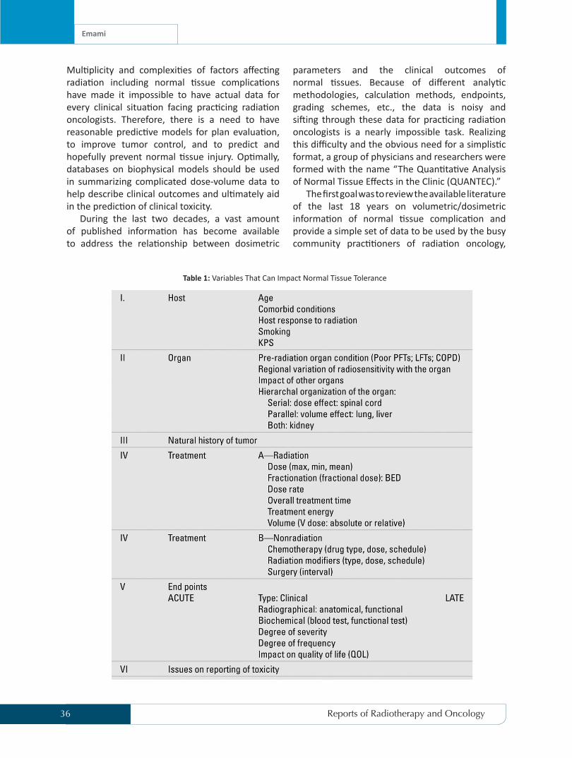
Table 1: Variables That Can Impact Normal Tissue Tolerance
Emami
37Spring 2013No.1Vol.1
physicists, and dosimetrists. The second goal of the QUANTEC group was to provide reliable predictive models on relationships between dose-volume parameters and the normal tissue complications to be utilized during the planning of radiation oncology. The result of several years of work by this group has recently been published (4–27). Although these publications contain a comprehensive review of published information and can be a guide for future research on this issue, they still have many shortcomings mainly due to the basic complexity of the subject. This shortcoming has been clearly indicated in the QUANTEC publication and the need for much more data in the future has been emphasized. However, the presented data in the publication is still cumbersome and lacks the “user-friendliness,” which is required to be used in the day-to-day practice of a busy community clinician.
As shown in Table.1 there are numerous factors that affect the radiation-induced complications of normal tissues on any given clinical situation. Thus, the experience and judgment of the clinician still plays the most important role in treating patients. As for predictive models, the problem lies in finding a reasonable model, acquiring sufficient data, and applying the statistical methods properly.
So far, in spite of major efforts, there is no model that has been demonstrated to predict radiation responses with sufficient accuracy for widespread clinical use. Most of the modeling at this point is still phenomenological and “descriptive” rather than predictive. The development of reliable and user-friendly predictive models is quite unlikely in the near future.
After reviewing the publication by the QUANTEC group, we attempt to provide the clinicians and the practitioners of radiation oncology a comprehensive but simpler, user-friendly set of data (Tables 2 and 3). It should be noted that the data is not intended to be extrapolated to pediatric patients. The data should be used only as a guide and does not substitute for a physician’s clinical judgment. We believe, as indicated in the original paper of “Emami et al.” and in the QUANTEC publication, that there is an urgent need for systematic research on this issue, which we hope will be forthcoming.
Word of Caution About BEDRecently, it has become popular (as in many
sections of QUANTEC publication) to convert the
dose-fractionation to a biological equivalent dose (BED) in order to compare various dosimetric parameters. A practical version of isoeffect formula based on the linear quadratic (LQ) model is:
D/Dref ( / + dref )/( / + d) =
The index of a/b is calculated based on information from cell survival curves that has been extrapolated and extended to human tumor and normal tissues by some computerized scientists. Unverified assignment of an a/b ratio and using it to calculate a normal tissue tolerance dose can be misleading or at least should be experimentally validated before being recommended for routine clinical use (7,9,10). The following are some basic facts based on current knowledge:
The following example depicts the basic fallacies of using BED, calculated from the above formula in clinics. Example:
If we arbitrarily choose 1 Gy/fraction/day of brain tissue, then the conversion of BED to dose/fractionation of 1 Gy/day:
Tolerance of Normal Tissue to Therapeutic Radiation

38 Reports of Radiotherapy and Oncology
In the authors’ limited informal survey, no radiation oncologists would use 90 Gy at 1 Gy/day or 51 Gy at 3 Gy/day (despite being the same BED as 60 Gy in 30 fractions using an a/b of 1), thus limiting the applicability of BED for routine clinical use. The following descriptive paragraphs of Tables 2 and 3 are presented as general guidelines.
Standard FractionationCentral Nervous SystemBrain
Radiation necrosis of the brain typically occurs 3 months to several years after radiotherapy (median 1–2 years) (3,7).
The original Emami publication estimated a 5% risk of radionecrosis at 5 years with a dose of 60 Gy to one-third of the brain with standard fractionation (3). More recently, QUANTEC conducted an extensive review of the modern literature and published new dose constraints for the brain (6,7). The review was based on a heterogeneous group of studies with varied dose and fractionation schemes. Studies were compared using the BED with an a∕b ratio of 3. A dose–response relationship was found to exist. For standard fractionation, the incidence of radionecrosis appears to be <3% for a dose of <60 Gy. The incidence increases to 5% with a dose of 72 Gy and 10% with a dose of 90 Gy. However, these doses were based on studies with widely varying parameters (target volumes, sample size, brain region, etc.).
It should be noted that an a∕b ratio of 3 is greater than the values frequently used in the literature and caution should be used when converting to BED (see above discussion). In our practice, we strive to achieve very homogeneous dose distributions with a Dmax (point dose) ≤ 65 Gy with only rare occurrences of symptomatic radiation necrosis.
Brainstem
RT-induced brainstem toxicity can be incapacitating and potentially lethal. The initial estimates by Emami et al. (3)
were of a TD 5/5 of 50 Gy to the entire brainstem and 60 Gy to one-third of the brainstem. These estimates were based on
the scant amount of data in the literature at that time and on clinical experience. The QUANTEC review identified additional modern series focusing on brainstem dose and dosevolume measures (6,9). The review included series that treated patients with photons, protons, or both. The QUANTEC review concluded that the original Emami constraint of 50 Gy was overly conservative. The entire brainstem can tolerate up to 54 Gy with a <5% risk of brainstem necrosis or neurologic toxicity. Small volumes (1–10 cc) can tolerate up to 59 Gy while a point (<<1 cc) may receive up to 64 Gy.
Spinal CordSpinal cord injury due to irradiation, though
rare, can be extremely debilitating resulting is paralysis, sensory, deficits, pain, and bowel/bladder incontinence (10,30). Schultheiss (30) published an extensive review of the literature regarding de novo irradiation of the spinal cord. Among the reviewed studies, a wide range of fractionation regimens were used (2–9 Gy/fraction). An a∕b ratio of 0.87 was estimated for the spinal cord and corresponding 2-Gy equivalent doses were calculated. The review estimated the risk of myelopathy to be 0.2% at 50 Gy and 5% at 59.3 Gy.
Similar conclusions regarding a∕b ratio and dose-volume limits were published by QUANTEC (6,10). It should be noted that an a∕b ratio of 0.87 is less than the values frequently used in the literature and caution should be used when converting to BED (see above discussion).
Chiasm and Optic NervesRadiation-induced optic neuropathy (RION)
is infrequent but usually results in rapid painless visual loss (8).
The initial Emami review listed a TD 5/5 of 50 Gy to the whole organ without partial volume tolerance data (3).
Again, this was based primarily on clinical experience and sparse published data. Many more studies are now published and were reviewed by QUANTEC (6,8). Based on the QUANTEC review, a
Emami

39Spring 2013No.1Vol.1
Table 2: Normal Tissue Tolerance for Standard Fractionation
Tolerance of Normal Tissue to Therapeutic Radiation

40 Reports of Radiotherapy and Oncology
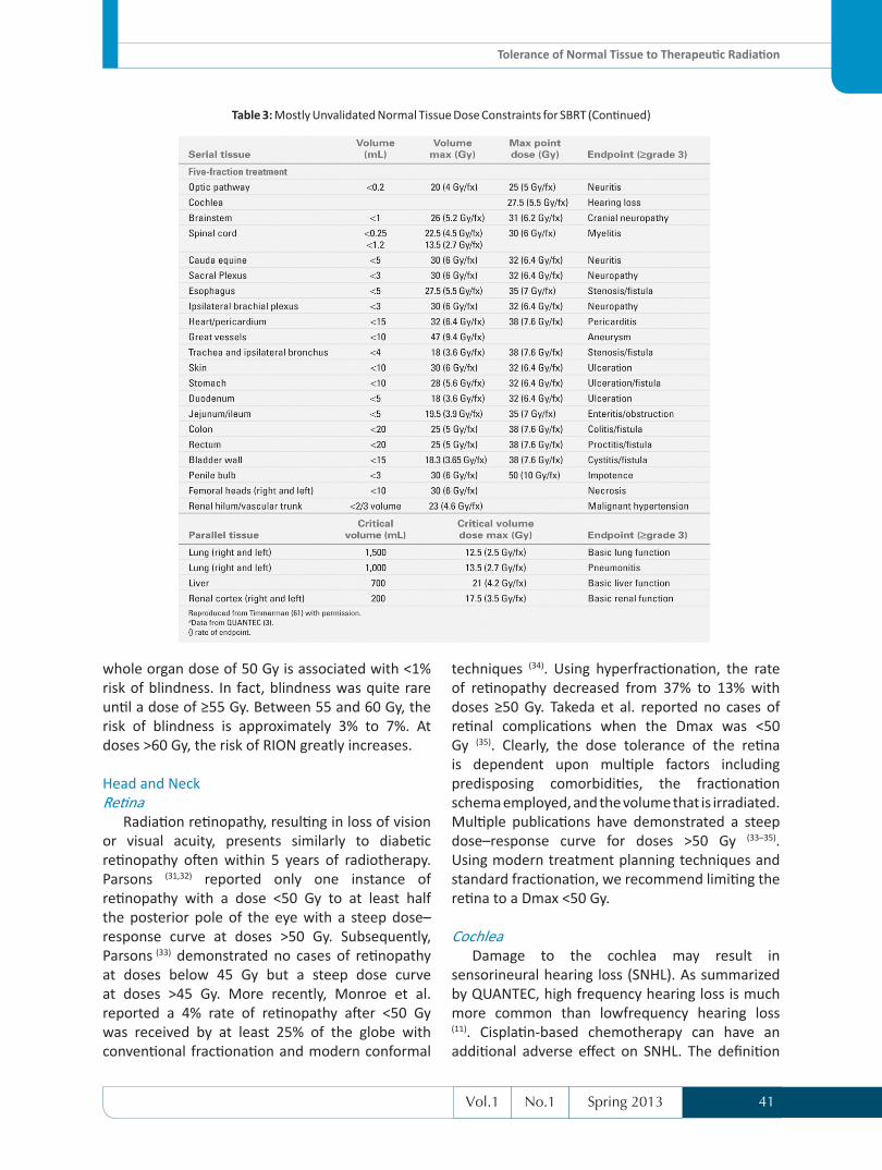
Table 3:Mostly Unvalidated Normal Tissue Dose Constraints for SBRT
Emami
41Spring 2013No.1Vol.1
whole organ dose of 50 Gy is associated with <1% risk of blindness. In fact, blindness was quite rare until a dose of ≥55 Gy. Between 55 and 60 Gy, the risk of blindness is approximately 3% to 7%. At doses >60 Gy, the risk of RION greatly increases.
Head and NeckRetina
Radiation retinopathy, resulting in loss of vision or visual acuity, presents similarly to diabetic retinopathy often within 5 years of radiotherapy. Parsons (31,32) reported only one instance of retinopathy with a dose <50 Gy to at least half the posterior pole of the eye with a steep dose–response curve at doses >50 Gy. Subsequently, Parsons (33) demonstrated no cases of retinopathy at doses below 45 Gy but a steep dose curve at doses >45 Gy. More recently, Monroe et al. reported a 4% rate of retinopathy after <50 Gy was received by at least 25% of the globe with conventional fractionation and modern conformal
techniques (34). Using hyperfractionation, the rate of retinopathy decreased from 37% to 13% with doses ≥50 Gy. Takeda et al. reported no cases of retinal complications when the Dmax was <50 Gy (35). Clearly, the dose tolerance of the retina is dependent upon multiple factors including predisposing comorbidities, the fractionation schema employed, and the volume that is irradiated. Multiple publications have demonstrated a steep dose–response curve for doses >50 Gy (33–35). Using modern treatment planning techniques and standard fractionation, we recommend limiting the retina to a Dmax <50 Gy.
CochleaDamage to the cochlea may result in
sensorineural hearing loss (SNHL). As summarized by QUANTEC, high frequency hearing loss is much more common than lowfrequency hearing loss (11). Cisplatin-based chemotherapy can have an additional adverse effect on SNHL. The definition
Table 3: Mostly Unvalidated Normal Tissue Dose Constraints for SBRT (Continued)
Tolerance of Normal Tissue to Therapeutic Radiation

42 Reports of Radiotherapy and Oncology
of clinically significant SNHL varies throughout the literature but is generally considered to be an increase in bone conduction threshold of 10 to 30 dB. The QUANTEC review examined several series and suggested a mean dose constraint of ≤45 Gy (6,11). Chan et al. conducted a longitudinal study of 87 consecutive patients treated for nasopharyngeal carcinoma, mostly treated with cisplatin-based chemoradiotherapy (36). A mean dose of ≤47 Gy to the cochlea resulted in <15% rate of SNHL. Therefore, based on a review of the literature with modern treatment planning techniques and concurrent cisplatin chemoradiotherapy we believe that a cochlear mean dose constraint of ≤45 Gy will result in a <15% rate of SNHL.
ParotidLate salivary dysfunction is a common toxicity
from radiotherapy for head and neck cancer that can take up to Late salivary dysfunction is a common toxicity from radiotherapy for head and neck cancer that can take up to 2 years to recover (37,38). Xerostomia has been widely defined in the literature from patient-reported outcomes to objective salivary flow. Quantifiably, xerostomia is defined by the LENT-SOMA scale. Grade 4 xerostomia consists of an objective reduction of ≥75% of baseline salivary function. The QUANTEC review (6,12) summarized the literature including the Washington University experience (37). Blanco demonstrated that sparing (mean dose <20 Gy) of at least one parotid gland minimized the incidence grade 4 xerostomia. Likewise, limiting both parotids to a mean dose of <25 Gy resulted in minimal grade 4 xerostomia. Dose to the parotids should be reduced as much as clinically allowable as lower mean doses generally result in better salivary function (12).
MandibleThe rates of osteoradionecrosis (ORN) of the
mandible have decreased over the past few decades (39). The risk of ORN is dependent on several factors including radiation dose, use of chemotherapy, dental hygiene, tumor location, mandibular surgery, and radiation technique (39–44). Ben-David et al. (40) demonstrated a steep dose falloff across the mandible when IMRT is employed. In their study, ≥50% of the patients in their study received ≥70 Gy to ≥1% of the mandibular volume with no cases of
grade ≥2 ORN. Additional studies, including IMRT for oral cavity cancers, demonstrate a rate of ORN near 5% (41–43). Therefore, we recommend limiting the mandible to a Dmax (point dose) ≤70 Gy when using IMRT.
Pharyngeal ConstrictorsTreatment intensification for head and neck
cancer has resulted in an increased rate of late sequela including dysphagia and aspiration. Modern treatment planning has allowed the study of various components of the swallowing apparatus. The results in the literature in this burgeoning area of research are variable as summarized in the QUANTEC review (6,13). Several groups have found the dose to the superior and/or middle pharyngeal constrictor muscles to be of paramount importance. Others have demonstrated the dose to the inferior pharyngeal constrictors or larynx to be of importance. Feng et al. (45) found no patients to have aspiration when the pharyngeal constrictors were limited to a mean dose of <60 Gy. We base our practice primarily on the findings from the University of Michigan and limit the superior pharyngeal constrictors to a mean dose of <60 Gy whenever clinically possible.
LarynxToxicity from radiotherapy to the larynx can
include vocal dysfunction and laryngeal edema. The original Emami publication (3) addressed the risk of cartilage necrosis; however, this is rarely seen in modern radiotherapy and is not as relevant of an endpoint as vocal function and laryngeal edema. The QUANTEC publication reviewed several studies involving vocal dysfunction, concluding doses to multiple structures (e.g., larynx, pharynx, and oral cavity) play an important role in voice function (6,13). Radiotherapy is commonly used for treatment of early-stage glottic cancer, employing doses >60 Gy, with a good voice outcome. A single publication (46) on laryngeal edema was reviewed, which found <20% incidence of ≥grade 2 edema when the mean laryngeal dose was <43.5 Gy and the V50 <27%.
ThoraxBrachial Plexus
Brachial plexopathy can manifest as pain, paresthesias, or motor deficits of the upper
Emami

43Spring 2013No.1Vol.1
extremity (47). Muscular atrophy and edema may develop. Emami et al. (3) suggested that the TD 5/5 to the entire brachial plexus was 60 Gy. More recently, several studies with over 20 years of followup have suggested that the incidence of brachial plexopathy continues to rise after 5 years and may not be apparent for up to 20 years after radiotherapy
(47,48). The brachial plexus appears to be especially sensitive to fractionation schedules, with the risk of injury much higher for larger fractions despite equivalent BED (49). With standard fractionation the risk of clinically apparent nerve damage seems to be <5%, after 5 years of completing radiotherapy, when the brachial plexus is limited to 60 Gy.
LungSymptomatic radiation pneumonitis (RP) is one
of the most common toxicities in patients treated with radiation for cancers of the lung, breast, and mediastinal lymphatics. The risk of RP often limits the dose delivered for treatment of these malignancies. Since the initial Emami publication (3) there has been an extensive amount of research attempting to relate many different dose-volume parameters to RP. The QUANTEC publication reviewed >70 published articles looking at both mean lung doses and Vx parameters (6,14). This comprehensive review demonstrated no clear threshold dose for symptomatic RP. The compiled data showed a mean dose–response curve with a 20% risk of RP for a mean lung dose of 20 Gy. In addition, multiple Vx values have been investigated for predicting RP but the data are not as consistent as the data for mean lung doses. Using 3D techniques, Graham found the V20 to be the most useful parameter for predicting the risk of RP (50). When Vx values are used, the V20 is the most commonly incorporated parameter.
EsophagusAcute esophagitis commonly occurs during
radiotherapy for thoracic malignancies and can lead to hospitalizations, procedures, and treatment breaks (16). Most series in the literature report rates of RTOG grade ≥2 esophagitis. The QUANTEC review summarized 11 studies that used 3D treatment planning (6,16). A single best parameter was not identified due to the diverse range of dose-volume metrics that correlated with acute esophagitis (51–53). As demonstrated in the
QUANTEC publication, there appears to be a trend demonstrating increased rates of acute esophagitis for volumes receiving >40 to 50 Gy. Currently, the ongoing RTOG 0617 is collecting V60 data on all patients and recommends keeping the mean dose <34 Gy (54).
HeartClinical pericarditis and long-term cardiac
mortality are the two most relevant cardiac toxicities. Since the original Emami publication (3), there remains a paucity of data reporting rates of pericarditis with dose-volume parameters. Indeed, several current RTOG protocols continue to use constraints similar to the original Emami TD 5/5 dose-volume estimates for the heart (54–56). As reviewed by QUANTEC (6,15), two esophageal cancer studies (57,58) assessed 3D-derived data with both studies demonstrating a rate of pericarditis <15% when the mean pericardial dose was <26 Gy. In addition, Wei found the pericardial V30 <46% to be significant on multivariate analysis. Long-term cardiac mortality has been demonstrated in multiple studies, most commonly in the treatment of breast cancer and Hodgkin’s lymphoma (15). A joint analysis of the Hodgkin’s and breast cancer data (59,60), summarized by QUANTEC, produced a dose– response curve for cardiac mortality. QUANTEC proposed a conservative approach, predicting that a V25 <10% of the heart will be associated with a <1% probability of cardiac mortality at 15 years after radiotherapy.
AbdomenLiver
Radiation-induced liver disease (RILD) typically occurs between 2 weeks and 3 months after radiotherapy. Preexisting liver disease may render patients more susceptible to RILD (17). The findings by QUANTEC (6,17) are very similar to the original estimates by Emami et al. (3), suggesting a <5% rate of RILD when the mean liver dose is ≤30 Gy in patients without preexisting liver disease or primary liver cancer. The mean liver dose should be ≤28 Gy in those patients with preexisting liver disease.
KidneyRadiation-induced renal dysfunction can be
expressed in various ways including symptomatic
Tolerance of Normal Tissue to Therapeutic Radiation
44 Reports of Radiotherapy and Oncology
expression, biochemical changes, or radiologic findings. As summarized by QUANTEC, a wide array of endpoints has been used in the literature from a decrease in creatinine clearance to renal failure (6,19). For bilateral whole kidney irradiation, a pooled analysis by Cassady (61) concluded a mean dose of 18 Gy corresponded to a 5% risk of injury at 5 years. For bilateral partial kidney irradiation, the data is less clear with a multitude of dose-volume metrics studied by several investigators (19). Small volumes of the kidney can tolerate relatively high doses of radiation. QUANTEC estimated a <5% risk of injury when the mean kidney dose is limited to <18 Gy. In addition, the current common practice of limiting the equivalent of one kidney to <20 Gy seems to be reasonable and is frequently used in our practice.
StomachLate radiation-induced toxicity to the stomach
can include dyspepsia and ulceration. Since the original Emami publication (3), few studies have reported severe RT-related gastric toxicity. The QUANTEC publication reviewed these studies, primarily pancreatic cancer trials, and concluded that a whole organ dose of 50 Gy has been associated with a 2% to 6% risk of severe late toxicity (6,18) (similar to Emami et al.).
Small BowelSmall-bowel toxicity can be greatly affected
by the use of concurrent chemotherapy and prior abdominal surgery. In particular, concurrent chemotherapy can impact the rates of acute small-bowel toxicity. Modern series employing 3D-conformal RT or IMRT have demonstrated that the volume of small bowel receiving relatively low doses of radiation plays a significant role in the rate of acute toxicity (18). When contouring individual bowel loops, the most robust dose-volume metric is the V15. The rate of grade ≥3 acute toxicity is <10% when the V15 <120 cc (62,63). When the entire potential space within the peritoneal cavity is contoured, a V45 <195 cc results in <10% acute toxicity (64). Late small-bowel toxicity, consisting of obstruction or perforation, can be influenced by prior abdominal surgery. Modern series reviewed by QUANTEC generally confirm the Emami et al. (3)
TD5/5 estimate for partial organ irradiation (6,18). In practice, we limit the volume of the small bowel
receiving 50 Gy to much less than one-third. We generally limit the V50 <5% based on the clinical scenario.
PelvisRectum
The treatment of prostate cancer has evolved such that the
great majority of patients will be alive for many years after radiotherapy. Late rectal toxicities from radiotherapy can significantly impact quality of life (QOL). Since Emami et al. (3), numerous studies have employed dose escalation using 3D-CRT or IMRT for the treatment of prostate cancer. These trials have resulted in the publication of many dose-volume analyses as summarized by the QUANTEC review (6,21). The dose-volume results are surprisingly consistent suggesting that high doses are most important in determining risk of toxicity.
BladderThe bladder frequently receives radiation
during the treatment of commonly encountered pelvic malignancies such as prostate, cervical, and bladder cancer. Due to the distensibility of the bladder it is difficult to conduct robust dose-volume analyses. The QUANTEC publication was unable to find any reliable data for partial bladder volume constraints in the treatment of prostate cancer and recommended using RTOG 0415 dose limits (6,20,65). In the treatment of bladder cancer, where the entire organ is targeted, rates of severe late bladder toxicity are varied (20). Shipley et al. (66) reported the pooled results of multiple RTOG trials demonstrating a grade ≥3 toxicity rate of ≤6% when treating the bladder to a dose of 64 to 65 Gy.
Penile BulbErectile dysfunction can have a significant
detrimental effect on QOL after treatment for prostate cancer. QUANTEC summarized the published studies correlating the dose and volume of the penile bulb that is irradiated with rates of erectile dysfunction (22). The results for various dosevolume parameters are conflicting. There is some data to support limiting the D70 <70 Gy and D90 <50 Gy. However, the strongest data supports the recommendation of limiting the penile bulb to a mean dose of <52 Gy without compromising target coverage (67).
Emami

45Spring 2013No.1Vol.1
Femoral HeadToxicity of radiation treatment to the pelvis
includes femoral head necrosis, femoral neck fracture, or long-term sequela resulting in hip replacement surgery. Besides radiation dose and volume, additional risk factors may include preexisting osteoporosis/osteopenia and androgen deprivation therapy (68–70). Emami et al. (3) suggested a TD 5/5 of 52 Gy to the whole femoral head. Grigsby et al. published the Washington University experience and documented a 4.8% incidence of femoral neck fracture following groin irradiation (68). Of note, there was only one case of femoral neck fracture when the whole femoral neck received ≤50 Gy. There is0 little data describing femoral toxicity when higher doses are delivered to small volumes of the femoral head or neck (71–74). We generally limit the entire femoral head to <50 Gy in an attempt to limit femoral head/neck toxicity to <<5%.
HypofractionationSome of the earliest radiotherapy treatments
were delivered in a hypofractionated fashion. As technology and radiobiology advanced, protracted fractionation schemes became the norm. Eventually, hypofractionation was again pursued and used to treat intracranial lesions. Stereotactic radiosurgery (SRS) has been used for decades and its success led to the use of hypofractionated treatment outside the brain. Over the past 15 years, the use of stereotactic body radiotherapy (SBRT) has become widespread and utilized to treat a number of cancers. The QUANTEC group reviewed the literature pertaining to SRS and published tolerance doses for some CNS organs at risk (6–11). The most comprehensive review to date was published by Timmerman (75). Both intracranial and extracranial organ tolerances were reviewed and adjusted for either single-fraction, three-fraction, or five-fraction treatments. Because the data in this burgeoning modality is relatively limited, the dose constraints are not validated. Rather, they are based on a combination of published data, clinical observations, modeling, and educated guessing. Despite these caveats, the dose constraints published by Timmerman provide an excellent starting point for clinical use.
ConclusionFrom the pioneering work of Rubin and
Cassarett, to the monumental work by Emami et al. and now the exhaustive review by QUANTEC, great progress has been made in the field of normal tissue tolerance to therapeutic radiation. Despite these efforts, many questions still remain. Normal tissue tolerance is an extremely complex issue and multifactorial in nature. There continues to be an urgent need for comprehensive and collaborative research. The dose-volume parameters within this chapter should be used only as a guide. For instance, there are clinical scenarios where a 5% rate of a particular toxicity is unacceptable. In contrast, there may be cases where one is willing to accept the risk a 20% rate of a particular side effect in order to obtain a desired clinical outcome. Therefore, it is imperative that the clinical judgment of the treating physician prevails in the treatment decision-making process.
References1. Halperin EC, Perez CA, Brady, LW. Preface to the
first edition. In: Halperin EC, Perez CA, Brady LW, eds. Perez and Brady’s principles and practice of radiation oncology, 5th ed. Philadelphia, PA: Lippincott Williams & Wilkins, 2008:xxi.
2. Rubin P, Cassarett G. A direction for clinical radiation pathology. In: Vaeth JM, et al., eds. Frontiers of radiation therapy and oncology VI. Baltimore, MD: University Park Press, 1972:1–16.
3. Emami B, Lyman J, Brown A, et al. Tolerance of normal tissue to therapeutic irradiation. Int J Radiat Oncol Biol Phys 1991;21(1):109–122.
4. Marks LB, Ten Haken RK, Martel MK. Guest editor’s introduction to QUANTEC: a users guide. Int J Radiat Oncol Biol Phys 2010;76(3 Suppl):S1–S2.
5. Bentzen SM, Constine LS, Deasy JO, et al. Quantitative Analyses of Normal Tissue Effects in the Clinic (QUANTEC): an introduction to the scientific issues. Int J Radiat Oncol Biol Phys 2010;76(3 Suppl):S3–S9.
6. Marks LB, Yorke ED, Jackson A, et al. Use of normal tissue complication probability models in the clinic. Int J Radiat Oncol Biol Phys 2010;76(3 Suppl):S10–S19.
7. Lawrence YR, Li XA, el Naqa I, et al. Radiation dosevolume effects in the brain. Int J Radiat Oncol Biol Phys 2010;76(3 Suppl):S20–S27.
8. Mayo C, Martel MK, Marks LB, et al. Radiation dosevolume effects of optic nerves and chiasm. Int
Tolerance of Normal Tissue to Therapeutic Radiation

46 Reports of Radiotherapy and Oncology
J Radiat Oncol Biol Phys 2010;76(3 Suppl):S28–S35.9. Mayo C, Yorke E, Merchant TE. Radiation associated
brainstem injury. Int J Radiat Oncol Biol Phys 2010;76 (3 Suppl):S36–S41.
10. Kirkpatrick JP, Van Der Kogel AJ, Schultheiss TE. Radiation dose-volume effects in the spinal cord. Int J Radiat Oncol Biol Phys 2010;76(3 Suppl):S42–S49.
11. Bhandare N, Jackson A, Eisbruch A, et al. Radiation therapy and hearing loss. Int J Radiat Oncol Biol Phys 2010; 76(3 Suppl):S50–S57.
12. Deasy JO, Moiseenko V, Marks L, et al. Radiotherapy dose-volume effects on salivary gland function. Int J Radiat Oncol Biol Phys 2010;76(3 Suppl):S58–S63.
13. Rancati T, Schwarz M, Allen AM, et al. Radiation dosevolume effects in the larynx and pharynx. Int J Radiat Oncol Biol Phys 2010;76(3 Suppl):S64–S69.
14. Marks LB, Bentzen SM, Deasy JO, et al. Radiation dosevolume effects in the lung. Int J Radiat Oncol Biol Phys 2010;76(3 Suppl):S70–S76.
15. Gagliardi G, Constine LS, Moiseenko V, et al. Radiation dose-volume effects in the heart. Int J Radiat Oncol Biol Phys 2010;76(3 Suppl):S77–S85.
16. Werner-Wasik M, Yorke E, Deasy J, et al. Radiation dosevolume effects in the esophagus. Int J Radiat Oncol Biol Phys 2010;76(3 Suppl):S86–S93.
17. Pan CC, Kavanagh BD, Dawson LA, et al. Radiationassociated liver injury. Int J Radiat Oncol Biol Phys 2010;76(3 Suppl):S94–S100.
18. Kavanagh BD, Pan CC, Dawson LA, et al. Radiation dose-volume effects in the stomach and small bowel. Int J Radiat Oncol Biol Phys 2010;76(3 Suppl):S101–S107.
19. Dawson LA, Kavanagh BD, Paulino AC, et al. Radiationassociated kidney injury. Int J Radiat Oncol Biol Phys 2010;76(3 Suppl):S108–S115.
20. Viswanathan AN, Yorke ED, Marks LB, et al. Radiation dose-volume effects of the urinary bladder. Int J Radiat Oncol Biol Phys 2010;76(3 Suppl):S116–S122.
21. Michalski JM, Gay H, Jackson A, et al. Radiation dosevolume effects in radiation-induced rectal injury. Int J Radiat Oncol Biol Phys 2010;76(3 Suppl):S123–S129.
22. Roach M 3rd, Nam J, Gagliardi G, et al. Radiation dosevolume effects and the penile bulb. Int J Radiat Oncol Biol Phys 2010;76(3 Suppl):S130–S134.
23. Jaffray DA, Lindsay PE, Brock KK, et al. Accurate accumulation of dose for improved understanding of radiation effects in normal tissue. Int J Radiat Oncol Biol Phys 2010;76(3 Suppl):S135–S139.
24. Jeraj R, Cao Y, Ten Haken RK, et al. Imaging for
assessment of radiation-induced normal tissue effects. Int J Radiat Oncol Biol Phys 2010;76(3 Suppl):S140–S144.
25. Bentzen SM, Parliament M, Deasy JO, et al. Biomarkers and surrogate endpoints for normal-tissue effects of radiation therapy: the importance of dose-volume effects. Int J Radiat Oncol Biol Phys 2010;76(3 Suppl):S145–S150.
26. Deasy JO, Bentzen SM, Jackson A, et al. Improving normal tissue complication probability models: the need to adopt a “data-pooling” culture. Int J Radiat Oncol Biol Phys 2010;76(3 Suppl):S151–S154.
27. Jackson A, Marks LB, Bentzen SM, et al. The lessons of QUANTEC: recommendations for reporting and gathering data on dose-volume dependencies of treatment outcome. Int J Radiat Oncol Biol Phys 2010;76(3 Suppl): S155–S160.
28. Veninga T, Langendijk HA, Slotman BJ, et al. Reirradiation of primary brain tumours: survival, clinical response and prognostic factors. Radiother Oncol 2001;59:127–137.
29. Mayer R, Sminia P. Reirradiation tolerance of the human brain. Int J Radiat Oncol Biol Phys 2008;70(5): 1350–1360.
30. Schultheiss TE, Kun LE, Ang KK, et al. Radiation response of the central nervous system. Int J Radiat Oncol Biol Phys 1995;31:1093–1112.
31. Parsons F. The effect of radiation on normal tissues in management of head and neck cancer. In: Million R, Cassisi N, eds. Management of head and neck cancer: a multidisciplinary approach, chapter 14. Philadelphia, PA: Lippincott Williams & Wilkins, 1984:183–184.
32. Parsons JT, Fitzgerald CR, Hood CI, et al. The effects of irradiation on the eye and optic nerve. Int J Radiat Oncol Biol Phys 1983;9609–9622.
33. Parsons JT, Bova FJ, Fitzgerald CR, et al. Radiation retinopathy after external-beam irradiation: analysis of time-dose factors. Int J Radiat Oncol Biol Phys 1994;30(4):765–773.
34. Monroe AT, Bhandare N, Morris CG, et al. Preventing radiation retinopathy with hyperfractionation. Int J Radiat Oncol Biol Phys 2005;61(3):856–864.
35. Takeda A, Shigematsu N, Suzuki S, et al. Late retinal complications of radiation therapy for nasal and paranasal malignancies: relationship between irradiated-dose area and severity. Int J Radiat Oncol Biol Phys 1999;44(3): 599–605.
36. Chan SH, Ng WT, Kam KL, et al. Sensorineural hearing loss after treatment of nasopharyngeal carcinoma:
Emami

47Spring 2013No.1Vol.1
a longitudinal analysis. Int J Radiat Oncol Biol Phys 2009;73(5): 1335–1342.
37. Blanco AI, Chao KSC, El Naqa I, et al. Dose-volume modeling of salivary function in patients with headand- neck cancer receiving radiotherapy. Int J Radiat Oncol Biol Phys 2005;62:1055–1069.
38. Eisbruch A, Kim KM, Terrell JE, et al. Xerostomia and its predictors following parotid-sparing irradiation of head-and-neck cancer. Int J Radiat Oncol Biol Phys 2001; 50:695–704.
39. Jereczek-Fossa BA, Orecchia R. Radiotherapy-induced mandibular bone complications. Cancer Treat Rev 2002;28(1):65–74.
40. Ben-David MA, Diamante M, Radawski JD, et al. Lack of osteoradionecrosis of the mandible after intensitymodulated radiotherapy for head and neck cancer: likely contributions of both dental care and improved dose distributions. Int J Radiat Oncol Biol Phys 2007;68(2):396–402.
41. Gomez DR, Zelefsky MJ, Wolden SL, et al. Osteoradionecrosis (ORN) of the mandible in head/neck cancer treated with intensity modulated radiation therapy (IMRT). Int J Radiat Oncol Biol Phys 2008;72(1 Suppl):S410.
42. Gomez DR, Zhung JE, Gomez J, et al. Intensity-modulated radiotherapy in postoperative treatment of oral cavity cancers. Int J Radiat Oncol Biol Phys 2009;73(4): 1096–1103.
43. Eisbruch A, Harris J, Garden AS, et al. Multi-institutional trial of accelerated hypofractionated intensitymodulated radiation therapy for early-stage oropharyngeal cancer (RTOG 00–22). Int J Radiat Oncol Biol Phys 2010;76(5):1333–1338.
44. Lee IJ, Koom WS, Lee CG, et al. Risk factors and doseeffect relationship for mandibular osteoradionecrosis in oral and oropharyngeal cancer patients. Int J Radiat Oncol Biol Phys 2009;75(4):1084–1091.
45. Feng FY, Kim HM, Lyden TH, et al. Intensity-modulated radiotherapy of head and neck cancer aiming to reduce dysphagia: Early dose–effect relationships for the swallowing structures. Int J Radiat Oncol Biol Phys 2007;68:1289–1298.
46. Sanguineti G, Adapala P, Endres EJ, et al. Dosimetric predictors of laryngeal edema. Int J Radiat Oncol Biol Phys 2007;68:741–749.
47. Bajrovic A, Rades D, Fehlauer F, et al. Is there a life-long risk of brachial plexopathy after radiotherapy of supraclavicular lymph nodes in breast cancer patients? Radiother Oncol 2004;71(3):297–301.
48. Johansson S, Svensson H, Denekamp J. Dose response and latency for radiation-induced fibrosis, edema, and neuropathy in breast cancer patients. Int J Radiat Oncol Biol Phys 2002;52(5):1207–1219.
49. Powell S, Cooke J, Parsons C. Radiation-induced brachial plexus injury: follow-up of two different fractionation schedules. Radiother Oncol 1990;18(3):213–220.
50. Graham MV, Purdy JA, Emami B, et al. Clinical dosevolume histogram analysis for pneumonitis after 3D treatment for non-small cell lung cancer (NSCLC). Int J Radiat Oncol Biol Phys 1999;45:323–329.
51. Bradley J, Deasy JO, Bentzen S, et al. Dosimetric correlates for acute esophagitis in patients treated with radiotherapy for lung carcinoma. Int J Radiat Oncol Biol Phys 2004;58:1108–1113.
52. Singh AK, Lockett MA, Bradley JD. Predictors of radiation- induced esophageal toxicity in patients with nonsmall cell lung cancer treated with three-dimensional conformal radiotherapy. Int J Radiat Oncol Biol Phys 2003;55:337–341.
53. Kim TH, Cho KH, Pyo HR, et al. Dose–volumetric parameters of acute esophageal toxicity in patients with lung cancer treated with three-dimensional conformal radiotherapy. Int J Radiat Oncol Biol Phys 2005;62:995–1002.
54. RTOG Protocol. Available from: http://rtog.org/members/ protocols/0617/0617.pdf. Last accessed September 20, 2010.
55. RTOG Protocol. Available from: http://rtog.org/members/ protocols/0436/0436.pdf. Last accessed September 20, 2010.
56. RTOG Protocol. Available from: http://rtog.org/members/ protocols/0623/0623.pdf. Last accessed September 20, 2010.
57. Martel MK, Sahijdak WM, Ten Haken RK, et al. Fraction size and dose parameters related to the incidence of pericardial effusions. Int J Radiat Oncol Biol Phys 1998;40: 155–161.
58. Wei X, Liu HH, Tucker SL, et al. Risk factors for pericardial effusion in inoperable esophageal cancer patients treated with definitive chemoradiation therapy. Int J Radiat Oncol Biol Phys 2008;70:707–714.
59. Gagliardi G, Lax I, Ottolenghi A, et al. Long-term cardiac mortality after radiotherapy of breast cance— application of the relative seriality model. Br J Radiol 1996;69:839–846.
60. Eriksson F, Gagliardi G, Liedberg A, et al. Long-term
Tolerance of Normal Tissue to Therapeutic Radiation

48 Reports of Radiotherapy and Oncology
cardiac mortality following radiation therapy for Hodgkin’s disease: analysis with the relative seriality model. Radiother Oncol 2000;55:153–162.
61. Cassady JR. Clinical radiation nephropathy. Int J Radiat Oncol Biol Phys 1995;31:1249–1256.
62. Baglan KL, Frazier RC, Yan D, et al. The dose-volume relationship of acute small bowel toxicity from concurrent 5-FU-based chemotherapy and radiation therapy for rectal cancer. Int J Radiat Oncol Biol Phys 2002;52: 176–183.
63. Robertson JM, Lockman D, Yan D, et al. The dose-volume relationship of small bowel irradiation and acute grade 3 diarrhea during chemoradiotherapy for rectal cancer. Int J Radiat Oncol Biol Phys 2008;70:413–418.
64. Roeske JC, Bonta D, Mell LK, et al. A dosimetric analysis of acute gastrointestinal toxicity in women receiving intensity modulated whole-pelvic radiation therapy. Radiother Oncol 2003;69:201–207.
65. RTOG Protocol. Available from: http://rtog.org/members/ protocols/0415/0415.pdf. Last accessed September 20, 2010.
66. Shipley WU, Bae K, Efstathiou JA, et al. Late pelvic toxicity following bladder-sparing therapy in patients with invasive bladder cancer: analysis of RTOG 89–03, 95–06, 97–06, 99–06. Int J Radiat Oncol Biol Phys 2007;69 (3 Suppl):S8.
67. Roach M, Winter K, Michalski JM, et al. Penile bulb dose and impotence after three-dimensional conformal radiotherapy for prostate cancer on RTOG 9406: Findings from a prospective, multi-institutional, phase I/II dose-escalation study. Int J Radiat Oncol Biol Phys 2004;60:1351–1356.
68. Grigsby PW, Roberts HL, Perez CA. Femoral neck fracture following groin irradiation. Int J Radiat Oncol Biol Phys 1995;32(1):63–67.
69. Katz A, Eifel PJ, Jhingran A, et al. The role of radiation therapy in preventing regional recurrences of invasive squamous cell carcinoma of the vulva. Int J Radiat Oncol Biol Phys 2003;57(2):409–418.
70. Shahinian VB, Kuo YF, Freeman JL, et al. Risk of fracture after androgen deprivation for prostate cancer. N Engl J Med 2005;352:154–164.
71. Fiorino C, Valdagni R, Rancati T, et al. Dose-volume effects for normal tissues in external radiotherapy: pelvis. Radiother Oncol 2009;93(2):153–167.
72. Bedford JL, Khoo VS, Webb S, et al. Optimization of coplanar six-field techniques for conformal radiotherapy of the prostate. Int J Radiat Oncol Biol Phys 2000;46(1): 231–238.
73. Marion CM, Zelefsky MJ, Paoli J, et al. Predictors of late femoral head toxicity after high-dose 3D-conformal radiotherapy and intensity modulated radiotherapy. Int J Radiat Oncol Biol Phys 2002;54(2 Suppl):S111.
74. Jereczek-Fossa BA, Vavassori A, Fodor C, et al. Dose escalation for prostate cancer using the three-dimensional conformal dynamic arc technique: analysis of 542 consecutive patients. Int J Radiat Oncol Biol Phys 2008;71(3): 784–794.
75. Timmerman RD. An overview of hypofractionation and introduction to this issue of seminars in radiation oncology. Semin Radiat Oncol 2008;18(4):215–222.
Emami