TLR2 and TLR4 mediated host immune responses in major ... · and TLR4 mediated host immune...
12
braz j infect dis 2 0 1 6; 2 0(2) :193–204 www.elsevier.com/locate/bjid The Brazilian Journal of INFECTIOUS DISEASES Review Article TLR2 and TLR4 mediated host immune responses in major infectious diseases: a review Suprabhat Mukherjee 1 , Subhajit Karmakar 1 , Santi Prasad Sinha Babu * Parasitology Laboratory, Department of Zoology (Centre for Advanced Studies), Visva-Bharati University, Santiniketan, India a r t i c l e i n f o Article history: Received 31 August 2015 Accepted 16 October 2015 Available online 14 January 2016 Keywords: Toll like receptor (TLR) Trypanosomiasis Malaria Filariasis a b s t r a c t During the course of evolution, multicellular organisms have been orchestrated with an efficient and versatile immune system to counteract diverse group of pathogenic orga- nisms. Pathogen recognition is considered as the most critical step behind eliciting adequate immune response during an infection. Hitherto Toll-like receptors (TLRs), especially the surface ones viz. TLR2 and TLR4 have gained immense importance due to their extreme ability of identifying distinct molecular patterns from invading pathogens. These pattern recognition receptors (PRRs) not only act as innate sensor but also shape and bridge innate and adaptive immune responses. In addition, they also play a pivotal role in regulating the balance between Th1 and Th2 type of response essential for the survivability of the host. In this work, major achievements rather findings made on the typical signalling and immunopathological attributes of TLR2 and TLR4 mediated host response against the major infectious diseases have been reviewed. Infectious diseases like tuberculosis, trypanosomia- sis, malaria, and filariasis are still posing myriad threat to mankind. Furthermore, increasing resistance of the causative organisms against available therapeutics is also an emerging problem. Thus, stimulation of host immune response with TLR2 and TLR4 agonist can be the option of choice to treat such diseases in future. © 2016 Published by Elsevier Editora Ltda. This is an open access article under the CC BY-NC-ND license (http://creativecommons.org/licenses/by-nc-nd/4.0/). Introduction Antimicrobial inflammatory response primarily onsets through initial sensing of distinct pathogen associated molecular patterns (PAMPs) by pattern recognition receptors (PRRs) of hosts. These receptors serve as crucial innate PRRs that sense microbial or endogenous products released from damaged or dying cells and trigger innate immunity through the activation of intracellular signal transduction pathways. 1 * Corresponding author. E-mail address: [email protected] (S.P.S. Babu). 1 These authors contributed equally to this work. Amongst the innate immune PRRs, Toll-like receptors (TLRs) have the unique capacity to sense the initial infection and are the most potent inducers of the inflammatory responses. 1 Depending on their cellular localization or respective PAMPs they identify, TLRs can be divided into two sub groups such as transmembrane (TLR1, TLR2, TLR4, TLR5, TLR6, and TLR11) and intracellular (TLR3, TLR7, TLR8, and TLR9). 1 These evo- lutionary conserved type-I transmembrane proteins (TLRs) can recognize ligand from almost all types of pathogenic organisms including viruses, bacteria, fungi, protozoa, http://dx.doi.org/10.1016/j.bjid.2015.10.011 1413-8670/© 2016 Published by Elsevier Editora Ltda. This is an open access article under the CC BY-NC-ND license (http:// creativecommons.org/licenses/by-nc-nd/4.0/).
Transcript of TLR2 and TLR4 mediated host immune responses in major ... · and TLR4 mediated host immune...

braz j infect dis 2 0 1 6;2 0(2):193–204
www.elsev ier .com/ locate /b j id
The Brazilian Journal of
INFECTIOUS DISEASES
Review Article
TLR2 and TLR4 mediated host immune responses
in major infectious diseases: a review
Suprabhat Mukherjee1, Subhajit Karmakar1, Santi Prasad Sinha Babu ∗
Parasitology Laboratory, Department of Zoology (Centre for Advanced Studies), Visva-Bharati University, Santiniketan, India
a r t i c l e i n f o
Article history:
Received 31 August 2015
Accepted 16 October 2015
Available online 14 January 2016
Keywords:
Toll like receptor (TLR)
Trypanosomiasis
Malaria
Filariasis
a b s t r a c t
During the course of evolution, multicellular organisms have been orchestrated with an
efficient and versatile immune system to counteract diverse group of pathogenic orga-
nisms. Pathogen recognition is considered as the most critical step behind eliciting adequate
immune response during an infection. Hitherto Toll-like receptors (TLRs), especially the
surface ones viz. TLR2 and TLR4 have gained immense importance due to their extreme
ability of identifying distinct molecular patterns from invading pathogens. These pattern
recognition receptors (PRRs) not only act as innate sensor but also shape and bridge innate
and adaptive immune responses. In addition, they also play a pivotal role in regulating
the balance between Th1 and Th2 type of response essential for the survivability of the
host. In this work, major achievements rather findings made on the typical signalling and
immunopathological attributes of TLR2 and TLR4 mediated host response against the major
infectious diseases have been reviewed. Infectious diseases like tuberculosis, trypanosomia-
sis, malaria, and filariasis are still posing myriad threat to mankind. Furthermore, increasing
resistance of the causative organisms against available therapeutics is also an emerging
problem. Thus, stimulation of host immune response with TLR2 and TLR4 agonist can be
the option of choice to treat such diseases in future.
© 2016 Published by Elsevier Editora Ltda. This is an open access article under the CC
BY-NC-ND license (http://creativecommons.org/licenses/by-nc-nd/4.0/).
Introduction
Antimicrobial inflammatory response primarily onsets
through initial sensing of distinct pathogen associated
molecular patterns (PAMPs) by pattern recognition receptors
(PRRs) of hosts. These receptors serve as crucial innate PRRs
that sense microbial or endogenous products released from
damaged or dying cells and trigger innate immunity through
the activation of intracellular signal transduction pathways.1
∗ Corresponding author.
E-mail address: [email protected] (S.P.S. Babu).1 These authors contributed equally to this work.
Amongst the innate immune PRRs, Toll-like receptors (TLRs)
have the unique capacity to sense the initial infection and are
the most potent inducers of the inflammatory responses.1
Depending on their cellular localization or respective PAMPs
they identify, TLRs can be divided into two sub groups such
as transmembrane (TLR1, TLR2, TLR4, TLR5, TLR6, and TLR11)
and intracellular (TLR3, TLR7, TLR8, and TLR9).1 These evo-
lutionary conserved type-I transmembrane proteins (TLRs)
can recognize ligand from almost all types of pathogenic
organisms including viruses, bacteria, fungi, protozoa,
http://dx.doi.org/10.1016/j.bjid.2015.10.011
1413-8670/© 2016 Published by Elsevier Editora Ltda. This is an open access article under the CC BY-NC-ND license (http://
creativecommons.org/licenses/by-nc-nd/4.0/).

194 b r a z j i n f e c t d i s . 2 0 1 6;2 0(2):193–204
IL-2
IL-4
IL-4,IL-10
IFN-γ
IL-2,IFN-γ,TNF-β
IL-3,IL-4,IL-5,IL-10
IFN-γ
IL-4
,IL-1
0,IL-1
3
Th1
Th2
Type-1
effector functionsType-2
effector functions
Fig. 1 – Cytokine mediated proinflammatory (Th1) or anti-inflammatory (Th2) polarization of immune cells.
helminths, etc. Structurally, TLRs located on cell membranes
possesses an extracellular domain containing leucine-rich
repeats that recognize distinct PAMPs and a toll-interleukin
1 (IL-1) receptor (TIR) domain required for downstream
signalling that guides activation of transcription factor
nuclear factor-�B (NF-�B) for inducing pro-inflammatory
cytokines and chemokines as well as the up-regulation of
co-stimulatory molecules on antigen presenting cells, such
as macrophages (M�s) and dendritic cells (DCs) that in turn
sensitize T-cell activation. Inflammation signalled from TLR
is a protective measure of the host body to ensure removal
of detrimental threats posed by infectious agents as well as
to accelerate the healing process. However, the Th1 biased
inflammatory consequences orchestrated by TLRs not only
involve in eliminating pathogenic infections but also can
induce fatal pathological outcomes like septic shock (Fig. 1).
Similarly, pathogen modulated TLR signalling develops a
Th2 based response beneficial for the pathogen i.e. disease
progression (Fig. 1). Thus, an adequate balance between pro-
and anti-inflammatory immune responses is of immense
importance to restore the normal physiological conditions
of the host body during and after a pathogenic infection.2
Herein, major research findings exploring the role of TLR2 and
TLR4 in the induction of host immunity against major par-
asitic diseases such as tuberculosis, leishmaniasis, malaria,
trypanosomiasis, and filariasis have been reviewed.
Immunobiology of TLR2 and 4
Since their discovery, TLR2 and 4 have gained much atten-
tion due to their extreme ability of identifying diversified array
of pathogenic ligands.1 Alike Drosophila protein ‘Toll’, mam-
malian TLR2/4 possesses a cytosolic IL-1 receptor homolog
domain but heterologous extracellular leucine-rich repeats.3
Interestingly, the mode of signalling is highly similar for
e.g. transcription factor ‘Dorsal’ activated by Toll pathway in
Drosophila is a functional homologue of NF-�B.4 The mode of
activation of NF-�B or Dorsal also share high degree of simi-
larity in terms of signalling intermediates like protein kinases
such as ‘Pelle’ of Drosophila5,6 and mammalian IL-1 receptor-
associated kinase.7 A comparative homology in the signal
pathway transduced by Drosophila ‘Toll’ and mammalian “TLR”
have been depicted in Fig. 2.
The functional features of “Toll signaling” are primarily dif-
ferent from insect to mammals. As obvious, ‘Toll’ receptors
in insect (Drosophila) perform developmental roles primarily
but it serves as innate immune receptor majorly in mammals.
The proteins involved in the dorso-ventral polarity also play
crucial role in the antimicrobial response in the fly as well.8
Interestingly, intermediates of dorso-ventral polarity deter-
mining ‘Toll’ pathway of insect share high degree of homology
with vertebrate TLRs (specifically TLR4) that performs pattern
recognition.8 Thus, the functional ancestry between develop-
ment and immune pathway has been emerged as a major
question. In particular, the discovery of immune function of
‘Toll’ in Drosophila also suggested towards the fact that the
immune function of the Toll gene product was not adapted
by the higher animals rather it can be revealed that the
coordination and integration and/or cross talk between the
intermediates of ‘Toll signaling’ has been improved with the
increase in complexity among animals most likely during the
course of evolution.8 In addition, unrelenting selective pres-
sure exerted by the rapidly evolving pathogenic organism also
contributed in this adaptive evolution of ‘Toll’ receptor.8 Partic-
ularly for TLR4, evolution of the gene (mostly due to mutation)
led to differential expression of TLR4 with different affinity
and specificity to its PAMP8 which may be the reason behind

b r a z j i n f e c t d i s . 2 0 1 6;2 0(2):193–204 195
Spatzle
dMyD88
Pelle
IRAK
MyD88
Ligands
(viral, fungal, bacterial,
protozoan)
TRAF6
IKK
NF -κB
NF -κB
Cytokines
Dorsal
Cactus
IκB
DIF
DIF
Cactus
kinase
Fig. 2 – Signalling homology between the Toll-pathway of Drosophila (left) and mammalian TLR pathway (right) in response
to extracellular ligand (s).
resistance/susceptibility to an infectious disease. However, the
exact molecular explanation of the mechanism involved in
the introduction of immune functioning in the “Toll pathway”
alongside its developmental function or its functional diver-
gence still remains as a shaded area in our understanding.
Being among the cell surface TLRs, TLR2 and 4 shape
pathogen specific innate immunity through distinct ligand
binding which in turn develops antigen-specific acquired
immunity as well. These two TLRs are the best characterized
PRRs which not only identify invading pathogens outside the
cell but also intracellular pathogens captured in endosomes
or lysosomes.9 TLR2 and 4 can sense PAMPs from various
infectious micro and macro organisms as summarized in
Table 1. The best possible explanation of cell surface TLR
activation is the presentation and binding of lipopolysac-
charide (LPS) to TLR4. Under pathogenic condition, a soluble
plasma protein namely LPS-binding protein (LBP) interacts
and binds with LPS.9 The entire LPS-LBP complex handed over
to glycosyl phosphatidyl inositol linked CD14 firstly and then
on to the TLR4-MD2 complex.9 TLR4 forms a complex with
MD2 on the cell surface which serves as the main LPS-binding
component.1 Five out of the six lipid chains of LPS occupy
the hydrophobic pocket of MD2 and the remaining lipid chain
exposed to the surface on MD2 associates with TLR4.1 The
phosphate groups on sugar moieties also interact with the
positively charged residues of TLR4. The multimeric receptor
composed of two copies of the TLR4-MD2-LPS complex result-
ing initiation of signal transduction by recruiting intracellular
adaptor molecules such as Myeloid differentiation factor
88 (MyD88), TIR-related adaptor protein inducing interferon
(TRIF), TRIF-related adaptor molecule (TRAM), TIR domain
containing adaptor protein (TIRAP) or MyD88 adaptor like
(MAL), and Sterile-alpha and Armadillo motif-containing
protein (SARM). The summary of mode of ligand recognition
by TLR4 has been depicted in Fig. 3.
TLR2 forms an m-shaped heterodimers with either TLR1
or TLR6 for recognizing tri- or di-acylated lipopeptides
respectively from the pathogens (Gram-negative bacteria,
mycoplasma, etc.).3 Out of the three lipid chains of tri-acylated
ligand (lipopeptide e.g. Pam3CSK4), two interact with TLR2 in
the TLR2-TLR1 heterodimer whereas the third chain occupies
the hydrophobic channel of TLR1.7 An absence of hydrophobic

196 b r a z j i n f e c t d i s . 2 0 1 6;2 0(2):193–204
Table 1 – Recognition of ligand and functional immunobiology of different TLRs against array of pathogens.
Organism Ligand(s) Targeted
receptor
Localization of ligands Effector function
Bacteria
Gram-negative bacteria Lipopolysaccharide (LPS) TLR4 Outer membrane Induces proinflammatory
response10
Gram-positive bacteria Peptidoglycans TLR2 Cell wall Enhances inflammatory
response10
Streptococcus B Lipoteichoic acid TLR2 Protoplast membranes Initiates inflammatory
response10
Staphylococcus aureus Phenol soluble modulin(PMS) TLR2 Extracellular Inhibit proinflammatory
cytokines TNF, IL-12 and IL-611
Trepanema maltophilum Glycolipids TLR2 Outer membrane surface Including the
pro-inflammatory cytokines
interleukin (IL)-1, IL-6, and
tumour necrosis factor
(TNF)-�12
Wolbachia Wolbachia surface protein (WSP) TLR2 Membrane surface Induces TNF-�, IL-12, and IL-813
Wolbachia Wolbachial liporotein TLR2 Bacterial membranes Induces TNF-� production and
up-regulates surface markers
of human lymphatic
endothelium13
Borrelia burgdorferi Outer surface protein A
lipoprotein (OspAL)
TLR2 Outer membrane surface Induces the inflammatory
response & down-regulate the
cell response to flagellin14,15
Staphylococcus epidermidis PSM TLR2 Bacterial cell surface Induce cytokine release16
Mycobacterium tuberculosis 19 kDa lipoprotein TLR2 Cell wall Induces apoptosis and inhibits
IFN-� induced expression of
several immune function
genes17
Pseudomonas aeruginosa LPS TLR4, TLR2 Outer membrane Induces TNF-� and IL-618
Pseudomonas aeruginosa Lipoprotein (OprI) TLR2, TLR4 Outer membrane Modulate allergen-specific Th2
effector cells19
Virus
Measles Hemagglutinin (HA) TLR2 Cytoplasm Suppresses IL-12 production20
Herpes Herpes Simplex Virus (HSV) TLR2 Golgi complex TNF-�and the IFN-stimulated
gene CXCL921
Respiratory syncytial
virus (RSV)
F protein TLR4 Membrane of the
endoplasmic reticulum (ER)
Inflammatory cytokines
(TNF-�, IL-6, etc.), type-1
interferons22
Mouse mammary tumour
virus (MMTV)
Envelope protein TLR4 Nucleolus Inflammatory cytokines
(TNF-�, IL-6, etc.), type-1
interferons22
Fungi
Saccharomyces cerevisiae Zymosan TLR2 Cell wall Inflammatory cytokines
(TNF-�, IL-6, etc.), type-1
interferons22
Saccharomyces cerevisiae Mannan TLR4 Cell wall Triggers TNF-� production23
Candida albicans Mannan TLR4 Cell wall Induces secrction of tumour
necrosis factor(TNF)-�23
Candida albicans Phospholipomannan TLR2 Cell wall Stimulate tumour necrosis
factor (TNF)-� production24
Cryptococcus neoformans Glucuronoxylomannan (GXM) TLR4 Capsule Up-regulation of FasL25
Aspergillus fumigatus Galactomannan TLR4 Hyphae/Mycelium Downregulates the IL-6 and
TNF-� production26
Protozoa
Trypanosoma cruzi GPI anchor TLR2 Outer leaflet of the lipid
bilayer
Proinflammatory cytokine
release, interleukin12 (IL-12),
nitric oxide (NO) and tumour
necrosis factor (TNF)
production27 Cytokine
induction
Trypanosoma cruzi Glycoinositolphospholipids TLR4 Outer leaflet of the
membrane
Induces IL12, TNF-˛, and nitric
oxide (NO) production28

b r a z j i n f e c t d i s . 2 0 1 6;2 0(2):193–204 197
Table 1 – (Continued)
Organism Ligand(s) Targeted
receptor
Localization of ligands Effector function
Plasmodium falciprum GPI anchor TLR2, TLR4 Cell membrane Induces TNF-�and reduced
IL-10 production29
Toxoplasma gondii GPI anchors TLR2, TLR4 Microneme Induces TNF-� production22
Leishmania donovani Galbetal 1,
4Manalpha-po(4)-containing
phosphoglycans
TLR2 Surface Induces nitric oxide (NO) and
proinflammatory Cytokines30
Leishmania major Lipophosphoglycan (LPG) TLR2, TLR4 Entire surface of the
parasite
Induces IL-12, TNF-�, IFN-�, NO
production31
Leishmania major Ecotin-like serine peptidase
inhibitor
TLR2, TLR4 Cytosol Up-regulation of
proinflammatory cytokines31
Leishmana sp. SHP-1 TLR2 Nuclear envelope Inhibition of pro inflammatory
gene expression32
Leishmania mexicana SHP-1 TLR2 Nuclear envelope Block IFN-�mediated nitric
oxide (NO) production in
macrophages33
Entamoeba histolytica Lipopeptidophosphoglycans
(LPPG)
TLR2, TLR4 Cell surface of the
trophozoites
Leads to TNF-�, IL-12, IL-10,
and nitric oxide release in
phagocytes34
Nematode
Filarial nematode Lacto-N-fucopentaose III
(LNFPIII)
TLR4 Surface Promote type 2 responses35
Acanthocheilonema Viteae Phosphatidyl Choline moiety
(ES-62)
TLR4 Surface Blockade of IL-12 and TNF-�
Production36
Ascaris lumbricoides phospholipids TLR2 Muscle, cuticle Induces Th2 response37
Trematode
Schistosoma mansoni Lysophosphatidylserine
(lyso-PS)
TLR2 Tegumental surface
membrane
Induce the development of
IL-10-producing regulatory T
cells38
Schistosoma mansoni Glycolipid (LFNPIII) TLR2 Surface membrane Induction of Treg& IL-1035
Schistosoma mansoni Schistosoma soluble egg
antigen (SEA)
TLR2 Egg and adult tegument Up-regulation of OX40L in DC
and block the Th2 response35
Schistosoma mansoni Lysophosphatidylserine [LPhS
(SWA)]
TLR2 Surface membrane Generation of IL-10-producing
Treg cells35
Schistosoma japonicum HSP60 (SjHSP60)-derived
peptide SJMHE1
TLR2 Surface membrane InducesTreg with
immunosuppressive activity39
Cestode
Taenia crassiceps Taeniacrassiceps
excreted/secreted antigens
(TcES)
TLR4 Excretory or secretory
products
Impaired pro-inflammatory
cytokine production and
increased Th2 biasing ability of
dendritic cells40
Taenia solium N-linkedglycans TLR2 Membrane surface Diminish the production of
proinflammatory cytokines41
channel in TLR6, TLR2-TLR6 heterodimer cannot recognize
triacylated lipopeptides. Other than discrimination between
lipoproteins, TLR2 has the ability to act together with co-
receptor (CD36) by working with the heterodimer to mediate
the sensing of some but not all TLR2 agonists which induces
internalization.42 Thus TLR2 is involved in the recognition of
a wide range of PAMPs derived from bacteria, fungi, parasites
and viruses also. TLR2, together with its co-receptors, TLR1
and TLR6, requires internalization to trigger NF-�B activation
in response to Lipoteichoic acid (LTA) and Pam3CSK4 that pro-
vides a novel accepting mechanism on how TLRs coordinate
ligand recognition and subsequent triggering of a specific
signal.43 Despite the expression of TLR2 at the cell surface, the
bacterial TLR2 ligands namely Pam3CSK4 and LTA can induce
NF-�B-dependent signalling from endosomal compartments
reported for human monocytes and for a NF-�B sensitive
reporter cell line.43
TLR2 and 4 are expressed in various immune cells includ-
ing neutrophils, monocytes/M�s and DCs.44 Amongst these,
neutrophils first migrate to the site of infection, sense the
pathogen and elicit immune response. However, coordinated
activation of adaptive response is mediated through binding
of specific ligand to monocytes or DCs that are also medi-
ated principally by TLR2 and 4.45 Moreover, these TLRs are
also expressed on classical adaptive immune cells viz. B and
T lymphocytes.46
After recognizing PAMPs by the TLR2 and 4 in a particu-
lar cell type, cytosolic TIR domain and associated adaptors
(MyD88, TRIF, and SARM) are activated that in turn drive acti-
vation of the transcription factors such as NF-�B and/or IRF3.
Activation of these TLRs triggers two distinct signalling path-
ways viz. MyD88 dependent and MyD88 independent or TRIF
dependent pathway. MyD88, TRIF, TRAM, MAL, and SARM are
the five important adaptors of TLR activation. MyD88 is the

198 b r a z j i n f e c t d i s . 2 0 1 6;2 0(2):193–204
Leucine-rich repeats(LRRs)
MD2PBP
CD14 CD14
MD2MD2
Exterior
domain
TIR
domain
Box-1
Box-2
Box-3
Cell membrane
Pathogenic
ligand
Pathogenic
ligand
Pathogenic
ligand
Pathogenic
ligand
Fig. 3 – Presentation of ligand (LPS) to TLR4 through the coordinated actions of serum LBP, membrane bound CD14 and MD2.
first amongst the adaptors used by all TLRs except TLR3. Upon
TLR activation, MyD88 recruits interleukin 1 receptor asso-
ciated kinase 4 (IRAK-4) that phosphorylates IRAK1 which
in turn activate TNF Receptor-Associated Factor 6 (TRAF6).
Both proteins leave the receptor complex and interact with
TGF-�-activated kinase 1 (TAK1) and two TAK1 binding pro-
teins TAB1 and TAB2. TAK1 becomes phosphorylated and
activates the I-�B kinase (IKK) complex comprising IKK�,
IKK�, and NEMO/IKK�, and Mitogen-activated protein kinase
(MAPK) leading to the activation of NF-�B and c-Jun N-terminal
kinases (JNK) signalling pathways respectively for triggering
the expression of cytokines (IL-6, IL-12, TNF-�).47 I-�B is then
phosphorylated allowing NF-�B translocation to the nucleus
and induces expression of pro-inflammatory cytokines. TAK1
is also capable of phosphorylating MKK6 and 7 which leads
to the activation of p38 and JNK. MAL or TIRAP is the second
adaptor that transmits signal from TLR4 and TLR2 by facili-
tating movement of MyD88 to TLR4 and/or possibly to TLR2
for transcription factor activation like NF-�B, JNK and Extra-
cellular regulated kinases 1(ERK-1).48 TRIF is the third adaptor
for TLR4 and TLR3 that activates Interferon regulatory factor
3 (IRF3) by activating the kinases TBK1 and the I-�B kinase D .
Fourth adaptor is TRAM which is one of the interacting part-
ners of TRIF. TRAM involves in transmitting TLR4 signalling
and results in MyD88-independent interferon-� production.49
The fifth adaptor is SARM which mainly interact with TRIF and
negatively regulate NF-�B and IRF3 activation.50
Activation of IRF3 and NF-�B are the principal targets
of TRIF-dependent pathway.51 TRIF recruits TRAF6 and
activates TAK1 for NF-�B activation through ubiquitina-
tion dependent mechanisms similar to those of the MyD88
dependent pathway.45 TRIF forms a multiprotein signalling
complex along with TRAF6, TNFR-associated death domain
(TRADD), Pellino-1 and Receptor-interacting protein1 (RIP1)
required for the activation of TAK1, which further activates
NF-�B and MAPK.1 In addition to NF-�B activation, the
TRIF-dependent pathway also leads to IRF3 activation and
interferon-� transcription.52 TRIF recruits another signalling
complex involving the non-canonical IKKs, TBK1 and IKKi
(IKK�), which catalyze the phosphorylation of IRF3 and induce
nuclear translocation to mediate transcription of target
genes.53 TRAF3 is also incorporated into the MyD88 complex
during signalling. Degradation of TRAF3 results in the translo-
cation of the membrane proximal signalling complex to the
cytoplasm leading to TAK1 activation that phosphorylates the
IKK complex to mediate NF-�B activation.54 The mechanism
of signalling induced from TLR-ligand interface to mediate
inflammatory response has been presented in Fig. 4.
Involvement of TLR2 and TLR4 in infectiousdiseases
Diseases caused by parasites/microbes are just not the burden
for mankind; they are economically considered as biohazards.
Major infectious diseases like tuberculosis (TB), malaria, leish-
maniasis, trypanosomiasis, and filariasis are the deadliest for
the extent of harm they result. In this section, current sce-
nario of the diseases of our interests has been presented with
up-to-date information.
Tuberculosis
Tuberculosis (TB) caused by the intracellular bacterium
Mycobacterium tuberculosis, capable of surviving within host
mononuclear cells, remains one of the world’s deadliest
communicable diseases. TB is a major public health prob-
lem causing 10 million new infected cases diagnosed each
year whereas deaths of 2 million victims.55 In 2013, an esti-
mated 9.0 million people developed TB and 1.5 million died

b r a z j i n f e c t d i s . 2 0 1 6;2 0(2):193–204 199
TLR4
RIPI1
IRF3
DNANucleus
IKK MAPK
Chemokines &
cytokines
AP1NF-κB
TRAF6
Lys63-Ub
TLR2
TRIF
Peli1
Peli1
Inflammatory genes
MyD88 MyD88
Pathogenic ligand
Fig. 4 – Signalling pathways and their crosstalk in response to specific ligand from pathogen.
from the disease worldwide (WHO, 2014). TLR2 and 4 sig-
nificantly contribute in the typical immune response in TB
and their interaction with Mycobacterium tuberculosis believed
to be the principal event. The decision behind progression
or elimination depends what type of microbacterial ligand
binds to these receptors.56 In general, mycobacteria have
adopted sophisticated evasion strategy to bypass M�s through
alternative activation whereas elimination of mycobacterial
infection is predominantly occurring by classically activated
M�s.57 Kusner58 has reported a 19 kDa mycobacterial sec-
retary lipoprotein that activates murine and human M�s
to secrete TNF-� and nitric oxide (NO) via interaction with
TLR2, but not TLR4. Mycobacterial glycolipids like lipoara-
binomannan (LAM), lipomannans (LM), phosphatidylinositol
mannosides (PIM2, PIM6), and a 19-kDa lipoprotein, are the
mycobacterial PAMPs believed to cause M� activation via the
TLR2 activating innate pathway by regulating tumour necro-
sis factor (TNF-�) and IL-10 secretion reported for human
monocyte-derived M�s.57 The interaction of M�s with M.
tuberculosis through TLRs is critical in defining the cytokine
profile that may or may not control disease progression. This
interaction promotes the synthesis of antibacterial molecules
(e.g., NO) and several cytokines, including TNF-�, interleukin-
6 (IL-6), IL-1�, and IL-12.57 In DCs, the engagement of TLR2
with these PAMPs induces the secretion of anti-inflammatory
cytokines such as IL-10, a cytokine that promotes disease
progression, by mechanisms that limit the inflammatory
response.57 Along with cytokine expression TLR2 activation
leads to NO dependent and independent killing of intracellular
M. tuberculosis in mouse and human M�s respectively.59 Apo-
ptosis by infected host cells is also mediated by TLR2 mediated
pathway involving MyD88, Fas-Associated protein with Death
Domain (FADD) and caspase-8 in which MyD88 associates with
FADD via their respective death domains.60 A scheme on TB
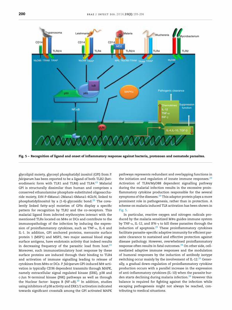
induced TLR activation has been shown in Fig. 5.
Malaria
Malaria is a mosquito borne infectious disease of humans and
other animals caused by parasitic protozoan of genus Plasmo-
dium (WHO, 2014) causing huge number of death each year.61
The host response during malaria infection, expresses high
levels of many proinflammatory cytokines, including TNF-�,
IL-1, IL-6, IL-12 and IFN-� that have important roles in control-
ling parasite growth.62 Such types of responses are primarily
originated from interaction of malarial surface molecules and
TLRs of host immune cells. Till date, search for TLR activating
PAMPs from malarial parasites have been the major high-
lighted objective for last few decades. In this connection, a
200 b r a z j i n f e c t d i s . 2 0 1 6;2 0(2):193–204
Trypanosoma Leishmania MalariaWuchereria Mycrobacterium
MD2
TLR2/4
IκB
MAPKs Pathogenic clearance
Suppression
functionChemokines
IL-12
TNF-α
INF-γ
IL-4,IL-10, TGF-β
p65
JUN
p50
NF-κB
MyD88 / TRAM MyD88 MyD88 /TRAM TRAM TIRAP MyD88MALTIRAP TIRAP
TLR2/4TLR4 TLR2TLR4
CD14 CD14 CD14
Fig. 5 – Recognition of ligand and onset of inflammatory response against bacteria, protozoan and nematode parasites.
glycolipid moiety, glycosyl phosphatidyl inositol (GPI) from P.
falciparum has been reported to be a ligand of both TLR2 (het-
erodimeric form with TLR1 and TLR6) and TLR4.63 Malarial
GPI is structurally dissimilar than human and comprises a
conserved ethanolamine phosphate-substituted oligosaccha-
ride moiety, EtN-P-6Mana1-2Mana1-6Mana1-4GlcN, linked to
phosphatidylinositol by a (1-6)-glycosidic bond.29 The cova-
lently linked fatty-acyl moieties of GPIs display a specific
pattern for recognition by TLR2 and the co-receptors. This
malarial ligand from infected erythrocytes interact with the
mentioned TLRs located on M�s or DCs and contribute to the
immunopathology of the infection by inducing the expres-
sion of proinflammatory cytokines, such as TNF-�, IL-6 and
IL-1. In addition, GPI anchored proteins, merozoite surface
protein 1 (MSP1) and MSP2, two major asexual blood stage
surface antigens, have endotoxin activity that indeed results
in decreasing frequency of the parasitic load from host.63
Moreover, such immunostimulatory host response by these
surface proteins are induced through their binding to TLR4
and activation of immune signalling leading to release of
cytokines from M�s or DCs. P. falciparum GPI-induced M� acti-
vation is typically CD36 dependent transmits through MAPK,
namely extracellular signal regulated kinase (ERK), p38 and
c-Jun N-terminal kinase (JNK) pathways as well as through
the Nuclear factor- kappa B (NF-�B).29 In addition, studies
using inhibitors of p38 activity and ERK1/2 activation indicated
towards significant crosstalk among the GPI activated MAPK
pathways represents redundant and overlapping functions in
the initiation and regulation of innate immune responses.29
Activation of TLR4/MyD88 dependent signalling pathway
during the malarial infection results in the excessive proin-
flammatory cytokine production responsible for the several
symptoms of the diseases.29 This adaptor protein plays a more
prominent role in pathogenesis, rather than in protection. A
scheme on malaria induced TLR activation has been shown in
Fig. 5.
In particular, reactive oxygen and nitrogen radicals pro-
duced by the malaria sensitized M�s guides immune system
by TNF-�, IL-12, and IFN-� to kill these parasites through the
induction of apoptosis.29 These proinflammatory cytokines
facilitate parasite-specific adaptive immunity for efficient par-
asite clearance to sustained and effective protection against
disease pathology. However, overwhelmed proinflammatory
response often results in fatal outcomes.29 On other side, cell-
mediated adaptive immune responses and the modulation
of humoral responses by the induction of antibody isotype
switching occur mainly by the involvement of IL-12.29 Gener-
ally, a gradual down-regulation of proinflammatory cytokine
production occurs with a parallel increase in the expression
of anti-inflammatory cytokines (IL-10) when the parasite bur-
den starts declining during malaria infection.29 However this
balance is required for fighting against the infection while
escaping pathogenesis might not always be reached, con-
tributing to medical situations.

b r a z j i n f e c t d i s . 2 0 1 6;2 0(2):193–204 201
Leishmaniasis
Leishmaniasis is caused by a protozoa parasite and transmit-
ted to humans by the bite of infected female phlebotomus
sandflies. This disease is the second-largest parasitic killer
in the world (after malaria), responsible for an estimated
200,000–400,000 infections each year worldwide (WHO, 2015).
The anti-leishmanial host response typically depends on the
quality of the adaptive immune response primarily induced
from host-parasite interaction. Especially, interaction between
leishmanial parasite and innate immune receptors rather
TLRs results generation of inflammatory mediators to deprive
parasite burden and develops efficient adaptive responses.
Lipophosphoglycan (LPG), the major identified ligand from
these parasites, has been reported to induce inflammation
through TLR2 and 4.64,65 LPG occurs both in the parasite sur-
face as well as in the secretory form and these two forms
are structurally similar but differ in the average number of
phosphorylated oligosaccharide repeat units present and in
the sugar types present in the glycan. Up-regulation and acti-
vation of TLR2 on human Natural killer (NK) cells by LPG of
L. major results enhancement of TNF-� and IFN-�. However,
interaction of LPGs with the TLRs is also influenced by the
structural diversity of the phosphoglycan. Likewise, LPG of L.
major, L. mexicana, L. aethiopica, and L. tropica were reported as
classical TLR2 ligand of M�s whereas performance of L. trop-
ica LPG is not so much satisfactory.65 Study on the induction of
TNF-� in M� by L. major LPG showed need of a lipid anchor and
a functional MyD88 adaptor.65 Such MyD88-dependent path-
ways are particularly important in developing protective IL-12-
mediated Th1 response against the parasite.65 The L. infantum
protein related to the silent information regulator 2 (SIR2)
family has been reported for stimulating the proliferation of
activated B lymphocytes, overexpression of major histocom-
patibility complex- (MHC) II and the co-stimulatory molecules
CD40 and CD86 as well as maturation of DCs accompanied
by the secretion of IL-12 and TNF-� are principally governed
through TLR2.64 In spite of its proinflammatory activity, LPG
has been shown to inhibit the progression of inflammatory sig-
nalling originated from TLR2/TLR4 by inducing the expression
of suppressors of the cytokine signalling (SOCS) family pro-
teins viz. SOCS-1 and SOCS-3.65,66 Another exciting fact about
LPG is that production of ROS is induced through the interac-
tion between membrane LPG and immune cell TLR-2 leading
to the differentiation of Th2 subsets whereas Th1-promoting
cytokines are reported to be induced solely by soluble LPG.65
LPG potentiated inflammatory responses through TLR2 stimu-
lation and IL-12 expression followed by Th1 responses in mice
was also reported after treating with synthetic oligosaccharide
analogues of LPG glycan structure indicating the importance
of glycan in the immunostimulatory activity of LPG.65 Interest-
ingly, parasites also use the same ligand as well as the TLRs to
supress the immune cell activation required for parasites’ ben-
efit i.e. disease progression. Faria et al.65 reported L. donovani
infected human THP1-derived M�s showed suppression in the
TLR2 stimulated IL-12 release with increased production of IL-
10 resulted from the inhibition of MAPKp38 phosphorylation
and activating ERK1/2 phosphorylation. In addition, L. dono-
vani, L. mexicana, and L. major have been reported to exploit
the M� tyrosine phosphatase SHP-1 to inactivate kinases
(e.g. IRAK) involved in TLR signalling.32 A graphical summary
on leishmanial parasite induced TLR activation is shown in
Fig. 5.
Proteoglycolipid complex (P8) composed of acysteine and
serine metalloprotease, host-derived Apolipoprotein E (ApoE),
and four glycolipids has been reported as another potential
TLR4 ligand of Leishmania detected in L. pifanoi amastig-
otes that influence phagocytosis through TLR activation.65
Activated TLR4 at the parasitophorous vacuole affects the
microenvironment surrounding the parasite for its control.
Particularly for the neutrophils, TLR4 induces IRAK4 for exo-
cytosis of neutrophil secretory granules.65 Mechanistically,
P8 sensitized M�s induce proinflammatory cytokines viz. IL-
1� and TNF-� through TLR4, MD2, CD14, and MyD88.65 In
addition, metacyclic promastigotes have been reported to
induce phosphorylation of the MAP kinases such as ERK,
p38, and JNK through TLR4 in L. mexicana.67 During such
course of inflammation of M�, iNOS, Cyclooxygenase-2 (COX-
2), Prostaglandin E2 (PGE2), NO and arginase-1 are been the
mediators.67
Trypanosomiasis (Chagas disease)
Chagas disease or American trypanosomiasis, is a tropical par-
asitic disease caused by the protozoan Trypanosoma cruzi (CDC,
2015) and transmitted by insects vector like Triatominae or
kissing bugs. In Latin America it causes a health threat for an
estimated 10 million people and more than 25 million people
are at risk. Typical immunity against Chagas disease is princi-
pally mediated by innate recognition through TLRs. Although
studied less, TLRs have been characterized as crucial determi-
nants in evoking anti- T. cruzi immune response in which both
TLR2 and TLR4 are the key mediators.68 In particular, these
receptors mediate internalization of trypanosomatid through
phagocytosis required for initiating the immune response by
phagosomal maturation to kill the parasite followed by anti-
gen presentation.69 Researchers have demonstrated fusion of
early endosomes, and phagocytosis induced by trypomastigo-
tes in M�s by ligation of guanine phosphonucleotide- binding
proteins Ras-related protein- (Rab-) 5 solely through TLR2.69
TLR2 activation has been reported to proceed via MyD88-
dependent pathway.69 Secretion of chemokines signalled from
TLR2 activation further recruit leukocytes to control the infec-
tion in a broad way.28 In particular, TLR2 signalling is targeted
by the trypanosomatids to hinder the immune response
induced by the parasite.
However, TLR activation resulted from immune cells-T.
cruzi interaction may also contribute to the disease pathology
as well.69 Involvement of TLR activation in the immunopathol-
ogy of chronic Chagas’ cardiopathy has been reported to be
due to a single-nucleotide polymorphism in the genes cod-
ing for the signalling proteins of TLR signalling and leads to
differential susceptibility to Chagas disease.69 For example,
T. cruzi-infected individuals have heterozygous MAL/TIRAP
S180L variant shows poorer signal transduction after binding
of the trypanosomal ligand to TLR2 or TLR4 resulting in lesser
chance of developing chronic Chagas’ cardiomyopathy.69 A

202 b r a z j i n f e c t d i s . 2 0 1 6;2 0(2):193–204
summary on trypanosomatid induced TLR activation has been
shown in Fig. 5.
Filariasis
Lymphatic filariasis, a vector borne neglected tropical dis-
ease, mainly caused by three species of nematode parasites-
Wuchereria bancrofti, Brugia malayi, and B. timori is considered
to be a major public health burden (WHO, 2015). Pathological
manifestations of this disease mainly include lymphedema,
elephantiasis and rarely a greater degree of neurological
manifestation.70 Recent studies also showed existence of co-
infection of filarial parasite with bacteria (TB),49 malaria 49,71
and opportunistic yeast.72 In general, the immunoresponse
in filariasis is complicated and unresolved especially the
involvement and functional role played by TLR2 and TLR4.49
Filarial intracellular symbiotic bacterium, Wolbachia has been
reported to cause inflammation through its lipopolysaccha-
ride (of outer membrane) after binding with TLR4. TLR2 and
TLR4 are also capable to bind other Wolbachial products like
Wolbachia surface protein (WSP) and induce inflammatory
response by secreting pro-inflammatory cytokines from M�s
and DCs.13 Such proinflammatory responses are of immense
importance to eliminate the infection from host. However,
filarial nematodes also target TLR2 and TLR4, especially TLR4,
for modulating host immune response. A phosphorylcholine-
conjugated glycoprotein molecule namely ES-62 binds TLR4
and create anti-inflammatory/Th2 biased immune response
to prolong survivability of the parasite inside host.36 In chronic
infection, filarial nematode induces apoptotic death of T
cells for down regulating host response and interestingly
the process is also operative through TLR4.73 Live micro-
filarae of B. malayi have been reported to modulate TLR4
expression in monocyte derived human DCs (mhDCs) to
induce an immunosuppressive environment in host through
interference of MyD88-dependent suppression of NF-�B sig-
nalling to hinder production of proinflammatory cytokines
and type-1 interferons. In addition to such inhibitory effect
of microfilarae over DCs, these also results partial inhibi-
tion of the function of human Langerhans cells (LCs) and
prevent proliferation of CD4+ T cells.74 Interestingly, TLR2
and TLR4 have been reported to be expressed on circulating
B cells during helminthic infection that uphold and main-
tain Th2 type-immune responses to make “worm favourable”
conditions.75 These specialized B cell subsets are known as
regulatory B cells (Breg) and their immunosuppressive func-
tion is also regulated through TLR4.75 TLR4 on these cells
trigger induction of IL-10 secretion from B cell through Myd88-
dependent pathway.76 Particularly in filariasis, Breg cells are
responsible for the induction as well as maintenance of hypo-
responsive immune status with elevated levels of Treg, IL-10
and filarial-specific IgG4.75 This Breg mediated responses
is recompensing as patients remain asymptomatic due to
devoid of excessive immunopathology.75 However, this typ-
ical Th2 environment by Breg results secondary infections
by bacteria and/or fungi due to lack of protective immune
cells repertoires in filarial subjects.75 A probable mechanism
of TLR activation by filarial parasites has been shown in
Fig. 5.
Conclusion and future prospects
Studies on the recognition of infectious pathogens through
TLRs have significantly contributed to our understanding
of the underlying mechanisms of innate immune defense
against the deadliest parasites. In this review, we have
put forward the findings made on the role played by two
crucial members of the TLR family viz. TLR2 and TLR4
against the most threatening infectious diseases. We have
discussed different previously reported findings on types of
pathogenic ligand alongside the mechanism of action of
inflammatory cascade originated from TLR4 and TLR2 in
tuberculosis, malaria, leishmaniasis, filariasis, and trypanoso-
miasis. Interesting outcomes achieved in the experimental
vaccine adjuvants and/or immunotherapy against active
leishmaniasis promises for effective future applications as
well as opens new possibilities regarding the use of TLR based
immunotherapeutic control strategies for malaria or other dis-
eases. Similarly, immunomodulatory TLR4 ligand from filarial
parasites has also shown to control allergic reaction by inhib-
iting proinflammatory mediators and this strategy could be
used effectively to combat inflammatory pathology of malaria
or other disease to reduce mortality. Similarly, TLR4 has been
emerged as a target of therapeutics for immunopharmacolog-
ical control of infectious and/or inflammatory diseases.77
However, many questions remain to be answered for this
aspect. Thus more concluding information rather inferences
are need to be gathered through population based studies.
Similarly, there is a need to search for other microbial ligands
for a same microbe/parasite as well as interplay between the
TLRs and obviously the signalling crosstalk. From immuno-
logical perspectives, study of mechanism of parasite/microbe
induced polarization of major antigen presenting cells may
offer us new target for developing therapeutic strategy to
restore Th1 (to eliminate infection) or Th2 biased (to protect
host from overt inflammation) immune response.
Conflicts of interest
The authors declare no conflicts of interest.
Acknowledgement
SM acknowledges University Grants Commission for the
award of his Senior Research Fellowship.
r e f e r e n c e s
1. Kawai T, Akira S. Toll-like receptors and their crosstalk with
other innate receptors in infection and immunity. Immunity.
2011;34:637–50.
2. Zeytun A, Chaudhary A, Pardington P, Cary R, Gupta G.
Induction of cytokines and chemokines by Toll-like receptor
signaling strategies for control of inflammation. Crit Rev
Immunol. 2010;30:53–67.
3. Botos I, Segal DM, Davies DR. The structural biology of
Toll-like receptors. Structure. 2011;19:447–59.
b r a z j i n f e c t d i s . 2 0 1 6;2 0(2):193–204 203
4. Valanne S, Wang JH, Ramet M. The Drosophila Toll signaling
pathway. J Immunol. 2011;186:649–56.
5. Ganesan S, Aggarwal K, Paquette N, Silverman N.
NF-kappaB/Rel proteins and the humoral immune responses
of Drosophila melanogaster. Curr Top Microbiol Immunol.
2011;349:25–60.
6. Janeway CA Jr, Medzhitov R. Innate immune recognition.
Annu Rev Immunol. 2002;20:197–216.
7. Maeshima N, Fernandez RC. Recognition of lipid A variants by
the TLR4-MD-2 receptor complex. Front Cell Infect Microbiol.
2013;3:3.
8. Kimbrell DA, Beutler B. The evolution and genetics of innate
immunity. Nat Rev Genet. 2001;2:256–67.
9. Park BS, Lee JO. Recognition of lipopolysaccharide pattern by
TLR4 complexes. Exp Mol Med. 2013;45:e66.
10. Aderem A, Ulevitch RJ. Toll-like receptors in the induction of
the innate immune response. Nature. 2000;406:782–7.
11. Schreiner J, Kretschmer D, Klenk J, et al. Staphylococcus aureus
phenol-soluble modulin peptides modulate dendritic cell
functions and increase in vitro priming of regulatory T cells. J
Immunol. 2013;190:3417–26.
12. Opitz B, Schroder NW, Spreitzer I, et al. Toll-like receptor-2
mediates Treponema glycolipid and lipoteichoic acid-induced
NF-kappaB translocation. J Biol Chem. 2001;276:
22041–7.
13. Brattig NW, Bazzocchi C, Kirschning CJ, et al. The major
surface protein of Wolbachia endosymbionts in filarial
nematodes elicits immune responses through TLR2 and TLR4.
J Immunol. 2004;173:437–45.
14. Bulut Y, Faure E, Thomas L, Equils O, Arditi M. Cooperation of
Toll-like receptor 2 and 6 for cellular activation by soluble
tuberculosis factor and Borrelia burgdorferi outer surface
protein A lipoprotein role of Toll-interacting protein and IL-1
receptor signaling molecules in Toll-like receptor 2 signaling.
J Immunol. 2001;167:987–94.
15. Cabral ES, Gelderblom H, Hornung RL, Munson PJ, Martin R,
Marques AR. Borrelia burgdorferi lipoprotein-mediated TLR2
stimulation causes the down-regulation of TLR5 in human
monocytes. J Infect Dis. 2006;193:849–59.
16. Strunk T, Power Coombs MR, Currie AJ, et al. TLR2 mediates
recognition of live Staphylococcus epidermidis and clearance of
bacteremia. PLoS ONE. 2010;5:e10111.
17. Pennini ME, Pai RK, Schultz DC, Boom WH, Harding CV.
Mycobacterium tuberculosis 19-kDa lipoprotein inhibits
IFN-gamma-induced chromatin remodeling of MHC2TA by
TLR2 and MAPK signaling. J Immunol. 2006;176:4323–30.
18. McIsaac SM, Stadnyk AW, Lin TJ. Toll-like receptors in the
host defense against Pseudomonas aeruginosa respiratory
infection and cystic fibrosis. J Leukoc Biol. 2012;92:
977–85.
19. Revets H, Pynaert G, Grooten J, De Baetselier P. Lipoprotein I, a
TLR2/4 ligand modulates Th2-driven allergic immune
responses. J Immunol. 2005;174:1097–103.
20. Hahm B, Cho JH, Oldstone MB. Measles virus-dendritic cell
interaction via SLAM inhibits innate immunity selective
signaling through TLR4 but not other TLRs mediates
suppression of IL-12 synthesis. Virology. 2007;358:251–7.
21. Sorensen LN, Reinert LS, Malmgaard L, Bartholdy C, Thomsen
AR, Paludan SR. TLR2 and TLR9 synergistically control herpes
simplex virus infection in the brain. J Immunol.
2008;181:8604–12.
22. Uematsu S, Akira S. Toll-Like receptors (TLRs) and their
ligands. Handb Exp Pharmacol. 2008:1–20.
23. Tada H, Nemoto E, Shimauchi H, et al. Saccharomyces
cerevisiae- and Candida albicans-derived mannan induced
production of tumor necrosis factor alpha by human
monocytes in a CD14- and Toll-like receptor 4-dependent
manner. Microbiol Immunol. 2002;46:503–12.
24. Jouault T, Ibata-Ombetta S, Takeuchi O, et al. Candida
albicans phospholipomannan is sensed through toll-like
receptors. J Infect Dis. 2003;188:165–72.
25. Monari C, Pericolini E, Bistoni G, Casadevall A, Kozel TR,
Vecchiarelli A. Cryptococcus neoformans capsular
glucuronoxylomannan induces expression of fas ligand in
macrophages. J Immunol. 2005;174:3461–8.
26. Chai LY, Kullberg BJ, Vonk AG, et al. Modulation of Toll-like
receptor 2 (TLR2) and TLR4 responses by Aspergillus fumigatus.
Infect Immun. 2009;77:2184–92.
27. Aoki MP, Carrera-Silva EA, Cuervo H, Fresno M, Girones N, Gea
S. Nonimmune cells contribute to crosstalk between immune
cells and inflammatory mediators in the innate response to
Trypanosoma cruzi infection. J Parasitol Res. 2012;2012:737324.
28. Coelho PS, Klein A, Talvani A, et al.
Glycosylphosphatidylinositol-anchored mucin-like
glycoproteins isolated from Trypanosoma cruzi
trypomastigotes induce in vivo leukocyte recruitment
dependent on MCP-1 production by
IFN-gamma-primed-macrophages. J Leukoc Biol.
2002;71:837–44.
29. Gowda DC. TLR-mediated cell signaling by malaria GPIs.
Trends Parasitol. 2007;23:596–604.
30. Flandin JF, Chano F, Descoteaux A. RNA interference reveals a
role for TLR2 and TLR3 in the recognition of Leishmania
donovani promastigotes by interferon-gamma-primed
macrophages. Eur J Immunol. 2006;36:411–20.
31. Faria MS, Calegari-Silva TC, de Carvalho Vivarini A, Mottram
JC, Lopes UG, Lima AP. Role of protein kinase R in the killing
of Leishmania major by macrophages in response to neutrophil
elastase and TLR4 via TNFalpha and IFNbeta. FASEB J.
2014;28:3050–63.
32. Abu-Dayyeh I, Shio MT, Sato S, Akira S, Cousineau B, Olivier
M. Leishmania-induced IRAK-1 inactivation is mediated by
SHP-1 interacting with an evolutionarily conserved KTIM
motif. PLoS Negl Trop Dis. 2008;2:e305.
33. Abu-Dayyeh I, Hassani K, Westra ER, Mottram JC, Olivier M.
Comparative study of the ability of Leishmania mexicana
promastigotes and amastigotes to alter macrophage signaling
and functions. Infect Immun. 2010;78:2438–45.
34. Wong-Baeza I, Alcantara-Hernandez M, Mancilla-Herrera I,
et al. The role of lipopeptidophosphoglycan in the immune
response to Entamoeba histolytica. J Biomed Biotechnol.
2010;2010:254521.
35. Diaz A, Allen JE. Mapping immune response profiles the
emerging scenario from helminth immunology. Eur J
Immunol. 2007;37:3319–26.
36. Goodridge HS, Marshall FA, Else KJ, et al. Immunomodulation
via novel use of TLR4 by the filarial nematode
phosphorylcholine-containing secreted product, ES-62. J
Immunol. 2005;174:284–93.
37. van Riet E, Everts B, Retra K, et al. Combined TLR2 and TLR4
ligation in the context of bacterial or helminth extracts in
human monocyte derived dendritic cells molecular correlates
for Th1/Th2 polarization. BMC Immunol. 2009;10:9.
38. van der Kleij D, Latz E, Brouwers JF, et al. A novel
host-parasite lipid cross-talk. Schistosomal
lyso-phosphatidylserine activates toll-like receptor 2 and
affects immune polarization. J Biol Chem. 2002;277:48122–9.
39. Wang X, Zhou S, Chi Y, et al. CD4+CD25+ Treg induction by an
HSP60-derived peptide SJMHE1 from Schistosoma japonicum is
TLR2 dependent. Eur J Immunol. 2009;39:3052–65.
40. Terrazas CA, Gomez-Garcia L, Terrazas LI. Impaired
pro-inflammatory cytokine production and increased
Th2-biasing ability of dendritic cells exposed to Taenia
excreted/secreted antigens: a critical role for carbohydrates
but not for STAT6 signaling. Int J Parasitol. 2010;40:
1051–62.

204 b r a z j i n f e c t d i s . 2 0 1 6;2 0(2):193–204
41. Johnston MJ, MacDonald JA, McKay DM. Parasitic helminths a
pharmacopeia of anti-inflammatory molecules. Parasitology.
2009;136:125–47.
42. Oliveira-Nascimento L, Massari P, Wetzler LM. The role of
TLR2 in infection and immunity. Front Immunol. 2012;3:
79.
43. Brandt KJ, Fickentscher C, Kruithof EK, de Moerloose P. TLR2
ligands induce NF-kappaB activation from endosomal
compartments of human monocytes. PLoS ONE.
2013;8:e80743.
44. O’Mahony DS, Pham U, Iyer R, Hawn TR, Liles WC. Differential
constitutive and cytokine-modulated expression of human
Toll-like receptors in primary neutrophils, monocytes, and
macrophages. Int J Med Sci. 2008;5:1–8.
45. Blasius AL, Beutler B. Intracellular toll-like receptors.
Immunity. 2010;32:305–15.
46. Muzio M, Bosisio D, Polentarutti N, et al. Differential
expression and regulation of toll-like receptors (TLR) in
human leukocytes selective expression of TLR3 in dendritic
cells. J Immunol. 2000;164:5998–6004.
47. Oeckinghaus A, Hayden MS, Ghosh S. Crosstalk in NF-kappaB
signaling pathways. Nat Immunol. 2011;12:695–708.
48. Kagan JC, Medzhitov R. Phosphoinositide-mediated adaptor
recruitment controls Toll-like receptor signaling. Cell.
2006;125:943–55.
49. Babu S, Anuradha R, Kumar NP, George PJ, Kumaraswami V,
Nutman TB. Filarial lymphatic pathology reflects augmented
toll-like receptor-mediated, mitogen-activated protein
kinase-mediated proinflammatory cytokine production.
Infect Immun. 2011;79:4600–8.
50. Carpenter S, O’Neill LA. How important are Toll-like receptors
for antimicrobial responses. Cell Microbiol. 2007;9:1891–901.
51. Iwanaszko M, Kimmel M. NF-kappaB and IRF pathways
cross-regulation on target genes promoter level. BMC
Genomics. 2015;16:307.
52. Tamassia N, Bazzoni F, Le Moigne V, et al. IFN-beta expression
is directly activated in human neutrophils transfected with
plasmid DNA and is further increased via TLR-4-mediated
signaling. J Immunol. 2012;189:1500–9.
53. Kawai T, Akira S. Regulation of innate immune signalling
pathways by the tripartite motif (TRIM) family proteins. EMBO
Mol Med. 2011;3:513–27.
54. Kawasaki T, Kawai T. Toll-like receptor signaling pathways.
Front Immunol. 2014;5:461.
55. Kleinnijenhuis J, Oosting M, Joosten LA, Netea MG, Van Crevel
R. Innate immune recognition of Mycobacterium tuberculosis.
Clin Dev Immunol. 2011;2011:405310.
56. Stenger S, Modlin RL. Control of Mycobacterium tuberculosis
through mammalian Toll-like receptors. Curr Opin Immunol.
2002;14:452–7.
57. Rocha-Ramirez LM, Estrada-Garcia I, Lopez-Marin LM, et al.
Mycobacterium tuberculosis lipids regulate cytokines, TLR-2/4
and MHC class II expression in human macrophages.
Tuberculosis (Edinb). 2008;88:212–20.
58. Kusner DJ. Mechanisms of mycobacterial persistence in
tuberculosis. Clin Immunol. 2005;114:239–47.
59. Herbst S, Schaible UE, Schneider BE. Interferon gamma
activated macrophages kill mycobacteria by nitric oxide
induced apoptosis. PLoS ONE. 2011;6:e19105.
60. Aliprantis AO, Yang RB, Weiss DS, Godowski P, Zychlinsky A.
The apoptotic signaling pathway activated by Toll-like
receptor-2. EMBO J. 2000;19:3325–36.
61. Caraballo H, King K. Emergency department management of
mosquito-borne illness: malaria, dengue, and West Nile virus.
Emerg Med Pract. 2014;16:1–23.
62. Punsawad C. Effect of malaria components on blood
mononuclear cells involved in immune response. Asian Pac J
Trop Biomed. 2013;3:751–6.
63. Gun SY, Claser C, Tan KS, Renia L. Interferons and interferon
regulatory factors in malaria. Mediators Inflamm.
2014;2014:243713.
64. Silvestre R, Silva AM, Cordeiro-da-Silva A, Ouaissi A. The
contribution of Toll-like receptor 2 to the innate recognition
of a Leishmania infantum silent information regulator 2
protein. Immunology. 2009;128:484–99.
65. Faria MS, Reis FC, Lima AP. Toll-like receptors in leishmania
infections guardians or promoters? J Parasitol Res.
2012;2012:930257.
66. de Veer MJ, Curtis JM, Baldwin TM, et al. MyD88 is essential
for clearance of Leishmania major possible role for
lipophosphoglycan and Toll-like receptor 2 signaling. Eur J
Immunol. 2003;33:2822–31.
67. Shweash M, Adrienne McGachy H, Schroeder J, et al.
Leishmania mexicana promastigotes inhibit macrophage IL-12
production via TLR-4 dependent COX-2, iNOS and arginase-1
expression. Mol Immunol. 2011;48:1800–8.
68. Rodrigues MM, Oliveira AC, Bellio M. The immune response to
Trypanosoma cruzi role of Toll-like receptors and perspectives
for vaccine development. J Parasitol Res. 2012;2012:507874.
69. Ramasawmy R, Cunha-Neto E, Fae KC, et al. Heterozygosity
for the S180L variant of MAL/TIRAP, a gene expressing an
adaptor protein in the Toll-like receptor pathway, is
associated with lower risk of developing chronic Chagas
cardiomyopathy. J Infect Dis. 2009;199:1838–45.
70. Mukherjee S, Sinha Babu SP. Neurofilariasis. In: Chopra J,
editor. Neurology in tropics. Asia Pacific: Elsevier; 2015.
71. Hoerauf A, Satoguina J, Saeftel M, Specht S.
Immunomodulation by filarial nematodes. Parasite Immunol.
2005;27:417–29.
72. Mukherjee S, Mukherjee N, Saini P, Gayen P, Roy P, Sinha Babu
SP. Molecular evidence on the occurrence of co-infection with
Pichia guilliermondii and Wuchereria bancrofti in two filarial
endemic districts of India. Infect Dis Poverty. 2014;3:13.
73. Babu S, Nutman TB. Immunopathogenesis of lymphatic
filarial disease. Semin Immunopathol. 2012;34:847–61.
74. Venugopal PG, Nutman TB, Semnani RT. Activation and
regulation of toll-like receptors (TLRs) by helminth parasites.
Immunol Res. 2009;43:252–63.
75. Ludwig-Portugall I, Layland LE. TLRs, Treg, and B Cells, an
Interplay of Regulation during Helminth Infection. Front
Immunol. 2012;3:8.
76. Yanaba K, Bouaziz JD, Matsushita T, Tsubata T, Tedder TF. The
development and function of regulatory B cells expressing
IL-10 (B10 cells) requires antigen receptor diversity and TLR
signals. J Immunol. 2009;182:7459–72.
77. Mukherjee N, Mukherjee S, Saini P, Roy P, Sinha Babu SP.
Phenolics and terpenoids the promising new search for
anthelmintics: a critical review. Mini Rev Med Chem. 2015.