Title: Phase II Study of the Beta-Blocker Propranolol With ...
23
Title: Phase II Study of the Beta-Blocker Propranolol With Chemotherapy in Patients Receiving Neoadjuvant Treatment for Newly Diagnosed Breast Cancer NCT Number: NCT01847001 Protocol Version Date: March 03, 2014
Transcript of Title: Phase II Study of the Beta-Blocker Propranolol With ...

Title: Phase II Study of the Beta-Blocker Propranolol With
Chemotherapy in Patients Receiving Neoadjuvant Treatment for Newly Diagnosed Breast Cancer
NCT Number: NCT01847001 Protocol Version Date: March 03, 2014

Page 1 Protocol dated 03/03/2014
Columbia University Medical Center IRB # AAAI9158
Phase II Study of the Beta-Blocker Propranolol With Chemotherapy in Patients
Receiving Neoadjuvant Treatment for Newly Diagnosed Breast Cancer
Principal Investigator: Kevin Kalinsky, M.D. Co-Investigators: Dawn Hershman, M.D.
Katherine Crew, M.D. Matthew Maurer, M.D. Andreas Hielscher, Ph.D. Hyun Keol Kim, MD
While a number of therapeutic options exist for patients with breast cancer (BC), breast tumor biology is heterogeneous and not all BCs respond to treatment. Identifying a marker predicting response could spare non-responders unnecessary side effects, cost, and time. A recent example in BC is bevacizumab, an expensive anti-angiogenic monoclonal antibody, for which the FDA revoked approval for patients with metastatic BC1. While this targeted therapy may benefit some patients, no appropriate predictive marker has been identified in the drug development process1. An ideal biologic marker would be easy to perform, reliable, low-cost and non-invasive. A limitation of assessing tumor-based markers in metastatic BC is the inability to procure tumor tissue at different treatment times. To circumvent this issue, anti-cancer agents can be assessed pre-operatively, where women with newly diagnosed BC receive a study drug, alone or with chemotherapy, between diagnostic breast biopsy and surgical resection. In addition, tumor changes can be directly compared to modulation of non-invasive markers, such as functional radiographs or blood, to identify a non-invasive marker predicting tumor response. We are conducting a neoadjuvant single-institution trial with the non-selective, inexpensive β –blocker propranolol alone and with chemotherapy in locally advanced BC. β-blockade regulates angiogenesis in primary breast tumors2,3. In these trials, we plan to evaluate treatment-related microvascular response via changes in breast Diffuse Optical Tomography (DOT), a non-invasive, fast, safe, and inexpensive breast imaging tool. As the optical property contrast from endogenous chromophores (oxyhemoglobin, deoxyhemoglobin, water, and lipid) provides information on tissue vascularity, it can monitor response to anti-angiogenic agents4. DOT changes occur as early as 1 week after starting pre-operative therapy5. The dynamic DOT system incorporated in these trials is unique to CUMC, as it has been designed by Columbia biomedical engineers. Our laboratory collaborators have measured this DOT system with anti-angiogenesis agents in animal models6, demonstrating the translational nature of this project. While non-dynamic DOT has been assessed in other neoadjuvant trials with small cohorts receiving heterogeneous chemotherapy agents5,7-10, none have evaluated DOT response to anti-angiogenic agents. Design: We are performing a phase II study investigating neoadjuvant propranolol alone, followed by propranolol with cytotoxic chemotherapy (taxane, followed by adriamycin/cytoxan) in 20 patients with locally advanced breast cancer. Once enrolled, patients will start propranolol in combination with weekly treatments of taxane therapy (+ every 3 week Herceptin and every 3 week Pertuzumab, if HER2+) followed by adriamycin/cytoxan + propranolol. Patients will continue to take propranolol through the day before surgery (last dose of propranolol: night before surgery). Primary Endpoint: 1. To assess the feasibility of giving neoadjuvant propranolol alone and in combination with chemotherapy. Our hypothesis is that it will be feasible for patients to take propranolol with chemotherapy in the neoadjuvant setting.

Page 2 Protocol dated 03/03/2014
Secondary Endpoints: 1. To evaluate deoxyhemoglobin reduction as a non-invasive, image-based biomarker of angiogenic response after 2 weeks of neoadjuvant propranolol administration in combination with paclitaxel, as measured by DOT. These images will be compared to DOT collected for the ongoing prospective IRB-AAAI0480 trial. 2. To explore the modulation of DOT markers at 5 time-points with propranolol: a)baseline,; b) before starting taxane week #3; c) before starting AC cycle #1; d) before starting AC cycle #2; and e) before surgery. These images will be compared to DOT collected for the ongoing prospective IRB-AAAI0480 trial. 3. To evaluate the association between changes in optical images and markers of angiogenesis [tumor (VEGF, CD31, CD105, microvessel density and area), serum (VEGF, V-cam, and PIGF expression)], proliferation (tumor KI-67 levels), and tumor macrophage infiltration (CD163 expression) compared to baseline levels. 4. To explore whether acute stress levels, as measured by urinary cortisol and responses to the Questionnaire on Stress in Cancer Patients - revised version (QSC-R23), associate with image- or blood-based biomarker changes after propranolol. 5. To assess the safety and tolerability of neoadjuvant propranolol along with chemotherapy A.Background: A.1. Angiogenesis in BC: A number of studies have demonstrated that angiogenesis, the process of blood vessel formation, is essential in the role in the growth, progression and metastatic potential of BC cells11,12. The most clinical experience with anti-angiogenic agents in BC has been with bevacizumab (Avastin), the humanized recombinant monoclonal antibody that specifically blocks the binding of vascular endothelial growth factor (VEGF) to high-affinity receptors. Three randomized control trials have demonstrated an improvement in progression free-survival and response rates with the addition of bevacizumab to front-line chemotherapy13-15. However, while significant, the improvement in these outcomes in the AVADO14 and Ribbon-115trials has not been clinically compelling, given the median progression-free survival of 1-2 months when adding bevacizumab to chemotherapy. Combined with the potential side effects and lack of overall survival advantage in these trials and subsequent meta-analyses16, there remains uncertainty about how to use bevacizumab in advanced BC. In July 2011, The ODAC Committee of the FDA revoked approval for patients with metastatic BC1. In addition, bevacizumab is an expensive drug. An economic analysis in 2009, based on efficacy data from ECOG E210013 and European cost-estimates, suggests an incremental cost-effectiveness ratio of 189,427 euros per quality-adjusted life-year17. While some women benefit from bevacizumab, it seems that neither prior adjuvant therapy nor tumor subtype serve as selection factors for bevacizumab-based treatment. A remaining major issue is that bevacizumab lacks a robust, predictive, biologic, or clinical marker of activity. This experience has highlighted the need for developing clear predictive biomarkers for anti-angiogenic agents in BC. A.2. Propranolol in Breast Cancer: Anti-angiogenic Effects: There is emerging pre-clinical and observational data that neuro-endocrine signaling influences BC metastatic potential. Used to treat cardiovascular disease, β -blockade associates with a reduction in distant BC metastases in observational studies18-21. β-blocker use independently predicts metastatic risk (HR=0.43; p=0.031) and BC-specific mortality at 10 years (HR=0.29; p=0.007)19. Compared to other anti-hypertensives, β-blocker use significant reduces BC recurrence (p=0.026). In a retrospective study of patients receiving neoadjuvant therapy, β-blocker use is an independent predictor of improved relapse free survival (HR: 0.52; p=0.015), with a trend in overall survival (HR 0.64; p=0.09)21. In another study, BC-specific

Page 3 Protocol dated 03/03/2014
Figure 1: Tumor samples from mice treated with a placebo (HeyA): isoproterenol (nonspecific β agonist), terbutaline (β2 agonist), xamoterol (β1 agonist), or isoproterenol plus propranolol. Stained for CD31 and VEGF (IHC and ISH). Tumor VEGF protein levels (ELISA). Microvessel Density is higher in the isoproterenol and terbutaline groups than in controls or in the combination group.
mortality is significantly lower for propranolol users (median: 80 mg daily) as compared to nonusers (HR: 0.19)22. Non-BC mortality is similar. No effect is seen with atenolol (β1 antagonist), suggesting that the β2 adrenergic receptor is the likely mediator. These studies provide evidence that propranolol should be evaluated prospectively. There is strong pre-clinical data in accordance with these observations, suggesting that β-blockers offer this advantage in BC by impacting the primary BC microenvironment2,3. Activation of cellular β-adrenergic receptors by norepinephrine triggers transcription factor activation, regulating tumor cell proliferation23 and apoptosis inhibition24. Norepinephrine stimulates the migration and chemotaxis of MDA-MB-468 (triple negative: TN) BC cells23. This effect is completely inhibited by β2-specific blockade and only partially reduced by the β1-specific blocker atenolol. The functionality of the β-adrenergic receptor is also observed in CG-5 BC (ER+) cells25. In BALB/c mice syngeneic to 66cl4 BC cells (TN), the β-agonist isoproterenol increases distant metastasis by 22-fold vs. control (p=0.03), and the β-blocker propranolol completely blocks stress-enhanced metastasis (p<0.0001). While primary BC size does not change with propranolol, propranolol largely abrogates primary tumor macrophage infiltration (F4/80+), as well as stress-induced vascularization of primary mammary tumors (CD31) (p <0.001). In SKOV3ip1 ovarian and MDA-MB-231 (TN) BC orthotropic models, less visceral metastases are seen in mice treated with propranolol (0%) vs. placebo (50%) (p = 0.01)3. While the β-agonist isoproterenol and β2-agonist terbutaline increase tumor weight and metastatic number, the β1 agonist xamoterol does not (Figure 1), which, similar to observational data3, implicates the role of β2. In SKOV3ip1 cells, VEGF mRNA expression increases by 842% after norepinephrine and promoter activity increases by 1,240%. This effect is blocked by propranolol. This supports the evaluating propranolol as an angiogenic modulator in BC. β-adrenergic receptor functionality is observed in ER+25 and HER2+ cells26. Given these consistent findings, we plan to investigate propranolol in a prospective, translational clinical trial.
A.3. Diffuse Optical Tomography (DOT) as an Image-based Marker of Tumor Angiogenic Response: As DOT provides information on tissue vascularity, it can monitor response to anti-angiogenic agents4. DOT utilizes optical transmission measurements with low-intensity light (i.e. non-ionizing radiation) to provide 3-dimensional information on tissue vascularity. The optical property contrast from endogenous chromophores [oxyhemoglobin (Hb02), deoxyhemoglobin (Hb), water, and

Page 4 Protocol dated 03/03/2014
Figure 2: Change in DOT Parameters - Oxygenated Hemoglobin (Hb02) and Deoxyhemoglobin (Hb): Baseline vs. 2 weeks of Paclitaxel in Patient #1 (A1) and Patient #2 (B1)
lipid] can distinguish malignant from normal tissue4,27. Total hemoglobin levels, which directly relate to blood vessel density in tumors, are double those in benign breast lesions28. Vascular changes precede measurable structural changes in mouse models29. A limited number of small series (n=7-16 patients) report DOT changes in patients undergoing neoadjuvant chemotherapy5,7-10. None incorporate targeted therapy beyond trastuzumab. Total hemoglobin levels significantly decreases with pathologic complete response (p=0.001)5,10. Changes occur as early as 1 week after starting pre-operative chemotherapy, with tumor Hb levels dropping 33% +/-7% in responders vs. non-responders (18% +/- 10%: z=0.008)5. In addition, pre-treatment total hemoglobin levels correlate with pre-treatment mean vessel density of CD105-expressing vessels, a neoangiogenesis marker (p<0.001)9. We will evaluate DOT as an early marker of angiogenic response in this neoadjuvant trial with propranolol. In an ongoing pilot (IRB-AAAD7133), 50 subjects with BCs are being recruited, with 10 subjects with healthy breasts as controls. The primary objective is to evaluate if DOT can differentiate between BC-bearing and healthy breasts. In the first 29 patients, there is a significant difference between deoxyhemoglobin levels in BC-bearing vs. healthy (p=0.017). The current data suggests a sensitivity of 91% and a specificity of 83%. In our ongoing study of 20 women receiving neoadjuvant taxane followed by adriamycin plus cytoxan (IRB-AAAI0480), we are assessing the percentage change in DOT parameters and pCR. This study has enrolled 15 patients (open: 8/2011). Figure 2 demonstrates DOT change in the first 2 analyzable patients after 2 weeks of paclitaxel. In the first 4 patients, the mean Hb02 and Hb decrease is 70% and 50%, respectively.
B. Study Design B.1. Overview: We propose a phase II study investigating the impact of neoadjuvant propranolol alone and in combination with cytotoxic chemotherapy (taxane, followed by adriamycin/cytoxan). Once enrolled, patients will start propranolol 20 mg po bid, ultimately increasing the dose of propranolol up to a maximum 80 mg po daily ER if tolerating propranolol (Section F.2 for details), when receiving 12 weeks of taxane therapy + propranolol (+ Herceptin and Pertuzumab, if HER2+) followed by adriamycin/cytoxan + propranolol (Figure 3). This dose of propranolol has been chosen as it is the median propranolol dose in the Barron study, in which propranolol is associated with reduced breast cancer progression and mortality22. We chose to start with a lower dose at initiation to make sure that patients are tolerating the dose, prior to titrating up. This was discussed with cardiologists at CUMC and agreed to be the safest approach. B.2. Intervention: We will also be evaluating changes in tumor-, image- and blood- biomarkers. The main inclusion criteria will be English or Spanish speaking women over the age of 18 with recently diagnosed invasive breast cancer deemed eligible to receive neoadjuvant propranolol plus cytotoxic chemotherapy. Patients should have a tumor size ≥ 1 cm by clinical measurement and scheduled to
Baseline Two-week Hb02
Hb
2 weeks after paclitaxel Baseline 2 weeks after
paclitaxel A1
Baseline
Hb02
Hb
A1 B1

Page 5 Protocol dated 03/03/2014
undergo 12 cycles of weekly paclitaxel (80mg/m2) (T) followed by 4 cycles of adriamycin (60mg/m2) and cyclophosphamide (600 mg/m2)(AC) given every 2 weeks with growth-factor support (i.e. neulasta). Full details are in Section D.6.Paclitaxel is administered as an IV infusion over 1 hour. Adriamycin is given by slow IV injection over 5-10 minutes. Cyclophosphamide is given by IV infusion over 30-60 minutes. The day following chemotherapy, the patient will receive neulasta, which stimulates the production of white blood cells and reduces the likelihood of developing a low white blood cell count and fever after chemotherapy. It will be given as an injection under the skin on the day after each AC. Neulasta may cause bone or joint pain and reactions at the injection sites. Prior to each infusion of paclitaxel, patients are administered diphenhydramine, dexamethasone, and an H2 blocker. Prior to each infusion of AC, patients are administered dexamethasone and outpatient anti-emetics. If paclitaxel is not available secondary to drug supply issues, weekly nab-paclitaxel (abraxane) can be substituted at a dose of 100 mg/m2. The first cycle of AC should start no more than 3 weeks after the last taxane dose unless the physician deems a longer break necessary. Subjects will be recruited from CUMC. B.3. Measures: In addition to DOT, we will collect tumor tissue and blood for changes in angiogenesis, macrophage infiltration, proliferation, and adrenergic signaling. For angiogenesis, we will explore modulation of tumor angiogenesis: CD31 (IHC: Dako), CD 105 (Vector labs), mean vessel density and area as described9. Serum VEGF, V-CAM levels, and PIGF (ELISA) will be assessed in the Irving Institute for Clinical and Translational Research Biuomarkers Core. For tumor macrophage infiltration, we will investigate tumor expression of CD163 by immunohistochemistry, as previously described30. If there is an issue with this signal, we will consider CD68 (PGM-1). Tumor proliferation will be assessed by immunohistochemistry, (Ki-67: Dako) In addition to serum norepinephrine levels to assess for adrenergic signaling , we will assess serum IL-6, since this cytokine is implicated in tumor progression and metastasis and increases with β -adrenergic receptor activation (all: Columbia Biomarker Core: ELISA)31.Because stressed animals demonstrate the most significant benefit to propranolol in pre-clinical models, we plan to explore measures of stress and their association with other biomarker changes. This will be measured at a) baseline, b) prior to week #3 taxane, c) prior to cycle #1 AC, and d) before surgery. We will also explore responses to the Questionnaire on Stress in Cancer Patients revised version (QSC-R23), a disease-specific questionnaire to assess psychosocial stress in cancer patients32. This questionnaire, which is grouped into 5 five homogeneous scale (psychosomatic complaints, fears, information deficits, everyday life restrictions and social strains), has been measured at serial time periods (regardless of menopausal status)33. Future tissue marker considerations will be rt-pcr on FFPE for pro-metastatic gene expression [candidates including COX2 (PTGS2), MMP9, ARG1, TGFB, VEGF, alpha2 integrin, gelsolin, Vcam1, Csf1]2. Figure 3: Schema C. Statistical Analysis C.1: Primary Endpoint: We will evaluate the feasibility of neoadjuvant propranolol, along with cytotoxic chemotherapy. The combination will be deemed feasible if at least 75% of patients (15 patients) are compliant with taking > 80% take the drug on a daily basis as prescribed (nonadherence/medication possession ratio < 20%)34.This rate is higher than the 52% compliance rate, defined by dose reduction or interruption, in other series with oral therapies in the neoadjuvant setting35.
Sign Consent
Weekly Paclitaxel x 12 + Every 3
week Herceptin and Perjeta (if HER2+) + Daily
Propranolol
DDACx4+ Daily
Propranolol Surgery

Page 6 Protocol dated 03/03/2014
C.2: Secondary Endpoints: We will evaluate the modulation of DOT deoxyhemoglobin measurements between the baseline and 2 weeks after propranolol plus paclitaxel . Assuming different correlation levels (0.1,0.2,0.3,0.4,0.5,0.6,0.7,0.8 and 0.9) between the baseline and 2 week measurements, we have over 95% power to detect a difference of 0.39 (from 10.82456/+-0.5455374 to 10.43346/+-0.2017752) at 5% significance level using paired t-tests. These statistical considerations are based on recently reviewed data in patients with repeated DOT measures. We will use the Spearman rank correlation coefficient to analyze relationships between angiogenic markers, tumor proliferation, and adrenergic signaling. The Wilcoxon signed-rank (matched-pairs) test will be used to compare expression levels between matched pre-and post-samples treated with propranolol alone. We will explore modulation of biomarkers collected at different time periods (Table 1). SAS Version 9.2 and STATA Version 9.0 will be used. This trial is feasible to conduct, as we treat 4-6 patients/month with neoadjuvant chemotherapy at Columbia. We expect half to enroll in this study. We plan to compare this data to the 20 patients treated with neoadjuvant chemotherapy (ongoing - no propranolol: IRB-AAAI0480) as a non-randomized concurrent control. We will evaluate the safety and tolerability of propranolol plus chemotherapy. The addition of propranolol to taxane therapy does not significantly alter biologic response, as measured by cellular apoptosis36. In the literature, there is various reporting on the cardiac safety of propranolol with adriamycin, ranging from protective37-39 to harmful40,41. However, the majority of the data suggests a cardioprotective effect, leading to its prospective investigation as a beneficial agent in cancer. In patients with lymphoma treated with adriamycin, β-blockade has demonstrated cardioprotective effects in a randomized controlled trial42. There are ongoing studies investigating β-blockade in the breast cancer adjuvant setting to determine if β-blockers reduce the risk of cardiomyopathy, including those receiving trastuzumab43. In this trial, we plan to closely monitor side effects, per the CTCAE v 4.0, as well as closely monitoring for cardiac dysfunction with echocardiogram or MUGA (Table 1). Stopping Rule: We will stop the trial if 20% of the patients (i.e. 4/20) need to be removed from the study due to toxicities. D. Study Procedure D.1. Anthropometric Measures: Upon or prior to enrollment, each study participant will have a targeted physical exam: height, weight, blood pressure, pulse, and breast exam including bra cup size as reported by the patient. Measurement of the breast tumor size will be documented using standardized calipers in metric notation with the patient lying in the supine position with the ipsilateral arm placed behind the head (or in the same position on all subsequent exams). D.2. Imaging: During the baseline visit, each participant will undergo static and dynamic DOT evaluation of their tumor for determination of oxyhemoglobin, deoxyhemoglobin, water, and fat content in the affected breast. Static and dynamic DOT readings will be obtained at 5 time points: at baseline, before starting taxane week #3, before starting AC cycle #1, before starting AC cycle #2, and before surgery. During treatment, patients will be followed every two weeks with clinic visits and breast exam measuring tumor size with calipers. D.3. Tissue-Based Biomarker Collection: For patients that provide consent at the time of enrollment, there is an optional breast biopsy for research purposes only. This biopsy will be a core needle biopsy of the known breast mass. The procedure will be performed by a member of the study team, surgical unit or radiology unit at CUMC. Breast core biopsy is optional prior to the third week of taxane therapy. A core biopsy gun will be used and four to six passes will be made to obtain tissue from the mass. Tissue will be placed in cold formalin prior to paraffin embedding and processing. Once the tissue is processed, it will be reviewed by the CUMC Department of Pathology and stored.

Page 7 Protocol dated 03/03/2014
D.4. Blood-Based Biomarker collection: We plan to collect blood at 4 different time periods. At each time period, we will collect one 10 ml SST tube, one 3 ml green-top tube, and one 7 ml EDTA tube. We plan on evaluating plasma norepinephrine. Blood will be collected and stored at -80. We will evaluate the blood in collaboration with Serge Cremers (CTSA). Currently, we plan on evaluating norepinephrine (plasma: HPLC/ECD), epinephrine (plasma: LCMSMS), and IL-6 (plasma: ELISA). We will consider future evaluation of other blood markers, such as CRP (serum: turbidimetric) D.5. Safety: Safety will be monitored with toxicity interviews performed every 2 weeks while a patient is on study (except if a patient has a 3-week delay between taxane therapy and AC, per the investigator’s discretion). Steps should be taken as per Tables2-6. D.6. Toxicity Monitoring: All unresolved adverse events greater than grade 1 should be followed by the investigator until the event has returned to baseline or grade 1, or the adverse event is otherwise explained. Patients will complete a pill diary during the study. This diary will be collected at the post-treatment follow-up. The purpose of the diary is to document that the patient appropriately takes propranolol and ensure an accurate toxicity assessment. Toxicities will be graded per CTCAE v. 4.0. The study will be monitored by the HICCC DSMB. D.7. Treatment Plan Agent Administration and Study Design Treatment will be administered on an outpatient basis. No investigational or commercial agents or therapies other than those described below may be administered with the intent to treat the patient's malignancy. Propranolol. Each dose of propranolol should taken within 30 minutes of food. The dosing of propranolol is described in Section F. Paclitaxel: 80 mg/m2 IV infusion over 60 min (or institutional guidelines) weekly x 12 consecutive weeks. Paclitaxel premedication: administer dexamethasone 20 mg PO BID approximately 12 and 6 hours prior the first dose of paclitaxel only; or 10 mg IV prior to the first paclitaxel dose. If there is no taxane-induced reaction after the first dose, administer dexamethasone 6-10 mg IV (or lower doses if the taxane is well tolerated without reaction), diphenhydramine 25-50 mg IV, and an H2 blocker (cimetidine 300 mg, ranitidine 20 mg, or an equivalent) approximately 30-60 minutes prior to each subsequent paclitaxel dose. Nab-paclitaxel (Abraxane): In the event paclitaxel is not available due to manufacturing and supply shortages, nab-paclitaxel will be substituted for paclitaxel. The dose is 100 mg/m2 IV infusion over 30 minutes weekly (or institutional standard). No premedications are given with nab-paclitaxel. Trastuzumab (only if HER2-positve): 8 mg/kg IV loading dose over 90 minutes, then 6 mg/kg weekly over 30-60 minutes x 3 doses (covering 12 weeks, including loading dose). To be administered after paclitaxel. Pertuzumab (only if HER2-positive): 840 mg IV loading dose over 60 minutes, then 420 mg every 3 weeks x 3 doses (covering 12 weeks, including loading dose). To be administered before paclitaxel. Granulocyte colony stimulating factor (G-CSF; filgrastim): G-CSF should not be used during paclitaxel +/- trastuzumab/pertuzumab unless: (1) treatment with paclitaxel is delayed due to neutropenia, (2) the patient has a serious or life-threatening documented or suspected infection associated with neutropenia (absolute neutrophil count [ANC]< 1000/mm3), or (3) or another complication that in the judgment of the treating physician could derive potential benefit from G-CSF. The drug should be used at a dose/schedule specified in the package insert. Pegfilgrastim should not

Page 8 Protocol dated 03/03/2014
be used. Doxorubicin and Cyclophosphamide: Doxorubicin 60 mg/m2 IV over 5-10 minutes, Cyclophosphamide 600 mg/m2 IV infusion over 30-60 minutes. The first cycle should be initiated with 3 weeks after the last paclitaxel and/or trastuzumab/pertuzumab dose. Premedication: Administer antiemetic therapy including a 5-HT3 antagonist and dexamethasone (12 mg) IVSS 30-60 minutes before AC chemotherapy plus aprepitant (125 mg PO before AC) in accordance with NCCN guidelines (www.nccn.org); dexamethasone (8 mg BID) and aprepitant (80 mg QD) is recommended on days 2-3 to reduce delayed emesis. Pegfilgrastim 6 mg SC on day 2 of each cycle of AC.

Page 9 Protocol dated 03/03/2014
E. Study Calendar Table 1
Base-line1
Taxane ± Herceptin and Perjeta2 Adriamycin & Cyclophosphamide
Pre-Surgery
3 Surgery
W1 W2 W3 W4 W5 W6 W7 W8 W9 W10 W11 W12 AC1 AC2 AC3 AC4 Physical Exam4: - vital signs - clinical breast
exam with caliper measurements
- adverse event assessment
x x x x x x x x x x x x
Systemic Imaging5 x
Echocardiogram and/or MUGA6 x x x
ECG7 x x x
Diffuse Optical Tomography (DOT)8
x x x x x
CBC with differential9 x x x x x x x x x x x x x x x x x x
Serum βHCG10 x
Propranolol11 x x x x x x x x x x x x x x x x x
Pill count and diary check x x x x x x x x x x x
Serum & Plasma12 x X x x
Questionnaire13 x x x
Tumor tissue collection14 x X
(optional) x
Thank you Gift card x
1. Baseline parameters should be completed within 8 weeks prior to the first dose of propranolol. Baseline parameters may be completed the same
day as the patient initiated propranolol, as long as they are completed prior to the patient starting the medication. Bra cup size per patient report
Page 10 Protocol dated 03/03/2014
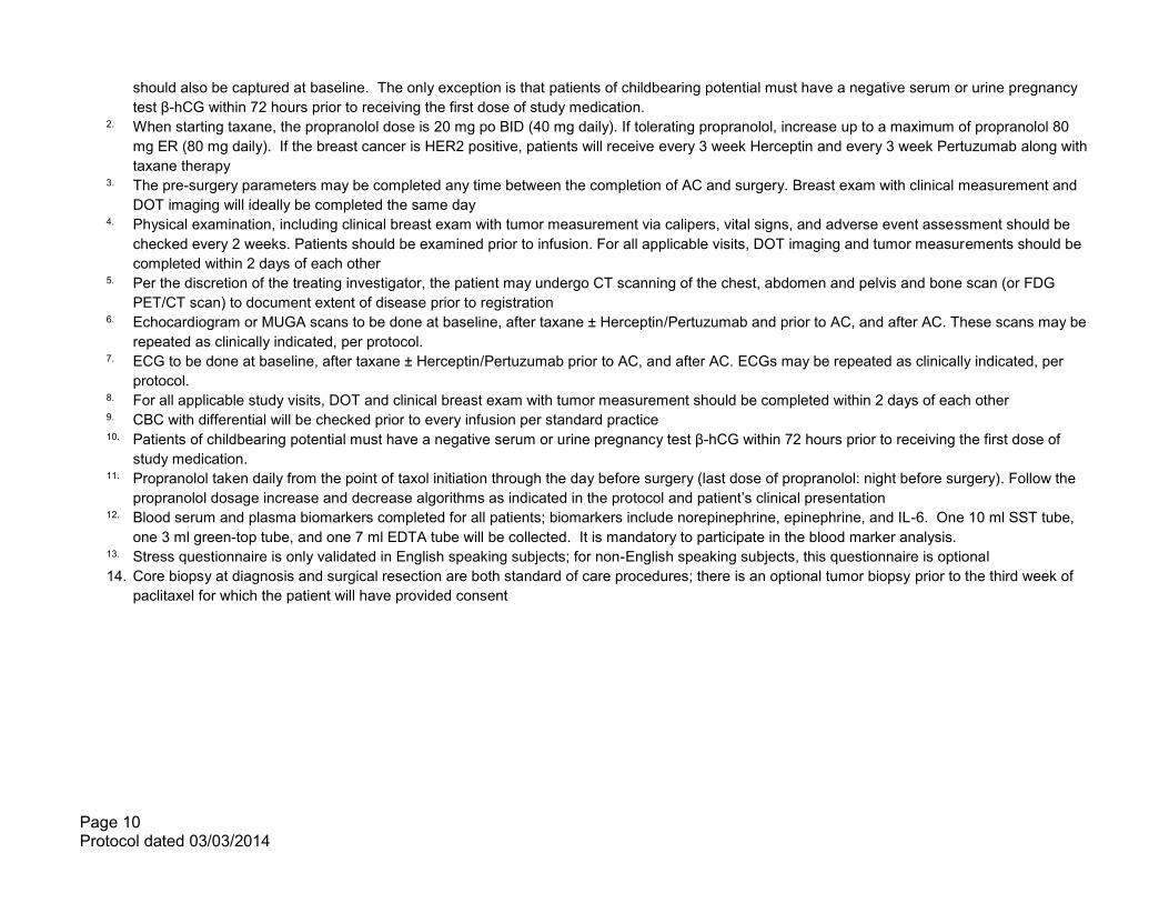
should also be captured at baseline. The only exception is that patients of childbearing potential must have a negative serum or urine pregnancy test β-hCG within 72 hours prior to receiving the first dose of study medication.
2. When starting taxane, the propranolol dose is 20 mg po BID (40 mg daily). If tolerating propranolol, increase up to a maximum of propranolol 80 mg ER (80 mg daily). If the breast cancer is HER2 positive, patients will receive every 3 week Herceptin and every 3 week Pertuzumab along with taxane therapy
3. The pre-surgery parameters may be completed any time between the completion of AC and surgery. Breast exam with clinical measurement and DOT imaging will ideally be completed the same day
4. Physical examination, including clinical breast exam with tumor measurement via calipers, vital signs, and adverse event assessment should be checked every 2 weeks. Patients should be examined prior to infusion. For all applicable visits, DOT imaging and tumor measurements should be completed within 2 days of each other
5. Per the discretion of the treating investigator, the patient may undergo CT scanning of the chest, abdomen and pelvis and bone scan (or FDG PET/CT scan) to document extent of disease prior to registration
6. Echocardiogram or MUGA scans to be done at baseline, after taxane ± Herceptin/Pertuzumab and prior to AC, and after AC. These scans may be repeated as clinically indicated, per protocol.
7. ECG to be done at baseline, after taxane ± Herceptin/Pertuzumab prior to AC, and after AC. ECGs may be repeated as clinically indicated, per protocol.
8. For all applicable study visits, DOT and clinical breast exam with tumor measurement should be completed within 2 days of each other
9. CBC with differential will be checked prior to every infusion per standard practice
10. Patients of childbearing potential must have a negative serum or urine pregnancy test β-hCG within 72 hours prior to receiving the first dose of study medication.
11. Propranolol taken daily from the point of taxol initiation through the day before surgery (last dose of propranolol: night before surgery). Follow the propranolol dosage increase and decrease algorithms as indicated in the protocol and patient’s clinical presentation
12. Blood serum and plasma biomarkers completed for all patients; biomarkers include norepinephrine, epinephrine, and IL-6. One 10 ml SST tube, one 3 ml green-top tube, and one 7 ml EDTA tube will be collected. It is mandatory to participate in the blood marker analysis.
13. Stress questionnaire is only validated in English speaking subjects; for non-English speaking subjects, this questionnaire is optional 14. Core biopsy at diagnosis and surgical resection are both standard of care procedures; there is an optional tumor biopsy prior to the third week of
paclitaxel for which the patient will have provided consent
Page 11 Protocol dated 03/03/2014
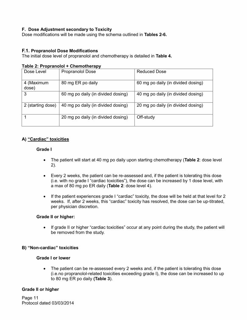
F. Dose Adjustment secondary to Toxicity Dose modifications will be made using the schema outlined in Tables 2-6. F.1. Propranolol Dose Modifications The initial dose level of propranolol and chemotherapy is detailed in Table 4. Table 2: Propranolol + Chemotherapy Dose Level Propranolol Dose Reduced Dose
4 (Maximum dose)
80 mg ER po daily 60 mg po daily (in divided dosing)
3 60 mg po daily (in divided dosing) 40 mg po daily (in divided dosing)
2 (starting dose) 40 mg po daily (in divided dosing) 20 mg po daily (in divided dosing)
1 20 mg po daily (in divided dosing) Off-study
A) “Cardiac” toxicities
Grade I
The patient will start at 40 mg po daily upon starting chemotherapy (Table 2: dose level 2).
Every 2 weeks, the patient can be re-assessed and, if the patient is tolerating this dose
(i.e. with no grade I “cardiac toxicities”), the dose can be increased by 1 dose level, with a max of 80 mg po ER daily (Table 2: dose level 4).
If the patient experiences grade I “cardiac” toxicity, the dose will be held at that level for 2
weeks. If, after 2 weeks, this “cardiac” toxicity has resolved, the dose can be up-titrated, per physician discretion.
Grade II or higher:
If grade II or higher “cardiac toxicities” occur at any point during the study, the patient will
be removed from the study.
B) “Non-cardiac” toxicities
Grade I or lower
The patient can be re-assessed every 2 weeks and, if the patient is tolerating this dose (i.e.no propranolol-related toxicities exceeding grade I), the dose can be increased to up to 80 mg ER po daily (Table 3).
Grade II or higher

Page 12 Protocol dated 03/03/2014
If grade II toxicity occurs, no change in dose is required, per physician discretion.
If grade III or IV toxicity and thought to be related to propranolol, reduce by 1 dose level. This toxicity should be monitored every 2 weeks, with a dose reduction by 1 dose level every 2 weeks until <grade II.
Every 2 weeks, the patient can be re-assessed. If all “non-cardiac” toxicities thought to
be related to propranolol are reduced to< grade I, the dose can be increased by one dose level, per physician discretion. The maximum dose is 80 mg ER po daily.
Table 3: Dose Modification for Propranolol based upon Toxicity Grade
Toxicity Grade
“Cardiac” “Non-cardiac”
I No change in dose while grade I; If resolved after 2 weeks (i.e. grade 0), the dose can be increased by 1 dose level (max: 80 mg daily)
Dose can be increased by 1 dose level every 2 weeks (max: 80 mg daily)
II Off-study No change in dose required; Upon toxicity reduction to < grade I, can increase by 1 dose level every 2 weeks (max: 80 mg daily)
III Off-study Reduce dose by 1 level every 2 weeks until < grade II; Upon reduction to < grade I, can increase by 1 dose level every 2 weeks (max: 80 mg daily)
IV Off-study Same as grade III
F.2. Chemotherapy Dose Modifications Patients remain at starting dose (Table 4: 3b), unless a dose reduction is required for the following toxicities below. Each dose reduction is defined as one dose level reduction (Table 4):
Grade III or IV febrile neutropenia (fever > 38.5C and ANC < 1,000/mm3) between courses. Filgrastim may also be used with weekly paclitaxel
Platelet nadir < 50,000
Grade III or IV neuropathy (hold paclitaxel until < grade 2 neuropathy, reduce paclitaxel dose one dose level, keep propranolol dose stable)
Patients with grade III or IV non-hematologic toxicity (excluding neuropathy and “cardiac”).NOTE: For patients with overlapping toxicities (i.e. possibly related to propranolol and chemotherapy, such as fatigue or dizziness), the timing, nature, and degree of toxicity must be considered. Patients may have modification of propranolol alone, chemotherapy alone, or both depending upon these factors, per physician discretion.
Patients who require a > 4 week delay in therapy due to toxicity should be removed from the protocol.
Page 13 Protocol dated 03/03/2014
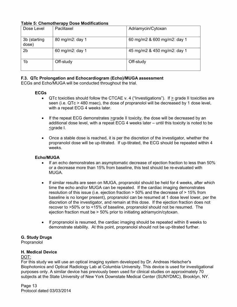
Table 5: Chemotherapy Dose Modifications Dose Level Paclitaxel Adriamycin/Cytoxan
3b (starting dose)
80 mg/m2: day 1 60 mg/m2 & 600 mg/m2: day 1
2b 60 mg/m2: day 1 45 mg/m2 & 450 mg/m2: day 1
1b Off-study Off-study
F.3. QTc Prolongation and Echocardiogram (Echo)/MUGA assessment ECGs and Echo/MUGA will be conducted throughout the trial. ECGs
QTc toxicities should follow the CTCAE v. 4 (“Investigations”). If > grade II toxicities are seen (i.e. QTc > 480 msec), the dose of propranolol will be decreased by 1 dose level, with a repeat ECG 4 weeks later.
If the repeat ECG demonstrates >grade II toxicity, the dose will be decreased by an
additional dose level, with a repeat ECG 4 weeks later – until this toxicity is noted to be <grade I.
Once a stable dose is reached, it is per the discretion of the investigator, whether the
propranolol dose will be up-titrated. If up-titrated, the ECG should be repeated within 4 weeks.
Echo/MUGA
If an echo demonstrates an asymptomatic decrease of ejection fraction to less than 50% or a decrease more than 15% from baseline, this test should be re-evaluated with MUGA.
If similar results are seen on MUGA, propranolol should be held for 4 weeks, after which
time the echo and/or MUGA can be repeated. If the cardiac imaging demonstrates resolution of this issue (i.e. ejection fraction > 50% and the decrease of > 15% from baseline is no longer present), propranolol can be resumed at 1 dose level lower, per the discretion of the investigator, and remain at this dose. If the ejection fraction does not recover to >50% or to <15% of baseline, propranolol should not be resumed. The ejection fraction must be > 50% prior to initiating adriamycin/cytoxan.
If propranolol is resumed, the cardiac imaging should be repeated within 8 weeks to
demonstrate stability. At this point, propranolol should not be up-titrated further. G. Study Drugs Propranolol H. Medical Device DOT: For this study we will use an optical imaging system developed by Dr. Andreas Hielscher's Biophotonics and Optical Radiology Lab at Columbia University. This device is used for investigational purposes only. A similar device has previously been used for clinical studies on approximately 70 subjects at the State University of New York Downstate Medical Center (SUNYDMC), Brooklyn, NY.

Page 14 Protocol dated 03/03/2014
Another system has been used with approximately 40 volunteers here at the Columbia University Medical Center. Measurements were performed on breast, limbs, human heads, and finger joints. These studies were approved by the IRB boards at SUNYDMC and Columbia University Medical Center (current protocol IRB-AAAB0510), respectively. The light intensities and exposure times are within required limits as set by the American National Standard Institute (ANSI) and recommended by the Occupational and Safety & Health Administration (OSHA) of the US Department of Labor. According to these standards (see ANSI Z136.12000, page 48, Table 7, entry for Visible and Near-Infrared light) the maximum permissible exposure (MPE) before any tissue damage occurs is given by MPE = 200x10^[0.002(x700)] mW/cm^2, where x is the wavelength of the light. For example, at x = 800 nm the MPE = 316 mW/cm^2, which is at least 10 times higher than intensities used in this study. Therefore, no adverse effects on the exposed tissues are expected. The Diffuse Optical Tomography machine is located in the Herbert Irving Pavilion, on the same floor as the Breast Oncology clinic. DOT measurements will be made by placing each subject’s breasts into a measurement head containing 96 optical fibers. During the optical measurements, light from four laser diodes (wavelengths 765 - 905nm, with light intensity comparable to that emitted by a conventional light bulb) will be sequentially coupled into 32 fibers that are contacting the breast. Transmitted light intensities will be collected by 64 detection fibers that are coupled to individual silicon photodiodes, which record the light intensities. The measured data is then processed and produces a cross sectional image of the breast’s optical properties from which values for water, oxyhemoglobin, deoxyhemoglobin and fat concentrations can be determined. Levels of each component will be quantified by selecting a region of interest (the area surrounding the highest concentration) and calculating the mean concentration for that region. Dynamic measurement of DOT will be made during a 30 second breath hold, and the recovery period 5 minutes after breath hold. Explaining the procedure, positioning the breast in the imaging head, setting up and conducting the measurements takes about 30 minutes and produces minimal discomfort. I. Study Subjects Inclusion criteria:
1. English or Spanish speaking women age ≥18
2. Heart Rate > 60 bpm 3. Systolic Blood Pressure > 100 mm/Hg 4. Deemed eligible to receive neoadjuvant chemotherapy with 12 cycles of weekly taxane therapy
(paclitaxel 80mg/m2 or abraxane 100 mg/m2 if there is a shortage of paclitaxel) followed by 4 cycles of adriamycin (60mg/m2) and cyclophosphamide (600 mg/m2) given every 2 weeks with growth-factor support.
5. Echo or MUGA with ejection fraction > 50%. 6. Patients with hormone receptor +/- and HER2 +/- breast cancer are eligible 7. If a patient has HER2-positive breast cancer, Herceptin and Pertuzumab will be given along with
taxane therapy 8. Any stage invasive breast cancer provided the primary breast tumor size is ≥ 1 cm 10. Agree to participate in research blood collection at 4 different time periods (20 ml = 4 teaspoons) 11. Agree to the evaluation of already collected core biopsy, as well as surgical resection tissue, for predictive biomarkers. The biopsy prior to Taxol #1 is optional.
Exclusion criteria:
1. Patients failing to meet the inclusion criteria

Page 15 Protocol dated 03/03/2014
2. QTc prolongation as defined by > 470 milliseconds on ECG 3. First degree AV block on ECG in which PR interval lengthened > 200 milliseconds; Second
Degree; or Third Degree 4. On beta-blocker treatment. If discontinued, patients must have been off beta-blockers for at
least 3 months. 5. History of asthma, given concern for β-blockade in this population
J. Recruitment of Subjects Patients will be recruited directly by their surgical or medical oncologists at Columbia University Medical Center (CUMC). In the Breast Oncology Clinic of CUMC, we estimate an accrual rate of 2 patients per month over a 12- month period. Therefore, we anticipate the accrual of 20 patients is feasible and that the recruitment period will be completed within 1 year. K. Confidentiality of Study Data The research file that links subject's name to the code number will be kept in a locked file cabinet on the 9th floor of the Herbert Irving Pavilion and only the investigator and study staff will have access to the file. The study data collected, specimens and questionnaire responses will be assigned a code number, and separated from the patient name or any other information that could identify them. If the results of this research project are published or presented at a scientific or medical meeting, the patient will not be identified. Otherwise, all results will be kept confidential and will not be divulged (except as required by law) without permission. Except when required by law, study information shared with persons and organizations outside of Columbia University Medical Center will not identify the patient by name, social security number, address, telephone number, or any other direct personal identifier. L. Potential Conflict of Interest None M. Location of the Study The study will be conducted at Columbia University Medical Center (CUMC). N. Potential Risk Potential Side effects include the following: Propranolol Less Likely (5-10%): Dizziness Joint Ache (Arthralgia) Headache Chest Discomfort Coughing Fatigue Back Pain Rare (1-4%): Depression Belly Pain (Abdominal Pain) Shortness of Breath (Dyspnea) Muscle Ache (Myalgia) Heart Rate Slowing (Bradycardia) Nausea
Page 16 Protocol dated 03/03/2014
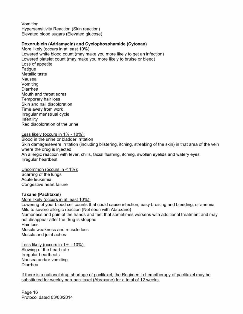
Vomiting Hypersensitivity Reaction (Skin reaction) Elevated blood sugars (Elevated glucose) Doxorubicin (Adriamycin) and Cyclophosphamide (Cytoxan) More likely (occurs in at least 10%): Lowered white blood count (may make you more likely to get an infection) Lowered platelet count (may make you more likely to bruise or bleed) Loss of appetite Fatigue Metallic taste Nausea Vomiting Diarrhea Mouth and throat sores Temporary hair loss Skin and nail discoloration Time away from work Irregular menstrual cycle Infertility Red discoloration of the urine Less likely (occurs in 1% - 10%): Blood in the urine or bladder irritation Skin damage/severe irritation (including blistering, itching, streaking of the skin) in that area of the vein where the drug is injected An allergic reaction with fever, chills, facial flushing, itching, swollen eyelids and watery eyes Irregular heartbeat Uncommon (occurs in < 1%): Scarring of the lungs Acute leukemia Congestive heart failure Taxane (Paclitaxel) More likely (occurs in at least 10%): Lowering of your blood cell counts that could cause infection, easy bruising and bleeding, or anemia Mild to severe allergic reaction (Not seen with Abraxane) Numbness and pain of the hands and feet that sometimes worsens with additional treatment and may not disappear after the drug is stopped Hair loss Muscle weakness and muscle loss Muscle and joint aches Less likely (occurs in 1% - 10%): Slowing of the heart rate Irregular heartbeats Nausea and/or vomiting Diarrhea If there is a national drug shortage of paclitaxel, the Regimen I chemotherapy of paclitaxel may be substituted for weekly nab-paclitaxel (Abraxane) for a total of 12 weeks.

Page 17 Protocol dated 03/03/2014
Abraxane (Nab-Paclitaxel) More Likely (at least 10%) Lowering of your blood cell counts that could cause infection, easy bruising and bleeding, or anemia Numbness and pain of the hands and feet that sometimes worsens with additional treatment and may not disappear after the drug is stopped Hair loss Muscle weakness and muscle loss Muscle and joint aches Less Likely (1-10%) Slowing of the heart rate (bradycardia) Irregular heartbeats Nausea and/or vomiting Diarrhea Sores in the mouth or throat Fatigue Lightheadedness Headaches Sensation of flashing lights or spots Changes in kidney function tests Increase in triglyceride (blood lipid) levels Changes in liver enzymes Skin irritation and swelling if the drug leaks from the vein into which it is being injected into the surrounding skin Rare (<1%) Liver damage or failure Seizure Herceptin (Trastuzumab: if breast cancer is HER2 positive) More Likely
Chills and/or fever with the infusion Nausea Vomiting Chills Headache Dizziness Shortness of breath Rash Pain (generalized, abdominal, back and muscle) Diarrhea Swelling in the ankles/legs Weakness Allergic reactions that include rash, itching and hives/swelling of the skin
Rare Hypersensitivity reaction (skin reaction) Abnormal liver function blood tests, hepatitis (inflammation of the liver) Bone pain

Page 18 Protocol dated 03/03/2014
Pain in the tumor Decreased red blood cells, which can cause anemia Decreased white blood cells, which can cause increased risk of developing an infection
Decreased platelets, which can cause an increased tendency to bleed Difficulty breathing Fluid leaking into the lungs making it difficult to breathe. In the most severe form, this could
require oxygen from a ventilator to breathe. This could lead to death Serious heart failure that may not be reversible.
Perjeta (Pertuzumab: if breast cancer is HER2 positive) More Likely
Diarrhea Tiredness
Less Likely Anemia which may require blood transfusion Pain Heartburn, nausea, vomiting Sores in mouth which may cause difficulty swallowing Chills, fever Allergic reaction which may cause rash, low blood pressure, wheezing, shortness of breath, Swelling of the face or throat Infection, especially when white blood cell count is low Change in heart function Loss of appetite Headache Shortness of breath Itching, rash, hives
Rare Heart failure, heart attack which may cause shortness of breath, swelling of ankles, and
tiredness Filgrastim (Neupogen) or pegfilgrastim (Neulasta) More likely (occurs in at least 10%) Bone pain Increase in some blood tests (uric acid, alkaline phosphatase, lactate dehydrogenase) Rare (< 1%) Temporarily decreased blood pressure Other Risks: Dexamethasone (Decadron) More likely (> 20%) Decreased or increased appetite Difficulty sleeping An excess of sugar in the blood An exaggerated feeling of well-being Headache, dizziness
Page 19 Protocol dated 03/03/2014
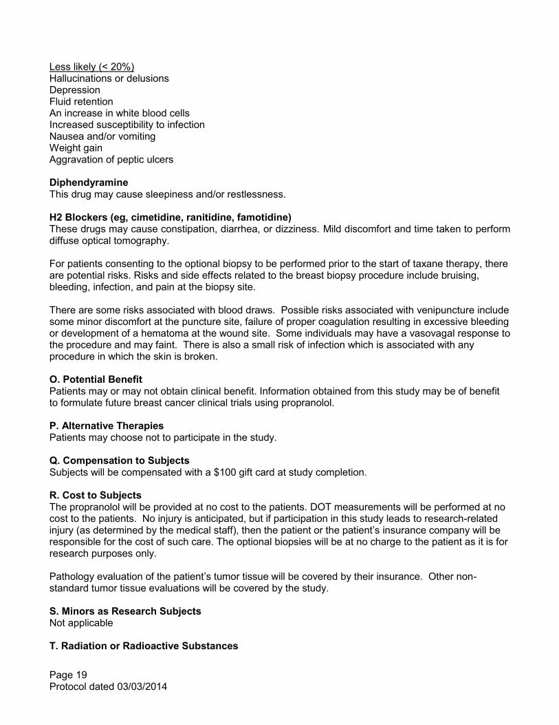
Less likely (< 20%) Hallucinations or delusions Depression Fluid retention An increase in white blood cells Increased susceptibility to infection Nausea and/or vomiting Weight gain Aggravation of peptic ulcers Diphendyramine This drug may cause sleepiness and/or restlessness. H2 Blockers (eg, cimetidine, ranitidine, famotidine) These drugs may cause constipation, diarrhea, or dizziness. Mild discomfort and time taken to perform diffuse optical tomography. For patients consenting to the optional biopsy to be performed prior to the start of taxane therapy, there are potential risks. Risks and side effects related to the breast biopsy procedure include bruising, bleeding, infection, and pain at the biopsy site. There are some risks associated with blood draws. Possible risks associated with venipuncture include some minor discomfort at the puncture site, failure of proper coagulation resulting in excessive bleeding or development of a hematoma at the wound site. Some individuals may have a vasovagal response to the procedure and may faint. There is also a small risk of infection which is associated with any procedure in which the skin is broken. O. Potential Benefit Patients may or may not obtain clinical benefit. Information obtained from this study may be of benefit to formulate future breast cancer clinical trials using propranolol. P. Alternative Therapies Patients may choose not to participate in the study. Q. Compensation to Subjects Subjects will be compensated with a $100 gift card at study completion. R. Cost to Subjects The propranolol will be provided at no cost to the patients. DOT measurements will be performed at no cost to the patients. No injury is anticipated, but if participation in this study leads to research-related injury (as determined by the medical staff), then the patient or the patient’s insurance company will be responsible for the cost of such care. The optional biopsies will be at no charge to the patient as it is for research purposes only. Pathology evaluation of the patient’s tumor tissue will be covered by their insurance. Other non-standard tumor tissue evaluations will be covered by the study. S. Minors as Research Subjects Not applicable T. Radiation or Radioactive Substances

Page 20 Protocol dated 03/03/2014
The patients will be exposed to radiation or radioactive substances only with mammography and MUGA scans (if they undergo MUGA). DOT does not use ionizing radiation. With DOT the intensity of light coupled into each breast is approximately 10-30mW/cm2 for about 10 seconds at each fiber end. This light intensity if comparable to that emitted by a conventional light bulb and is well below the threshold for injury as specified by ANSI and OSHA. U. Plans for Monitoring Data and Safety The HICCC NCI approved Data Safety Monitoring Committee (DSMC) will oversee the conduct of this trial. This protocol will adhere to the policies of the HICCC Cancer Center Data and Safety Monitoring Plan, version 2 guidelines in accordance with NIC regulations. The committee is led by Dr. Gregory Mears and consists of HICCC members. The DSMC meets monthly to review adverse event reporting and the timeliness of adverse event reporting. The PI will submit data and safety monitoring reports to the DSMC. All unanticipated problems will be reported to the CUMC IRB as per the latest IRB policy and according to the FDA 21 CFR 812.46. V. References: 1. Twombly R. Avastin's Uncertain Future in Breast Cancer Treatment. Journal of the National Cancer Institute 2011. 2. Sloan EK, Priceman SJ, Cox BF, et al. The sympathetic nervous system induces a metastatic switch in primary breast cancer. Cancer Res 2010;70:7042-52. 3. Thaker PH, Han LY, Kamat AA, et al. Chronic stress promotes tumor growth and angiogenesis in a mouse model of ovarian carcinoma. Nat Med 2006;12:939-44. 4. Lee K. Optical mammography: Diffuse optical imaging of breast cancer. World J Clin Oncol 2011;2:64-72. 5. Cerussi A, Hsiang D, Shah N, et al. Predicting response to breast cancer neoadjuvant chemotherapy using diffuse optical spectroscopy. Proc Natl Acad Sci U S A 2007;104:4014-9. 6. Molly L. Flexman HKK, Jonghwan Lee, Sonia L. Hernandez, Jianzhong Huang, Tessa J. Johung, Darrell J. Yamashiro, Jessica J. Kandel, Andreas H. Hielscher, "Optical tomographic monitoring of vascular responses to anti-angiogenic drugs in preclinical tumor models," in Optical Tomography and Spectroscopy of Tissue IX, edited by Bruce J. Tromberg, Arjun G. Yodh, Mamoru Tamura, Eva M. Sevick-Muraca, Robert R. Alfano, Proc. SPIE 7896, 789627(2011). 7. Soliman H, Gunasekara A, Rycroft M, et al. Functional Imaging Using Diffuse Optical Spectroscopy of Neoadjuvant Chemotherapy Response in Women with Locally Advanced Breast Cancer. Clinical Cancer Research 2010;16:2605-14. 8. Zhu Q, Tannenbaum S, Hegde P, Kane M, Xu C, Kurtzman SH. Noninvasive monitoring of breast cancer during neoadjuvant chemotherapy using optical tomography with ultrasound localization. Neoplasia 2008;10:1028-40. 9. Pakalniskis MG, Wells WA, Schwab MC, et al. Tumor angiogenesis change estimated by using diffuse optical spectroscopic tomography: demonstrated correlation in women undergoing neoadjuvant chemotherapy for invasive breast cancer? Radiology 2011;259:365-74. 10. Jiang S, Pogue BW, Carpenter CM, et al. Evaluation of breast tumor response to neoadjuvant chemotherapy with tomographic diffuse optical spectroscopy: case studies of tumor region-of-interest changes. Radiology 2009;252:551-60. 11. Weidner N, Folkman J, Pozza F, et al. Tumor angiogenesis: a new significant and independent prognostic indicator in early-stage breast carcinoma. J Natl Cancer Inst 1992;84:1875-87. 12. Uzzan B, Nicolas P, Cucherat M, Perret GY. Microvessel density as a prognostic factor in women with breast cancer: a systematic review of the literature and meta-analysis. Cancer Res 2004;64:2941-55. 13. Miller KD. E2100: a phase III trial of paclitaxel versus paclitaxel/bevacizumab for metastatic breast cancer. Clin Breast Cancer 2003;3:421-2.

Page 21 Protocol dated 03/03/2014
14. Pivot X, Schneeweiss A, Verma S, et al. Efficacy and safety of bevacizumab in combination with docetaxel for the first-line treatment of elderly patients with locally recurrent or metastatic breast cancer: Results from AVADO. Eur J Cancer 2011;47:2387-95. 15. Robert NJ, Diéras V, Glaspy J, et al. RIBBON-1: Randomized, Double-Blind, Placebo-Controlled, Phase III Trial of Chemotherapy With or Without Bevacizumab for First-Line Treatment of Human Epidermal Growth Factor Receptor 2–Negative, Locally Recurrent or Metastatic Breast Cancer. Journal of Clinical Oncology 2011. 16. Valachis A, Polyzos NP, Patsopoulos NA, Georgoulias V, Mavroudis D, Mauri D. Bevacizumab in metastatic breast cancer: a meta-analysis of randomized controlled trials. Breast Cancer Res Treat 2010;122:1-7. 17. Dedes KJ, Matter-Walstra K, Schwenkglenks M, et al. Bevacizumab in combination with paclitaxel for HER-2 negative metastatic breast cancer: An economic evaluation. European Journal of Cancer 2009;45:1397-406. 18. Benish M, Bartal I, Goldfarb Y, et al. Perioperative use of beta-blockers and COX-2 inhibitors may improve immune competence and reduce the risk of tumor metastasis. Ann Surg Oncol 2008;15:2042-52. 19. Powe DG, Voss MJ, Zanker KS, et al. Beta-blocker drug therapy reduces secondary cancer formation in breast cancer and improves cancer specific survival. Oncotarget 2010;1:628-38. 20. Ganz PA, Habel LA, Weltzien EK, Caan BJ, Cole SW. Examining the influence of beta blockers and ACE inhibitors on the risk for breast cancer recurrence: results from the LACE cohort. Breast Cancer Res Treat 2011. 21. Melhem-Bertrandt A, Chavez-Macgregor M, Lei X, et al. Beta-blocker use is associated with improved relapse-free survival in patients with triple-negative breast cancer. J Clin Oncol 2011;29:2645-52. 22. Barron TI, Connolly RM, Sharp L, Bennett K, Visvanathan K. Beta blockers and breast cancer mortality: a population- based study. J Clin Oncol 2011;29:2635-44. 23. Drell TLt, Joseph J, Lang K, Niggemann B, Zaenker KS, Entschladen F. Effects of neurotransmitters on the chemokinesis and chemotaxis of MDA-MB-468 human breast carcinoma cells. Breast Cancer Res Treat 2003;80:63-70. 24. Sood AK, Armaiz-Pena GN, Halder J, et al. Adrenergic modulation of focal adhesion kinase protects human ovarian cancer cells from anoikis. J Clin Invest 2010;120:1515-23. 25. Badino GR, Novelli A, Girardi C, Di Carlo F. Evidence for functional beta-adrenoceptor subtypes in CG-5 breast cancer cell. Pharmacol Res 1996;33:255-60. 26. Shi M, Liu D, Duan H, et al. The beta2-adrenergic receptor and Her2 comprise a positive feedback loop in human breast cancer cells. Breast Cancer Res Treat 2011;125:351-62. 27. Wang J, Jiang S, Li Z, et al. In vivo quantitative imaging of normal and cancerous breast tissue using broadband diffuse optical tomography. Med Phys 2010;37:3715-24. 28. Zhu Q, Cronin EB, Currier AA, et al. Benign versus malignant breast masses: optical differentiation with US-guided optical imaging reconstruction. Radiology 2005;237:57-66. 29. Scholl SM, Fourquet A, Asselain B, et al. Neoadjuvant versus adjuvant chemotherapy in premenopausal patients with tumours considered too large for breast conserving surgery: preliminary results of a randomised trial: S6. Eur J Cancer 1994;30A:645-52. 30. Shabo I, Stal O, Olsson H, Dore S, Svanvik J. Breast cancer expression of CD163, a macrophage scavenger receptor, is related to early distant recurrence and reduced patient survival. Int J Cancer 2008;123:780-6. 31. Fredriksson JM, Lindquist JM, Bronnikov GE, Nedergaard J. Norepinephrine induces vascular endothelial growth factor gene expression in brown adipocytes through a beta -adrenoreceptor/cAMP/protein kinase A pathway involving Src but independently of Erk1/2. J Biol Chem 2000;275:13802-11.
Page 22 Protocol dated 03/03/2014
32. Herschbach P, Keller M, Knight L, et al. Psychological problems of cancer patients: a cancer distress screening with a cancer-specific questionnaire. Br J Cancer 2004;91:504-11. 33. Hartl K, Engel J, Herschbach P, Reinecker H, Sommer H, Friese K. Personality traits and psychosocial stress: quality of life over 2 years following breast cancer diagnosis and psychological impact factors. Psychooncology 2010;19:160-9. 34. Neugut AI, Subar M, Wilde ET, et al. Association Between Prescription Co-Payment Amount and Compliance With Adjuvant Hormonal Therapy in Women With Early-Stage Breast Cancer. Journal of Clinical Oncology 2011;29:2534-42. 35. Baselga J, Semiglazov V, van Dam P, et al. Phase II Randomized Study of Neoadjuvant Everolimus Plus Letrozole Compared With Placebo Plus Letrozole in Patients With Estrogen Receptor–Positive Breast Cancer. Journal of Clinical Oncology 2009;27:2630-7. 36. Schwarz ER, Kersting PH, Reffelmann T, et al. Cardioprotection by Carvedilol: antiapoptosis is independent of beta-adrenoceptor blockage in the rat heart. J Cardiovasc Pharmacol Ther 2003;8:207-15. 37. Somberg J, Cagin N, Levitt B, et al. Blockade of tissue uptake of the antineoplastic agent, doxorubicin. J Pharmacol Exp Ther 1978;204:226-9. 38. Wikman-Coffelt J, Rapcsak M, Sievers R, Rouleau JL, Parmley WW. Verapamil, propranolol, and hydralazine protect against the acute cardiac depression induced by adriamycin. Cardiovasc Res 1983;17:43-9. 39. Kawabata H, Ryomoto T, Ishikawa K. Effect of beta-blocker on metabolism and contraction of doxorubicin-induced cardiotoxicity in the isolated perfused rabbit heart. Angiology 2000;51:405-13. 40. Choe JY, Combs AB, Folkers K. Potentiation of the toxicity of adriamycin by propranolol. Res Commun Chem Pathol Pharmacol 1978;21:577-80. 41. Bernstein D, Fajardo G, Zhao M, et al. Differential cardioprotective/cardiotoxic effects mediated by beta-adrenergic receptor subtypes. Am J Physiol Heart Circ Physiol 2005;289:H2441-9. 42. Georgakopoulos P, Roussou P, Matsakas E, et al. Cardioprotective effect of metoprolol and enalapril in doxorubicin-treated lymphoma patients: A prospective, parallel-group, randomized, controlled study with 36-month follow-up. American Journal of Hematology 2010;85:894-6. 43. Pituskin E, Haykowsky M, Mackey J, et al. Rationale and design of the Multidisciplinary Approach to Novel Therapies in Cardiology Oncology Research Trial (MANTICORE 101 - Breast): a randomized, placebo-controlled trial to determine if conventional heart failure pharmacotherapy can prevent trastuzumab-mediated left ventricular remodeling among women with HER2+ early breast cancer using cardiac MRI. BMC Cancer 2011;11:318.