Title: Bacterial membrane vesicles deliver peptidoglycan ... · 1 Title: Bacterial membrane...
54
1 Title: Bacterial membrane vesicles deliver peptidoglycan to NOD1 in epithelial cells. Maria Kaparakis 1* , Lynne Turnbull 2 , Leticia Carneiro 3 , Stephen Firth 4 , Harold A. Coleman 5 , Helena C. Parkington 5 , Lionel Le Bourhis 6 , Abdulgader Karrar 1 , Jérôme Viala 7 , Johnson Mak 8 , Melanie L. Hutton 1* , John K. Davies 1,9 , Peter J. Crack 10 , Paul J. Hertzog 11 , Dana J. Philpott 6 , Stephen E. Girardin 3 , Cynthia B. Whitchurch 2 and Richard L. Ferrero 1* 1 Department of Microbiology, Monash University, Clayton, Victoria 3800, Australia 2 Institute for the Biotechnology of Infectious Diseases, University of Technology, Broadway, New South Wales 2007, Australia 3 Department of Laboratory Medicine and Pathobiology, University of Toronto, Ontario M5S1A8, Canada 4 Monash Micro Imaging, Monash University, Clayton, Victoria 3800, Australia 5 Department of Physiology, Monash University, Clayton, Victoria 3800, Australia 6 Department of Immunology, University of Toronto, Ontario M5S1A8, Canada 7 Service de Gastroentérologie, Nutrition et Mucoviscidose, Hôpital Robert Debré, Paris 75935, France 8 The Macfarlane Burnet Institute for Medical Research and Public Health, Melbourne, Victoria 3004, Australia 9 Australian Bacterial Pathogenesis Program 10 Department of Pharmacology, The University of Melbourne, Parkville, Victoria 3010, Australia 11 Centre for Innate Immunity and Infectious Disease, Monash Institute of Medical Research, Monash University, Victoria 3800, Australia
Transcript of Title: Bacterial membrane vesicles deliver peptidoglycan ... · 1 Title: Bacterial membrane...

1
Title: Bacterial membrane vesicles deliver peptidoglycan
to NOD1 in epithelial cells.
Maria Kaparakis1*, Lynne Turnbull2, Leticia Carneiro3, Stephen Firth4, Harold A.
Coleman5, Helena C. Parkington5, Lionel Le Bourhis6, Abdulgader Karrar1, Jérôme
Viala7, Johnson Mak8, Melanie L. Hutton1*, John K. Davies1,9, Peter J. Crack10, Paul J.
Hertzog11, Dana J. Philpott6, Stephen E. Girardin3, Cynthia B. Whitchurch2 and
Richard L. Ferrero1*
1 Department of Microbiology, Monash University, Clayton, Victoria 3800, Australia
2 Institute for the Biotechnology of Infectious Diseases, University of Technology,
Broadway, New South Wales 2007, Australia
3 Department of Laboratory Medicine and Pathobiology, University of Toronto,
Ontario M5S1A8, Canada
4 Monash Micro Imaging, Monash University, Clayton, Victoria 3800, Australia
5 Department of Physiology, Monash University, Clayton, Victoria 3800, Australia
6 Department of Immunology, University of Toronto, Ontario M5S1A8, Canada
7 Service de Gastroentérologie, Nutrition et Mucoviscidose, Hôpital Robert Debré,
Paris 75935, France
8 The Macfarlane Burnet Institute for Medical Research and Public Health,
Melbourne, Victoria 3004, Australia
9 Australian Bacterial Pathogenesis Program
10 Department of Pharmacology, The University of Melbourne, Parkville, Victoria
3010, Australia
11 Centre for Innate Immunity and Infectious Disease, Monash Institute of Medical
Research, Monash University, Victoria 3800, Australia

2
* Current address, Centre for Innate Immunity and Infectious Disease, Monash
Institute of Medical Research, Clayton Victoria, Australia
Contact Dr Richard Ferrero
Centre for Innate Immunity and Infectious Disease, Monash Institute of Medical
Research, 27-31 Wright St, Clayton Victoria 3168, Australia
Tel. 61 3 9594 7721; Fax. 61 3 9594 7211
Running title NOD1 detects bacterial vesicles

3
Summary
Gram negative bacterial peptidoglycan is specifically recognized by the host
intracellular sensor NOD1, resulting in the generation of innate immune responses.
Although epithelial cells are normally refractory to external stimulation with
peptidoglycan, these cells have been shown to respond in a NOD1 dependent manner
to Gram negative pathogens that can either invade or secrete factors into host cells. In
the present work, we report that Gram negative bacteria can deliver peptidoglycan to
cytosolic NOD1 in host cells via a novel mechanism involving outer membrane
vesicles (OMVs). We purified OMVs from the Gram negative mucosal pathogens:
Helicobacter pylori, Pseudomonas aeruginosa and Neisseria gonorrhoea and
demonstrated that these peptidoglycan containing OMVs upregulated NF-!B and
NOD1 dependent responses in vitro. These OMVs entered epithelial cells through
lipid rafts thereby inducing NOD1 dependent responses in vitro. Moreover, OMVs
delivered intragastrically to mice induced innate and adaptive immune responses via a
NOD1 dependent but TLR independent mechanism. Collectively, our findings
identify OMVs as a generalized mechanism whereby Gram-negative bacteria deliver
peptidoglycan to cytosolic NOD1. We propose that OMVs released by bacteria
in vivo may promote inflammation and pathology in infected hosts.

4
Introduction
The cytosolic host protein nucleotide binding oligomerization domain 1 (NOD1) has
emerged as a key pathogen recognition molecule (PRM) for innate immune responses
in epithelial cells (Fritz et al., 2006). This protein acts as an intracellular “sensor” of
bacterial pathogens through its recognition of cell wall peptidoglycan (PG). As a
result of detailed molecular studies, human NOD1 was shown to exhibit exquisite
specificity for a diaminopimelate containing GlcNAc-MurNAc tripeptide (GM-TriDAP)
motif that is almost exclusively found in Gram negative bacterial PG (Magalhaes et
al., 2005). Although the specificity of the NOD1 ligand has been determined, the
mechanisms whereby PG enters host cells and induces innate immune signaling
during physiological conditions remain poorly understood.
Epithelial cells are generally refractory to external stimulation with microbial
products, such as PG and lipopolysaccharide (LPS). The addition of synthetic or
natural NOD1 agonists to the exterior of non-phagocytic epithelial cells in culture has
no effect on initiating NOD1 signaling in these cells (Girardin et al., 2003). Certain
bacterial pathogens, however, are able to trigger cytosolic NOD1 signaling in
epithelial cells by either cell invasion (Girardin et al., 2001), or via the actions of a
bacterial secretion system (Viala et al., 2004), suggesting that the intracellular
presentation of PG may be required for signaling in these cells.
We previously reported that H. pylori utilizes the Type IV secretion system
(T4SS) encoded by the “cag” pathogenicity island (cagPAI), to deliver PG to host
cytosolic NOD1. The process of H. pylori transferring its PG into the host epithelial
cell, and its subsequent recognition by NOD1, results in the activation of NF-!B and
the production of interleukin-8 (IL-8) (Viala et al., 2004). However, findings of that
study also indicated that another mechanism independent of the T4SS may exist

5
which is utilized by bacteria to transport PG into non-phagocytic host epithelial cells
(Viala et al., 2004). Specifically, we reported that H. pylori 251 harboring a non-
functional cagPAI, were still able to deliver radiolabeled PG to epithelial cells albeit
with lower efficiency to bacteria with a functional T4SS (Viala et al., 2004). This
finding would be consistent with the ability of H. pylori bacteria without a functional
T4SS to induce weak NOD1 responses in epithelial cells (Chaouche-Drider et al.,
2009). Moreover, the existence of a T4SS independent mechanism for NOD1
activation may provide and explanation for the as yet unresolved question of how
cagPAI negative H. pylori are able to initiate inflammation and pathology in vivo
(Backert et al., 2004; Yamaoka et al., 1997).
Previous studies showed that the microinjection of bacteria free supernatants
of Shigella flexneri induced pro-inflammatory responses in epithelial cells (Girardin
et al., 2003; Philpott et al., 2000), suggesting that bacteria may secrete PG in a form
that is suitable for NOD1 recognition. Although the supernatant associated material
responsible for this activity was not identified, it was hypothesized that these bacteria
free supernatants may contain outer membrane vesicles (OMVs) (Philpott et al.,
2000).
OMVs, or “blebs,” are shed by Gram negative bacteria during normal growth,
and have been reported to enter and transport virulence factors into host cells (Kuehn
and Kesty, 2005). Given that OMVs contain numerous components of the bacterial
cell wall (Kuehn and Kesty, 2005; Keenan et al., 2000), including PG hydrolyzing
enzymes (Li et al., 1996), we speculated that OMVs may be involved in the
intracellular delivery of PG to NOD1. In this study, we identify OMVs as a novel
mechanism whereby all H. pylori irrespective of their cagPAI status, and indeed all
Gram negative bacteria, may transport PG intracellularly so as to initiate NOD1

6
dependent NF-!B responses in non-phagocytic epithelial cells. We report that OMVs
enter host epithelial cells via lipid rafts to transport their PG to NOD1 and induce pro-
inflammatory responses. Using MyD88 and MAL knockout animals we determined
that TLRs do not play a role in OMV induced innate immune responses in vivo.
Furthermore, we identified NOD1 as being essential for the development of innate
and adaptive immune responses to bacterial OMVs in vivo. Therefore, we propose
that OMVs are a novel mechanism whereby Gram negative bacteria can transport
their PG into the cytoplasm of non-phagocytic epithelial cells and initiate NOD1
dependent innate and adaptive immune responses in vivo.
Results
OMVs activate NF-!B dependent responses in non-phagocytic cells.
To determine whether OMVs can initiate NOD1 signaling in non-phagocytic
cells, we purified OMVs from clinical and laboratory isolates of the Gram negative
bacterium, H. pylori (Fig. S1A). This pathogen was previously shown to induce
NOD1 signaling in epithelial cells via a bacterial T4SS encoded by the cagPAI (Viala
et al., 2004). As we wished to identify a potentially T4SS independent mechanism for
NOD1 signaling, we purified OMVs from cagPAI positive and cagPAI negative
H. pylori strains, as well as from a cagPAI isogenic mutant (see below). The OMVs
purified from H. pylori bacteria displayed a spherical, bi-layered morphology. All
H. pylori OMVs separated by SDS-PAGE and subjected to Western blot analysis
displayed a similar protein content and were largely devoid of the abundant
cytoplasmic protein, urease, which can induce pro-inflammatory effects on gastric
epithelial cells (Beswick et al., 2006) (Fig. S1B and C).

7
Next, the pro-inflammatory activities of H. pylori OMVs were determined by
measuring nuclear factor-!B (NF-!B) dependent responses in AGS and HEK239
epithelial cell lines with functional NOD1 signaling (Girardin et al., 2001; Girardin et
al., 2003). As a control, AGS cells were stimulated with live cagPAI positive
H. pylori 251 bacteria, which were shown previously to activate NF-!B responses via
a NOD1 dependent mechanism (Viala et al., 2004) (Fig. 1). The external application
of H. pylori OMVs, from cagPAI positive and negative strains, to epithelial cells
induced significant NF-!B reporter activity compared to non-stimulated control cells
(Fig. 1A). Therefore, OMVs purified from all H. pylori strains, irrespective of their
cagPAI status, were capable of inducing NF-!B reporter activity. The variability in
NF-!B reporter activity induced by OMVs from different strains correlated with
previous findings describing the ability of these individual isolates to induce varying
levels of NF-!B reporter activity and IL-8 production (Philpott et al., 2002).
To confirm the role of a cagPAI independent mechanism for OMV induced
responses in cells, we prepared OMVs from an isogenic cagPAI deletion mutant of
the H. pylori 251 clinical isolate. H. pylori 251 cagPAI OMVs induced the up-
regulation of several NF-!B dependent pro-inflammatory responses, as measured by
human-"-defensin 2 (hBD2) and hBD3 reporter activity, as well as IL-8 production
(Fig. 1B and 1C). In contrast, spent OMV free culture media or killed H. pylori 251
bacteria added directly to cells had no effect on IL-8 production (Fig. 1C).
Furthermore, the microinjection of H. pylori 251 cagPAI OMVs, but not the NOD1
active motif of PG (GM-TriDAP) or culture medium alone, induced the nuclear
translocation of the p65 subunit of NF-!B in epithelial cells (Fig. S2). These data
demonstrated the ability of internalized OMVs to initiate NF-!B-dependent
pro-inflammatory responses in epithelial cells.

8
OMVs specifically induce NOD1 dependent responses in non-phagocytic
cells.
As the epithelial cell lines in the preceding studies express functional NOD1,
and that OMVs were postulated to contain PG, we next sought to investigate the role
of this PRM in OMV induced responses in epithelial cells and mouse embryonic
fibroblasts (MEFs), with normal or impaired NOD1 signaling. NOD1 functionality
was altered in these cells by either: expression of a dominant negative NOD1
construct (Viala et al., 2004) (Fig. 2A); stable knock-down of NOD1 expression
(unpublished data) (Fig. 2B and C); or by gene disruption (Fig. 2D-F). In all
instances, NF-!B dependent responses to H. pylori OMVs were significantly reduced
in cells with impaired NOD1 signaling, when compared with wild type cells (P<0.05;
Fig. 2A-F). These responses were shown to be independent of a cagPAI encoded
secretion system (Fig. 2A-F). NOD1 dependency was also observed for AGS and
MEF cell responses to OMVs from the Gram negative bacteria Neisseria gonorrhoea
and Pseudomonas aeruginosa, which harbor PG with NOD1 agonist activity
(Girardin et al., 2003; Travassos et al., 2005) (Fig. S3A and B respectively; P<0.05,
Fig. 2B and 2D-F). Taken together, the data highlight the broad relevance of Gram
negative bacterial OMVs as mediators of NOD1 dependent NF-!B responses in non-
phagocytic cells. Furthermore, the findings reveal for the first time a potential
mechanism by which H. pylori may drive the gastritis observed in cagPAI negative
infections (Backert et al., 2004; Yamaoka et al., 1997).
Gram negative bacterial PG associated with OMVs is responsible for the NOD1
dependent NF-!B response induced in epithelial cells.

9
Consistent with previous reports (Kuehn and Kesty, 2005; Keenan et al.,
2000) OMVs from H. pylori, P. aeruginosa and N. gonorrhoea contained proteins,
whereas those from the latter two bacteria also contained DNA (Fig. S3E and F). To
investigate the potential role of these OMV associated molecules in NF-!B activation,
OMVs were subjected to heat, Proteinase K, or DNase treatment prior to stimulation
of HEK293 cells (Fig. 3A and B). The effects of these treatments were assessed by
electron microscopy, Western blot and agarose gel electrophoresis, respectively (Fig.
S3C-F). Heat treatment did not significantly alter the morphology of OMVs (Fig.
S3C and D), nor the ability of H. pylori and N. gonorrhoea OMVs to induce NF-!B
activity. In contrast, heat treated P. aeruginosa OMVs displayed a small but
significant reduction in immunostimulatory ability (Fig. 3A, P<0.05). This may be
consistent with the reported redundancy in PRM signaling to P. aeruginosa infection
(Skerrett et al., 2007).
OMVs were also subjected to a continuous sucrose gradient, to remove any
bacterial contaminants contained within the preparation. After separation by
ultracentrifugation, fractions were analyzed by Western blotting (Figure S4A).
Standardized amounts of each fraction (corresponding to approximately 10 µg
protein) were tested for their ability to induce NF-!B-luciferase activity in HEK293
cells (Figure S4B). The fraction containing the peak NF-!B-inducing activity
(Fraction 6) corresponded to that in which purified OMVs are normally isolated (at a
density of 1.15 g/ml, 35% w/w sucrose) (Shang et al., 1998). The presence of OMVs
within this fraction was confirmed by electron microscopy (Figure S4C). Relatively
few proteins were found in this fraction, whereas a gradual increase in protein
concentration was evident within heavier and less immunogenic fractions, indicating
that many proteins associated with OMVs were removed during this purification
10
process (Figure S4A). Collectively, these data suggest that proteins are unlikely to
play a major role in the NOD1dependent activity of H. pylori OMVs.
Nevertheless, to eliminate DNA and/or protein as mediators of H. pylori OMV
activity, Proteinase K and DNase treated OMVs were added to HEK293 cells stably
expressing Toll like receptor 2 (TLR2) or TLR9. These PRMs respond to bacterial
lipoproteins and unmethylated DNA, respectively. The responsiveness of these cells
was confirmed using H. pylori LPS, which atypically signals via TLR2 (Yokota et al.,
2007; Lepper et al., 2005; Mandell et al., 2004) and CpG DNA, respectively (Fig.
S5). Treated OMVs did not exhibit significantly altered NF-!B inducing activity in
these cells (Fig. 3B). Furthermore, as neither HEK293 (Girardin et al., 2003) nor
AGS cells (Kurt-Jones et al., 2004) possess a functional form of TLR2, and AGS cells
cannot produce IL-8 in response to LPS stimulation (Backhed et al., 2003), we can
conclude that H. pylori LPS was not responsible for activation of NF-!B by H. pylori
OMVs. Collectively, these findings indicated that neither lipoproteins, DNA nor LPS
were the prime agonists for NF-!B activation in epithelial cells stimulated by
H. pylori OMVs.
Given the demonstrated role for NOD1 in OMV induced responses (Fig. 2), it
seemed likely that PG should be present within OMVs. To confirm this hypothesis,
we prepared OMVs from an H. pylori strain (251 lysA) in which tritiated meso-
diaminopimelic substrate is specifically incorporated into the GM-TriDAP motif of
Gram negative PG (Viala et al., 2004). Silver deposits, corresponding to tritiated PG,
were associated with AGS cells that had been co-cultured with OMVs from either
H. pylori 251 lysA or isogenic cagPAI mutant (251lysAcagM) bacteria (Fig. 3C).
These deposits were absent from non-treated cells. In agreement with the
radiolabeling data, OMV preparations were found to contain approximately 0.3 to 0.5

11
ng of the muramic acid moiety of PG, per µg OMV protein (0.45 ng ± 0.053 of
muramic acid per µg of OMVs, mean ± SEM; n=3 independent samples).
OMVs enter non-phagocytic cells via lipid rafts to induce NOD1 dependent
responses.
The requirement for PG entry in cytosolic NOD1 signaling (Girardin et al.,
2001; Inohara et al., 2001) suggested that OMVs must enter the intracellular
compartment of cells. To investigate this question, AGS cells were co-cultured with
Alexa Fluor 568 labeled OMVs and then permeabilized or not with Triton X-100.
H. pylori OMVs were detected using anti-H. pylori OMVs and Alexa Fluor 488
antibodies. Confocal microscopy revealed the co-localization of Alexa Fluor 488 and
568 fluorochromes only within permeabilized AGS cells, thus indicating the
intracellular location of OMVs (Fig. 4A and C. Movie S1 and S2).
As bacterial OMVs have been reported to deliver virulence factors into host
cells via lipid rafts (Kesty et al., 2004), we next examined whether these cholesterol
enriched domains may similarly be involved in OMV mediated NOD1 signaling. For
this, lipid rafts from the cell membranes of AGS cells were disrupted by treatment
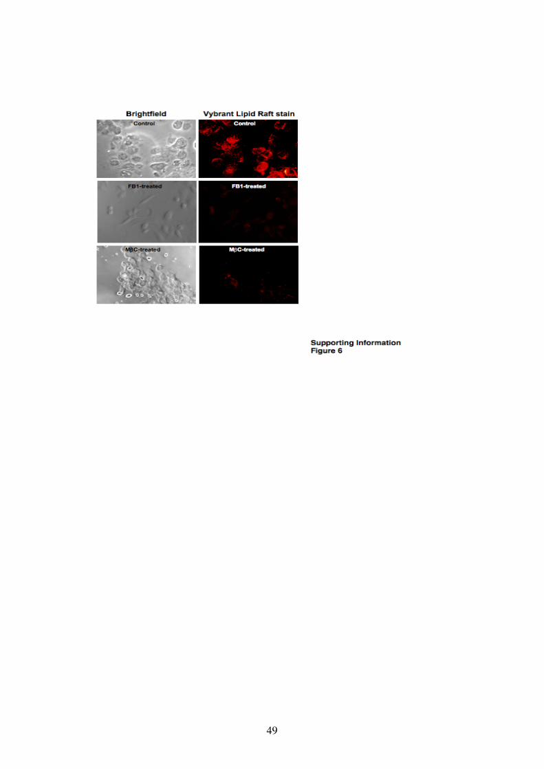
with Fumonisin B1 (FB1), an inhibitor of sphingomyelin incorporation into lipid rafts,
or methyl-"-cyclodextrin (M"C), a cholesterol depleting agent. A lipid raft stain was
used to confirm the reduction of lipid rafts on the surface of FB1 and M"C treated
AGS cells (Fig. S6). After incubation of cells with Alexa Fluor 568 labeled OMVs,
FB1 treated AGS cells exhibited very low levels of red fluorescence, when compared
to non-treated cells, and less intracellular co-localization when permeabilized and
stained with Alexa Fluor 488 labeled anti-H. pylori OMV antibodies (confocal; Fig.
S7, epifluorescence; Fig. S8). These results strongly suggest that sequestration of

12
sphingomyelin from lipid rafts abrogates OMV internalization. In concordance with
FB1 treated AGS cells, M"C treated cells exhibited significantly reduced amounts of
Alexa Fluor labeled OMVs within their intracellular compartment, compared to non-
treated cells, further suggesting that the disruption of lipid rafts abrogates OMV
internalization (confocal; Fig. 4A-D, epifluorescence; Fig. S9).
M"C, but not FB1 (Gopee and Sharma, 2004), has no effect on NF-!B
signaling in cells, thus allowing us to also determine the role of lipid rafts in NOD1
responses to OMVs. First, we established that M"C treatment did not affect cell
viability in AGS and HEK293 cells (AGS; P= 0.1835, HEK293 cells; P=0.096), nor
NOD1 independent signaling with phorbol myristate acetate (PMA) (Fig. S10). Next,
we determined the role of lipid rafts in NF-!B responses by treating HEK293 and
AGS cells with M"C prior to co-culture with H. pylori 251 cagPAI OMVs. M"C
treatment of HEK293 and AGS cells abrogated OMV induced NF-!B activity and
IL-8 production, respectively, when compared to non-treated controls (Fig. 4E; Fig.
S10 respectively). Moreover, replenishment of cholesterol on M"C treated HEK293
cells completely restored the immunostimulatory capacity of OMVs (Fig. 4F), thereby
excluding pleiotropic effects of M"C. Taken together, the data show that
pharmacological disruption of lipid rafts prevents both OMV entry and the induction
of innate immune signaling in host cells.
NOD1 is essential for innate and adaptive immune responses against H. pylori
OMVs in vivo.
Having demonstrated the ability of OMVs to induce NOD1 dependent
signaling in vitro, we next examined the effect of these structures on host immune
responses in vivo. For this, we established a model to determined the gastric

13
expression levels of the NOD1 responsive chemokine gene Cxcl2 (Viala et al., 2004)
in C57BL/6 mice that were intragastrically fed either a single dose of H. pylori 251
cagPAI OMVs or H. pylori SS1 bacteria, as a positive control. Gastric Cxcl2 mRNA
was detected as early as 1 day post feeding, in both H. pylori OMV or H. pylori SS1
fed animals, compared to PBS controls (Fig. S11). These responses were maintained
in H. pylori infected animals during the period of infection, whereas OMV induced
gastric Cxcl2 responses declined to basal levels by day 7 post feeding (Fig. S11).
To determine the potential role of TLR ligands in the up-regulation of gastric
Cxcl2 mRNA expression in vivo by H. pylori OMVs, we fed H. pylori 251 cagPAI
OMVs to Mal (MyD88 adaptor like/TIRAP; TIR domain containing adaptor protein)
and MyD88 knockout mice (KO) (Fig. 5A). Mal is an adaptor molecule required for
TLR2 and TLR4 signaling (Kenny and O'Neill, 2008; Fitzgerald et al., 2001),
whereas MyD88 is the key adaptor protein required for signaling by all TLRs,
excluding TLR3 (Rakoff-Nahoum and Medzhitov, 2009). Oral administration of
OMVs to Mal and MyD88 KO mice resulted in the up-regulation of gastric Cxcl2
mRNA to similar levels as those detected in wild type OMV fed mice, at 1 day post
feeding (Fig. 5A) (P=0.52 and P=0.75, respectively, when compared to OMV fed WT
mice). These data suggested that TLRs were not required for the observed gastric
responses to H. pylori OMVs. Next, we examined the requirement of NOD1 for OMV
induction of innate and adaptive immune responses in NOD1 wild type (WT;
Card4+/+) and NOD1 KO (Card4-/-) mice. Gastric Cxcl2 expression levels were
significantly increased in WT animals that had been intragastrically fed H. pylori 251
cagPAI OMVs, when compared to those of both PBS fed WT and OMV fed KO
animals, at one day post feeding (P<0.05; Fig. 5B). Furthermore, to examine the
requirement of NOD1 for the development of an OMV specific humoral immune

14
response, mice were fed a second dose of OMVs at day 28 and antibody responses
were measured 3 weeks later at day 49. The WT but not KO mice, displayed
significant anti-H. pylori OMV IgG responses in their sera in response to mucosally
administered H. pylori OMVs, when compared to PBS fed animals 49 days post
feeding (P=0.0311 and P=0.696, respectively; Fig. 5C). Collectively, these data
definitively prove that while there may be other bacterial products associated with
H. pylori OMVs, TLR recognition of these bacterial components is not responsible
for the initiation of inflammatory responses observed in vivo. Therefore, these
findings clearly identify that NOD1 is essential for the generation of OMV dependent
innate immune responses in the mucosal compartment in vivo, as well as the
development of systemic OMV specific adaptive immune responses.
Discussion
Since the discovery of NOD1 as the receptor responsible for the recognition of
Gram negative PG (Girardin et al., 2003), numerous studies have examined the role
of NOD1 in the initiation of innate immune responses. Most of these studies have
used purified PG or NOD1 agonists that were introduced into epithelial cells by cell
permeabilization, (Girardin et al., 2003) or by direct incubation with phagocytic cells,
such as macrophages (Magalhaes et al., 2005). There have been limited examples,
however, describing the physiological mechanisms by which Gram negative bacterial
pathogens may transport their PG to cytosolic NOD1 within host cells, particularly
non-phagocytic cells. To date, the two known mechanisms whereby Gram negative
bacteria can deliver PG to cytosolic host NOD1 involve either cellular invasion
(Girardin et al., 2001) or delivery via a bacterial secretion system (Viala et al., 2004).

15
However, PG molecules from almost all Gram negative pathogens, irrespective of
their ability to invade host cells or to express a bacterial secretion system, can be
detected by NOD1 and are able to initiate NOD1 dependent pro-inflammatory
responses in host cells (reviewed in (Kaparakis et al., 2007; Fritz et al., 2006;
Sansonetti, 2006)). Furthermore, it has been previously reported that in the absence of
a T4SS, H. pylori is still able to transfer PG into host epithelial cells albeit via a less
efficient, unknown mechanism (Viala et al., 2004). Hence, a fundamental question in
the area of NOD1 research has remained unanswered: how might all Gram negative
pathogens, irrespective of their mode of infection, initiate NOD1 signaling in non-
phagocytic epithelial cells? In this study, we have identified OMVs released by Gram
negative bacteria as a generalized mechanism for the delivery of PG to host cytosolic
NOD1.
In all multicellular organisms, membrane vesicles seem to function as a
mechanism for intercellular communication and transportation of virulence
determinants between cells during normal and stressed conditions (reviewed in
(Stoorvogel et al., 2002)). We have now identified for the first time a novel
physiological role for bacterial OMVs as initiators of immune responses in host cells.
We suggest that membrane vesicles represent a new virulence mechanism by which
both non-invasive and invasive bacteria initiate inflammatory processes in host cells.
Stimulation of cells with altered NOD1 functionality identified that OMV induced
immune responses were initiated via NOD1. We found that OMVs containing PG
enter host cells and initiate NOD1 dependent inflammatory responses. Furthermore,
the introduction of PG or GM-TriDAP into the cytoplasm of NOD1 expressing cells,
via microinjection, does not result in NOD1 dependent signaling (this study and SEG,
unpublished data). Hence, administration of PG directly into the host cytosol is not

16
sufficient for NOD1 recognition. Our data suggest that OMVs, which are comprised
of a bi-layered lipid membrane, facilitate the intracellular trafficking of PG in an
appropriate form to cytosolic NOD1 and thus, the initiation of an immune response.
Indeed, a previous study has identified that lipophilic acryl residues associated with
iE-DAP, the core immunostimulatory component of NOD1, enhanced the NOD1
stimulatory activity of iE-DAP (Hasegawa et al., 2007), further supporting our
hypothesis that lipid associated with PG can facilitate NOD1 signaling. The
intracellular trafficking and interaction of NOD1 with OMV associated PG forms the
basis of future research.
In this study, we report that lipid rafts located on the cell membrane, are utilized by
OMVs as portals of entry into host cells. Indeed, depletion of lipid rafts on the
surface of epithelial cells reduced OMV entry and NOD1 dependent responses.
Conversely, cholesterol replenishment of the cells restored both the entry and
immunostimulatory capacities of OMVs, thereby confirming the requirement of lipid
rafts for OMV induced NF-!B responses. A possible explanation for the inability of
microinjected NOD1 agonists to initiate signaling is due to the lipid membrane
association of NOD1 and its potential inability to access its ligand. We speculated that
OMV entry via lipid rafts may render PG accessible to membrane associated NOD1.
Although bacterial OMVs have been reported to deliver toxins via lipid rafts
(Kesty et al., 2004), this is the first report to our knowledge in which lipid rafts have
been shown to be critical for OMV induced innate immune responses in host cells.
Interestingly, however, a recent report described the association of NOD1 and its
downstream signaling molecule NEMO with the plasma membrane at the apical
surface of human epithelial cells (Kufer et al., 2008). Moreover, the addition of
invasive Shigella to cells provoked the further recruitment of membrane associated

17
NOD1 to the focal points of bacterial entry i. e. lipid raft domains (Kufer et al., 2008).
These data are therefore consistent with our findings suggesting that lipid rafts are
essential for OMV entry and NOD1 signaling in non-phagocytic epithelial cells.
We performed various studies to exclude the role of OMV associated TLR agonist
in the induction of NF-!B immune responses. HEK293 and AGS cells are devoid of a
functional form of TLR2 (Girardin et al., 2003; Kurt-Jones et al., 2004) and AGS
cells do not produce IL-8 in response to LPS stimulation (Nilsson et al., 2008;
Backhed et al., 2003). Therefore, we postulated that H. pylori LPS and lipoproteins
associated with OMVs were not responsible for activation of NF-!B or NF-!B
induced responses, such as hBDs and IL-8. Furthermore, by a process of exclusion,
using either Proteinase K or DNAse treated OMVs, we established that lipoproteins
and bacterial DNA were not required for OMV induced NF-!B responses in TLR2
and TLR9 expressing HEK293 cells.
Finally, we confirmed that TLRs are redundant for the induction of innate immune
response against OMVs in vivo by administering H. pylori 251 cagPAI OMVs orally
to WT and KO mice. We showed that MyD88 and Mal KO mice, which are defective
in all of the known TLR pathways involved in signaling to bacterial pathogens,
generated rapid inflammatory responses to OMVs, thus excluding a requirement for
TLRs in these responses. In contrast, NOD1 KO mice did not produce any Cxcl2
mRNA responses, nor any OMV specific IgG antibodies, in response to oral
administration of H. pylori OMVs. These findings provide an immunological basis for
the known efficacy of OMV based vaccines, such as the one developed against the
NOD1 signaling pathogen, N. meningitidis (Antignac et al., 2003; Saunders et al.,
1999). We also propose that OMVs may play a role in the inflammatory conditions
associated with persistently colonizing pathogens, such as H. pylori (Fiocca et al.,

18
1999). This would explain how H. pylori cagPAI negative strains, which lack a
functional T4SS, are still able to induce inflammation in vivo (Ohnita et al., 2005;
Crabtree et al., 2002; Lee et al., 1997).

19
Experimental procedures
Bacteria H. pylori 189, 249, 26695, 251 and 251 cagPAI isolates were cultured on
blood agar medium or in Brain Heart Infusion (BHI) broth supplemented with 0.2 %
(w/v) "-cyclodextrin (Sigma) (Philpott et al., 2002). The H. pylori 251 cagPAI
deletion mutant was constructed by natural transformation with pJP46 (Odenbreit et
al., 2001). The mouse adapted H. pylori Sydney Strain 1 (SS1) was cultured using
blood agar (Ferrero et al., 1998). N. gonorrhoeae MS11 and P. aeruginosa
PA103pilA were cultured as described previously (Gunesekere et al., 2006;
Whitchurch et al., 2005). P. aeruginosa PA103pilA does not produce many of the
known virulence factors (e. g. pilin and flagellin).
Bacterial OMVs OMVs were purified from log phase bacterial cultures (Keenan et
al., 2000) and protein concentrations were determined by Bradford assay (BioRad).
OMVs were stained using Alexa Fluor 488, 568 (Molecular Probes) or 0.5 % (w/v)
uranyl acetate. Muramic acid contained within OMVs and standard solutions (MDP,
Invivogen), was quantified (Hadzija, 1974). OMV associated PG was tritiated as
previously described (Viala et al., 2004). OMVs were heat treated by boiling at 100˚C
for 20 minutes. DNA and proteins were removed using 10 units/ml of DNase
(Promega) or 100 µg/ml of Proteinase K (Epicentre), respectively. The enzymes were
inactivated at 75˚C for 20 minutes. Proteinase K was further inactivated using an
inhibitor (Cocktail Set I, Calbiochem). The effectiveness of treatments was
confirmed by Western blot or agarose gel electrophoresis, respectively. OMVs from
H. pylori strains were probed with antibodies to either: in-house rabbit anti-H. pylori,
or rabbit anti-H. pylori urease subunits A or B.

20
Sucrose gradient purification of OMVs
OMV preparations were washed 3 times with PBS using an Amicon YM-10 column
(Millipore), prior to layering onto discontinuous sucrose gradients that were subjected
to centrifugation at 100,000 g for 16 hours as previously described (Shang et al.,
1998). Fractions (3 ml) were collected, washed 3 times with PBS using an Amicon
YM-10 column and concentrated to a final volume of 500 µl. Each fraction was tested
for their protein concentration, their protein profile by Western blot analysis, and their
ability to induce NF-!B luciferase activity in HEK293 cells. The presence of OMVs
within Fraction 6 was confirmed by electron microscopy (Jeol 200CX 200KV
transmission electron microscope).
Epithelial cell culture assays HeLa, HEK293 and AGS cells were cultured using
standard techniques. Stable AGS NOD1 knockdown and control cell lines were
generated by integration of an expression vector containing siRNA directed to either
NOD1 or EGFP, respectively (manuscript in preparation, R.L.F). Reporter assays
were performed with Ig! luciferase (Viala et al., 2004), hBD2 or hBD3, dTK Renilla
(Promega) or #CARD NOD1 (Viala et al., 2004) constructs. Cells transfected with
luciferase constructs were stimulated for 8 hours with OMVs (10 µg protein) or
H. pylori 251 bacteria at a MOI of 10:1 (Viala et al., 2004; Philpott et al., 2002), then
lyzed (Reporter lysis buffer, Promega) and the luminescence measured using a
FLUOstar Optima luminometer (BMG Labtech). H. pylori 251 bacteria were killed by
fixing with 1 % (w/v) formaldehyde, and subsequently plated on blood agar to
confirm they were no longer viable (Huang et al., 1995). Digitonin permeabilized
HEK293 cells were stimulated with equivalent amounts of phenol water extracted,
bacteria free S. flexneri supernatants. AGS cells were stimulated with 160 nM phorbol
21
myristate acetate (PMA, Invitrogen). TLR2 expressing HEK293 cells were stimulated
with 6.25 ng of H. pylori (Dr A. Moran, The National University of Ireland, Galway,
Ireland) or 125 ng ultrapure E. coli LPS (Invivogen), and TLR9 expressing HEK293
cells were stimulated with 100 nM CpG DNA (a gift from Dr A. Mansell, Monash
Institute for Medical Research, Monash University, Australia) for 8 hours. AGS
culture supernatants were analyzed for IL-8 production (OptEIATM, BD Biosciences).
Cell microinjection HeLa cells, grown on 10 mm coverslips in serum free
conditions, were microinjected using an approach adapted from our earlier studies
(Philpott et al., 2000; Coleman et al., 2001). Coverslips were mounted in a recording
chamber (Warner Instruments) on a Leica DMLFS microscope and cell filling was
monitored using 0.2 % (w/v) Lucifer yellow and 0.05 % (w/v) fluorescein dextran
(Molecular Probes, 3 kDa). Micropipettes were mounted on a micromanipulator
(MP-285, Sutter), connected to a picospritzer (General Valve) to facilitate cell
loading. After microinjection, cells were incubated at 37 °C for 2 hours, fixed and
NF-!B was detected using anti-p65 NF-!B antibody (Cell Signaling Technologies)
and anti-rabbit Alexa-568 labeled secondary antibody (Molecular Probes).
Internalization of OMVs AGS cells grown on coverslips were co-cultured with
Alexa Fluor labeled OMVs, for 16 hours, fixed in 4 % (w/v) paraformaldehyde and
permeabilized using 0.1 % (v/v) Triton-X 100 in PBS. Slides were blocked in a 5 %
(v/v) FCS and 0.1 % (w/v) BSA solution, with or without the addition of 0.1 % (v/v)
Triton-X-100, then incubated with a combination of: either in-house rabbit anti-
H. pylori or rabbit anti-H. pylori OMV and goat anti-rabbit IgG Alexa-568 or Alexa
488 labeled antibodies (Invitrogen). Slides were visualized using AX70 or BX51

22
upright UV microscopes (Olympus). Confocal images were obtained using a Leica
SP5 multiphoton confocal microscope. Images were prepared for publication using
Analysis software (Olympus, Tokyo, Japan).
Depletion of lipid rafts using methyl-"-cyclodextrin or Fumonisin B1 and
staining of lipid rafts. Cells were cholesterol or sphingolipid depleted using methyl-
"-cyclodextrin (M"C; Sigma), or Fumonisin B1 (FB1; Sigma), respectively. Cells
were depleted of cholesterol by a 30 minute treatment with 4 nM M"C in serum free
media. Cell viability after M"C treatment was confirmed using the luminescence
based Cell Titer Glow assay (Promega). Cells were depleted of sphingomyelin by
growing them in the presence of 100 µM FB1 for 2 days. After either treatment, the
cells were subjected to OMV stimulation, or had their lipid rafts stained using the
Vybrant® lipid raft labeling kit (Molecular probes). Cholesterol was added to
cholesterol depleted cells by treating with 250 µM cholesterol (5-cholesten-3"-ol;
Sigma) and 4 nM M"C for 30 minutes.
Co-culture studies with tritiated OMV associated PG
PG within OMVs from H. pylori lysA or lysAcagM bacteria was specifically tritiated
using a previously described technique (Viala et al., 2004). AGS cells were co-
cultured overnight with tritiated OMVs (300 µg protein), and the presence of tritiated
PG detected. The slides were counter stained with Giemsa stain.
MEF isolation and cell culture All NOD1 animal experimentation was performed at
the University of Toronto (protocol number: 20006359). MEFs were isolated from
C57BL/6 WT and NOD1 (Card4-/-) KO mice, which had been backcrossed more than

23
eight times onto a C57BL/6 background (Girardin et al., 2003). MEFs were cultured
in 96 well plates (4 x 10⁴ cells/ well), then stimulated for 24 hours with either: OMVs,
highly purified Escherichia coli LPS (100 ng/ml; Lausen, Switzerland); Gram-
positive PG from Staphylococcus aureus (100 ng/ml; Sigma) or Pam3Cys (100 ng/ml;
Sigma). Chemokine and cytokine levels were determined by ELISA (DuoSet, R&D
Systems).
Mouse immunization and measurement of immune responses Age matched male
and female animals were used in all experiments. Immunization of NOD1, MyD88
and Mal knockout animals was performed at The University of Toronto, The
University of Melbourne and Monash University, respectively. All animal
experimentation was performed in accordance with the animal ethics approval
obtained from the relevant institution. Animals received either a single intragastrically
delivered 100 µl dose of 50 µg of H. pylori 251 cagPAI OMVs in PBS, 3x10⁷
H. pylori SS1 bacteria in 100 µl of PBS or PBS alone (Ferrero et al., 1998). Their
stomachs were removed 16 hours later, washed with PBS and stored in RNAlater
(Ambion). In some experiments, stomachs from animals were analyzed at later
timepoints, as indicated in the figure legends. Gastric RNA was purified using TRIzol
reagent (Invitrogen). RNA was treated with RNAse free DNAse (Promega), prior to
generation of cDNA (SuperScript III, Invitrogen). The cDNA was amplified using
primers for murine actin (forward primer: 5’ ACGGCCAGGTCATCACTATTG,
reverse primer 5’CAAGAAGGAAGGCTGGAAAAGA) and murine Cxcl2 (forward
primer: 5’ GGGCGGTCAAAAAGTTTGC, reverse primer 5’
ATTTGTTCAGTATCTTTTGGATGATTTTC). Up-regulation of Cxcl2 mRNA in
the stomach tissue, normalized to actin mRNA, was detected using SYBR GREEN

24
PCR mastermix and ABI7300 (ABI) or Mastercycler ep realplex (Eppendorf) real
time machines.
Antibody production in response to OMV stimulation OMV fed mice were
gavaged at 28 days with a second 100 µl dose of 50 µg of H. pylori 251 cagPAI
OMVs in PBS and antibody responses were determined 21 days later, at day 49. PBS
control mice were gavaged with PBS at both timepoints. Mouse anti-H. pylori OMV
IgG responses were detected by ELISA using a method adapted from our earlier
studies (Kaparakis et al., 2006). Maxisorb 96 well plates were coated with 250 µg/ml
of H. pylori 251 cagPAI OMVs. Sera were initially diluted 1:50 and serial dilutions of
these were performed to determine the endpoint titre. OMV specific mouse anti-IgG
antibodies were detected using anti-mouse IgG biotin-labeled antibody (Chemicon)
and streptavidin-HRP (Chemicon). ELISAs were developed using BD OptEIA TMB
substrate reagent (BD biosciences). Absorbances were read at OD450nm using a
FLUOstar Optima (BMG Labtech). Serum endpoint titres were expressed as the
reciprocal of the dilution of serum that gave an OD450nm value five times the value of
the background. Mice with an antibody titre below the detection limit of the assay
(log10 1.7) were assigned a titre of log10 1.7. Positive and negative control sera were
included in all ELISA experiments.
Statistical analysis IL-8 responses were analyzed using the Student’s t-test.
Luciferase activity, antibody titres and mRNA levels were analyzed using the Mann
Whitney U-test. Differences were considered significant when P<0.05.

25
References
Antignac, A., Rousselle, J.C., Namane, A., Labigne, A., Taha, M.K. and Boneca, I.G. (2003) Detailed structural analysis of the peptidoglycan of the human pathogen Neisseria meningitidis. J Biol Chem. 278: 31521-31528. Backert, S., Schwarz, T., Miehlke, S., Kirsch, C., Sommer, C., Kwok, T., et al (2004) Functional analysis of the cag pathogenicity island in Helicobacter pylori isolates from patients with gastritis, peptic ulcer, and gastric cancer. Infect Immun. 72: 1043-1056. Backhed, F., Rokbi, B., Torstensson, E., Zhao, Y., Nilsson, C., Seguin, D., et al (2003) Gastric mucosal recognition of Helicobacter pylori is independent of Toll-like receptor 4. J Infect Dis. 187: 829-836. Beswick, E.J., Pinchuk, I.V., Minch, K., Suarez, G., Sierra, J.C., Yamaoka, Y. and Reyes, V.E. (2006) The Helicobacter pylori urease B subunit binds to CD74 on gastric epithelial cells and induces NF-kappaB activation and interleukin-8 production. Infect Immun. 74: 1148-1155. Chaouche-Drider, N., Kaparakis, M., Karrar, A., Fernandez, M.I., Carneiro, L.A., Viala, J., et al (2009) A commensal Helicobacter sp. of the rodent intestinal flora activates TLR2 and NOD1 responses in epithelial cells. PLoS One. 4: e5396. Coleman, H.A., Tare, M. and Parkington, H.C. (2001) K+ currents underlying the action of endothelium-derived hyperpolarizing factor in guinea-pig, rat and human blood vessels. J Physiol. 531: 359-373. Crabtree, J.E., Ferrero, R.L. and Kusters, J.G. (2002) The mouse colonizing Helicobacter pylori strain SS1 may lack a functional cag pathogenicity island. Helicobacter. 7: 139-140; author reply 140-131. Ferrero, R.L., Thiberge, J.M., Huerre, M. and Labigne, A. (1998) Immune responses of specific-pathogen-free mice to chronic Helicobacter pylori (strain SS1) infection. Infect Immun. 66: 1349-1355. Fiocca, R., Necchi, V., Sommi, P., Ricci, V., Telford, J., Cover, T.L. and Solcia, E. (1999) Release of Helicobacter pylori vacuolating cytotoxin by both a specific secretion pathway and budding of outer membrane vesicles. Uptake of released toxin and vesicles by gastric epithelium. J Pathol. 188: 220-226. Fitzgerald, K.A., Palsson-McDermott, E.M., Bowie, A.G., Jefferies, C.A., Mansell, A.S., Brady, G., et al (2001) Mal (MyD88-adapter-like) is required for Toll-like receptor-4 signal transduction. Nature. 413: 78-83. Fritz, J.H., Ferrero, R.L., Philpott, D.J. and Girardin, S.E. (2006) Nod-like proteins in immunity, inflammation and disease. Nat Immunol. 7: 1250-1257.

26
Girardin, S.E., Tournebize, R., Mavris, M., Page, A.L., Li, X., Stark, G.R., et al (2001) CARD4/Nod1 mediates NF-kappaB and JNK activation by invasive Shigella
flexneri. EMBO Rep. 2: 736-742. Girardin, S.E., Boneca, I.G., Carneiro, L.A., Antignac, A., Jehanno, M., Viala, J., et al (2003) Nod1 detects a unique muropeptide from gram-negative bacterial peptidoglycan. Science. 300: 1584-1587. Gopee, N.V. and Sharma, R.P. (2004) The mycotoxin fumonisin B1 transiently activates nuclear factor kB, tumor necrosis factor a and caspase 3 via protein kinase Ca-dependent pathway in porcine renal epithelial cells. Cell Biology and Toxicology
20: 197-212. Gunesekere, I.C., Kahler, C.M., Powell, D.R., Snyder, L.A., Saunders, N.J., Rood, J.I. and Davies, J.K. (2006) Comparison of the RpoH-dependent regulon and general stress response in Neisseria gonorrhoeae. J Bacteriol. 188: 4769-4776. Hadzija, O. (1974) A simple method for the quantitative determination of muramic acid. Anal Biochem. 60: 512-517. Hasegawa, M., Kawasaki, A., Yang, K., Fujimoto, Y., Masumoto, J., Breukink, E., et
al (2007) A Role of Lipophilic Peptidoglycan-related Molecules in Induction of Nod1-mediated Immune Responses. J Biol Chem. 282: 11757-11764. Huang, J., O'Toole, P.W., Doig, P. and Trust, T.J. (1995) Stimulation of interleukin-8 production in epithelial cell lines by Helicobacter pylori. Infect Immun. 63: 1732-1738. Inohara, N., Ogura, Y., Chen, F.F., Muto, A. and Nunez, G. (2001) Human Nod1 confers responsiveness to bacterial lipopolysaccharides. J Biol Chem. 276: 2551-2554. Kaparakis, M., Philpott, D.J. and Ferrero, R.L. (2007) Mammalian NLR proteins; discriminating foe from friend. Immunol Cell Biol. 85: 495-502. Kaparakis, M., Laurie, K.L., Wijburg, O., Pedersen, J., Pearse, M., van Driel, I.R., et
al (2006) CD4+ CD25+ regulatory T cells modulate the T-cell and antibody responses in helicobacter-infected BALB/c mice. Infect Immun. 74: 3519-3529. Keenan, J., Day, T., Neal, S., Cook, B., Perez-Perez, G., Allardyce, R. and Bagshaw, P. (2000) A role for the bacterial outer membrane in the pathogenesis of Helicobacter
pylori infection. FEMS Microbiol Lett. 182: 259-264. Kenny, E.F. and O'Neill, L.A. (2008) Signalling adaptors used by Toll-like receptors: an update. Cytokine. 43: 342-349. Kesty, N.C., Mason, K.M., Reedy, M., Miller, S.E. and Kuehn, M.J. (2004) Enterotoxigenic Escherichia coli vesicles target toxin delivery into mammalian cells. Embo J. 23: 4538-4549.

27
Kuehn, M.J. and Kesty, N.C. (2005) Bacterial outer membrane vesicles and the host-pathogen interaction. Genes Dev. 19: 2645-2655. Kufer, T.A., Kremmer, E., Adam, A.C., Philpott, D.J. and Sansonetti, P.J. (2008) The pattern-recognition molecule Nod1 is localized at the plasma membrane at sites of bacterial interaction. Cell Microbiol. 10: 477-486. Kurt-Jones, E.A., Sandor, F., Ortiz, Y., Bowen, G.N., Counter, S.L., Wang, T.C. and Finberg, R.W. (2004) Use of murine embryonic fibroblasts to define Toll-like receptor activation and specificity. J Endotoxin Res. 10: 419-424. Lee, A., O'Rourke, J., De Ungria, M.C., Robertson, B., Daskalopoulos, G. and Dixon, M.F. (1997) A standardized mouse model of Helicobacter pylori infection: introducing the Sydney strain. Gastroenterology. 112: 1386-1397. Lepper, P.M., Triantafilou, M., Schumann, C., Schneider, E.M. and Triantafilou, K. (2005) Lipopolysaccharides from Helicobacter pylori can act as antagonists for Toll-like receptor 4. Cell Microbiol. 7: 519-528. Li, Z., Clarke, A.J. and Beveridge, T.J. (1996) A major autolysin of Pseudomonas
aeruginosa: subcellular distribution, potential role in cell growth and division and secretion in surface membrane vesicles. J Bacteriol. 178: 2479-2488. Magalhaes, J.G., Philpott, D.J., Nahori, M.A., Jehanno, M., Fritz, J., Bourhis, L.L., et
al (2005) Murine Nod1 but not its human orthologue mediates innate immune detection of tracheal cytotoxin. EMBO Rep. 6: 1201-1207. Mandell, L., Moran, A.P., Cocchiarella, A., Houghton, J., Taylor, N., Fox, J.G., et al (2004) Intact gram-negative Helicobacter pylori, Helicobacter felis, and Helicobacter
hepaticus bacteria activate innate immunity via toll-like receptor 2 but not toll-like receptor 4. Infect Immun. 72: 6446-6454. Nilsson, C., Skoglund, A., Moran, A.P., Annuk, H., Engstrand, L. and Normark, S. (2008) Lipopolysaccharide diversity evolving in Helicobacter pylori communities through genetic modifications in fucosyltransferases. PLoS ONE. 3: e3811. Odenbreit, S., Gebert, B., Puls, J., Fischer, W. and Haas, R. (2001) Interaction of Helicobacter pylori with professional phagocytes: role of the cag pathogenicity island and translocation, phosphorylation and processing of CagA. Cell Microbiol. 3: 21-31. Ohnita, K., Isomoto, H., Honda, S., Wada, A., Wen, C.Y., Nishi, Y., et al (2005) Helicobacter pylori strain-specific modulation of gastric inflammation in Mongolian gerbils. World J Gastroenterol. 11: 1549-1553. Philpott, D.J., Yamaoka, S., Israel, A. and Sansonetti, P.J. (2000) Invasive Shigella
flexneri activates NF-kappa B through a lipopolysaccharide-dependent innate intracellular response and leads to IL-8 expression in epithelial cells. J Immunol. 165: 903-914.

28
Philpott, D.J., Belaid, D., Troubadour, P., Thiberge, J.M., Tankovic, J., Labigne, A. and Ferrero, R.L. (2002) Reduced activation of inflammatory responses in host cells by mouse-adapted Helicobacter pylori isolates. Cell Microbiol. 4: 285-296. Rakoff-Nahoum, S. and Medzhitov, R. (2009) Toll-like receptors and cancer. Nat Rev
Cancer. 9: 57-63. Sansonetti, P.J. (2006) The innate signaling of dangers and the dangers of innate signaling. Nat Immunol. 7: 1237-1242. Saunders, N.B., Shoemaker, D.R., Brandt, B.L., Moran, E.E., Larsen, T. and Zollinger, W.D. (1999) Immunogenicity of intranasally administered meningococcal native outer membrane vesicles in mice. Infect Immun. 67: 113-119. Shang, E.S., Skare, J.T., Exner, M.M., Blanco, D.R., Kagan, B.L., Miller, J.N. and Lovett, M.A. (1998) Isolation and characterization of the outer membrane of Borrelia
hermsii. Infect Immun. 66: 1082-1091. Skerrett, S.J., Wilson, C.B., Liggitt, H.D. and Hajjar, A.M. (2007) Redundant Toll-like receptor signaling in the pulmonary host response to Pseudomonas aeruginosa. Am J Physiol Lung Cell Mol Physiol. 292: L312-322. Stoorvogel, W., Kleijmeer, M.J., Geuze, H.J. and Raposo, G. (2002) The biogenesis and functions of exosomes. Traffic. 3: 321-330. Travassos, L.H., Carneiro, L.A., Girardin, S.E., Boneca, I.G., Lemos, R., Bozza, M.T., et al (2005) Nod1 participates in the innate immune response to Pseudomonas
aeruginosa. J Biol Chem. 280: 36714-36718. Viala, J., Chaput, C., Boneca, I.G., Cardona, A., Girardin, S.E., Moran, A.P., et al (2004) Nod1 responds to peptidoglycan delivered by the Helicobacter pylori cag pathogenicity island. Nat Immunol. 5: 1166-1174. Whitchurch, C.B., Beatson, S.A., Comolli, J.C., Jakobsen, T., Sargent, J.L., Bertrand, J.J., et al (2005) Pseudomonas aeruginosa fimL regulates multiple virulence functions by intersecting with Vfr-modulated pathways. Mol Microbiol. 55: 1357-1378. Yamaoka, Y., Kita, M., Kodama, T., Sawai, N., Kashima, K. and Imanishi, J. (1997) Induction of various cytokines and development of severe mucosal inflammation by cagA gene positive Helicobacter pylori strains. Gut. 41: 442-451. Yokota, S., Ohnishi, T., Muroi, M., Tanamoto, K., Fujii, N. and Amano, K. (2007) Highly-purified Helicobacter pylori LPS preparations induce weak inflammatory reactions and utilize Toll-like receptor 2 complex but not Toll-like receptor 4 complex. FEMS Immunol Med Microbiol. 51: 140-148.

29
Figure legends
Fig. 1. Bacterial OMVs induce NF-!B dependent responses in cells.
(A) NF-!B reporter activity in HEK293 cells stimulated with 10 µg OMVs from
cagPAI positive (251, 26695) or negative (189, 249) H. pylori strains, H. pylori
bacteria (251) or culture medium (control). (B) hBD2 and hBD3 reporter activities in
HEK293 cells. (C) IL-8 production by AGS cells stimulated with viable or killed
H. pylori 251 bacteria, 10 µg H. pylori 251 cagPAI OMVs or OMV control
corresponding to spent media. Error bars indicate ± SEM between triplicates. All data
are representative of ! 3 independent experiments.
Fig. 2. OMVs induce NOD1 dependent NF-!B responses in non-phagocytic cells.
(A) NF-!B activity in OMV stimulated (10 µg) HEK293 cells co-transfected with
20-100 ng of dominant negative NOD1 (#CARD). (B) NF-!B activity and (C) IL-8
responses in AGS siRNA stable knockdown cells stimulated with 10 µg OMVs from
H. pylori 251 cagPAI, N. gonorrhoeae or P. aeruginosa. OMV control corresponds to
spent media (D) KC, (E) MIP-2 and (F) IL-6 production in wild-type and NOD1
knockout (NOD1 KO) MEFs stimulated with 100 ng E. coli LPS, Gram positive PG,
Pam3Cys (P3Cys) or 10 µg OMVs from H. pylori 251 cagPAI (Hp), N. gonorrhoeae
(Ng) or P. aeruginosa (Pa). Error bars indicate ± SEM between triplicates. All data
are representative of ! 3 independent experiments. * denotes P<0.05 versus controls.
Fig. 3. DNase and protease treatment of OMVs does not abolish their
immunostimulatory ability.
(A) NF-!B reporter activity in HEK293 cells stimulated with 10 µg of non-treated or
heat treated OMVs from H. pylori 251 cagPAI (Hp), N. gonorrhoeae (Ng) or
30
P. aeruginosa (Pa). (B) NF-!B reporter activity in TLR2 and TLR9 expressing
HEK293 cells stimulated with 10 µg of non-treated, heat, Proteinase K or DNase
treated H. pylori 251 cagPAI OMVs. Percentage values reported with respect to cells
stimulated with non-treated OMVs. Error bars indicate ± SEM between triplicates.
Data are representative of ! 3 independent experiments. * denotes P<0.05 versus
controls. (C) AGS cells co-cultured with 300 µg tritiated OMVs (observed as brown
granules) from H. pylori lysA or lysAcagM strains (100 x magnification). Data are
representative of three independent experiments.
Fig. 4. OMVs enter epithelial cells via lipid rafts.
AGS cells were co-cultured with 10 µg Alexa 568 labeled H. pylori cagPAI OMVs
(red). Lipid raft intact (A, C), or M"C treated (B, D) cells were permeabilized with
Triton X-100 (C-D), or left as controls (A-B). OMVs were detected using rabbit anti-
H. pylori OMVs and anti-rabbit Alexa Fluor 488 antibodies, respectively (green).
Cells were visualized by confocal microscopy. Labeled OMVs (red) were
predominantly contained within the intracellular compartment of AGS cells whereas
fewer were within M"C treated cells. Only permeabilized cells exhibited areas of
intracellular dual fluorescence (yellow; indicated by the arrows). Scale bar represents
20 µm. (E) NF-!B activity in HEK293 cells pre-treated or not with M"C prior to
stimulation with 10 µg OMVs from H. pylori 251 cagPAI (Hp), N. gonorrhoeae (Ng)
or P. aeruginosa (Pa). (F) NF-!B activity in control, M"C treated, or M"C treated
and cholesterol replenished HEK293 cells stimulated with 10 µg H. pylori 251
cagPAI OMVs. Error bars indicate ± SEM between triplicates. All data are
representative ! 3 independent experiments. * denotes P<0.05 versus M"C untreated
cells.

31
Fig. 5. NOD1 (Card4-/-) KO mice do not respond to OMV stimulation.
(A) Gastric Cxcl2 mRNA responses in C57BL/6 WT (WT), Mal KO (Mal) and
MyD88 KO (MyD88) mice orally administered PBS (open symbols) or 50 µg
H. pylori 251 cagPAI OMVs (filled symbols). Responses were measured 1 day post
feeding. Horizontal lines indicate the mean ± SEM values corresponding to each
group of animals. Data for Mal and MyD88 KO animals were pooled from two
independent experiments, whereas the data for WT mice were pooled from four
independent experiments. (B) Gastric Cxcl2 mRNA responses in C57BL/6 WT (WT)
and NOD1 KO (NOD1) mice orally administered PBS (open symbols) or 50 µg
H. pylori 251 cagPAI OMVs (filled symbols). Responses were measured 1 day post
feeding. Line indicates average response of WT OMV fed animals. Results are from
two experiments pooled, minimum n=3 mice per group in each experiment. * denotes
P<0.05 versus WT OMV mice. (C) H. pylori OMV specific IgG antibody titre of
C57BL/6 WT (WT) and NOD1 KO (NOD1) mice. n=3 mice per PBS control groups.
n=5 and n=4 mice per OMV fed WT and KO groups respectively. * denotes P<0.05
versus WT PBS mice.

32
Acknowledgements
This project was supported by grants from The ANZ Charitable trust (R.L.F),
The National Health and Medical Research Council (NHMRC; to R.L.F., H.C.P,
H.A.C., J.K.D., C.B.W., P.J.C., P.J.H.), the Australian Research Council (H.C.P.), the
Victorian Neurotrauma Initiative and Heart Foundation (P.J.C.) and The Canadian
Institutes of Health (S.E.G. and D.J.P.). D.J.P. is an International Research Scholar of
the Howard Hughes Medical Institute. C.B.W. is an NHMRC Senior Research
Fellow. The Australian Bacterial Pathogenesis Program was supported by a Program
Grant from the NHMRC.
We thank Miss S. Walker (Monash Micro Imaging, Monash University), for
microscopy image processing; Mrs K. Hoe (Micromon, Clayton, Australia) for
electron microscopy; Dr J. Keenan (Christchurch School of Medicine and Health
Sciences, Christchurch, New Zealand) and Ms S. Lyons-Schindler (Monash
University) for advice regarding OMV purification and Neisseria culture,
respectively. Drs R. Haas (Ludwig-Maximilians-University, Munich, Germany), A.
Mansell (Monash University), D. Golenbock (University of Massachusetts,
Worcester, MA) A. Moran (The National University of Ireland, Galway, Ireland), N.
Mangan (Monash University) and Ms R. Parry (Promega Australia) are thanked for
providing reagents and assistance.
Fig. S1. Purification of H. pylori OMVs and analysis of their protein content.
(A) Representative transmission electron micrograph (TEM) of H. pylori OMVs
(indicated by arrows), which were purified from H. pylori (strain 189, 27,500x
magnification). Scale bar indicates 500 nm. H. pylori OMVs were analyzed for

33
protein content by Western blotting. OMVs from H. pylori strains: 1) 26695, 2) 251,
3) 251 cagPAI, 4) 189 and 5) 249, were probed with antibodies to either: (B) H. pylori
total extracts, or (C) H. pylori urease subunits A (UreA) or B (UreB). As a control,
the antibodies were also reacted against whole bacteria of H. pylori 251 (lane 6).
Molecular weight markers (Panel B) are indicated on the left hand side of the
membrane. Arrows (Panel C) indicate the molecular weights and positions of the
urease subunits on an SDS-PAGE gel.
Fig. S2. Microinjection of HeLa cells with H. pylori OMVs.
Representative images of HeLa cells co-microinjected with FITC-Dextran, Lucifer
Yellow and either: BHI broth, GM-TriDAP (TriDap) or H. pylori OMVs. Lucifer
Yellow and FITC-Dextran were used to identify microinjected cells both during
microinjection and after immunofluorescence staining, respectively. NF-!B
localization within microinjected cells was determined using rabbit anti-NF-!B and
anti-rabbit Alexa Fluor 568 antibodies, respectively. As a control, HeLa cells that
were not injected were reacted with an anti-rabbit Alexa 568 antibody to determine
the level of non-specific binding. All microinjected cells (identified by the white
arrows) displayed diffuse NF-!B staining, whereas only OMV injected cells had
NF-!B staining localised in their nucleus. Images are representative of two
independent experiments. (Scale bar indicates 50 µm.)
Fig. S3. Purification of N. gonorrhoeae and P. aeruginosa OMVs and
confirmation of the removal of proteins and DNA from OMV preparations.
Representative TEMs of OMVs (indicated by the arrows) purified from (A)
N. gonorrhoeae and (B) P. aeruginosa. Scale bar indicates 100 nm. Representative

34
TEMs of (C) control and (D) heat treated H. pylori OMVs (indicated by the arrows).
Scale bar indicates 100 nm. (E) Western blot analysis of total proteins in H. pylori
OMV preparations subjected to heat denaturation (lane 1), Proteinase K digestion
(lane 2) or no treatment (lane 3). Treated and non-treated H. pylori OMVs were
transferred to nitrocellulose membrane and probed with antisera against an H. pylori
total extract. (F) OMV preparations from N. gonorrhoeae (lanes 1 and 2) and
P. aeruginosa (lanes 3 and 4) were subjected to DNase treatment (lanes 2 and 4) or
mock treated (lanes 1 and 3). OMVs was separated by agarose gel electrophoresis
and stained with SYBR Green (Invitrogen).
Fig. S4. Sucrose gradient purified OMVs contain reduced amounts of bacterial
proteins but retain their ability to induce NF-!B activity in HEK293 cells.
OMVs were purified by continuous gradient sucrose ultracentrifugation. Fractions
were collected (3 mls) and concentrated to 500 µl. (A) Equivalent amounts (25 µl) of
each fraction were analyzed for their protein content by Western blotting. Fractions
were numbered F1 to F13, with F1 being the lightest fraction. OMVs prior to sucrose
gradient purification (O) were also analyzed for their protein profile. Molecular
weight markers are indicated on the left-hand side of the membrane. (B) Standardized
amounts of each fraction (approximately 10 µg protein) were tested for their ability to
induce NF-!B-luciferase activity in HEK293 cells. Error bars indicate ± SEM
between triplicates. Data is representative of two independent experiments. (C)
Representative TEM of the highest NF-!B-luciferase inducing fraction (F6)
containing sucrose purified H. pylori OMVs (indicated by the arrows). Scale bar
indicates 100 nm.

35
Figure S5. TLR2 and TLR9 expressing HEK293 cells respond to H. pylori LPS
and CpG DNA stimulation.
NF-!B reporter activity in TLR2 and TLR9 expressing HEK293 cells stimulated with
6.25 ng H. pylori LPS or 100 nM CpG DNA for 8 hours. Error bars indicate ± SEM
between triplicates. Data are representative of 3 independent experiments.
Fig. S6. FB1 and M"C treatments disrupt lipid raft domains on host cell
membranes.
Lipid rafts on AGS cell membranes of FB1 treated, M"C treated and control (non-
treated) cells were examined using the Vybrant® lipid raft stain (Molecular Probes).
Stained cells were examined by phase contrast and fluorescence microscopy (left and
right panels, respectively; 100 x magnification). Lipid rafts are stained red. Images
are representative of three independent experiments.
Fig. S7. OMVs enter epithelial cells via lipid rafts and entry is hindered in FB1
treated AGS cells.
AGS cells were co-cultured with 10 µg of Alexa 568 labeled H. pylori cagPAI OMVs
(red). Lipid raft intact (A, C), or FB1 treated (B, D) AGS cells were permeabilized
with Triton X-100 (C-D), or left as controls (A-B). OMVs were detected using rabbit
anti-H. pylori OMVs and anti-rabbit Alexa Fluor 488 antibodies, respectively (green).
Cells were visualized by confocal microscopy. Labelled OMVs (red) were
predominantly contained within the intracellular compartment of AGS cells and fewer
OMVs were located within FB1 treated cells, highlighting the requirement of lipid
rafts for OMV entry. Only permeabilized cells exhibited areas of intracellular dual
fluorescence (yellow; arrows). Scale bar represents 20 µm.

36
Fig. S8. FB1-treatment reduces the ability of OMVs to enter the intracellular
compartment of epithelial cells.
Alexa Fluor 568 labeled H. pylori OMVs (10 µg) (red) were co-cultured with FB1-
treated (B and D) or -untreated (A and C) AGS cells and analyzed by epifluorescence.
Cells were permeabilized with Triton X-100 (C-D), or left as controls (A-B). OMVs
were detected using rabbit anti-H. pylori OMVs and anti-rabbit Alexa Fluor 488
antibodies, respectively (green). Cells were visualized by epifluorescence. FB1
treatment significantly reduced the level of fluorescent OMVs associated with AGS
cells. Co-localisation (yellow) is indicated by the arrows (100 x magnification). Data
are representative of more than two independent experiments.
Fig. S9. M"C -treatment reduces the ability of OMVs to enter the intracellular
compartment of epithelial cells via lipid rafts.
Alexa Fluor 488 labeled H. pylori OMVs (10 µg) (green) were co-cultured with
M"C-treated (B and D) or -untreated (A and C) AGS cells and analyzed by
epifluorescence. Cells were permeabilized with Triton X-100 (C-D), or left as
controls (A-B). OMVs were detected using rabbit anti-H. pylori OMVs and anti-
rabbit Alexa Fluor 568 antibodies (red), respectively. Cells were visualized by
epifluorescence. M"C treatment significantly reduced the level of fluorescent OMVs
associated with AGS cells. Co-localisation (yellow) is indicated by the arrows (100 x
magnification). Data are representative of more than three independent experiments.
Fig. S10. M"C treatment of AGS cells reduces OMV-induced IL-8 responses but
not NOD1 independent responses.

37
AGS cells were pre-treated or not with M"C prior to stimulation for 24 hours with
either H. pylori 251 (Hp), OMVs (10 µg) isolated from H. pylori 251 cagPAI
(OMVs), or 160 nM phorbol myristate acetate (PMA, Invitrogen). Cell culture
supernatants were analyzed for IL-8 production by ELISA. Error bars indicate ± SEM
between triplicates. Data are representative of two independent experiments. *
denotes P=0.04, ** denotes P=0.001 versus controls. P=0.115 PMA versus M"C +
PMA.
Fig. S11. H. pylori OMVs induce gastric Cxcl2 mRNA responses in WT C57BL/6
mice.
Gastric Cxcl2 mRNA responses in WT C57BL/6 female mice orally administered
with A) 3x10⁷ H. pylori SS1 (filled symbols), B) 50 µg H. pylori cagPAI OMVs
(filled symbols) or PBS (open symbols). Column indicates average response of
animals. Error bars indicate ± SEM between samples from individual mice. n=3 mice
per group at each timepoint.
Movie S1 Legend Co-localization of internalized H. pylori cagPAI OMVs and
anti-H. pylori antibody within permeabilized AGS cells.
AGS cells were co-cultured with 10 µg of Alexa 568 labeled H. pylori cagPAI OMVs
(red). Cells were permeabilized with Triton X-100 and OMVs were detected using
rabbit anti-H. pylori OMVs and anti-rabbit Alexa Fluor 488 antibodies, respectively
(green). Cells were visualized by confocal microscopy. Labeled OMVs (red) were
identified as being contained within the intracellular compartment of AGS cells by the
presence of areas of intracellular dual fluorescence (yellow) in permeabilized cells.

38
Images were acquired using a 40 x objective and 4x zoom, rendered using Imaris x64
and a threshold applied to render isosurfaces.
Movie S2 Legend Absence of co-localization of internalized H. pylori cagPAI
OMVs and anti-H. pylori antibody within intact AGS cells.
AGS cells were co-cultured with 10 µg of Alexa 568 labeled H. pylori cagPAI OMVs
(red). Cell membranes were left intact (not treated with Triton X-100) and
extracellular OMVs were detected using rabbit anti-H. pylori OMVs and anti-rabbit
Alexa Fluor 488 antibodies, respectively (green). Cells were visualized by confocal
microscopy. Labeled OMVs (red) were identified as being contained within the
intracellular compartment of AGS cells, as there is an absence of intracellular dual
fluorescence (yellow) within cells. Images were acquired using a 40 x objective and
4x zoom, rendered using Imaris x64 and a threshold applied to render isosurfaces.

39

40

41

42

43

44

45

46

47

48
49

50

51

52

53

54