Tissue. In order to understand how coordination is carried out in the human body we must first...
28
Tissue
-
Upload
jane-lilian-baker -
Category
Documents
-
view
218 -
download
0
Transcript of Tissue. In order to understand how coordination is carried out in the human body we must first...

Tissue

In order to understand how coordination is carried out in the human body we must first understand the organization of the body system. Each system contains a number of organs and each organ is composed of different types of tissue.

Types of tissue
A tissue is composed of similarly specialized cells that perform a common function in the body.
1. Epithelial tissue-----Covers body surfaces and lines body cavities.
2. Connective tissue-----Binds and supports body parts.
3. Muscular tissue-----moves body parts.4. Nervous tissue----receives stimuli and
conducts impulses from one body part to another.

Cancer is classified according to the type of tissue
Carcinoma-----the most common type of cancers of the epithelial tissue.
Sarcoma-----arising from muscles or connective tissue (especially bone and cartilage)
Leukemia-----cancer of the blood Lymphomas----cancer of the lymphoid
tissue.

Epithelial tissue tightly packed cells that form a continuous layer
or sheet lining the entire body surface and most of the body inner activities.
It is on the external surface to protects the body from:
injury, drying out and possible pathogen (virus or bacterium invasion)
It has other functions such as: Secretes mucus along the digestive tract and
sweeps up impurities from the lungs by means of cilia.
Efficiently absorbs molecules from kidney tubules and from the intestine because of minute cellular extensions (microvillus).

Types They can be simple or stratifiedSimple-----tissue has a single layer of cells Example: Capillaries as the permeability of capillaries
allow exchange of substances between the blood and tissue.
Stratified----layers of cells piled one on top of the other.
Nose, mouth, esophagus, anal, vaginaPsedostratified epithelium----appears to be layered
but true layers don’t exist. As in the lining of trachea called psedostraified ciliated columnar epithelium.
Glandular epithelium-----Secrets a product and can be a single epithelial cell or contain many cells.

Glands Exocrine glands-----Secretes their products
into ducts Endocrine glands----Secrets their products
directly into blood stream.
The pancreas is both an exocrine gland as it secretes digestive juices into the small intestine via ducts, and an endocrine gland because it secrets insulin into the blood stream.

Junctions between cells1.Tight junctions:An impermeable barrier Example: In the intestine, the digestive juice stay out of the
body. In the kidneys the urine stays within kidney tubules.2.Gap junctions: Forms when two adjacent plasma membrane channel as join.
This lends strength but it also allows ions, sugars and small molecules to pas between the two cells.
Example: In the heart and smooth muscle gap junctions ensure synchronized contraction.
3.Adhesion junction: The adjacent plasma membranes do not touch but are held
together. Example: In the heart, stomach, bladder, where tissues get
stretched adhesion junctions hold the cells together.

Connective tissue Binds organs together, provides support and
protection, fills spaces produces blood cells and stores fat.
Connective tissue cells are widely separated by a matrix consisting a no cellular material that vary in consistency from solid to semi fluid to fluid. It may have fibers of three possible types:
1.White collagen-----contain collagen, a protein that gives them flexibility and strength.
2.Reticular fibers-----very thin collagen that are highly branched and form delicate supporting networks.
3.Yellow elastic fibers----contain elastin, a protein not as strong as collagen but is more elastic.

Types1.Loose fibrous and dense fibrous tissue:They both contain cells called fibroblasts located
some distance from one another, separated by a jellylike matrix containing white collagen fibers and yellow elastic fibers.
2.Adipose tissue and reticular tissue:The fibroblasts enlarge and store fat. The body uses
this stored fat energy, insulation and organ protection.
Adipose tissue found beneath the skin, around the kidneys and on the surface of the heart.
Reticular connective tissue forms a supporting mesh work of lymphoid tissue present in lymph nodes, the spleen, the thymus and bone marrow.

3.CartilageThe cells lie in small chambers called lacunae, separated
by a matrix that is solid yet flexible.Because this tissue lacks a direct blood supply, it heals
very slowly.Types:1.Hyaline cartilage----most common type, contain very
fine collagen fibers. Found in the nose and at the end of long bones and ribs, it forms the rings of the respiratory passage.
2.Elastic cartilage----has more elastic fibers, therefore it is more flexible found in the frame work of the outer ear.
3.Fibrocartilage----Contain strong collagen fibers.It is found in structures that withstand tension and pressure, such as the pads between the vertebrae in the back bone and the wedges in the knee joint.
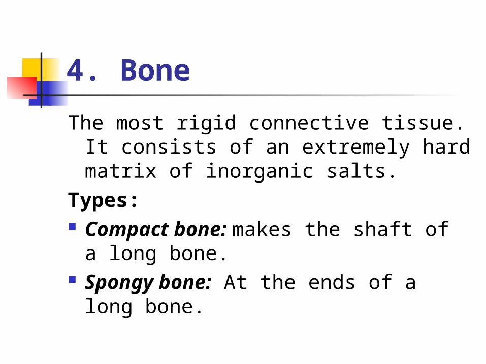
4. Bone
The most rigid connective tissue. It consists of an extremely hard matrix of inorganic salts.
Types: Compact bone: makes the shaft of
a long bone. Spongy bone: At the ends of a
long bone.

Continue5.Muscular tissue: Composed of cells that are called muscle fibers.
Muscle fibers contain actin filaments and myosin filaments, whose interaction accounts for movement.
6.Nervous tissue: Contains nerve cells (neurons) present in the brain
and spinal cord. A neuron has three parts: dendrites, a cell body
and axon. The nervous system has three functions: Sensory input, integration of data, motor output.

Body cavities and membranes
The internal organs are located within specific body cavities. During human development, there is a large ventral cavity called a coelom , which becomes divided into the thoracic and abdominal cavities. Membranes divide the thoracic cavity into the pleural cavities, containing the right and left lung, and the pericardial cavity containing the heart.

Continue
The thoracic cavity is separated from the abdominal cavity by a horizontal muscle called diaphragm.
The stomach, liver, spleen, gallbladder and most of the small and large intestine are the upper portion of the abdominal cavity.
The lower portion contains the rectum, the urinary bladder, the internal reproductive organs and the rest of the large intestine.

The dorsal cavity has also two parts
The cranial cavity within the skull containing the brain and the vertebral canal formed by the vertebrae contains the spinal cord.
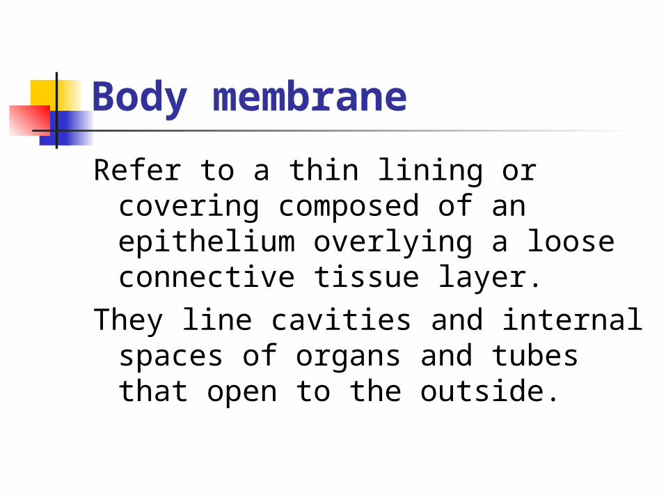
Body membrane
Refer to a thin lining or covering composed of an epithelium overlying a loose connective tissue layer.
They line cavities and internal spaces of organs and tubes that open to the outside.

Types Mucouse membrane
Mucouse membrane Serous membrane Pleural membranes Peritoneum Synovial membranes

1.Mucouse membrane:
Lines the tubes of the digestive, respiratory, urinary and productive systems.
The epithelium of this membrane contains goblet cells that secrete mucus which protects the body from invasion by bacteria and viruses, hence more mucus is secreted and expelled when a person has a cold.
mucous protects the walls of the stomach and small intestine from digestive juices, this protection breakdown when a person has ulcer.

2.Serous membrane:
Line the thoracic and abdominal cavities and the organs they contain.
They secrete a watery fluid that keeps the membranes lubricated.

3. Pleural membranes:
serous membranes that line the pleural cavity and lungs.
Pleurisy is a well-known infection of this membrane.

4.Peritoneum:
Lines the abdominal cavity and its organs. In between the organs, there is a double layer of peritoneum called mesentery.
Peritonitis is a life-threatening infection of the peritoneum.

5.Synovial membranes
Line freely movable joint cavities. They secrete synovial fluids into the
joint cavity, this fluid lubricates the ends of the bones so they can move freely.
In rheumatoid arthritis the synovial membrane becomes inflamed and grows thicker restricting movement.

6.Meninges
Found within the dorsal cavity. Composed of connective tissue
and it is a protective covering for the brain and spinal cord.
Meningitis is a life-threatening infection of the meninges.

Organ systemThe body contain a number of systems that work
together to maintain homeostasis. Homeostasis; It is the relative constancy of the body's internal
environment. Because of homeostasis even though external conditions may change dramatically internal conditions still stay within a narrow range.
Example: -No matter how hot it gets the body temperature stays around 37-degrees
-No matter how acidic your meal pH of blood stays 7.4
-No matter if you eat a candy bar, the amount of sugar in blood is about 0.1%

ContinueInternal conditions are not constant the fluctuate
below and above a particular level. Therefore, the internal state of the body is often described as one of dynamic equilibrium.
If any great changes occur, illness results.Homeostatic mechanism in the body has three
components:1. a sensor---------detect changes in the internal
environment2. a regulator center------------activates the effector3. an effector----------reveres the change and brings
conditions back to normal again. Now the sensor is no longer activated.

Mechanisms
Negative feed back: Keeps a variable, such as body temperature
close to a particular value, or set pointExample: 1-When blood pressure falls, sensory receptors
signal a regulatory center in the brain. This center send out nerve impulses to the arterial walls so that they constrict, once the blood pressure rises, the system is inactivated.
2-Regulation of body temperature.

ContinuePositive feed back:Brings about an ever great change in the same
direction. It allows changes in one direction and does not achieve relative stability.
Example: Birth stimulation.Positive feed back can be harmful, as when fever
causes metabolic changes that push fever still higher, causing death at 45-degrees because cellular protein denature and metabolism stops.
The internal environment consists of blood and tissue fluid. All organ systems contribute to the constancy of tissue fluid and blood. Special contributions are made by the liver, which keeps blood glucose constant, the kidney regulates pH.