Tissue Doppler and strain imaging of left ventricle in ... · Tissue Doppler imaging (TDI) ......
9
Original Article J Vet Sci 2015, 16(3), 357-365ㆍhttp://dx.doi.org/10.4142/jvs.2015.16.3.357 J VS Received 7 Nov. 2014, Revised 11 Mar. 2015, Accepted 4 Apr. 2015 *Corresponding author: Tel: +82-62-530-2821; Fax: +82-62-530-2809; E-mail: [email protected] Journal of Veterinary Scienceㆍⓒ 2015 The Korean Society of Veterinary Science. All Rights Reserved. This is an Open Access article distributed under the terms of the Creative Commons Attribution Non-Commercial License (http://creativecommons.org/licenses/ by-nc/4.0) which permits unrestricted non-commercial use, distribution, and reproduction in any medium, provided the original work is properly cited. pISSN 1229-845X eISSN 1976-555X Tissue Doppler and strain imaging of left ventricle in Beagle dogs with iatrogenic hypercortisolism Heejin Oui, Sunghoon Jeon, Gahyun Lee, Seungjo Park, Kyoung-Oh Cho, Jihye Choi * College of Veterinary Medicine, Chonnam National University, Gwangju 500-757, Korea Changes in radial and longitudinal left ventricular (LV) function were investigated in beagles with iatrogenic hypercortisolism. A total of 11 normal dogs were used, and 2 mg/kg prednisone was administered per oral q12 h for 28 days to 7 out of 11 dogs to induce iatrogenic hypercortisolism. Body weight, blood pressure, conventional echocardiography and tissue Doppler imaging (TDI) of normal and iatrogenic hypercortisolism groups were conducted. The myocardial wall velocity of the LV was measured using color TDI and myocardial deformation was determined by the strain and strain rate. Conventional echocardiography revealed that the diastolic LV free wall and interventricular septum in the hypercortisolism group were thickened relative to those in the normal group. The peak early diastolic myocardial velocity and early to late diastolic myocardial velocity ratio of TDI in the hypercortisolism group were significantly lower than those in the normal group. The strain values in the hypercortisolism group were significantly lower than those in the normal group, particularly for longitudinal wall motion. The lower values of myocardium from TDI and strain imaging could be used to investigate subclinical LV systolic and diastolic dysfunction in dogs with the iatrogenic hypercortisolism. Keywords: dog, hypercortisolism, left ventricle, strain, tissue Doppler imaging Introduction Tissue Doppler imaging (TDI) quantitatively assesses global and regional systolic function and abnormal relaxation of the heart by measuring tissue velocity, strain and strain rate [24,27]. Tissue velocity directly describes wall motion, which is a tethering motion of the heart regardless of whether regional myocardium movement is active or passive. Strain is an index of the length change in a myocardial segment during the cardiac cycle, and strain rate is calculated as the time-derivative of strain [5]. If two adjacent points in one myocardial segment show different lengths of change with respect to time, the myocardial segment shows a change in its own shape. Using this property, strain and strain rate reflect myocardial deformation and deformation rate, respectively. However, these two parameters are less affected by the tethering movement of the heart and may discriminate between passive and active cardiac motion. Hypercortisolism is one of the most common endocrine diseases in geriatric dogs [26]. This disorder can cause various side effects such as steroid hepatopathy, hypertension, impaired glucose tolerance of diabetes, and hypercoagulability [1,18,26]. The effects of hypercortisolism on cardiac function have been investigated in humans, and hypertrophy and concentric remodeling of the left ventricle (LV) and systolic and diastolic dysfunction have been suggested by conventional echocardiography and TDI studies [2,3,8,13,17]. In particular, impairment of LV systolic dysfunction, which is not clearly identified in conventional echocardiography, can be detected by TDI. Moreover, the association between hypercortisolism and cardiac function has not yet been studied in dogs using conventional echocardiography or TDI. In this study, intra-observer within- and between-day TDI variability was assessed and LV function was investigated in dogs with iatrogenic hypercortisolism using TDI, including strain imaging, and the reliability of these methods for LV function was determined. Materials and Methods This study protocol was approved by the Institutional Animal Care and Use Committee of Chonnam National University and the animals were cared for in accordance with the Guidelines
Transcript of Tissue Doppler and strain imaging of left ventricle in ... · Tissue Doppler imaging (TDI) ......
Original ArticleJ Vet Sci 2015, 16(3), 357-365ㆍhttp://dx.doi.org/10.4142/jvs.2015.16.3.357 JVS
Received 7 Nov. 2014, Revised 11 Mar. 2015, Accepted 4 Apr. 2015*Corresponding author: Tel: +82-62-530-2821; Fax: +82-62-530-2809; E-mail: [email protected] of Veterinary Scienceㆍⓒ 2015 The Korean Society of Veterinary Science. All Rights Reserved.This is an Open Access article distributed under the terms of the Creative Commons Attribution Non-Commercial License (http://creativecommons.org/licenses/ by-nc/4.0) which permits unrestricted non-commercial use, distribution, and reproduction in any medium, provided the original work is properly cited.
pISSN 1229-845XeISSN 1976-555X
Tissue Doppler and strain imaging of left ventricle in Beagle dogs with iatrogenic hypercortisolism
Heejin Oui, Sunghoon Jeon, Gahyun Lee, Seungjo Park, Kyoung-Oh Cho, Jihye Choi*
College of Veterinary Medicine, Chonnam National University, Gwangju 500-757, Korea
Changes in radial and longitudinal left ventricular (LV) function were investigated in beagles with iatrogenic hypercortisolism. A total of 11 normal dogs were used, and 2 mg/kg prednisone was administered per oral q12 h for 28 days to 7 out of 11 dogs to induce iatrogenic hypercortisolism. Body weight, blood pressure, conventional echocardiography and tissue Doppler imaging (TDI) of normal and iatrogenic hypercortisolism groups were conducted. The myocardial wall velocity of the LV was measured using color TDI and myocardial deformation was determined by the strain and strain rate. Conventional echocardiography revealed that the diastolic LV free wall and interventricular septum in the hypercortisolism group were thickened relative to those in the normal group. The peak early diastolic myocardial velocity and early to late diastolic myocardial velocity ratio of TDI in the hypercortisolism group were significantly lower than those in the normal group. The strain values in the hypercortisolism group were significantly lower than those in the normal group, particularly for longitudinal wall motion. The lower values of myocardium from TDI and strain imaging could be used to investigate subclinical LV systolic and diastolic dysfunction in dogs with the iatrogenic hypercortisolism.
Keywords: dog, hypercortisolism, left ventricle, strain, tissue Doppler imaging
Introduction
Tissue Doppler imaging (TDI) quantitatively assesses global and regional systolic function and abnormal relaxation of the heart by measuring tissue velocity, strain and strain rate [24,27]. Tissue velocity directly describes wall motion, which is a tethering motion of the heart regardless of whether regional myocardium movement is active or passive. Strain is an index of the length change in a myocardial segment during the cardiac cycle, and strain rate is calculated as the time-derivative of strain [5]. If two adjacent points in one myocardial segment show different lengths of change with respect to time, the myocardial segment shows a change in its own shape. Using this property, strain and strain rate reflect myocardial deformation and deformation rate, respectively. However, these two parameters are less affected by the tethering movement of the heart and may discriminate between passive and active cardiac motion.
Hypercortisolism is one of the most common endocrine diseases in geriatric dogs [26]. This disorder can cause various side effects such as steroid hepatopathy, hypertension, impaired glucose tolerance of diabetes, and hypercoagulability [1,18,26].
The effects of hypercortisolism on cardiac function have been investigated in humans, and hypertrophy and concentric remodeling of the left ventricle (LV) and systolic and diastolic dysfunction have been suggested by conventional echocardiography and TDI studies [2,3,8,13,17]. In particular, impairment of LV systolic dysfunction, which is not clearly identified in conventional echocardiography, can be detected by TDI. Moreover, the association between hypercortisolism and cardiac function has not yet been studied in dogs using conventional echocardiography or TDI.
In this study, intra-observer within- and between-day TDI variability was assessed and LV function was investigated in dogs with iatrogenic hypercortisolism using TDI, including strain imaging, and the reliability of these methods for LV function was determined.
Materials and Methods
This study protocol was approved by the Institutional Animal Care and Use Committee of Chonnam National University and the animals were cared for in accordance with the Guidelines
358 Heejin Oui et al.
Journal of Veterinary Science
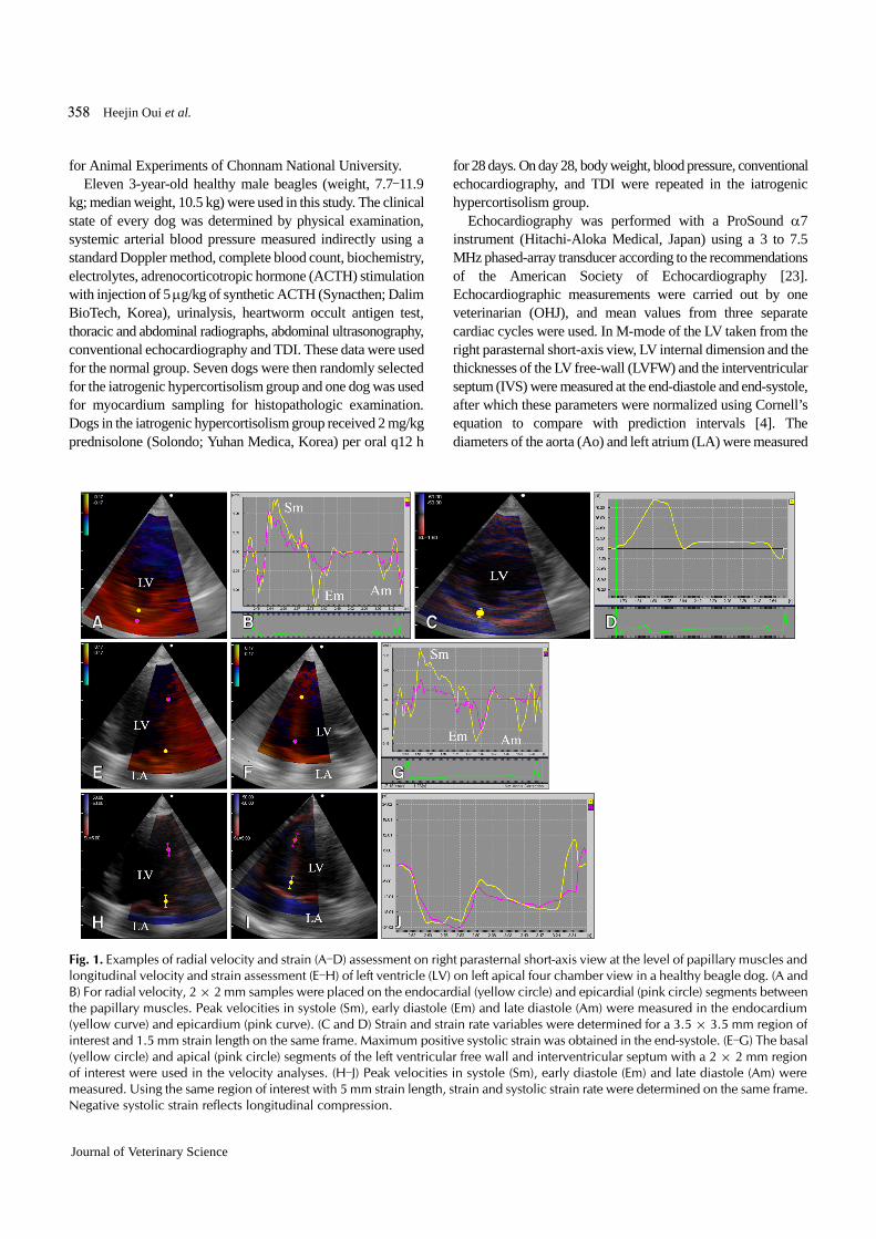
Fig. 1. Examples of radial velocity and strain (A–D) assessment on right parasternal short-axis view at the level of papillary muscles andlongitudinal velocity and strain assessment (E–H) of left ventricle (LV) on left apical four chamber view in a healthy beagle dog. (A andB) For radial velocity, 2 × 2 mm samples were placed on the endocardial (yellow circle) and epicardial (pink circle) segments betweenthe papillary muscles. Peak velocities in systole (Sm), early diastole (Em) and late diastole (Am) were measured in the endocardium (yellow curve) and epicardium (pink curve). (C and D) Strain and strain rate variables were determined for a 3.5 × 3.5 mm region of interest and 1.5 mm strain length on the same frame. Maximum positive systolic strain was obtained in the end-systole. (E–G) The basal(yellow circle) and apical (pink circle) segments of the left ventricular free wall and interventricular septum with a 2 × 2 mm regionof interest were used in the velocity analyses. (H–J) Peak velocities in systole (Sm), early diastole (Em) and late diastole (Am) were measured. Using the same region of interest with 5 mm strain length, strain and systolic strain rate were determined on the same frame.Negative systolic strain reflects longitudinal compression.
for Animal Experiments of Chonnam National University.Eleven 3-year-old healthy male beagles (weight, 7.7–11.9
kg; median weight, 10.5 kg) were used in this study. The clinical state of every dog was determined by physical examination, systemic arterial blood pressure measured indirectly using a standard Doppler method, complete blood count, biochemistry, electrolytes, adrenocorticotropic hormone (ACTH) stimulation with injection of 5 g/kg of synthetic ACTH (Synacthen; Dalim BioTech, Korea), urinalysis, heartworm occult antigen test, thoracic and abdominal radiographs, abdominal ultrasonography, conventional echocardiography and TDI. These data were used for the normal group. Seven dogs were then randomly selected for the iatrogenic hypercortisolism group and one dog was used for myocardium sampling for histopathologic examination. Dogs in the iatrogenic hypercortisolism group received 2 mg/kg prednisolone (Solondo; Yuhan Medica, Korea) per oral q12 h
for 28 days. On day 28, body weight, blood pressure, conventional echocardiography, and TDI were repeated in the iatrogenic hypercortisolism group.
Echocardiography was performed with a ProSound 7 instrument (Hitachi-Aloka Medical, Japan) using a 3 to 7.5 MHz phased-array transducer according to the recommendations of the American Society of Echocardiography [23]. Echocardiographic measurements were carried out by one veterinarian (OHJ), and mean values from three separate cardiac cycles were used. In M-mode of the LV taken from the right parasternal short-axis view, LV internal dimension and the thicknesses of the LV free-wall (LVFW) and the interventricular septum (IVS) were measured at the end-diastole and end-systole, after which these parameters were normalized using Cornell’s equation to compare with prediction intervals [4]. The diameters of the aorta (Ao) and left atrium (LA) were measured
TDI of LV in dogs with iatrogenic hypercortisolism 359
www.vetsci.org
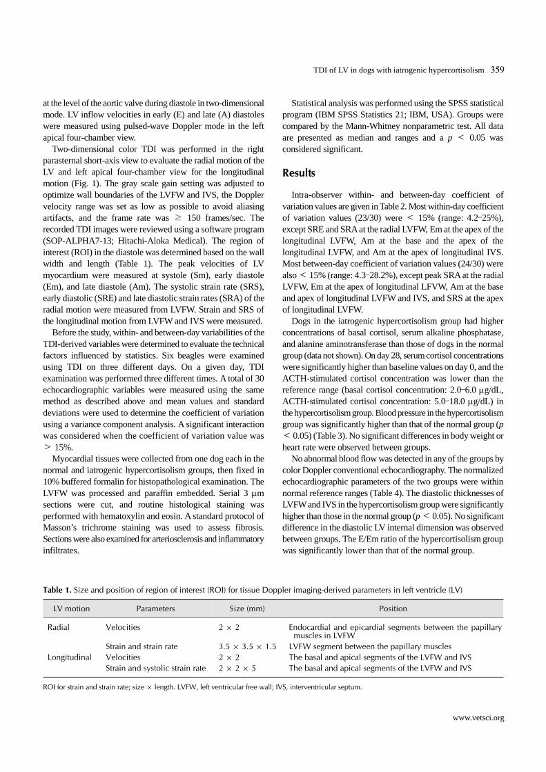
Table 1. Size and position of region of interest (ROI) for tissue Doppler imaging-derived parameters in left ventricle (LV)
LV motion Parameters Size (mm) Position
Radial Velocities 2 × 2 Endocardial and epicardial segments between the papillary muscles in LVFW
Strain and strain rate 3.5 × 3.5 × 1.5 LVFW segment between the papillary musclesLongitudinal Velocities 2 × 2 The basal and apical segments of the LVFW and IVS
Strain and systolic strain rate 2 × 2 × 5 The basal and apical segments of the LVFW and IVS
ROI for strain and strain rate; size × length. LVFW, left ventricular free wall; IVS, interventricular septum.
at the level of the aortic valve during diastole in two-dimensional mode. LV inflow velocities in early (E) and late (A) diastoles were measured using pulsed-wave Doppler mode in the left apical four-chamber view.
Two-dimensional color TDI was performed in the right parasternal short-axis view to evaluate the radial motion of the LV and left apical four-chamber view for the longitudinal motion (Fig. 1). The gray scale gain setting was adjusted to optimize wall boundaries of the LVFW and IVS, the Doppler velocity range was set as low as possible to avoid aliasing artifacts, and the frame rate was ≥ 150 frames/sec. The recorded TDI images were reviewed using a software program (SOP-ALPHA7-13; Hitachi-Aloka Medical). The region of interest (ROI) in the diastole was determined based on the wall width and length (Table 1). The peak velocities of LV myocardium were measured at systole (Sm), early diastole (Em), and late diastole (Am). The systolic strain rate (SRS), early diastolic (SRE) and late diastolic strain rates (SRA) of the radial motion were measured from LVFW. Strain and SRS of the longitudinal motion from LVFW and IVS were measured.
Before the study, within- and between-day variabilities of the TDI-derived variables were determined to evaluate the technical factors influenced by statistics. Six beagles were examined using TDI on three different days. On a given day, TDI examination was performed three different times. A total of 30 echocardiographic variables were measured using the same method as described above and mean values and standard deviations were used to determine the coefficient of variation using a variance component analysis. A significant interaction was considered when the coefficient of variation value was > 15%.
Myocardial tissues were collected from one dog each in the normal and iatrogenic hypercortisolism groups, then fixed in 10% buffered formalin for histopathological examination. The LVFW was processed and paraffin embedded. Serial 3 m sections were cut, and routine histological staining was performed with hematoxylin and eosin. A standard protocol of Masson’s trichrome staining was used to assess fibrosis. Sections were also examined for arteriosclerosis and inflammatory infiltrates.
Statistical analysis was performed using the SPSS statistical program (IBM SPSS Statistics 21; IBM, USA). Groups were compared by the Mann-Whitney nonparametric test. All data are presented as median and ranges and a p < 0.05 was considered significant.
Results
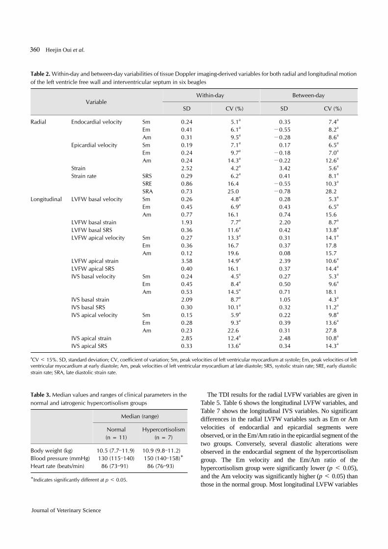
Intra-observer within- and between-day coefficient of variation values are given in Table 2. Most within-day coefficient of variation values (23/30) were < 15% (range: 4.2–25%), except SRE and SRA at the radial LVFW, Em at the apex of the longitudinal LVFW, Am at the base and the apex of the longitudinal LVFW, and Am at the apex of longitudinal IVS. Most between-day coefficient of variation values (24/30) were also < 15% (range: 4.3–28.2%), except peak SRA at the radial LVFW, Em at the apex of longitudinal LFVW, Am at the base and apex of longitudinal LVFW and IVS, and SRS at the apex of longitudinal LVFW.
Dogs in the iatrogenic hypercortisolism group had higher concentrations of basal cortisol, serum alkaline phosphatase, and alanine aminotransferase than those of dogs in the normal group (data not shown). On day 28, serum cortisol concentrations were significantly higher than baseline values on day 0, and the ACTH-stimulated cortisol concentration was lower than the reference range (basal cortisol concentration: 2.0–6.0 g/dL, ACTH-stimulated cortisol concentration: 5.0–18.0 g/dL) in the hypercortisolism group. Blood pressure in the hypercortisolism group was significantly higher than that of the normal group (p < 0.05) (Table 3). No significant differences in body weight or heart rate were observed between groups.
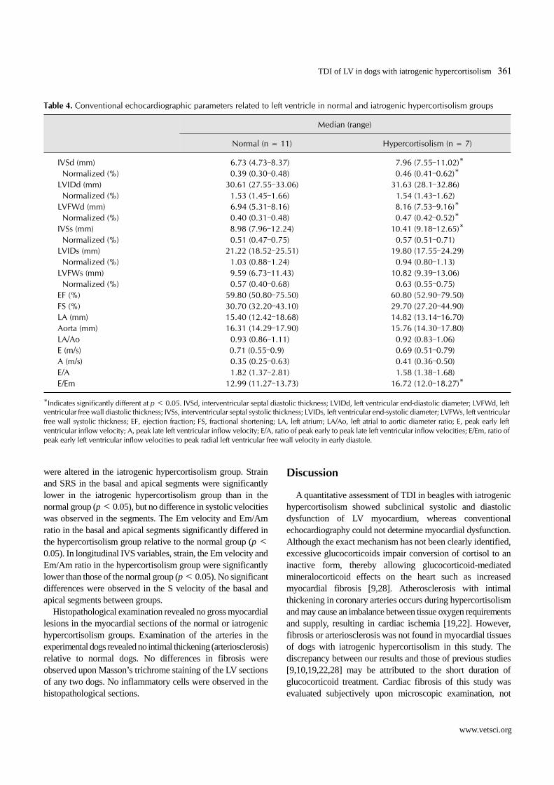
No abnormal blood flow was detected in any of the groups by color Doppler conventional echocardiography. The normalized echocardiographic parameters of the two groups were within normal reference ranges (Table 4). The diastolic thicknesses of LVFW and IVS in the hypercortisolism group were significantly higher than those in the normal group (p < 0.05). No significant difference in the diastolic LV internal dimension was observed between groups. The E/Em ratio of the hypercortisolism group was significantly lower than that of the normal group.
360 Heejin Oui et al.
Journal of Veterinary Science
Table 3. Median values and ranges of clinical parameters in the normal and iatrogenic hypercortisolism groups
Median (range)
Normal (n = 11)
Hypercortisolism (n = 7)
Body weight (kg) 10.5 (7.7–11.9) 10.9 (9.8–11.2) Blood pressure (mmHg) 130 (115–140) 150 (140–158)*Heart rate (beats/min) 86 (73–91) 86 (76–93)
*Indicates significantly different at p < 0.05.
Table 2. Within-day and between-day variabilities of tissue Doppler imaging-derived variables for both radial and longitudinal motion of the left ventricle free wall and interventricular septum in six beagles
VariableWithin-day Between-day
SD CV (%) SD CV (%)
Radial Endocardial velocity Sm 0.24 5.1a 0.35 7.4a
Em 0.41 6.1a −0.55 8.2a
Am 0.31 9.5a −0.28 8.6a
Epicardial velocity Sm 0.19 7.1a 0.17 6.5a
Em 0.24 9.7a −0.18 7.0a
Am 0.24 14.3a −0.22 12.6a
Strain 2.52 4.2a 3.42 5.6a
Strain rate SRS 0.29 6.2a 0.41 8.1a
SRE 0.86 16.4 −0.55 10.3a
SRA 0.73 25.0 −0.78 28.2Longitudinal LVFW basal velocity Sm 0.26 4.8a 0.28 5.3a
Em 0.45 6.9a 0.43 6.5a
Am 0.77 16.1 0.74 15.6LVFW basal strain 1.93 7.7a 2.20 8.7a
LVFW basal SRS 0.36 11.6a 0.42 13.8a
LVFW apical velocity Sm 0.27 13.3a 0.31 14.1a
Em 0.36 16.7 0.37 17.8Am 0.12 19.6 0.08 15.7
LVFW apical strain 3.58 14.9a 2.39 10.6a
LVFW apical SRS 0.40 16.1 0.37 14.4a
IVS basal velocity Sm 0.24 4.5a 0.27 5.3a
Em 0.45 8.4a 0.50 9.6a
Am 0.53 14.5a 0.71 18.1IVS basal strain 2.09 8.7a 1.05 4.3a
IVS basal SRS 0.30 10.1a 0.32 11.2a
IVS apical velocity Sm 0.15 5.9a 0.22 9.8a
Em 0.28 9.3a 0.39 13.6a
Am 0.23 22.6 0.31 27.8IVS apical strain 2.85 12.4a 2.48 10.8a
IVS apical SRS 0.33 13.6a 0.34 14.3a
aCV < 15%. SD, standard deviation; CV, coefficient of variation; Sm, peak velocities of left ventricular myocardium at systole; Em, peak velocities of left ventricular myocardium at early diastole; Am, peak velocities of left ventricular myocardium at late diastole; SRS, systolic strain rate; SRE, early diastolic strain rate; SRA, late diastolic strain rate.
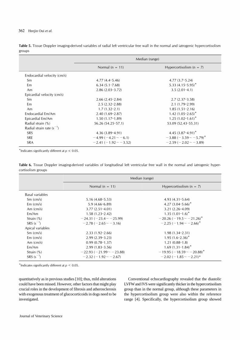
The TDI results for the radial LVFW variables are given in Table 5. Table 6 shows the longitudinal LVFW variables, and Table 7 shows the longitudinal IVS variables. No significant differences in the radial LVFW variables such as Em or Am velocities of endocardial and epicardial segments were observed, or in the Em/Am ratio in the epicardial segment of the two groups. Conversely, several diastolic alterations were observed in the endocardial segment of the hypercortisolism group. The Em velocity and the Em/Am ratio of the hypercortisolism group were significantly lower (p < 0.05), and the Am velocity was significantly higher (p < 0.05) than those in the normal group. Most longitudinal LVFW variables
TDI of LV in dogs with iatrogenic hypercortisolism 361
www.vetsci.org
Table 4. Conventional echocardiographic parameters related to left ventricle in normal and iatrogenic hypercortisolism groups
Median (range)
Normal (n = 11) Hypercortisolism (n = 7)
IVSd (mm) 6.73 (4.73–8.37) 7.96 (7.55–11.02)* Normalized (%) 0.39 (0.30–0.48) 0.46 (0.41–0.62)*LVIDd (mm) 30.61 (27.55–33.06) 31.63 (28.1–32.86) Normalized (%) 1.53 (1.45–1.66) 1.54 (1.43–1.62)LVFWd (mm) 6.94 (5.31–8.16) 8.16 (7.53–9.16)* Normalized (%) 0.40 (0.31–0.48) 0.47 (0.42–0.52)*IVSs (mm) 8.98 (7.96–12.24) 10.41 (9.18–12.65)* Normalized (%) 0.51 (0.47–0.75) 0.57 (0.51–0.71)LVIDs (mm) 21.22 (18.52–25.51) 19.80 (17.55–24.29) Normalized (%) 1.03 (0.88–1.24) 0.94 (0.80–1.13)LVFWs (mm) 9.59 (6.73–11.43) 10.82 (9.39–13.06) Normalized (%) 0.57 (0.40–0.68) 0.63 (0.55–0.75)EF (%) 59.80 (50.80–75.50) 60.80 (52.90–79.50)FS (%) 30.70 (32.20–43.10) 29.70 (27.20–44.90)LA (mm) 15.40 (12.42–18.68) 14.82 (13.14–16.70)Aorta (mm) 16.31 (14.29–17.90) 15.76 (14.30–17.80)LA/Ao 0.93 (0.86–1.11) 0.92 (0.83–1.06)E (m/s) 0.71 (0.55–0.9) 0.69 (0.51–0.79)A (m/s) 0.35 (0.25–0.63) 0.41 (0.36–0.50)E/A 1.82 (1.37–2.81) 1.58 (1.38–1.68)E/Em 12.99 (11.27–13.73) 16.72 (12.0–18.27)*
*Indicates significantly different at p < 0.05. IVSd, interventricular septal diastolic thickness; LVIDd, left ventricular end-diastolic diameter; LVFWd, left ventricular free wall diastolic thickness; IVSs, interventricular septal systolic thickness; LVIDs, left ventricular end-systolic diameter; LVFWs, left ventricular free wall systolic thickness; EF, ejection fraction; FS, fractional shortening; LA, left atrium; LA/Ao, left atrial to aortic diameter ratio; E, peak early left ventricular inflow velocity; A, peak late left ventricular inflow velocity; E/A, ratio of peak early to peak late left ventricular inflow velocities; E/Em, ratio of peak early left ventricular inflow velocities to peak radial left ventricular free wall velocity in early diastole.
were altered in the iatrogenic hypercortisolism group. Strain and SRS in the basal and apical segments were significantly lower in the iatrogenic hypercortisolism group than in the normal group (p < 0.05), but no difference in systolic velocities was observed in the segments. The Em velocity and Em/Am ratio in the basal and apical segments significantly differed in the hypercortisolism group relative to the normal group (p < 0.05). In longitudinal IVS variables, strain, the Em velocity and Em/Am ratio in the hypercortisolism group were significantly lower than those of the normal group (p < 0.05). No significant differences were observed in the S velocity of the basal and apical segments between groups.
Histopathological examination revealed no gross myocardial lesions in the myocardial sections of the normal or iatrogenic hypercortisolism groups. Examination of the arteries in the experimental dogs revealed no intimal thickening (arteriosclerosis) relative to normal dogs. No differences in fibrosis were observed upon Masson’s trichrome staining of the LV sections of any two dogs. No inflammatory cells were observed in the histopathological sections.
Discussion
A quantitative assessment of TDI in beagles with iatrogenic hypercortisolism showed subclinical systolic and diastolic dysfunction of LV myocardium, whereas conventional echocardiography could not determine myocardial dysfunction. Although the exact mechanism has not been clearly identified, excessive glucocorticoids impair conversion of cortisol to an inactive form, thereby allowing glucocorticoid-mediated mineralocorticoid effects on the heart such as increased myocardial fibrosis [9,28]. Atherosclerosis with intimal thickening in coronary arteries occurs during hypercortisolism and may cause an imbalance between tissue oxygen requirements and supply, resulting in cardiac ischemia [19,22]. However, fibrosis or arteriosclerosis was not found in myocardial tissues of dogs with iatrogenic hypercortisolism in this study. The discrepancy between our results and those of previous studies [9,10,19,22,28] may be attributed to the short duration of glucocorticoid treatment. Cardiac fibrosis of this study was evaluated subjectively upon microscopic examination, not
362 Heejin Oui et al.
Journal of Veterinary Science
Table 5. Tissue Doppler imaging-derived variables of radial left ventricular free wall in the normal and iatrogenic hypercortisolism groups
Median (range)
Normal (n = 11) Hypercortisolism (n = 7)
Endocardial velocity (cm/s) Sm 4.77 (4.4–5.46) 4.77 (3.7–5.24) Em 6.34 (5.1–7.68) 5.33 (4.15–5.95)* Am 2.86 (2.03–3.72) 3.5 (2.01–4.1)Epicardial velocity (cm/s) Sm 2.66 (2.45–2.84) 2.7 (2.37–3.58) Em 2.5 (2.32–2.88) 2.1 (1.79–2.99) Am 1.7 (1.32–2.1) 1.85 (1.51–2.16)Endocardial Em/Am 2.40 (1.69–2.87) 1.42 (1.05–2.65)*Epicardial Em/Am 1.50 (1.17–1.89) 1.25 (1.02–1.61)*Radial strain (%) 56.26 (54.25–57.1) 53.09 (52.43–55.31)Radial strain rate (s−1) SRS 4.36 (3.89–4.91) 4.45 (3.87–4.91)* SRE −4.99 (−4.21∼−6.1) −3.88 (−3.59∼−5.79)* SRA −2.41 (−1.92∼−3.52) −2.59 (−2.02∼−3.89)
*Indicates significantly different at p < 0.05.
Table 6. Tissue Doppler imaging-derived variables of longitudinal left ventricular free wall in the normal and iatrogenic hyper-cortisolism groups
Median (range)
Normal (n = 11) Hypercortisolism (n = 7)
Basal variables Sm (cm/s) 5.16 (4.68–5.53) 4.93 (4.31–5.64) Em (cm/s) 5.9 (4.66–6.89) 4.27 (3.04–5.66)* Am (cm/s) 3.77 (2.51–4.01) 3.21 (2.26–4.09) Em/Am 1.58 (1.23–2.42) 1.35 (1.01–1.6)* Strain (%) −24.31 (−23.4∼−25.99) −20.26 (−19.5∼−21.26)* SRS (s−1) −2.78 (−2.65∼−3.16) −2.25 (−1.94∼−2.66)*Apical variables Sm (cm/s) 2.33 (1.92–2.66) 1.98 (1.34–2.31) Em (cm/s) 2.99 (2.39–3.23) 1.95 (1.6–2.36)* Am (cm/s) 0.99 (0.78–1.37) 1.21 (0.88–1.8) Em/Am 2.99 (1.83–3.56) 1.69 (1.31–1.84)* Strain (%) −22.93 (−21.99∼−23.88) −19.95 (−18.59∼−20.88)* SRS (s−1) −2.32 (−1.92∼−2.67) −2.02 (−1.85∼−2.21)*
*Indicates significantly different at p < 0.05.
quantitatively as in previous studies [10]; thus, mild alterations could have been missed. However, other factors that might play crucial roles in the development of fibrosis and atherosclerosis with exogenous treatment of glucocorticoids in dogs need to be investigated.
Conventional echocardiography revealed that the diastolic LVFW and IVS were significantly thicker in the hypercortisolism group than in the normal group, although these parameters in the hypercortisolism group were also within the reference range [4]. Specifically, the hypercortisolism group showed
TDI of LV in dogs with iatrogenic hypercortisolism 363
www.vetsci.org
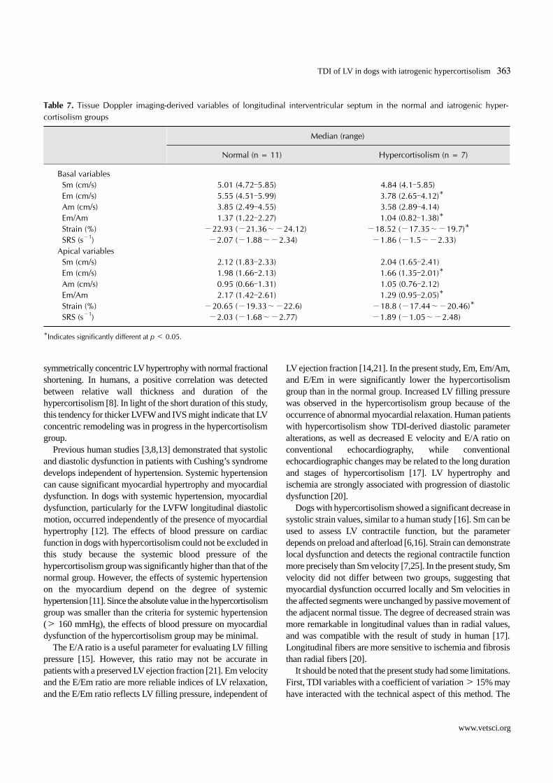
Table 7. Tissue Doppler imaging-derived variables of longitudinal interventricular septum in the normal and iatrogenic hyper-cortisolism groups
Median (range)
Normal (n = 11) Hypercortisolism (n = 7)
Basal variables Sm (cm/s) 5.01 (4.72–5.85) 4.84 (4.1–5.85) Em (cm/s) 5.55 (4.51–5.99) 3.78 (2.65–4.12)* Am (cm/s) 3.85 (2.49–4.55) 3.58 (2.89–4.14) Em/Am 1.37 (1.22–2.27) 1.04 (0.82–1.38)* Strain (%) −22.93 (−21.36∼−24.12) −18.52 (−17.35∼−19.7)* SRS (s−1) −2.07 (−1.88∼−2.34) −1.86 (−1.5∼−2.33) Apical variables Sm (cm/s) 2.12 (1.83–2.33) 2.04 (1.65–2.41) Em (cm/s) 1.98 (1.66–2.13) 1.66 (1.35–2.01)* Am (cm/s) 0.95 (0.66–1.31) 1.05 (0.76–2.12) Em/Am 2.17 (1.42–2.61) 1.29 (0.95–2.05)* Strain (%) −20.65 (−19.33∼−22.6) −18.8 (−17.44∼−20.46)* SRS (s−1) −2.03 (−1.68∼−2.77) −1.89 (−1.05∼−2.48)
*Indicates significantly different at p < 0.05.
symmetrically concentric LV hypertrophy with normal fractional shortening. In humans, a positive correlation was detected between relative wall thickness and duration of the hypercortisolism [8]. In light of the short duration of this study, this tendency for thicker LVFW and IVS might indicate that LV concentric remodeling was in progress in the hypercortisolism group.
Previous human studies [3,8,13] demonstrated that systolic and diastolic dysfunction in patients with Cushing’s syndrome develops independent of hypertension. Systemic hypertension can cause significant myocardial hypertrophy and myocardial dysfunction. In dogs with systemic hypertension, myocardial dysfunction, particularly for the LVFW longitudinal diastolic motion, occurred independently of the presence of myocardial hypertrophy [12]. The effects of blood pressure on cardiac function in dogs with hypercortisolism could not be excluded in this study because the systemic blood pressure of the hypercortisolism group was significantly higher than that of the normal group. However, the effects of systemic hypertension on the myocardium depend on the degree of systemic hypertension [11]. Since the absolute value in the hypercortisolism group was smaller than the criteria for systemic hypertension (> 160 mmHg), the effects of blood pressure on myocardial dysfunction of the hypercortisolism group may be minimal.
The E/A ratio is a useful parameter for evaluating LV filling pressure [15]. However, this ratio may not be accurate in patients with a preserved LV ejection fraction [21]. Em velocity and the E/Em ratio are more reliable indices of LV relaxation, and the E/Em ratio reflects LV filling pressure, independent of
LV ejection fraction [14,21]. In the present study, Em, Em/Am, and E/Em in were significantly lower the hypercortisolism group than in the normal group. Increased LV filling pressure was observed in the hypercortisolism group because of the occurrence of abnormal myocardial relaxation. Human patients with hypercortisolism show TDI-derived diastolic parameter alterations, as well as decreased E velocity and E/A ratio on conventional echocardiography, while conventional echocardiographic changes may be related to the long duration and stages of hypercortisolism [17]. LV hypertrophy and ischemia are strongly associated with progression of diastolic dysfunction [20].
Dogs with hypercortisolism showed a significant decrease in systolic strain values, similar to a human study [16]. Sm can be used to assess LV contractile function, but the parameter depends on preload and afterload [6,16]. Strain can demonstrate local dysfunction and detects the regional contractile function more precisely than Sm velocity [7,25]. In the present study, Sm velocity did not differ between two groups, suggesting that myocardial dysfunction occurred locally and Sm velocities in the affected segments were unchanged by passive movement of the adjacent normal tissue. The degree of decreased strain was more remarkable in longitudinal values than in radial values, and was compatible with the result of study in human [17]. Longitudinal fibers are more sensitive to ischemia and fibrosis than radial fibers [20].
It should be noted that the present study had some limitations. First, TDI variables with a coefficient of variation > 15% may have interacted with the technical aspect of this method. The
364 Heejin Oui et al.
Journal of Veterinary Science
TDI-derived variables obtained from the apical segment had relatively higher coefficients of variation than those in the basal segment. This may have occurred because TDI is angle-dependent and sensitive to noise. A second limitation is that only intra- observer repeatability was evaluated. Finally, this study was performed in a relatively small population of adult dogs for a short period. Despite these limitations, we demonstrated the effects of iatrogenic hypercortisolism on myocardial function in dogs.
The LV showed diastolic and systolic dysfunction in dogs with iatrogenic hypercortisolism. These changes were detected more clearly with TDI-derived variables, particularly in longitudinal motion before becoming prominent on conventional echocardiography. The results presented herein will serve as fundamental data for investigating the effects of cortisol effect on myocardial changes in a larger population of dogs with hypercortisolism.
Acknowledgments
This study was supported, in part, by the Animal Medical Institute of Chonnam National University in Korea.
Conflict of Interest
There is no conflict of interest.
References
1. Badylak SF, van Vleet JF. Sequential morphologic and clinicopathologic alterations in dogs with experimentally induced glucocorticoid hepatopathy. Am J Vet Res 1981, 42, 1310-1318.
2. Baykan M, Erem C, Gedikli O, Hacihasanoglu A, Erdogan T, Kocak M, Kaplan S, Kiriş A, Orem C, Çelik S. Assessment of left ventricular diastolic function and Tei index by tissue Doppler imaging in patients with Cushing’s syndrome. Echocardiography 2008, 25, 182-190.
3. Bayram NA, Ersoy R, Aydin C, Gul K, Keles T, Topaloglu O, Durmaz T, Bozkurt E, Cakir B. Assessment of left ventricular functions by tissue Doppler echocardiography in patients with Cushing’s disease. J Endocrinol Invest 2009, 32, 248-252.
4. Cornell CC, Kittleson MD, Della Torre P, Häggström J, Lombard CW, Pedersen HD, Vollmar A, Wey A. Allometric scaling of M-mode cardiac measurements in normal adult dogs. J Vet Intern Med 2004, 18, 311-321.
5. D’Hooge J, Heimdal A, Jamal F, Kukulski T, Bijnens B, Rademakers F, Hatle L, Suetens P, Sutherland GR. Regional strain and strain rate measurements by cardiac ultrasound: principles, implementation and limitations. Eur J Echocardiogr 2000, 1, 154-170.
6. Drighil A, Madias JE, Mathewson JW, El Mosalami H, El Badaoui N, Ramdani B, Bennis A. Haemodialysis: effects of acute decrease in preload on tissue Doppler imaging indices
of systolic and diastolic function of the left and right ventricles. Eur J Echocardiogr 2008, 9, 530-535.
7. Edvardsen T, Skulstad H, Aakhus S, Urheim S, Ihlen H. Regional myocardial systolic function during acute myocardial ischemia assessed by strain Doppler echocardiography. J Am Coll Cardiol 2001, 37, 726-730.
8. Fallo F, Budano S, Sonino N, Muiesan ML, Agabiti-Rosei E, Boscaro M. Left ventricular structural characteristics in Cushing’s syndrome. J Hum Hypertens 1994, 8, 509-513.
9. Glorioso N, Filigheddu F, Parpaglia PP, Soro A, Troffa C, Argiolas G, Mulatero P. 11-Hydroxysteroid dehydrogenase type 2 activity is associated with left ventricular mass in essential hypertension. Eur Heart J 2005, 26, 498-504.
10. Hattori T, Murase T, Iwase E, Takahashi K, Ohtake M, Tsuboi K, Ohtake M, Miyachi M, Murohara T, Nagata K. Glucocorticoid-induced hypertension and cardiac injury: effects of mineralocorticoid and glucocorticoid receptor antagonism. Nagoya J Med Sci 2013, 75, 81-92.
11. Hillis LD, Izquierdo C, Davis C, Brotherton S, Eberhart R, Roan PG, Willerson JT. Effect of various degrees of systemic arterial hypertension on acute canine myocardial ischemia. Am J Physiol 1981, 240, H855-861.
12. Misbach C, Gouni V, Tissier R, Trehiou-Sechi E, Petit AMP, Carlos Sampedrano C, Pouchelon JL, Chetboul V. Echocardiographic and tissue Doppler imaging alterations associated with spontaneous canine systemic hypertension. J Vet Intern Med 2011, 25, 1025-1035.
13. Muiesan ML, Lupia M, Salvetti M, Grigoletto C, Sonino N, Boscaro M, Rosei EA, Mantero F, Fallo F. Left ventricular structural and functional characteristics in Cushing's syndrome. J Am Coll Cardiol 2003, 41, 2275-2279.
14. Nagueh SF, Middleton KJ, Kopelen HA, Zoghbi WA, Quiñones MA. Doppler tissue imaging: a noninvasive technique for evaluation of left ventricular relaxation and estimation of filling pressures. J Am Coll Cardiol 1997, 30, 1527-1533.
15. Nishimura RA, Tajik AJ. Evaluation of diastolic filling of left ventricle in health and disease: Doppler echocardiography is the clinician’s Rosetta Stone. J Am Coll Cardiol 1997, 30, 8-18.
16. Oki T, Fukuda K, Tabata T, Mishiro Y, Yamada H, Abe M, Onose Y, Wakatsuki T, Iuchi A, Ito S. Effect of an acute increase in afterload on left ventricular regional wall motion velocity in healthy subjects. J Am Soc Echocardiogr 1999, 12, 476-483.
17. Pereira AM, Delgado V, Romijn JA, Smit JWA, Bax JJ, Feelders RA. Cardiac dysfunction is reversed upon successful treatment of Cushing’s syndrome. Eur J Endocrinol 2010, 162, 331-340.
18. Peterson ME, Altszuler N, Nichols CE. Decreased insulin sensitivity and glucose tolerance in spontaneous canine hyperadrenocorticism. Res Vet Sci 1984, 36, 177-182.
19. Pivonello R, Faggiano A, Lombardi G, Colao A. The metabolic syndrome and cardiovascular risk in Cushing’s syndrome. Endocrinol Metab Clin North Am 2005, 34, 327-339.
20. Poulsen SH, Andersen NH, Ivarsen PI, Mogensen CE, Egeblad H. Doppler tissue imaging reveals systolic dysfunction in patients with hypertension and apparent
TDI of LV in dogs with iatrogenic hypercortisolism 365
www.vetsci.org
"isolated" diastolic dysfunction. J Am Soc Echocardiogr 2003, 16, 724-731.
21. Rivas-Gotz C, Manolios M, Thohan V, Naqueh SF. Impact of left ventricular ejection fraction on estimation of left ventricular filling pressures using tissue Doppler and flow propagation velocity. Am J Cardiol 2003, 91, 780-784.
22. Rosenfeld S, Marmorston J, Sobel H, White AE. Enhancement of experimental atherosclerosis by ACTH in the dog. Proc Soc Exp Biol Med 1960, 103, 83-86.
23. Sahn DJ, DeMaria A, Kisslo J, Weyman A; The committee on M-mode standardization of the American society of echocardiography. Recommendations regarding quantitation in M-mode echocardiography: results of a survey of echocardiographic measurements. Circulation 1978, 58, 1072-1083.
24. Simak J, Keller L, Killich M, Hartmann K, Wess G. Color-coded longitudinal interventricular septal tissue velocity imaging, strain and strain rate in healthy Doberman Pinschers. J Vet Cardiol 2011, 13, 1-11.
25. Skulstad H, Andersen K, Edvardsen T, Rein KA, Tønnessen TI, Hol PK, Fosse E, Ihlen H. Detection of ischemia and new insight into left ventricular physiology by strain Doppler and tissue velocity imaging: assessment during coronary bypass operation of the beating heart. J Am Soc Echocardiogr 2004, 17, 1225-1233.
26. Smets P, Meyer E, Maddens B, Daminet S. Cushing’s syndrome, glucocorticoids and the kidney. Gen Comp Endocrinol 2010, 169, 1-10.
27. Tidholm A, Ljungvall I, Höglund K, Westling AB, Häggström J. Tissue Doppler and strain imaging in dogs with myxomatous mitral valve disease in different stages of congestive heart failure. J Vet Intern Med 2009, 23, 1197-1207.
28. Young MJ, Moussa L, Dilley R, Funder JW. Early inflammatory responses in experimental cardiac hypertrophy and fibrosis: effects of 11-hydroxysteroid dehydrogenase inactivation. Endocrinology 2003, 144, 1121-1125.