Tissue Bath & Pe rfusion Systems - Harvard ApparatusSystems... · Sp yse temr sf &u As ppi lioc atn...
46
Tissue Bath & Perfusion Systems Selection Guide Precision Systems for Physiology Research
Transcript of Tissue Bath & Pe rfusion Systems - Harvard ApparatusSystems... · Sp yse temr sf &u As ppi lioc atn...

Tissue Bath &Perfusion Systems
Selection GuidePrecision Systems
for Physiology Research

Isolated Organ & Tissue
isolated organ & tissue
1Harvard Apparatus • phone 508.893.8999 • toll free U.S. 800.272.2775 • fax 508.429.5732 • www.harvardapparatus.com
Products Page No.Systems & Applications Overview 2-3A New Approach to Isolated Organ & Tissue Research 4Graz Tissue Bath System 5-6Schuler Tissue Bath System 7-8M Series Myograph 9Compact Organ Bath 10-11Tissue Bath Teaching Sets 12-13Mayflower Horizontal Tissue Bath System 14-15Perfusion Bath for Tubular Organs 16TIOX Bath for O2 Consumption 17Coleman Superfusion Bath 18Steiert Bath for Cardiac Tissue 19Precision Cut Lung Slice Chamber 20Marsh Ganglion Bath 21Moist Chambers for Organ Perfusion 22-24UP-100 Universal Perfusion System 25Perfusion System for Pig Liver/Kidney 26Isolated Heart Systems Overview 27-28IH-SR Isolated Heart for Small Rodent (Murine, Rat, Guinea Pig) 29-31IH-5 Isolated Heart for Rat, Guinea Pig, & Rabbit 32-34UP-100LD Basic Langendorff Heart System 35Student Isolated Heart Perfusion Apparatus 36PSCI Perfusion System for Cell Isolation 37Easy Set-up for Cell Extraction 38Isolated Lung Systems Overview 39IPL-1 Isolated Perfused Lung for Mouse 40-41IPL-2 Isolated Perfused Lung for Rat & Guinea Pig 42-43IPL-4 Isolated Perfused Lung for Rabbit 44-45IPL-16 Isolated Perfused Lung for Pigs & Large Animals 46
IH-SR Isolated Heart for Small Rodents, see page 29
IPL-16 Isolated Perfused Lung for Pigs and Large Animals,
see page 46
UP-100 Universal Perfusion System,
see page 25
Marsh Ganglion Bath, see page 21
Moist Chamber withEdema Balance,see page 24

perfusion & tissue bathSystems & Applications Overview Perfusion and Tissue Bath Systems Overview
2
perfusion & tissue bath
Isolated Organ &
Tissue
Harvard Apparatus • phone 508.893.8999 • toll free U.S. 800.272.2775 • fax 508.429.5732 • www.harvardapparatus.com
text
3Harvard Apparatus • phone 508.893.8999 • toll free U.S. 800.272.2775 • fax 508.429.5732 • www.harvardapparatus.com
perf
usio
n &
tiss
ue b
ath
Isol
ated
Org
an &
Tis
sue
perfusion & tissue bath> Schuler Vertical Tissue Bath for
> Graz Vertical Tissue Bath
> Mayflower Horizontal Tissue Bath
> Modular Organ Bath for Isolated Tissues
> Compact Organ Bath for Isolated Tissues
> M Series Myograph
Frequently Used for Studying
• Spasm
• Spasmolysis
• Vascular tone
• Edema
Frequently Used for Studying:
• Inotropism
• Chronotropism
• Dromotropism
• Refractory State
Frequently Used for Studying:
• Metabolism
• Neromuscular Transmission
Frequently Used for Studying:
• Neuro-Neuronal Transmission
• Neuromuscular Transmission
> Schuler Vertical Tissue Bath
> Graz Vertical Tissue Bath
> Mayflower Horizontal Tissue Bath
> Modular Organ Bath for Isolated Tissues
> Compact Organ Bath for Isolated Tissues
> M Series Myograph
> Schuler Vertical Tissue Bath
> Graz Vertical Tissue Bath
> Mayflower Horizontal Tissue Bath
> Coleman Superfusion Bath
> Modular Organ Bath for Isolated Tissues
> Compact Organ Bath for Isolated Tissues
> M Series Myograph
> Schuler Vertical Tissue Bath
> Graz Vertical Tissue Bath
> Mayflower Horizontal Tissue Bath
> Coleman Superfusion Bath
> Superfusion Cascade Bath
> M Series Myograph
> Schuler Vertical Tissue Bath for Strips or Rings
> Graz Vertical Tissue Bath for Strips or Rings
> Mayflower Horizontal Tissue Bath for Strips or Rings
• Ileum
• Stomach
• Duodenum
• Telia Coli
• Aorta
• Artery
• Vein
• Trachea
• Lung
• Bronchi
• Bladder
• Ureter
• Vas-deferens
• Uterus
• Placenta
• S-A node • A-V node • Atrium • Cardiac Muscle • Trabecula • Papillary Muscle
• Purkinjie Fiber
• Sartorious
• Gastrocnemius
• Nerve-muscle Preparations
• Nerve
• Ganglion
> Marsh Ganglion Bath for Whole Nerve and Ganglion
> Schuler Vertical Tissue Bath for Nerve-Muscle Preparation
> Schuler Vertical Tissue Bath
> Graz Vertical Tissue Bath
> Mayflower Horizontal Tissue Bath
> Steiert Tissue Bath for Cardiac AP and Force Measurement
systems for tissuestissues applications
Gastro-intestinal Tract
• Esophagus
• Stomach
• Liver/Pancreas
• Intestine
> Moist Chamber, Perfusion System for Liver, etc.
> UP-100, perfusion of isolated liver... in-situ or ex-vivo
> PBTO, Intestine perfusion
> Servo Controlled Perfusion System, Perfusion of Isolated Liver: In-Situ or Ex-Vivo
> IPR, Perfusion Bath for Ileum Peristaltic Reflex Studies
Vascular Musculature
• Hind Quarter
• Mesenteric Bed
• Coronary Vasculature
> IH-SR, HA-PL, UP-100, IH-5, IH-9 Isolated Heart System for Small Rodents, Rabbit, Small Pig
> Moist Chamber With Edema Balance
> PBTO, Blood Vessel Perfusion
Bronchial Musculature
• Lung> IPL-4, IPL-2, IPL-4, IPL-16 Isolated Perfused Lung of Mouse, Rat and Guinea Pig, Rabbit, Pig
> PBTO, Intraluminal Trachea Perfusion
> PCLS, Precision Cut Lung Slice Chamber
Uro-Genital Tract
• Kidney
• Placenta
> PS-1, Moist Chamber, Perfusion System for Kidney, etc.
> PBTO, Intraluminal Vas Deferens Perfusion
Heart
• Langendorff
• Working heart
• Heart-Lung Preparation
> IH-SR, BL-IH, HA-PL, BL-UP, UP-100, IH-5, IH-9 Isolated Heart System for Small Rodents, Rabbit, Small Pig
• Intact Limb/Hindquarter > UP-100
• Brain
• Spinal Cord
• Ganglion
Smooth Muscle
systems for organsorgans
Cardiac
Skeletal Muscle
Nerve Bundle
isolated organ & tissueThe Harvard Apparatus 3 Tier Approach to ResearchOur 3 Tier System is Organized in the Following Manner:
4
3 tier approach to researchIsolated O
rgan & Tissue
Harvard Apparatus • phone 508.893.8999 • toll free U.S. 800.272.2775 • fax 508.429.5732 • www.harvardapparatus.com
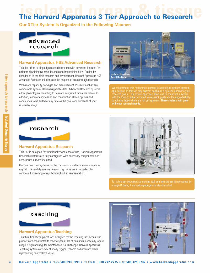
We recommend that researchers contact us directly to discuss specificapplications so that we may custom configure a system tailored to yourresearch goals. This proven approach allows us to construct a systemwith the tools to achieve immediate research goals and the upgradeabilityto achieve those which are not yet apparent. These systems will growwith your research needs.
To make these systems easy to order, each complete system is represented bya single Ordering # and option packages are clearly marked.
Isolated Heart forSmall Rodents
Harvard Apparatus HSE Advanced ResearchThis tier offers cutting edge research systems with advanced features forultimate physiological stability and experimental flexibility. Guided bydecades of in-the-field research and development, Harvard Apparatus HSEAdvanced Research solutions are the engines of breakthrough research.
With more capability packages and measurement possibilities than anycomparable system, Harvard Apparatus HSE Advanced Research systemsallow physiological recording to be more integrated than ever before. Inaddition, modular engineering and construction allows options andcapabilities to be added at any time as the goals and demands of yourresearch change.
Harvard Apparatus Research This tier is designed for functionality and ease of use, Harvard ApparatusResearch systems are fully configured with necessary components andaccessories already included.
It offers precision systems for the routine or standard measurements inany lab. Harvard Apparatus Research systems are also perfect forcompound screening or rapid-throughput experimentation.
Harvard Apparatus TeachingThis third tier of equipment was designed for the teaching labs needs. Theproducts are constructed to meet a special set of demands, especially whereusage is high and regular maintenance is a challenge. Harvard ApparatusTeaching systems are exceptionally rugged, reliable and accurate, whilerepresenting an excellent value.
advancedresearch
research
teaching

tissue bath systems
Isolated Organ & Tissue
tissue bath systems
5Harvard Apparatus • phone 508.893.8999 • toll free U.S. 800.272.2775 • fax 508.429.5732 • www.harvardapparatus.com
Graz Tissue Bath System
• Suitable for most standard pharmacological experiments• Permits experiments on smooth muscle preparations as well as on cardiac (atria) or skeletal muscle preparations, vascular rings etc
• Clear, 4-channel arrangement or single bath unit• Rigid construction• Contractions can be measured isometrically or isotonically• Interchangeable tissue vessels: 2, 5, 10 or 20 ml• Experiments with minimum test substance quantities are possible using small tissue vessels• Adaptable to different tissue preparations with appropriate tissue supports• Tissue supports with integral field stimulating electrode available• Simple to operate, easy to clean
Figure 1: Tissue Support
for Strips
Figure 2: Tissue Support
for Rings
Figure 3: Core Holder withField Stimulation
Electrodes
Tissue Bath System
advancedresearch
Single Chan-nel Graz Tis-sue BathSystem

tissue bath systems
6
tissue bath systems
Isolated Organ &
Tissue
Harvard Apparatus • phone 508.893.8999 • toll free U.S. 800.272.2775 • fax 508.429.5732 • www.harvardapparatus.com
Graz Tissue Bath System (continued)
Order # Product 73-2369 Basic Unit for 4-Position Tissue Bath, Model GRAZ 73-0559 Mounting Material for Tissue Vessel and Holder 73-0560 Holder for 10 or 20ml GRAZ Glass Tissue Bath 73-2271 Glass Tissue Bath, 10ml for GRAZ Tissue Bath 73-2276 Glass Tissue Bath, 20ml for GRAZ Tissue Bath 73-0561 Holder for 2 or 5ml GRAZ Glass Tissue Bath 73-2273 Glass Tissue Bath, 2ml for GRAZ Tissue Bath 73-2275 Glass Tissue Bath, 5ml for GRAZ Tissue Bath 73-3531 Core Tissue Holder Without Electrical Stimulation 73-3533 Core Tissue Holder With Field Stimulation Electrodes for 10 or 20 ml GRAZ Tissue Vessel73-3532 Core Tissue Holder With Field Stimulation Electrodes for 2 or 5 ml GRAZ Tissue Vessel73-3535 Support Set for Rings 0.5mm Wire (Large Version only 10 and 20 ml Vessels)73-3537 Support Set for Rings 0.3 mm Wire 73-3534 Support Set for Strips 0.5 mm Wire 73-2117 Gas Distribution Block for Aerating
Graz Tissue Bath SystemThe Graz Organ Bath can be used for many standard pharmacological experiments.This apparatus was originally developed for experiments on small isolatedvascular rings (1 to 2 mm dia.) with special attention to a low incubationvolume of the medium. The smallest tissue vessel available has a volume of 2ml. The muscle contractions produced in these experiments can be measuredeither as forces (isometrically) or as displacements (isotonically).
The organ bath is also available in variable versions. Larger tissue vessels of5, 10 or 20 ml are available for larger vessels and other muscle preparations.These baths can also be used for experiments on papillary muscle or isolatedatria (e.g. guinea-pig), with provision for electrical stimulation.
The recommended F30 isometric transducer can easily be replaced by othertypes of transducers without any modification of the apparatus.
The main consideration in designing the bath has been a simple and cleararrangement, without neglecting the necessary stability. A rigid constructionis an essential requirement for measuring small contraction forces. A rigidPlexiglas baseplate carriers 4 vertical rods on which the individual componentsare mounted. Tissue vessels and suitable holders are available in 4 differentsizes. The perfusion solution is aerated by glass frits fused into the vesselbottom. A needle valve is provided for each tissue vessel to permit accurateadjustment of the gas flow.
The tissue supports consist of a core holder and support sets.
Two types of core holder are available with or without field stimulationelectrodes. The field stimulation electrodes are two parallel Platinum platesof 15 x 5 mm separated by 11 mm for the 10 or 20 ml tissue vessels or 12 x 5mm and separated by 6 mm for the 2 and 5 ml glassware.
Four types of support sets are available. A support set consists of the anchorfor the tissue, mounted on the core holder and the connecting wire to thetransducer. The anchors can be easily interchanged to adapt to the currentexperiment.
– Support set for muscle strips (see Fig. 1) wire diameter 0.5 mm
– Support set for rings (see Fig 2) exist in two versions wire diameter 0.5 mm fixed pin lenght 6 mm and wire diameter 0.3 mm fixed pin length 6 mm for very small vessels.
– Support set for rings with 0.5 mm diameter fixed pin length 11 mm can only be used with 10 or 20 ml vessels.
Additional items required are a thermociculator, a reservoir for the solution,transducers, monitoring system setup using the PLUGSYS Amplifier System.Recording and Evaluation of the signals using ACAD software, in case ofelectrical stimulation a multiple output stimulator is required.
For a system description according your requirements please use:www.hugo-sachs.de/timail.html
or contact our technical experts at: [email protected]
For a custom configuration and full system quotation.
advancedresearch

tissue bath systems
Isolated Organ & Tissue
tissue bath systems
7Harvard Apparatus • phone 508.893.8999 • toll free U.S. 800.272.2775 • fax 508.429.5732 • www.harvardapparatus.com
Schuler Tissue Bath System
• 4-Channel tissue bath• 5, 10, 20 and 50 ml tissue vessels available• Choice of tissue holders to adapt to any application• Two flush modes: – Overflow – Drain/Refill• Software for data acquisition• Wide range of Force or Displacement Transducers Available
A selection of force (F30, F10, K30) and displacement (B40) transducers isavailable which are mounted to Vernier positioners. The Vernier micropositionerpermits precise adjustment of the preload (pre-stretch) on force transducers,or setting a suitable zero within the range of the displacement transducer.
Electrical Stimulation of isolated tissue requires the use of tissue holders withstimulation electrodes of the appropriate volume and a suitable stimulatorwhich has a separate output for each tissue and also produces stimulus trainsfor stimulating smooth muscle. The amplitude on each stimulus output shouldbe individually adjustable for each tissue.
Components are available individually or as complete solutions.
Additional items required: thermociculator, a reservoir for the solution,transducers, monitoring system setup using the PLUGSYS Amplifier System.Recording and Evaluation of the signals using ACAD software, Optionalsoftware and hardware modules are available for computer controlled fieldstimulation, tissue pretension using electronically controlled Vernierpositioners and tissue bath flushing. In case of electrical stimulation amultiple output stimulator is required.
Order # Product73-2001 Plexiglass Stand for 4-Position Schuler Organ Bath73-2193 Heat Exchanger
Tissue Bath System
advancedresearch
The Schuler tissue bath system is our most advanced and feature rich tissuebath system used for the study of force or displacement from a wide variety oftissue preparations such as: cardiac (atria, papillary muscle), skeletal andsmooth muscle (intestine, bladder, uterus). Isolated intact blood vessels andnerve-muscle preparation experiments are also possible with our speciallydesigned tissue holders.
The rigid construction and ergonomic design of the Schuler Bath allows forrapid tissue mounting and adjustment to minimize tissue drying and hypoxia.Tissue bath volumes of 5, 10, 20 and 50 ml are available along with bath andtissue specific holders. Tissue supports are available for rings, strips andspecialty applications, with or without platinum plate field stimulation electrodes.All tissue holders include an integrated oxygenating frit at the back of the holderto minimize disruption of force and displacement due to bath oxygenation.
Positioning of the tissue is greatly simplified by the integrated platform carryingthe tissue holder, the micropositioner and the transducer; the platform canbe raised so that the tissue holder comes out of the glass vessel to provideready access to the tissue. The flushing solution is warmed by flow-throughheat exchangers mounted on the Plexiglass Base. A central connection forgas addition is provided. Six needle valves provide individual adjustment ofthe gas flow to the four tissue vessels as well as the handy preparation dishand the solution reservoir.
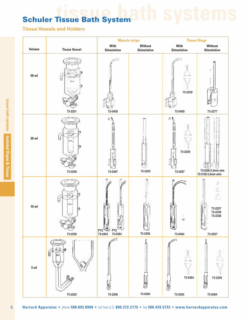
tissue bath systemsSchuler Tissue Bath SystemTissue Vessels and Holders
8
tissue bath systems
Isolated Organ &
Tissue
Harvard Apparatus • phone 508.893.8999 • toll free U.S. 800.272.2775 • fax 508.429.5732 • www.harvardapparatus.com
Volume Tissue Vessel
Muscle strips Tissue RingsWithout
StimulationWith
StimulationWithout
StimulationWith
Stimulation
50 ml
20 ml
10 ml
5 ml
73-2201 73-0400 73-0400 73-2277
73-2259
73-2200 73-0397 73-0397 73-2256 0.3mm wire73-3102 0.2mm wire
73-2203
73-2200PY273-0404 73-0404 73-220773-2206
73-225773-224973-2258
73-225973-0364
73-2202 73-2206 73-0393 73-036473-0364
20ml
20ml
50 ml
PY273-0364
73-2259

tissue bath systems
Isolated Organ & Tissue
tissue bath systems
9Harvard Apparatus • phone 508.893.8999 • toll free U.S. 800.272.2775 • fax 508.429.5732 • www.harvardapparatus.com
M Series Myograph System For Force Measurements in Very Small Tissues
advancedresearch
Features and Benefits:• Precision Fabrication permits use of tissue samples with lumen diameters down to 60µm and segment lengths up to 3 mm • True Isometric Measurement: rigidly mounted transducer exhibits less than 12.5 µm deflection at 0.5gm force • Transducer Safety Lock to protect the integrated force transducer during the preparation phase• Lighted Chamber to assist in tissue mounting• Sliding Wrist Rest for easy manipulation of the chambers during use• Ideal 5ml working volume sample chamber• Integrated Temperature Controller for optimal physiological maintenance• Available as the M4 four channel and M1 single channel Myograph system configurations.• Complete Myography workstation ready for interface to any Harvard Apparatus Data Acquisi-tion System
The New M Series Myograph is designed for researchers performingcontractile force studies on small ring samples with sizes ranging from 60µm to over 1 mm diameter. That includes mouse aortic rings and smallintestinal ring samples down to micro-vessel preparations, such asmesenteric arteries.
Order # Product 72-9434 Radnoti M4 4-Channel Myograph System, 115VAC 72-9435 Radnoti M1 1-Channel Myograph System, 115VAC 72-9436 Radnoti M4 4-Channel Myograph System, 220VAC 72-9437 Radnoti M1 1-Channel Myograph System, 220VAC 72-9438 Peristaltic Pump Quad Bank 72-9430 Replacement Wire for Myograph, 25 micron, pack of 10 72-9431 Replacement Wire for Myograph, 50 micron, pack of 10 72-9432 Replacement Wire for Myograph, 75 micron, pack of 10 72-9433 Replacement Wire for Myograph, 125 micron, pack of 10
Using precision fabricated micro vessel support brackets, the systemaccommodates lumen diameters down to 60µm and segment lengths up to3mm. To mount the vessel segment, two individual parallel wires arepassed through the sample lumen and attached to the micro vesselsupport brackets. Each vessel support bracket incorporates a micro groove on the face to aid in maintaining the wire position when mounting.One bracket is connected to a precision 0-0.5 gm capacitance typeisometric force transducer (switchable to 0-5 gm and also available in 0-2 gm/0-20 gm configurations) that provides a true isometricmeasurement, i.e. less than 12.5 µm deflection at 0.5 gm force, which iscritical when working with small vessel samples. The opposing supportbracket is connected to a precision micrometer controlling the X axismovement, with Y and minor Z axis positioning controls convenientlypositioned on the Myograph chamber, allowing for precise alignment ofthe micro vessel support brackets. The wires are then anchored via alocking screw on each of the brackets. The locking screws feature a smallbushing that is incorporated under the screw head greatly improvingcapture of the support wire. A pre-load tension is then applied viaadjustment of the X axis micrometer.

tissue bath systems
10
tissue bath systems
Isolated Organ &
Tissue
Harvard Apparatus • phone 508.893.8999 • toll free U.S. 800.272.2775 • fax 508.429.5732 • www.harvardapparatus.com
Compact Organ Baths for Isolated Tissuesresearch
Components Included• Compact organ bath unit • Micropositioners (one per chamber) • Thermo regulation unit • Tissue holder • Oxygen outlet for petri chambers • Set of spare fuses • Instruction manual • Calibration certificate • 2 year warranty
Options• Tissue chambers • Stimulation electrodes• Stimulators• Isometric or isotonic transducers• Signal amplifiers• PROTOWIN Software
Key Features• Compact design reduces space requirements• Complete accessibility to all components facilitates maintenance • Independent perfusate inputs allow use of different physiological solutions in the same chamber • All the input-output connections are accessible on the rear panel of the device • Two modes for solution replacement: overflow/ emptying and filling • Turbulence-free water circulation • Use of safety sensors for both water level and water temperature (cut-off at 50°C) • Electronic heating resistance control system prevents thermal shock• Available accessories for continuous perfusion • Ensures optimal heat stability
Parameters Measured• Parameters given by the associated software (see PROTOWIN) Use of totally inert materials, Viton, Teflon, Delrin, Silicone and other materials that are not degraded by acids or salt
8-Channel Compact Organ Bath

tissue bath systems
Isolated Organ & Tissue
tissue bath systems
11Harvard Apparatus • phone 508.893.8999 • toll free U.S. 800.272.2775 • fax 508.429.5732 • www.harvardapparatus.com
Accessories to Use Continuous Perfusion The Compact Organ Baths have been designed and developed to satisfythe requirements of investigators by offering advanced characteristics thatrender them suitable for the in-vitro study of tissue behavior. Due to theircompact design minimum bench space is required.
The modular design allows up to eight individually mounted tissues to bestudied simultaneously and independently per system. Several models areavailable, from one up to eight chambers. All of them are supplied with anexternal heating control unit, transducer' stands & micropositioners andtissue holders. The main perspex reservoir houses the heater system, thechambers and coils, all submerged in thermostatetically controlled water.Each vessel is filled and emptied by means of a three-way tap (ManualSeries) or by electrically-operated valves (Automatic Series) which, besidesthe possibility of manual activation by means of frontal pushbuttons, canalso be remotely controlled by a Timer or Programmer (Software).
Tissue chambers come in 5, 10, 25 or 50 mL capacity. A range of isometricor isotonic force transducers are available.
The Compact Organ Baths are associated with the PROTOWIN softwarefor data storage and analysis.
SpecificationsChamber Capacites 5, 10, 25 or 50 ml
Coil Capacity 180 ml
Temperature Range +3°C to 50°C (Protection); Resolution 0.1°C Room Temperature
Temperature Stability ± 0.1°C in the Whole Water Heat Tank
Power Supply 115-220 V, 50-60 Hz; Consumption 1000 W
Order # Model Product76-0030 LE01002 2 Chambers Manual Compact Organ Bath, Chambers not Included 76-0031 LE01026 2 Chambers Automated Compact Organ Bath, Chambers not Included 76-0032 LE01004 4 Chambers Manual Compact Organ Bath, Chambers not Included 76-0033 LE01046 4 Chambers Automated Compact Organ Bath, Chambers not Included 76-0034 LE01006 6 Chambers Manual Compact Organ Bath, Chambers not Included 76-0035 LE01066 6 Chambers Automated Compact Organ Bath, Chambers not Included 76-0036 LE01008 8 Chambers Manual Compact Organ Bath, Chambers not Included 76-0037 LE01086 8 Chambers Automated Compact Organ Bath, Chambers not Included
Compact Organ Baths for Isolated Tissues (continued)
research
CitationsRodriguez A et al. (2007) Induced vasoconstriction in vascular smooth muscle cells is mediatedvia a nitric oxide-dependent mechanism. Endocrinology 148(1): 324-331. (aorta vascular smoothmuscle, Spain) Kim EJ et al. (2005) Safety pharmacology of sibutramine mesylate, an anti-obesity drug. Hum.Exp. Toxicol. 24(3): 109-119 (rat smooth muscle, Korea Shpak B et al. (2004) Inotropic and lusitropic effects induced by the inhibitory factor of theNa/Ca exchanger are not mediated by the (beta)-adrenergic activation. J. Cardiovasc. Pharm.44(4): 466-472. (guinea and rat ventricle strips, Israel) Hisbrunner G et al. (2003) An in vitro study on spontaneous cervical contractility in the cow dur-ing oestrus and diestrus. J. Vet. Med. Series A, 50(9): 442-446 (cervical smooth muscle,Switzerland).
Order # Model Product Options
76-0038 PROTOWIN Software for Organ Bath, Up to 8 Channels, Dose/Response 76-0039 ISO510 Amplifier for Isometric and Isotonic Transducer 76-0323 LE0140 10 Tissue Clamps Set 76-0324 LE0145 10 Tissue Hooks Set 76-0044 LE0105 5 ml Chamber for Compact Organ Bath 76-0045 LE0110 10 ml Chamber for Compact Organ Bath 76-0046 LE0125 25 ml Chamber for Compact Organ Bath 76-0047 LE0150 50 ml Chamber for Compact Organ Bath 76-0048 LE01030A Stainless Steel Electrode76-0049 LE01030B Stainless Steel Electrode, Two Rings, 20 mm Apart 76-0050 LE01030C Stainless Steel Electrode, Two Rings, 15 mm Apart 76-0051 LE01030D Stainless Steel Electrode, Surface Contact76-0052 LE01035A Platinum Electrode, Single Ring 76-0053 LE01035B Platinum Electrode, Two Rings, 20 mm Apart 76-0054 LE01035C Platinum Electrode, Two Rings, 15 mm Apart * Visit our STIMULATORS section for more details.* Visit our transducers section for more details.

tissue bath systems
12
tissue bath systems
Isolated Organ &
Tissue
Harvard Apparatus • phone 508.893.8999 • toll free U.S. 800.272.2775 • fax 508.429.5732 • www.harvardapparatus.com
Basic Double Tissue Bath Setwith 50-0652 Complete SimpleLever Assembly*
Basic Single Tissue Bath Setwith 50-0652 Complete SimpleLever Assembly*
Student Single Tissue Bath Setwith 50-0652 Complete SimpleLever Assembly
Research Double Tissue Bath Set• Temp. automatically held within 0.5°C• Dial graduated from 15° to 45°C in 5° increments• Large 11.2 L water bath capacity for stable temp. in coils and tissue vessels• Magnetic stirrer ensures uniform temp.• 250 watts of heating power for fast heat-up time• Warming coils lie flat against sides of tank, leaving center free for procedures• 400 ml capacity flat perfusate warming coils ensure sufficient supply of temp. controlled perfusate for lengthy assays• Detachable stem tissue vessel, for increased convenience when changing tissue vessels (tissue chamber can be removed without disturbing stem or disconnecting tubing for warmed perfusate and drainage
Basic Double/Basic Single Tissue Bath Sets• Temperature automatically held within 0.5°C• Dial graduated from 15° to 45°C in 5° increments• Fixed stem tissue vessels supplied as standard
Student Single Tissue Bath Set• Manual temperature control• Offers superior visibility and access for student• Side limb of the special fixed stem student tissue vessel is curved upward making it easy to connect to the spiral perfusate warming coil
Specifications and Ordering Information for Harvard Apparatus Tissue Bath Sets Research Double Basic Double Basic Single Student Single Tissue Bath Set Tissue Bath Set Tissue Bath Set Tissue Bath Set 115 VAC, 60 Hz/230 VAC, 50 Hz 115 VAC, 60 Hz/230 VAC, 50 Hz 115 VAC, 60 Hz/230 VAC, 50 Hz 115 VAC, 60 Hz/230 VAC, 50 Hz
Heated Water BathHeater Power 250 W 80 W 80 W 50 WTemperature Control Automatic within 0.5°C Automatic within 0.5°C Automatic within 0.5°C ManualMagnetic Stirrer Yes No No NoCapacity 11.2 L 4.7 L 3.6 L 0.8 LDimensions, H x W x D 18 x 29 x 21.5 cm 15.5 x 21.5 x14 cm 15.5 x 16.5 x 14 cm 12.5 x 10 cm (H x D) (7.25 x 11.5 x 8.5 in) (6.25 x 8.5 x 5.5 in) (6.25 x 6.5 x 5.5 in) (5 x 4 in)Tissue Vessel(s)Quantity per Set 2 2 1 1Type Supplied Detachable Stem Fixed Stem Fixed Stem Student Fixed StemPerfusate Warming CoilsType Flat Coil Spiral Coil Spiral Coil Spiral CoilCapacity per Vessel 200 ml 35 ml 35 ml 35 mlThermometer Length 15 cm 10 cm 10 cm 10 cmOrder # 50-2146 / 50-2153 50-2120 / 50-2138 50-2104 / 50-2112 50-0306 / 50-0314
Tissue Bath Setsteaching

tissue bath systems
Isolated Organ & Tissue
tissue bath systems
13Harvard Apparatus • phone 508.893.8999 • toll free U.S. 800.272.2775 • fax 508.429.5732 • www.harvardapparatus.com
Tissue Bath Set Componentsteaching
Research Double Single StudentOrder # Description TissueBath Set Tissue Bath Set Tissue Bath Set Tissue Bath Set
50-2146 50-2153 50-2120 50-2138 50-2104 50-2112 50-0306 50-0314
50-2328 Research Heated Water Bath, 115 VAC, 60 Hz 1 - - - - - - -
50-2336 Research Heated Water Bath, 230 VAC, 50 Hz - 1 - - - - - -
50-2302 Double Heated Water Bath, 115 VAC, 60 Hz - - 1 - - - - -
50-2310 Double Heated Water Bath, 230 VAC, 50 Hz - - - 1 - - - -
50-2286 Single Heated Water Bath, 115 VAC, 60 Hz - - - - 1 - - -
50-2294 Single Heated Water Bath, 230 VAC, 50 Hz - - - - - 1 - -
50-2260 Student Heated Water Bath, 115 VAC, 60 Hz - - - - - - 1 -
50-2278 Student Heated Water Bath, 230 VAC, 50 Hz - - - - - - - 1
50-0322 Rod for Student Tissue Bath Set - - - - - - 1 1
50-0330 Student Tissue Vessel - - - - - - 1 1
50-0348 Spiral Perfusate Warming Coil - - 2 2 1 1 1 1
50-0355 Thermometer, 0-50°C, 100 mm - - - - - - 1 1
50-0363 Oxygenation Tubes 2 2 2 2 1 1 1 1
50-2203 Fixed Stem Tissue Vessel, 50 ml - - 2 2 1 1 - -
50-2237 Detachable Stem Tissue Vessel Only, 50 ml 2 2 - - - - - -
50-2245 Detachable Stem Tissue Vessel Stem, 50 ml 2 2 - - - - - -
50-2369 Rubber Bung 2 2 2 2 1 1 1 1
50-2385 Flat Perfusate Warming Coil 1 1 - - - - - -
50-2419 Thermometer Holder - - - - - - 1 1
50-2427 Upright Rod for Water Baths 4 4 4 4 2 2 - -
50-2575 Rod Clamp 4 4 4 4 2 2 2 2
50-6485 Frontal Writing Point 2 2 2 2 1 1 1 1
50-6519 Simple Lever 2 2 2 2 1 1 1 1

tissue bath systems
14
textIsolated O
rgan & Tissue
Harvard Apparatus • phone 508.893.8999 • toll free U.S. 800.272.2775 • fax 508.429.5732 • www.harvardapparatus.com
tissue bath systems
The Mayflower tissue bath is a horizontal tissue chamber with an integralcontraction force measurement. It is completely open at the top and has asmall bath volume of 3 - 5 ml. The compact and modular construction pro-vides ideal conditions for investigations on small muscle preparations(urethra, papillary muscle, cavernous body), vascular rings, or trachealrings.
Basic Equipment: – Acrylate support with a movable mounting platform for preload adjustment through a micrometer screw. This platform receive the tissue chamber and acts as holder for the force transducer. The acrylate support also includes a glass heat exchanger for the pre-heating of the perfusate solution. – The tissue chamber differentiates the two versions.
Flow Through System (73-2155)The tissue chamber is carved out of an acrylate block and placed on thesupport stand. This chamber includes the tissue holder, the connections forelectrical stimulation electrodes, a frit for aeration and a draw-off tubewith height adjustment to set the bath volume. As it is not jacketed,continuous flow through the chamber is required.
• Horizontal tissue bath for isometric contraction measurements using a F10 or F30 force transducer• Two versions are available: – Flow-through superfusion system – Incubation system• Possibility of electrical stimulation• Exchangeable holders depending on tissue used (for vascular or tracheal rings, for strips)
Incubation System (73-3600)The tissue chamber is jacketed. The bottom of the chamber is made of ateflon coated stainless steel plate to optimize the temperature stability ofthe solution. A additional small stainless steel heat exchanger is also builtin to avoid temperature fluctuation.
The incubation chamber can also be used as a flow-through superfusionchamber. The superfusion chamber cannot be used for incubation.
Additional Equipment: – A multi-channel roller pump for the perfusate circuit – A thermocirculator for keeping the perfusate solution at constant temperature
The modular concept of this apparatus offers a wide range of differentarrangements to meet individual requirements regarding bath geometry,tissue holders and stimulation electrodes. The horizontal arrangement ofthe tissue and the open top provide ideal conditions during preparationand experiment.
The various parts of the apparatus are made mainly from Plexiglas orstainless steel and are therefore inert and corrosion-resistant. The solutionflows in from a roller pump and passes through a heat exchanger, theoutflow is under suction through a suction tube with height adjustment bythe same pump. A frit for aeration is located in the tissue chamber. Twoconnection sockets for the stimulation electrodes are located on eitherside of the tissue chamber.
Mayflower Tissue Bath System 73-2155 Mayflower TissueBath Configured as a flow-through system
Mayflower Tissue Bath Systemadvancedresearch

tissue bath systems
Isolated Organ & Tissue
tissue bath systems
15Harvard Apparatus • phone 508.893.8999 • toll free U.S. 800.272.2775 • fax 508.429.5732 • www.harvardapparatus.com
Mayflower Tissue Bath System (continued)advancedresearch
Electrical Stimulation ElectrodesThe stimulation electrodes can be of different types:
– Coaxial electrode for contact stimulation (stainless steel).
If point stimulation is required, a Miniature Coaxial Stimulation Electrode is installed using our Mini Ball Joint Postioning System. The electrode can therefore precisely and easily be placed.
– Two plate electrodes for field stimulation (platinum or AgAgCI)
– Flexible platinum wires for direct contact with the tissue
73-3600 MAYFLOWER “Fully Thermostated Bath Top” In-cubation System
MAYFLOWER Chamber withField Electrodes
MAYFLOWER Chamber withdirect Stimulation Electrode
Special holder for vascular or tracheal rings. The rings are slidover the holder jaws.
Tissue HoldersThree different tissue holders are available. Tissue holders can be exchanged.The holders consist of two parts, a fixed part positioning the tissue inside thebath and the part transmitting the contractile force to the transducer.
Order # Product 73-2155 Small Volume Horizontal Tissue Bath, System MAYFLOWER73-3600 Small Volume Horizontal Tissue Bath, System MAYFLOWER “Fully Thermostated Bath Top”
IMPORTANT NOTE:Mayflower Tissue Bath is especially designed forHSE-HA F30 or F10 Force Transducers and it is not possible to modifyTransducer Holder to suit other transducer models.
For a system description according your requirements please use:www.hugo-sachs.de/ihmail.html
or contact our technical experts at: [email protected]
For a custom configuration and full system quotation.
A standard tissue holder is usedfor small muscle preparations. The tissue is attached to bothhooks by threads.
Special holder for small musclepreparation. The tissue is clampedon the right-hand side by a springclip against the bottom of thechamber and attached to the transducer hook using a thread. A tool for lifting the clamping system is supplied.
isolated organ systems
16
isolated organ systems
Isolated Organ &
Tissue
Harvard Apparatus • phone 508.893.8999 • toll free U.S. 800.272.2775 • fax 508.429.5732 • www.harvardapparatus.com
Order # Product 73-2158 Horizontal Tissue Bath PBTO, Type 813/673-2044 Device for Afterload Pressure of 0-30 cmH2073-2333 Device for Afterload Pressure of 0-300 mmHg
Benefits and Features• Perfusion of tubular organs (trachea, blood vessels, intestines)
• Individual solutions for intraluminal perfusions and extraluminal superfusion• Controlled perfusion pressure
Applications• Intraluminal perfusion of tubular organs (trachea, blood vessels, intestines)• Testing circular-action musculature, vascular toneand stents in isolated vessels
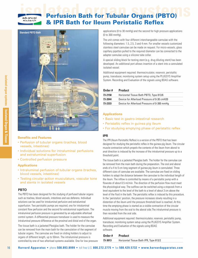
PBTOThe PBTO has been designed for the studying of perfused tubular organssuch as trachea, blood vessels, intestines and vas deferens. Individualsolutions can be used for intraluminal perfusion and extraluminalsuperfusion. Two peristaltic pumps are required, one for intraluminalconstant flow perfusion and the second for extraluminal superfusion. Theintraluminal perfusion pressure is generated by an adjustable afterloadcontrol system. A differential pressure transducer is used to measure theintraluminal pressure difference at the proximal and distal end of the organ.
The tissue bath is a jacketed Plexiglas bath. The holder for the cannulaecan be removed from the main bath for the cannulation of the segment oftubular organs. The cannulae are fixed on sliding holders to adjust toorgans of different length, up to 50mm. The intraluminal pressure iscontrolled by one of two afterload systems available. One for low pressure
Applications• Basic test in gastro-intestinal research• Peristaltic reflex in guinea pig ileum• For studying emptying phase of peristaltic reflex
IPRThe IPR (Ileum Peristaltic Reflex) is a version of the PBTO that has been designed for studying the peristaltic reflex in the guinea-pig ileum. The circularmuscle contraction which propels the contents of the ileum from aboral toanal direction is induced by the increase of the intraluminal pressure up to athreshold point.
The tissue bath is a jacketed Plexiglas bath. The holder for the cannulae canbe removed from the main bath during the preparation. The oral and aboralends of a 4 to 5 cm long segment of guinea pig ileum is cannulated. Three different sizes of cannulae are available. The cannulae are fixed on slidingholders to adapt the distance between the cannulae to the individual length ofthe ileum. The inflow is controlled by means of a peristaltic pump with aflowrate of about 0.5 ml/min. The direction of the perfusion flow must meetthe physiological way. The outflow can be switched using a stopcock from alevel equivalent to the level of the bath to a level of about 3 cm above thelevel of the fluid in the bath. The peristaltic reflex is induced by this procedure.In the ‘peristalsis’ position, the pressure increases slowly resulting in a distention of the ileum until the pressure threshold level is reached. At thistime the emptying phase is started as a visible contraction of the circular muscle moving from the oral to the aboral side.The intraluminal pressure isthen recorded from the oral side.
Additional equipment required: thermocirculator, reservoir, peristaltic pump,transducer, monitoring system setup using the PLUGSYS Amplifier System.Recording and Evaluation of the signals using BDAS software.
Order # Product 73-3013 Horizontal Tissue Bath IPR, Type 813/2
applications (0 to 30 mmHg) and the second for high pressure applications(0 to 300 mmHg).
The unit comes with four different interchangeable cannulae with the following diameters: 1.5, 2.5, 3 and 4 mm. For smaller vessels customizedstainless steel cannulae can be made on request. For micro-vessels, glasscapillary pipettes pulled to the required diameter can be connected to theadapter cannulae using a silicone tube collar.
A special sliding block for testing stent (e.g. drug diluting stent) has beendeveloped. An additional port allows insertion of a stent into a cannulatedisolated vessel.
Additional equipment required: thermocirculator, reservoir, peristalticpump, transducer, monitoring system setup using the PLUGSYS AmplifierSystem. Recording and Evaluation of the signals using BDAS software.
Perfusion Bath for Tubular Organs (PBTO) & IPR Bath for Ileum Peristalic Reflex
advancedresearch
Standard PBTO Bath
text
Isolated Organ & Tissue
tissue bath systems
17Harvard Apparatus • phone 508.893.8999 • toll free U.S. 800.272.2775 • fax 508.429.5732 • www.harvardapparatus.com
TIOX Tissue Bath for O2 Consumption & Contraction Force Measurement
advancedresearch
Benefits and Features• Unique sealed system to measure muscle contraction and oxygen consumption.• Oxygen electrode with very low rate of oxygen consumption• Small volume
Applications• Bioenergetics studies• Sarcopenia in senescence studies• Evaluation of metabolic - contractile caracteristics• Fatigue and muscle weakness studies• Sports physiology and biochemistry
Order # Product 73-3794 TIOX Tissue Bath 69-3006 Microcathode Oxygen Electrode72-1975 Magnetic Mini Stirrer72-1977 Micro Stirring Bar73-0831 HSE Isometric Force Transducer F30
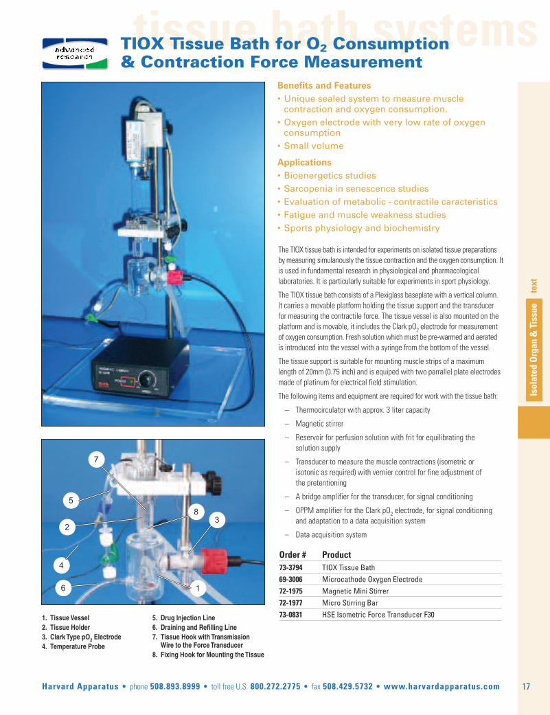
The TIOX tissue bath is intended for experiments on isolated tissue preparationsby measuring simulanously the tissue contraction and the oxygen consumption. Itis used in fundamental research in physiological and pharmacologicallaboratories. It is particularly suitable for experiments in sport physiology.
The TIOX tissue bath consists of a Plexiglass baseplate with a vertical column.It carries a movable platform holding the tissue support and the transducerfor measuring the contractile force. The tissue vessel is also mounted on theplatform and is movable, it includes the Clark pO2 electrode for measurementof oxygen consumption. Fresh solution which must be pre-warmed and aeratedis introduced into the vessel with a syringe from the bottom of the vessel.
The tissue support is suitable for mounting muscle strips of a maximumlength of 20mm (0.75 inch) and is equiped with two parrallel plate electrodesmade of platinum for electrical field stimulation.
The following items and equipment are required for work with the tissue bath:
– Thermocirculator with approx. 3 liter capacity
– Magnetic stirrer
– Reservoir for perfusion solution with frit for equilibrating the solution supply
– Transducer to measure the muscle contractions (isometric or isotonic as required) with vernier control for fine adjustment of the pretentioning
– A bridge amplifier for the transducer, for signal conditioning
– OPPM amplifier for the Clark pO2 electrode, for signal conditioning and adaptation to a data acquisition system
– Data acquisition system
1. Tissue Vessel2. Tissue Holder3. Clark Type pO2 Electrode4. Temperature Probe
5. Drug Injection Line6. Draining and Refilling Line7. Tissue Hook with Transmission Wire to the Force Transducer8. Fixing Hook for Mounting the Tissue
4
3
1
58
6
7
2

tissue bath systems
18
tissue bath systems
Isolated Organ &
Tissue
Harvard Apparatus • phone 508.893.8999 • toll free U.S. 800.272.2775 • fax 508.429.5732 • www.harvardapparatus.com
preparation. The lower part is funnel-shaped to allow the collection of theefluate for reperfusing, perfusing thenext tissue or bioassay analysis. Theisometric force transducer is fixed onthe rod using a Vernier control.
The tissue holder is a separate part thatis installed in the main superfusionchamber. The holder can easily beremoved to simplify the preparation.The holder consists of the body, thehook for fixing the tissue, the stainlesssteel cannulas for superfusion and thestimulation electrodes. The tissue isattached using a thread to the tissuehook. The thread at the other end of thetissue must be attached to thetransducer. A slide in the holder bodyallows easy positioning of the thread inthe central hole. The superfusioncannulae can be rotated in the main
body to optimize the superfusion after the holder is in place into the chamberand the thread fixed to the transducer. The stimulation electrodes are made ofplatinum and must be buckled to be close to the tissue. The electrodes do nottouch the tissue but must be parallel and close enough to the tissue so that thesuperfusing solution makes electrical contact.
The dual heat exchanger consists of Tygon tubing loops inside a jackettube connected to a thermocirculator.
In the Flow through mode each tissue is superfused individually with fresh solution or solution containing the test compound. The outcoming solution iscollected and goes to waste or is stored for later bioassay analysis.
In the Recirculating mode each tissue is superfused individually. Theoutcoming solution after superfusion is collected and used for superfusingagain in the same tissue.
In the Cascade mode the first tissue is superfused with fresh solution andall the subsequent tissues are superfused with the outcoming solution ofthe preceding chamber.
Additional equipement required: peristaltic pump, force transducer withVernier control, bridge amplifier for the force transducer, Data acquisitionACAD, electrical stimulator, thermocirculator if heat exchangers are used.
The Coleman superfusion system is suitable for a variety of applicationsinvolving the study of contraction and relaxation of smooth musclepreparations, the effect of drugs that interfere with these actions, as well asthose that interfere with autonomic neurotransmission. The system consistsof a main stand that can receive up to eight single superfusion chambers.Each chamber is equipped with electrical stimulation capability, a rod forfixing the force transducer and an optional two channel heat exchanger. Thelower end of the tissue is held by a stainless steel hook. The thread attachedto the top of the tissue passes through the opening directly to the forcetransducer. The superfusion solution is fed by a peristaltic pump through astainless steel cannula to the thread; it runs down the thread and superfusesthe tissue. Two separate solution paths are available for superfusion withand without test substance. Two platinum wires are placed parallel to thetissue to form stimulation electrodes. The solution running down provideselectrical contact between the electrodes and the tissue.
The superfusion chambers are made of Plexiglas and do not requirethermostating owing to the low thermal conductivity of the material. Thechamber is fixed on the frame in the horizontal position. All additional parts(tissue holder, dual heat exchanger, road for mounting the transducer) arefixed on the chamber. The upper part of the chamber receives the tissueholder. The lower part can be swung away to simplify access to the
VernierControl
ForceTransducer
SuperfusionLines
TissueHook
StimulationElectrodes
Order # Product 73-2221 Basic Unit for Superfusion Bath according to COLEMAN (stand for 4-Baths) Type 84073-2222 Superfusion Bath according to COLEMAN73-2917 Heat Exchanger to Superfusion Bath COLEMAN
Coleman SuperfusionBath
Benefits and Features• Suitable for virtually any tissue sample• Compact, allows the study of up to four tissue preparations in parallel• Rapid removal of potentially toxic metabolites• Obviates the necessity of repeated washing procedures
• Simple reclaimation of perfusate for recirculation or collection
Applications• Evaluation of the potency of labile substances• Determining the rates of onset and offset of drug action• Evaluation of spasmogenic and spasmolitic agents
For a system description according your requirements please use:www.hugo-sachs.de/timail.html
or contact our technical experts at: [email protected]
For a custom configuration and full system quotation.
Coleman Superfusion Bathadvancedresearch

tissue bath systems
Isolated Organ & Tissue
tissue bath systems
19Harvard Apparatus • phone 508.893.8999 • toll free U.S. 800.272.2775 • fax 508.429.5732 • www.harvardapparatus.com
Steiert Tissue Bath System For Simultaneous Action Potential & Contraction Force Measurements on Cardiac Tissue
advancedresearch
The Steiert Tissue bath system is a horizontal bath, open at the top, specificallydesigned for electrophysiological (Intracellular Action Potential) and mecanical(Contractile Force) studies of cardiac tissue preparations such as papillarymuscle and purkinje fibers. The unique design allows the user to record bothintracellular action potentials and contractile force from the same tissuepreparation. The core system includes the tissue bath, a peristaltic pump andthe DC-controller for temperature regulation and control of the perfusate, adirect contact coaxial stimulation electrode and the force transducer mountingwith micrometer control to maintain tissue tension.
The tissue bath has ports for fluid inlet and outlet. The port outlet is a sipperpipe connected to the peristaltic pump. The sipper pipe is adjustable in heightto adjust the bath volume (1-2 ml). The tissue under investigation is securedwith needles to a Silicone pad fitted in the bath bottom, the other end issecured, either with a thread or directly, to the hook of the force transducer.Only transducers F10 or F30 can be used.
Stimulation takes place through a miniature coaxial electrode which is mountedflexibly on a triple ball joint to the back of the bath. The stimulation electrodecan therefore be positioned to suit individual requirements.
The Temperature Controller DC has been specially developed for thermostatingthe perfusion solution supply on the organ bath after Steiert. Special attention
Applications and Features• Compact and easy to use setup for electrophysiology studies on heart muscles• Suitable for papillary muscle and Purkinje Fibre• Simultaneous recording of intracellular action potential and force development• Mini coaxial electrode for electrical stimulation
Order # Product 73-2152 Basic Unit, Organ Bath According to STEIERT Type 813, 230 VAC 50 HZ73-2892 Basic Unit, Organ Bath According to STEIERT Type 813, 115 VAC 60 HZ
has been paid during the development of this controller to a very low radiatedinterference level. The heating circuit (temperature controller and heatingelement) has excellent decoupling from the perfusion solution circuit. Theheating element is a long life halogen bulb. It is therefore excellently suitablefor the interference-free measurement of intracellular action potentials.
Additional equipment required: force transducer, bridge amplifier for the forcetransducer and microelectrode amplifier with headstage, Data acquisition,recording microelectrodes, electrical stimulator with isolation unit, tissuepreparation dish, anti-vibration table, microscopes, illumination,micromanipulators and many other options.
If contractile force measurement is not required, special versions are availbleon request.
A version with field stimulation is also available on request. Be aware that inthis case stimulation artefacts may interfere with the action potential signal.
For a system description according your requirements please use:www.hugo-sachs.de/timail.html
or contact our technical experts at: [email protected]
For a custom configuration and full system quotation.
Steiert Tissue Bath System

isolated lung systems
20
isolated lung systems
Isolated Organ &
Tissue
Harvard Apparatus • phone 508.893.8999 • toll free U.S. 800.272.2775 • fax 508.429.5732 • www.harvardapparatus.com
PCLS Precision Cut Lung Slice Chamber
• Assessment of lung functions under cell culture conditions• From any species (murine, rat, human)• Study of airways of different sizes • Allows quantification of the responsiveness of simple airway passages and simple vessels• Analyzing ciliary beating frequency
The incubation chamber was developed to allow incubation and observation ofslices by an inverted microscope. The chamber is made of Polycarbonate. It isconnected to a water bath to maintain constant temperature conditions. Twoincubation cells are positioned in the center of the chamber. The bottom of thecells is sealed by glass, the cover is made of acrylic glass. The slices are fixed inthe incubation cells by positioning them under nylon strings fixed to a bentplatinum wire. The incubation cells can be filled with buffer, medium or drugsolutions through the filling pipe. Buffer solution can be removed from the cellsover a vacuum pipe. In addition, it is possible to gas the incubation cells in orderto use bicarbonate buffered media.
The incubation chamber is placed on the stage of an inverted microscope andwarmed to 37°C. The slices are screened for airways and transferred to theincubation chamber. Lung slices are selected for study using predefined criteria(Martin et al. 1996). Airways and vessels are focused, and finally the images areanalyzed by image analysis software (e.g., Optimas or Metamorph).
Example of an ApplicationAs an example Figure 1 shows exposure of a slice to increasingconcentrations of endothelin-1. Shown is a lung slice containing an airway (B),a pulmonary artery (PA) and a pulmonary vein (PV). The pulmonary artery andthe airway contracted almost completely, while the pulmonary vein areadecreased to only 50% of its initial area. These responses are now easilyquantified by digital imaging technique.
It is a distinct advantage of this technique that in many ways precision-cutslices can be treated like a cell culture. Thus, the slices can be incubatedunder various conditions and gene as well as protein expression or mediatorrelease be determined. In contrast to cell culture models, in slices theanatomical structure of the lung is largely maintained, so that the functionalconsequences of gene expression and mediator release can be evaluated.
Figure 1. Exposure of a PCLS to increasing concentrations of endothelin-1.Shown is one lung slice containing a small airway (B), a pulmonary artery (PA)and a pulmonary vein (PV). The slice was imaged before (C) and after exposure toincreasing concentrations of endothelin-1, ranging from 10-10 M to 10-6 M.
Inlet FromWaterbath
Gas Inlet Filling Pipes
Vacuum Pipes
Return to waterbath
Platinum Wire withNylon Threads
Incubation Cells
Incubation Chamber Top View
Vacuum Pipes
Lung Functions Under the MicroscopePrecision-cut lung slices (PCLS) offer a novel and unique way to assess lungfunctions under cell culture conditions. They can be prepared from nearly anyspecies including mouse, rat and human lungs. The method allows the study ofthe response of airways of different size (down to the terminal bronchioles) andto relate these changes in lung functions to gene expression and mediatorrelease. Slices are viable for at least three days. They can be placed under aninverted microscope, where digital image techniques allow quantification notonly of the responsiveness of single airways, but also of single vessels. Inaddition, it is possible to analyze the ciliary beating frequency. More than 20slices can be obtained from one lung, thus this method is very economical interms of experimental costs and animal use. Tissue cores are prepared from thelungs filled with agarose solution, after cooling to 4°C. From the cores, slices(220 ± 20µm) are cut using a tissue slicer
Order # Product 73-2370 Precision Cut Lung Slice Chamber
Accessories
73-0125 Thermostatic Circulator E 103, 230 VAC, 73-2802 Thermostatic Circulator E 103, 115 VAC73-0113 Roller Pump Reglo Analogue ISM 827, | 4 Channels, 0.002 to 30 ml/min
advancedresearch

Isolated Organ & Tissue
tissue bath systems
21Harvard Apparatus • phone 508.893.8999 • toll free U.S. 800.272.2775 • fax 508.429.5732 • www.harvardapparatus.com
tissue bath systems
Marsh Ganglion Bathadvancedresearch
Marsh Ganglion Bath
For Studying• Synaptic transmission• Nerve conduction
Applications Include• Vagus nerve• Cervical ganglion
Order # Product 73-2414 MARSH Ganglion Bath Type 85873-0387 Set Electrode Components to MARSH Ganglion Bath (for 2 electrodes)
Parts for one elec-trode
Finishedelectrode
Marsh Bath set up for recording from rat superior cervical ganglia
The MARSH Ganglion Bath is intended to test the action of drugs on thesynaptic transmission or nerve conduction in the vagus nerve or the cervicalganglion.
The bath is an open-top Perspex bath which is divided into three compartmentsby two separators. Each of these separators consists of a removable uppersection and a lower section with a cutout to allow the nerve to pass betweenchambers without crushing and also support the tissue in position.
The first chamber contains two platinum electrodes for axonal stimulation ofthe nerve bundle or preganglionic nerve trunk. Recordings are made from thecentral chamber with the third chamber acting as reference chamber usingnon-polarizing silver/silver chloride electrodes. Drugs are applid via the conti-nously perfused central chamber.
The electrode set to the Marsh Ganglion Bath includes the necessary com-ponents to produce two recording electrodes to interconnect the recordingchamber with the PHDA headstage amplifier.
Additional equipement required: amplifier with high-impedance input and apeak height detector (PLUGSYS module PHDA), Data acquisition, electricalstimulator with isolation unit, thermocirculator, solution reservoir, illumina-tion, oscilloscope.
The elctrode set contains two 1ml syringes, 2 lengths of PE tubing with luer tip, 2Ag/AgCl pellets with taper plastic body and connecting socket at the backsuitable for 2 mm plug pin and Agar powder.
If stored properly (protected from light sources) electrodes can be used forabout 2 weeks, the Agar solution must then be replaced.
The electrodes are connected by short wires with 2mm pins to the headstageof the amplifier. These cables are supplied with the PHDA amplifier.
For a system description according your requirements please use:www.hugo-sachs.de/timail.html
or contact our technical experts at: [email protected]
For a custom configuration and full system quotation.

moist chambers
22
moist cham
bersIsolated O
rgan & Tissue
Harvard Apparatus • phone 508.893.8999 • toll free U.S. 800.272.2775 • fax 508.429.5732 • www.harvardapparatus.com
Moist Chamber Type 834/8
Order # Product 73-2901 Moist Chamber Type 834/8 with Metal Tube Heat Exchanger73-3692 Bubble Trap for Flow Rate up to 50 ml/min73-3094 Stainless Steel Mesh Electrode
Mesentric Bed Per-fusion System
Benefits and Features• Excellent temperature control for perfusate and organ• Precise positioning of cannulae and measurement probes
• Straight forward operation and compact dimensions• A large choice of cannulae, bubble traps and other acessories makes the moist chamber suitable for a huge range of perfusion applications• Provides a complete perfusion system in combination with the UP-100 or Perfusion Control System
Applications• For perfusion of organs from rodents like liver, kidney, pancreas and mesenteric bed• For investigating the tone of small blood vessels under the effect of vasoactive substances• Biochemistry, studying metabolic processes
Moist chamber equiped for isolatedrat liver perfusion under constantflow conditions
Mini ball joint holders support arterial and venous cannulae for rat liver perfusion
Moist Chamber73-2901
The popular Moist Chamber Type 834/8 is an exceptionally flexible anduseful tool for perfusion of most “simple” organs from typical rodentmodels. In its most basic configuration, the Moist Chamber consists of asuitably deep (110x40x35mm) organ chamber and tight-fitting cover. Bothcomponents are double-walled and water-jacketed to provide a stabletemperature controlled environment within the organ chamber. Perfusateis warmed by passage through a built-in heat exchanger and bubble trapimmediately before contact with the organ.
Inside the chamber, a flexible silicon platform acts as a rest for fixation(usually with standard fixing pins) of the organ. Anchors for our Mini BallJoint positioning system and precision arterial and venous cannulae arepre-drilled on both sides of the organ. In addition, several measurementand sample ports are provided for easy access to the inner chamber, evenwith the cover in place, making the chamber suitable for collecting a widerange of physiological data.
The Moist Chamber Type 834/8 can be part of a simple constant flowperfusion system. Used as such, a water-jacketed buffer reservoir, peristaltic
pump and appropriate cannulae are used to complete the perfusion circuit,while a thermocirculator feeds the water-jacketed components to maintainthe thermostating circuit. The Moist Chamber Type 834/8 can also be used toform the core of our UP-100 or Perfusion Control system to permit perfusion atconstant pressure as well as constant flow.
The basic Moist Chamber Type 834/8 is configured as shown above andconsists of the chamber, cover and silicon plate only. Contact HarvardApparatus for configuration of a complete perfusion system for specificapplications.
Additional equipment required: thermocirculator, bubble trap, cannulae,holders, peristaltic pump, transducers, monitoring system setup using thePLUGSYS Amplifier System. Recording and Evaluation of the signals usingBDAS software.
Special Application: The Rat Mesenteric BedThe key part of the perfusion system for the rat mesenteric bed is the moistchamber. The mesenteric tissue is placed into the moist chamber on astainless steel mesh (replaces the silicone plate) which also acts as anodeduring electrical stimulation.
advancedresearch
For a system description according your requirements please use:www.hugo-sachs.de/orgmail.html
or contact our technical experts at: [email protected]
For a custom configuration and full system quotation.

moist chambers
Isolated Organ & Tissue
moist chambers
23Harvard Apparatus • phone 508.893.8999 • toll free U.S. 800.272.2775 • fax 508.429.5732 • www.harvardapparatus.com
Moist Chamber Applicationsadvancedresearch
Moist chamber with peristaltic pump, SCP perfusion control system, perfusate reservoir and thermocirculator. Used forconstant pressure perfusion for organs such as kidney and pancreas. The SCP perfusion control system includes flowmeasurement capability. By disabling the pressure control, the system can also work as constant flow perfusion systemwith pressure measurement.
Moist chamber with peristaltic pump, perfusate reservoir and thermocirculator. Used for constant flow perfusion for organs such as Liver.

moist chambers
24
moist cham
bersIsolated O
rgan & Tissue
Harvard Apparatus • phone 508.893.8999 • toll free U.S. 800.272.2775 • fax 508.429.5732 • www.harvardapparatus.com
Additional equipment required: thermocirculator, peristaltic pump, syringepump, transducer for perfusion pressure, servo controller (SCP), monitoringsystem setup using the PLUGSYS Amplifier System. Recording and Evaluationof the signals using BDAS software.
The chamber can be used for any other organ that requires continuousweighing during perfusion.
The interchangeable adapter block holding the perfusion lines and cannulaecan be customized for other types of organ to be perfused.
Developed in cooperation with the Forschungszentrum Borstel, 23845, Borsteland Universitätsklinikum, Chirurgie, 24105 Kiel.
• Compact arrangement• Dual Perfusion System, vascular & intraluminal intestinal• Built-in balance for edema evaluation/organ weight measurement• Vascular bed is in warm and moist environment during perfusion• Optimized temperature control of perfusate and organ• Controlled perfusion conditions
• Cannulation block simplifies surgery
Applications• To study simultanuously vascular, luminal and lymphatic flows, arterial, venous and intraluminal pressures and bowel weight.• Septic multi-organ failure study in gastro intestinal area
Moist Chamber with Edema Balance Moist Chamber for Microvascular Permeability Studies
The system is based on a moist chamber with a built-in organ weighingsystem. The jacketed chamber maintains a warm and moist environment forthe organ. The chamber has been configured for studying the edema evolutionin a perfused intestine with attached mesenteric bed with two separateperfusion lines for simultaneous vascular and intraluminal perfusion.
The chamber is supplied with a movable cannulation block including all therequired heating coils and bubble traps. This block acts also as holder for thetubing and cannulae, it can be placed near the animal for easy in-situpreparation. After surgery, the block with the preparation is moved and fixedon the chamber. This ensures continuous perfusion during the entire durationof surgery and reduced risk of embolism or ischemia.
The chamber provides a number of ports for connecting the measurementsystem and perfusate collection. Measurement of perfusion pressures andflows are also available. A peristaltic pump is used for vascular perfusion. Aconstant pressure is maintained by controlling the pump speed with anelectronic controller (SCP) via pressure measurement. For the intraluminalIleum perfusion, a syringe pump is used.
Order # Product 73-3685 Moist Chamber with Edema Balance Type 802 (MCWEB)
Cannulating block
WeighingTransducer
VascularCannulaeIntraluminal
Cannulae
Cannulation block removed from chamber for surgery
73-3685
advancedresearch
CitationsA Model of the Isolated Perfused Rat Small IntestineIngmar Lautenschläger,1 Heike Dombrowsky,2 Inez Frerichs,3 Solveig-Carolin Kuchenbecker,3 Stef-fen Bade,2 Holger Schultz,2 Peter Zabel,2 Jens Scholz,3 Norbert Weiler,3,* and Stefan Uhlig2
1University Medical Centre Schleswig-Holstein, Campus Kiel 2Research Center Borstel 3UniversityHospital Schleswig-Holstein, Campus Kiel
Submitted 31 July 2009 ; revision received 8 October 2009 ; accepted in final form 8 November 2009
ABSTRACTIntestinal edema remains a serious clinical problem and novel approaches to study its pathophysiologyare needed. It was our aim to develop a long term stable isolated perfused rat small bowel prepara-tion permitting analysis of vascular, luminal, interstitial and lymphatic compartments and to demon-strate the utility of this model by studying the effects of the pro-inflammatory mediatorplatelet-activating factor (PAF). A temperature-controlled chamber with an integrated balance was
designed to perfuse isolated intestines through the mesenteric artery and the gut lumen. Steroidsor oxygen carriers were not needed. Functional and morphological integrity of the tissue was pre-served for several hours as confirmed by oxygen consumption, venous lactate-to-pyruvate ratio,arterial and venous pH, lactose digestion and galactose uptake, intravascular and luminal pres-sures, maintained fluid homeostasis, gut motility, and by quantitative light microscopic analysis.Administration of PAF caused typical effects such as vasoconstriction, gut atony, and loss ofgalactose uptake. PAF also elicited a transient loss of 20% of the perfusate liquid from the mesen-teric vascular bed, two thirds of which were transferred to the lumen. All these responses were en-tirely reversible. This new model provides detailed insights into the physiology of the smallintestine, and will allow to study fundamental processes such as fluid homeostasis, barrier func-tions, transport mechanisms and immune responses in this organ. Using this model, here weshow a dramatic and yet reversible response of the rat small bowel to PAF suggesting luminalwater clearance as a novel safety factor in the intestine that may be of clinical relevance. Intes-tine physiology; fluid balance; platelet activating factor.

isolated organ
systems
Isolated Organ & Tissue
isolated organ systems
25Harvard Apparatus • phone 508.893.8999 • toll free U.S. 800.272.2775 • fax 508.429.5732 • www.harvardapparatus.com
UP-100 Equipped with Moist Chamber
The UP-100 is a multi-purpose perfusion system best utilized when differenttypes of organs must be perfused either in situ or ex vivo. The modular designof this system allows easy adaptation to different applications using additionsor extensions to the base unit.
System Extensions for Perfusion Ex-VivoInternal organs (kidney, liver, mesenteric bed) must obviously be kept underoptimal physiological conditions; moist and at defined temperature duringperfusion. For these applications the UP-100 is combined with the MoistChamber Type 834/8.
System Extensions for Perfusion in SituFor in situ perfusion of organs such as liver and kidney, or for perfusion ofregional vascular systems like hindquarter, an operating table can be placed onthe main Plexiglas plate below the UP-100 mounting platform. The compactarrangement allows the connection line between organ and heat exchanger toremain short to ensure consistent perfusate temperature.
System Extensions for Perfusion of Mouse, Rat,Guinea-pig or Small Rabbit Heart For perfusion of the isolated heart according to Langendorff, the UP-100 can beequipped with a jacketed heart chamber and additional measurement systemfor isovolumetric LVP.
Additional equipment required: thermocirculator, electrical stimulator, peristalticpump, transducer for perfusion pressure, monitoring system setup using thePLUGSYS Amplifier System. Recording and Evaluation of the signals usingBDAS software.
• Multi-purpose system for perfusing isolated organs in-situ or ex-vivo• Ideal for perfusing isolated organs such as: – Liver – Rabbit Ear – Heart – Kidney – Rat hind limb – Mesenteric bed• Perfusion can be performed at constant pressure or constant flow without involving any modification of apparatus• Ideal replacement for perfusion system with hydrostatic pressure generation, low perfusate volume in use, perfusion pressure up to 300 mmHg possible• Can be equipped with an oxygenator for optimal aeration of perfusate containing albumin or erythrocytes
Applications• In situ perfusion of hind limb, hindquarter mesenteric bed, liver, kidney: – Blood vessel tone in peripheral vascular bed – Balance tests by muscle work (glucose/lactate/pyruvate, high energy phosphates/orthophosphate, etc.) – Test of vasodilative drugs in occlusive diseases of legs – Test of muscle relaxants (end-plate pharmacology)• Ex-vivo perfusion of liver, kidney, mesenteric bed, by using the additional Moist Chamber Type 834/8 – Test of vasodilative drugs – Studying metabolic processes – Neural vascular tone – Organ preservation for transplant• Special version for Langendorff heart perfusion – Optimized for LVP measurement – Ideal for compound screening
Order # Product 73-2316 Universal Perfusion System Basic Unit UP-100, Type 834The UP-100 is a universal system. It needs to be adapted to the specific application by addingother equipment.
UP-100 Equipped withHeated Surgery Table
For a system description according your requirements please use:www.hugo-sachs.de/orgmail.html
or contact our technical experts at: [email protected]
For a custom configuration and full system quotation.
Universal Perfusion System UP-100advancedresearch

isolated organ systems
26
isolated organ systems
Isolated Organ &
Tissue
Harvard Apparatus • phone 508.893.8999 • toll free U.S. 800.272.2775 • fax 508.429.5732 • www.harvardapparatus.com
Perfusion System for Isolated Pig Liver or Pig Kidney
advancedresearch
Basic SystemThe pig liver to be perfused is placed in a moist, thermostated chamber(inside dimensions: 400 x 300 x 180 mm) and perfused with blood orerythrocyte containing perfusate under constant-flow conditions via theportal vein. A centrifugal pump with a gentle action on blood is employed toreduce hemolysis. As this type of pump does not supply a constant flow orpressure, the constant flow is maintained by an electronic controller (SCP).For the kidney instead of the liver chamber, a smaller chamber (insidedimensions 260 x 200 x 210 mm) is used. The kidney is mainly perfused atconstant pressure, which is also controlled by the SCP.
Components for a complete system:
• Thermostated moist chamber for pig liver or pig kidney
The following additional items are required for operating the apparatus:
• Thermocirculator
• Pump with electrical control. For blood we recommend a Pump Drive BVP-ZX with centrifugalpump head, see the Pump Section A
• SCP, Servo Controlled Perfusion System
• Measurement system for Flow and Pressure
• Oxygenator with heat exchanger, e.g. Terumo CapioxSX10® or Medtronic Minimax Plus PRF®
Additional monitoring equipment can be added to the system as required tocreate a custom, application specific, system for your unique research needs.
Monitoring System is set up using the PLUGSYS Amplifier System. Recording andEvaluation of the signals using BDAS software.
• For use in physiological or pharmacological research for the perfusion of a pig liver or kidney with blood or erythrocyte containing perfusate• For liver or kidney transplantation studies• For liver or kidney xenotransplantation studies
Order # Product 73-2804 Moist Chamber for Isolated Pig Liver Type 69/273-2994 Moist Chamber for Isolated Pig Kidney
HSE Perfusion System for Isolated Pig Liver orPig Kidney
For a system description according your requirements please use:www.hugo-sachs.de/orgmail.html
or contact our technical experts at: [email protected]
For a custom configuration and full system quotation.

isolated heart systems
Isolated Organ & Tissue
isolated heart systems Isolated Heart System Overview
27Harvard Apparatus • phone 508.893.8999 • toll free U.S. 800.272.2775 • fax 508.429.5732 • www.harvardapparatus.com
Use the chart below to find the system(s) that matchyour specific research requirements. Then, contactour technical experts with any questions or for acustom configuration to your requirements.
advancedresearch research teaching
YourRequirments
Mouse Rat/Guinea Pig Rabbit Minipig/Small Pig
• Included in Basic Unit + Available Option
Langendorff IH-SR IH5 IH-9 UP-100 HA-PL StudentPerfusion Systems Langendorff IHHeart Constant Flow • • • • • • • • • • • Constant Pressure • • • • • • • • • • • Working Heart Capability + + + + + General Features
Optimal Temperature Conditions • • • • • Solid State Perfusion Circuit • • • • • High Perfusion Pressure Possible • • • • • Adjustable Flow Resistance • • • • • • • Features in WH Mode
True Cardiac Afterload System • • • • • Measurements in LD Mode
Coronary Flow (Direct Measurement) + + + + + + + CoronaryFlow(Indirect Measurement) • • + + + + + + • Perfusion Pressure • • • • • • • • • • • • Isovolumetric LVP Contraction • • + + + + + + • • + + Contractile Force • • Measurements in WH Mode
Preload Pressure + + + + + Afterload Pressure + + + + + Flow into Atrium + + + + + Aortic Flow + + + + + LVP Pressure + + + + + Pressure-Volume Loop + + + + + Measurements in All Modes
Single ECG + + + + + + + + + + Multi-lead ECG + + Single MAP + + + + + + + + + + Multiple MAP + + + + + + + + Heart Dimensions + + + + + + Blood Gasses (pH, pO2, pCO2) + + + + + + Data Acquisition and Analysis Software + + + + + + + + • • + +

isolated heart systems Isolated Heart Systems Overview
28
isolated heart systems
Isolated Organ &
Tissue
Harvard Apparatus • phone 508.893.8999 • toll free U.S. 800.272.2775 • fax 508.429.5732 • www.harvardapparatus.com
TYPICAL APPLICATIONS
• Cardiology
• Physiology
• Pharmacology
• Biochemistery
• Cardiac preload and afterload dependent studies
• Low-flow/ischemia studies
• Metabolic studies
• Cardiolplegia recovery
• Hypoxic studies
TYPICAL APPLICATIONS
• Cardiology
• Physiology
• Pharmacology
• Biochemistery
• Testing vasoactive substances
• Testing cardiac rhythm
• Ischemia/Reperfusion studies
• Dispersion of ventricular repolarization
• Cardiovascular screening
• Hypoxic studies
TYPICAL APPLICATIONS
• Cardiology
• Physiology
• Pharmacology
• Biochemistery
• Testing vasoactive substances
• Ischemia / Reperfusion studies
• Testing cardiac rhythm
• Dispersion of ventricular repolarization
• Cardiovascular screening
• Hypoxic studies
DISADVANTAGES
• Usually higher cost
• Since flow is not constant, ef-fective dose is difficult to cal-culate
DISADVANTAGES
• Non physiological mode
• Heart does only Isovolumetric work
• Risk of massive pressure may cause heart damage
ADVANTAGES
• Heart produces pressure volume work
• Ultimate physiological mode
• No risk of isolated heart damaging by unintentionally high pressure
• Allows study of cardiac function and metabolism
ADVANTAGES
• Lower cost
• Simple methodology usually employed in compound screening
ADVANTAGES
• Physiological mode
• No risk of damaging heart byunintentionally high pressure
• Most commonly use method, facilitates comparison w/large body of literature
DISADVANTAGES
• Heart does only Isovolumetric work
• Can involve higher cost
• Since flow is not constant effective dose is difficult tocalculate
Perfusion Modes
Constant Pressure Perfusion Constant Flow Perfusion
Langendorff Perfusion Mode
Ejecting WorkingHeart

isolated heart systems
Isolated Organ & Tissue
isolated heart systems
29Harvard Apparatus • phone 508.893.8999 • toll free U.S. 800.272.2775 • fax 508.429.5732 • www.harvardapparatus.com
IH-SR Isolated Heart for Small Rodents Mouse, Rat and Guinea Pig
Isolated Heart for Small Rodents
Features and Benefits• Langendorff and Optional Working Heart Perfusion in a Single System• Compact Design, Optimized Individually for: Mouse, Rat or Guinea Pig• Constant Pressure or Constant Flow Perfusion in one unit, easily switch between the two modes• Compact, doesn’t require wall-installed high water columns, suitable for hearts from hypertensive rats (perfusion pressures up to 300 mmHg are possible)• Unique integrated small volume Aortic Block with built-in Bubble Trap and Windkessel• Large Fully-Heated Heart Chamber – Natural physiological environment for the isolated heart – Hearts are kept alive for hours in a very stable physiologic environment – Easy-to-use with smooth open and closed operation – All electrodes, catheters and probes are fully enclosed for easy access and to maintain physiological conditions while performing the experiments• Unique cannula design – Cannula resistance is optimized according to Hagen-Poiseuilles physical law – Different cannula sizes available, all metal, no fragile glass cannulae• Proprietary Mini Holders allow easy and stress-free positioning of electrodes, catheters and probes
Applications• Treatment with Inotropic Substances• Cardiac Rhythm• Ischemia and Reperfusion• Cardiac Preconditioning• Cardiovascular Screening Performance• Phenotyping of Transgenic Animals• Compound Screening
IH-SR Aortic Block
The Aortic Block really is the core ofthe IH-SR. It is the attachment pointfor the heart and the central hub forperfusion control, physiological moni-toring and drug infusion. Over 20years of experience, research and de-velopment have resulted in this sys-tem core which simply cannot beduplicated.
advancedresearch
In either Langendorff or Working Heart configuration, the IH-SR can beoutfitted as a complete Cardiac Physiology Workstation with a range ofmeasurement devices, signal conditioning equipment and acquisition &analysis software to provide a superior solution for virtually any study.
What makes the IH-SR unique is the revolutionary Perspex construction whichsimply cannot be matched. The use of Perspex instead of traditional glassallows our engineers to mill the perfusion pathway directly into the solidPerspex blocks. The result is what we call a Solid State PhysiologicalPerfusion Circuit (S2P2C). This patented perfusion technology ensures aprecisely repeatable non-turbulent perfusion pathway for the highest fidelitypressure and flow measurement. This combined with the naturally excellentthermal properties of Perspex, creates a system that allows control,maintenance and monitoring of circulatory parameters in a way that is morephysiologically relevant than any conventional perfusion system.

isolated heart systems
30
isolated heart systems
Isolated Organ &
Tissue
Harvard Apparatus • phone 508.893.8999 • toll free U.S. 800.272.2775 • fax 508.429.5732 • www.harvardapparatus.com
For a system description according your requirements please use:www.hugo-sachs.de/ihmail.html
or contact our technical experts at: [email protected]
For a custom configuration and full system quotation.
IH-SR Isolated Heart for Small Rodents Mouse, Rat and Guinea Pig (continued)
Working Heart Extension
Benefits• Assessment of External Heart Work Under Adjustable Load (Preload and Afterload)• Optional Continuous Measurement of Metabolic Parameters, i.e. pH, pO2, pCO2
• Compact Design, Optimized for Each Species: Mouse, Hamster, Rat, Guinea Pig• Modular Design to Integrate More Applications in the Future• Unsurpassed Physiological Environment • Unique Aortic Block for Precision Preload and Afterload Control• Small Flow Resistance and Low Dead Space Volume for Highly Reproducible and Accurate Results
• Low Volume Drug Injection Pathway• Solid State Physiological Circuit Mimics In-vivo
Perfusion and Prevents Measurement Artifact• Provides a valuable tool for the researcher who
wants to study cardiac function and metabolism• Combines the advantages of an isolated organ
preparation with in situ-like perfusion features• Allows a more comprehensive monitoring of
functional parameters, the calculation of the ex-ternal heart work and mechanical efficiency, and a considerably higher sensitivity for variousexperimental manipulations
The Working Heart model is the ultimate Ex-Vivo physiological model. TheIH-SR Working Heart allows the researcher to take full advantage of thismodel with physiological simulation that cannot be matched.
SpecificationsOperating Modes:
Version LH Only Langendorff Mode at constant pressure or constant flow
Version WH Working Heart Mode (after Neely)
Limiting Data:
Aorta, ID 0.8 to 2.5 mm
Pulmonary Artery, ID 0.8 to 2.5 mm
Aortic Flow (WH) Up to 100 ml/min
Aortic Pressure Up to 300 mmHg
Atrial Pressure Up to 10 mmHg approx. with special option up to 30 mmHg
On the Langendorff Preparation Following Signals Can Be Measured:LVP Isovolumetric LVP or Contraction ForcePP Perfusion Pressure (Aortic Pressure)CF Coronary Flow ECG Electrocardiogram MAP Monophasic Action PotentialpO2a / e Oxygen Partial Pressure in the Affluent /EffluentpH pH in the EffluentT Temperature
On the Working Heart Preparation Following Signals Can Be Measured:PrP Preload PressureAoP Aortic Pressure (Afterload) LVP Left Ventricular PressurePV Loops LV Pressure-Volume LoopsAF Aortic Flow CO Cardiac Output (Flow into Left Atrium)ECG Electrocardiogram MAP Monophasic Action PotentialpO2a / e Oxygen Partial Pressure in the Affluent/EffluentpH pH in the EffluentT Temperature
Position of the Left Atrial Block andAortic Block in a Working Heart System,shown with optional MAP Electrode.
advancedresearch
The modular nature of the IH-SR allows for initial integration or laterupgrade with the Working Heart perfusion pathway. This enablesorthograde perfusion: entering the left atrium, flowing through the leftventricle and exiting the aorta. In this way, more physiological assay ofventricular contractility is possible as the left ventricle is now fully-ejectingand performing pressure volume work. In order to fully exploit thisadvantage, the IH-SR includes a specialized pathway which easily allowsintroduction of a Pressure or Pressure Volume catheter directly into the leftventricle via the aorta.

isolated heart systems
Isolated Organ & Tissue
isolated heart systems
31Harvard Apparatus • phone 508.893.8999 • toll free U.S. 800.272.2775 • fax 508.429.5732 • www.harvardapparatus.com
IH-SR Isolated Heart for Small Rodents Mouse, Rat and Guinea Pig (continued)
System DescriptionThe apparatus by design is constructed to be very compact, improved forlow volume and high temperature stability.
All of the components are mounted on a basic Plexiglas stand except for thethermostatic circulator and the measuring instruments. Special attention hasbeen made to ensure constant and defined temperature conditions in the heart.All critical parts in this respect are arranged inside the unique temperaturestable heart chamber, thus avoiding any cooling through a temperature gradientto ambient temperature.
The key part of the system is the unique aortic block. A clever arrangementof balls around the aortic cannula allows to mount easily by means of theminiature ball joint holders all the available measurement electrodes (ECG,MAP, electrical stimulation...) and collecting cannulae. This system can beupgraded from a simple Langendorff system to Working Heart and for manyspecific applications. The modular setup allows a continuously evolutingsystem. Many customized version are also available.
The connection to the heart is made through interchangeable aortic cannulaeand atrial cannulae in working heart extension. The perfusate is aerated bybubbling in the reservoir. When the perfusate contains erhytrocytes or albumine,a special membrane oxygenator to prevent foaming can equip the unit.
To get started, select the basic system then add specific options to suit theparticular needs of your study. If any questions arise, our expert technical teamof scientists and engineers is always ready to assist with system configuration,application support or custom design requests.
Order # Product Langendorff System & Options
73-4018 Basic IH-SR (Core System requires option 73-4019 or 73-4020)73-4019 Additions to IH-SR core system for Mouse hearts 73-4020 Additions to IH-SR core system for Rat hearts 73-4021 Direct Flow Measurement for Mouse Hearts73-4022 Direct Flow Measurement for Rat Hearts73-4023 Measurement of ECG Lead II73-4024 Pacing 73-4025 Measurement of One Epicardial Monophasic Action Potential on Mouse Hearts73-4026 Measurement of One Epicardial Monophasic Action Potential on Rat Hearts73-4027 Measurement of Endocardial Monophasic Action Potential on Rat Hearts73-4028 Cannulating System for Pulmonary Artery on Rat Hearts for Metabolic Studies73-4029 pO2 Measurement73-4030 pH Measurement73-4031 pCO2 Measurement
Additions to the Core System for a Working Heart
73-4032 Working Heart Option (requires 73-4033 or 73-4034)73-4033 Addition to WH-Option for Mouse Hearts73-4034 Addition to WH-Option for Rat Hearts73-4035 Measurement of Cardiac Output for Mouse Hearts73-4036 Measurement of Cardiac Output for Rat Hearts73-4037 LVP Measurement in Mouse Hearts73-4038 LVP Measurement in Rat Hearts73-4043 Pressure-Volume Loop Measurement in Mouse Hearts73-4044 Pressure-Volume Loop Measurement in Rat Hearts
Data Acquisition Options
73-4039 Data Acquisition and Evaluation System ISOHEART73-4040 Data Acquisition and Evaluation System PowerLab
Drug Application Options
73-4041 Drug Application73-4042 Drug Application Flow Controlled in Constant Pressure Mode
Other Additions or Options
73-4045 Additions for Cardioplegia Solution Application73-2481 Low-Temperature Thermociculator, 230V73-2981 Low-Temperature Thermociculator, 115VAC73-4046 Cell Isolation with IH-SR Basic System73-4047 Temperature Measurement73-4048 Oxygenating System for Foaming Solutions Containing Albumin or Fatty Acids73-3329 Effluate Fraction Collection73-4049 Multi-Mapping ECG, 32 Microelectrodes73-4050 Multi-Mapping ECG, 64 Microelectrodes
advancedresearch
Contact us to receive the NEW detailed IH-SR Isolated Heart Systembrochure. Please visit ourwebsite or call us to requestyour FREE copy today!

isolated heart systems
32
isolated heart systems
Isolated Organ &
Tissue
Harvard Apparatus • phone 508.893.8999 • toll free U.S. 800.272.2775 • fax 508.429.5732 • www.harvardapparatus.com
IH-5 Isolated Heart Size 5 Rat, Guinea Pig and Rabbit
Features and Benefits• Langendorff and Optional Working Heart Perfusion in a Single System• Compact Design, Optimized Individually for: Rabbit, Rat or Guinea Pig• Constant Pressure or Constant Flow Perfusion in One Unit, Easily Switch Between the Two Modes• Compact, Doesn’t Require Wall Installed High Water Columns, Suitable for Hearts from Hypertensive animals (Perfusion Pressures up to 300 mmHg are Possible)• Unique Integrated Small Volume Aortic Block with Built-in Bubble Trap and Windkessel• Unique Cannulae Design – Cannula Resistance is Optimized According to Hagen-Poiseuilles Physical Law – Different Cannulae Sizes Available, All Metal, No Weak Glass Cannulae• Proprietary Mini Holders Allow Easy and Stress Free Access to Hold Electrodes and Catheters in Position• Drug injection pathway built directly into aortic perfusate stream• Can easily be upgraded to a working heart system
Applications • Electrophysiology studies Monophasic Action Potential (MAP) single and multiple ECG measurements• Dispersion of ventricular repolarization• In-depth hemodynamic applications including study of cardiac flow, LVP and pressure-volume relationships• Treatment with Inotropic and Vasoactive Substances• Cardiac Rhythm
Isolated Heart Size 5
Aortic Blockand HeartChamberArranged forLangendorffperfusion
advancedresearch
In either Langendorrf or Working Heart configuration, the IH-5 can beoutfitted as a complete Cardiac Physiology Workstation with a range ofmeasurement devices, signal conditioning equipment and acquisition &analysis software to provide a superior solution for virtually any study.
To get started, select the basic system then add specific options to suit theparticular needs of your study. If any questions arise, our expert technicalteam of scientists and engineers is always ready to assist with systemconfiguration, application support or custom design requests.
Langendorff Heart System (Retrograde Perfusion)The IH-5 utilizes the proven architecture of our ground-breaking IH-SR toset the standard for isolated heart perfusion in rabbits, adult guinea pigsand adult rats. Engineered for the increased flow produced by thesespecies, the IH-5 offers ultimate perfusion stability and real physiologicalconditions for longer, more relevant recordings with fewer artifacts.
Both Langendorff and Working Heart versions allow a choice ofmeasurement capabilities unmatched by any existing system, withdedicated capability packages available for specialized applications.
Perfusion of larger hearts creates the opportunity for detailed study ofcardiac electrophysiology. With dedicated electrodes, positioners,amplifiers and software for a complete cardiac electrophysiologyworkstation, the IH-5 is the only choice for measurement and analysis ofmultiple ECG and MAP signals.
Applications (continued)• Ischemia and Reperfusion• Cardiac Preconditioning• Cardiovascular Screening Performance• Phenotyping of Transgenic Animals• Compound Screening
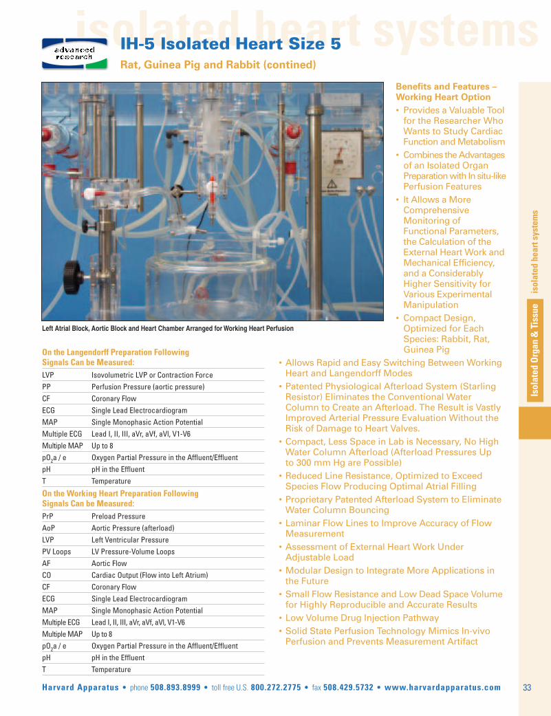
isolated heart systems
Isolated Organ & Tissue
isolated heart systems
33Harvard Apparatus • phone 508.893.8999 • toll free U.S. 800.272.2775 • fax 508.429.5732 • www.harvardapparatus.com
IH-5 Isolated Heart Size 5 Rat, Guinea Pig and Rabbit (contined)
Benefits and Features –Working Heart Option• Provides a Valuable Tool for the Researcher Who Wants to Study Cardiac Function and Metabolism• Combines the Advantages of an Isolated Organ Preparation with In situ-like Perfusion Features• It Allows a More Comprehensive Monitoring of Functional Parameters, the Calculation of the External Heart Work and Mechanical Efficiency, and a Considerably Higher Sensitivity for Various Experimental Manipulation• Compact Design, Optimized for Each
Species: Rabbit, Rat, Guinea Pig• Allows Rapid and Easy Switching Between Working Heart and Langendorff Modes• Patented Physiological Afterload System (Starling Resistor) Eliminates the Conventional Water Column to Create an Afterload. The Result is Vastly Improved Arterial Pressure Evaluation Without the Risk of Damage to Heart Valves.• Compact, Less Space in Lab is Necessary, No High Water Column Afterload (Afterload Pressures Up to 300 mm Hg are Possible)• Reduced Line Resistance, Optimized to Exceed Species Flow Producing Optimal Atrial Filling• Proprietary Patented Afterload System to Eliminate Water Column Bouncing• Laminar Flow Lines to Improve Accuracy of Flow Measurement• Assessment of External Heart Work Under Adjustable Load• Modular Design to Integrate More Applications in the Future• Small Flow Resistance and Low Dead Space Volume for Highly Reproducible and Accurate Results• Low Volume Drug Injection Pathway• Solid State Perfusion Technology Mimics In-vivo Perfusion and Prevents Measurement Artifact
On the Langendorff Preparation Following Signals Can be Measured:LVP Isovolumetric LVP or Contraction ForcePP Perfusion Pressure (aortic pressure)CF Coronary Flow ECG Single Lead Electrocardiogram MAP Single Monophasic Action PotentialMultiple ECG Lead I, II, III, aVr, aVf, aVl, V1-V6 Multiple MAP Up to 8pO2a / e Oxygen Partial Pressure in the Affluent/EffluentpH pH in the EffluentT Temperature
On the Working Heart Preparation Following Signals Can be Measured:PrP Preload PressureAoP Aortic Pressure (afterload) LVP Left Ventricular PressurePV Loops LV Pressure-Volume LoopsAF Aortic Flow CO Cardiac Output (Flow into Left Atrium)CF Coronary Flow ECG Single Lead Electrocardiogram MAP Single Monophasic Action PotentialMultiple ECG Lead I, II, III, aVr, aVf, aVl, V1-V6 Multiple MAP Up to 8pO2a / e Oxygen Partial Pressure in the Affluent/EffluentpH pH in the EffluentT Temperature
Left Atrial Block, Aortic Block and Heart Chamber Arranged for Working Heart Perfusion
advancedresearch

isolated heart systems
34
isolated heart systems
Isolated Organ &
Tissue
Harvard Apparatus • phone 508.893.8999 • toll free U.S. 800.272.2775 • fax 508.429.5732 • www.harvardapparatus.com
IH-5 Isolated Heart Size 5 Rat, Guinea Pig and Rabbit (contined)
SpecificationsPerfusion Kit
Body Weight Up to 2.5 kg
Heart Diameter 25 mm
Heart Length (Base - Apex) 30 mm
Aorta, ID Up to 5 mm
Aortic Flow (in WH Mode) Up to 500 ml/min
Perfusion Pressure (Afterload) Up to 300 mmHg
Order # Product 73-3604 Basic Unit for Isolated Heart Size 5 Type 843/173-2916 Heart Chamber for IH5 D=135/100 mm H=115 mm73-3072 Aortic Cannulae Set to IH-5 for Rat/Guinea Pig Hearts 73-0719 Aortic Cannulae Set to IH-5 for Rabbit Hearts73-3064 Working Heart Option to IH-5 Basic Unit73-3066 Left Atrium Cannula for IH-5 for Rat/Guinea Pig Heart,
OD=2.3 mm73-2880 Left Atrium Cannula for IH-5 for Rabbit Heart, OD=6.0 mm
System Description
The IH-5 is very compact, improved for low volume and high environmentstability.All of the components are mounted on a basic Plexiglas stand except for thethermocirculator, and the PLUGSYS amplifier system. Special attention hasbeen made to ensure that all critical connecting tubing are maintained short,thus avoiding any cooling through a temperature gradient to ambienttemperature.
The key part of the system is the unique aortic block which is the interfacebetween the heart and the IH-5. The bubble trap, compliance chamber,patented flow resistance membrane, infusion and measurement ports are allintegrated into this single specialized component. In addition, the aortic blockfeatures adjustable attachment points for our proprietary Mini Ball Joint Holderpositioning system. This permits precise placement of measurement probesfor ultra-stable recordings.
The modular nature of the IH-5 allows the system to evolve along with yourresearch. A Langendorff system can easily be upgraded to full Working Heart;and any IH-5 can be upgraded with special purpose capabilies like multi-channel ECG and MAP for a complete cardiac electrophysiology workstation.
The connection to the heart is made through interchangeable aortic cannulaeand atrial cannulae in working heart extension. The perfusate is aerated bybubbling in the reservoir. When the perfusate contains erythrocytes or albumine,a special membrane oxygenator to prevent foaming can equip the unit. In theLangendorff mode, heart contractility is measured using a balloon insertedinto the left ventricle and connected to a pressure transducer.
Additional equipment required: thermocirculator, peristaltic pumps, transducers,monitoring system setup using the PLUGSYS Amplifier System. Data acquisitionand Evaluation using ISOHEART software.
advancedresearch
Multi-Lead ECG RecordingThe heart is immersed in the heart chamber. The ECG-electrodes are mountedon an insert placed into the heart chamber. The ECG-signals are picked up fromthe heart surface using the conductivity of the perfusate. The three electrodesare arranged on the insert to mimic an ‘Einthoven’ derivation with the referenceelectrode located beneath the heart. The additional six electrodes are mountedaround the heart on a special ring to record unipolar Wilson leads V1 to V6.The electrodes are connected to the input box located on top of the unit (on the same box as the MAP electrodes). The box is connected to the PLUGSYSEGM (Einthoven Goldberger Module) and the PLUGSYS WLA (Wilson LeadAmplifier). The twelve leads recorded are I, II, III, aVL, aVR, aVF, V1 to V6.
If only a single ECG derivation (II) is required two single elctrodes are simplymounted with ball joint holders on the aortic block. In this case the insert isnot required.
Multiple MAP RecordingA special holder for up to 8 MAP-electrodes has been designed. On this holderare located eight evenly spaced cantilever arms holding spring-loadedmonophasic action potential electrodes. The spring-force (pressure of the electrodes against the epicardium) can be adjusted individually for each electrode. Even the position of the electrode on the heart can be adjusted individually. The electrodes are connected to the main input box type 701/1fixed on the top of the unit. This input box is connected to the MAPM(Monophasic action potential module) type 703 an especially designed PLUGSYSmodule. One MAPM can handle six electrodes, two modules are requested.
A special electrode for endocardial MAP is also available. This electrode ismounted with ball joint holders on the aortic block
Circular Holder for Placement of MAP Electrodes and Multi Lead ECG

isolated heart systems
Isolated Organ & Tissue
isolated heart systems
35Harvard Apparatus • phone 508.893.8999 • toll free U.S. 800.272.2775 • fax 508.429.5732 • www.harvardapparatus.com
UP-100 Basic Langendorff Heart System For Isovolumetric LVP Measurement
For the perfusion of the isolated heart according to Langendorff, the universalperfusion system UP-100 can be equipped with a jacketed heart chamber andadditional measurement system for isovolumetric LVP, using a latex balloonwith spindle syringe for diastolic pressure adjustment. The setup representsa compact unit. It can be used for constant pressure or constant flow perfusionof isolated hearts of mice, guinea pigs, rats and small rabbits as long as thecoronary flow is below 100 ml/min. The heart is connected via the aortic cannula to the heat exchanger which acts also as bubble trap. Provides anaccurate, low-cost way to indirectly measure coronary flow. The system can be adapted for direct, real-time coronary flow measurement allowing the study of myogene autoregulation (reactive hyperemia) and/or for electrogram recording.
If perfusate containing albumin or erythrocytes is used, the oxygenation canbe optimized with the additional fiber oxygenator.
The complete system includes the perfusion apparatus, the transducers andamplifiers for perfusion pressure, the servo controller and integrated peristalticpump, the isovolumetric left ventricular pressure measurement (isovolumetricballoon method) using a second pressure transducer and amplifier.
Order # Product Langendorff System
73-4128 Basic System UP-100H (Core System requires at least option 73-4129 or 73-4130)73-4129 Additions to UP-100H core system for Mouse hearts73-4130 Additions to UP-100H core system for Rat hearts
Options
73-4131 Direct Flow Measurement 73-4132 Measurement of ECG Lead II 73-4134 Pacing 73-4133 Oxygenating System for Foaming Solutions Containing Al-
bumin or Fatty Acids73-4047 Temperature Measurement
Data Acquisition
73-4039 Data Acquisition and Evaluation System ISOHEART73-4040 Data Acquisition and Evaluation System POWERLAB
Drug Application
73-4041 Drug Application 73-4042 Drug Application Flow Controlled in Constant Pressure Mode
UP-100 Basic Set-up for Langendorff Heart PerfusionUP-100 Basic Set-up for Lan-gendorff Heart Perfusion
• For isolated mouse, rat, guinea pig or small rabbit heart perfusion according to Langendorff
• Constant flow or constant pressure perfusion using the same equipment• Easy and compact
Applications• Simple Langendorff heart preparation optimized for LVP measurement using the balloon method • Ideal for compound screening in multi-channel configuration• In-line flow and ECG measurement available
research

isolated heart systems
36
isolated heart systems
Isolated Organ &
Tissue
Harvard Apparatus • phone 508.893.8999 • toll free U.S. 800.272.2775 • fax 508.429.5732 • www.harvardapparatus.com
Student Isolated Heart Perfusion Apparatusteaching
The ‘Langendorff’ preparation offers extreme flexibility for handling theperfusing fluids:
• The perfusing fluid reservoir serves as a Mariotteflask that maintains a constant perfusion pressureas the reservoir empties
• The perfusion pressure can be varied easily and rap-idly by varying the height of the reservoir above theperfused heart. The height of the reservoir can bevaried through 62.5 cm (24 in)
• To rapidly change perfusing fluids a second reser-voir (offered as an accessory) can be added to thesecond frame upright and a ‘Y’ connector can con-nect it to the glass warming coil
Order # ProductSystem Components50-0496 Constant Head 1 L Reservoir50-0488 Reservoir Holder50-0587 Aeration Reservoir50-0595 Aeration Stone with Tubing50-0537 Bath Assembly50-2898 Tubing 1 m (3-1/4 ft)50-0554 Warming Coil50-2369 Rubber Bung50-2914 Screwclips, 250-0562 Red Rubber Bung50-0563 Cannula50-6287 Thermometer50-0588 Manometer50-7624 Plug50-0596 Tying Rod53-2262 Pulleys, qty. of 253-2512 Rod, 150 mm (6 in)
Order # ProductSystem Components53-2522 Rod, 250 mm (10 in)53-2530 Rod, 500 mm (20 in)53-2550 Rods, 750 mm (29 in), qty. of 353-2336 Extra Weight Rectangular Laboratory Stand Base, qty. of 253-2102 Small Clamp with Extension Stem53-2012 Closed Connectors, qty. of 553-2032 ‘T’ Connectors, qty. of 353-2062 Large 360° Rotation Connector53-2244 Male-Female Hinged Adapter50-0686 Ink Pen with Lever50-7681 Replacement Ink Pen50-0678 Brodie Lever
• For the perfusion of the excised mammalian heart• Large capacity warming bath with temperature controlled to 0.5°C• Free-standing apparatus uses minimal bench space
This Systemincludes the componentsbelow
The 1 liter reservoir supplies a constant head of perfusate. The perfusate flowrate is controlled by one of two methods. If the Reservoir cap is off, the flowrate is controlled directly by a pinch clamp on the outlet tubing. If the reservoircap is tightly sealed, a Mariotte flask is created and the flow rate is set by theperfusion pressure. The height of the reservoir determines this pressure whichin turn sets the rate of air allowed to enter the reservoir. As air enters, theperfusate flows out. This reservoir is placed in the three-prong, spring clipreservoir holder that has a 20 cm (8 in) long tube that slips on the top of a rodand locks in place by a knurled set screw. Using this set screw the height of theReservoir can be varied from 15 cm (6 in) above the top of the rod down to thetop of the rod. If it is necessary to lower the reservoir even more, the holder hasa clamp mounted on one side that can locate the holder at any point on the rod.The total vertical movement of the reservoir is 62.5 cm (24 in) above the top ofthe fluid warming coil. The aeration reservoir is mounted to the side of thereservoir holder. The perfusing fluid is led from the main reservoir to theaeration reservoir by tubing provided. The aeration stone is mounted on the endof 2 m (6.6 ft) tubing. This aeration stone fits inside the aeration reservoir andprovides a high level of oxygenation to the perfusant.
The transparent, heavy plastic water bath which warms the perfusant measures,H x W x D, 15.5 x 16.5 x 14 cm (6-1/4 x 6-1/2 x 5-1/2 in) and has a capacity of 3.6liters. The bath is heated by two simple sturdy cartridge elements that aremounted through the front of the tank near the bottom and extend 5 cm (2 in) intoit. These heating elements have a combined rating of 80 watts. The temperatureof the water is controlled by a front panel dial graduated from 15° to 45°C by 5°Cgraduations. A metal tube mounted inside the bath carries a sensitive thermistorsensing bead. Once stabilized, this system maintains the water temperaturewithin 0.5°C of the desired temperature. The front panel of the water bath has
Order # Product 50-2864 Student Isolated Heart Perfusion Apparatus, 115 VAC, 60 Hz50-2872 Student Isolated Heart Perfusion Apparatus, 230 VAC, 50 Hz50-2880 Second Reservoir Set Includes Reservoir Holder, 1 L Reservoir, glass Aeration Reservoir and Aeration Stone. Set comes complete with tubing and connectors for mounting on second rod of Perfusion Apparatus
two indicator lights. One illuminates when the bath is plugged into the AC line.The second illuminates whenever the heating circuit is energized. A screw-infuse holder is also mounted on the front panel for easy access. A drain hole withtubing, a plug and a tapered plastic cone are located in the bottom of the watertank. The soft rubber bung that carries the stem of the warming coil fits into thecone making a leakproof joint. The glass warming coil for the perfusing fluid is 5cm (2 in) diameter by 9 cm (3-1/2 in) long. It has a capacity of approximately 35ml. The stem of the warming coil connects to the heart cannula with tubing anda pinch clamp controls the flow of the warmed perfusate. A clamp and groovedrod are supplied for securing the bottom of the excised mammalian heart.
The heart cannula has openings to accommodate both a manometer andthermometer. The manometer has a fixed clamp for mounting directly to acrossbar and is connected to the cannula by tubing. The scale reads 0 to250 mmHg and is free to move up and down behind the tubing to facilitatezero adjustment. The glass thermometer is straight, 10 cm (4 in) long andgraduated from 0° to 50°C by 0.1°C increments. It is mounted in a rubberbung and fits into the side arm of the heart cannula. All of the clamps,stands and rods supplied with this apparatus are from our Stronghold™ line.Two light pulleys are supplied and carry a thread from the heart to atransducer (not supplied) or writing point.

cell isolation
Isolated Organ & Tissue
cell isolation
37Harvard Apparatus • phone 508.893.8999 • toll free U.S. 800.272.2775 • fax 508.429.5732 • www.harvardapparatus.com
PSCI Perfusion System for Cell Isolation
The PSCI is specially designed for harvesting individual cells from isolatedorgans like mouse, rat or guinea-pig hearts, liver and other organs. Individualcells are released from the cell structure of the tissue through perfusion withenzyme solution and are then flushed out.
The apparatus has two separate perfusion circuits. The organ can be switchedbetween the two circuits by means of a specifically designed changeoverstopcock. One circuit is filled with conventional perfusion solution and is used inthe initial phase to flush out all blood cells from the organ. During the secondphase the apparatus is switched to the second circuit which operates withenzyme solution and where disintegration takes place.
In case of using a heart, atmospheric bacteria are kept away from the heartthrough suitable construction of the apparatus and by creating a small positivepressure in the heart chamber. Throughout the entire perfusion process, theheart is contained within a system closed to the outside atmosphere. It is onlyopened after the termination of perfusion in order to remove the heart.
Perfusion takes place under constant-flow conditions. The suitable constantflow is adjusted on the peristaltic pump. The limits of the apparatus are a flowrate of about 50 or 100 ml/min depending on the configuration.
Perfusion pressure measurement can easily be added. By using our SCPcontroller constant pressure perfusion is possible.
The apparatus is so designed that the individual steps required for preparing thecells can proceed as simply and clearly as possible. The components wetted bythe perfusion solutions are made from materials resistant to alcohol so that theapparatus can be filled with alcohol for sterilisation.
The dual heat exchanger is mounted on a vertical panel which in turn issecured to a rigid main vertical stainless steel column. A platform for theroller pump is also secured to the vertical column. The remainingcomponents are not fixed to the basic setup but are positioned on thebaseplate and are suitably connected by tubing. All components securedto the vertical steel column can be moved to suitable operating positionsafter releasing clamping screws. The protease reservoir and the holder for
• For cell isolation from mouse, rat and guinea pig organs by enzymatic disintegration• Specifically engineered dual perfusion system for blood cell flushand enzymatic disintegration
• Dedicated extension for cardiomyocite isolation
PSCI with operating tablefor in-situ perfusion
the perfusion pressure transducer are also part of the main system.
The main system is universal and must be adapted to the organ used byadding options.
The following options are available:
The dual heat exchanger:
• Stainless steel tube ID = 1.5 mm,Volume approx.1ml for the PSCI-M
• Stainless steel tube ID = 2 mm ,Volume approx. 1.5ml for the PSCI-R
The heat exchanger steel tubing can be interchanged.
Adaptation for Heart perfusion (not shown)In case of using the heart version, a jacketed heart chamber is mounted on aslide which is clamped to the vertical column. A slow gas flow into this chamberdoes ensure a positive pressure inside the heart chamber at all times, thuspreventing ingress of bacteria from the surroundings during operation.
Adaptation for in-situ perfusion (shown above)By adding an operating table and relevant cannulae depending on the organ.
Adaptation for in-vitro perfusionBy adding a jacketed moist chamber and the relevant cannulae dependingon the organ.
Additional equipment required: thermocirculator, peristaltic pumps,reservoir, magnetic stirrer, cannulae, operating table and others dependingon version selected, eventualy transducers, monitoring system setup usingthe PLUGSYS Amplifier System.
SpecificationsHeat Exchanger Inside Diameter 1.5 mm (0.06 in) for M Version 2.0 mm (0.08 in) for R Version
Maximum Flowrate 50 ml/min for the M Version 100 ml/min for the R Version
Prime Volume < 3ml for the M Version < 5ml for the R Version
Dimensions, W x D x H 600 x 400 x 570 mm (23.6 x 15.8 x 22.5 in)
Weight 8 kg (17.6 lbs)
Order # Product73-3659 PSCI-M Perfusion System for Cell Isolation from Mice Organs73-3639 PSCI-R Perfusion System for Cell Isolation from Rat Organs73-3638 PSCI-MH Perfusion System for Cell Isolation from Mice Hearts73-3672 PSCI-RH Perfusion System for Cell Isolation from Rat or Guinea Pig Hearts
advancedresearch
For a system description according your requirements please use:www.hugo-sachs.de/ihmail.html
or contact our technical experts at: [email protected]
For a custom configuration and full system quotation.

cell isolation
38
cell isolationIsolated O
rgan & Tissue
Harvard Apparatus • phone 508.893.8999 • toll free U.S. 800.272.2775 • fax 508.429.5732 • www.harvardapparatus.com
This perfusion system has been specifically created to meet the needs ofindividuals who wish to isolate primary cells from organs of species in thesize range of mouse to guinea pigs. The system can be configured foreither In Situ or Ex Vivo organ perfusion.
The system is first used with perfusate for the blanching and than with theprotease solution for tissue disintegration. The perfusate for blanching iswarmed and oxygenated in separate buffer reservoir and supplied via theperistaltic pump and warming coil. The protease solution is in a separatereservoir oxygenated and stirred. For In Situ perfusion an operating table canbe placed just below the warming coil, for Ex Vivo application (heart, liver...)the system can be equipped with a jacketed chamber. We offer a completeline of custom perfusion cannula (Aortic, In Situ and Ex Vivo) toaccommodate vessels from 1.0 to 3.5 mm ID. The In Situ and Ex Vivocannulae feature our unique tip basket to prevent vessel occlusion.
Easy Set-up for Cell Extraction
Easy Setup for Cell Extraction by ConstantFlow Perfusion• Cell isolation from mouse, rat, guinea pig organs by disintegration using protease solution• Designed for cardiomyocyte isolation• Can be used for other organs In Situ or Ex Vivo
Order # Product 73-3756 Easy Setup for Cell Extraction by Organ Disintegration Type 80373-3761 Heart Chamber to Easy Cell Extraction System
Perfusion Systemfor Cell Isolation
This system is simple to setup, operate and maintain. Its compact designminimizes the bench space required for the system. The wettedcomponents of the perfusion circuit can be flushed with a 70% ethanolsolution or autoclaved for sterilization purposes.
Additional equipment required: thermocirculator, peristaltic pumps,magnetic stirrer, reservoir, cannulae, operating table and others dependingon verison selected, eventualy transducers, monitoring system setup usingthe PLUGSYS Amplifier System.
research
For a system description according your requirements please use:www.hugo-sachs.de/ihmail.html
or contact our technical experts at: [email protected]
For a custom configuration and full system quotation.

isolated lung systems
Isolated Organ & Tissue
isolated lung systems Isolated Lung Systems Overview
39Harvard Apparatus • phone 508.893.8999 • toll free U.S. 800.272.2775 • fax 508.429.5732 • www.harvardapparatus.com
Constant PressurePerfusion
Constant Flow Perfu-sion
Negative Pressure Ventilation Positive Pressure Ventilation
Constant PressurePerfusion
Constant Flow Perfu-sion
TYPICAL APPLICATIONS
• Studies on primarily vascular effects
• Physiology:
– Hypoxic vasoconstriction
– Edema formation
• Pharmacology
– Action of histamine, arachidonic acid metabolites
– Immunopharmacology (cytokines...)
• Biochemistery
– Metabolic activity (biogenic amines, prostacyclin, angiotensin..)
– Surfactant biochemistry
TYPICAL APPLICATIONS
• Studies on primarily respiratory effects or combinedrespiratory and vascular effects
• Physiology:
– Hypoxic vasoconstriction
– Edema formation
• Pharmacology
– Action of histamine, arachidonic acid metabolites
– Immunopharmacology (cytokines...)
• Inhalation toxicology
– Environmental toxins (ozone, sulphur dioxide...)
• Biochemistery
– Metabolic activity (biogenic amines, prostacyclin, angiotensin..)
– Surfactant biochemistry
DISADVANTAGES
• Risk for baro traumaticventilation
• Higher cost
• Since flow is not con-stant effective dose isdifficult to calculate
DISADVANTAGES
• Non physiologic venti-lation
• Risk for baro traumatic ventilation
• Risk of damaging thevascular system by overpressure
DISADVANTAGES
• Higher cost
• Since flow is not con-stant effective dose isdifficult to calculate
ADVANTAGES
• No risk of damagingthe vascular system byoverpressure
ADVANTAGES
• Lower cost
• Easy quantification ofdrug dose application
ADVANTAGES
• Physiological mode
• No risk for baro traumatic ventilation
• No risk of damaging the vascular system by overpressure
ADVANTAGES
• Advanced physiolgicalmode
• No risk for barotrau-matic ventilation
• Lower cost
• Easy quantification ofdrug dose application
DISADVANTAGES
• Higher cost

isolated lung systems
40
textIsolated O
rgan & Tissue
Harvard Apparatus • phone 508.893.8999 • toll free U.S. 800.272.2775 • fax 508.429.5732 • www.harvardapparatus.com
IPL-1 Isolated Perfused Lung for Mouse
isolated lung systems
Isolated Perfused Lung System
Additional Options Include:• Dedicated PLUGSYS ventilator module for negative or positive pressure ventilation VCM• Dedicated PLUGSYS module for regular induction of hyperinflation of the lung TCM (sigh, augmented breath)• Transducers and amplifiers for respiratory mechanics measurement: airflow, tracheal pressure, thoracic chamber pressure• Transducers and amplifiers for perfusion measurement: realtime flow, perfusion pressure, venous pressure, temperature etc• Dedicated software PULMODYN for data acquisition and analysis of respiratory mechanics and perfusion• Optional connection for external ventilator (e.g. MINIVENT for testing barotraumatic ventilation• Optional connection for supplying alternative gas mixtures and/or aerosols during negativpressure ventilation
• Sensor and amplifier for continuous perfusate analysis (pO2, pCO2, pH)• Deoxygenation using membrane oxygenator of blood or erythrocyte containing perfusion• Large choice of accessories: thermocirculator, jacketed reservoir, holders etc
Benefits and Features• Exclusive artifical thorax chamber for isolated lung – Integrated surgery table to reduce damage during preparation phase – Integrated changeover system for switch between physiological negative-pressure ventilation and simple positive-pressure ventilation• Low flow resistance and dead space volume, minimize perfusion artifacts• Unique built-in pneumotachometer and air humidifier with small dead volume• Drug injection pathway built directly into pul monary perfusate stream • Unique compensation system for vascular transmural pressure changes and simulation of hypertensive cardiac afterload
Applications• Studies on combined respiratory and vascular effects• Physiology: – Hypoxic vasoconstriction• Pharmacology – Action of histamine, arachidonic acid metabolites – Immunopharmacology (cytokines...)• Inhalation toxicology – Environmental toxins (ozone, sulphur dioxine...)• Biochemistery – Metabolic activity (biogenic amines, prostacyclin, angiotensin..) – Surfactant biochemistry
advancedresearch

text
Isolated Organ & Tissue
isolated lung systems
41Harvard Apparatus • phone 508.893.8999 • toll free U.S. 800.272.2775 • fax 508.429.5732 • www.harvardapparatus.com
isolated lung systems
IPL-1 Isolated Perfused Lung for Mouse (continued)
DescriptionVentilation of the lung is possible under negative pressure as well asunder positive pressure (the latter is essential during the preparationphase). The ventilation parameters are controlled with a special PLUGSYSVentilator, the "Ventilation Control Module" (VCM, Type 681).
The apparatus consists essentially of a flat jacketed Plexiglass thoracicchamber and a jacketed cover. The size has been chosen so that acomplete mouse can be mounted inside it. The walls of the Plexiglass dishcarry all the auxiliary devices, connections and ports for perfusion andventilation. This arrangement offers the advantage that connecting tubingand cannulae can be kept short, resulting in a very small dead volume andensuring optimum thermal stability.
Perfusion takes place at constant flow. The perfusate is passed by meansof a roller pump at constant flow through the heat exchanger, through abubble trap to the pulmonary artery and finally into the lung vascular bed.The venous discharge is provided by cannulating the pulmonary vein, thecannula is placed in the left atrium of the heart. The venous dischargepasses into the pressure balancing vessel (compensation for the vasculartransmural pressure) and is drawn off from there by the second channel ofthe roller pump.
During the preparation phase the animal is ventilated at positive pressure.After the connections of the trachea and the perfusion supply have beenmade ventilation can be changed over to negative pressure. The chambernow forms an artificial thorax.
ExtensionsSeveral adapters are available to work with standard ventilators (positivepressure ventilation only), to supply different air mixtures to the lungs or toconnect a nebulizer for aerosol challenge.
Order # Product 73-4292 IPL-1 Core System, 115 V73-4291 IPL-1 Core System, 230 VVCM is a PLUGSYS module and requires a system maincase.
The Following Signals Can be Measured:FL Respiratory AirflowTCP Thoracal Chamber Pressure (intrapleural pressure)TP Tracheal PressurePP Perfusion Pressure VP Venous Outflow Pressure pO2a/e Oxygen Partial Pressure in the Affluent/EffluentpCO2a/e CO2 Partial Pressure in the Affluent/EffluentpH a/e pH in the Affluent/EffluentT Temperature
The Following Parameters Can be Evaluated Using PULMODYN:-Peak Inspiratory and Expiratory Airflow-Tidal Volume, Minute Volume-Dynamic Lung Resistance, Dynamic Compliance-Vascular Resistance-O2, CO2 Exchange
advancedresearch
For a system description according your requirements please use:www.hugo-sachs.de/ilmail.html
or contact our technical experts at: [email protected]
For a custom configuration and full system quotation.

isolated lung systems
42
textIsolated O
rgan & Tissue
Harvard Apparatus • phone 508.893.8999 • toll free U.S. 800.272.2775 • fax 508.429.5732 • www.harvardapparatus.com
IPL-2 Isolated Perfused Lung for Rat & Guinea Pig
isolated lung systems
Isolated Perfused Lung System
Benefits and Features• Optimized temperature conditions for the isolated lung, unique jacketed thoracic chamber• Optional operating table for non-damaging in-situ preparation• Negative-pressure ventilation similar to in-vivo condition or positive pressure ventilation available• Low flow resistance and dead space volume, mini mize perfusion artifacts• Unique built-in pneumotachometer and air humidifier with small dead volume• Drug injection pathway built directly into pulmonary perfusate stream
• Unique compensation system for vascular transmural pressure changes• Dedicated option for continuous measurement of lung weight (Edema)• More measurement parameters than any other system
Applications• Studies on combined respiratory and vascular effects• Physiology: – Hypoxic vasoconstriction – Edema formation• Pharmacology – Action of histamine, arachinodic acid metabolites• Inhalation toxicology – Environmental toxins (ozone, sulphur dioxine)
advancedresearch
• Biochemistery – Metabolic activity (biogenic amines, prostacyclin, angiotensin..) – Surfactant biochemistry
Additional Options Include:• Dedicated PLUGSYS ventilator module for negative or positive pressure ventilation VCM • Dedicated PLUGSYS module for regular induction of hyperinflation of the lung TCM (sigh, augmented breath)• Transducers and amplifiers for respiratory mechanics measurement: airflow, tracheal pressure, thoracic chamber pressure• Transducers and amplifiers for perfusion measurement: realtime flow, perfusion pressure, venous pressure, temperature etc... • Dedicated software PULMODYN for data acquisition and analysis of respiratory mechanics and perfusion• Optional connection for external ventilator (see section F) for testing barotraumatic ventilation• Optional connection for supplying alternative gas mixtures and/or aerosols• Sensor and amplifier for continuous perfusate analysis (pO2, pCO2, pH)• Deoxygenation using membrane oxygenator of blood or erythrocyte containing perfusion• Large choice of accessories: thermocirculator, jacketed reservoir, holders etc...

text
Isolated Organ & Tissue
isolated lung systems
43Harvard Apparatus • phone 508.893.8999 • toll free U.S. 800.272.2775 • fax 508.429.5732 • www.harvardapparatus.com
isolated lung systems
IPL-2 Isolated Perfused Lung for Rat & Guinea Pig (continued)
Order # Product 73-4276 IPL-2 Core System, 115 V73-4275 IPL-2 Core System, 230 VVCM is a PLUGSYS module and requires a system maincase.
The Following Signals Can Be Measured:FL Respiratory AirflowTCP Thoracal Chamber Pressure (intrapleural pressure)TP Tracheal PressurePP Perfusion Pressure VP Venous Outflow Pressure W Lung Weight ChangespO2a/e Oxygen Partial Pressure in the Affluent/EffluentpCO2a/e CO2 Partial Pressure in the Affluent/EffluentpH a/e pH in the Affluent/EffluentT Temperature
The Following Parameters Can be Evaluated Using PULMODYN:Peak Inspiratory and Expiratory AirflowTidal Volume, Minute VolumeLung Resistance, Dynamic ComplianceVascular ResistancePrecapillary and Postcapillary ResistanceFiltration CoefficientO2, CO2 Exchange
Basic SystemThe basic setup consists of a Plexiglass base with two stable vertical steelcolumns. The main system is mounted on a Plexiglass panel which is fixedon the two steel columns. This allows the system to be modified orextended to suit different versions or for special applications.
The essential part of the system is the artificial thorax. It consists of awater-jacketed glass chamber sealed by a special cover. The cover ismounted on the Plexiglass panel, the glass chamber is fixed underneath itwith two metal clips. The inner part of the cover is the organ holder withthe connections for cannulating the trachea (ventilation), the pulmonaryartery and the left atrium (perfusion).
Ventilation of the lung is possible under negative pressure as well asunder positive pressure (the latter is essential during the preparationphase). The ventilation parameters are controlled with a special PLUGSYSVentilator, the "Ventilation Control Module" (VCM, Type 681).Theventilation medium (air or gas mixture) is constantly humidified by thebuilt-in humidifier.
An optional operating table with trough is mounted on the base plate infront of the artificial thoracic chamber. The organ holder with theconnections for the trachea (ventilation) and for the pulmonary artery andpulmonary vein (perfusion) can be removed from the main cover to allowan easier preparation and optimal non traumatic lung transfer from theanimal to the system. The lung is continuously ventilated at positivepressure until it is placed into the artificial thorax. Then a simple valvepermits instantaneous changeover to negative pressure ventilation.
PerfusionLungs can be perfused by either constant flow or constant pressure.Although constant flow perfusion may mimic the in vivo situation moreclosely, it has the disadvantage that hydrostatic edema becomesinevitable during vasoconstriction. Constant pressure perfusion permitshigher perfusate flow rates since vasoconstriction decreases perfusateflow and hydrostatic edema is less likely to occur.
Can be configured in one of three different perfusion modes:
1. Constant Flow Perfusion
2. Constant Pressure Perfusion
3. Dual System to Switch from Constant Flow to Con-stant Pressure and Vice Versa
Lung Weight ChangesContinuous measurement of weight changes of the lung is required formonitoring edema formation. This is possible during negative pressureventilation inside the thoracic chamber by using the HSE-Edema balanceLS 30 (weight transducer) (73-0593). Weight changes induced by raising orlowering the perfusion pressure can be used to assess vascularpermeability by determining the filtration coefficient.
ExtensionsSeveral adapters are available to work with ventilators, to supply differentair mixtures to the lungs or to connect aerosol nebulizer for aerosolchallenge.
advancedresearch
For a system description according your requirements please use:www.hugo-sachs.de/ilmail.html
or contact our technical experts at: [email protected]
For a custom configuration and full system quotation.
Application Note:IPL-2 option for modificationThe IPL-2 can also be specially modified to measure lung weightchanges in mouse models.
Speak with a technical specialist for details on this option.
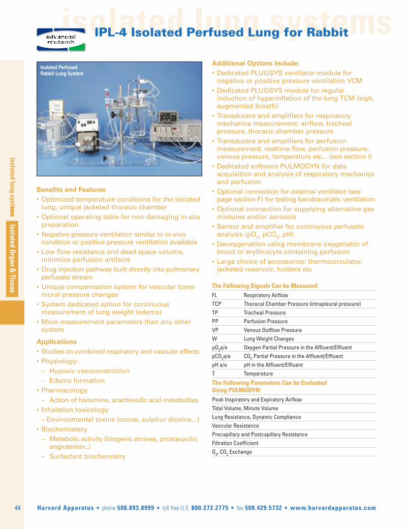
isolated lung systems
44
textIsolated O
rgan & Tissue
Harvard Apparatus • phone 508.893.8999 • toll free U.S. 800.272.2775 • fax 508.429.5732 • www.harvardapparatus.com
isolated lung systems
Isolated PerfusedRabbit Lung System
Benefits and Features• Optimized temperature conditions for the isolated lung, unique jacketed thoracic chamber
• Optional operating table for non-damaging in-situ preparation
• Negative-pressure ventilation similar to in-vivo condition or positive pressure ventilation available• Low flow resistance and dead space volume, mini mize perfusion artifacts• Drug injection pathway built directly into pul monary perfusate stream • Unique compensation system for vascular trans- mural pressure changes• System dedicated option for continuous measurement of lung weight (edema)• More measurement parameters than any other system
Applications• Studies on combined respiratory and vascular effects• Physiology: – Hypoxic vasoconstriction – Edema formation• Pharmacology – Action of histamine, arachinodic acid metabolites• Inhalation toxicology – Environmental toxins (ozone, sulphur dioxine...)• Biochemistery – Metabolic activity (biogenic amines, prostacyclin, angiotensin..) – Surfactant biochemistry
Additional Options Include:• Dedicated PLUGSYS ventilator module for negative or positive pressure ventilation VCM • Dedicated PLUGSYS module for regular induction of hyperinflation of the lung TCM (sigh, augmented breath)• Transducers and amplifiers for respiratory mechanics measurement: airflow, tracheal pressure, thoracic chamber pressure• Transducers and amplifiers for perfusion measurement: realtime flow, perfusion pressure, venous pressure, temperature etc... (see section I)• Dedicated software PULMODYN for data acquisition and analysis of respiratory mechanics and perfusion• Optional connection for external ventilator (see page section F) for testing barotraumatic ventilation• Optional connection for supplying alternative gas mixtures and/or aerosols• Sensor and amplifier for continuous perfusate analysis (pO2, pCO2, pH)• Deoxygenation using membrane oxygenator of blood or erythrocyte containing perfusion• Large choice of accessories: thermocirculator, jacketed reservoir, holders etc
The Following Signals Can be Measured:FL Respiratory AirflowTCP Thoracal Chamber Pressure (intrapleural pressure)TP Tracheal PressurePP Perfusion Pressure VP Venous Outflow Pressure W Lung Weight ChangespO2a/e Oxygen Partial Pressure in the Affluent/EffluentpCO2a/e CO2 Partial Pressure in the Affluent/EffluentpH a/e pH in the Affluent/EffluentT Temperature
The Following Parameters Can be Evaluated Using PULMODYN:Peak Inspiratory and Expiratory AirflowTidal Volume, Minute VolumeLung Resistance, Dynamic ComplianceVascular ResistancePrecapillary and Postcapillary ResistanceFiltration CoefficientO2, CO2 Exchange
advancedresearch
IPL-4 Isolated Perfused Lung for Rabbit

isolated lung systems
Isolated Organ & Tissue
isolated lung systems
45Harvard Apparatus • phone 508.893.8999 • toll free U.S. 800.272.2775 • fax 508.429.5732 • www.harvardapparatus.com
IPL-4 Isolated Perfused Lung for Rabbit (continued)
Order # Product 73-4297 IPL-4 Core System, 115 V73-4296 IPL-4 Core System, 230 VVCM is a PLUGSYS module and requires a system maincase.
73-3376 NMR Cover for IPL-4 73-3377 Pulmonary Artery Cannula for IPL-4, NMR73-3378 Left Atrium Cannula for IPL-4, NMR73-3379 Jacketed Tubing for IPL-4 NMR73-3469 Pressure Equilibration System for IPL-4 NMR
MRI-Coverfor IPL-4
MRI-Cover for IPL-4 There is an increasing interest in studying isolated lungs by using MRI. Aspecial metal free cover for the IPL-4 lung thoracic chamber has beendesigned. The regular cover is replaced by the MRI cover, and thecomplete lung chamber can be removed from the stand after surgery. Thelung chamber with cover is mounted on the MRI e.g. in a head holder. Allparts are non-metalic and made of plexiglas, Teflon and carbon fiber. Inthe NMR version only positive pressure ventilation can be used.
Basic SystemThe basic setup consists of a Plexiglass base with a stable vertical steelcolumn. The main system is fixed on the steel column. This allows thesystem to be modified or extended to suit different versions or for specialapplications.
The essential part of the system is the artificial thoracic chamber. Itconsists of a water-jacketed glass chamber sealed by a special cover. Thecover is mounted on the steel column, the glass chamber is fixedunderneath it with two metal clips. The cover is the organ holder with theconnections for cannulating the trachea (ventilation), the pulmonary arteryperfusion and the pulmonary venous effluent (perfusion).
Ventilation of the lung is possible under negative pressure as well asunder positive pressure (the latter is essential during the preparationphase). The ventilation parameters are controlled with a special PLUGSYSVentilator, the "Ventilation Control Module" (VCM, Type 681). Theventilation medium (air or gas mixture) is constantly humidified by thebuilt-in humidifier.
An optional operating table is mounted on the base plate in front of theartificial thorax cover. The cover with the connections for the trachea(ventilation) and for the pulmonary artery and pulmonary vein (perfusion)can easily be inclined in order to have the tracheal cannula in line with thetrachea and allow easier preparation. After cannulation the cover can begradually rotated and moved up by turning a knob, thus the lung can beremoved from the animal with minimal stretch damage. During surgery,the cover is equipped with a special ventilation head, and the lung iscontinuously ventilated at positive pressure until the artificial thorax is inplace. Then a simple valve permits instantaneous changeover to negativepressure ventilation.
Constant Flow PerfusionThe constant flow rate of perfusate into the pulmonary artery isdetermined by a roller pump. The perfusate is stored in a water-jacketedcontainer to maintain constant temperature. Aeration is provided to keepthe pH constant. A heat exchanger and a bubble trap are placed next tothe pulmonary artery connection for exact adjustment of the perfusatetemperature just before the thorax chamber and to prevent air bubblesentering the lung.
Lung Weight ChangesContinuous measurement of weight changes of the lung is possible duringnegative pressure ventilation inside the thoracic chamber by using theHSE-Edema balance LS 30 (weight transducer) (73-3468).
advancedresearch
For a system description according your requirements please use:www.hugo-sachs.de/ilmail.html
or contact our technical experts at: [email protected]
For a custom configuration and full system quotation.

isolated lung systems
46
isolated lung systems
Isolated Organ &
Tissue
Harvard Apparatus • phone 508.893.8999 • toll free U.S. 800.272.2775 • fax 508.429.5732 • www.harvardapparatus.com
IPL-16 Isolated Perfused Lung forPigs and Large Animals
DescriptionThe apparatus consists essentially of a jacketed Plexiglass thoracicchamber (inside dimensions 400 x 500 x 300 mm) and a jacketed cover.The size has been chosen so that a complete pig lung (both lobes) can bemounted inside it. The lung is placed on a platform. The platform can bemoved out of the chamber to facilitate the surgery. All connections forperfusion and ventilation are mounted on the plattform. This arrangementoffers the advantage that connecting tubing and cannulae can be keptshort, resulting in a very small dead volume and ensuring optimum thermalstability.
PerfusionConstant flow perfusion may mimic the in vivo situation more closely,however, it has the disadvantage that hydrostatic edema becomesinevitable during vasoconstriction. Constant pressure perfusion permitshigher perfusate flow rates since vasoconstriction decreases perfusateflow and hydrostatic edema is less likely to occur. The chamber is theperfusate reservoir (whole blood or erythrocyte containing perfusate).Perfusion takes place at constant flow. The perfusate is taken from thereservoir and is passed by means of a peristaltic pump at constant flowthrough the deoxygenator and primary heat exchanger, through a secondheat exchanger and a bubble trap to the pulmonary artery and finally intothe lung vascular bed. The venous discharge returns to the reservoir. Forventilation, a large animal ventilator Model 665 or 663 or a humanventilator is used.
The chamber also includes our exclusive lung weight measurement systemfor continuous monitoring of edema formation.
ExtensionsAerosol nebulizer for inhaled compound challenge Controller for constantpressure perfusion
Additional Options Include• Transducers and amplifiers for respiratory mechanics
measurement: airflow, tracheal pressure, thoracicchamber pressure
• Transducers and amplifiers for perfusion measure-ment: realtime flow, perfusion pressure, venouspressure, temperature etc.
• Dedicated software PULMODYN for data acquisi-tion and analysis of respiratory mechanics and perfusion
• Sensor and amplifier for continuous perfusateanalysis (pO2, pH)
• Deoxygenation using membrane oxygenator ofwhole blood or erythrocyte containing perfusion
• Large choice of accessories: ventilators, thermocir-culator, peristaltic pumps, holders etc...
Benefits and Features• Optimized temperature and humidity conditions for the isolated lung, unique jacketed thoracic chamber• Continuous measurement of lung weight to monitor edema formation• Very low flow resistance and minimal dead space volume virtually eliminate perfusion artifacts• Drug injection pathway built directly into pulmonary perfusate stream for precision compound dosing and screening• Removable platform to facilitate surgery• Standard connection for ventilator• More measurement parameters with greater precision than any other system
Applications• Respiratory mechanics – Airway Flow, Tidal Volume, dynamic resistance and compliance• Vascular permeability studies – Arterial and venous resistance calculations – Inflammation (?) and Tissue Edema monitoring• Pharmacology – Compound screening, e.g. action of histamine, arachinodic acid metabolites• Inhalation toxicology – Environmental toxins, e.g. Ultrafine particles, ozone, sulphur dioxin...• Biochemistery – Metabolic activity, e.g of biogenic amines, prostacyclin, angiotensin.. – Surfactant bioactivity• Therapy studies – Study of chemotherapy applications using a human lung lobe after lobectomy of the lung is performed in lung cancer patient
Order # Product 73-4302 IPL-16 Core System, 115 V73-4301 IPL-16 Core System, 230 V
advancedresearch