Timely Visualization of the Collaterals Formed during...
9
FULL PAPER 1800573 (1 of 9) www.small-journal.com © 2018 WILEY-VCH Verlag GmbH & Co. KGaA, Weinheim Timely Visualization of the Collaterals Formed during Acute Ischemic Stroke with Fe 3 O 4 Nanoparticle-based MR Imaging Probe Ting Wang, Yi Hou, Bo Bu, Wenxin Wang, Tiancong Ma, Chunyan Liu, Lan Lin, Lin Ma, Xin Lou,* and Mingyuan Gao DOI: 10.1002/smll.201800573 1. Introduction Ischemic stroke due to large vessel blocked is a devastating event and one of the leading causes of mortality and neu- rologic disability. For example, it caused ≈6.7 million deaths worldwide in 2012 according to a recent statistics. [1,2] Collat- eral vessels provide alternative routes for cerebral blood supply to preserve the brain tissue at the risk of infarction after arterial occlusion. Collaterals differ greatly among patients with acute ischemic stroke (AIS) and largely determine the size of the final infarct and the growth of penumbra. [2,3] Good collateral status may effectively extend the time window for clinical thera- pies, thus improving the probability of positive outcome. [4,5] Therefore, timely vis- ualizing and grading the status of collat- eral vessels are mandatory in the clinical practice, but remain currently challenging. At present, digital subtraction angiog- raphy (DSA), [6] X-ray computed tomog- raphy (CT), [7] and magnetic resonance imaging (MRI) [8–13] are widely used in detecting the cerebral collaterals in vivo. In addition, other imaging techniques such as positron emission tomography and single-emission computed tomography are also adopted for pro- viding some relevant information on the cerebral collaterals. [14] Among all these methods, DSA remains the gold standard for the evaluation of collateral vessels, but it is an invasive approach. CT perfusion and CT angiography are quick and easy-to-use in evaluating the functional and anatomic aspects of the collat- erals, respectively, while MRI assesses the collateral vessels with multiple imaging parameters through different sequences. [3] For example, MR angiography can be used to evaluate the collaterals in the circle of Willis; [8] fluid attenuated inversion recovery can provide information on collateral status; [9,10] while MR perfusion imaging including dynamic susceptibility con- trast enhanced perfusion-weighted imaging, arterial spin labe- ling, and territorial arterial spin labeling have been developed for reflecting the collateral perfusion. [11–13] Although MR-based approaches are much safer than X-ray-based methods since no ionizing radiation is induced, it remains difficult to evaluate the leptomeningeal collaterals with MRI. In the clinic, the cerebral Ischemic stroke is one of the major leading causes for long-term disability and mortality. Collateral vessels provide an alternative pathway to protect the brain against ischemic injury after arterial occlusion. Aiming at visualizing the collaterals occurring during acute ischemic stroke, an integrin α v β 3 -specific Fe 3 O 4 –Arg-Gly-Asp (RGD) nanoprobe is prepared for magnetic resonance imaging (MRI) of the collaterals. Rat models are constructed by occluding the middle cerebral artery for imaging studies of cerebral ischemia and ischemia– reperfusion on 7.0 Tesla MRI using susceptibility-weighted imaging sequence. To show the binding specificity to the collaterals, the imaging results acquired with the Fe 3 O 4 –RGD nanoprobe and the Fe 3 O 4 mother nanoparticles, respectively, are carefully compared. In addition, an RGD blocking experiment is also carried out to support the excellent binding specificity of the Fe 3 O 4 – RGD nanoprobe. Following the above experiments, cerebral ischemia– reperfusion studies show the collateral dynamics upon reperfusion, which is very important for the prognosis of various revascularization therapies in the clinic. The current study has, for the first time, enabled the direct observation of collaterals in a quasi-real time fashion and further disclosed that the antegrade flow upon reperfusion dominates the blood supply of primary ischemic tissue during the early stage of infarction, which is significantly meaningful for clinical treatment of stroke. MRI Probes T. Wang, L. Lin, Prof. L. Ma, Prof. X. Lou Department of Radiology Chinese PLA General Hospital No. 28 Fuxing Road, Beijing 100853, P. R. China E-mail: [email protected] Y. Hou, T. C. Ma, C. Y. Liu, Prof. M. Y. Gao Key Laboratory of Colloid Interface and Chemical Thermodynamics Institute of Chemistry Chinese Academy of Sciences Bei Yi Jie 2, Zhong Guan Cun, Beijing 100190, P. R. China Prof. B. Bu, W. X. Wang Department of Neurosurgery Chinese PLA General Hospital No. 28 Fuxing Road, Beijing 100853, P. R. China T. C. Ma, Prof. M. Y. Gao School of Chemistry and Chemical Engineering University of Chinese Academy of Sciences Beijing 100049, China The ORCID identification number(s) for the author(s) of this article can be found under https://doi.org/10.1002/smll.201800573. Small 2018, 14, 1800573
Transcript of Timely Visualization of the Collaterals Formed during...

FULL PAPER
1800573 (1 of 9)
www.small-journal.com
© 2018 WILEY-VCH Verlag GmbH & Co. KGaA, Weinheim
Timely Visualization of the Collaterals Formed during Acute Ischemic Stroke with Fe3O4 Nanoparticle-based MR Imaging Probe
Ting Wang, Yi Hou, Bo Bu, Wenxin Wang, Tiancong Ma, Chunyan Liu, Lan Lin, Lin Ma, Xin Lou,* and Mingyuan Gao
DOI: 10.1002/smll.201800573
1. Introduction
Ischemic stroke due to large vessel blocked is a devastating event and one of the leading causes of mortality and neu-rologic disability. For example, it caused ≈6.7 million deaths worldwide in 2012 according to a recent statistics.[1,2] Collat-eral vessels provide alternative routes for cerebral blood supply to preserve the brain tissue at the risk of infarction after arterial occlusion. Collaterals differ greatly among patients with acute ischemic stroke (AIS) and largely determine the size of the final infarct and the growth of penumbra.[2,3] Good collateral status may effectively extend the time window for clinical thera-pies, thus improving the probability of positive outcome.[4,5] Therefore, timely vis-ualizing and grading the status of collat-eral vessels are mandatory in the clinical practice, but remain currently challenging.
At present, digital subtraction angiog-raphy (DSA),[6] X-ray computed tomog-raphy (CT),[7] and magnetic resonance imaging (MRI)[8–13] are widely used in
detecting the cerebral collaterals in vivo. In addition, other imaging techniques such as positron emission tomography and single-emission computed tomography are also adopted for pro-viding some relevant information on the cerebral collaterals.[14] Among all these methods, DSA remains the gold standard for the evaluation of collateral vessels, but it is an invasive approach. CT perfusion and CT angiography are quick and easy-to-use in evaluating the functional and anatomic aspects of the collat-erals, respectively, while MRI assesses the collateral vessels with multiple imaging parameters through different sequences.[3] For example, MR angiography can be used to evaluate the collaterals in the circle of Willis;[8] fluid attenuated inversion recovery can provide information on collateral status;[9,10] while MR perfusion imaging including dynamic susceptibility con-trast enhanced perfusion-weighted imaging, arterial spin labe-ling, and territorial arterial spin labeling have been developed for reflecting the collateral perfusion.[11–13] Although MR-based approaches are much safer than X-ray-based methods since no ionizing radiation is induced, it remains difficult to evaluate the leptomeningeal collaterals with MRI. In the clinic, the cerebral
Ischemic stroke is one of the major leading causes for long-term disability and mortality. Collateral vessels provide an alternative pathway to protect the brain against ischemic injury after arterial occlusion. Aiming at visualizing the collaterals occurring during acute ischemic stroke, an integrin αvβ3-specific Fe3O4–Arg-Gly-Asp (RGD) nanoprobe is prepared for magnetic resonance imaging (MRI) of the collaterals. Rat models are constructed by occluding the middle cerebral artery for imaging studies of cerebral ischemia and ischemia–reperfusion on 7.0 Tesla MRI using susceptibility-weighted imaging sequence. To show the binding specificity to the collaterals, the imaging results acquired with the Fe3O4–RGD nanoprobe and the Fe3O4 mother nanoparticles, respectively, are carefully compared. In addition, an RGD blocking experiment is also carried out to support the excellent binding specificity of the Fe3O4–RGD nanoprobe. Following the above experiments, cerebral ischemia–reperfusion studies show the collateral dynamics upon reperfusion, which is very important for the prognosis of various revascularization therapies in the clinic. The current study has, for the first time, enabled the direct observation of collaterals in a quasi-real time fashion and further disclosed that the antegrade flow upon reperfusion dominates the blood supply of primary ischemic tissue during the early stage of infarction, which is significantly meaningful for clinical treatment of stroke.
MRI Probes
T. Wang, L. Lin, Prof. L. Ma, Prof. X. LouDepartment of RadiologyChinese PLA General HospitalNo. 28 Fuxing Road, Beijing 100853, P. R. ChinaE-mail: [email protected]. Hou, T. C. Ma, C. Y. Liu, Prof. M. Y. GaoKey Laboratory of ColloidInterface and Chemical ThermodynamicsInstitute of ChemistryChinese Academy of SciencesBei Yi Jie 2, Zhong Guan Cun, Beijing 100190, P. R. ChinaProf. B. Bu, W. X. WangDepartment of NeurosurgeryChinese PLA General HospitalNo. 28 Fuxing Road, Beijing 100853, P. R. ChinaT. C. Ma, Prof. M. Y. GaoSchool of Chemistry and Chemical EngineeringUniversity of Chinese Academy of SciencesBeijing 100049, China
The ORCID identification number(s) for the author(s) of this article can be found under https://doi.org/10.1002/smll.201800573.
Small 2018, 14, 1800573

1800573 (2 of 9)
www.advancedsciencenews.com www.small-journal.com
© 2018 WILEY-VCH Verlag GmbH & Co. KGaA, Weinheim
collaterals are classified into primary, secondary, and tertiary collaterals.[2] The primary collaterals refer to the circle of Willis, the secondary collaterals mainly include leptomeningeal col-laterals, and the tertiary collaterals refer to arteriogenesis and angiogenesis that largely occur during chronic ischemia. It is believed that the leptomeningeal collaterals of 50–400 µm,[15] as important routes, promote the retrograde perfusion of adjacent territories, especially during the acute arterial occlusion. How-ever, timely evaluating the status of leptomeningeal collaterals remains practically very challenging due to their small size. Although CT and MR can provide indirect information on lep-tomeningeal collaterals through perfusion, they hardly provide anatomical information about cerebral collaterals.
Recent achievements in molecular imaging have offered new approaches for noninvasively studying the nononcological dis-eases, such as the hindlimb ischemic and myocardial infarction diseases.[16–19] For example, magnetic iron oxide nanoparticles and upconversion nanoparticles have been used for imaging the atherosclerotic plaques and even for differentiating the vulnerable plaques from stable ones.[17,18] However, to the best of our knowl-edge, there is no similar study on the cerebral collaterals, par-ticularly the leptomeningeal collaterals formed in acute ischemic stroke. The development of the cerebral collaterals involves neo-vascularization, that is a complex pathophysiology process and the molecules and factors involved in this process may be taken as potential targets for angiogenesis imaging. To date, integrin αvβ3, vascular endothelial growth factor (VEGF), matrix metal-loproteinases, and extra domain B (ED-B) domain of fibronectin isoforms have been selected as targets in different studies, while integrin αvβ3 is most often used since it is highly expressed by human brain microvascular endothelial cells and plays a critical role in promoting postischemic angiogenesis.[20–22] With a cyclic Arg-Gly-Asp (RGD) peptide as specific ligand, versatile molecular imaging probes have been constructed for targeting integrin αvβ3, but in most cases for tumor imaging.[23–26] Therefore, it can be deduced that the RGD peptide sequence may also be used as a targeting moiety for in vivo imaging of the cerebral collaterals.
In the current paper, we report our imaging studies on the collaterals developed in acute ischemic stroke. Previous inves-tigations on blood–brain barrier (BBB)[27] and toxoplasmic brain lesions[28] have demonstrated that Fe3O4 nanoparticles are superior for brain imaging due to their unique super-paramagnetism. The long blood half-life of nanoparticles with suitable antifouling modification is also greatly in favor of the diagnosis of vascular diseases.[27] Therefore, Fe3O4 nano-particles were chosen to construct an integrin αvβ3-specific nan-oprobe by covalently attaching RGD on the surface of biocom-patible Fe3O4 nanoparticles and the resulting probe is denoted as Fe3O4–RGD. A rat model of right middle cerebral artery occlusion (rMCAO) was built for imaging studies on a 7.0 T animal MRI scanner with a susceptibility-weighted imaging (SWI) sequence. Typically, the rats were subjected to MR imaging right after rMCAO in order to acquire timely informa-tion on the leptomeningeal collaterals developed in acute phase upon enhancement with the αvβ3-specific Fe3O4–RGD probes. Careful imaging studies in combination with histochemical analysis of brain tissues were carried out for showing the potential of the Fe3O4–RGD probes for enhanced imaging of the leptomeningeal collaterals. It was for the first time observed
that the collaterals formed shortly after the artery occlusion van-ished upon revascularization within only a few hours, which well highlights the current studies.
2. Results and Discussion
2.1. PEGylated Fe3O4 Nanoprobes
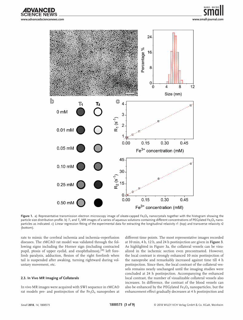
As shown in Figure 1a, the oleate-capped Fe3O4 nanocrystals of 6.4 ± 0.4 nm were prepared through a conventional thermal decomposition method.[29] By replacing the oleate ligand with an asymmetric polyethylene glycol (PEG) ligand bearing a diphosphate group and a maleimide group on different ends (dp–PEG–mal), PEGylated Fe3O4 nanoparticles were obtained. The MRI contrast enhancement effects of the PEGylated Fe3O4 nanoparticles in aqueous solutions are shown in Figure 1b. By linear regression fitting of the experimental data, the molar relaxivity r1 and r2 were extracted as 6.03 and 85.8 mm−1 s−1, respectively (Figure 1c).
Through the particle surface maleimide residues, an inte-grin αvβ3-specific peptide sequence (i.e., Arg-Gly-Asp-Cys (RGDC)) was further covalently attached onto the surface of the PEGylated Fe3O4 nanoparticles through click reaction to obtain the nanoprobe (Fe3O4–RGD). Dynamic light scattering was adopted to evaluate the effectiveness of the coupling reaction. The results shown in Figure 2a reveal that the hydrodynamic size is slightly decreased after the conjugation reaction, in the meantime, the size distribution profile of Fe3O4 nanoparticles gets a little narrowed. The small variation in the hydrodynamic size can be explained by the low molecular weight of RGD and the slight decrease in hydrodynamic size can probably be attrib-uted to the different hydration properties of the RGDC moiety in comparison with the PEG segment. The most important information conveyed by Figure 2a is that no unwanted coagu-lation occurred during the conjugation reaction, which is fore-most for the construction of any nanoprobes.
The bioactivity of the Fe3O4–RGD conjugates was quanti-tatively evaluated through cell binding assays in which human umbilical vein endothelial cells (HUVECs) and mice fibroblast cells 3T3 were used as positive and negative controls, respec-tively. The cellular uptake of the Fe3O4–RGD probes was analyzed through Prussian blue staining and compared with that obtained with the mother PEGylated Fe3O4 nanoparticles. The staining results in Figure 2b clearly reveal that the RGD as the specific ligand can dramatically increase the binding affinity of the Fe3O4 nanoparticles to RGD positive cells (left and middle frames of Figure 2b), while the Fe3O4–RGD probes do not show specific binding affinity to RGD negative cells (mice fibroblast cells 3T3) (right frame of Figure 2b). All these differences are well reflected by the quantified data on the cellular uptake of Fe3O4–RGD nanoprobes or Fe3O4 nanoparticles, as given in Figure 2c.
2.2. rMCAO Rat Model
To validate the potential of Fe3O4–RGD nanoprobe in targeting the leptomeningeal collaterals through integrin αvβ3, the right middle cerebral artery occlusion was mechanically induced in
Small 2018, 14, 1800573
1800573 (3 of 9)
www.advancedsciencenews.com www.small-journal.com
© 2018 WILEY-VCH Verlag GmbH & Co. KGaA, Weinheim
rats to mimic the cerebral ischemia and ischemia–reperfusion diseases. The rMCAO rat model was validated through the fol-lowing signs including the Horner sign (including contracted pupil, ptosis of upper eyelid, and enophthalmos),[30] left fore-limb paralysis, adduction, flexion of the right forelimb when tail is suspended after awaking, turning rightward during vol-untary movement, etc.
2.3. In Vivo MR Imaging of Collaterals
In vivo MR images were acquired with SWI sequence in rMCAO rat models pre- and postinjection of the Fe3O4 nanoprobes at
different time points. The most representative images recorded at 10 min, 4 h, 12 h, and 24 h postinjection are given in Figure 3. As highlighted in Figure 3a, the collateral vessels can be visu-alized in the ischemic section even precontrasted. However, the local contrast is strongly enhanced 10 min postinjection of the nanoprobe and remarkably increased against time till 4 h postinjection. Since then, the local contrast of the collateral ves-sels remains nearly unchanged until the imaging studies were concluded at 24 h postinjection. Accompanying the enhanced local contrast, the number of visualizable collateral vessels also increases. In difference, the contrast of the blood vessels can also be enhanced by the PEGylated Fe3O4 nanoparticles, but the enhancement effect gradually decreases at 4 h postinjection and
Small 2018, 14, 1800573
Figure 1. a) Representative transmission electron microscopy image of oleate-capped Fe3O4 nanocrystals together with the histogram showing the particle size distribution profile. b) T1 and T2 MR images of a series of aqueous solutions containing different concentrations of PEGylated Fe3O4 nano-particles as indicated. c) Linear regression fitting of the experimental data for extracting the longitudinal relaxivity r1 (top) and transverse relaxivity r2 (bottom).

1800573 (4 of 9)
www.advancedsciencenews.com www.small-journal.com
© 2018 WILEY-VCH Verlag GmbH & Co. KGaA, Weinheim
completely vanishes after 24 h postinjection, as highlighted in Figure 3b. To further show the binding specificity of the Fe3O4–RGD nanoprobe, RGD was injected 30 min prior to the injec-tion of the Fe3O4–RGD nanoprobe. The final imaging results given in Figure 3c reveal that the enhancement effect of the
Fe3O4–RGD nanoprobe is dramatically sup-pressed, as evidenced by the quick recovery of the T2 value of the blood vessels, which makes the imaging results from the third group much closer to those obtained from the second group based on the PEGylated Fe3O4 nanoparticles, as indicated in the frames on the right-hand side.
Previous studies on the cerebral blood flow caused by transient ipsilateral common carotid artery occlusion in rats through laser-Doppler flowmetry indicated that the leptomeningeal arteries got quickly dilated and then became stable 60 ± 24 s after the ischemic onset. In the meantime, the cer-ebral blood flow was decreased by a factor of 0.9.[31] In consequence of the reduced blood flow velocity, the level of deoxyhemoglobin is increased owing to the enhanced compensa-tory oxygen extraction fraction, which leads to the conspicuity of vessels on SWI,[32,33] and explains the visualization of the collat-eral vessels precontrasted shown in Figure 3, because SWI as a high-resolution and 3D gra-dient-echo T2* MR technique is very sensi-tive to the magnetic susceptibility difference, particularly suitable for blood, hemorrhage, and iron storage sensitive imaging.[34]
A recent animal study using laser speckle contrast imaging technique revealed that the anastomoses connecting anterior cerebral artery and middle cerebral artery quickly devel-oped after rMCAO and became persistent for at least 24 h,[35] while another baboon study indicated that the expression of VEGF, integrin αvβ3, and proliferating cell nuclear antigen (PCNA) is provoked in the microvessels beginning within 1 h after rMCAO and gets enhanced within 1–2 h.[36] The results given in Figure 3 clearly reveal that the enhancement effect on the collaterals is remarkably retained 24 h postinjection of Fe3O4–RGD nanoprobes (Figure 3a). In contrast, neither PEGylated Fe3O4 nanoparticles (Figure 3b) nor RGD and Fe3O4–RGD nanoprobes (Figure 3c) exhibit such a delayed collateral enhancement, irre-spective of the early enhancement. Based on the aforementioned literature results and the current experimental observations, it can there-fore be concluded that the current Fe3O4–RGD nanoprobe can specifically target the collaterals by recognizing αvβ3 through its RGD motif.
Based on the Doppler shift induced by the red blood cells, the laser-Doppler flow-
metry has been developed for hemodynamics research, espe-cially in cerebral blood flow studies.[37] In comparison with the laser-Doppler flowmetry, the laser speckle contrast imaging technique produces 2D perfusion mapping of large areas with significantly improved temporal and spatial resolutions. It
Small 2018, 14, 1800573
Figure 2. a) Hydrodynamic size distribution profiles of the PEGylated Fe3O4 nanoparticles recorded before (black line) and after conjugated with RGD (red line). b) Microscopy images of HUVECs (left and middle) and 3T3 cells (right) captured after incubated with the mother PEGylated Fe3O4 nanoparticles (left) and Fe3O4–RGD nanoprobes (middle, right), respectively, followed by Prussian blue staining. c) Fe contents in cell samples incubated with the mother PEGylated nanoparticles and Fe3O4–RGD nanoprobe, respectively, determined through ICP-AES (Blank represents the Fe content in the untreated HUVECs).

1800573 (5 of 9)
www.advancedsciencenews.com www.small-journal.com
© 2018 WILEY-VCH Verlag GmbH & Co. KGaA, Weinheim
is thus widely used for blood perfusion studies, especially in highly vascularized tissues like cerebral tissues.[35,38] In addi-tion, in vivo confocal fluorescence microscopy is also very pow-erful in visualizing blood flow even in single blood vessels such as cerebral capillaries, arterioles, and venules.[39,40] Although the optical imaging techniques are unique in the acquisition of real-time information, the penetration depth undoubtedly limits their clinical applications. Even with rat, a cranial window is indispensable for cerebral perfusion studies. Photoacoustic imaging as a newly emerging noninvasive technique is based on the photoacoustic effect that incorporates optical imaging with sound to achieve imaging depths substantially higher than that reached by the conventional optical imaging.[41,42] Never-theless, the imaging sensitivity may face challenges in detecting the collaterals. In huge contrast, the experimental results given in Figure 3 suggest that with the help of specific nanoprobe, the dynamics of collateral formations can be visualized with MRI even in a quasi-real time fashion. As MR imaging is a nonin-vasive approach and not limited by tissue depth, the current approach is thus potentially clinic-translatable for evaluating the collateral status.
2.4. Prussian Blue and Eosin Staining
To further verify the targeting specificity of the Fe3O4–RGD nanoprobe, Prussian blue staining for showing the distribution of the Fe3O4 nanoparticles was carried out and compared with eosin-stained collateral vessels. According to the results given in Figure 4, the Fe3O4–RGD nanoprobes are mainly distributed over the wall of the collateral vessels that can clearly be identi-fied upon eosin staining (Figure 4a). Different from Fe3O4–RGD nanoprobes, the mother Fe3O4 nanoparticles are rarely observed in the control sample (Figure 4b), which further supports the specificity of the Fe3O4–RGD nanoprobes for targeting the cer-ebral collaterals in vivo through RGD/αvβ3 interactions.
2.5. Evolution of the Collaterals upon Reperfusion
It is well-known that the revascularization therapies via throm-bolysis and/or mechanical thrombectomy are widely adopted for the treatment of AIS,[1,43] while the clinical outcomes are believed to be associated with the collateral status.[44] However, the collateral perfusion dynamics imposed by the revasculari-zation remains very unclear and may lead to the diverse con-sequences to the treatments. Inspired by the above successful experiments, we then performed the following imaging studies on ischemia–reperfusion by removing the suture 2 h after the occlusion of the middle cerebral artery, to mimic clinical revas-cularization treatments. With the aid of Fe3O4–RGD nanoprobes intravenously delivered, the MR images were recorded and the most representative images acquired at 30 min, 80 min, 260 min, 12 h, and 24 h postreperfusion are shown in Figure 5. It is quite obvious that the number of the collateral vessels vis-ualizable in the ischemic region starts to decrease right after the reperfusion, which happens so quickly that this tendency already becomes very obvious within the initial 30 min of rep-erfusion. Then, most collaterals vanish at around 80 min after reperfusion, and never get recovered even after 24 h. All these variations can be seen from the quantified T2 values of the local blood vessels given in the frame in the right-bottom corner. Moreover, in very good agreement with previous study that no obvious hemorrhage can be found in brain parenchyma after reperfusion.[45] To date, intracranial hemorrhage is the major complication in both thrombolysis[46] and mechanical recanali-zation[47] treatments, resulting in high morbidity and mortality. Previous rat study revealed that the probability of hemorrhagic transformation was low during the initial 2 h of rMCAO, while it was greatly enhanced if the thrombolysis therapy was applied 3 h after the occlusion.[45] It was also previously observed in both animal[48] and clinical studies[49] that the development of hemo-rrhagic transformation in AIS is strongly associated with the disruption of the BBB integrity, probably because the collateral
Small 2018, 14, 1800573
Figure 3. Susceptibility-weighted MR images of rMCAO rat brains recorded before (precontrast) and at different time points after intravenous injection of a) Fe3O4–RGD nanoprobes, b) PEGylated Fe3O4 nanoparticles, or c) RGD 30 min prior to the injection of Fe3O4–RGD nanoprobes. The temporal variation of the T2 value of the blood vessels in the highlighted area is provided on the right-hand side in each corresponding frame. The red solid lines highlight the ischemia regions where the collaterals developed after rMCAO and the highlighted areas were color-coded to show the better contrast enhancement effect of the Fe3O4–RGD nanoprobes.

1800573 (6 of 9)
www.advancedsciencenews.com www.small-journal.com
© 2018 WILEY-VCH Verlag GmbH & Co. KGaA, Weinheim
vessels quickly developed during AIS are incomplete. Therefore, the absence of hemorrhage, as shown in Figure 5, indicates that if the reperfusion is initiated quickly enough, the antegrade flow, referring to the flow through the MCA to the distal brain tissue, may dominate the blood supply of primary ischemic tissue and thus greatly lower the probability of the hemorrhage due to the quick close of the collaterals in response to the reperfusion.
Until now, the brain perfusion is mainly assessed in the clinic through the cerebral blood flow before and after the recanaliza-tion of vessels through thrombolysis, carotid angioplasty with stent placement, and/or carotid endarterectomy.[50–52] Direct visualiza-tion of the collaterals remains in great need. Although remaining at animal study level, the current study, for the first time, demon-strates that the dynamic variation of the col-laterals upon reperfusion leaves a valuable time window for encouraging antegrade blood flow to lower the risk of hemorrhage in AIS patients.
3. Conclusion
In summary, an integrin αvβ3-specific Fe3O4–RGD nanoprobe is constructed by covalently attaching RGD on the surface of biocompat-ible Fe3O4 nanoparticles for noninvasively in vivo visualizing the collaterals developed in acute ischemic stroke. Systematic studies combining both in vitro and in vivo experi-mental results clearly reveal that αvβ3 is an appropriate target of the collaterals in AIS, which thus enables the following MR imaging studies on the ischemia–reperfusion
process to mimic the revascularization therapies. It is found that the occurrence of the cerebral collaterals is a very quick process in acute phase of ischemia stroke, while the disappear-ance of the cerebral collaterals is also very quick upon reper-fusion during the early occlusion of the cerebral artery, which provides a valuable chance for reducing the clinical implica-tions of revascularization treatments. In brief, the current
Small 2018, 14, 1800573
Figure 5. Susceptibility-weighted MR images of rMCAO rat brains recorded right after rMCAO (post-rMCAO), the removal of the suture 2 h post-rMCAO (postreperfusion), and then at different time points postreperfusion after intravenous injection of the Fe3O4–RGD nanoprobes (the highlighted areas were color-coded to show the contrast enhancement effect of the Fe3O4–RGD nanoprobes). The temporal variation of the T2 value of the blood vessels in the highlighted area is provided in the last frame and the T2 value marked by a red star is recorded right after rMCAO (post-rMCAO).
Figure 4. Prussian blue staining (left) and eosin staining (right) of brain tissues between hip-pocampus and striatum sections extracted 24 h postinjection of a) Fe3O4–RGD nanoprobes and b) the mother Fe3O4 nanoparticles, respectively.

1800573 (7 of 9)
www.advancedsciencenews.com www.small-journal.com
© 2018 WILEY-VCH Verlag GmbH & Co. KGaA, Weinheim
studies provide an effective nanoprobe for timely visualizing the cerebral collaterals in vivo and may pave a noninvasive approach for in-depth studies of cerebral collaterals, such as individually evaluating the cerebral collaterals among different patients, monitoring the collateral changes during treatment, and even assessing the risk of hemorrhage after reperfusion in AIS patients through the collateral status.
4. Experimental SectionChemicals: The following materials were purchased from Sigma-
Aldrich, i.e., FeCl3·6H2O, oleic acid, 1-octadecene, and 2-iminothiolane hydrochloride. Analytical grade chemical reagents such as ethanol, cyclohexane, and tetrahydrofuran (THF) were purchased from Sinopharm Chemical Reagent Beijing, Co., Ltd. The dp–PEG–mal ligand was a customized product provided by Beijing Oneder Hightech Co. Ltd., the molecular weight of PEG segment is 2000. Iron oleate complex was prepared according to a previous report.[29]
Synthesis of Hydrophobic Fe3O4 Nanoparticles: Fe3O4 nanoparticles of 6.4 nm were synthesized according to ref. [29]. In brief, 3.6 g (4 mmol) of freshly prepared iron oleate and 3.39 g (4 mmol) of oleic acid were dissolved in 25 mL of 1-octadecene. The resultant solution was heated to 310 °C with a rate of 3.3 °C min−1, and then maintained at 310 °C for 30 min under nitrogen protection. The preparation was terminated by cooling the reaction mixture down to room temperature. The resultant nanoparticles were precipitated by acetone, collected by magnetic separation, washed with acetone several times, and finally redispersed in THF or cyclohexane for further experiments.
Ligand Exchange: As a typical example, 150 mg of dp–PEG–mal was dissolved in 10 mL of THF containing 10 mg hydrophobic Fe3O4 nanoparticles. Then, the reaction mixture was heated to 60 °C and kept at this temperature for 12 h under stirring. After that, the Fe3O4 nanoparticles were precipitated and washed with cyclohexane for three times, and then dried under vacuum at room temperature. To remove excess PEG ligand, the PEGylated Fe3O4 nanoparticles dissolved in Milli-Q water were further purified through ultrafiltration with 100 kDa molecular weight cut off (MWCO) centrifugal filter (Millipore YM-100) for 4 cycles at 6000 × g.
Preparation of the Fe3O4–RGD Conjugates: Typically, 3.13 mg of RGDC was dissolved in 3.0 mL Tris buffer under stirring at room temperature, and then 2.5 mL of aqueous solution containing 10 mg of (dp–PEG–mal)-coated Fe3O4 nanoparticle was quickly introduced. Through the click reaction between the thiol group of RGDC and the maleimide moieties on the surface of the PEGylated Fe3O4 nanoparticles, the RGD-modified nanoparticles were formed under stirring for ≈1 h. The product was then purified by ultrafiltration with 1× phosphate buffered saline (PBS) for 4 cycles using 100 kDa MWCO centrifugal filter (Millipore YM-100) to remove the uncoupled peptide.
Cell Culture and Staining: HUVECs and mice fibroblast cells 3T3 were cultured in a medium of dulbecco modified eagle medium (DMEM)–high glucose supplemented with 10% fetal bovine serum (FBS), 100 U mL−1 penicillin, and 0.1 mg mL−1 streptomycin at 37 °C under 5% CO2 atmosphere. Approximately 2 × 10⁵ HUVECs and 3T3 cells were seeded in the wells of two 8-well chamber slides and incubated overnight at 37 °C under 5% CO2 to allow a firm adherence, respectively. Then, the cells were further cultured in a medium without FBS for 12 h. After that, the cells were fixed by adding 4% paraformaldehyde. Typically, 500 µL of the nanoprobe or mother PEGylated nanoparticle (concentration of Fe is 15 mg L−1) was added into the wells and incubated with the fixed cells for 4 h. After the supernatant was decanted, the cells were rinsed three times with PBS buffer to remove the unbound particles. The resulting cells were incubated with Prussian blue staining solution for 15 min, washed with ultrapure water, and then subjected to microscopy observation.
Inductively coupled plasma-atomic emission spectrometry (ICP-AES) was further used to quantitatively evaluate the targeting
ability of nanoprobes through the cellular uptake of the particle probes. The cells treated according to above procedures were first prepared. Then, the Fe content was determined after the collected cells were eroded with 3 m HNO3.
Animals and Surgical Procedure: All the animal experiments were conducted according to the guideline for Care and Use of Laboratory Animals approved by the Institutional Animal Care and Use Committee at Chinese PLA General Hospital. No distress vocalization, prostration, or hyperactivity was observed throughout the experiments. Seventeen male adult Sprague Dawley rats of 270 ± 20 g were used to construct the rMCAO model according to the protocols reported previously.[53] Typically, the rats were in abrosia for 12 h besides water. Abdominal anesthesia was conducted with 1% pentobarbital. During surgery, the rectal temperature was monitored and kept at 37 ± 0.5 °C using a heating blanket. A 4-0 nylon monofilament suture (total length: 5 cm; silicon-coated tip length and diameter: 6 and 0.43 ± 0.02 mm, respectively) with a slightly enlarged round tip was inserted via external carotid artery into internal carotid artery and pushed ≈18–20 mm from the carotid bifurcation for building the rMCAO model. After the skin was sutured, the rat was subjected to MR scanning for brain imaging immediately. With respect to the reperfusion experiment, the monofilament was carefully removed 2 h after the occlusion of the right middle cerebral artery to restore blood flow for an ischemia–reperfusion study. In total, 12 model rats with successful rMCAO were obtained and used for the current studies.
In Vivo MRI Assessment: The brain MR imaging was carried out on a 7.0 T MRI system (Bruker BioSpec70/20 USR) equipped with a rat head coil to receive signals. The MR images pre- and postcontrasted with Fe3O4 nanoparticles and Fe3O4–RGD nanoprobes were acquired using a gradient echo SWI sequence, respectively. The imaging parameters are set as follows: repetition time (TR) = 399 ms, echo time (TE) = 6.76 ms, field of view = 35 × 35 mm, matrix = 384 × 384, slice thickness = 1 mm, number of slices = 25, acquisition time = 130 s.
During the MRI experiments, the animals were anesthetized with 2% isoflurane in oxygen-mixed air via a facemask. Rectal temperature was maintained at 37 ± 1 °C. The Fe3O4–RGD nanoparticles or PEGylated Fe3O4 nanoparticles were injected into the tail vain at the injection dose of 15 mg Fe per kg body weight, and the final concentration of Fe3O4 was 7.5 mg Fe mL−1 in PBS. The injection dose of RGD for the blocking experiments was 125 mg per kg body weight.
The MR imaging experiments on cerebral collaterals were divided into three groups. The first group of rats received intravenous injection of Fe3O4–RGD nanoprobe shortly after the construction of the rMCAO, the second group received injection of PEGylated Fe3O4 nanoparticles, while the third group received intravenous injection of RGD 30 min prior to Fe3O4–RGD nanoprobe. The serial time points for data acquisition were 10 min, 30 min, 1 h, 2 h, 3 h, 4 h, 6 h, 8 h, 12 h, and 24 h postinjection of Fe3O4–RGD nanoprobe or PEGylated Fe3O4 nanoparticles, respectively. The images acquired before the injection of any imaging agents were denoted as precontrast.
Regarding the cerebral ischemia–reperfusion experiments, the imaging studies were carried out according to the following timeline: (1) the rat was first imaged right after rMCAO; (2) it was then imaged shortly after the removal of the suture, typically at 2 h after rMCAO; (3) subsequently, the Fe3O4–RGD nanoprobe was intravenously delivered and then the rat was imaged again at the time points including 30 min, 50 min, 80 min, 140 min, 200 min, 260 min, 6 h, 8 h, 12 h, and 24 h postreperfusion.
To show the contrast enhancement effect of various Fe3O4 particle probes on collaterals, the T2 values of three different locations of each blood vessel were extracted, and then 3 to 5 blood vessels were selected from each image and then averaged. Descriptive statistical analysis of the results was conducted for all data analyses.
Prussian Blue and Eosin Staining: Rats were decapitated immediately after MRI scans. The brain tissues between hippocampus and striatum section were harvested, then fixed with formalin, and embedded in paraffin. After that, the brain tissues were sliced with an ultrathin semiautomatic microtome to obtain coronal sections of 4 µm. Typically, two adjacent slices were selected for Prussian blue staining and eosin staining, respectively.
Small 2018, 14, 1800573
1800573 (8 of 9)
www.advancedsciencenews.com www.small-journal.com
© 2018 WILEY-VCH Verlag GmbH & Co. KGaA, Weinheim
AcknowledgementsT.W. and Y.H. contributed equally to this work. T.W., Y.H., B.B., W.X.W., T.C.M., C.Y.L., and L.L. performed the experiments, including synthesis, characterization, and in vivo imaging. T.W. and Y.H. performed data analysis and wrote the paper. L.M., X.L., and M.Y.G. conceived the research, provided guidance, discussed the data, revised and improved the paper. This research was supported by the National Natural Science Foundation of China (81730048, 81530057, 81671754, 81720108024, 81671126, 81101034), the Ministry of Science and Technology through National Key Research Program of China (2016YFC0100104).
Conflict of InterestThe authors declare no conflict of interest.
Keywordsαvβ3, antegrade flow, collaterals, Fe3O4 nanoparticles, MRI
Received: February 9, 2018Revised: February 19, 2018
Published online: April 17, 2018
[1] C. F. Madelung, C. Ovesen, C. Trampedach, A. Christensen, I. Havsteen, C. K. Hansen, H. Christensen, Acta Neurol. Scand. 2018, 137, 125.
[2] H. C. Alves, F. T. Pacheco, A. J. Rocha, Arq. Neuro-Psiquiatr. 2016, 74, 662.
[3] O. Y. Bang, M. Goyal, D. S. Liebeskind, Stroke 2015, 46, 3302.[4] M. Ribo, A. Flores, M. Rubiera, J. Pagola, S. J. argento-Freitas,
D. Rodriguez-Luna, P. Coscojuela, O. Maisterra, S. Piñeiro, F. J. Romero, J. Alvarez-Sabin, C. A. Molina, Stroke 2011, 42, 3465.
[5] Y. H. Hwang, D. H. Kang, Y. W. Kim, Y. S. Kim, S. P. Park, D. S. Liebeskind, Am. J. Neuroradiol. 2015, 36, 495.
[6] E. Martinon, P. H. Lefevre, P. Thouant, G. V. Osseby, F. Ricolfi, A. Chavent, J. Neuroradiol. 2014, 41, 97.
[7] A. W. Keedy, W. S. Fischette, B. P. Soares, S. Arora, B. C. Lau, R. Magge, J. Bredno, S. Cheng, M. Wintermark, Stroke 2012, 43, 1295.
[8] F. McVerry, D. S. Liebeskind, K. W. Muir, Am. J. Neuroradiol. 2012, 33, 576.
[9] S. J. Kim, Y. S. Ha, S. Ryoo, H. J. Noh, S. Y. Ha, O. Y. Bang, G. M. Kim, C. S. Chung, K. H. Lee, Stroke 2012, 43, 386.
[10] K. Y. Lee, L. L. Latour, M. Luby, A. W. Hsia, J. G. Merino, S. Warach, Neurology 2009, 72, 1134.
[11] G. Zaharchuk, Neuroimaging Clin. North Am. 2011, 21, 285.[12] J. H. Lyu, N. Ma, D. S. Liebeskind, D. J. Wang, L. Ma, Y. Xu, T. Wang,
Z. Miao, X. Lou, Stroke 2016, 47, 428.[13] B. Wu, X. Wang, J. Guo, S. Xie, E. C. Wong, J. Zhang, X. Jiang,
J. Fang, Am. J. Neuroradiol. 2008, 29, 1855.[14] L. Ruan, B. Wang, Q. ZhuGe, K. Jin, Brain Res. 2015, 1623, 166.[15] M. Brozici, A. van der Zwan, B. Hillen, Stroke 2003, 34, 2750.[16] J. C. Schwarz, M. G. van Lier, E. N. Bakker, J. de Vos, J. A. Spaan,
E. VanBavel, M. Siebes, Microsc. Microanal. 2017, 23, 77.[17] H. Qiao, Y. Wang, R. Zhang, Q. Gao, X. Liang, L. Gao, Z. Jiang,
R. Qiao, D. Han, Y. Zhang, Y. Qiu, J. Tian, M. Y. Gao, F. Cao, Bioma-terials 2017, 112, 336.
[18] R. R. Qiao, H. Qiao, Y. Zhang, Y. Wang, C. Chi, J. Tian, L. Zhang, F. Cao, M. Y. Gao, ACS Nano 2017, 11, 1816.
[19] J. Hedhli, A. Czerwinski, M. Schuelke, A. Płoska, P. Sowinski, L. Hood, S. B. Mamer, J. A. Cole, P. Czaplewska, M. Banach, I. T. Dobrucki, L. Kalinowski, P. Imoukhuede, L. W. Dobrucki, Sci. Rep. 2017, 7, 3185.
[20] M. R. Stacy, M. W. Maxfield, A. J. Sinusas, Yale J. Biol. Med. 2012, 85, 75.
[21] R. Haubner, Eur. J. Nucl. Med. Mol. Imaging 2006, 33, 54.[22] H. Huang, Q. Huang, F. Wang, R. Milner, L. Li, J. Neuroinflamma-
tion 2016, 13, 227.[23] M. V. Backer, J. M. Backer, Theranostics 2012, 2, 502.[24] M. Piras, A. Testa, I. N. Fleming, S. Dall’Angelo, A. Andriu,
S. Menta, M. Mori, G. D. Brown, D. Forster, K. J. Williams, M. Zanda, ChemMedChem 2017, 12, 1142.
[25] S. Liu, Z. Liu, K. Chen, Y. Yan, P. Watzlowik, H. J. Wester, F. T. Chin, X. Chen, Mol. Imaging Biol. 2010, 12, 530.
[26] A. J. Schraa, R. J. Kok, H. E. Moorlag, E. J. Bos, J. H. Proost, D. K. Meijer, L. F. de Leij, G. Molema, Int. J. Cancer 2002, 102, 469.
[27] R. Qiao, Q. Jia, S. Hüwel, R. Xia, T. Liu, F. Gao, H. J. Galla, M. Y. Gao, ACS Nano 2012, 6, 3304.
[28] L. Wei, G. Zhou, Z. Li, L. He, M. Y. Gao, J. Tan, H. Lei, Magn. Reson. Imaging 2007, 25, 1442.
[29] J. Park, K. An, Y. Hwang, J. G. Park, H. J. Noh, J. Y. Kim, J. H. Park, N. M. Hwang, T. Hyeon, Nat. Mater. 2004, 3, 891.
[30] E. Z. Longa, P. R. Weinstein, S. Carlson, R. Cummins, Stroke 1989, 20, 84.
[31] Y. Morita, Y. Fukuuchi, A. Koto, N. Suzuki, K. Isozumi, J. Gotoh, T. Shimizu, M. Takao, M. Aoyama, Keio J. Med. 1997, 46, 120.
[32] D. Kaya, A. Dinçer, M. E. Yildiz, M. O. Cizmeli, C. Erzen, Am. J. Neuroradiol. 2009, 30, 1227.
[33] Y. K. Tsui, F. Y. Tsai, A. N. Hasso, F. Greensite, B. V. Nguyen, J. Neurol. Sci. 2009, 287, 7.
[34] E. M. Haacke, Y. Xu, Y. C. Cheng, J. R. Reichenbach, Magn. Reson. Med. 2004, 52, 612.
[35] G. A. Armitage, K. G. Todd, A. Shuaib, I. R. Winship, J. Cereb. Blood Flow Metab. 2010, 30, 1432.
[36] T. Abumiya, J. Lucero, J. H. Heo, M. Tagaya, J. A. Koziol, B. R. Copeland, G. J. del Zoppo, J. Cereb. Blood Flow Metab. 1999, 19, 1038.
[37] B. A. Sutherland, T. Rabie, A. M. Buchan, Methods Mol. Biol. 2014, 1135, 237.
[38] P. G. Vaz, A. Humeau-Heurtier, E. Figueiras, C. Correia, J. M. R. Cardoso, Phys. Med. Biol. 2017, 63, 015024.
[39] A. G. Hudetz, G. M. Weigle, F. J. Fenoy, R. J. Roman, Microvasc. Res. 1992, 43, 334.
[40] C. M. Rovainen, T. A. Woolsey, N. C. Blocher, D. B. Wang, O. F. Robinson, J. Cereb. Blood Flow Metab. 1993, 13, 359.
[41] J. J. Zhang, X. Zhen, P. K. Upputuri, M. Pramanik, P. Chen, K. Y. Pu, Adv. Mater. 2017, 29, 1604764.
[42] Y. Y. Jiang, K. Y. Pu, Small 2017, 13, 1700710.[43] T. Dalkara, E. M. Arsava, J. Cereb. Blood Flow Metab. 2012, 32, 2091.[44] O. Y. Bang, J. L. Saver, B. H. Buck, J. R. Alger, S. Starkman,
B. Ovbiagele, D. Kim, R. Jahan, G. R. Duckwiler, S. R. Yoon, F. Viñuela, D. S. Liebeskind, J. Neurol., Neurosurg. Psychiatry 2008, 79, 625.
[45] S. C. Fagan, T. N. Nagaraja, J. D. Fenstermacher, J. Zheng, M. Johnson, R. A. Knight, Neurol. Res. 2003, 25, 377.
[46] P. Khatri, L. R. Wechsler, J. P. Broderick, Stroke 2007, 38, 431.[47] W. S. Smith, Am. J. Neuroradiol. 2006, 27, 1177.[48] C. Neumann-Haefelin, G. Brinker, U. Uhlenkuken, F. Pillekamp,
K. A. Hossmann, M. Hoehn, Stroke 2002, 33, 1392.[49] R. I. Aviv, C. D. d’Esterre, B. D. Murphy, J. J. Hopyan, B. Buck,
G. Mallia, V. Li, L. Zhang, S. P. Symons, T. Y. Lee, Radiology 2009, 250, 867.
Small 2018, 14, 1800573

1800573 (9 of 9)
www.advancedsciencenews.com www.small-journal.com
© 2018 WILEY-VCH Verlag GmbH & Co. KGaA, Weinheim
[50] S. Okazaki, M. Griebe, J. Gregori, M. Günther, J. Sauter-Servaes, M. E. Wolf, A. Gass, M. G. Hennerici, K. Szabo, R. Kern, Stroke 2016, 47, 247.
[51] Z. Xue, D. Peng, Z. Sun, C. Wu, B. Xu, F. Wang, D. Zhou, T. Dong, Med. Sci. Monit. 2016, 22, 3362.
[52] P. J. Van Laar, J. Hendrikse, W. P. Mali, F. L. Moll, H. B. van der Worp, M. J. van Osch, J. van der Grond, J. Vasc. Surg. 2007, 45, 1155.
[53] H. Y. Yang, M. S. Jang, G. H. Gao, J. H. Lee, D. S. Lee, Nanoscale 2016, 8, 12588.
Small 2018, 14, 1800573