Thyroid Gland Development and Function
203
description
glándula tiroides.
Transcript of Thyroid Gland Development and Function


Thyroid Gland Development and Function

Endocrine DevelopmentVol. 10
Series Editor
Martin O. Savage London

Thyroid GlandDevelopment andFunction
Basel · Freiburg · Paris · London · New York ·
Bangalore · Bangkok · Singapore · Tokyo · Sydney
Volume Editors
Guy Van Vliet Montreal, Que.
Michel Polak Paris
41 figures, 10 in color, and 11 tables, 2007
Guy Van Vliet, MD Michel Polak, MD, PhDEndocrinology Service and Research Center Endocrinologie pédiatrique
Department of Pediatrics INSERM U845
Sainte-Justine Hospital Hopital Necker Enfants Malades
University of Montreal Paris, France
Montreal, Quebec, Canada
Bibliographic Indices. This publication is listed in bibliographic services, including Current Contents® and
Index Medicus.
Disclaimer. The statements, options and data contained in this publication are solely those of the individ-
ual authors and contributors and not of the publisher and the editor(s). The appearance of advertisements in the
book is not a warranty, endorsement, or approval of the products or services advertised or of their effectiveness,
quality or safety. The publisher and the editor(s) disclaim responsibility for any injury to persons or property
resulting from any ideas, methods, instructions or products referred to in the content or advertisements.
Drug Dosage. The authors and the publisher have exerted every effort to ensure that drug selection and
dosage set forth in this text are in accord with current recommendations and practice at the time of publication.
However, in view of ongoing research, changes in government regulations, and the constant flow of information
relating to drug therapy and drug reactions, the reader is urged to check the package insert for each drug for
any change in indications and dosage and for added warnings and precautions. This is particularly important when
the recommended agent is a new and/or infrequently employed drug.
All rights reserved. No part of this publication may be translated into other languages, reproduced or
utilized in any form or by any means electronic or mechanical, including photocopying, recording, microcopying,
or by any information storage and retrieval system, without permission in writing from the publisher.
© Copyright 2007 by S. Karger AG, P.O. Box, CH–4009 Basel (Switzerland)
www.karger.com
Printed in Switzerland on acid-free and non-aging paper (ISO 9706) by Reinhardt Druck, Basel
ISSN 1421–7082
ISBN 978–3–8055–8296–4
Library of Congress Cataloging-in-Publication Data
Thyroid gland development and function / volume editors, Guy Van Vliet,
Michel Polak.
p. ; cm. – (Endocrine development, ISSN 1421-7082 ; v. 10)
Includes bibliographical references and indexes.
ISBN-13: 978-3-8055-8296-4 (hard cover : alk. paper)
1. Thyroid gland–Diseases. 2. Thyroid gland–Pathophysiology.
3. Thyroid gland–Growth. I. Van Vliet, Guy. II. Polak, Michel. III. Series.
[DNLM: 1. Thyroid Gland–growth & development. 2. Thyroid
Gland–physiology. 3. Thyroid Gland–physiopathology. 4. Thyroid
Diseases–genetics. W1 EN3635 v.10 2007 / WK 200 T5472 2007]
RC655.T4844 2007
616.4�4–dc22
2007019947

V
VII ForewordSavage, M.O. (London)
IX PrefaceVan Vliet, G. (Montreal, Que.); Polak, M. (Paris)
Disorders of Thyroid Gland Development
1 Murine Models for the Study of Thyroid Gland DevelopmentDe Felice, M.; Di Lauro, R. (Naples/Ariano Irpino)
15 Familial Forms of Thyroid DysgenesisCastanet, M.; Polak, M.; Léger, J. (Paris)
29 Possible Non-Mendelian Mechanisms of Thyroid DysgenesisDeladoëy, J. (Montreal, Que.); Vassart, G. (Brussels); Van Vliet, G. (Montreal, Que.)
43 Thyroid Imaging in ChildrenGarel, C.; Léger, J. (Paris)
Disorders of Thyroid Function
62 Clinical and Biological Consequences of Iodine Deficiency during PregnancyGlinoer, D. (Brussels)
Contents

86 Ontogenesis of Thyroid Function and Interactions with Maternal FunctionObregon, M.J.; Calvo, R.M.; Escobar del Rey, F.; Morreale de Escobar, G. (Madrid)
99 New Phenotypes in Thyroid Dyshormonogenesis: Hypothyroidism due to DUOX2 MutationsMoreno, J.C.; Visser, T.J. (Rotterdam)
Disorders of Thyroid Hormone Metabolism
118 Thyroid Hormone Transporter DefectsGrüters, A. (Berlin)
127 Novel Biological and Clinical Aspects of Thyroid Hormone MetabolismDumitrescu, A.M.; Refetoff, S. (Chicago, Ill.)
Pediatric Thyroid Tumors
140 Papillary and Follicular Thyroid Cancers in ChildrenVasko, V. (Bethesda, Md.); Bauer, A.J. (Bethesda, Md./Washington, D.C.);
Tuttle, R.M. (New York, N.Y.); Francis, G.L. (Richmond, Va.)
173 Hereditary Medullary Thyroid Carcinoma: How Molecular GeneticsMade Multiple Endocrine Neoplasia Type 2 a Paediatric DiseaseSzinnai, G. (Paris/Basel); Sarnacki, S.; Polak, M. (Paris)
188 Author Index
189 Subject Index
Contents VI

Foreword
This volume in the Endocrine Development series entitled Thyroid GlandDevelopment and Function fits perfectly into the primary aim of the series,
which is to discuss the physiology and clinically relevant pathophysiology of
key endocrine systems. Scientific and clinical interests are given prominence in
this volume. Professor Polak and Professor Van Vliet are highly experienced,
both from experimental and clinical standpoints, to edit this issue. They have
chosen subjects of major interest and contributors of very high quality.
Human thyroid development and its defects are described with the help of
genetic studies in mouse models. The metabolic aspects of thyroid hormone
action are also discussed. Genetic defects of thyroid hormone synthesis are cov-
ered and their clinical relevance debated. The important field of thyroid cancer
in the context of spontaneous occurrence and as part of familial neoplasia syn-
dromes is described in detail. Finally the important problem of environmental
iodine deficiency which has emerged as a global public health concern is
rightly included.
Overall, this excellent volume will inform scientists and clinicians of key
areas in the field of thyroid disorders. I enthusiastically welcome this latest
addition to the series.
Martin O. Savage, London
VII

Preface
In 1985, Karger published a book entitled Pediatric Thyroidology which
had been edited by F. Delange, D.A. Fisher and P. Malvaux. Since then, tremen-
dous advances have taken place in developmental and molecular biology. These
advances have had a major impact on all fields of medicine, and pediatric thy-
roidology is no exception. Consistent with its publication in the EndocrineDevelopment series edited by Martin Savage, this book starts with chapters
focusing on developmental abnormalities of the thyroid gland in genetically
engineered mice. Studies are described on the possible mendelian and non-
mendelian mechanisms involved in abnormalities of thyroid development in
humans and on their proper classification by imaging. Ironically, the common-
est developmental abnormality, a defect in migration resulting in thyroid
ectopy, remains an enigma in the field.
The recent advances in our understanding of thyroid hormone metabolism
and transport into the cells, which have been revealed by astute observations of
‘experiments of nature’ observed in children, followed by sophisticated molec-
ular investigations, are reviewed next.
What makes the thyroid rather unique in the field of endocrinology is its
critical dependence on an environmental factor, iodine. Pregnant women are
particularly sensitive to a low nutritional supply of iodine. Within the thyroid,
iodine needs to be oxidized, a process which requires H2O2; genetic lesions
resulting in decreased function of the protein involved in the generation of H2O2
lead to a form of hypothyroidism that may be exacerbated during pregnancy and
the newborn period. The intricate relationships between maternal and fetal thy-
roid function may result in major consequences of maternal hypothyroidism on
IX

the psychomotor development of the offspring. These aspects are reviewed in
the next section.
The biology of tumors arising from thyroid follicular cells in childhood
differs from those arising from the same cells later in life. Tumors arising from
the parafollicular or C cells represent the first example of the major impact that
DNA-based diagnosis has had on the practice of pediatric endocrinology; in
this area, we stand on the verge of ‘codon-specific’ medicine. These pediatric
thyroid tumors are reviewed in the last section of this book.
We are well aware that our choice of topics may seem rather arbitrary. It
was not our aim to produce a complete overview of pediatric thyroid diseases
and their consequences, but rather to focus on selected topics which fell under
the general umbrella of Endocrine Development and in which we felt that major
advances had recently been made, usually through a combination of clinical
observations and patient-oriented basic biological investigations. We thank all
the authors for their outstanding contributions and sincerely hope the readers
will learn from perusing this book as much as we have from editing it.
Guy Van Vliet, Montreal, Que.
Michel Polak, Paris
Preface X
Van Vliet G, Polak M (eds): Thyroid Gland Development and Function.
Endocr Dev. Basel, Karger, 2007, vol 10, pp 1–14
Murine Models for the Study ofThyroid Gland Development
Mario De Felicea,b, Roberto Di Lauroa,b
aDipartimento di Biologia e Patologia Molecolare e Cellulare,
Università Federico II, Napoli, e bIRGS, Biogem s.c.a r.l., Ariano Irpino (AV), Italia
AbstractGene targeting technology has allowed the generation of mouse mutants which lack
specific genes. These mice represent a valuable tool for identifying the in vivo functions of
proteins and for dissecting the pathways that control the development and differentiation of
the numerous structures of the body. What we know about thyroid morphogenesis is largely
due to studies on murine models generated in gene-targeting experiments. Although several
points remain to be elucidated, a number of genes involved in thyroid organogenesis have
been identified in recent years. In addition, this information has greatly improved our knowl-
edge of the pathogenetic mechanisms of thyroid dysgenesis in humans. This review sum-
marizes the complex processes leading to thyroid development mostly by describing the
phenotype of currently available knockout animals.
Copyright © 2007 S. Karger AG, Basel
Introduction
Fifteen years ago it was reported that transcription factors, identified for
their relevance in the expression of genes specific for differentiated thyrocytes,
were present in the thyroid primordium [1]. This discovery made it possible to
begin the exploration of the genetic basis of thyroid development. In recent
years genes and mechanisms involved in thyroid morphogenesis have been par-
tially identified. Many of the steps of thyroid development have been eluci-
dated, but a number of crucial issues of this process are still obscure. At the
same time, the generation of murine strains in which thyroid-relevant genes
have been disrupted has provided useful animal models of human thyroid dys-
genesis (TD), the most frequent cause of congenital hypothyroidism [2]. These
Disorders of Thyroid Gland Development

De Felice/Di Lauro 2
models have been valuable in elucidating the molecular pathology of TD, con-
firming that it is a genetically heterogeneous disease. In addition, the study of
patients affected by TD is providing insights into the molecular mechanisms
involved in normal thyroid development.
The aim of this review is to summarize what we know to date and to high-
light what is still unknown about the processes regulating the normal and dis-
turbed development of the thyroid, mostly as deduced from the phenotype of
knockout animals.
Genetics of Early Stages of Thyroid Development
Specification of the Thyroid AnlageIn mammals the developing thyroid is first visible as a thickening of the
endodermal epithelium emerging at the most anterior part of the foregut. This
structure, called thyroid anlage, is evident by E8–8.5 in mice and by E20–22 in
humans. The endodermal cells of the thyroid anlage, acquiring a specific mole-
cular signature – the co-expression of the four transcription factors Hhex [3],
Titf1 [1], Pax8 [1] and Foxe1 [4] – distinguish themselves from their neighbors.
The process leading to the establishment of the thyroid anlage is defined as thy-
roid specification; it is one of the effects of the morphogenetic events that
regionalize the undifferentiated original endodermal tube into anatomically
defined compartments which undertake a defined developmental program [5].
The ‘prime mover’ of the initial specification of the thyroid anlage is obscure. It
has been hypothesized that the cells are destined to a thyroid fate as a conse-
quence of short-range inductive signals originating either from the surrounding
mesenchyme or from the endothelial lining of the adjacent aortic sac. However,
the molecules involved in this process are unknown.
Defects in thyroid specification should result in thyroid agenesis, i.e. total
absence of the gland as a consequence of impaired organogenesis. Any gene that
regulates formation of the foregut, such as Nodal, transcription factors down-
stream of Nodal signaling, FGF4, members of the GATA family or Sox genes,
could play a role in the initial specification of the thyroid. However, mutant mice
with targeted inactivation of genes involved in this process are not informative in
dissecting a thyroid-specific network. Actually, in most cases, these mice show
developmental arrest at stages that preclude assessment of thyroid specification.
Other candidate genes may be those encoding factors responsible for the onset of
Titf1, Pax8 and Hhex expression in the thyroid bud. These genes are still to be
identified and how they affect thyroid specification is still to be determined [6].
Up to now, we do not have any murine model that displays bona fide thyroid age-
nesis due a defective initiation of thyroid morphogenesis.

Murine Models for the Study of Thyroid Gland Development 3
Budding of the Thyroid PrimordiumBy E9.5, the thyroid anlage evaginates from the floor of the pharynx,
invades the surrounding mesenchyme and forms a definite bud that appears as
a flask-like structure at E10.5 (table 1). It has been recently demonstrated that
cell proliferation is low or lacking in the thyroid bud [7]. These data have been
used to suggest that to some extent the growth of the thyroid primordium could
be due, at least in part, to the recruitment of cells from the pharyngeal endo-
derm. The bud assumes an elongated shape and it rapidly becomes an endodermal-
lined diverticulum that begins to migrate caudally into the mesenchyme. The
thyroid primordium is still connected to the epithelium of the pharynx by a nar-
row structure, the thyroglossal tract, that gradually regresses and by E11.5 dis-
appears. Thyroid morphogenesis follows the same pattern in humans [8], in
whom the thyroid diverticulum begins its migration at E26 and loses its conti-
nuity with the pharynx at E37.
Disturbances in the genetic network controlling any step following the spec-
ification of the thyroid anlage could impair survival or proliferation of the thyroid
precursor cells causing athyreosis, a dysgenesis characterized by the disappear-
ance of the thyroid primordium and, as consequence, of the adult gland. Thyroid
precursor cells are hallmarked by the presence of the transcription factors Hhex,
Table 1. Different phases of thyroid development in mice: morphological features, expression of
relevant genes and capacity to produce thyroid hormones
Embryonic Stages of Controller Functional Thyroid
day morphogenesis genes differentiation hormones
Titf1 Ffgr2 Tg NISFoxe1 TPOPax8 TshrHhex
E8 undifferentiated endoderm � � � � �E8.5 thyroid anlage appears � � � � �E9.5 thyroid bud evaginates � � � � �E10.5 thyroid bud migration begins � � � � �E11.5 thyroglossal duct disappears � � � � �E12.5 expansion of thyroid primordium � � � � �E13.5 thyroid migration is complete � � � � �E14.5–15 definitive bilobed shape � � � � �E15.5–16 onset of folliculogenesis � � � � �E16.5 terminal differentiation � � � � �E17–18.5 expansion of the thyroid � � � � �

De Felice/Di Lauro 4
Titf1, Pax8 and Foxe1; each of them plays an essential role in the morphogenesis
of the gland [6]. Accordingly, mice carrying null mutations in each of these genes
are good models of athyreosis. Actually, in these mice, the morphogenesis of the
gland begins but the thyroid bud disappears. Hhex, Titf1 and Pax8 mutants will be
discussed below, while the features of Foxe1 null mice will be described in the
next section.
Hhex
Hhex (formerly known as Hex for hematopoietically expressed homeobox
or Prh for proline-rich homeobox) is a homeodomain-containing transcription
factor, first identified in multipotent hematopoietic cells. It is encoded by a
gene located on chromosome 19 in mice and chromosome 10q23.32 in humans.
In embryos, at early stages of development, Hhex is detected in the primitive
and definitive endoderm. It is then expressed in the primordium of several
organs derived from the foregut, including the thyroid bud [3].
Studies of Hhex�/� embryos [9] show that this protein plays a critical role in
the development of the liver, forebrain, heart and thyroid. In Hhex null embryos
at E9, the thyroid anlage is present and the expression of Titf1, Pax8 and Foxe1 is
not affected. At E10, in the absence of Hhex, thyroid budding is severely
impaired and the thyroid primordium is represented only by a few nonmigrating
cells which do not express Titf1, Pax8 or Foxe1 mRNA. At later stages, the pri-
mordium disappears. These data strongly suggest that Hhex has no role in thy-
roid specification but is involved in the survival of already determined thyroid
precursors. Since Hhex is required to maintain Titf1, Pax8 and Foxe1 expression
in the developing thyroid [6], we cannot exclude that the absence of these factors
is the direct cause of the thyroid phenotype displayed by Hhex�/� embryos.
No HHEX mutations in humans have been described so far.
Titf1
Titf1 (formerly known as TTF-1 for thyroid transcription factor-1, or
Nkx2-1 or T/EBP) is a homeodomain-containing transcription factor member
of the Nkx2 family, initially identified as a protein able to bind to specific
sequences in the thyroglobulin (Tg) and thyroid peroxidase (TPO) promoters.
Titf1 is encoded by a gene located on chromosome 12 in mice and on chromo-
some 14q13 in humans. During embryonic life, in addition to the thyroid pri-
mordium, Titf1 is detected in the endodermal cells of trachea and lungs and in
selected areas of the forebrain, including the developing posterior pituitary [1].
In the developing thyroid, Titf1 is expressed in the precursors of both follicular
and C cells and in the epithelial cells of the ultimobranchial body [10].
Mice carrying a null mutation for the Titf1 gene die at birth and display
hypoplastic lungs, alterations in the forebrain, lack of pituitary and thyroid [11].

Murine Models for the Study of Thyroid Gland Development 5
The thyroid anlage is comparable in wild-type and Titf1�/� embryos up to E9.
However, already at E10.5 the thyroid primordium shows a reduced expression
of Pax8, Foxe1 and Hhex [6] and appears much smaller in size compared to
wild type. Subsequently, thyroid cells disappear probably through apoptosis.
Consistently with the expression pattern of Titf1, in the absence of this factor,
calcitonin-producing C cells and epithelial cells of the ultimobranchial body
disappear. The ultimobranchial body is correctly formed but undergoes apop-
totic degeneration by E12 in the early phase of migration [12]. It is worth not-
ing that the thyroid parenchyma is composed of three epithelial cell populations
of different embryological origin – follicular, parafollicular and ultimobranchial
body-derived cells; Titf1 is dispensable for the initial specification but is
absolutely required for the survival of all three cell types. Titf1 functions are in
part dosage-sensitive. Indeed Titf1�/� mice display decreased coordination,
mild hyperthyrotropinemia [13] and an abnormal fusion of the ultimobranchial
body with the thyroid diverticulum [12].
The genetic pathway controlled by Titf1 in the thyroid primordium is still
elusive, even if we know that the absence of this transcription factor abolishes
the expression of Bmp4 and Fgf8 in the developing lungs and in the posterior
pituitary, respectively. Titf1 could regulate the survival of the thyroid precursor
cells through Fgf-dependent mechanisms. Consistent with this hypothesis are
the findings that Fgfr2 is expressed in the thyroid bud starting at E11 and that
mice deficient for this receptor lack a thyroid gland [14].
In humans, TITF1 loss-of-function mutations present a remarkable domi-
nant effect. These patients are affected by a syndrome characterized by neuro-
logical disturbances (choreoathetosis) [13, 15], respiratory distress and generally
mild hypothyroidism, while scintigraphy shows variable results, ranging from
normal to absent uptake.
Pax8
Pax8 (paired box gene 8) is a member of a family of transcription factors
characterized by the presence of a DNA binding domain called paired domain
encoded by a gene located on chromosome 2 in both humans and mice. Pax8 rec-
ognizes and binds to specific sequences present in both Tg and TPO promoters
and, in differentiated thyroid follicular cells, directly interacts with Titf1. This
cooperation could be relevant in the stimulation of thyroid genes. During
embryonic life, Pax8 is expressed in the myelencephalon, in the kidneys and in
the endodermal cells of the developing thyroid since E8.5 [16].
Pax8�/� embryos show a thyroid anlage which cannot be distinguished
morphologically from that of the wild type. The thyroid bud evaginates from
the endoderm and migrates into the mesenchyme. However, in absence of Pax8
by E11.5 the thyroid primordium appears hypoplastic [10] and does not express

De Felice/Di Lauro 6
Foxe1 and Hhex [6]. A day later, the follicular cells are essentially undetectable.
Pax8�/� pups present a rudimentary gland, composed almost completely of
calcitonin-producing C cells and die within 2–3 weeks of birth. Thus Pax8, like
Titf1, is required for the survival of thyroid cell precursors and to maintain the
expression of other thyroid-specific regulatory genes.
In humans, individuals carrying heterozygous loss-of-function mutations
in the PAX8 gene show hypothyroidism with TD [17]. Thyroid alterations are
variable, from mild hypoplasia of the gland to absence of the thyroid.
Migration of the Thyroid PrimordiumBy E12 proliferative activity is detected in the thyroid primordium which
begins to expand laterally; at E13–14 it reaches its definitive pretracheal posi-
tion where it merges with the ultimobranchial bodies containing the precursors
of C cells derived from the neural crest. In humans, the thyroid primordium
reaches its destination, anterior to the trachea and inferior to the cricoid carti-
lage, by E44–48, after a ‘journey’ requiring almost 4 weeks.
The genetic basis of the migration of the thyroid primordium has been only
partially elucidated. It is hard to attribute a role to Hhex, Titf1 or Pax8: on one
hand, in Hhex or Titf1 null embryos thyroid morphogenesis stops before the
start of the migration; on the other hand, in the absence of Pax8, the thyroid pri-
mordium correctly migrates into the mesenchyme. In contrast, Foxe1, although
expressed along the entire endodermal epithelium of foregut, has a specific
function in this process.
Foxe1 (formerly called TTF-2 for thyroid transcription factor-2) is a tran-
scription factor, a member of the winged helix/forkhead family, encoded by a
gene located on chromosome 4 in mice and on chromosome 9q22 in humans.
At an early stage of embryonic life, it is expressed in the developing thyroid,
Rathke’s pouch, tongue and esophagus; at a later stage, it is detected in the sec-
ondary palate, definitive choanae, whiskers and hair follicles [18]. Analysis of
Foxe1 null mice shows that in the absence of this factor the specification of the
thyroid anlage is correct. However, the migration of thyroid precursor cells is
impaired: at E10 in Foxe1�/�, the thyroid primordium is still on the floor of the
pharynx while in wild-type embryos it is already descending towards its final
location. At later stages, Foxe1 null thyroid cells either disappear or form an
ectopic small thyroid remnant able to synthesize Tg [19].
These data indicate that Foxe1, in addition to cooperating in the control of
the survival of thyroid cells, is specifically involved in the migration of the thy-
rocytes. Its crucial role in promoting thyroid migration is confirmed by studies
on another mouse model where the expression of this factor is restricted only to
the developing thyroid. In such a mutant mouse, the thyroid bud migrates,
demonstrating that this phenomenon is a cell-autonomous event that depends

Murine Models for the Study of Thyroid Gland Development 7
on Foxe1-controlled features intrinsic to the thyroid precursor cells [6].
However, genes that accomplish the migration program remain to be identified.
Furthermore, in addition to these mechanisms, other morphogenetic events
occurring in the neck region and in the mouth can contribute to drive the thyroid
primordium towards its final location [20].
Homozygous defects in the FOXE1 gene in humans are associated with
Bamforth syndrome, characterized by cleft palate, bilateral choanal atresia,
spiky hair and athyreosis [21].
Gene Interactions at Early Stages of Thyroid MorphogenesisThe phenotype of the knockout mice summarized above not only demon-
strates that Titf1, Hhex, Pax8, and Foxe1 are required for correct thyroid devel-
opment but also indicates that these genes are linked in a complex network of
reciprocal regulatory interactions. In the thyroid anlage the expression of Titf1,
Hhex, and Pax8 is not dependent on the expression of each other, since at E9 in
each knockout mouse the other two factors are unaffected. In contrast, at a later
stage, each of them controls the maintenance of the expression of the others [6].
For this reason, we cannot exclude that the athyreosis displayed by each of these
mutants is due to the removal of the entire regulatory network. Interestingly,
Foxe1 holds a lower position in the genetic regulatory cascade controlling thy-
roid development. Actually, the simultaneous presence of Titf1, Hhex and Pax8
is required for its expression, while in the Foxe1 null mouse, Titf1, Hhex and
Pax8 are correctly expressed in the thyroid bud. These data are consistent with
the finding that in the developing human thyroid the expression of both TITF1and PAX8 precedes the onset of FOXE1 expression [8].
Genetics of Late Stages of Thyroid Development
LobulationAt E14–15 the developing thyroid is composed of a semicircular midline
portion and two rudimentary paratracheal lobes. Then, the lateral lobes enlarge;
as a consequence of this process the gland assumes its definitive shape: two
lobes connected by a narrow isthmus. At the same time, the thyroid parenchyma
begins to reorganize into cords of cells and small rudimentary follicles become
evident; in addition migrating C cells, derived from the ultimobranchial bodies,
disseminate into the parenchyma and eventually assume a parafollicular posi-
tion. In humans, around E50 the thyroid is already separated into two lobes and
begins to form rudimentary follicles.
When the process of lobulation is disturbed, the correct organogenesis of
the gland is impaired: the thyroid fails to separate into two symmetric lobes and

De Felice/Di Lauro 8
forms a unique mass (hemiagenesis). Some animal models are revealing them-
selves to be a useful tool to dissect the mechanisms controlling the formation of
the lobes.
Sonic hedgehog (Shh) is a member of the hedgehog family, soluble ligands
for Patched receptor, encoded by a gene located on chromosome 5 in mice and
on chromosome 7q36 in humans. In embryos, Shh is expressed in many tissues
derived from all three germ layers. Remarkably, it is detected along the entire
foregut epithelium including the pharyngeal floor but is excluded from cells of
the thyroid anlage [6]. The complex phenotype of Shh�/� embryos testifies that
this molecule is a key regulator of embryogenesis. Recent studies on these
mutants have proved a role of Shh in thyroid organogenesis. In Shh null
embryos the early steps of thyroid morphogenesis are unaffected but the whole
lobulation process seems to be impaired: at E15, the developing thyroid appears
as a single midline tissue mass which is located laterally to the trachea at the
end of organogenesis [22]. Since neither Shh nor its receptor have been detected
in thyroid cells, these data strongly suggest that the lobulation process is
instructed by other structures whose correct patterning is disturbed in the
absence of Shh. Candidate structures could be vessels located close to the thy-
roid tissue which display an aberrant development in Shh null embryos. This
hypothesis is consistent with the report of thyroid hemiagenesis in patients
affected by diseases characterized by congenital anomalies of the heart and
great vessels, such as the Di George or truncus arteriosus syndrome. However,
the genetic basis of the formation of symmetric lobes is far from being entirely
elucidated. Actually, a high frequency of thyroid hemiagenesis has also been
reported in mice double heterozygous for the null allele of Titf1 and Pax8 [23],
genes which are not expressed in structures close to the developing thyroid.
Thus autonomous events restricted to the thyroid cells must interact with sig-
nals from adjacent tissues to complete the lobulation process.
Functional DifferentiationBy E14.5 thyroid cells go through a differentiative program that is com-
pleted, 2 days later, with the synthesis of thyroxine. The functional differentia-
tion of thyroid cells is a consequence of the expression, according to a precise
temporal pattern, of a series of proteins essential for thyroid hormone biosyn-
thesis: Tg, TPO, TSH receptor (Tshr), sodium/iodide symporter (NIS), thyroid
oxidases (Thoxs) and pendrin (PDS). The mechanisms controlling the func-
tional differentiation of thyroid cells at this stage are under investigation. In
adults, the most important regulator of thyroid physiology is TSH, acting
through its receptor Tshr; how and to what extent TSH/Tshr signaling is
involved in thyroid cell differentiation has been only recently studied in animal
models.

Murine Models for the Study of Thyroid Gland Development 9
Tshr (thyroid-stimulating hormone receptor) is a member of the family of
G protein-coupled receptors encoded by a gene localized on chromosome12 in
mice and 14q31 in humans. Tshr is detected in thyroid cells by E15 – at the same
time as when TSH is produced by the fetal pituitary – and its expression strongly
increases by E17. The availability of mice that carry mutations in the Tshr gene
has provided a powerful tool to elucidate the role of TSH/Tshr signaling during
embryonic life. Both Tshrhyt/Tshrhyt (carrying a spontaneous loss-of-function
mutation in the Tshr gene) [24] and Tshr�/� mice [25] (in which the Tshr gene
has been disrupted by gene targeting) display severe hypothyroidism after birth.
A detailed analysis on E17 embryos has shown that in the absence of a func-
tional Tshr, NIS and TPO are almost undetectable while the expression of Tg
appears to be only slightly decreased in the mutant thyroid in comparison with
wild-type ones. These data indicate that the TSH/Tshr pathway plays an essential
role in the differentiative program of the thyroid cell exerting a coordinated and
tight control of two proteins that are key to the process of Tg iodination.
Among the molecular defects causing TD in humans, mutations in the
TSHR gene represent the most frequent finding. Individuals homozygous or
compound heterozygous for loss-of-function mutations in the TSHR gene show
mild or severe thyroid hypoplasia [26]. In many cases, the thyroid size appears
normal and only increased TSH levels characterize these subjects.
Expansion of the Fetal ThyroidIn the last stages of embryonic life thyroid cells show a high proliferation
rate so that the thyroid increases in size. At the same time, the gland develops its
peculiar highly organized architecture; by E17.5, the gland is composed of
small follicles accumulating Tg in the lumen and surrounded by a capillary net-
work. In the mouse, the regulation of growth and function of the thyroid by the
hypothalamic-pituitary axis is fully active only after birth. In humans, the thy-
roid displays an evident follicular organization after 10–11 weeks of gestation
but the gland continues to grow until term; furthermore, the hypothalamic-
pituitary-thyroid axis is operative at midgestation.
In adult mice, the TSH-induced cAMP pathway is the main regulator of thy-
roid growth. This is confirmed by the hypoplastic adult thyroid displayed by all
animal models carrying natural or induced mutations in Tshr or its cognate lig-
and. In addition, transgenic mice overexpressing in the thyroid the A2 adenosine
receptor, which causes a constitutive activation of adenylyl cyclase, show a dra-
matic hyperplasia of the gland [27]. In contrast, at E17, in the absence of a func-
tional Tshr, the size of the gland and number of proliferating thyreocytes are
comparable in mutant and wild-type embryos [24]. Furthermore, at birth, the thy-
roid in A2 adenosine receptor transgenic mice is comparable to that of wild-type
newborn mice [27]. These data indicate that during embryonic life the growth of
De Felice/Di Lauro 10
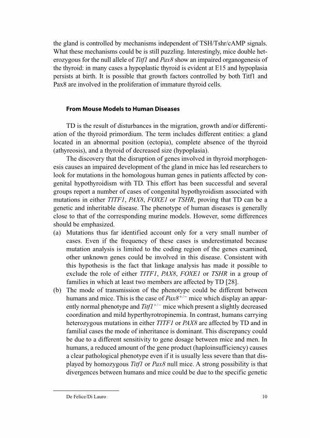
the gland is controlled by mechanisms independent of TSH/Tshr/cAMP signals.
What these mechanisms could be is still puzzling. Interestingly, mice double het-
erozygous for the null allele of Titf1 and Pax8 show an impaired organogenesis of
the thyroid: in many cases a hypoplastic thyroid is evident at E15 and hypoplasia
persists at birth. It is possible that growth factors controlled by both Titf1 and
Pax8 are involved in the proliferation of immature thyroid cells.
From Mouse Models to Human Diseases
TD is the result of disturbances in the migration, growth and/or differenti-
ation of the thyroid primordium. The term includes different entities: a gland
located in an abnormal position (ectopia), complete absence of the thyroid
(athyreosis), and a thyroid of decreased size (hypoplasia).
The discovery that the disruption of genes involved in thyroid morphogen-
esis causes an impaired development of the gland in mice has led researchers to
look for mutations in the homologous human genes in patients affected by con-
genital hypothyroidism with TD. This effort has been successful and several
groups report a number of cases of congenital hypothyroidism associated with
mutations in either TITF1, PAX8, FOXE1 or TSHR, proving that TD can be a
genetic and inheritable disease. The phenotype of human diseases is generally
close to that of the corresponding murine models. However, some differences
should be emphasized.
(a) Mutations thus far identified account only for a very small number of
cases. Even if the frequency of these cases is underestimated because
mutation analysis is limited to the coding region of the genes examined,
other unknown genes could be involved in this disease. Consistent with
this hypothesis is the fact that linkage analysis has made it possible to
exclude the role of either TITF1, PAX8, FOXE1 or TSHR in a group of
families in which at least two members are affected by TD [28].
(b) The mode of transmission of the phenotype could be different between
humans and mice. This is the case of Pax8�/� mice which display an appar-
ently normal phenotype and Titf1�/� mice which present a slightly decreased
coordination and mild hyperthyrotropinemia. In contrast, humans carrying
heterozygous mutations in either TITF1 or PAX8 are affected by TD and in
familial cases the mode of inheritance is dominant. This discrepancy could
be due to a different sensitivity to gene dosage between mice and men. In
humans, a reduced amount of the gene product (haploinsufficiency) causes
a clear pathological phenotype even if it is usually less severe than that dis-
played by homozygous Titf1 or Pax8 null mice. A strong possibility is that
divergences between humans and mice could be due to the specific genetic

Murine Models for the Study of Thyroid Gland Development 11
background of the strain used in generating the corresponding animal
models.
(c) Murine models have proved that Foxe1 is specifically involved in thyroid
bud migration. Although in humans an ectopic thyroid is the commonest
form of TD, FOXE1 mutations associated with an ectopic thyroid have not
yet been described: all subjects carrying loss-of-function mutations in this
gene show an absence of the thyroid. However, since only few patients
have been reported, it is premature to conclude that FOXE1 defects in
humans do not cause ectopia.
Interestingly, a recent study reports 3 subjects with an ectopic gland car-
rying heterozygous missense mutations in the NKX2-5 gene [29]. This fac-
tor could be involved, at least in part, in thyroid development; actually, in
Nkx2-5 null mouse embryos the thyroid bud appears smaller when com-
pared to that of wild-type embryos.
(d) In the familial cases of TD, patients do not display a clear mendelian trans-
mission. There is a higher incidence of subclinical thyroid developmental
abnormalities in those families with at least one case of TD. In addition,
patients carrying mutations in either TITF1 or PAX8 genes are affected by
syndromes characterized by incomplete penetrance and variability of the
phenotype even among the affected members of the same family. Taken
together, these observations strongly suggest that in humans also TD could
be a multigenic disease. This working hypothesis has received strong sup-
port from a novel multigenic model of TD. Mice double heterozygous for
the null allele of Titf1 and Pax8 gene display an overt thyroid phenotype
(hypoplasia or hemiagenesis of the gland and reduced synthesis of Tg)
provided the mutations are present on a specific genetic background,
C57BL/6. The same mutations in a different mouse strain (129/Sv) are
unable to cause any thyroid defects, indicating that other C57BL/6 specific
alleles, in addition to the null mutations in Titf1 and Pax8, are responsible
for the emergence of TD. This model establishes that TD can be caused by
multiple minor genetic defects [23]. The pool of genes that can be affected
is rather large. As table 2 shows, defects in several genes, mostly transcrip-
tion factors or growth factors, have been demonstrated to impair the devel-
opment of the thyroid in animal models. Minor defects in these genes or in
their targets could also be involved in TD in humans.
(e) Congenital hypothyroidism with TD occurs mostly as a sporadic disease.
Furthermore, TD has been found to be discordant in monozygotic twins [30].
These observations strongly suggest that this disease, even though genetic,
can be noninheritable. The frequency of these entities is difficult to assess.
Noninheritable mechanisms, such as somatic mutations or postzygotic epige-
netic events, could be involved in the pathogenesis of human TD [31].

De Felice/Di Lauro 12
References
1 Lazzaro D, Price M, De Felice M, Di Lauro R: The transcription factor TTF-1 is expressed at the
onset of thyroid and lung morphogenesis and in restricted regions of the foetal brain. Development
1991;113:1093–1104.
2 De Felice M, Di Lauro R: Thyroid development and its disorders: genetics and molecular mecha-
nisms. Endocr Rev 2004;25:722–746.
3 Thomas PQ, Brown A, Beddington R: Hex: a homeobox gene revealing peri-implantation asym-
metry in the mouse embryo and an early transient marker of endothelial cell precursors.
Development 1998;125:85–95.
4 Zannini MS, Francis-Lang H, Plachov D, Di Lauro R: Pax-8, a paired domain-containing protein,
binds to a sequence overlapping the recognition site of a homeodomain and activates transcription
from two thyroid-specific promoters. Mol Cell Biol 1992;12:4230–4241.
5 Grapin-Botton A, Melton M: Endoderm development: from patterning to organogenesis. Trends
Genet 2000;16:124–130.
6 Parlato R, Rosica A, Rodriguez-Mallon A, Affuso A, Postiglione MP, Arra C, Mansouri A, Kimura S,
Di Lauro R, De Felice M: An integrated regulatory network controlling survival and migration in
thyroid organogenesis. Dev Biol 2004;276:464–475.
7 Fagman H, Andersson L, Nilsson M: The developing mouse thyroid: embryonic vessel contacts
and parenchymal growth pattern during specification, budding, migration, and lobulation. Dev
Dyn 2006;235:444–455.
8 Trueba SS, Auge J, Mattei G, Etchevers H, Martinovic J, Czernichow P, Vekemans M, Polak M,
Attie-Bitach T: PAX8, TITF1 and FOXE1 gene expression patterns during human development:
new insights into human thyroid development and thyroid dysgenesis associated malformations.
J Clin Endocrinol Metab 2004;90:455–462.
9 Martinez Barbera JP, Clements M, Thomas P, Rodriguez T, Meloy D, Kioussis D, Beddington RS:
The homeobox gene Hex is required in definitive endodermal tissues for normal forebrain, liver
and thyroid formation. Development 2000;127:2433–2445.
Table 2. Currently available mouse models of TD
Gene symbol Chromosome Features of the gene product Thyroid phenotype of null mice embryos
Hhex 19 transcription factor athyreosis
Titf1 12 transcription factor athyreosis
Pax8 2 transcription factor athyreosis
Foxe1 4 transcription factor athyreosis or ectopia
Tshr 12 G protein-coupled receptor defects in functional differentiation
Fgfr2 7 tyrosine kinase receptor athyreosis
Fgf10 13 peptide growth factor athyreosis
Nkx2-5 17 transcription factor thyroid bud hypoplasia
Hoxa3 6 transcription factor hypoplasia; persistent ultimobranchial body
Eya 1 1 transcription factor hypoplasia; persistent ultimobranchial body
Edn-1 13 signaling peptide hypoplasia; absence of isthmus
Tbx1 16 transcription factor hypoplasia; impaired lobulation
Pax3 1 transcription factor hypoplasia
Shh 5 morphogen impaired lobulation
Hoxa5 6 transcription factor defects in functional differentiation

Murine Models for the Study of Thyroid Gland Development 13
10 Mansouri A, Chowdhury K, Gruss P: Follicular cells of the thyroid gland require Pax8 gene func-
tion. Nat Genet 1998;19:87–90.
11 Kimura S, Hara Y, Pineau T, Fernandez-Salguero P, Fox CH, Ward JM, Gonzalez FJ: The T/ebp
null mouse: thyroid-specific enhancer-binding protein is essential for the organogenesis of the
thyroid, lung, ventral forebrain, and pituitary. Genes Dev 1996;10:60–69.
12 Kusakabe T, Hoshi N, Kimura S: Origin of the ultimobranchial body cyst: T/ebp/Nkx2.1 expres-
sion is required for development and fusion of the ultimobranchial body to the thyroid. Dev Dyn
2006;235:1300–1309.
13 Pohlenz J, Dumitrescu A, Zundel D, Martine U, Schonberger W, Koo E, Weiss RE, Cohen RN,
Kimura S, Refetoff S: Partial deficiency of thyroid transcription factor 1 produces predominantly
neurological defects in humans and mice. J Clin Invest 2002;109:469–473.
14 Revest JM, Spencer-Dene B, Kerr K, De Moerlooze L, Rosewell I, Dickson C: Fibroblast growth
factor receptor 2-IIIb acts upstream of Shh and Fgf4 and is required for limb bud maintenance but
not for the induction of Fgf8, Fgf10, Msx1, or Bmp4. Dev Biol 2001;231:47–62.
15 Krude H, Schutz B, Biebermann H, von Moers A, Schnabel D, Neitzel H, Tonnies H, Weise D,
Lafferty A, Schwarz S, De Felice M, von Deimling A, van Landeghem F, Di Lauro R, Gruters A:
Choreoathetosis, hypothyroidism, and pulmonary alterations due to human NKX2-1 haploinsuffi-
ciency. J Clin Invest 2002;109:475–480.
16 Plachov D, Chowdhury K, Walther C, Simon D, Guenet JL, Gruss P: Pax8, a murine paired box gene
expressed in the developing excretory system and thyroid gland. Development 1990;110:643–651.
17 Macchia PE, Lapi P, Krude H, Pirro MT, Missero C, Chiovato L, Souabni A, Baserga M, Tassi V,
Pinchera A, Fenzi G, Gruters A, Busslinger M, Di Lauro R: PAX8 mutations associated with con-
genital hypothyroidism caused by thyroid dysgenesis. Nat Genet 1998;19:83–86.
18 Dathan N, Parlato R, Rosica A, De Felice M, Di Lauro R: Distribution of the titf2/foxe1 gene
product is consistent with an important role in the development of foregut endoderm, palate, and
hair. Dev Dyn 2002;224:450–456.
19 De Felice M, Ovitt C, Biffali E, Rodriguez-Mallon A, Arra C, Anastassiadis K, Macchia PE,
Mattei MG, Mariano A, Schoeler H, Macchia V, Di Lauro R: A mouse model for hereditary thy-
roid dysgenesis and cleft palate. Nat Genet 1998;19:395–398.
20 Fagman H, Grande M, Edsbagge J, Semb H, Nilsson M: Expression of classical cadherins in thyroid
development: maintenance of an epithelial phenotype throughout organogenesis. Endocrinology
2003;144:3618–3624.
21 Clifton-Bligh RJ, Wentworth JM, Heinz P, Crisp MS, John R, Lazarus JH, Ludgate M, Chatterjee V:
Mutation of the gene encoding human TTF-2 associated with thyroid agenesis, cleft palate and
choanal atresia. Nat Genet 1998;19:399–401.
22 Fagman H, Grande M, Gritli-Linde A, Nilsson M: Genetic deletion of sonic hedgehog causes hemi-
agenesis and ectopic development of the thyroid in mouse. Am J Pathol 2004;164:1865–1872.
23 Amendola E, De Luca P, Macchia PE, Terracciano D, Rosica A, Chiappetta G, Kimura S,
Mansouri A, Affuso A, Arra C, Macchia V, Di Lauro R, De Felice M: A mouse model demonstrates
a multigenic origin of congenital hypothyroidism. Endocrinology 2005;146:5038–5047.
24 Postiglione MP, Parlato R, Rodriguez-Mallon A, Rosica A, Mithbaokar P, Maresca M, Marians RC,
Davies TF, Zannini MS, De Felice M, Di Lauro R: Role of the thyroid-stimulating hormone recep-
tor signaling in development and differentiation of the thyroid gland. Proc Natl Acad Sci USA
2002;99:15462–15467.
25 Marians RC, Ng L, Blair HC, Unger P, Graves PN, Davies TF: Defining thyrotropin-dependent
and -independent steps of thyroid hormone synthesis by using thyrotropin receptor-null mice. Proc
Natl Acad Sci USA 2002;99:15776–15781.
26 Refetoff S: Resistance to thyrotropin. J Endocrinol Invest 2003;26:770–779.
27 Ledent C, Dumont JE, Vassart G, Parmentier M: Thyroid expression of an A2 adenosine receptor
transgene induces thyroid hyperplasia and hyperthyroidism. EMBO J 1992;11:537–542.
28 Castanet M, Sura Trueba S, Chauty A, Carré A, Heath S, Léger J, Lyonnet S, Czernichow P, Polak M:
Linkage and mutational analysis of familial thyroid dysgenesis suggest genetic heterogeneity. Eur
J Hum Genet 2005;13:232–239.
29 Dentice M, Cordeddu V, Rosica A, Ferrara AM, Santarpia L, Salvatore D, Chiovato L, Perri A,
Moschini L, Fazzini C, Olivieri A, Costa P, Stoppioni V, Baserga M, De Felice M, Sorcini M, Fenzi G,

De Felice/Di Lauro 14
Di Lauro R, Tartaglia M, Macchia PE: Missense mutation in the transcription factor NKX2-5: a
novel molecular event in the pathogenesis of thyroid dysgenesis. J Clin Endocrinol Metab 2006;91:
1428–1433.
30 Perry R, Heinrichs C, Bourdoux P, Khoury K, Szots F, Dussault JH, Vassart G, Van Vliet G:
Discordance of monozygotic twins for thyroid dysgenesis: implications for screening and for mol-
ecular pathophysiology. J Clin Endocrinol Metab 2002;87:4072–4077.
31 Vassart G, Dumont JE: Thyroid dysgenesis: multigenic or epigenetic, or both. Endocrinology
2005;146:5035–5037.
Prof. Roberto Di Lauro
IRGS, Biogem s.c.a r.l.
Via Camporeale
IT–83031 Ariano Irpino (AV) (Italy)
Tel. �39 0825 881630, Fax �39 0825 881637, E-Mail [email protected]

Van Vliet G, Polak M (eds): Thyroid Gland Development and Function.
Endocr Dev. Basel, Karger, 2007, vol 10, pp 15–28
Familial Forms of Thyroid Dysgenesis
Mireille Castaneta, Michel Polaka, Juliane Légerb
aPaediatric Endocrinology Unit and INSERM U845, Hôpital Necker-Enfants Malades
and bPaediatric Endocrinology Unit, Hôpital Robert Debré, Paris, France
AbstractIn many instances, the pathophysiology of thyroid dysgenesis (TD) remains as yet unclear
and until relatively recently the disorder was usually regarded as occurring in a sporadic manner.
However, over the past few years, a small but significant proportion of familial cases has been
identified (2%) through the study of subjects with congenital hypothyroidism and more recent
work has revealed an even higher proportion of familial TD in both symptomatic or asympto-
matic individuals (7.9%). Together, these studies strongly point to a significant genetic compo-
nent of this disorder. Moreover, detailed observations of members affected by different types of
TD in the same family suggest that TD could be an entity with a common underlying mecha-
nism for all the etiological groups. To date, molecular genetic studies have implicated four genes
in thyroid development and some mutations have been reported in affected subjects. Three of
these encode transcription factors while the forth encodes the thyrotropin hormone receptor.
However, their involvement in the general TD population remains questionable, as only a few
mutations have been reported so far and as linkage analysis has demonstrated the relevance of
other genes. Therefore, further work is required to fully understand the pathophysiology of TD.
Copyright © 2007 S. Karger AG, Basel
Introduction
Thyroid dysgenesis (TD) is the most frequent cause of congenital hypothy-
roidism (CH; 85% of cases) and results from abnormalities of thyroid gland
development including a spectrum of embryogenetic defects such as ectopic
thyroid gland, athyreosis and more rarely, thyroid hypoplasia and hemiagenesis
[1]. Other variations of thyroid anatomy such as cysts of the thyroglossal duct
and additional thyroid tissue have been occasionally described, usually in
asymptomatic patients with normal thyroid function [2].
The pathogenesis of TD is as yet not well-understood and until relatively
recently the disorder was typically regarded as arising in a sporadic manner.
Disorders of Thyroid Gland Development

Castanet/Polak/Léger 16
Over the last 50 years, reports of some familial cases of CH by either athyreosis
[3–5] or ectopic gland [6–8] suggested an inherited disease and two recent
familial studies have confirmed this hypothesis [1, 9, 10]. In this chapter, we
describe current knowledge regarding the familial forms of TD and explore the
molecular basis of this disorder.
Thyroid Development
In the human embryo the thyroid gland primordium first appears late in the
4th week in the midline of the floor of the primitive pharynx at a point that is
later known as the foramen cecum on the tongue. Thereafter, this initially round
cluster of cells begins its migration from the pharyngeal floor through the ante-
rior midline of the neck during which time the cells multiply. At 24–32 days,
this median anlage has already become a bilobed structure, but it only reaches
its final position at around 48–50 days. At the same time, connection of the
median anlage with the ultimobranchial body, developed from the endoderm of
the 4th pharyngeal pouch, occurs, resulting in the incorporation of the C cells
into the thyroid. At 51 days, the gland exhibits its definitive external form, with
an isthmus connecting the two lateral lobes and reaches its final position below
the thyroid cartilage by the 7th week of embryonic life. During its descent, the
developing thyroid gland retains an attachment to the pharynx by a narrow
epithelial stalk known as the thyroglossal duct [11]. By 37 days, this structure
that connects the median thyroid anlage with the point of origin of its migration
on the floor of the pharynx has generally disappeared [12] and normally the
only remnant of the thyroglossal duct is the foramen cecum itself (fig. 1).
Usually, the terminal differentiation of thyroid follicular cells [as evi-
denced by expression of the genes encoding the TSH receptor (TSHR), the Na/I
symporter (NIS), thyroglobulin (Tg) and thyroperoxidase (TPO)] and the for-
mation of follicles occur in the normal embryo only once migration is complete
[13].
Different Types of TD
The vast majority of CH patients with TD have a defect in thyroid migra-
tion which results in the presence of ectopic thyroid tissue (�65% of CH
patients). This arrest in the normal descent of part or all of the thyroid gland can
result in a lingual, suprahyoid or infrahyoid location. Most frequently, thyroid
tissue is found at the base of the tongue. Note that double ectopic thyroid tissues
have also been exceptionally reported [14]. Although usually, ectopically sited

Familial Forms of Thyroid Dysgenesis 17
thyroid tissues manifest with CH, a proportion of cases are found incidentally
in asymptomatic subjects raising the possibility that many are never diagnosed
[15]. For example, it has been shown in 200 consecutive necropsies on euthy-
roid Caucasian individuals that 10% of this population exhibited ectopic lingual
thyroid tissue, in addition to a normal thyroid; the size of this ectopic tissue var-
ied from a few acini to a nodule of 1 cm in diameter with males and females
equally affected [16]. The fact that some subjects appear to remain asympto-
matic suggests that the ectopically sited thyroid tissues could retain normal
function. This hypothesis is supported by the finding of uptake of radioactive
iodine on scintiscan and of typical colloid-filled follicles under microscopic
examination following surgical removal [17]. Moreover, although the thyroxine-
producing capacity of patients with ectopically sited thyroid tissue is generally
limited, it appears to remain constant, suggesting normal postnatal survival of
ectopic cells [18].
The second most common variant of TD (�30%) is the absence of
detectable thyroid follicular cells (commonly named athyreosis, although this
designation is not entirely correct since these patients, like those with ectopic
4th week
Foramencecum
Thyroid gland embryology
Primitivepharynx
Foramencecum
Foramencecum
Hyoid bone Hyoid bone
Trachea
Esophagus
Tongue
Thyroglossal ductbreaks down
Trachea
Esophagus
Respiratorydiverticulum
Thyroiddiverticulum
Thyroid cartilage
Thyroidgland
Thyroidgland
Late 5th week
Early 5th week 7th week
Fig. 1. Thyroid development [11].

Castanet/Polak/Léger 18
thyroids, have functional C cells). Whether thyroid follicular cells disappear
through apoptosis after initial differentiation [as demonstrated in knockout
mice in the chapter by De Felice and Di Lauro, pp. 1–14] or fail to differentiate
initially is unknown. Such developmental failure can also affect only one lobe
of the gland resulting in hemiagenesis; more rarely it involves the majority, but
not all, of the follicular cells resulting in very hypoplastic glands which are nor-
mally situated. Note that these small glands are sometimes so hypofunctional
that they may be undetectable by nuclear medicine studies: in these cases of
apparent athyreosis, careful ultrasonographic evaluation of the neck may reveal
a very small thyroid gland of normal shape and in the orthotopic position. This
failure to detect any thyroid tissue by scintiscan usually occurs in patients with
severe thyrotropin resistance due to complete loss of TSHR function [19, 20].
Therefore, a certain degree of misclassification might exist and to make an
accurate diagnosis of athyreosis, it is important to combine a good thyroid
scintigraphy with high definition ultrasonography. In addition, undetectable
serum thyroglobulin levels in hypothyroid subjects could provide an additional
clue in athyreosis cases [21].
Developmental thyroid abnormalities may also include persistence of the
thyroglossal duct giving rise to ‘thyroglossal duct cysts’ which become gener-
ally clinically apparent during infancy or childhood. Alternatively, cell residues
may remain giving rise to a pyramidal lobe at the distal portion of the duct
which remains attached to the thyroid gland, usually to the left lobe [12, 22].
Cysts within the empty thyroid area in CH patients with TD have also been
found, suggesting possible cystic degeneration of clusters of thyroid follicular
cells that have completed their normal migration, even when the thyroid gland
has otherwise remained incompletely descended or has entirely disappeared
[14]. Additionally, other adjacent developmental abnormalities have been reported,
for example additional thymic tissue within the empty thyroid bed in patients
with either ectopic thyroid tissue or athyreosis [23].
Note that the 2 major forms of TD, i.e. ectopic thyroid gland and athyreo-
sis, are often associated with CH, whilst other variations as outlined above are
usually found in asymptomatic subjects with normal thyroid function, hence
making it difficult to establish their true incidence. Thyroid hemiagenesis and
the presence of a pyramidal lobe are indeed usually incidentally discovered in
patients with other thyroid disorders when thyroid ultrasound or scintigraphy
is performed [22, 24]. So far, only one study has reported a prevalence for thy-
roid hemiagenesis of 0.2% derived from a systematic ultrasound investigation
of normal children [25]. Thyroglossal duct cysts are usually diagnosed through
an asymptomatic neck mass, acute infection, chronic inflammation or hemor-
rhage and account for approximately 70% of congenital neck abnormalities
[26].
Familial Forms of Thyroid Dysgenesis 19
Familial Forms of CH due to TD
A thorough survey of the literature covering the past 40 years has revealed
that almost 20 families with multiple affected individuals with CH due to TD
have been described in different countries, suggesting that TD could be a famil-
ial disease in some instances [3–8]. Therefore, over the past few years, a French
national survey was performed to corroborate this hypothesis. In this survey
covering more than 2,500 CH patients with TD diagnosed in France between
1980 and 1998, 67 patients with a positive family history of CH from TD
belonging to 32 multiplex families (i.e. with at least two affected members)
were referred. These data made it possible to determine a prevalence of familial
cases of 2%. Additionally, statistical analysis revealed that, among first-degree
relatives, the number of familial cases of CH with TD was significantly higher
(by �15-fold) than expected by chance alone, indicating that TD revealed by
CH had a significant familial component [1, 9].
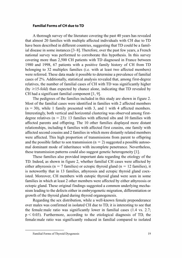
The pedigrees of the families included in this study are shown in figure 2.
Most of the familial cases were identified in families with 2 affected members
(n � 30), while 1 family presented with 3, and 1 with 4 affected members.
Interestingly, both vertical and horizontal clustering was observed among first-
degree relatives (n � 23): 13 families with affected sibs and 10 families with
affected parents and offspring. The 10 other families displayed more distant
relationships, including 6 families with affected first cousins, one family with
affected second cousins and 2 families in which more distantly related members
were affected. This high proportion of transmissions from parent to offspring
and the possible father to son transmission (n � 2) suggested a possible autoso-
mal dominant mode of inheritance with incomplete penetrance. Nevertheless,
these transmission patterns could also suggest genetic heterogeneity [1].
These families also provided important data regarding the etiology of the
TD. Indeed, as shown in figure 2, whether familial CH cases were affected by
either athyreosis (n � 7 families) or ectopic thyroid gland (n � 12 families), it
is noteworthy that in 13 families, athyreosis and ectopic thyroid gland coex-
isted. Moreover, CH members with eutopic thyroid gland were seen in some
families in which at least 2 other members were affected by either athyreosis or
ectopic gland. These original findings suggested a common underlying mecha-
nism leading to the defects either in embryogenetic migration, differentiation or
growth of the thyroid gland during thyroid organogenesis.
Regarding the sex distribution, while a well-known female preponderance
over males was confirmed in isolated CH due to TD, it is interesting to see that
the female:male ratio was significantly lower in familial cases (1.4 vs. 2.7;
p � 0.03). Furthermore, according to the etiological diagnosis of TD, the
female:male ratio was significantly reduced in familial compared to isolated
Castanet/Polak/Léger 20
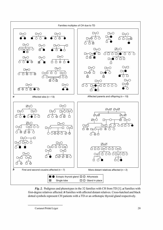
Fig. 2. Pedigrees and phenotypes in the 32 families with CH from TD [1]. a Families with
first-degree relatives affected. b Families with affected distant relatives. Cross-hatched and black
dotted symbols represent CH patients with a TD or an orthotopic thyroid gland respectively.
Families multiplex of CH due to TD
Affected parents and offspring (n�10)Affected sibs (n�13)
More distant relatives affected (n�2)First and second cousins affected (n�7)
Ectopic thyroid gland
Single lobe
Athyreosis
Gland in place
a
b

Familial Forms of Thyroid Dysgenesis 21
cases in CH with athyreosis (0.9 vs. 2.7; p � 0.02), whereas only a slightly
lower proportion (nonsignificant) of females was observed in cases of ectopic
thyroid gland (1.9 vs. 2.7). These data could suggest the possible involvement
of sex-modified etiological factors in familial cases, particularly in athyreosis.
In conclusion, this family study demonstrated for the first time that TD
revealed by CH had a significant familial component, with a potentially com-
mon underlying mechanism at least in athyreosis and ectopic thyroid gland.
Although common unidentified environmental factors cannot be totally ruled
out, this report strongly suggested the existence of genetic factors contributing
to the risk of TD that might be controlled by sex. Nevertheless, marked clinical
variability both within and between families could reflect genetic heterogeneity
and further genetic studies are required to better elucidate the physiopathology
of thyroid defects.
Familial Forms of TD in First-Degree Relatives of Children with CH
On the basis that some thyroid tract abnormalities might be totally asymp-
tomatic and that familial cases have been reported with either major forms
(i.e. with CH) [1] or TDA (asymptomatic forms of TD) [27] or both in affected
members of the same family [6, 28], a further study was performed to deter-
mine whether the prevalence of familial cases of TD might be higher than pre-
viously reported. Systematic screening by neck ultrasound and measurement of
serum TSH and FT4 concentrations were performed in all first-degree relatives
of 84 CH children with TD. Moreover, when ectopic or additional thyroid tissue
was found by ultrasound, radioiodine thyroid scanning (radioactive iodide 123I)
was performed to identify functional thyroid tissue. Among the 241 screened
first-degree relatives of the 84 studied patients, 19 relatives (7.9% of cases)
belonging to 18 families were found with asymptomatic TDA, i.e. without any
clinical complaints and serum-free thyroid hormone and TSH levels in the nor-
mal range. This proportion of affected individuals in the nuclear families of CH
patients with TD was significantly higher than that seen in the control popula-
tion (7.9 vs. 0.9%; p � 0.001), and pointed to a familial disorder [10]. As
shown in figure 3, the 19 subjects affected by TDA carried a total of 21 detected
anomalies as 2 subjects exhibited 2 different disorders, respectively, hemiagen-
esis and thyroglossal duct cyst (n � 1) and additional thyroid tissue and thy-
roglossal duct cyst (n � 1). Thyroglossal duct cyst was the main TDA found in
14 subjects (7 males, 7 females) who were the sibs (n � 6) or the parents
(n � 8) of 13 CH children with either ectopic thyroid tissue (n � 5), athyreosis
(n � 7) or hemiagenesis (n � 1). In all of these 14 patients, the thyroid gland

Castanet/Polak/Léger 22
Fig. 3. Pedigrees of the 18 families with TD, both with CH and asymptomatic cases [54].
a Families with affected sibs (n � 8). b Families with affected parents and offsprings (n � 10).
was normally located. Additional thyroid tissue with the presence of a pyrami-
dal lobe along the left lobe of the normally located thyroid gland was found in
3 mothers of CH children with ectopic thyroid tissue. Thyroid hemiagenesis with
the presence of a single well-located lobe (left: n � 2, right: n � 1) was found
in 3 sibs (2 males, 1 females) of 3 CH children with ectopic thyroid tissue.
Finally, surprisingly, cervical ectopic thyroid gland was found in a healthy clin-
ically and biologically euthyroid sister of 1 CH child with athyreosis without
any evidence of thyroid tissue in the normal location determined by ultrasonog-
raphy. This observation is in accordance with a substantial function of ectopic
thyroid tissue.
Regarding the sex ratio, as for the familial cases of athyreosis, an equal
proportion of boys and girls were found in TDA again suggesting possible
involvement of a sex-modified gene in thyroid development. Note that these
data were in accordance with the equal sex ratio reported in a larger series of
symptomatic thyroglossal duct cysts [29].
Regarding the pedigrees reported in this study, a similar proportion of
affected parents (6.5%) and sibs (10.5%) with asymptomatic TDA was found,
which was again in favor of a dominant mode of inheritance. Moreover, this
unique sample of families including members affected with either asympto-
matic or major forms of TD made a segregation analysis possible, demonstrating
Affected sibs (n�8)
Affected parents and offsprings (n�10)
E*
A
A EH
A A
*
E
Congenital hypothyroidismwith thyroid dysgenesisA = AthyreosisE = Ectopic thyroid tissueH = HemiagenesisThyroglossal duct cystPyramidal lobeThyroid hemiagenesisEctopic thyroid tissue
E EA
A E A
EE A
E
*
*
a b

Familial Forms of Thyroid Dysgenesis 23
a low penetrance of the disease at 21% and a prediction of the occurrence risk
after an isolated case of 10.5% for TDA.
This second familial study demonstrated that among first-degree relatives
of a CH population with TD, there is an elevated rate of asymptomatic thyroid
developmental anomalies. The estimates of prevalence of families with both
minor and major forms of TD (21.4% of the investigated families) were much
higher than the proportion of families with only affected members with CH due
to TD. Moreover, it should be pointed out that this high proportion of familial
occurrence may be underestimated as only first-degree relatives were evaluated
by systematic screening in this study excluding more distant relatives which
cannot be examined extensively.
In conclusion, these two recent familial studies have revealed a relatively
high rate of familial TD including asymptomatic individuals (TDA). Although a
role of humoral or environmental factors in the thyroid development cannot be
totally excluded, these data strongly suggest a genetic component to the disor-
der. Moreover, the pedigrees in these 2 studies give support to the concept of a
common origin for embryogenesis, migration, differentiation or growth of the
thyroid gland during organogenesis. Taken together, these new findings of
familial TD cases should prompt us to perform systematic screening for thyroid
defects in relatives of patients affected by TD with or without CH.
Molecular Mechanisms of Thyroid Developmental Abnormalities
Studies in knockout mice have demonstrated a critical role for several
genes in the early events of thyroid organogenesis. To date, four genes have
been involved; three of them encode transcription factors [30] while the other
encodes the TSHR. Although the three highly conserved transcription factors
(�90% homology between mice and humans) are not thyroid-specific, it is
noteworthy that simultaneous expression of these three factors is unique to the
thyroid follicular cells from the beginning of their differentiation and is main-
tained throughout thyroid development and into adulthood [31]. In mice, Ttf-1
inactivation leads to absence of thyroid tissue associated with severe defects in
the lung and the forebrain, indicating a critical role of this factor in early events
of organogenesis [32]. Ttf-2 inactivation has revealed that this factor is required
for the downward migration of the thyroid gland as well as for palate closure,
with knockout mice showing either athyreosis or ectopic gland associated with
cleft palate. Note that this observation supported the previous hypothesis from
familial studies that ectopic thyroid gland and athyreosis could have a common
underlying mechanism [33]. Pax8 knockout mice demonstrated severe thyroid

Castanet/Polak/Léger 24
hypoplasia with complete absence of follicular structures [34] whilst defects in
TSH secretion and action are associated with a small orthotopic thyroid gland,
indicating that the action of TSH through its receptor is not required for migra-
tion but is essential for the proliferation and for the maintenance of the differ-
entiated function of the thyroid follicular cells [35, 36].
That these genetic factors may be involved in the pathogenesis of TD in
humans is supported by recent reports which have identified germline mutations
of these 4 candidate genes in about 20 patients with TD. Homozygous TTF-2 (or
FOXE1) mutations are reported in 2 familial cases of athyreosis associated with
cleft palate and choanal atresia in 1 case. This association, known as Bamforth
syndrome, is consistent with the spatio-temporal expression of TTF-2 that has
been detected in the thyroid gland and in the oropharyngeal epithelium during
development [37–39]. Heterozygous TTF-1 (or NKX2.1) mutations produce a
predominantly neurological phenotype (choreoathetosis) with possible pul-
monary lesions that is associated with generally mild thyroid dysfunction with a
normal or hypoplastic thyroid gland [40–42]. Heterozygous mutations of PAX8
have been identified in some families with an isolated thyroid phenotype (con-
sisting of an orthotopic thyroid hypoplasia) that may be associated with cystic
lesions and kidney malformations, e.g. unilateral renal agenesis and a left-sided
ureteropelvic obstruction [43, 44]. All of these associations are in keeping with
experimental evidence that the proteins are expressed in several other developing
organs, lung and ventral forebrain for Ttf1 [32] and kidney for Pax8 [39, 45].
Inactivating mutations of the TSHR gene, either homozygous or compound
heterozygous, have been observed in a few cases of thyroid hypoplasia [19, 20,
46, 47]. Therefore, taken together, these data argue in favor of a significant
genetic contribution in TD, implicating at least these 4 candidate genes.
Nevertheless, further genetic studies are needed to establish phenotype-genotype
correlations and to permit genetic counseling for this type of disorder.
However, despite intensive research programs throughout the world, abnor-
malities in these 4 genes have been found in only a small proportion of TD
cases, most of them with syndromic phenotypes, suggesting that none of them
is a major genetic factor in this disorder and that other genes may be involved.
A linkage analysis performed in 19 TD multiplex families supported this view
showing the exclusion of the 4 candidate genes in 5 families that demonstrated
for the first time the relevance of other genes [30]. On the basis of function and
spatiotemporal expression, several other genes may be involved in thyroid
development. For example, the Nkx2.5 gene (or CSX gene in humans), the
HOXB3 or HOXA3 genes, the divergent homeobox gene HEX, the hepatocyte
nuclear factor HNF3 gene, the GATA6 gene or the eyes absent gene (EYA1)
could be relevant candidate genes since they are expressed early during
embryogenesis of the thyroid gland and impairment of their function may be

Familial Forms of Thyroid Dysgenesis 25
responsible for TD. In addition, some of these genes may interact with the pre-
viously described candidate genes. However, none of these genes is entirely
thyroid specific and their inactivation in mice usually leads to extrathyroidal
malformations [48–51] that are not usually found in the TD patients. However,
reports of a significantly higher incidence of extrathyroid congenital abnormal-
ities in CH patients than in the general population (respectively, 9 vs. 2.5%)
could suggest the implication of genes interacting in the development of several
organs [1]. Therefore, careful studies of the thyroid and extrathyroid phenotype
in humans and in mice must be performed to identify further relevant candidate
genes. Recently, as a higher prevalence of congenital heart disease has been
documented in children with CH than in the general population, Dentice et al.
[52] investigated the Nkx2.5 gene and found 4 heterozygous mutations in
TD patients, suggesting the relevance of this gene to thyroid development.
Furthermore, the identification of additional candidate genes controlling early
events in thyroid organogenesis in humans and acting upstream of NKX2.1,
FOXE1 or PAX8 would be very helpful. Indeed, it is noteworthy that the initial
differentiation of thyroid follicular cells on the floor of the pharynx is normal in
mice with homozygous deletion of these three transcription factors. In addition,
performing the cloning of tissue-specific genes and/or a genomewide screening
in a significant number of familial cases of TD could be interesting strategies.
In conclusion, recent data have revealed for the first time the familial char-
acter of TD, strongly suggesting a genetically determined disorder. Moreover,
many arguments are in favor of a possible polygenic basis for thyroid defects with
the involvement of at least 4 genes. However, some of genes involved remain elu-
sive. Additionally, the role of environmental effects and/or the implication of a
noninheritable postzygotic event cannot currently be excluded as illustrated by
discordance for CH from TD in monozygotic twins [53]. Therefore, accumulating
evidence supports the view that the genetics of TD are complex, possibly with a
polygenic/multifactorial basis, and genetic susceptibility to TD could lack a sim-
ple mendelian pattern of inheritance. Accordingly, further studies are needed to
enhance our understanding of the pathophysiology of TD.
Acknowledgment
The authors would like to thank Dr. C. Garel (Hôpital Robert Debré, Paris) for re-reading
thyroid ultrasound scans in the familial studies and all the families and the pediatricians taking
part in these studies.
This work was supported by grants from Novo Nordisk and Evian, and from the
Fondation pour la Recherche Médicale. M.C. was awarded the Young Investigator Award at
the ESPE (European Society for Paediatric Endocrinology) Meeting 2006 in Rotterdam, The
Netherlands.
Castanet/Polak/Léger 26
References
1 Castanet M, Polak M, Bonaïti-Pellié C, Lyonnet S, Czernichow P, Léger J: Nineteen years of
national screening for congenital hypothyroidism: familial cases with thyroid dysgenesis suggest
the involvement of genetic factors. J Clin Endocrinol Metab 2001;86:2009.
2 Kaplan EL, Shukla M, Hara H, Ito K: Developmental abnormalities of the thyroid; in De Groot LG
(ed): Endocrinology. Philadelphia, Saunders, 1994, pp 893–899.
3 Ainger LE, Kelley VC: Familial athyreotic cretinism: report of 3 cases. J Clin Endocrinol Metab
1955;15:469.
4 Bamforth JS, Hugues IA, Lazarus JH, Weaver CM, Harper PS: Congenital hypothyroidism, spiky
hair, and cleft palate. J Med Genet 1989;26:49.
5 Stäger J, Froesch ER: Congenital familial thyroid aplasia. Acta Endocrinol (Copenh) 1981;96:188.
6 Orti E, Castells S, Qazi Q, Inamdar S: Familial thyroid disease: lingual thyroid in two siblings and
hypoplasia of a thyroid lobe in a third. J Pediatr 1971;78:675.
7 Kaplan M, Kauli R, Raviv U, Lubin E, Laron Z: Hypothyroidism due to ectopy in siblings. Am
J Dis Child 1977;131:1264.
8 Defoer FY, Malher C: Lingual thyroid in two natural brothers. J Clin Invest 1990;13:65.
9 Castanet M, Lyonnet S, Bonaïti-Pellié C, Polak M, Czernichow P, Léger J: Familial forms of thy-
roid dysgenesis among infants with congenital hypothyroidism. N Engl J Med 2000;343:441.
10 Leger J, Marinovic D, Garel C, Polak M, Czernichow P: High prevalence of thyroid developmental
anomalies in first degree relatives of children with congenital hypothyroidism. J Clin Endocrinol
Metab 2002;87:575–580.
11 O’Rahilly R: The timing and sequence of events in the development of the human endocrine
system during the embryonic period proper. Anat Embryol (Berl) 1983;166:439.
12 Sprinzl GM, Koebke J, Wimmers-Klick J, Eckel HE, Thumfart WF: Morphology of the human
thyroglossal tract: a histologic and macroscopic study in infants and children. Ann Otol Rhinol
Laryngol 2000;109:1135.
13 Macchia PE: Recent advances in understanding the molecular basis of primary congenital
hypothyroidism. Mol Med Today 2000;6:36.
14 Marinovic D, Garel C, Czernichow P, Léger J: Additional phenotypic abnormalities with presence
of cysts within the empty area in patients with congenital hypothyroidism with thyroid dysgenesis.
J Clin Endocrinol Metab 2003;88:1212–1216.
15 Gillis D, Brnjac L, Perlman K, Sochett EB, Daneman D: Frequency and characteristics of lingual
thyroid not detected by screening. J Pediatr Endocrinol Metab 1998;11:229.
16 Sauk JJ Jr: Ectopic lingual thyroid. J Pathol 1970;102:239.
17 Gallo A, Leonetti F, Torri E, Manciocco V, Simonelli M, DeVincentiis M: Ectopic lingual thyroid
as unusual cause of severe dysphagia. Dysphagia 2001;16:220.
18 Grant DB, Hulse JA, Jackson DB, Leung SP, Ng WK: Ectopic thyroid: residual function after
withdrawal of treatment in infancy and later childhood. Acta Paediatr Scand 1989;78:889.
19 Abramowicz MJ, Duprez L, Parma J, Vassart G, Heinrichs C: Familial congenital hypothyroidism
due to inactivating mutation of the thyrotropin receptor causing profound hypoplasia of the thyroid
gland. J Clin Invest 1997;99:3018.
20 Gagne N, Parma J, Deal C, Vassart G, Van Vliet G: Apparent congenital athyreosis contrasting
with normal plasma thyroglobulin levels and associated with inactivating mutations in the thy-
rotropin receptor gene: are athyreosis and ectopic thyroid distinct entities? J Clin Endocrinol
Metab 1998;83:1771.
21 Czernichow P, Schlumberger M, Pomarede R, Fragu P: Plasma thyroglobulin measurements help
determine the type of thyroid defect in congenital hypothyroidism. J Clin Endocrinol Metab
1983;56:242.
22 Siraj QH, Aleem N, Inam-Ur-Rehman A, Qaisar S, Ahmad M: The pyramidal lobe: a scintigraphic
assessment. Nucl Med Commun 1989;10:685.
23 Bubuteishvili L, Garel C, Czernichow P, Leger J: Thyroid abnormalities by ultrasonography in
neonates with congenital hypothyroidism. J Pediatr 2003;143:759.

Familial Forms of Thyroid Dysgenesis 27
24 McHenry CR, Walfish PG, Rosen IB, Lawrence AM, Paloyan E: Congenital thyroid hemiagene-
sis. Am Surg 1995;61:634.
25 Shabana W, Delange F, Freson M, Osteaux M, de Schepper J: Prevalence of thyroid hemiagenesis
ultrasound screening in normal children. Eur J Pediatr 2000;159:456.
26 Rapidis AD, Economidis J, Goumas PD, Langdon JD, Skordalakis A, Tzortzatou F, Anagnostopoulos D,
Matsaniotis N: Tumours of the head and neck in children. A clinico-pathological analysis of 1,007
cases. J Craniomaxillofac Surg 1988;16:279.
27 Issa MM, deVries P: Familial occurrence of thyroglossal duct cyst. J Pediatr Surg 1991;26:30.
28 Rosenberg T, Gilboa Y: Familial thyroid ectopy and hemiagenesis. Arch Dis Child 1980;55:639.
29 Solomon JR, Rangecroft L: Thyroglossal-duct lesions in childhood. J Pediatr Surg 1984;19:555.
30 Castanet M, Sura-Trueba S, Chauty A, Carre A, de Roux N, Heath S, Leger J, Lyonnet S,
Czernichow P, Polak M: Linkage and mutational analysis of familial thyroid dysgenesis demon-
strate genetic heterogeneity implicating novel genes. Eur J Hum Genet 2005;13:232.
31 Damante G, Tell G, Di Lauro R: A unique combination of transcription factors controls differenti-
ation of thyroid cells. Prog Nucleic Acid Res Mol Biol 2001;66:307.
32 Kimura S, Hara Y, Pineau T, Fernandez-Salguero P, Fox CH, Ward JM, Gonzalez FJ: The T/ebp
null mouse: thyroid-specific enhancer-binding protein is essential for the organogenesis of the
thyroid, lung, and ventral forebrain. Genes Dev 1996;10:60.
33 De Felice M, Ovitt C, Biffali E, Rodriguez-Mallon A, Arra C, Anastassiadis K, Macchia PE,
Mattei MG, Mariano A, Schöler H, Macchia V, Di Lauro R: A mouse model for hereditary thyroid
dysgenesis and cleft palate. Nat Genet 1998;19:399.
34 Mansouri A, Chowdhury K, Gruss P: Follicular cells of the thyroid gland require Pax8 gene func-
tion. Nat Genet 1998;19:87.
35 Postiglione MP, Parlato R, Rodriguez-Mallon A, Rosica A, Mithbaokar P, Maresca M, Marians
RC, Davies TF, Zannini MS, De Felice M, Di Lauro R: Role of the thyroid-stimulating hormone
receptor signaling in development and differentiation of the thyroid gland. Proc Natl Acad Sci
USA 2002;99:15462.
36 Beamer WJ, Eicher EM, Maltais LJ, Southard JL: Inherited primary hypothyroidism in mice.
Science 1981;212:61.
37 Clifton-Bligh RJ, Wentworth JM, Heinz P, Crisp MS, John R, Lazarus JH, Ludgate M, Chatterjee
K: Mutation of the gene encoding human TTF-2 associated with thyroid agenesis, cleft palate and
choanal atresia. Nat Genet 1998;19:399.
38 Castanet M, Park SM, Smith A, Bost M, Léger J, Lyonnet S, Pelet A, Czernichow P, Chatterjee K,
Polak M: A novel loss-of-function mutation in TTF-2 is associated with congenital hypothy-
roidism, thyroid agenesis, and cleft palate. Hum Mol Genet 2002;11:2051.
39 Trueba SS, Auge J, Mattei G, Etchevers H, Martinovic J, Czernichow P, Vekemans M, Polak M,
Attie-Bitach T: PAX8, TITF1, and FOXE1 gene expression patterns during human development:
new insights into human thyroid development and thyroid dysgenesis-associated malformations.
J Clin Endocrinol Metab 2005;90:455.
40 Krude H, Schuetz B, Biebermann H, von Moers A, Schnabel D, Neitzel H, Tönnies H, Weise D,
Lafferty A, Schwarz S, DeFelice M, von Deimling A, van Landeghem F, DiLauro R, Grüters A:
Choreoathetosis, hypothyroidism, and pulmonary alterations due to human NKX2.1 haploinsuffi-
ciency. J Clin Invest 2002;109:475.
41 Pohlenz J, Dumitrescu A, Zundel D, Martiné U, Schönberger W, Koo E, Weiss RE, Cohen RN,
Kimura S, Refetoff S: Partial deficiency of thyroid transcription factor 1 produces predominantly
neurological defects in humans and mice. J Clin Invest 2002;109:469.
42 Simon-Carré A, Szinnai G, Castanet M, Barat P, Delrue MA, Moutard ML, Raynaud C, Romana
S, Sarda P, Ythier H, Léger J, Polak M: Clinical and molecular aspects of five TITF1 gene anom-
alies (abstract). Horm Res 2006;65(suppl 4):CF2-101.
43 Macchia PE, Lapi P, Krude H, Pirro MT, Missero C, Chiovato L, Souabni A, Baserga M, Tassi V,
Pinchera A, Fenzi G, Grüters A, Busslinger M, Di Lauro R: Pax8 mutations associated with con-
genital hypothyroidism caused by thyroid dysgenesis. Nat Genet 1998;19:83.
44 Meeus L, Gilbert B, Rydlewski C, Parma J, Roussie AL, Abramowicz M, Vilain C, Christophe D,
Costagliola S, Vassart G: Characterization of a novel loss of function mutation of PAX8 in a familial

Castanet/Polak/Léger 28
case of congenital hypothyroidism with in-place, normal-sized thyroid. J Clin Endocrinol Metab
2004;89:4285.
45 Plachov D, Chowdhury K, Walther C, Simon D, Guenet JL, Gruss P: Pax8, a murine paired box
gene expressed in the developing excretory system and thyroid gland. Development 1990;110:643.
46 Bretones P, Duprez L, Parma J, David M, Vassart G, Rodien P: A familial case of congenital
hypothyroidism caused by a homozygous mutation of the thyrotropin receptor gene. Thyroid
2002;11:977.
47 Park SM, Clifton-Bligh RJ, Betts P, Chatterjee VK: Congenital hypothyroidism and apparent
athyreosis with compound heterozygosity or compensated hypothyroidism with probable hemizy-
gosity for inactivating mutations of the TSH receptor. Clin Endocrinol (Oxf) 2004;60:220.
48 Lints TJ, Parsons LM, Hartley L, Lyons I, Harvey RP: Nkx2.5: A novel murine homeobox gene
expressed in early heart progenitor cells and their myogenic descendants. Development 1993;
119:419.
49 Martinez Barbera JP, Clements M, Thomas P, Rodriguez T, Meloy D, Kioussis D, Beddington
RSP: The homeobox gene Hex is required in definitive endodermal tissues for normal forebrain,
liver and thyroid formation. Development 2000;127:2433.
50 Xu PX, Zheng W, Laclef C, Maire P, Maas RL, Peters H, Xu X: Eya1 is required for the morpho-
genesis of mammalian thymus, parathyroid and thyroid. Development 2002;129:3033.
51 Ang SL, Rossant J: HNF3b is essential for node and notochord formation in mouse development.
Cell 1994;78:561.
52 Dentice M, Cordeddu V, Rosica A, Ferrara AM, Santarpia L, Salvatore D, Chiovato L, Perri A,
Moschini L, Fazzini C, Olivieri A, Costa P, Stoppioni V, Baserga M, De Felice M, Sorcini M, Fenzi
G, Di Lauro R, Tartaglia M, Macchia PE: Missense mutation in the transcription factor NKX2–5:
a novel molecular event in the pathogenesis of thyroid dysgenesis. J Clin Endocrinol Metab
2006;91:1428.
53 Perry R, Heinrichs C, Bourdoux P, Khoury K, Szotz F, Dussault JH, Vassart G, Van Vliet G:
Discordance of monozygotic twins for thyroid dysgenesis: implications for screening and for
molecular pathophysiology. J Clin Endocrinol Metab 2002;87:4072.
Juliane Léger, MD
Paediatric Endocrinology Unit, Hôpital Robert Debré
48 boulevard Sérurier
FR–75019 Paris (France)
Tel. �33 1 4003 2354, Fax �33 1 4040 2429, E-Mail [email protected]

Van Vliet G, Polak M (eds): Thyroid Gland Development and Function.
Endocr Dev. Basel, Karger, 2007, vol 10, pp 29–42
Possible Non-Mendelian Mechanisms of Thyroid Dysgenesis
Johnny Deladoëya, Gilbert Vassartb, Guy Van Vlieta
aEndocrinology Service and Research Center, Department of Pediatrics, Sainte-Justine
Hospital, University of Montreal, Montreal, Que., Canada; bGenetics Service, Erasme
Hospital and Institute of Interdisciplinary Research (IRIBHM), Free University of
Brussels (U.L.B.), Brussels, Belgium
AbstractMost research on the molecular mechanisms of thyroid dysgenesis over the past decade
has focussed on the Mendelian mechanisms that may account for the few (�5%) cases in
which there is an affected relative. This chapter first reviews methodological issues in the
imaging techniques used to classify thyroid dysgenesis into its various forms (ectopic thy-
roid, agenesis, orthotopic hypoplasia and hemiagenesis). It then reviews the evidence that
non-Mendelian mechanisms must be involved in the vast majority of cases of this disease, for
which the percentage of sporadic cases and of discordance between monozygotic twins
exceeds 95%. Among the mechanisms reviewed are early somatic mutations and epigenetic
changes in genes involved in thyroid development such as the thyroid transcription factors
TTF-1, TTF-2 and PAX-8. The possible role of extrathyroid genes involved in the control of
migration of the median thyroid bud during embryogenesis, such as adhesion molecules, and
of vascular factors involved in the stabilization of the bilobed structure of the thyroid is also
discussed.
Copyright © 2007 S. Karger AG, Basel
Introduction
While considerable progress has been made over the past decade in our
understanding of single gene disorders affecting the thyroid axis, the molecular
mechanisms underlying defects in thyroid development represent one of the
remaining enigmas in the pathophysiology of thyroid diseases [1]. Yet, these
defects collectively account for about 80–85% of cases of congenital hypothy-
roidism, which affects 1 in 3,000 newborns [2].
Disorders of Thyroid Gland Development

Deladoëy/Vassart/Van Vliet 30
There are several arguments against a simple Mendelian basis for congenital
hypothyroidism from thyroid dysgenesis (CHTD): (1) it affects females more
often than males, and this female preponderance is especially pronounced for
defects in thyroid migration (resulting in thyroid ectopy), which affects 3
females for 1 male [3]; this sex ratio is not compatible with simple dominant or
recessive mechanisms; (2) it is a sporadic disease in more than 95% of the cases
diagnosed by neonatal screening [2], and (3) the corresponding figure for the
rate of discordance of monozygotic twins is very similar to that of sporadicity
(92%) [4]. These clinical observations explain the low yield of the search for
germline mutations in genes encoding transcription factors known to be
involved in thyroid development such as TTF-1, TTF-2 and PAX-8 in patients
with CHTD (about 20 of 500 tested patients) [5, 6] [reviewed in the chapter by
Castanet et al., pp. 15–28].
In spite of the observations described above, the field has remained dom-
inated by the Mendelian line of thought for the past 10 years: for instance, it
has been argued that the condition could in fact be dominant but that, until the
advent of biochemical screening of neonates, the affected subjects were too
mentally deficient to reproduce, leading to the apparent sporadicity [7]. Yet,
the mean IQ scores in congenital hypothyroidism patients before screening
were about 80, and whether this degree of loss of intellectual potential would
be sufficient to affect reproductive fitness is questionable. More recently, a
multigenic model has been proposed based on studies of mice of different
strains that are double heterozygotes for Pax-8 and Ttf-1 [8]. Genes other than
those encoding the three thyroid transcription factors mentioned above may be
involved in some families [9]. In this respect, it is worth mentioning that thy-
roid migration may be a passive phenomenon [10], although this is controver-
sial [11], so that genes expressed by the mesenchyme surrounding the thyroid
during its migration should be considered as possible candidates for thyroid
ectopy. By way of analogy, X-linked Kallman syndrome is a condition in
which the migration of endocrine cells (gonadotropin-releasing hormone-
secreting neurons) is defective due to a loss of function of a protein extrinsic to
these cells [12]. Lastly, recent evidence for the role of embryonic vessels in
thyroid organogenesis [13, 14] suggests that vascular factors should be added
to the list of candidate genes.
However, it is clear that mechanisms other than Mendelian inheritance
need to be considered to explain the thyroid developmental defect in the vast
majority of patients with CHTD. Among these, early somatic mutations or epi-
genetic modifications have been proposed 10 years ago [15] but have not been
adequately explored. Before discussing these two possible mechanisms, a brief
review of the different types of thyroid dysgenesis in humans, and of how to
distinguish them, is presented.

Non-Mendelian Mechanisms of Thyroid Dysgenesis 31
How to Define and Classify Patients with Thyroid Dysgenesis?
The term ‘dysgenesis’, most often used for the gonads, means ‘defective or
abnormal development of an organ’. It is often, but not always, associated with
abnormal function of that organ. Thyroid dysgenesis usually encompasses four
variants, two common and two very uncommon ones. Whether these variants
are part of a spectrum or are distinct entities is unclear at present [16].
(1) Ectopic thyroid (75–80% of cases): In these cases, the only thyroid tissue
present is either lingual (and may occasionally be visible) or sublingual (in
which case it can only be reliably demonstrated by nuclear medicine
scintigraphy; fig. 1). It is assumed that this results from a defect in migra-
tion. Because active migration of the thyroid primordial cells has not been
conclusively proven, some have suggested that the term relocalization be
used [14] but we prefer to use the ‘migration’ terminology. A premature
arrest in migration should be distinguished from the finding of aberrant
thyroid tissue, in addition to a normal orthotopic gland, beyond the normal
path of migration (usually in the mediastinum). In addition to their abnor-
mal location, ectopic (sub)lingual thyroids have an abnormal shape, being
round rather than bilobulated. In normal human embryos, the thyroid
already has lateral expansions at 32 days [17]. The round appearance of
ectopic thyroids therefore implies either that migration stopped before 32
days or that the lateral expansions disappeared. The role of the vasculature
and of the ultimobranchial bodies in stabilizing the lateral lobes has been
suggested by studies in mice [13]. Whether the atrophy of the thyroid arter-
ies seen in humans with lingual thyroids [18] represents a cause or a con-
sequence of the regression of the lateral expansions remains unknown.
(2) Agenesis or athyreosis (15–20% of cases): In this situation, there is no
uptake of radioisotope (technetium or iodine) in the thyroid bed, along the
normal path of descent or anywhere in the cervical area. Interestingly, up
to 50% of such patients have a detectable plasma thyroglobulin concentra-
tion and their condition is best described as ‘apparent’ athyreosis [19].
Whether their thyroglobulin-producing tissue is orthotopic or ectopic can-
not be determined using current imaging techniques. In patients with
undetectable radioisotope uptake and plasma thyroglobulin (‘true’ athyreo-
sis), it is unknown whether their thyroid follicular cells never differentiated
or whether they disappeared after initial differentiation. Along this line, it
should be mentioned that apoptosis of thyroid cells after initial differentia-
tion has been shown in Ttf-1�/� embryos [20].
(3) Hypoplasia of a bilobed and orthotopic thyroid (less than 5% of cases):
These hypoplastic glands typically take up radioisotopes rather poorly or
not at all. The reported cases of heterozygous mutations in PAX8 or of

Deladoëy/Vassart/Van Vliet 32
homozygous or compound heterozygous mutations in the TSH receptor
(TSHR) have generally presented with this phenotype [16, 21, 22].
(4) Hemiagenesis: This situation, in which one of the thyroid lobes (usually
the left), and sometimes the isthmus, are absent, accounts for less than 1%
of cases of congenital hypothyroidism [3]. However, it is observed in 1 in
a
b
c
Fig. 1. The two commonest variants of thyroid dysgenesis as documented by sodium
pertechnetate scintigraphy. a Ectopic sublingual thyroid; note the lack of lateral lobes, giving
a round appearance, and an absence of uptake in the area where the thyroid normally lies.
b No detectable thyroid tissue; note that the background level has been increased to ascertain
that no thyroid tissue could be visualized, which results in some visible uptake in the salivary
glands and mediastinum; true athyreosis was confirmed by an undetectable plasma thy-
roglobulin concentration. c For comparison, the normal appearance of the thyroid gland in a
newborn is shown. Left panels are anterior views, right panels are lateral (right) views.

Non-Mendelian Mechanisms of Thyroid Dysgenesis 33
500 euthyroid children examined by ultrasound [23]. This is because
compensatory growth of the remaining tissue permits adequate thyroid
hormone production in the vast majority of cases.
The gold standard for distinguishing between these subtypes of thyroid
dysgenesis is radioisotope scanning (fig. 1) [reviewed in the chapter by Garel
and Léger, pp. 43–61]. The sublingual thyroids can be readily visualized if the
child is fed between the administration of the isotope and imaging (to decrease
uptake by the salivary glands, in the case of technetium scanning) [24].
However, when a low or absent uptake is seen, there are a number of potential
pitfalls that have to be considered (failure to inject or ingest the isotope, pres-
ence of maternally derived TSHR blocking antibodies, massive iodine over-
load, and mutations in the TSHR or in the sodium-iodide symporter).
Variations in the technical quality of the isotope scanning likely account for
wide differences in the proportion of ectopy and athyreosis between centers.
Ultrasound has been extensively studied, including with color Doppler
imaging. It has so far been found to be inferior to isotope scanning in detecting
sublingual thyroids [25]. In the area where the thyroid normally lies in cases of
proven sublingual thyroid or of true athyreosis, the hyperechogenic structures
that can be identified by very experienced radiologists likely represent the ulti-
mobranchial bodies [26].
Given theses caveats, the phenotypic description of patients with a muta-
tion purportedly causing a certain type of thyroid dysgenesis should be care-
fully scrutinized. That apparent athyreosis or orthotopic thyroid hypoplasia may
result from heterozygous PAX8 mutations [21] or from homozygous or com-
pound heterozygous TSHR mutations [16, 22] is well documented. Likewise,
well-documented cases illustrate that homozygous TTF-2 mutations lead to true
athyreosis [27–29], while mutations in GLI3 lead to apparent athyreosis [30], in
both situations with a number of extrathyroid manifestations. However, claims
that thyroid tissue is present on ultrasound when plasma thyroglobulin and
radioisotope uptake are undetectable [31] should be viewed with great scepti-
cism. So should statements that the thyroid was ectopic without specifying
which type of imaging was used [7, 32]. Thus, because no case of thyroid
ectopy documented by scintigraphy has been linked to a germline mutation in
any transcription factor, this disease entity is the prime category for searching
other disease mechanisms, which are discussed below.
Early Somatic Mutations
Postzygotic events, such as an early somatic mutation with a dominant
effect leading to a loss of function in a gene important for thyroid development,

Deladoëy/Vassart/Van Vliet 34
could theoretically explain a sporadic occurrence and the habitual discordance
between monozygotic twins. A conceptual criticism of this hypothesis has been
that somatic mutations usually lead to a phenotype because they result in a gain
of function, the mutated cell having acquired a competitive advantage over its
neighbors such as an increased rate of proliferation or increased function. The
best example of such a phenomenon in the field of thyroid diseases is the role of
somatic gain-of-function mutations in the pathophysiology of hyperfunctioning
thyroid adenomas [33]. If a loss of function occurred in 1 of the about 50 cells
when they start forming the median thyroid bud, that cell may die, fail to
migrate and/or to undergo terminal differentiation, but all the others will follow
their normal developmental program and there would therefore be no pheno-
type. Thus, the somatic mutation hypothesis implies that the mutational event
occurred very early during development, in the cell that is the common ancestor
of the cells destined to become thyroid follicular cells. In this respect, it is
important to mention that studies of the clonality of the thyroid have revealed
that it appears to have a large ‘embryonic patch size’ (in other words, it is
derived from only a few cell clones – as few as two, one for each lobe in one of
the specimens reported [34]). Furthermore, examples of postzygotic loss-of-
function events leading to a phenotype do exist. These include: (1) the occur-
rence of 45,X/46,XY [35] or 45,X/46,XX [36] mosaic karyotypes, which have
been shown to result in variable features of Turner syndrome; (2) the observa-
tion of somatic mosaicism in some individuals with androgen insensitivity syn-
drome [37, 38], and (3) the report of a somatic mutation in IRF6 in the affected
twin of a monozygotic pair discordant for the Van der Woude and popliteal
pterygium syndromes [39].
On the other hand, if migration and terminal differentiation are mutually
exclusive phenomena, as suggested by the sequence of events observed during
normal thyroid development [reviewed in the chapter by De Felice and Di
Lauro, pp. 1–14], a gain-of-function mutation leading to a premature start of the
terminal differentiation program could result in a secondary arrest in migration.
It should be emphasized that ectopic thyroid glands have undergone complete
terminal differentiation, as shown by their capacity to trap and organify iodine
and to synthesize thyroid hormone. The hypothyroidism most often observed in
patients with ectopic thyroid may be due to a reduced capacity of ectopic tissue
to respond adequately to the growth-stimulating effects of TSH, whereas it is
fully responsive for activation of function. However, TSH and its receptor are
not involved in migration itself [5]. On the basis of results of the perchlorate
discharge tests, it has been suggested that some newborns with ectopic thyroids
also have impaired iodine organification [40]. However, this is unlikely to play
a role in the associated hypothyroidism because hypothyroidism was permanent
whereas the ‘organification defect’ was no longer present after the age of 2 years.

Non-Mendelian Mechanisms of Thyroid Dysgenesis 35
Rather, this suggests that the use of the same normative data to interpret the per-
chlorate discharge test in newborns and in older children and adults is inappro-
priate. The histological appearance of ectopic thyroids is essentially normal
[41] (our own unpubl. observations), the only abnormality being their round
shape and their position.
A practical difficulty in exploring the somatic mutation hypothesis is that
it would require that the sequence of candidate genes be compared between
DNA extracted from the affected tissue (that is, the ectopic thyroid) and leuko-
cytes from the same individual. Interestingly, Wilkins [42] in 1965 no longer
stated, as in previous editions of his textbook, that ectopic thyroids were to be
removed. This earlier recommendation had been based on the fear of malig-
nant transformation of ectopic thyroid tissue; the concept that ectopic thyroids
would be more prone to malignant degeneration was based on an analogy
between ‘undescended thyroid’ and undescended testes. While there are cases
of cancer occurring in an ectopic gland in the literature [43], these likely rep-
resent a reporting bias and there is no evidence that malignant transformation
is more common in ectopic than in orthotopic thyroids. Also, nowadays most
patients with ectopic thyroids are identified in the neonatal period because
they have an increased TSH on neonatal screening. Treatment with thyroxine
normalizes TSH and prevents proliferation and growth of the ectopic thyroid
which therefore will generally never cause obstructive symptoms. Thus,
ectopic thyroids are very seldom surgically removed. Furthermore, the very
few paraffin-embedded ectopic thyroids that can be retrieved from pathology
departments is a poor source of DNA because the DNA that can be extracted is
fragmented [44] and therefore not suitable for mutation screening. The somatic
mutation hypothesis is also pursued by investigators interested in congenital
heart malformations [45]. However, we think that the reports of multiple
sequence variants in several genes in the diseased area of the heart and not in
the normal area are likely artifactual, due to the use of formalin-embedded
specimens.
In spite of these conceptual and practical difficulties, we remain con-
vinced that the somatic mutation hypothesis still deserves consideration.
However, a second hypothesis, that epigenetic factors are important for thyroid
development, needs to be addressed because it could also account for the pre-
dominantly sporadic occurrence of thyroid dysgenesis and for the discordance
between monozygotic twins for this malformation. Indeed, Mathis and Nicolas
[46] consider this second hypothesis to be more likely: according to their cal-
culations, the size of founder cell populations in the embryo is too low for the
frequency of spontaneous somatic mutations to be a major issue in the mor-
phogenesis of individuals and is more consistent with the frequency of errors
generated by epigenetic controls such as methylation of nucleotides.

Deladoëy/Vassart/Van Vliet 36
Epigenetic Modifications
The term epigenetic was initially coined to describe ‘the interaction of
genes with their environment that bring the phenotype into being’. Nowadays,
it encompasses the study of the mechanisms that control changes in gene
expression that can be transmitted through a somatic cell linage or even
through the germ cells but that cannot be explained by changes in DNA
sequence [47]. The most widely studied chemical substrate of epigenesis is the
methylation of the 5-carbon position of cytosine by a group of enzymes known
as methyltransferases. Methyltransferases specifically target cytosines within
the CpG dinucleotide, using S-adenosyl-L-methionine as the methyl donor.
Methylation of cytosine at the C5 position of adenine is the only methylation
reaction that is known to occur in vertebrates [48]. 5-Methyl cytosines com-
prise about 1% of the DNA bases in the genome (approximately 75% of the
CpG dinucleotides are methylated) and occur in a nonrandom distribution,
with a higher frequency in the 5� end of genes compared to their 3� end. CpG
dinucleotides are grouped into clusters called CpG islands [49]. Relatively
recently, the concept of epigenesis has been extended to encompass all mecha-
nisms associated with cellular differentiation. These include histone acetyla-
tion and methylation, chromatin modification and control of mRNA expression
by noncoding RNAs [47].
DNA methylation is involved in many normal processes including the regu-
lation of gene transcription, the maintenance of chromatin structure in a tightly
coiled configuration, the inactivation of the X chromosome, the silencing of
‘parasitic’ DNA elements and the marking of imprinted genes in which the tran-
scription of the gene occurs in a parent-of-origin-specific fashion [50].
Aberrant DNA methylation of imprinted genes is involved in an increasing
number of human diseases [47]. The disease process in which epigenetic mech-
anisms have been most widely studied is carcinogenesis: indeed, methylation-
induced silencing of tumor suppressor genes seems to play an important role in
tumor development and hypomethylating agents are being studied in chemother-
apy protocols [51].
During development, the establishment of tissue-specific patterns of gene
expression is correlated with specific methylation/demethylation patterns of
CpG islands associated with gene promoters [52]. As far as the thyroid is con-
cerned, tissue-specific expression of the thyroglobulin gene has been shown to
correlate with the unmethylated state of its promoter [53]. Also, GABP (a ubi-
quitous transcription factor) has been shown to regulate transcription of the
TSHR gene in a methylation-dependent manner. Both methylation of specific
CpG sites within the TSHR promoter and methylation sensitivity of GABP
appear to contribute to the failure of rat FRT thyroid cells to express the

Non-Mendelian Mechanisms of Thyroid Dysgenesis 37
endogenous TSHR. In contrast, these CpG sites are completely demethylated
and bind GABP in the rat thyroid cells FRTL-5 expressing the TSHR gene [54].
However, most relevant to developmental biology in general and to the
problem of thyroid dysgenesis in particular is the observation that the majority
of methylation ‘errors’ probably occur early in postzygotic development. On the
other hand, monozygotic twins, while genetically identical, show differences in
their patterns of DNA methylation and histone acetylation that increase during
their life span [55]. Specifically, in monozygotic twins discordant for either the
Beckwith-Wiedemann syndrome or the Silver-Russel syndrome (which are
both linked to the imprinted gene cluster at 11p15.5), different methylation pro-
files in the 11p15.5 region have been shown [56, 57]. In a monozygotic twin
pair discordant for a caudal duplication anomaly, the promoter region of the
AXIN gene was more methylated in the affected than in the unaffected twin
[58]. Monozygotic twinning by itself is associated with an increased rate of
both epigenetic alterations and of congenital malformations [1]. Whether
methylation differences exist in the promoter of thyroid-specific genes between
ectopic and normal thyroid or between monozygotic twins discordant for thy-
roid dysgenesis remains to be determined.
Towards a Unifying Hypothesis Combining Germline and Somatic Changes in a Two-Hit Model
We are still only at the beginning of our understanding of the molecular
mechanisms controlling normal and abnormal thyroid gland development in
humans. Studies of animal models and of the few human cases with an identi-
fied monogenic disorder responsible for a defect in thyroid gland development
have yielded important insights into these mechanisms. However, it is impor-
tant to consider both Mendelian and non-Mendelian mechanisms and to con-
sider not only molecules expressed in thyroid follicular cells but also those
expressed in the surrounding parenchyma and in the developing blood vessels
that supply the thyroid during its development.
A unifying hypothesis that could account for the observations reviewed
above is a two-hit model combining a germline mutation with a somatic muta-
tion or epigenetic difference in threshold-sensitive genes involved in critical
developmental steps such as migration of the thyroid anlage [59] (fig. 2).
However, this putative germline mutation is unlikely to be in PAX-8, TTF-1 or
TTF-2, because systematic screening of relatively large numbers of patients
with CHTD for mutations in these genes has yielded negative results [60–63].
Another sporadic congenital endocrine disorder that is much less common than
thyroid dysgenesis, focal hyperinsulinism, has been shown to result from such a

Deladoëy/Vassart/Van Vliet 38
two-hit model [64]: in the pancreatic lesions found in these patients, a pater-
nally inherited mutation in the SUR1 or KIR6.2 genes is found together with
loss of the maternal 11p15 allele (loss of heterozygosity). The loss of heterozy-
gosity is a somatic event restricted to the pancreatic lesion, which explains why
focal congenital hyperinsulinism is a sporadic disease. Whether such a model is
applicable to thyroid dysgenesis remains to be determined.
Acknowledgments
We would like to thank Dr. Cheri Deal for helpful discussions. Dr. Johnny Deladoëy is
supported by the ‘Fondation suisse pour les bourses en médecine et biologie’ and has received
support from the Department of Pediatrics of the University of Montreal and from the
Endocrine Fellows Foundation (USA). Research in pediatric thyroid diseases at the Sainte-
Justine Hospital is supported by generous donations from Mr. John H. McCall MacBain. Dr.
Gilbert Vassart is supported by the Belgian ‘Fonds National de la Recherche Scientifique’.
References
1 Vassart G, Dumont JE: Thyroid dysgenesis: multigenic or epigenetic or both? Endocrinology
2005;146:5035–5037.
2 Van Vliet G: Hypothyroidism in infants and children; in Braverman LE, Utiger RD (eds): The
Thyroid: a Fundamental and Clinical Text, ed 9. New York, Lippincott Williams & Wilkins, 2005,
pp 1029–1047.
3 Devos H, Rodd C, Gagne N, Laframboise R, Van Vliet G: A search for the possible molecular
mechanisms of thyroid dysgenesis: sex ratios and associated malformations. J Clin Endocrinol
Metab 1999;84:2502–2506.
4 Perry R, Heinrichs C, Bourdoux P, Khoury K, Szots F, Dussault JH, Vassart G, Van Vliet G:
Discordance of monozygotic twins for thyroid dysgenesis: implications for screening and for mol-
ecular pathophysiology. J Clin Endocrinol Metab 2002;87:4072–4077.
First hit: germline mutations
Second hit: ‘epimutations’ or somatic mutations
Thyroid dysgenesis
Fig. 2. Two-hit model for thyroid
dysgenesis.

Non-Mendelian Mechanisms of Thyroid Dysgenesis 39
5 Van Vliet G: Development of the thyroid gland: lessons from congenitally hypothyroid mice and
men. Clin Genet 2003;63:445–455.
6 De Felice M, Di Lauro R: Thyroid development and its disorders: genetics and molecular mecha-
nisms. Endocr Rev 2004;25:722–746.
7 Macchia PE, Lapi P, Krude H, Pirro MT, Missero C, Chiovato L, Souabni A, Baserga M, Tassi V,
Pinchera A, Fenzi G, Gruters A, Busslinger M, Di Lauro R: PAX8 mutations associated with con-
genital hypothyroidism caused by thyroid dysgenesis. Nat Genet 1998;19:83–86.
8 Amendola E, De Luca P, Macchia PE, Terracciano D, Rosica A, Chiappetta G, Kimura S,
Mansouri A, Affuso A, Arra C, Macchia V, Di Lauro R, De Felice M: A mouse model demonstrates
a multigenic origin of congenital hypothyroidism. Endocrinology 2005;146:5038–5047.
9 Castanet M, Sura-Trueba S, Chauty A, Carre A, de Roux N, Heath S, Leger J, Lyonnet S,
Czernichow P, Polak M: Linkage and mutational analysis of familial thyroid dysgenesis demon-
strate genetic heterogeneity implicating novel genes. Eur J Hum Genet 2005;13:232–239.
10 Fagman H, Grande M, Edsbagge J, Semb H, Nilsson M: Expression of classical cadherins in thyroid
development: maintenance of an epithelial phenotype throughout organogenesis. Endocrinology
2003;144:3618–3624.
11 Parlato R, Rosica A, Rodriguez-Mallon A, Affuso A, Postiglione MP, Arra C, Mansouri A, Kimura S,
Di Lauro R, De Felice M: An integrated regulatory network controlling survival and migration in
thyroid organogenesis. Dev Biol 2004;276:464–475.
12 Legouis R, Hardelin JP, Levilliers J, Claverie JM, Compain S, Wunderle V, Millasseau P, Le Paslier D,
Cohen D, Caterina D: The candidate gene for the X-linked Kallmann syndrome encodes a protein
related to adhesion molecules. Cell 1991;67:423–435.
13 Fagman H, Andersson L, Nilsson M: The developing mouse thyroid: embryonic vessel contacts
and parenchymal growth pattern during specification, budding, migration, and lobulation. Dev
Dyn 2006;235:444–455.
14 Alt B, Elsalini OA, Schrumpf P, Haufs N, Lawson ND, Schwabe GC, Mundlos S, Gruters A,
Krude H, Rohr KB: Arteries define the position of the thyroid gland during its developmental relo-
calisation. Development 2006;133:3797–3804.
15 Abramowicz MJ, Vassart G, Refetoff S: Probing the cause of thyroid dysgenesis. Thyroid
1997;7:325–326.
16 Gagne N, Parma J, Deal C, Vassart G, Van Vliet G: Apparent congenital athyreosis contrasting
with normal plasma thyroglobulin levels and associated with inactivating mutations in the thy-
rotropin receptor gene: are athyreosis and ectopic thyroid distinct entities? J Clin Endocrinol
Metab 1998;83:1771–1775.
17 O’Rahilly R: The timing and sequence of events in the development of the human endocrine sys-
tem during the embryonic period proper. Anat Embryol (Berl) 1983;166:439–451.
18 Declerck S, Casselman JW, Depondt M, Vandevoorde P: Lingual thyroid imaging. J Belge Radiol
1993;76:241–242.
19 Djemli A, Fillion M, Belgoudi J, Lambert R, Delvin EE, Schneider W, Van Vliet G: Twenty years
later: a reevaluation of the contribution of plasma thyroglobulin to the diagnosis of thyroid dysge-
nesis in infants with congenital hypothyroidism. Clin Biochem 2004;37:818–822.
20 Kimura S, Ward JM, Minoo P: Thyroid-specific enhancer-binding protein/thyroid transcription
factor 1 is not required for the initial specification of the thyroid and lung primordia. Biochimie
1999;81:321–327.
21 Meeus L, Gilbert B, Rydlewski C, Parma J, Roussie AL, Abramowicz M, Vilain C, Christophe D,
Costagliola S, Vassart G: Characterization of a novel loss of function mutation of PAX8 in a famil-
ial case of congenital hypothyroidism with in-place, normal-sized thyroid. J Clin Endocrinol
Metab 2004;89:4285–4291.
22 Abramowicz MJ, Duprez L, Parma J, Vassart G, Heinrichs C: Familial congenital hypothyroidism
due to inactivating mutation of the thyrotropin receptor causing profound hypoplasia of the thyroid
gland. J Clin Invest 1997;99:3018–3024.
23 Shabana W, Delange F, Freson M, Osteaux M, De Schepper J: Prevalence of thyroid hemiagenesis:
ultrasound screening in normal children. Eur J Pediatr 2000;159:456–458.
24 Verelst J, Chanoine JP, Delange F: Radionuclide imaging in primary permanent congenital
hypothyroidism. Clin Nucl Med 1991;16:652–655.

Deladoëy/Vassart/Van Vliet 40
25 Perry RJ, Maroo S, Maclennan AC, Jones JH, Donaldson MD: Combined ultrasound and isotope
scanning is more informative in the diagnosis of congenital hypothyroidism than single scanning.
Arch Dis Child 2006;91:972–976.
26 Chanoine JP, Toppet V, Body JJ, Van Vliet G, Lagasse R, Bourdoux P, Spehl M, Delange F:
Contribution of thyroid ultrasound and serum calcitonin to the diagnosis of congenital hypothy-
roidism. J Endocrinol Invest 1990;13:103–109.
27 Bamforth JS, Hughes IA, Lazarus JH, Weaver CM, Harper PS: Congenital hypothyroidism, spiky
hair, and cleft palate. J Med Genet 1989;26:49–51.
28 Clifton-Bligh RJ, Wentworth JM, Heinz P, Crisp MS, John R, Lazarus JH, Ludgate M, Chatterjee
VK: Mutation of the gene encoding human TTF-2 associated with thyroid agenesis, cleft palate
and choanal atresia. Nat Genet 1998;19:399.
29 Castanet M, Park SM, Smith A, Bost M, Leger J, Lyonnet S, Pelet A, Czernichow P, Chatterjee K,
Polak M: A novel loss-of-function mutation in TTF-2 is associated with congenital hypothy-
roidism, thyroid agenesis and cleft palate. Hum Mol Genet 2002;11:2051–2059.
30 Senee V, Chelala C, Duchatelet S, Feng D, Blanc H, Cossec JC, Charon C, Nicolino M, Boileau P,
Cavener DR, Bougneres P, Taha D, Julier C: Mutations in GLIS3 are responsible for a rare syn-
drome with neonatal diabetes mellitus and congenital hypothyroidism. Nat Genet 2006;38:
682–687.
31 Baris I, Arisoy AE, Smith A, Agostini M, Mitchell CS, Park SM, Halefoglu AM, Zengin E,
Chatterjee VK, Battaloglu E: A novel missense mutation in human TTF-2 (FKHL15) gene associ-
ated with congenital hypothyroidism but not athyreosis. J Clin Endocrinol Metab 2006;91:
4183–4187.
32 Dentice M, Cordeddu V, Rosica A, Ferrara AM, Santarpia L, Salvatore D, Chiovato L, Perri A,
Moschini L, Fazzini C, Olivieri A, Costa P, Stoppioni V, Baserga M, De Felice M, Sorcini M, Fenzi
G, Di Lauro R, Tartaglia M, Macchia PE: Missense mutation in the transcription factor NKX2-5:
a novel molecular event in the pathogenesis of thyroid dysgenesis. J Clin Endocrinol Metab
2006;91:1428–1433.
33 Parma J, Duprez L, Van Sande J, Cochaux P, Gervy C, Mockel J, Dumont J, Vassart G: Somatic
mutations in the thyrotropin receptor gene cause hyperfunctioning thyroid adenomas. Nature
1993;365:649–651.
34 Jovanovic L, Delahunt B, McIver B, Eberhardt NL, Grebe SK: Thyroid gland clonality revisited:
The embryonal patch size of the normal human thyroid gland is very large, suggesting X-chromosome
inactivation tumor clonality studies of thyroid tumors have to be interpreted with caution. J Clin
Endocrinol Metab 2003;88:3284–3291.
35 Costa T, Lambert M, Teshima I, Ray PN, Richer CL, Dallaire L: Monozygotic twins with
45,X/46,XY mosaicism discordant for phenotypic sex. Am J Med Genet 1998;75:40–44.
36 Lebl J, Zahradnikova M, Vlasak I, Neuhuber F: Discordant growth pattern and ovarian function in
monozygotic twins with 45,X/46,XX mosaicism. Horm Res 2001;55:102–105.
37 Holterhus PM, Bruggenwirth HT, Hiort O, Kleinkauf-Houcken A, Kruse K, Sinnecker GH,
Brinkmann AO: Mosaicism due to a somatic mutation of the androgen receptor gene deter-
mines phenotype in androgen insensitivity syndrome. J Clin Endocrinol Metab 1997;82:
3584–3589.
38 Kohler B, Lumbroso S, Leger J, Audran F, Grau ES, Kurtz F, Pinto G, Salerno M, Semitcheva T,
Czernichow P, Sultan C: Androgen insensitivity syndrome: somatic mosaicism of the androgen
receptor in seven families and consequences for sex assignment and genetic counseling. J Clin
Endocrinol Metab 2005;90:106–111.
39 Kondo S, Schutte BC, Richardson RJ, Bjork BC, Knight AS, Watanabe Y, Howard E, de Lima RL,
Daack-Hirsch S, Sander A, Donald-McGinn DM, Zackai EH, Lammer EJ, Aylsworth AS,
Ardinger HH, Lidral AC, Pober BR, Moreno L, Arcos-Burgos M, Valencia C, Houdayer C,
Bahuau M, Moretti-Ferreira D, Richieri-Costa A, Dixon MJ, Murray JC: Mutations in IRF6 cause
Van der Woude and popliteal pterygium syndromes. Nat Genet 2002;32:285–289.
40 al-Jurayyan NA, el-Desouki MI: Transient iodine organification defect in infants with ectopic thy-
roid glands. Clin Nucl Med 1997;22:13–16.
41 Gallo A, Leonetti F, Torri E, Manciocco V, Simonelli M, DeVincentiis M: Ectopic lingual thyroid
as unusual cause of severe dysphagia. Dysphagia 2001;16:220–223.

Non-Mendelian Mechanisms of Thyroid Dysgenesis 41
42 Wilkins L: The Diagnosis and Treatment of Endocrine Disorders in Childhood and Adolescence.
Springfield, Charles C. Thomas, 1965.
43 Perez JS, Munoz M, Naval L, Blasco A, Diaz FJ: Papillary carcinoma arising in lingual thyroid.
J Craniomaxillofac Surg 2003;31:179–182.
44 Ando T, Imaizumi M, Graves PN, Unger P, Davies TF: Intrathyroidal fetal microchimerism in
Graves’ disease. J Clin Endocrinol Metab 2002;87:3315–3320.
45 Reamon-Buettner SM, Borlak J: Somatic mutations in cardiac malformations. J Med Genet
2006;43:e45.
46 Mathis L, Nicolas JF: Cellular patterning of the vertebrate embryo. Trends Genet 2002;18:
627–635.
47 Rakyan VK, Beck S: Epigenetic variation and inheritance in mammals. Curr Opin Genet Dev
2006;16:573–577.
48 Bheemanaik S, Reddy YV, Rao DN: Structure, function and mechanism of exocyclic DNA
methyltransferases. Biochem J 2006;399:177–190.
49 Attwood JT, Yung RL, Richardson BC: DNA methylation and the regulation of gene transcription.
Cell Mol Life Sci 2002;59:241–257.
50 Robertson KD: DNA methylation and chromatin – unraveling the tangled web. Oncogene
2002;21:5361–5379.
51 Stebbing J, Bower M, Syed N, Smith P, Yu V, Crook T: Epigenetics: an emerging technology in the
diagnosis and treatment of cancer. Pharmacogenomics 2006;7:747–757.
52 Klose RJ, Bird AP: Genomic DNA methylation: the mark and its mediators. Trends Biochem Sci
2006;31:89–97.
53 Libert F, Vassart G, Christophe D: Methylation and expression of the human thyroglobulin gene.
Biochem Biophys Res Commun 1986;134:1109–1113.
54 Yokomori N, Tawata M, Saito T, Shimura H, Onaya T: Regulation of the rat thyrotropin receptor
gene by the methylation-sensitive transcription factor GA-binding protein. Mol Endocrinol
1998;12:1241–1249.
55 Fraga MF, Ballestar E, Paz MF, Ropero S, Setien F, Ballestar ML, Heine-Suner D, Cigudosa JC,
Urioste M, Benitez J, Boix-Chornet M, Sanchez-Aguilera A, Ling C, Carlsson E, Poulsen P, Vaag A,
Stephan Z, Spector TD, Wu YZ, Plass C, Esteller M: Epigenetic differences arise during the life-
time of monozygotic twins. Proc Natl Acad Sci USA 2005;102:10604–10609.
56 Weksberg R, Shuman C, Caluseriu O, Smith AC, Fei YL, Nishikawa J, Stockley TL, Best L,
Chitayat D, Olney A, Ives E, Schneider A, Bestor TH, Li M, Sadowski P, Squire J: Discordant
KCNQ1OT1 imprinting in sets of monozygotic twins discordant for Beckwith-Wiedemann syn-
drome. Hum Mol Genet 2002;11:1317–1325.
57 Gicquel C, Rossignol S, Cabrol S, Houang M, Steunou V, Barbu V, Danton F, Thibaud N,
Le Merrer M, Burglen L, Bertrand AM, Netchine I, Le Bouc Y: Epimutation of the telomeric
imprinting center region on chromosome 11p15 in Silver-Russell syndrome. Nat Genet 2005;37:
1003–1007.
58 Oates NA, van Vliet J, Duffy DL, Kroes HY, Martin NG, Boomsma DI, Campbell M, Coulthard MG,
Whitelaw E, Chong S: Increased DNA methylation at the AXIN1 gene in a monozygotic twin
from a pair discordant for a caudal duplication anomaly. Am J Hum Genet 2006;79:155–162.
59 Grasberger H, Vaxillaire M, Pannain S, Beck JC, Mimouni-Bloch A, Vatin V, Vassart G, Froguel P,
Refetoff S: Identification of a locus for nongoitrous congenital hypothyroidism on chromosome
15q25.3-26.1. Hum Genet 2005;118:348–355.
60 Lanzerath K, Bettendorf M, Haag C, Kneppo C, Schulze E, Grulich-Henn J: Screening for Pax8
mutations in patients with congenital hypothyroidism in South-West Germany. Horm Res 2006;66:
96–100.
61 Lapi P, Macchia PE, Chiovato L, Biffali E, Moschini L, Larizza D, Baserga M, Pinchera A, Fenzi G,
Di Lauro R: Mutations in the gene encoding thyroid transcription factor-1 (TTF-1) are not a fre-
quent cause of congenital hypothyroidism (CH) with thyroid dysgenesis. Thyroid 1997;7:
383–387.
62 Perna MG, Civitareale D, De Fillipis V, Sacco M, Cisternino C, Tassi V: Absence of mutations in
the gene encoding thyroid transcription factor-1 (TTF-1) in patients with thyroid dysgenesis.
Thyroid 1997;7:377–381.

Deladoëy/Vassart/Van Vliet 42
63 Tonacchera M, Banco M, Lapi P, Di Cosmo C, Perri A, Montanelli L, Moschini L, Gatti G,
Gandini D, Massei A, Agretti P, De Marco G, Vitti P, Chiovato L, Pinchera A: Genetic analysis of
TTF-2 gene in children with congenital hypothyroidism and cleft palate, congenital hypothy-
roidism, or isolated cleft palate. Thyroid 2004;14:584–588.
64 Giurgea I, Bellanne-Chantelot C, Ribeiro M, Hubert L, Sempoux C, Robert JJ, Blankenstein O,
Hussain K, Brunelle F, Nihoul-Fekete C, Rahier J, Jaubert F, de Lonlay P: Molecular mechanisms
of neonatal hyperinsulinism. Horm Res 2006;66:289–296.
Guy Van Vliet, MD
Endocrinology Service and Research Center
Department of Pediatrics
Sainte-Justine Hospital
University of Montreal, 3175 Côte Ste-Catherine
Montreal, Que. H3T 1C5 (Canada)
Tel. �1 514 345 4735, Fax �1 514 345 4988
E-Mail [email protected]

Van Vliet G, Polak M (eds): Thyroid Gland Development and Function.
Endocr Dev. Basel, Karger, 2007, vol 10, pp 43–61
Thyroid Imaging in Children
Catherine Garela, Juliane Légerb
Departments of aPaediatric Imaging and bPaediatric Endocrinology, Hôpital Robert
Debré, Assistance Publique-Hôpitaux de Paris, Paris, France
AbstractUltrasonography (US) and radionuclide thyroid scanning are the imaging modalities of
choice in the evaluation of the thyroid gland in children. The main normal US patterns of the
thyroid gland are reviewed. In early infancy, thyroid imaging is usually performed in the con-
text of congenital hypothyroidism (CH). The aetiological diagnosis of CH is usually based on
the combined use of US and radionuclide thyroid scanning. A detailed description of thyroid
abnormalities observed in CH is given. Thyroid dysgenesis includes athyreosis, ‘empty’ thy-
roid area with ectopic thyroid tissue and thyroid hypoplasia (global hypoplasia and thyroid
hemiagenesis). Mild thyroid developmental anomalies (pyramidal lobe, thyroglossal duct
cysts, thyroid hemiagenesis) may be observed in euthyroid patients or among first-degree
relatives of a CH population with thyroid dysgenesis. A normally located gland (goitre,
normal-sized thyroid gland or hypoplastic thyroid gland) may also be observed in patients
with CH. The main patterns observed in chronic and acute thyroiditis, in hyperthyroidism
and thyroid tumours are described. Moreover, fetuses and neonates born to mothers with
Graves’ disease are at risk of thyroid dysfunction and goitre due to hypothyroidism in rela-
tion to excessive maternal treatment or to maternal hyperthyroidism which may be observed
with fetal US.
Copyright © 2007 S. Karger AG, Basel
Ultrasonography (US) and radionuclide thyroid scanning are the imaging
modalities of choice in the evaluation of the thyroid gland in children. A good
knowledge of thyroid embryology is required in order to understand the differ-
ent patterns observed in congenital hypothyroidism (CH) as well as in certain
thyroid developmental anomalies (TDAs). Some maternal thyroid hormones,
antibodies and antithyroid drugs cross the placenta, so that maternal thyroid
disease may induce thyroid dysfunction in fetuses and neonates. In early
infancy, thyroid imaging is usually performed in the context of CH, whereas
later, during childhood, imaging is more focused on the evaluation of thyroid
Disorders of Thyroid Gland Development

Garel/Léger 44
nodules or goitre in relation with hyperthyroidism (Graves’ disease), hypothy-
roidism or euthyroidism (thyroiditis).
Normal Appearance of the Thyroid Gland on US
TechniquePatients are examined in the supine position with their neck well extended.
Images are obtained with a high frequency probe (10–15 MHz) in transverse
and longitudinal planes. A study of the thyroid gland by US must always
include a systematic survey of the whole anterior cervical area from the fora-
men caecum (base of the tongue) to the normal anatomical position of the thy-
roid gland and even lower above the sternal manubrium.
The thyroid gland is easily identified at the lower part of the neck and
appears as a homogeneous bilobed structure. Its echogenicity is greater than
that of the neck muscles. Colloid follicles, appearing as small cystic areas
(1–3 mm in diameter), are occasionally seen within the parenchyma. The folli-
cles may contain echogenic foci representing the colloid. The lobes are bor-
dered laterally by the common carotid artery and the internal jugular vein and
medially by the trachea. Anterior to each lobe are the anterior muscles of the
neck and posterior to the thyroid are the longus colli muscles. The oesophagus,
with its echogenic mucosa, is visible medially, adjacent to the trachea [1, 2].
In the neonate, the thyroid lobes appear as ovoids. In the older child, their
shape is more triangular [3] (fig. 1).
US volumetry is considered the most reliable method for determining thyroid
volume. The volume of a thyroid lobe is usually calculated by using the formula:
depth � length � width � �/6. The total thyroid volume is obtained by adding up
the volumes of both lobes. The isthmus is not taken into account. Thyroid volume
is best correlated with the body surface area [3]. Interobserver errors may occur to
a rate of up to 30% in the assessment of thyroid volume by US, with the widest
variation encountered in determining the length (craniocaudal diameter) of the thy-
roid gland. Intraobserver variation is smaller (about 8%) [4].
Normal values have been established in children [3, 5]. At all ages, the vol-
ume of the right lobe is slightly higher than that of the left lobe. In neonates, the
volume ranges from 0.38 to 1.42 ml (mean volume � 0.84). From 7 days to
8 years, the volume of the gland increases slowly, with no significant difference
between girls and boys. Above the age of 8, the volume increases more rapidly,
approaching adult values at the end of puberty (mean value � 2.7 ml in prepu-
bertal children and 11.6 ml in late pubertal patients over 17 years) [6]. Thyroid
growth in adolescents with a normal iodine supply is mainly influenced by
growth factors involved in somatic development, and is further modulated by

Thyroid Imaging in Children 45
sex steroids. Specifically, the effect of oestrogens may account for the higher
incidence of goitre in girls with mild iodine deficiency [7].
Modest differences in iodine intake may lead to marked differences in the
incidence of goitre; when iodine supplementation is implemented, thyroid
enlargement is readily reversible in children [8].
Developmental Abnormalities of the Thyroid Gland with or without Hypothyroidism
CH is a relatively common disorder, occurring in approximately 1 in 3,000
to 1 in 4,000 live births [9, 10]. Patients with CH are classified as having thy-
roid dysgenesis (85% of cases) or as having a normally located gland with or
without goitre.
The aetiological diagnosis of CH is usually based on the combined use of
US and radionuclide thyroid scanning. Both modalities are complementary
tools. A detailed description of thyroid abnormalities is essential because it
a
c
b
Fig. 1. Normal sonographic appearance of the thyroid gland in a neonate: width �7 mm, depth � 5 mm, height � 20 mm (a); a 6-year-old boy: width � 10 mm, depth �10 mm, height � 30 mm (b), and a 15-year-old boy: width � 16 mm, depth � 11.5 mm,
height � 40 mm (c).

Garel/Léger 46
contributes to widening the knowledge of CH, and also because it plays an
important role in determining disease severity and outcome.
Thyroid dysgenesis comprises a heterogeneous group of TDAs. It includes
athyreosis (no thyroid tissue can be seen in normal or ectopic location), ectopic
thyroid tissue without iodine uptake in the thyroid area, and thyroid hypoplasia
(global hypoplasia and thyroid hemiagenesis).
US correctly establishes whether thyroid tissue is present in its normal
location. Normal thyroid tissue is more echogenic than the muscles but less
echogenic than fat. In the absence of thyroid lobes, small hyperechoic struc-
tures are located laterally on both sides of the trachea. Their appearance clearly
differs from thyroid tissue because they are smaller than the normal thyroid
lobes (about 5 mm deep and wide), do not increase in size during childhood
and, more importantly, have a marked hyperechogenicity. The echogenicity
of these structures is approximately the same as that of fat [6, 11–13] (fig. 2).
Of course, the older the child, the easier the diagnosis with US, since the
volume of these structures remains small and stable, but trained sonographers
can quite easily establish this diagnosis even in neonates. It has been suggested
that these structures could represent remnants of the ultimobranchial bodies,
which are known to contain calcitonin-secreting cells [6].
In a recent series of 57 cases of children with CH due to thyroid dysgene-
sis diagnosed during the neonatal period, a prospective evaluation by US was
performed at the age of 10.5 � 4.5 years. Cysts were found in the ‘empty’ thy-
roid area in 68% of patients presenting with either ectopic thyroid tissue,
Fig. 2. ‘Empty’ thyroid area in a neonate with ectopic thyroid tissue. Hyperechoic
structures (arrows; width � 7 mm; depth � 5 mm) are visible on both sides of the trachea.
Their echogenicity is approximately the same as that of fat (arrowhead).

Thyroid Imaging in Children 47
athyreosis, or hemiagenesis. They were located close to the midline, were single
or multiple, uni- or bilateral, vertically oval or round, with a mean size of
3.5 mm. They were unrelated to the age of the patients or the quality of treat-
ment [12] (fig. 3). The exact nature of these cysts is unknown. They may result
from the persistence of ultimobranchial bodies as a cystic structure. They might
also be due to a persistence of the thyroglossal duct with cystic degeneration of
the thyroid follicular cells that have completed their normal migration, even if
the thyroid tissue remains incompletely descended or the thyroid gland has dis-
appeared entirely [12].
Additional thymus tissue has also been observed within the ‘empty’ thy-
roid area in 4 out 42 patients with athyreosis or ectopic thyroid tissue. The pre-
cise histological nature of this tissue has not been established with certainty, but
its echogenicity was exactly the same as that of the thymus: the structure was
hypoechoic with multiple linear echoes and discrete echogenic foci. The thy-
mus could be seen in its normal location in all the children. During embryonic
life, the thymus glands migrate to their definitive location inferior and ventral
to the developing thyroid and fuse to form a single, bilobed thymus structure.
Occasionally during this descent, remnants of thymic tissue can be implanted
along the cervical pathway [13].
Whether the molecular mechanisms governing development, morphogene-
sis, or migration of the embryonic thyroid and thymus share dependent factors
remains to be explored.
In children with CH and thyroid dysgenesis, ‘empty’ thyroid area can also
be associated with ectopic thyroid tissue, which is the most frequent cause of
CH, accounting for approximately two thirds of cases. Ectopic or maldescended
thyroid tissue represents an arrest in the usual descent of the thyroid tissue. It is
not uncommon, and the presence of lingual thyroid tissue has been demonstrated
in 10% of autopsy subjects who had a thyroid gland in the normal location [14].
Fig. 3. ‘Empty’ thyroid area in a 1-month-old girl with athyreosis. One cyst (arrow) is
visible within the ‘empty’ thyroid area.

Garel/Léger 48
A significant proportion of ectopic thyroid glands is also found incidentally in
asymptomatic patients, thus suggesting that many are never diagnosed [15, 16].
These patients present with an ‘empty’ thyroid area, and ectopic tissue is
observed at any site along the thryoglossal duct pathway. The presence of such
ectopic thyroid tissue emphasizes the necessity to obtain imaging of the thyroid
area in all patients with an anterior neck mass before removing it, in order to
avoid hypothyroidism as a consequence of surgical excision [17].
The diagnosis of ectopic thyroid tissue can be established by radionuclide
thyroid scanning (ectopic thyroid tissue shows a marked iodine uptake), US or
magnetic resonance imaging. With US, it is mandatory to systematically survey
the entire anterior cervical area, including the base of the tongue. The ectopic
tissue demonstrates an echogenicity and vascularity similar to those of the nor-
mal thyroid gland. Any tissue of homogeneous appearance lying along the nor-
mal pathway of the thyroglossal duct is considered to be suggestive of ectopic
thyroid tissue. The tissue must be assessed in size and echogenicity (compared
with muscle) and its location must be defined in relation to the hyoid bone. In
addition, the vascularity of this tissue must be evaluated with colour and/or
power Doppler imaging. Using this method, ectopic thyroid tissue can be
detected in about one quarter of children with CH and thyroid ectopia [18]. This
detection rate is far lower than with radionuclide scanning but higher than pre-
viously reported in older US series, which have certainly led to a marked under-
estimation of the possibilities of US because of the poor quality of US units at
that time [11, 19–22]. It is interesting to note that the detection of ectopic thy-
roid tissue by US (possibly related to a larger amount of thyroid tissue) seems to
correspond to less severe forms of CH, as demonstrated by the higher epiphy-
seal knee surface and higher mean serum FT4 and FT3 levels [18]. Ectopic thy-
roid tissue is visible with the same rate of detection in neonates and older
children. In two thirds of the patients, the ectopic thyroid tissue is located at the
suprahyoid level. Moreover, US reveals different echogenicity and vascularity
in patients with visible ectopic thyroid tissue, depending on thyroid function. In
neonates, prior to treatment for CH, ectopic tissue appears hyperechoic and
hypervascular in Doppler US, i.e. identical to that observed in the thyroid gland
of normal neonates (fig. 4). Conversely, in older children who have been treated
for several years, ectopic thyroid tissue is mainly hypoechoic with no vascular-
ity (fig. 5). Reduction in blood flow may correspond to the normalization of
TSH values after treatment, hence the reduction in stimulation of remnant thy-
roid tissue and the decreased activity. Variations in echogenicity could also be
related to variations in activity [18]. The presence of a double ectopia is a very
rare condition that has been reported by very few authors [18, 23–25].
Ectopic thyroid tissue can also be detected by magnetic resonance imag-
ing. It appears as a mass of higher intensity than the surrounding tissue on both

Thyroid Imaging in Children 49
T1-weighted and T2-weighted images. Few cases have been reported in adults
and children [22, 26–28]. The usefulness of this imaging modality in the aetio-
logical diagnosis of CH has not been studied.
Mild TDAs, including ectopic thyroid tissue as mentioned above, can be
observed in euthyroid patients or among first-degree relatives of a CH popula-
tion with thyroid dysgenesis [29]. US is superior to radionuclide thyroid scan-
ning in the assessment of mild TDAs [13].
a b
Fig. 4. Ectopic thyroid tissue in a neonate. a The ectopic tissue (arrow) is hyperechoic
and is located below the hyoid bone (arrowhead) which is well depicted on this midline sagit-
tal slice. b Hypervascularization of the thyroid ectopic tissue is observed with colour
Doppler US.
Fig. 5. Thyroid ectopic tissue in a 14-year-old girl treated for CH. Midline sagittal
slice at the level of the base of the tongue. The thyroid ectopic tissue is hypoechoic (arrow).

Garel/Léger 50
A pyramidal lobe represents the persistence of the caudal portion of the
thyroglossal duct. It appears as additional thyroid tissue, usually lying in the
midline and attached to the thyroid gland, but it can also arise from either lobe
(more commonly from the left lobe). It is isoechoic to the thyroid gland and
shows iodine uptake at radionuclide thyroid scanning [29] (fig. 6).
Thyroglossal duct cysts are located at any site along the pathway of the thy-
roglossal duct. They result from cell residues remaining along this pathway.
Familial cases of thyroglossal duct cysts have been reported [30–32]. On US,
these cysts are anechoic (unless they are infected) and are located on the mid-
line or in a parasagittal location (fig. 7).
Thyroid hemiagenesis is a disorder thought to result either from a failure of
the thyroid anlage to become bilobed and to spread out on both sides, or from
involution of one side of the bilobed structure. It is believed to be the rarest of all
TDAs, its prevalence during routine thyroid US evaluation being estimated
between 0.05 and 0.2% [33–35]. The missing lobe is usually the left one (three
quarters of cases) and the isthmus is absent in 60% of cases (fig. 8). Hypoplasia
of one lobe can also be observed (fig. 9). An enlargement of the existing lobe has
been described in some series [34], but no major compensatory enlargement was
found in other studies [33, 35]. US has been found to be clearly superior over
radionuclide thyroid scanning for the diagnosis of thyroid hemiagenesis [33].
Thyroid function is within the normal range in the majority of patients but
hypothyroidism can also be observed in those patients as mentioned above
[35, 36]. Familial forms of thyroid hemiagenesis have been reported [35, 37].
Fig. 6. Pyramidal lobe detected with US in the mother of a child treated for CH with
athyreosis. This additional thyroid tissue (arrow) is attached to the left thyroid gland and has
the same echogenicity as normal thyroid tissue.

Thyroid Imaging in Children 51
The pathogenesis of TDAs is unknown and TDAs have usually been con-
sidered sporadic. Associations among these anomalies have been reported:
hemiagenesis accompanying an ectopic gland [38], or a thyroglossal duct cyst
[39], or an ectopic thyroid with thyroglossal duct cyst [40]. The existence of
such associations, the presence of familial forms and the report of TDAs among
first-degree asymptomatic relatives of a CH population with thyroid dysgenesis
[29] argue in favour of genetically determined mechanisms. When thyroid dys-
genesis is diagnosed in a child presenting with CH, US in first-degree relatives
may reveal asymptomatic TDAs.
A normally located gland is observed in 15% of patients with CH and rep-
resents a heterogeneous group. The thyroid gland may be hypertrophied
(goitre), have a normal size, or be globally hypoplastic. In addition, in this
group of patients, hypothyroidism may be permanent or transient. In the group
a b
Fig. 7. Intralingual thyroglossal duct cyst (arrow) in a 7-year-old euthyroid boy.
a Coronal slice at the level of the base of the tongue. b Midline sagittal slice at the same level.
The hyoid bone (arrowhead) is hyperechoic and the subhyoid component of the cyst (dotted
arrow) is visible.
Fig. 8. Thyroid hemiagenesis. The left lobe is absent but the isthmus (arrow) is present.

Garel/Léger 52
with transient hypothyroidism, the most common causes are iodine deficiency
(the prevalence of which varies widely in the world), iodine overload, particu-
larly in premature newborns, or transplacental passage of antithyroid antibodies
or antithyroid drugs. In a recent study of 79 patients with CH and a normally
located thyroid gland, transient hypothyroidism was observed in 38% of cases.
Among permanent CH cases, 55% had goitre, 29% had a normal-sized and
shaped thyroid gland and 16% showed a hypoplastic gland (either global or par-
tial hypoplasia or asymmetry of the two lobes or hemiagenesis). Patients with a
normal-sized and shaped thyroid gland had a significantly less severe form of
hypothyroidism than those with goitre or a hypoplastic thyroid gland [36].
Goitre is usually associated with inborn abnormalities of thyroid hormone
synthesis, iodine organification defects and defects of thyroglobulin synthesis
being the most frequent causes. This type of disease, which has an autosomal
recessive mode of inheritance, is assessed by means of a thyroid radionuclide
scanning with perchlorate discharge test. Pendred disease has also been
reported in association with CH and goitre, but in 25% of cases no aetiology
can be determined [36]. An absence of iodine uptake can also be observed in
patients with goitre related to a defect of the sodium/iodine symporter [36].
Nodules may develop within goitre related to enzyme deficiencies, and make it
necessary to follow up these children with US [41].
Permanent CH with a normal-sized thyroid gland can be observed in
pseudohypoparathyroidism and Down’s syndrome [36].
In several patients with permanent CH and normal or hypoplastic thyroid
gland, inactivating mutations in the TSH receptor gene have been found
[36, 42–44]. In these patients with severe TSH resistance, no iodine uptake is
observed with radionuclide scanning despite the presence of a thyroid gland;
Fig. 9. Hypoplasia of the left thyroid lobe in a neonate. The left lobe (arrow) is much
smaller than the right one. Behind the left lobe, the same hyperechogenicity as in ‘empty’
thyroid area can be seen (arrowhead).

Thyroid Imaging in Children 53
therefore, this entity can be misdiagnosed as a true athyreosis, which empha-
sizes the necessity of combining radionuclide scanning and US in the evalua-
tion of CH patients [45].
Regarding the role of both US and radionuclide scanning in the aetiologi-
cal diagnosis of CH, it can be said that:
• US is useful in identifying a normally located thyroid gland and in delin-
eating its anatomy, but it is less accurate than radionuclide thyroid scan-
ning in the diagnosis of ectopic glands.
• Radioiodine thyroid scanning is still required in the clinical evaluation of
these patients, especially of patients where, with US, the thyroid gland can-
not be seen in its normal location, and in patients with goitre in whom a
positive perchlorate discharge test makes it possible to diagnose an organ-
ification defect [13].`
Chronic Autoimmune Thyroiditis
Chronic autoimmune thyroiditis (or Hashimoto’s thyroiditis) is defined as
the association of circulating thyroid antibodies with a morphological abnor-
mality of the thyroid gland or hypothyroidism. Its prevalence is higher in girls
than in boys and a family history of thyroid disease is present in about one
quarter of cases. Other autoimmune diseases are found in one fifth of the chil-
dren. At initial presentation, goitres are observed in 80% of cases. Imaging by
US or radionuclide scanning is usually not required to make a diagnosis of
Hashimoto’s thyroiditis. If US is performed, it will show an enlarged, hypoe-
choic gland (compared to muscles) with a coarse heterogeneous echogenicity.
These findings are more commonly observed in children with hypothyroidism.
Nodules are present in one fifth of the children [46–49] (fig. 10). Radionuclide
scanning may reveal inhomogeneous uptake, but is generally normal [48].
Acute Thyroiditis
Acute thyroiditis is a rare condition in children. It may be related to a congen-
ital fistula between the pyriform sinus and the ipsilateral lobe of the thyroid gland
or the perithyroidal space. Such a fistula is considered a remnant of the fourth pha-
ryngeal pouch [50]. Congenital fistulas are more common on the left side than on
the right. On US, the thyroid gland is enlarged and heterogeneous with multiple
hypoechoic and complex masses. Abscess formation and involvement of the
perithyroid area can also be observed [1, 51, 52] (fig. 11). The diagnosis can be
established by endoscopy showing the fistula at the bottom of the pyriform sinus.

Garel/Léger 54
Fig. 11. Acute thyroiditis of the left thyroid lobe in relation with a fistula between this
lobe and the ipsilateral pyriform sinus. The left thyroid lobe is enlarged, hypoechoic and het-
erogeneous. The borders of the lobe are ill defined because of the involvement of the perithy-
roid area.
Fig. 10. Hashimoto’s thyroiditis associated with diabetes mellitus in an 11-year-old
girl. The thyroid gland is enlarged and hypoechoic compared with the surrounding muscles
(arrows). It presents a coarse heterogeneous echogenicity.
Thyroid Imaging in Children 55
Hyperthyroidism
Fetuses and neonates born to mothers with Graves’ disease are at risk of
thyroid dysfunction if the mother tests positive for TSH receptor antibodies
and/or receives antithyroid drugs during pregnancy. The fetal thyroid gland
starts secreting thyroid hormones at about 12 weeks of development, and fetal
TSH receptors become responsive to TSH around 20 weeks. Before 12 weeks,
the mother is the only source of thyroid hormones for the fetus. Maternal T4
crosses the placenta to some extent while TSH does not cross the placenta at all.
The maternal thyroid gland has to adjust the output of thyroid hormones to the
state of pregnancy and maintain this equilibrium until term [53, 54].
Both TSH receptor antibodies and antithyroid drugs cross the placenta.
One quarter of the fetuses born to high-risk mothers present goitre on fetal US
(fig. 12). In these fetuses, the main issue is to determine whether goitre is due to
hypothyroidism in relation to excessive maternal treatment or related to hyper-
thyroidism, the fetal thyroid gland being stimulated by TSH receptor antibodies
due to maternal Graves’ disease. Doppler examination of the fetal thyroid gland
proves useful since, when hypervascularization is confined to the periphery of
the gland, the pattern is suggestive of hypothyroidism [53].
Graves’ disease is an autoimmune disease caused by thyroid-stimulated
immunoglobulin binding to the TSH receptor. It is more frequent in girls than in
boys and peaks in adolescence. Sonographically, the thyroid gland is diffusely
enlarged, often with lobulated contours. The gland may show a normal
echogenicity or may be hypoechoic as in thyroiditis (fig. 13). Increased diffuse
parenchymal hypervascularity (named ‘thyroid inferno’) is observed [1, 55].
High-graded hypervascularization is not observed to such an extent in patients
Fig. 12. Homogeneous goitre in a neonate born to a mother with Graves’ disease.

Garel/Léger 56
with chronic autoimmune thyroiditis [56]. The importance of the goitre is vari-
able and may be small, moderate or large [57]. In exceptional cases, the volume
of the thyroid is normal.
Thyroid Nodules
Thyroid nodules are rare in children as compared to adults. Benign follicular
adenoma is the most common cause of thyroid nodules in children. Most nodules
are hypoechoic compared with the rest of the gland [1], but iso- and hyperechoic
nodules may also be observed. A peripheral hypoechoic halo is commonly seen
around adenomas: it is caused by the fibrous capsule and blood vessels [51].
Some toxic adenomas may be responsible for associated hyperthyroidism. A cir-
cumscribed hypervascularization is observed with colour Doppler US [55] (fig.
14). Fine-needle biopsy has a 90–95% accuracy of diagnosis in children [51].
Thyroid Tumours
Cervical teratomas are usually large masses easily recognizable at birth;
they can also be detected with US in fetuses (fig. 15). They may contain fat,
teeth and soft-tissue elements and have a non-homogeneous echogenicity. They
develop within or adjacent to the thyroid gland [1, 58].
Carcinoma of the thyroid is uncommon in children. On US, the thyroid
gland shows a hypoechoic solid mass, sometimes with coarse calcifications
ba
Fig. 13. Graves’ disease in a 5-year-old boy. a The thyroid gland is enlarged and dif-
fusely hypoechoic. b Marked hypervascularization of the thyroid gland is demonstrated with
colour Doppler US.

Thyroid Imaging in Children 57
14
15
Fig. 14. Huge thyroid nodule developed in the right lobe. The nodule (arrow) is
hypoechoic.
Fig. 15. Cervical teratoma developed in the right thyroid lobe of a 27-week-old fetus.
Midline sagittal slice. This huge heterogeneous mass was responsible for polyhydramnios.
The teratoma (arrow) is visible above the thorax (dotted arrow), before the cervical spine
(small arrowhead). The fetal head is indicated by the large arrowhead.

Garel/Léger 58
[51]. Papillary carcinoma [see chapter by Vasko et al., pp. 140–172] is more
common than medullary carcinoma. In paediatric patients, the latter is gener-
ally familial and observed in children of parents with multiple endocrine neo-
plasia type 2 [see chapter by Szinnai et al., pp. 173–187].
Conclusion
US is a non-invasive imaging tool, and is superior to radionuclide scanning
in the assessment of normally located thyroid glands, as it provides a good eval-
uation of their anatomy and echogenicity. US is less accurate than radionuclide
scanning in the diagnosis of ectopic thyroid tissue but gives a more detailed
description (location, echogenicity, vascularization) of such tissue.
The ultrasound findings in some thyroid diseases (chronic autoimmune
thyroiditis, Graves’ disease, thyroid nodules) are similar in children and
adults. Conversely, some patterns (mainly those related to CH) are discovered
mainly during the neonatal period. The pathogenesis of a vast majority of
these diseases remains unknown, even though a genetic implication has been
proved in some cases. Screening for TDAs among first-degree relatives of CH
patients might be useful to evaluate the familial component of the disease. A
detailed description of thyroid abnormalities in CH is essential, in order to
improve the knowledge of the disease, and also to predict its permanence and
severity.
References
1 Siegel M: Face and neck; in Siegel M (ed): Pediatric Sonography. Philadelphia, Lippincott
Williams & Wilkins, 2002, pp 123–166.
2 Butch RJ, Simeone JF, Mueller PR: Thyroid and parathyroid ultrasonography. Radiol Clin North
Am 1985;23:57–71.
3 Chanoine JP, Toppet V, Lagasse R, Spehl M, Delange F: Determination of thyroid volume by ultra-
sound from the neonatal period to late adolescence. Eur J Pediatr 1991;150:395–399.
4 Ozgen A, Erol C, Kaya A, Ozmen MN, Akata D, Akhan O: Interobserver and intraobserver variations
in sonographic measurement of thyroid volume in children. Eur J Endocrinol 1999;140:328–331.
5 Zimmermann MB, Hess SY, Molinari L, De Benoist B, Delange F, Braverman LE, Fujieda K, Ito Y,
Jooste PL, Moosa K, Pearce EN, Pretell EA, Shishiba Y: New reference values for thyroid volume
by ultrasound in iodine-sufficient schoolchildren: A World Health Organization/Nutrition for Health
and Development Iodine Deficiency Study Group Report. Am J Clin Nutr 2004;79:231–237.
6 Chanoine JP, Toppet V, Body JJ, Van Vliet G, Lagasse R, Bourdoux P, Spehl M, Delange F:
Contribution of thyroid ultrasound and serum calcitonin to the diagnosis of congenital hypothy-
roidism. J Endocrinol Invest 1990;13:103–109.
7 Fleury Y, Van Melle G, Woringer V, Gaillard RC, Portmann L: Sex-dependent variations and tim-
ing of thyroid growth during puberty. J Clin Endocrinol Metab 2001;86:750–754.

Thyroid Imaging in Children 59
8 Knudsen N, Bulow I, Jorgensen T, Laurberg P, Ovesen L, Perrild H: Goitre prevalence and thyroid
abnormalities at ultrasonography: a comparative epidemiological study in two regions with
slightly different iodine status. Clin Endocrinol (Oxf) 2000;53:479–485.
9 Delange F: Neonatal screening for congenital hypothyroidism: results and perspectives. Horm Res
1997;48:51–61.
10 Kaplan EL, Shukla M, Hara H, Ito K: Developmental abnormalities of the thyroid; in De Groot LJ
(ed): Endocrinology. Philadelphia, Saunders, 1994, pp 893–899.
11 Poyhonen L, Lenko HL: Ultrasonography in congenital hypothyreosis. Acta Paediatr Scand
1984;73:523–526.
12 Marinovic D, Garel C, Czernichow P, Leger J: Additional phenotypic abnormalities with presence
of cysts within the ‘empty’ thyroid area in patients with congenital hypothyroidism with thyroid
dysgenesis. J Clin Endocrinol Metab 2003;88:1212–1216.
13 Bubuteishvili L, Garel C, Czernichow P, Leger J: Thyroid abnormalities by ultrasonography in
neonates with congenital hypothyroidism. J Pediatr 2003;143:759–764.
14 Sauk JJ Jr: Ectopic lingual thyroid. J Pathol 1970;102:239–243.
15 Hung W, Randolph JG, Sabatini D, Winship T: Lingual and sublingual thyroid glands in euthyroid
children. Pediatrics 1966;38:647–651.
16 Gillis D, Brnjac L, Perlman K, Sochett EB, Daneman D: Frequency and characteristics of lingual
thyroid not detected by screening. J Pediatr Endocrinol Metab 1998;11:229–233.
17 Holland AJ, Sparnon AL, LeQuesne GW: Thyroglossal duct cyst or ectopic thyroid gland? J
Paediatr Child Health 1997;33:346–348.
18 Marinovic D, Garel C, Czernichow P, Leger J: Ultrasonographic assessment of the ectopic thyroid
tissue in children with congenital hypothyroidism. Pediatr Radiol 2004;34:109–113.
19 Muir A, Daneman D, Daneman A, Ehrlich R: Thyroid scanning, ultrasound, and serum thyroglo-
bulin in determining the origin of congenital hypothyroidism. Am J Dis Child 1988;142: 214–216.
20 De Bruyn R, Ng WK, Taylor J, Campbell F, Mitton SG, Dicks-Mireaux C, Grant DB: Neonatal
hypothyroidism: comparison of radioisotope and ultrasound imaging in 54 cases. Acta Paediatr
Scand 1990;79:1194–1198.
21 Ueda D, Mitamura R, Suzuki N, Yano K, Okuno A: Sonographic imaging of the thyroid gland in
congenital hypothyroidism. Pediatr Radiol 1992;22:102–105.
22 Takashima S, Nomura N, Tanaka H, Itoh Y, Miki K, Harada T: Congenital hypothyroidism: assess-
ment with ultrasound. AJNR Am J Neuroradiol 1995;16:1117–1123.
23 Hod N, Mindlin L, Cohenpour M, Horne T: Double ectopic thyroid. Pediatr Radiol 2002;32:
859–861.
24 Kumar R, Khullar S, Gupta R, Marwah A, Drm MA: Dual thyroid ectopy: case report and review
of the literature. Clin Nucl Med 2000;25:253–254.
25 Hazarika P, Siddiqui SA, Pujary K, Shah P, Nayak DR, Balakrishnan R: Dual ectopic thyroid: a
report of two cases. J Laryngol Otol 1998;112:393–395.
26 Ohnishi H, Sato H, Noda H, Inomata H, Sasaki N: Color Doppler ultrasonography: diagnosis of
ectopic thyroid gland in patients with congenital hypothyroidism caused by thyroid dysgenesis. J
Clin Endocrinol Metab 2003;88:5145–5149.
27 Johnson JC, Coleman LL: Magnetic resonance imaging of a lingual thyroid gland. Pediatr Radiol
1989;19:461–462.
28 Spinas GA, Staub JJ, Wey W, Nidecker A: Magnetic resonance imaging for the assessment of lin-
gual thyroid. J Endocrinol Invest 1989;12:429–431.
29 Leger J, Marinovic D, Garel C, Bonaiti-Pellie C, Polak M, Czernichow P: Thyroid developmental
anomalies in first degree relatives of children with congenital hypothyroidism. J Clin Endocrinol
Metab 2002;87:575–580.
30 Ashworth J: Three generations of thyroglossal duct remnant in one family. J Fam Pract
1979;8:524–525.
31 Millikan JS, Murr P, Moore EE, Moore GE: A familial pattern of thyroglossal duct cysts. JAMA
1980;244:1714.
32 Issa MM, deVries P: Familial occurrence of thyroglossal duct cyst. J Pediatr Surg 1991;26:30–31.
33 Shabana W, Delange F, Freson M, Osteaux M, De Schepper J: Prevalence of thyroid hemiagenesis:
ultrasound screening in normal children. Eur J Pediatr 2000;159:456–458.

Garel/Léger 60
34 Majorana R, Carta A, Floriddia G, Leonardi D, Buscema M, Sava L, Calaciura F, Vigneri R:
Thyroid hemiagenesis: prevalence in normal children and effect on thyroid function. J Clin
Endocrinol Metab 2003;88:1534–1536.
35 Castanet M, Leenhardt L, Leger J, Simon-Carre A, Lyonnet S, Pelet A, Czernichow P, Polak M:
Thyroid hemiagenesis is a rare variant of thyroid dysgenesis with a familial component but with-
out Pax8 mutations in a cohort of 22 cases. Pediatr Res 2005;57:908–913.
36 Gaudino R, Garel C, Czernichow P, Leger J: Proportion of various types of thyroid disorders
among newborns with congenital hypothyroidism and normally located gland: a regional cohort
study. Clin Endocrinol (Oxf) 2005;62:444–448.
37 Rajmil HO, Rodriguez-Espinosa J, Soldevila J, Ordonez-Llanos J: Thyroid hemiagenesis in two
sisters. J Endocrinol Invest 1984;7:393–394.
38 Hsu CY, Wang SJ: Thyroid hemiagenesis accompanying an ectopic sublingual thyroid. Clin Nucl
Med 1994;19:546.
39 Tsang SK, Maher J: Thyroid hemiagenesis accompanying a thyroglossal duct cyst: a case report.
Clin Nucl Med 1998;23:229–232.
40 Wang CY, Chang TC: Preoperative thyroid ultrasonography and fine-needle aspiration cytology in
ectopic thyroid. Am Surg 1995;61:1029–1031.
41 Niedziela M: Pathogenesis, diagnosis and management of thyroid nodules in children. Endocr
Relat Cancer 2006;13:427–453.
42 Sunthornthepvarakui T, Gottschalk ME, Hayashi Y, Refetoff S: Brief report: resistance to thy-
rotropin caused by mutations in the thyrotropin-receptor gene. N Engl J Med 1995;332:155–160.
43 Biebermann H, Schoneberg T, Krude H, Schultz G, Gudermann T, Gruters A: Mutations of the
human thyrotropin receptor gene causing thyroid hypoplasia and persistent congenital hypothy-
roidism. J Clin Endocrinol Metab 1997;82:3471–3480.
44 Alberti L, Proverbio MC, Costagliola S, Romoli R, Boldrighini B, Vigone MC, Weber G,
Chiumello G, Beck-Peccoz P, Persani L: Germline mutations of TSH receptor gene as cause of
nonautoimmune subclinical hypothyroidism. J Clin Endocrinol Metab 2002;87:2549–2555.
45 Castanet M, Polak M, Bonaiti-Pellie C, Lyonnet S, Czernichow P, Leger J: Nineteen years of
national screening for congenital hypothyroidism: familial cases with thyroid dysgenesis suggest
the involvement of genetic factors. J Clin Endocrinol Metab 2001;86:2009–2014.
46 Marinovic D, Leger J, Garel C, Czernichow P: Chronic autoimmune thyroiditis in the child. Arch
Pediatr 2000;7:1284–1292.
47 Marcocci C, Vitti P, Cetani F, Catalano F, Concetti R, Pinchera A: Thyroid ultrasonography helps
to identify patients with diffuse lymphocytic thyroiditis who are prone to develop hypothyroidism.
J Clin Endocrinol Metab 1991;72:209–213.
48 Alos N, Huot C, Lambert R, Van Vliet G: Thyroid scintigraphy in children and adolescents with
Hashimoto disease. J Pediatr 1995;127:951–953.
49 Ivarsson SA, Ericsson UB, Fredriksson B, Persson PH: Ultrasonic imaging in the differential diag-
nosis of diffuse thyroid disorders in children. Am J Dis Child 1989;143:1369–1372.
50 Benson MT, Dalen K, Mancuso AA, Kerr HH, Cacciarelli AA, Mafee MF: Congenital anomalies
of the branchial apparatus: embryology and pathologic anatomy. Radiographics 1992;12:
943–960.
51 Babcock DS: Thyroid disease in the pediatric patient: emphasizing imaging with sonography.
Pediatr Radiol 2006;36:299–308, quiz 372–393.
52 Lucaya J, Berdon WE, Enriquez G, Regas J, Carreno JC: Congenital pyriform sinus fistula: a
cause of acute left-sided suppurative thyroiditis and neck abscess in children. Pediatr Radiol
1992;21:27–29.
53 Luton D, Le Gac I, Vuillard E, Castanet M, Guibourdenche J, Noel M, Toubert ME, Leger J,
Boissinot C, Schlageter MH, Garel C, Tebeka B, Oury JF, Czernichow P, Polak M: Management of
Graves’ disease during pregnancy: the key role of fetal thyroid gland monitoring. J Clin
Endocrinol Metab 2005;90:6093–6098.
54 Serreau R, Polak M, Leger J, Vuillard E, Thurninger O, Chemouny S, Heid M, Guibourdenche J,
Jacqz-Aigrain E, Oury JF, Luton D: Fetal thyroid goiter after massive iodine exposure. Prenat
Diagn 2004;24:751–753.

Thyroid Imaging in Children 61
55 Bogazzi F, Bartalena L, Brogioni S, Burelli A, Manetti L, Tanda ML, Gasperi M, Martino E:
Thyroid vascularity and blood flow are not dependent on serum thyroid hormone levels: studies
in vivo by color flow Doppler sonography. Eur J Endocrinol 1999;140:452–456.
56 Schulz SL, Seeberger U, Hengstmann JH: Color Doppler sonography in hypothyroidism. Eur J
Ultrasound 2003;16:183–189.
57 Glaser NS, Styne DM: Predictors of early remission of hyperthyroidism in children. J Clin
Endocrinol Metab 1997;82:1719–1726.
58 Vazquez E, Enriquez G, Castellote A, Lucaya J, Creixell S, Aso C, Regas J: US, CT, and MR imag-
ing of neck lesions in children. Radiographics 1995;15:105–122.
Catherine Garel, MD
Service d’Imagerie Pédiatrique, Hôpital Robert Debré
48 boulevard Sérurier
FR–75019 Paris (France)
Tel. �33 140 032 247, Fax �33 140 032 245, E-Mail [email protected]
Van Vliet G, Polak M (eds): Thyroid Gland Development and Function.
Endocr Dev. Basel, Karger, 2007, vol 10, pp 62–85
Clinical and Biological Consequences ofIodine Deficiency during Pregnancy
Daniel Glinoer
Division of Endocrinology, Department of Internal Medicine, Thyroid Investigation
Clinic, University Hospital Saint Pierre, Brussels, Belgium
AbstractThe main change in thyroid function associated with the pregnant state is the require-
ment of an increased production of thyroid hormone that depends directly upon the adequate
availability of dietary iodine and integrity of the glandular machinery. In healthy pregnant
women, physiological adaptation takes place when the iodine intake is adequate, while this is
replaced by pathological alterations when there is a deficient iodine intake. Pregnancy acts
typically, therefore, as a revelator of underlying iodine restriction. Iodine deficiency has
important repercussions for both the mother and the fetus, leading to hypothyroxinemia, sus-
tained glandular stimulation and finally goitrogenesis. Furthermore, because severe iodine
deficiency may be associated with an impairment in the psychoneurointellectual outcome in
the progeny, because both mother and offspring are exposed to iodine deficiency during
gestation (and the postnatal period), and because iodine deficiency is still prevalent today in
several large regions of the world, iodine supplements should be given systematically to
pregnant and breastfeeding mothers. Particular attention is required to ensure that pregnant
women receive an adequate iodine supply, in order to reach the ideal recommended nutrient
intake of 250 �g iodine/day.
Copyright © 2007 S. Karger AG, Basel
Introduction
Iodine deficiency (ID) is a major threat to the health and development of
populations worldwide, with preschool children and pregnant women repre-
senting the target groups with the highest risks. When requirements for iodine
are not met, thyroid hormone (TH) synthesis is impaired, resulting in a series of
functional and developmental abnormalities collectively referred to as ID disor-
ders. Depending upon the degree of severity of ID in a given population, clini-
cal conditions associated therewith include goiter (referred to as ‘endemic
Disorders of Thyroid Function

Iodine Deficiency during Pregnancy 63
goiter’), miscarriage and still birth, hypothyroidism, impaired growth, as well
as mental and neurological disorders resulting form irreversible brain damage
(referred to as ‘endemic cretinism’). Although cretinism is the most dramatic
expression of severe ID, more subtle degrees of mental impairment with poor
learning abilities in schoolchildren as well as reduced intellectual performance
and impaired working capability are also of considerable significance [1].
For the thyroid gland, the pregnant state represents a prolonged condition
associated with alterations in the regulation of thyroid function and economy,
due to separate physiological events that take place at different time points dur-
ing gestation, and constitute a challenge for the maternal thyroid, because
together these events exert stimulatory effects on the glandular machinery [2].
Until the late 1980s, it was commonly accepted that the thyroid gland was capa-
ble of adapting physiologically to the pregnant state without much happening in
healthy euthyroid women. The main ‘apparent’ changes in thyroid function tests –
that had been identified since the early 1960s – were an increase in serum total
T4 and T3 concentrations following the marked increase in serum thyroxine-
binding globulin (TBG) concentrations, itself resulting from sustained estrogen
stimulation [3]. In these early days, it was also considered that the production of
TH was not modified during pregnancy [4]. Nowadays however, this view has
been completely modified, as several important metabolic changes are known
to take place during pregnancy.
Regulation of Thyroid Function during Pregnancy
Beginning already in early gestation, reaching a plateau at midgestation
that is maintained thereafter until term, there is a 2- to 3-fold increase in serum
TBG concentrations under the influence of a sustained rise in the concentration
of estrogen [5]. Also starting in early gestation, there is an increase in renal
blood flow and glomerular filtration which leads to an increased iodide clear-
ance from plasma, and thus to an obligatory loss of iodine. Occurring tran-
siently near the end of the first trimester, there is direct stimulation of the
maternal thyroid gland by an increase in the concentration of human chorionic
gonadotropin that may lead temporarily to a slight increase in the concentration of
free T4 [6, 7]. Finally, significant changes occur in the peripheral metabolism of
maternal TH during the second half of gestation, mainly under the influence of
placental type 3 iodothyronine deiodinase [8, 9]. Together, these events repre-
sent a profound metabolic change, associated with the progression of gestation
already during its first half, which constitutes a transition from the preconcep-
tion thyroidal steady state to the pregnancy steady state.

Glinoer 64
In order to be met, these metabolic changes require an increase in hormone
production by the maternal thyroid gland (fig. 1). Once a new equilibrium has
been reached, the increased demands of TH are sustained until term. For healthy
pregnant women with a sufficient iodine intake, the challenge for the thyroid
gland is to adjust the hormonal output in order to achieve the new equilibrium
and maintain it until term: this corresponds to physiological adaptation of the
thyroidal economy to the pregnant state. When pregnancy takes place in women
who are otherwise healthy but reside in an area with a restricted iodine intake,
physiological adaptation is progressively replaced by pathological alterations.
Thus, pregnancy typically acts to reveal the underlying iodine restriction: the
more severe the ID, the more pronounced the maternal (and also fetal) thyroidal
consequences [10–15].
Regulation of thyroid function in normal pregnancy
High estrogen levels
Transient decreasein free hormones
Rise in serum TSHconcentrations
(within normal limits)
Stimulation effects onmaternal thyroid gland
The thyrotropicactivity of hCG
transiently stimulatesthe thyroid gland
Peak hCG levelsoccur near the
end of firsttrimester
Human chorionicgonadoptropin(hCG) levels are
elevatedIncrease in TBG levels
(leading to markedincrease in hormone-
binding capacity of serum)
Modifications in the peripheral metabolism of thyroid hormones
through transplacental passage
and deiodinationMID I: no changeMID II: maintains T3 production locally MID III: increases the turnover of maternal T4
Fig. 1. The scheme shows the three series of separate events which together concur in
exerting stimulatory effects on thyroid function during a normal pregnancy. The first event is
linked to the progressive rise in TBG concentrations during the first trimester; the second
event takes place transiently near the end of the first trimester and is related to the thyrotropic
action of peak hCG concentrations; the third event takes place mainly during the second half
of gestation and is related to pregnancy-specific modifications in peripheral metabolism of
maternal TH, mainly at placental level. MID � Monoiodothyronine deiodinase (type I, II, III).

Iodine Deficiency during Pregnancy 65
Metabolism of Iodine during Pregnancy
After being reduced to iodide, dietary iodine is rapidly absorbed from the
gut. Iodide of dietary origin then mixes rapidly with iodide derived from the
peripheral catabolism of TH and iodothyronines by deiodination, and together
they constitute the extrathyroidal pool of plasma inorganic iodide. This pool
exists in a dynamic equilibrium with two organs, the thyroid gland and the kid-
neys. A schematic representation of the kinetics of iodine in healthy nonpregnant
and pregnant women is shown in figure 2. A normal adult uses some 80 �g of
iodide/day to produce TH and the system is balanced to fulfil these daily needs.
When the iodine intake by nonpregnant women is adequate (�150 �g/day), the
kinetic balance is achieved by thyroid uptake of 35% of the available iodine. Of
the 80 �g of hormonal iodide produced each day by the catabolism of TH, 15 �g
of iodide is lost in the feces, leaving 65 �g to be redistributed between the
Intake (70 �g)
Thyroid (50% uptake)
Hormone
Feces (15 �g)
Feces (15 �g)
Urine (67 �g)
35 �g35 �g
80 �g
33 �g32 �g
b
Thyroid (35% uptake)
Hormone
Urine (135 �g)
55 �g95 �g
80 �g
25 �g40 �g
Intake (150 �g)
a
Intake (70 �g)
Thyroid (60% uptake)
Hormone
cFeces (15 �g)
Urine (70 �g)
42 �g28 �g
120 �g
62 �g42 �g
Fig. 2. Schematic representation of the kinetics of iodide in healthy nonpregnant
and pregnant adults. a Nonpregnant adult with an adequate iodine intake of 150 �g/day.
b Nonpregnant adult with a restricted iodine intake corresponding to 70 �g/day. c The latter
condition is compared with an identically restricted level of iodine intake (70 �g/day) in a
pregnant woman. Daily production of TH was set at 80 �g of iodide/day (in nonpregnant
adults) and increased by 1.5-fold to 120 �g/day during pregnancy.

Glinoer 66
thyroid gland and irreversible urinary losses. In these conditions the metabolic
balance remains in equilibrium and the body is able to maintain abundant
intrathyroidal iodine stores ranging from 10 to 20 mg [16]. In contrast, when the
iodine intake is restricted before the onset of a pregnancy to 70 �g/day or less,
the body must increase iodide trapping through the pituitary-thyroid feedback
mechanism to compensate for iodine restriction and maintain the necessary
absolute iodine intake. Augmentation of iodide trapping is the fundamental
mechanism through which the thyroid gland adapts to changes in the daily iodine
supply, and this mechanism is the key to understanding the thyroid adaptation to
ID [17, 18]. In such conditions, there is a shortfall of some 10 �g of iodine a day
and the thyroid gland uses stored iodine which is therefore progressively
depleted to low amounts of 2–5 mg of stable iodine. Over time, if the nutritional
situation remains unchanged, the metabolic balance of iodine tends to become
negative.
Two fundamental changes take place during pregnancy. First, there is a sig-
nificant increase in renal iodide clearance by some 30–50%. Since the renal
iodide clearance already increases in the first weeks of gestation and persists
thereafter, this constitutes an obligatory iodine ‘leakage’ which tends to lower
circulating plasma inorganic iodide concentration and, in turn, induce a com-
pensatory increase in the thyroidal clearance of iodide. Second, there is a con-
comitant and sustained increase in the production of TH by 50%, which
corresponds to an incremental requirement from 80 to 120 �g of hormonal
iodide/day. These two mechanisms underscore the increased physiological
activity of the thyroid gland during the first half of pregnancy [19–23].
Calculations show that when the daily intake is restricted to only some 70 �g of
iodine during pregnancy and despite an increment in thyroidal uptake to 60%,
the equilibrium becomes more or less rapidly lost, with an absolute iodide entry
into the gland that is insufficient to fulfil the increased requirements of TH pro-
duction. In such conditions, there is a shortfall of some 20 �g of iodine/day. As
figure 2 shows, in order to sustain an increased production of TH, the glandular
machinery must draw from already depleted intrathyroidal iodine stores [2, 13].
An additional mechanism of maternal iodine deprivation occurs during the sec-
ond half of gestation, from the passage of a part of the available iodine from the
maternal circulation to the fetal-placental unit. The absolute extent of iodine
that is transferred from the mother to the fetus has not yet been precisely estab-
lished but, at midgestation, the fetal thyroid gland has already started to pro-
duce TH that are indispensable for the adequate development of the fetus.
In summary, during pregnancy in conditions with ID, the already lowered
intrathyroidal iodine stores become even more depleted within one trimester
after conception; furthermore, when the iodine deprivation prevails already dur-
ing the first half of gestation, ID tends to become even more severe with the

Iodine Deficiency during Pregnancy 67
progression of gestation to its final stages. This is the rationale for the excessive
stimulation of the thyroid gland that is observed during a pregnancy that takes
place in conditions with ID. The consequences of this are relative hypothyrox-
inemia and hypothyroidism with an increased concentration in serum thyroid-
stimulating hormone (TSH) and thyroglobulin (TG), and finally an increase in
thyroid volume (TV) leading to goiter formation in both the mother and new-
born [24–26].
Epidemiology and Management of ID during Pregnancy
Epidemiological AspectsCountries such as the United States, Japan, and a limited number of
European regions have set up national programs of dietary iodine supplementa-
tion in the population that have been in place for many years. Therefore, ID dis-
orders are believed not to present problems. This view is however probably too
optimistic. A recent survey in the United States has shown that the average
iodine intake has markedly decreased during the period 1988–1994, compared
with a similar survey carried out in 1971–1974. The median urinary iodine
excretion is presently 150 �g/l compared with over 300 �g/l in the previous
period. Even though such a level of iodine intake in the USA may at first glance
be considered almost ideal and ‘comfortably above the recommended mini-
mum’, the survey showed that as many as 15% of women of child-bearing age
and almost 7% during pregnancy had iodine excretion levels which were in the
range of moderate ID, namely below 50 �g/l [27, 28].
A second important epidemiological consideration is that the risk of iodine
deprivation during pregnancy needs to be assessed locally and monitored over
time, because mild to moderate ID occurs in areas that are not immediately rec-
ognized to be iodine-deficient. For instance, the southwestern region of France
was not particularly known to be iodine deficient because of the relative proxim-
ity to the sea and the fish-eating habit of the population. Nevertheless, a study
performed in 1997 in a cohort of pregnant women in the city of Toulouse clearly
showed that urinary iodine excretion levels (UIE) were too low, with over 75% of
pregnant women having iodine excretion levels below 100 �g/l [29].
A third important concept relates to the notion of unexpected geographical
variations in the iodine intake within a given country, because ID in general,
and mild-to-moderate ID more specifically, may frequently show significant
variations from one area to another. A good example is illustrated by a Danish
study. Pregnant women without iodine supplementation had a median iodine
excretion level of 62 �g/g creatinine in Copenhagen compared with only 33 �g/g
creatinine in another area of Denmark (Jutland). Furthermore, these striking

Glinoer 68
differences were not alleviated in pregnant women from the same two areas
who received iodine supplements, indicating that the supplementation was
not sufficient enough, presumably because the iodine supplements were
entirely taken up by the maternal (and perhaps fetal?) iodine-deprived thyroid
glands [30].
In summary, the degree of ID should therefore be assessed specifically in
each area concerned and the local situation correctly evaluated before embark-
ing on medical recommendations for iodine fortification programs [31].
Managerial AspectsIn 2001, the World Health Organization has officially endorsed the recommen-
dations made by international organizations such as the ICCIDD (International
Council for Control of Iodine Deficiency Disorders) and UNICEF (United
Nations Children’s Fund) to eliminate ID disorders, on the basis that ID present
at critical stages during pregnancy and early childhood resulted in impaired
development of the brain and consequently in impaired mental function. The
recommended nutrient intake (RNI) for iodine in adults and children above the
age of 12 years is 150 �g/day. While a variety of methods exists for the correc-
tion of ID, the most commonly applied method is universal salt iodization
(USI), that is the addition of suitable amounts of potassium iodide (or iodate) to
all salt for human and livestock consumption [32]. In January 2005, a commit-
tee of international experts met in Geneva under the auspices of the World
Health Organization, and the 2001 recommendations were revised [33] (table
1). The consensus reached by the panel was that the RNI for iodine during preg-
nancy and breastfeeding should range between 200 and 300 �g/day, with an
average of 250 �g/day. During breastfeeding, the physiology of TH production
returns to normal but iodine is efficiently concentrated by the mammary gland
into milk. Since breast milk provides approximately 100 �g of iodine per day to
the infant, the WHO recommendation is that breastfeeding mothers should con-
tinue to take 250 �g of iodine/day. An excessive iodine intake may potentially
cause more disease, especially in patients with known (or underlying) autoim-
mune thyroid disorders or autonomous thyroid tissue [34]. Since there is no
strong evidence to define clearly ‘how much more iodine may become too
much iodine’, the present consensus is to indicate that there is no proven further
benefit in providing pregnant women with more than twice the daily RNI, i.e.
500 �g of iodine/day.
To implement the RNI for iodine in the pregnant state, the natural iodine
intake level in a population must be taken into account. Therefore, multiple tai-
lored means must be used to reach the RNI for iodine. The overall consideration
is that the sooner the iodine fortification is implemented, the better is the result-
ing adaptation of thyroid function to the pregnant state. It is also important to

Iodine Deficiency during Pregnancy 69
emphasize that USI cannot be used for this purpose in pregnancy because of the
necessary salt restriction. Practically for the implementation of iodine fortifica-
tion during pregnancy, several epidemiological situations must be distin-
guished. In countries with a long-standing and well-established USI program,
pregnancies are not at risk of having ID and, therefore, no systematic dietary
fortification ought to be organized in these populations. It can however be rec-
ommended individually to pregnant women to use multivitamin tablets prepared
specifically for the needs of pregnancy and containing iodine supplements,
since it is known that even in such apparently satisfactory iodine intake condi-
tions, a fraction of pregnant women may still have an insufficient dietary iodine
intake [27]. In countries without an efficient USI program or an established USI
program where the coverage is known to be only partial, complementary
approaches are required to reach the RNI for iodine. Such approaches include
the use of oral iodine supplements in the form of potassium iodide
(100–200 �g/day) or the inclusion of KI (125–150 �g/day) in multivitamin
tablets specifically designed for pregnancy. Finally in areas with no accessible
USI program and difficult socioeconomic conditions generally, it is recom-
mended to administer orally iodized oil as early during gestation as possible:
400 mg of iodine will cover thyroidal needs for about a 1-year period [35].
Table 1. Recommendations for iodine nutrition during pregnancy
• It is recommended that women take 150 �g of iodine/day before pregnancy
• It is recommended that women increase their iodine intake to 250 �g/day during preg-
nancy and breastfeeding
• It is recommended to start iodine fortification as early in pregnancy as possible
• The maximum iodine intake should not exceed 500 �g/day to prevent the risk of iodine-
induced thyroid disease in women with a predisposition (autoimmune thyroid disease,
autonomous thyroid tissue)
• USI cannot achieve target supplementation levels because of salt restriction in pregnant
women
• To achieve the RNI level for iodine supplementation, it is recommended to use:
• Oral iodine supplements with KI (100–200 �g/day) or multivitamin tablets contain-
ing iodine (125–150 �g/day) in countries with a partial USI coverage
• Oral iodized oil (400 mg of iodine once): this dose supplements for a 1-year period and
is adequate for pregnant women who cannot afford or adhere to daily supplementation
• To monitor the adequacy of iodine supplementation at a population level, it is recom-
mended to use measurements of urinary iodine excretion
• To monitor the adequacy of iodine supplementation at an individual level, it is recom-
mended to use thyroid function parameters, such as serum TSH, free T4, TG, T3/T4
ratio, and TV measured by ultrasonography

Glinoer 70
Monitoring the Adequacy of Iodine IntakeThe best single test to evaluate the adequacy of iodine nutrition in a popu-
lation is provided by UIE. In conditions with an adequate iodine intake during
pregnancy, the UIE should ideally range between 150 and 250 �g/day (or
100–200 �g/l, based on an average 1.5 liters of daily urine output) [36].
However, although UIE is highly useful for public health estimations of iodine
intake levels in populations, it alone is not a valid diagnostic criterion in indi-
viduals. Therefore, to assess the situation at the level of an individual, it is rec-
ommended to use thyroid function parameters which constitute valid markers of
the thyroidal consequences of ID during pregnancy (table 1).
Consequences of ID during Pregnancy
Biological ConsequencesThe increase in TBG during gestation causes an increase in total serum TH
(T4 and T3). To estimate the free hormone concentration, a TH binding ratio,
free T4 index, or direct free T4 measurement must be obtained. Because the
reduction in the free fraction of T3 is approximately equal to that of T4, the stan-
dard approach for these determinations using T3 as a tracer can still be used.
However, it is important to recognize that as the free fraction is reduced, the
resin T3 uptake (and similar assessments of the free hormone fraction) asymp-
totically approaches a fixed lower limit. This is not linearly related to the
increase in unoccupied TBG binding sites [37]. Thus, the decrease in the TH
binding ratio usually does not match the quantitative decrease in the T4- and
T3-free fractions estimated directly, and in some sera the free T4 index or estimate
will end up being slightly elevated relative to the true free T4 or T3 [38]. Direct
measurements of serum free T4 using the older ‘analogue’ technologies often
resulted in an artifactually decreased free T4 estimate in euthyroid pregnant sub-
jects. Such artifacts have been attributed to the influence of the physiological
serum albumin decrease that commonly occurs in pregnancy. Even nowadays
and despite technical progress, these artifacts need to be taken into account in
the interpretation of thyroid function tests in pregnant women [39, 40].
In healthy pregnant women with an adequate iodine supply, the increase in
total serum T4 is less marked than the increase in serum TBG. As a conse-
quence, TBG is slightly less saturated with T4 and the free T4 concentration
decreases physiologically during gestation by 10–15% [2]. To differentiate
between the ‘normal’ and an abnormal decrement in free T4 concentrations, it
has recently been suggested to establish trimester-specific ranges for free T4
measurements in the pregnant state [41]. Concerning serum TSH concentra-
tions, these may be transiently lowered – and hence become infranormal – during

Iodine Deficiency during Pregnancy 71
the first trimester in about one fifth of pregnant women in response to eleva-
tions of human chorionic gonadotropin. Thus, such a lowering in serum TSH
should not lead automatically to a diagnosis of thyroid dysfunction. During the
second and third trimester, serum TSH returns to the normal range of
0.4–2.5 mU/l [2, 5, 7, 42] (see table 2).
The main impact of ID occurring before and during pregnancy, even when
it is considered only mild or moderate, is to induce maternal hypothyroxinemia.
Hypothyroxinemia can be defined as any deviation of serum free T4, at any time
point during gestation, that is present for a prolonged period with free T4 con-
centrations significantly below those considered ideal for a given gestational
age. In these conditions, the abnormal lowering in free T4 leads, in turn, to
enhanced thyroidal stimulation via the pituitary (TSH) feedback mechanisms
and ultimately to goiter formation in both the mother and fetus (see below). In
clinical practice, enhanced glandular stimulation associated with ID can be
evaluated using simple biochemical parameters [13]. Five biochemical thyroid
parameters have been shown to provide useful markers for evaluating enhanced
glandular stimulation when pregnancy takes place in association with ID. The
first parameter is relative hypothyroxinemia, i.e. free T4 concentrations that
tend to cluster near (or below) the lower limit of normality. The second parame-
ter is preferential T3 secretion, reflected by an elevated total T3/T4 molar ratio.
Table 2. Thyroid physiology during pregnancy
Physiological change Thyroid-related consequences
in iodine sufficiency in ID
Increased renal iodine Increased thyroidal clearance Increased thyroidal
clearance with obligatory clearance (more pronounced)
iodine losses
Decreased plasma iodine Serum free T4 decreases Serum free T4 decreases
and placental transport of marginally and TSH significantly and TSH
iodine to the fetus remains unchanged increases progressively
until term
Increased serum TBG Increased serum total T4 and T3 Increased serum total T4
and T3 that do not reach
ideal levels
Increased plasma volume Increased T4 and T3 pool size Increased T4 and T3 pool size
Inner-ring deiodination of T4 Accelerated rates of T4 and Accelerated rates of T4 and
and T3 by placenta T3 degradation and T3 degradation and
production production

Glinoer 72
The third parameter is related to changes in serum TSH. While serum TSH
remains unchanged in iodine-sufficient pregnancies, TSH levels show a pro-
gressive and steady increase after the first trimester and until term in women
with ID. The fourth parameter concerns the changes in serum TG. In iodine-
deficient pregnancies, serum TG increases progressively to reach supranormal
values, mainly during late gestation. It is important to emphasize that monitor-
ing serum TG changes during pregnancy in conditions with ID is of particular
clinical value, because the increment in TG correlates well with goiter forma-
tion, hence constituting a useful prognostic marker of gestational goitrogenesis.
Finally, as already alluded to above, the fifth parameter is provided by the low-
ering in UIE, which broadly follow the degree of severity of ID. The successive
steps in the formation of a vicious circle associated with ID, and its prevention
by the early fortification of dietary iodine intake are schematically represented
in figure 3.
Goiter FormationIn the early 1990s, the concept was introduced that ID was a preponderant
causal factor to explain gestational goitrogenesis, affecting both the mother and
the fetus (fig. 4). While goiter formation was not noticeable in pregnant women
residing in iodine-sufficient areas, several European studies indicated that sig-
nificant changes in TV occurred in association with pregnancy. Together, these
studies have shown that pregnancy is frequently associated with goiter forma-
tion [2, 10, 24]. In regions with a sufficient iodine intake, the increments in TV
remain usually minimal, in the order of 10–15% on average above the precon-
ception TV; these minor changes are mainly consistent with vascular swelling
(intumescence) of the gland during pregnancy [43, 44]. In other regions known
Iodine-deficient status 1
2
3
5
4
Enhancedglandular
stimulation
Goiter formationin mother
in offspring
Correlationwith
iodine restriction
Prevention (or correction)by iodine supplementation
Fig. 3. Schematic representation of the formation of a vicious circle when pregnancy
takes place in conditions with ID to illustrate the concept of enhanced thyroidal stimulation
leading to goiter formation, and its prevention by fortification of dietary iodine intake.

Iodine Deficiency during Pregnancy 73
to have a lower iodine intake, increments in TV are significantly more marked,
ranging between 20 and 35% on average, with many women exhibiting a dou-
bling in TV between the first trimester and term. For instance in areas such as
Brussels (Belgium) and Toulouse (France), 10% of the women were shown to
develop a gestational goiter before iodine supplementation was systematically
prescribed, and the degree of glandular hyperplasia was directly correlated to
the severity of ID during pregnancy (see fig. 4b) [5, 24, 29]. Thus, goiter for-
mation during pregnancy is the hallmark of ID. Together, low intrathyroidal
iodine stores that prevail already before conception, increased needs for a
higher iodine availability once pregnancy begins, and finally, insufficiency of
daily iodine intake – that is maintained throughout gestation – constitute the
three major components of enhanced thyroidal stimulation and resulting goitro-
genesis in the pregnant state [45–47].
Concerning the newborns of mothers with ID, precise ultrasonographic
measurements of TV in neonates have indicated that thyroid sizes were 40%
larger in the newborns from nonsupplemented mothers compared with new-
borns from iodine-supplemented mothers (see fig. 4e). Moreover, glandular
hyperplasia was already present in 10% of these infants soon after birth, com-
pared with none in the newborns from iodine-receiving mothers. These data
show that ID is associated with goiter formation in the progeny, and emphasize
the exquisite sensitivity of the fetal thyroid to the consequences of maternal
iodine deprivation, indicating that the process of goiter formation starts already
during the earliest stages of development of the fetal thyroid gland [48].
Long-Term Consequences of Gestational GoitrogenesisAn important question concerns the long-term evolution of a goiter formed
during pregnancy, in the absence of iodine supplementation. Both prospective
and retrospective studies have shown that maternal goiters due to ID do not
revert entirely to baseline TV values after parturition [45, 49]. The women who
develop a goiter during gestation are prone to remain goitrous thereafter, and
the succession of consecutive pregnancies add to this detrimental effect (see
fig. 4d). Gestational goiter formation constitutes therefore one of the environ-
mental factors explaining the preponderance of goiter in the female population,
especially in multiparous women (see fig. 4c). In a recent study from Denmark,
it was also shown that parity had an influence on thyroid size in conditions with
ID, especially in women who smoked (see fig. 4e) [47].
In summary, pregnancy represents a strong goitrogenic stimulus for both
the mother and fetus, even in areas with only moderate ID. Several environ-
mental factors may play a role to explain gestational goiter formation, which
tend to reinforce each other: ID, successive pregnancies, and finally smoking
habits.
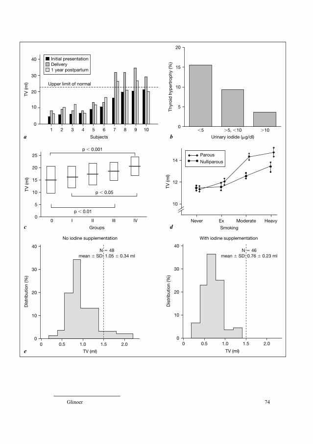
Glinoer 74
1
10
20
30
20
15
10
Thyr
oid
hyp
ertr
ophy
(%)
5
0�5 �5, �10
Urinary iodide (�g/dl)
14
12
10
Never
Smoking
Ex Moderate Heavy
�10
TV (m
l)
TV (m
l)
40
02 3 4 5 6 7 8 9
0
10
20
30
Dis
trib
utio
n (%
)
40
00.5 1.0 1.5
TV (ml)
No iodine supplementation
N � 48 mean � SD: 1.05 � 0.34 ml
2.0
10
Subjectsa b
c
e
d
Upper limit of normal
25
15
10TV (m
l)
5
00 I II III
p � 0.01
p � 0.05
p � 0.001
IV
Groups
20
0
10
20
30
Dis
trib
utio
n (%
)
40
00.5 1.0 1.5
TV (ml)
With iodine supplementation
N � 46 mean � SD: 0.76 � 0.23 ml
2.0
1 year postpartumDeliveryInitial presentation
Parous
Nulliparous

Iodine Deficiency during Pregnancy 75
Prevention of Gestational Goiter Due to IDAs already discussed, women should ideally be provided with an adequate
iodine intake (150 �g/day) long before they become pregnant in order to pre-
vent gestational goitrogenesis, since it is only by reaching a long-term steady
state with replenished intrathyroidal iodine stores that the triggering of the thy-
roid machinery can be avoided once gestation begins. To achieve such a goal,
national public health authorities need to develop iodine supplementation pro-
grams of the population’s diet. Correcting this public health problem has been
the aim of a massive global campaign, undertaken 10–15 years ago worldwide,
and that has shown remarkable progress so far [31, 50–52]. Until 1992, most
European countries were moderately to severely iodine deficient. A survey car-
ried out in twelve European countries in more recent years, using a mobile unit
(the ‘ThyroMobil’ van) equipped with a sonographic device and the facilities
for collecting urine samples, allowed for the determination of TV and urinary
iodine concentrations in almost 8,000 schoolchildren aged 7–15 years. The
main results indicated that the status of iodine nutrition was markedly improved
in many, albeit not all, of the European countries surveyed. Silent iodine pro-
phylaxis is not sufficient to restore an adequate iodine balance, and more strin-
gent prophylactic measures need to be taken by public health authorities to
achieve an ideal iodine nutrition status in the population [53–55].
In the mean time, the most appropriate preventive and therapeutic
approach to avoid gestational goitrogenesis is to systematically increase the
iodine supply as early as possible during gestation and continue the nutrition
fortification after parturition, particularly in mothers who breastfeed. This can
Fig. 4. a TV in 10 selected healthy pregnant women (numbered from 1 to 10) in
Brussels, measured by ultrasonography at initial presentation in the first trimester, then at
delivery, and finally 12 months postpartum, and showing goiter formation during pregnancy
in 4/10 women and only a partial TV normalization during the postpartum period (adapted
from Glinoer et al. [45]). b Inverse correlation between the degree of thyroid hypertrophy and
the severity of ID (measured as urinary iodine concentrations) in pregnant women in the
southwest of France (adapted from Caron et al. [29]). c Progressive changes in TV
(mean � SD) between group 0 (representing nulliparous women) and group IV (representing
women with previous pregnancies: group I had one pregnancy, group II two pregnancies,
group III three pregnancies, group IV four or more pregnancies) in a retrospective study of TV
in relation to parity in 208 nongoitrous healthy women with a mean age of 42 years from
southern Italy (adapted from Rotondi et al. [46]). d The effect of parity and smoking on TV in
women in Denmark (adapted from Knudsen et al. [47]). e Distribution frequencies of TVs in
neonates born to mothers without (left) and with (right) iodine supplementation during preg-
nancy. Iodine supplementation allowed for a marked 38% average reduction in mean neonatal
TV and for the complete prevention of thyroid hyperplasia at birth. The upper limit of normal
TV in newborns is indicated by the vertical dotted lines (adapted from Glinoer et al. [48]).

Glinoer 76
be achieved by the use of multivitamin pills, containing appropriate amounts of
iodine supplements. How much supplemental iodine should be given to prevent
goiter formation remains a matter of local assessment, since it depends mainly
on the extent of the preexisting iodine deprivation [56–59]. The ultimate goal
which is to restore and maintain a balanced iodine status can be reached in most
instances with 100–200 �g iodine given daily as a supplement during preg-
nancy. It should be remembered, however, that with long-standing iodine
restriction in the diet before the onset of pregnancy, a lag period (of about one
trimester) is inevitable before the benefits of the iodine supplementation
improving thyroid function are observed.
Pregnancy in Regions with Severe ID
In many regions in the world, ID is not only overtly present, but it is often
severe. Large areas remain in Central Africa and Asia, for instance, that still
have iodine intakes below 25 �g/day, characteristic of severe ID and endemic
goiter [51, 52]. In such regions, the thyroid status of pregnant women and their
offspring is frequently impaired. In addition, other factors, such as selenium
deficiency and thiocyanate excess (resulting from the staple use of foodstuffs
such as cassava) combine with severe ID, tending to complicate the thyroidal
situation even further [60]. In terms of thyroid function, the adult populations
usually exhibit a mixed pattern encompassing subjects with a normal thyroid
function and others who present various degrees of hypothyroidism. In women
of child-bearing age, severe ID and hypothyroidism play a role in reducing fer-
tility and increase the rate of spontaneous abortions.
When these women become pregnant, thyroid function tends to deteriorate
even further, as gestation progresses. Thus, the thyroidal stress associated with
pregnancy in conditions with mild to moderate ID cannot be compared, at least
in quantitative terms, with the thyroidal repercussions observed in countries
with long-standing and severe ID. Because of the obvious difficulties inherent
in careful field studies in most areas with severe ID, there have been only few
studies of thyroid function and no systematic study to assess the changes in goi-
ter size in pregnant women [61–63]. Until a few years ago, it was not feasible to
obtain echographic measurements of the thyroid gland on a large and represen-
tative scale; it was even more difficult to investigate prospectively goitrogenic
changes during pregnancy. Presently, this situation is rapidly evolving, because
of the possibility to adapt the ThyroMobil technology to large field studies even
in remote areas [64, 65].
In women of child-bearing age and during pregnancy, iodine supplements
have been administered in the form of iodized salt, potassium iodide drops, and

Iodine Deficiency during Pregnancy 77
also in the form of iodized oil (given intramuscularly or orally) as an emergency
prophylactic and therapeutic approach in areas with severe ID complicated by
endemic cretinism. Several such programs have conclusively demonstrated their
remarkable efficiency to treat endemic goiter, as well as to eradicate endemic
cretinism [31, 60, 66]. Also, the results of these studies have proved that the
pregnancy of women who reside in severely iodine-deficient regions can be
managed adequately with iodine supplementation. Except for emergency situa-
tions, there is presumably no need to use supraphysiological amounts of iodine
to improve significantly or even normalize thyroid function parameters. Although
it has not been possible so far in the setting of difficult field studies to evaluate
quantitatively the reduction in goiter size or goiter prevalence associated with the
improvement of thyroid function, it can be assumed that goiter reduction was
indeed a ‘side’ benefit of the improvement in the iodine status.
Fetal and Neonatal Consequences of Maternal ID and Thyroid Underfunction
The adequate functioning of both the maternal and fetal thyroid glands
plays an important part to ensure that the fetal neuropsychointellectual develop-
ment progresses normally. Globally, three sets of clinical disorders, schemati-
cally illustrated in figure 5, ought to be considered. In infants with a defect of
thyroid ontogenesis, leading to congenital hypothyroidism, the participation of
maternal TH to the fetal circulating thyroxine environment remains theoreti-
cally normal. Therefore, the risk of brain damage results exclusively from the
insufficient production of TH by the fetus, and will hence depend on the sever-
ity of the defect involved (for instance, total agenesis versus ectopia). In con-
trast, when only the maternal thyroid gland is functionally deficient (for
instance, in hypothyroidism due to chronic autoimmune thyroiditis), both the
severity and the temporal occurrence of maternal thyroid underfunction will
drive the resulting consequences for an impaired fetal neuronal development.
Clinical situations of this type may take place already at early gestational stages
(women with untreated or undertreated hypothyroidism), or appear during late
gestational stages (women with autoimmunity features who are euthyroid dur-
ing the first half of gestation). Finally, in conditions of ID, both maternal and
fetal thyroid functions are affected. Therefore, it is primarily the precocity and
severity of pregnancy-related ID that will drive the potential repercussions for
fetal neurological development.
Iodine is required for the synthesis of TH, and TH are crucial for brain
development both during fetal and early postnatal life [67–70]. When severe
enough, ID induces maternal and fetal hypothyroxinemia from early gestation

Glinoer 78
onwards [25]. Thus, any impairment in hormone availability during critical
periods of brain development may induce irreversible brain damage, with mental
retardation and neurological abnormalities as the final consequences (i.e. endemic
cretinism) that ultimately depend upon the timing and severity of the brain’s
insult [71–73]. The characteristic neurological picture of endemic cretinism is
presumably directly due to insults to the developing brain, occurring already
during the first trimester (i.e. deafness) and mostly during the second trimester,
while cerebellar abnormalities may result from a postnatal insult [74–76]. This
interpretation is supported by the observation that the full neurological picture
of endemic cretinism can only be prevented when ID is corrected before the
second trimester of pregnancy, and optimally prior to conception [71]. The par-
ticular pattern of myxedematous cretinism that is commonly found in Africa
might be explained by the fact that in this area severe ID is complicated by sele-
nium deficiency. Selenium deficiency results in the accumulation of peroxide
in the hyperstimulated thyroid glands, and excess peroxide induces thyroid cell
destruction, leading to parenchymal fibrosis and hypothyroidism [77]. Endemic
NormalFetus
Mother
Thyroxinemiain the fetus
Contributionarising frommaternalhormonetransfer
Clinical disorders
Normal
Conce
ption
Mid-g
esta
tion
Term
Normal
Defectiveontogenesis(congential
hypothyroidism)Normal
Iodinedeficiency
Hypothy-roxinemia
Iodinedeficiency
Fig. 5. Schematic representation of the three sets of clinical conditions that may affect
thyroid function in the mother alone, the fetus alone, or the fetomaternal unit, showing the
relative contributions of an impaired maternal and/or fetal thyroid function that may eventu-
ally lead to alterations in fetal thyroxinemia (from Glinoer and Delange [25]).
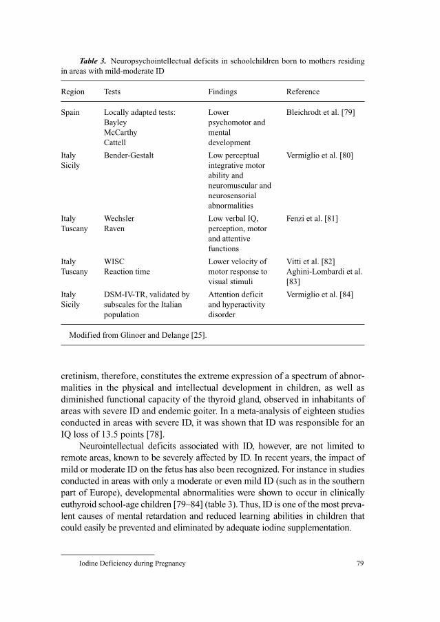
Iodine Deficiency during Pregnancy 79
cretinism, therefore, constitutes the extreme expression of a spectrum of abnor-
malities in the physical and intellectual development in children, as well as
diminished functional capacity of the thyroid gland, observed in inhabitants of
areas with severe ID and endemic goiter. In a meta-analysis of eighteen studies
conducted in areas with severe ID, it was shown that ID was responsible for an
IQ loss of 13.5 points [78].
Neurointellectual deficits associated with ID, however, are not limited to
remote areas, known to be severely affected by ID. In recent years, the impact of
mild or moderate ID on the fetus has also been recognized. For instance in studies
conducted in areas with only a moderate or even mild ID (such as in the southern
part of Europe), developmental abnormalities were shown to occur in clinically
euthyroid school-age children [79–84] (table 3). Thus, ID is one of the most preva-
lent causes of mental retardation and reduced learning abilities in children that
could easily be prevented and eliminated by adequate iodine supplementation.
Table 3. Neuropsychointellectual deficits in schoolchildren born to mothers residing
in areas with mild-moderate ID
Region Tests Findings Reference
Spain Locally adapted tests: Lower Bleichrodt et al. [79]
Bayley psychomotor and
McCarthy mental
Cattell development
Italy Bender-Gestalt Low perceptual Vermiglio et al. [80]
Sicily integrative motor
ability and
neuromuscular and
neurosensorial
abnormalities
Italy Wechsler Low verbal IQ, Fenzi et al. [81]
Tuscany Raven perception, motor
and attentive
functions
Italy WISC Lower velocity of Vitti et al. [82]
Tuscany Reaction time motor response to Aghini-Lombardi et al.
visual stimuli [83]
Italy DSM-IV-TR, validated by Attention deficit Vermiglio et al. [84]
Sicily subscales for the Italian and hyperactivity
population disorder
Modified from Glinoer and Delange [25].

Glinoer 80
Conclusions and Perspectives
The main changes in thyroid function associated with the pregnant state
are due to increased hormone requirements, which begin in the first trimester of
gestation. Increased hormone requirements can only be met by proportionally
increased hormone production, directly depending upon the availability of
iodine in the diet. When dietary iodine is deficient, adequate physiological
adaptation is difficult to achieve and is progressively replaced by pathological
alterations occurring in parallel with the degree of long-term iodine depriva-
tion, leading to enhanced glandular stimulation. Therefore, pregnancy typically
acts to reveal the underlying iodine restriction and gestation results in a defi-
cient iodine status, even in conditions with only a marginally restricted intake,
such as is observed in many European regions. ID during pregnancy has important
Table 4. Take home messages
• The challenge for the thyroid gland is to adjust the hormonal output to achieve a new
steady state corresponding to physiological adaptation to the pregnant state. When preg-
nancy takes place in women with a restricted iodine intake, physiological adaptation is
progressively replaced by pathological alterations
• The RNI for iodine is 250 �g/day in pregnant and lactating women
• The most important consequence of ID during pregnancy is maternal underfunction
(i.e. hypothyroxinemia), leading in turn to excessive thyroidal stimulation with a rise in
serum TSH and increased serum TG
• Gestational goiter formation is the clinical hallmark of ID. Together, low intrathyroidal
iodine stores before conception, increased needs for higher iodine availability once
pregnancy begins, and the insufficiency of daily iodine intake constitute the major com-
ponents of enhanced thyroidal stimulation leading to goitrogenesis affecting both the
mother and fetus
• A goiter formed during gestation regresses only partially after parturition. Pregnancy is
therefore an environmental factor that may help explain the higher prevalence of goiter
and thyroid disorders in women. Goiter formation takes place in the fetus, emphasizing
the exquisite sensitivity of the fetal thyroid to consequences of maternal iodine depriva-
tion, and indicating that the process of goiter formation begins during the earliest stages of
fetal gland development
• Iodine is required for the synthesis of TH, and TH are crucial for fetal brain develop-
ment. When severe enough, ID may induce fetal hypothyroxinemia from early gestation
onwards. Any impairment in hormone availability during critical periods of develop-
ment may induce brain damage, with irreversible neurological abnormalities
• In recent years, the impact of mild, moderate, or only borderline ID on the fetus has
been recognized with developmental abnormalities shown to occur and impaired school
achievements that may be present in apparently normal school-aged children

Iodine Deficiency during Pregnancy 81
repercussions for both mother and fetus, namely thyroid underfunction and
goitrogenesis. Furthermore, ID may be associated with alterations of the neu-
ropsychointellectual outcome in the progeny, and the risk for an abnormal
development of the progeny is further enhanced because both the mother and
offspring are exposed to the deficiency, not only during the entirety of gestation
but also the postnatal period. Iodine prophylaxis should be introduced system-
atically to women during pregnancy and the lactation period. Concerning areas
with a severe deficiency, the correction of the lack of iodine has proved highly
beneficial to prevent mental deficiency disorders: the many actions undertaken
to eradicate ID have prevented the occurrence of mental retardation in millions
of young infants throughout the world. In most public health programs dealing
with the correction of the ID disorder, iodized salt has been used as the pre-
ferred method of conveying iodine supplements to the household. Iodized salt,
however, is not the ideal vector in the specific instance of pregnancy and breast-
feeding, because of the necessity to restrict salt intake. Finally, it is with some
concern that the results of the recent nutritional survey in the United States have
disclosed that ID, thought to have been eradicated many years ago, may actually
show a risk of a resurgence, particularly in young women. This issue will need
to be considered seriously by the medical community and public health author-
ities, since similar situations may occur in other countries as well (see the main
‘take home messages’ in table 4).
Acknowledgments
The author acknowledgs the financial support of the Belgian Ministry for Education
and Scientific Research, within the framework of the ‘Actions de Recherche Concertée
2004–2009’ (ARC/Convention No. 04/09-314), which was of great help in preparing the pre-
sent manuscript.
References
1 Andersson M, Takkouche B, Egli I, Allen HE, de Benoist B: Current global iodine status and
progress over the last decade towards the elimination of iodine deficiency. Bull WHO 2005;83:
518–525.
2 Glinoer D: The regulation of thyroid function in pregnancy: pathways of endocrine adaptation
from physiology to pathology. Endocr Rev 1997;18:404–433.
3 Bartalena L: Recent achievements in studies on thyroid hormone-binding proteins. Endocr Rev
1990;11:47–64.
4 Dowling JT, Appleton WG, Nicoloff JT: Thyroxine turnover during human pregnancy. J Clin
Endocrinol Metab 1964;27:1749–1750.
5 Glinoer D, De Nayer P, Bourdoux P, Lemone M, Robyn C, Van Steirteghem A, Kinthaert J,
Lejeune B: Regulation of maternal thyroid function during pregnancy. J Clin Endocrinol Metab
1990;71:276–287.

Glinoer 82
6 Guillaume J, Schussler GC, Goldman J: Components of the total serum thyroid hormone concen-
trations during pregnancy: high free thyroxine and blunted thyrotropin (TSH) response to TSH-
releasing hormone in the first trimester. J Clin Endocrinol Metab 1985;60:678–684.
7 Glinoer D, De Nayer P, Robyn C, Lejeune B, Kinthaert J, Meuris S: Serum levels of intact human
chorionic gonadotropin (hCG) and its free alpha and beta subunits, in relation to maternal thyroid
stimulation during normal pregnancy. J Endocrinol Invest 1993;16:881–888.
8 Bianco AC, Salvatore D, Gereben B, Berry MJ, Larsen PR: Biochemistry, cellular and molecular
biology, and physiological roles of iodothyronine selenodeiodinases. Endocr Rev 2002;23:38–89.
9 Huang SA, Dorfman DM, Genest DR, Salvatore D, Larsen PR: Type 3 iodothyronine deiodinase is
highly expressed in the human uteroplacental unit and in fetal epithelium. J Clin Endocrinol
Metab 2003;88:1384–1388.
10 Glinoer D: Maternal and fetal impact of chronic iodine deficiency. Clin Obstet Gynecol 1997;40:
102–116.
11 Glinoer D: What happens to the normal thyroid during pregnancy? Thyroid 1999;9:631–635.
12 Glinoer D: Pregnancy and iodine. Thyroid 2001;11:471–481.
13 Glinoer D: The regulation of thyroid function during normal pregnancy – importance of the iodine
nutrition status. Best Pract Res Clin Endocrinol Metab 2004;18:133–152.
14 Glinoer D: Iodine nutrition requirements during pregnancy (editorial). Thyroid 2006;16:947–948.
15 Caron P, Glinoer D, Lecomte P, Orgiazzi J, Wémeau J-L: Apport iodé en France: prévention de la
carence iodée au cours de la grossesse et l’allaitement. Ann Endocrinol 2006;67:281–286.
16 Delange F, Bourdoux P, Chanoine JP, Ermans AM: Physiopathology of iodine nutrition during
pregnancy, lactation, and early postnatal life; in Berger H (ed): Vitamins and Minerals in
Pregnancy. New York, Raven Press, 1988, pp 205–214.
17 Ermans AM: Endemic goiter; in Ingbar SH, Braverman LE (eds): The Thyroid – a Fundamental
and Clinical Text, ed 5. Philadelphia, Lippincott, 1986, pp 705–721.
18 Robbins J: Iodine deficiency, iodine excess and the use of iodine for protection against radioactive
iodine. Thyroid Today 1994;3:1–5.
19 Pochin EE: The iodine uptake of the human thyroid throughout the menstrual cycle and in preg-
nancy. Clin Sci 1952;11:441–445.
20 Halnan KE: The radioiodine uptake of the human thyroid in pregnancy. Clin Sci 1958;17:
281–290.
21 Aboul-Khair SA, Crooks J, Turnbull AC, Hytten FE: The physiological changes in thyroid func-
tion during pregnancy. Clin Sci 1964;27:195–207.
22 Silva JE: Effects of iodine and iodine-containing compounds on thyroid function. Med Clin North
Am 1985;69:881–898.
23 Liberman CS, Pino SC, Fang SL, Braverman LE, Emerson CH: Circulating iodide concentrations
during and after pregnancy. J Clin Endocrinol Metab 1998;83:3545–3549.
24 Glinoer D, Lemone M: Goiter and pregnancy – a new insight into an old problem. Thyroid 1992;2:
65–72.
25 Glinoer D, Delange F: The potential repercussions of maternal, fetal, and neonatal hypothyroxine-
mia on the progeny. Thyroid 2000;10:871–887.
26 Glinoer D: The importance of iodine nutrition during pregnancy. Public Health Nutrition
April–May 2007, in press.
27 Hollowell JG, Staehling NW, Hannon WH, Flanders DW, Gunter EW, Maberly GF, Braverman LE,
Pino S, Miller DT, Garbe PL, DeLozier DM, Jacjson RJ: Iodine nutrition in the United States.
Trends and public health implications: iodine excretion data from national health and nutrition
examination surveys I and III (1971–1974 and 1988–1994). J Clin Endocrinol Metab 1998;83:
3401–3408.
28 The Public Health Committee of the American Thyroid Association: Iodine supplementation for
pregnancy and lactation – United States and Canada: Recommendations of the American Thyroid
Association. Thyroid 2006;16:949–951.
29 Caron P, Hoff M, Bazzi S, Dufor A, Faure G, Ghandour I, Lauzu P, Lucas Y, Maraval D, Mignot F,
Ressigeac P, Vertongen F, Grangé V: Urinary iodine excretion during normal pregnancy in healthy
women living in the southwest of France: correlation with maternal thyroid parameters. Thyroid
1997;7:749–754.

Iodine Deficiency during Pregnancy 83
30 Nohr SB, Laurberg P, Borlum K-G, Pedersen KM, Johannesen PL, Damm P, Fuglsang E, Johansen
A: Iodine deficiency in pregnancy in Denmark – regional variations and frequency of individual
iodine supplementation. Acta Obstet Gynecol Scand 1993;72:350–353.
31 Dunn JT, Delange F: Damaged reproduction: the most important consequence of iodine defi-
ciency. J Clin Endocrinol Metab 2001;86:2360–2363.
32 World Health Organization: Assessment of Iodine Deficiency Disorders and Monitoring Their
Elimination: a Guide for Programme Managers, ed 2. Geneva, WHO Press, 2001.
33 World Health Organization. Technical consultation of experts in Geneva (January 2005): The pre-
vention and control of iodine deficiency in pregnant and lactating women and in children under
two years of age – Recommendations of a WHO Technical Consultation. Public Health Nutrition
April–May 2007, in press.
34 Roti E, Vagenakis AG: Effect of excess iodide – clinical aspects; in Braverman LE, Utiger RD
(eds): The Thyroid – a Fundamental and Clinical Text, ed 9. Philadelphia, Lippincott Williams &
Wilkins, 2005, pp 288–305.
35 Chaouki ML, Benmiloud M: Prevention of iodine deficiency disorders by oral administration of
lipiodol during pregnancy. Eur J Endocrinol 1994;130:547–551.
36 Delange F: Optimal iodine nutrition during pregnancy, lactation and the neonatal period. Int
J Endocrinol Metab 2004;2:1–12.
37 Wang R, Nelson JC, Weiss RM, Wilcox RB: Accuracy of free thyroxine measurements across nat-
ural ranges of thyroxine binding to serum proteins. Thyroid 2000;10:31–39.
38 Osathanondh R, Tulchinsky D, Chopra IJ: Total and free thyroxine and triiodothyronine in normal
and complicated pregnancy. J Clin Endocrinol Metab 1976;42:98–103.
39 Roti E, Gardini E, Minelli R, Bianconi L, Flisi M: Thyroid function evaluation by different com-
mercially available free thyroid hormone measurement kits in term pregnant women and their
newborns. J Endocrinol Invest 1991;14:1–9.
40 Sapin R, D’Herbomez M, Schlienger JL: Free thyroxine measured with equilibrium dialysis and
nine immunoassays decreases in late pregnancy. Clin Lab 2004;50:581–584.
41 Demers LM, Spencer CA: Laboratory medicine practice guidelines: laboratory support for the
diagnosis and monitoring of thyroid disease. Clin Endocrinol (Oxf) 2003;58:138–140.
42 Haddow JE, Knight GJ, Palomaki GE, McClain MR, Pulkkinen AJ: The reference range and
within-person variability of thyroid stimulating hormone during the first and second trimesters of
pregnancy. J Med Screen 2004;11:170–174.
43 Levy RP, Newman DM, Rejali LS, Barford DA: The myth of goiter in pregnancy. Am J Obstet
Gynecol 1980;137:701–703.
44 Nelson M, Wickus GG, Caplan RH, Beguin EA: Thyroid gland size in pregnancy: an ultrasound
and clinical study. J Reprod Med 1987;32:888–890.
45 Glinoer D, Lemone M, Bourdoux P, De Nayer P, Delange F, Kinthaert J, Lejeune B: Partial
reversibility during late postpartum of thyroid abnormalities associated with pregnancy. J Clin
Endocrinol Metab 1992;74:453–457.
46 Rotondi M, Amato G, Biondi B, Mazziotti G, Del Buono A, Nicchio MR, Balzano S, Bellastella A,
Glinoer D, Carella C: Parity as a thyroid size-determining factor in areas with moderate iodine
deficiency. J Clin Endocrinol Metab 2000;85:4534–4537.
47 Knudsen N, Bülow I, Laurberg P, Ovesen L, Perrild H, Jorgensen T: Parity is associated with
increased thyroid volume solely among smokers in an area with moderate to mild iodine defi-
ciency. Eur J Endocrinol 2002;146:39–43.
48 Glinoer D, Delange F, Laboureur I, De Nayer P, Lejeune B, Kinthaert J, Bourdoux P: Maternal and
neonatal thyroid function at birth in an area with marginally low iodine intake. J Clin Endocrinol
Metab 1992;75:800–805.
49 Glinoer D, De Nayer P, Delange F, Lemone M, Toppet V, Spehl M, Grün JP, Kinthaert J, Lejeune B:
A randomized trial for the treatment of mild iodine deficiency during pregnancy: maternal and
neonatal effects. J Clin Endocrinol Metab 1995;80:258–269.
50 Delange F, Lecomte P: Iodine supplementation – benefits outweigh risks. Drug Safety 2000;2:
89–96.
51 Dunn JT: Correcting iodine deficiency is more than just spreading around a lot of iodine. Thyroid
2001;11:363–364.

Glinoer 84
52 Hetzel BS: Eliminating iodine deficiency disorders – the role of the International Council in the
global partnership. Bull WHO 2002;80:410–417.
53 Delange F, Benker G, Caron P, Eber O, Ott P, Peter F, Podoba J, Simescu M, Szybinsky Z,
Vertongen F, Vitti P, Wiersinga W, Zamrazil V: Thyroid volume and urinary iodine in European
schoolchildren: standardization of values for assessment of iodine deficiency. Eur J Endocrinol
1997;136:180–187.
54 Delange F, Van Onderbergen A, Shabana W, Vandemeulebroecke E, Vertongen F, Gnat D, Dramaix
M: Silent iodine prophylaxis in western Europe only partly corrects iodine deficiency: the case of
Belgium. Eur J Endocrinol 2000;143:189–196.
55 Ciardelli R, Haumont D, Gnat D, Vertongen F, Delange F: The nutritional iodine supply of Belgian
neonates is still insufficient. Eur J Pediatr 2002;161:519–523.
56 Romano R, Jannini EA, Pepe M, Grimaldi A, Olivieri M, Spennati P, Cappa F, D’Armiento M: The
effects of iodoprophylaxis on thyroid size during pregnancy. Am J Obstet Gynecol 1991;164:
482–485.
57 Kung AWC, Lao TT, Chau MT, Tam SC, Low LC: Goitrogenesis during pregnancy and neonatal
hypothyroxinemia in a borderline iodine sufficient area. Clin Endocrinol 2000;53:725–731.
58 Antonangeli L, Maccherini D, Cavaliere R, Di Giulio C, Reinhardt B, Pinchera A, Aghini-
Lombardi F: Comparison of two different doses of iodide in the prevention of gestational goiter in
marginal iodine deficiency: a longitudinal study. Eur J Endocrinol 2002;147:29–34.
59 Glinoer D: Feto-maternal repercussions of iodine deficiency during pregnancy. Ann Endocrinol
(Paris) 2003;64:37–44.
60 Delange F: The disorders induced by iodine deficiency. Thyroid 1994;4:107–128.
61 Thilly CH, Delange F, Lagasse R, Bourdoux P, Ramioul L, Berquist H, Ermans AM: Fetal
hypothyroidism and maternal thyroid status in severe endemic goiter. J Clin Endocrinol Metab
1978;47:354–360.
62 Silva JE, Silva S: Interrelationships among thyroxine, triiodothyronine, reverse triiodothyronine,
and thyroid-stimulating hormone in iodine-deficient pregnant women and their offspring: effects
of iodine supplementation. J Clin Endocrinol Metab 1981;52:671–677.
63 Boyages SC, Halpern JP, Maberly GF, Eastman CJ, Morris J, Collins J, Jupp JJ, Jin CE, Wang ZH,
You CY: A comparative study of neurological and myxedemetous endemic cretinism in Western
China. J Clin Endocrinol Metab 1988;67:1262–1271.
64 Djokomoeljanto R, Setyawan H, Dramaix M, Hadisaputro S: The thyromobil model for standard-
ized evaluation of iodine deficiency disorder control in Indonesia. Thyroid 2001;11:365–372.
65 Rossi AC, Tomimori E, Camargo R, Medeiros-Neto G: Searching for iodine deficiency in school-
children from Brazil: the Thyromobil project. Thyroid 2001;11:661–663.
66 Dunn JT: The use of iodized oil and other alternatives for the elimination of iodine deficiency dis-
orders; in Hetzel BS, Pandav CS (eds): The Conquest of Iodine Deficiency Disorders. New Delhi,
Oxford University Press Publications, 1994, pp 108–117.
67 Contempré B, Jauniaux E, Calvo R, Jurkovic D, Campbell S, Morreale de Escobar G: Detection of
thyroid hormones in human embryonic cavities during the first trimester of pregnancy. J Clin
Endocrinol Metab 1993;77:1719–1722.
68 Chan S, Kilby MD: Thyroid hormone and central nervous system development. J Endocrinol
2000;165:1–8.
69 Lavado-Autric R, Auso E, Garcia-Velasco JV, Arufe MC, Escobar del Rey F, Berbel P, Morreale de
Escobar G: Early maternal hypothyroxinemia alters histogenensis and cerebral cortex cytoarchi-
tecture of the progeny. J Clin Invest 2003;111:1073–1082.
70 Zoeller RT: Transplacental thyroxine and brain development. J Clin Invest 2003;111:954–957.
71 Pharoah PO, Butterfield IH, Hetzel BS: Neurological damage to the fetus resulting from severe
iodine deficiency during pregnancy. Lancet 1971;i:308–310.
72 Morreale de Escobar G, Obregon MJ, Escobar del Rey F: Is neuropsychological development
related to maternal hypothyroidism or to maternal hypothyroxinemia? J Clin Endocrinol Metab
2000;85:3975–3987.
73 Calvo RM, Jauniaux E, Gulbis B, Asuncion M, Gervy C, Contempre B, Morreale de Escobar G:
Fetal tissues are exposed to biologically relevant free thyroxine concentrations during early phases
of development. J Clin Endocrinol Metab 2002;87:1768–1777.

Iodine Deficiency during Pregnancy 85
74 Delange F: Endemic cretinism; in Braverman LE, Utiger RD (eds): The Thyroid – a Fundamental
and Clinical Text, ed 8. Philadelphia, Lippincott Williams & Wilkins, 2000, pp 743–754.
75 Halpern JP, Boyages SC, Maeberly GF, Collins JK, Eastman CJ, Morris J: The neurology of
endemic cretinism. Brain 1991;114:825–841.
76 DeLong GR, Stanbury JB, Fierro-Benitez R: Neurological signs in congenital iodine deficiency
disorder (endemic cretinism). Dev Med Child Neurol 1985;27:317–324.
77 Vanderpas JB, Contempré B, Duale NL, Goossens B, Bebe N, Thorpe R, Ntambue K, Dumont J,
Thilly CH, Diplock AT: Iodine and selenium deficiency associated with cretinism in Northern
Zaire. Am J Clin Nutr 1990;52:1087–1093.
78 Bleichrodt N, Born MP: A meta-analysis of research on iodine and its relationship to cognitive
development; in Stanbury JB (ed): The Damaged Brain of Iodine Deficiency. New York, Cognizant
Communication Corporation, 1994.
79 Bleichrodt N, Escobar del Rey F, Morreale de Escobar G, Garcia I: Iodine deficiency – implica-
tions for mental and psychomotor development in children; in DeLong GR, Robbins G, Condliffe
PG (eds): Iodine and the Brain. New York, Plenum Press, 1989, pp 269–287.
80 Vermiglio F, Sidoti M, Finocchiaro MD, Battiato S, Lo Presti VP, Benvenga S, Trimarchi F:
Defective neuromotor and cognitive ability in iodine-deficient schoolchildren of an endemic goi-
ter region in Sicily. J Clin Endocrinol Metab 1990;70:379–384.
81 Fenzi GF, Giusti LF, Aghini-Lombardi F, Bartalena L, Marcocci C, Santini F, Bargagna S,
Brizzolara D, Ferretti G, Falciglia G, Montelone L, Marcheschi M, Pinchera A: Neuropsychological
assessment in schoolchildren from an area of moderate iodine deficiency. J Endocrinol Invest
1990;13:427–431.
82 Vitti P, Aghini-Lombardi F, Antonangeli L, Rago T, Chiovato L, Pinchera A, Marcheschi M,
Bargagna S, Bertuccelli B, Ferretti G, Sbrana B: Mild iodine deficiency in fetal/neonatal life and
neuropsychological performances. Acta Med Austr 1992;19:57–59.
83 Aghini-Lombardi F, Pinchera A, Antonangeli L, Rago T, Chiovato L, Bargagna S, Bertucelli B,
Ferretti G, Sbrana B, Marcheschi M, Vitti P: Mild iodine deficiency during fetal/neonatal life and
neuropsychological impairment in Tuscany. J Endocrinol Invest 1995;18:57–62.
84 Vermiglio F, Lo Presti VP, Moleti M, Sidoti G, Tortorella G, Scaffidi G, Castagna MG, Mattina F,
Violi MA, Crisa A, Artemisia A, Trimarchi F: Attention deficit and hyperactivity disorders in the
offspring of mothers exposed to mild-moderate iodine deficiency: a possible novel iodine defi-
ciency disorder in developed countries. J Clin Endocrinol Metab 2004;89:6054–6060.
Prof. Daniel Glinoer
Division of Endocrinology, Department of Internal Medicine
Thyroid Investigation Clinic, University Hospital Saint Pierre
322, Rue Haute, BE–1000 Brussels (Belgium)
Tel. �32 2 535 4516, Fax �32 2 535 3137
E-Mail [email protected] or [email protected]

Van Vliet G, Polak M (eds): Thyroid Gland Development and Function.
Endocr Dev. Basel, Karger, 2007, vol 10, pp 86–98
Ontogenesis of Thyroid Function andInteractions with Maternal Function
M.J. Obregon, R.M. Calvo, F. Escobar del Rey, G. Morreale de Escobar
Instituto de Investigaciones Biomedicas, Centro mixto ‘Alberto Sols’ (CSIC-UAM),
Madrid, Spain
AbstractFetal and neonatal development of thyroid function involves the embryogenesis, differ-
entiation and maturation of the thyroid gland, of the hypothalamic-pituitary-thyroid axis and
of the systems controlling thyroid hormone metabolism. We focus here on aspects related to
neurodevelopment. Throughout gestation, thyroxine (T4) transferred from the mother, pre-
sent in embryonic fluids by 4 weeks, protects the fetal brain. Free T4 (FT4) in fetal fluids
increases rapidly, approaching adult levels by midgestation, in concentrations that are deter-
mined by the maternal serum T4. T3 remains very low throughout pregnancy. In the cerebral
cortex T3, generated from T4, reaches adult values by midgestation and is partly bound to spe-
cific nuclear receptor isoforms. The iodothyronine deiodinases are important for the spatial
and temporal presence of T3 in different fetal brain areas. After onset of fetal thyroid secre-
tion at midgestation, maternal transfer of T4 continues to contribute importantly to fetal
serum T4, protecting neurodevelopment until birth. In rats, even a transient period of mater-
nal hypothyroxinemia disrupts neurodevelopment irreversibly, supporting epidemiological
evidence for its negative role in human neurodevelopment. The prompt treatment of maternal
hypothyroidism or hypothyroxinemia should mitigate negative effects on neurodevelopment.
Neurodevelopmental deficits of preterm infants might also result from an untimely interrup-
tion of the maternal transfer of T4 [Morreale de Escobar et al: J Clin Endocrinol Metab
2000;85:3975–3987; Best Pract Res Clin Endocrinol Metab 2004;18:225–248; Eur J
Endocrinol 2004;151(suppl 3):U25–U37].
Copyright © 2007 S. Karger AG, Basel
Ontogenesis of Thyroid Function
The thyroid gland develops mostly during fetal and early postnatal life. The
thyrocytes forming the functional unit of the gland – the thyroid follicle – derive
from the embryonic endoderm in the floor of the primitive pharynx, forming a
Disorders of Thyroid Function

Ontogenesis of Thyroid Function and Interactions with Maternal Function 87
bud already visible at embryonic day 16 (E16) in humans. After proliferation of
the cells and migration at E24–E32 in humans, the thyroid reaches its final
position at E40–E50 [1]. An error during morphogenesis results in thyroid
abnormalities, such as agenesis or ectopies, as shown in null mice with targeted
deletions of thyroid transcription factors, fully described in the chapter by De
Felice and Di Lauro [pp. 1–14].
The histological differentiation of the thyrocytes and formation of the fol-
licle are accompanied by the progressive appearance of specific proteins:
thyroglobulin, thyroid peroxidase, sodium/iodine symporter (NIS) and TSH
receptor, all necessary for the synthesis and secretion of T4 and T3. The main
physiological regulator of thyroid gland activity is TSH, but TSH-independent
autoregulatory mechanisms also play an important role in the postnatal
adaptation to fluctuations in iodine availability. Thyroglobulin has been
detected in human thyroid as early as the 5th week of gestation, before the gland
reaches its final position. Under the artificial conditions of organ culture,
iodine uptake starts at 12–13 weeks after conception, coinciding with the clo-
sure of follicles. In vivo, however, significant uptake of iodine, a prerequisite
for the synthesis and secretion of fetal thyroid hormones, is minimal until
midgestation (18–20 weeks) coinciding with full development of the pituitary-
portal vascular system.
For obvious reasons, studies in human fetuses are scarce and much of our
knowledge of the role of thyroid hormones during fetal life comes from
experimental work performed in rats [table 3 in 2; 3–5], where thyroid hormone
secretion starts at E17.5–E18. Fetal serum T4 concentrations increase 10-fold
between E18 and the end of gestation (E22), a developmental period compara-
ble to that of a human fetus at the beginning of the third trimester. T3, however,
is very low throughout gestation.
In most fetal tissues, T4 increases in parallel to the increases in fetal plasma
T4 while T3 concentrations are highly variable in different tissues: in liver, as in
plasma, T3 is low throughout gestation, but in other tissues, such as brain or
brown adipose tissue (BAT), T3 almost reaches adult levels. This suggests that
T3 is required for the development and maturation of these tissues, as con-
firmed for certain brain areas or in BAT, which is being prepared for the transi-
tion, at birth, to lower environmental temperatures. The higher T3 observed in
some tissues is due to ontogenic increases in 5�-deiodinase activities: D2 in
fetal brain and BAT and D1 in lung, aimed at the production of T3 when and
where T3 is required [3, 5]. The presence of 5-deiodinase (D3) activity is high
during most of the fetal life. D3 deiodinates T3 and T4 in the inner ring, leading
to inactive compounds, and is a key enzyme during the fetal period. It is acti-
vated in proliferating states, and is present in high amounts in placenta, uterus
and fetal membranes [6, 7]. The role of D3 is to act as a ‘barrier’, which prevents

Obregon/Calvo/Escobar del Rey/Morreale de Escobar 88
excessive amounts of maternal thyroid hormones from reaching the conceptus
and keeps T3 low throughout fetal development.
Thyroid hormones are important for normal development and the role of
the deiodinases during development is to achieve the timely and tissue-specific
appropriate T3 levels. For example, D2 peaks in the mouse cochlea before the
onset of hearing; D2 is thought to be involved in the maturation of the auditory
function. In the rat brain, D2 activity peaks near birth at E21 and at day 15 after
birth, dates that coincide with important periods of neuronal and glial matura-
tion [4]. High D2 and T3 concentrations are found in fetal BAT during the active
recruitment of the tissue occurring before birth. Many other tissues such as the
skin, the retina or the bone are also being studied. The role of D2 and D3 during
fetal development is being studied in mice with targeted deletion of D2 and D3
[6]. The D2 knockout mice have pituitary resistance to T4, impaired auditory
and visual functions, become hypothermic in the cold and have reduced anxiety
levels. The D3 knockout mice are hypothyroid, have growth retardation and
impaired fertility associated with D3 being an imprinted gene.
In the present contribution we try to summarize what is known, and what is
still unknown, regarding early and late fetal thyroid hormone physiology, its
importance in neurodevelopment, its dependence on the production of T4 by the
mother, and the likely consequence of the untimely interruption caused by pre-
mature birth.
The Influence of Maternal Thyroid Hormones until Midgestation
Irreversible brain damage is found when thyroid hormone deficiency
occurs during brain development. Epidemiological findings in areas of neuro-
logical cretinism clearly indicate an early involvement of the maternal thyroid
hormones in fetal CNS development [2, 8, 9]. The severity of the CNS damage
found is related to the degree and timing of maternal T4 deficiency and is only
prevented when the maternal hypothyroxinemia is corrected before midgesta-
tion [tables 1 and 2 in 2]. These ideas were difficult to reconcile with the suc-
cess of the prompt postnatal treatment with T4 of children with congenital
hypothyroidism (CH). This success was considered proof that the developing
fetal brain did not need thyroid hormone until after birth, because no major
CNS damage was observed if the athyrotic newborn was promptly treated
with T4. However, new insights into maternal transfer of thyroid hormones
throughout gestation and its likely role in fetal neurodevelopment have
reconciled the findings in CH and in neurological cretinism caused by iodine
deficiency.

Ontogenesis of Thyroid Function and Interactions with Maternal Function 89
Findings from Experimental Rat ModelsBefore onset of fetal thyroid function (FTF), T4 and T3 of maternal origin
are present at very low concentrations in rat embryonic and fetal tissues, including
the brain, that are directly influenced by the maternal serum T4 [10]. The thy-
roid hormone receptor isoforms are already present long before the onset of
FTF, at neural tube closure, and are likely to mediate biological effects of the T3,
locally generated from T4 [11–13].
After onset of FTF, maternal transfer of thyroid hormones continues until
term and represents an important proportion of thyroid hormones available to the
fetus. In case of fetal thyroid failure, the amount of maternal T4 reaching the fetal
brain is enough to selectively prevent the cerebral T3 deficiency of a hypothyroid
rat [14]. Maternal T4 and T3, however, are not equivalent for the fetal brain,
because during fetal and early postnatal development, cerebral T3 depends on its
local generation from T4 – through D2 activity – and is not affected by circulating
T3 levels. Therefore, if the mother is hypothyroxinemic, the brain of a hypothyroid
fetus is T3-deficient, even if maternal and fetal circulating T3 is normal or actually
increased. Fetal brain T3 is also protected from an excess of maternal T4. Such
results suggest that overtreatment of the mother with T4 is less damaging for the
fetal brain than maternal hypothyroxinemia. In contrast, there is almost no pro-
tection of the fetal brain from an excess of circulating T3 [15].
If such experimental findings were relevant for man, they would explain
why in most cases of promptly treated CH there is no permanent severe CNS
damage. Most CH fetuses have a normal mother, supplying enough T4 to the
developing brain throughout gestation to avoid cerebral T3 deficiency. As a
result, the fetal brain has not been severely damaged before birth, and its nor-
mal development is thereafter achieved with T4 treatment. These observations
would also explain the irreversible damage caused by an insufficient supply of
T4 during early development, when the mother is the only source of hormone to
the brain. Indeed, in the rat important phases of the development of the neocor-
tex are altered by a period of maternal hypothyroxinemia preceding the onset of
FTF [16, 17], showing directly that T4 of maternal origin is important for early
neurodevelopment. The most severe damage would be expected to occur when
both the mother and fetus are hypothyroxinemic throughout pregnancy, as actu-
ally confirmed in humans [18].
Findings in the Human FetusDuring the last decades, our knowledge regarding thyroid hormone econ-
omy of the human fetus has increased both quantitatively and qualitatively and
contributed to important new insights.
Major technical advances have made this possible, among them a) the
development of highly sensitive methods to estimate very low concentrations of

Obregon/Calvo/Escobar del Rey/Morreale de Escobar 90
iodothyronines in fetal fluids and tissues (for which commercial kits are inade-
quate) and b) the development of transvaginal, ultrasound-guided puncture of
the embryonic cavities to obtain samples from the fetal compartment without
severing vascular connections with the mother [8, 9]. The latter option has
changed some previously held concepts, which had been based on findings
from aborted fetuses. We will artificially divide the information into two peri-
ods, namely the first half of gestation, when the mother is the major source of
thyroid hormones available to the fetus, and the second half, when active FTF
starts and the maternal contribution is still quite important. Most of the infor-
mation is focused on the developing brain.
From Conception to MidgestationThyroxine, T3 and reverse 3�,5�,3-triiodothyronine (rT3) have been found
in coelomic and amniotic fluids [19] from 5.8–11 weeks’ postmenstrual age
(PMA), which is 3.8–9 weeks’ postconceptional age. The concentration of T4 in
the coelomic fluid is positively correlated with the concentration of the hor-
mone in the mother’s circulation, but values are extremely low compared with
those of the mother [19]. The concentration of T3 is at least 10-fold lower than
that of T4, whereas that of rT3 is clearly higher. Concentrations in the coelomic
fluid were higher than in the amniotic compartment. These low concentrations
of T4 and T3 confirmed the efficiency of the placental ‘barrier’. Because of the
minute amounts of iodothyronines found in these fluids, their possible biologi-
cal significance was often questioned, and therefore a second study included
serum samples up to 17 weeks’ PMA [19]. It confirmed that the concentration
of T4 in fetal fluids, including serum, is more than 100-fold lower than in mater-
nal serum, and the concentration of T3 is even lower.
Free T4 (FT4), however, was found to reach concentrations that are biologi-
cally active in their mothers (see fig. 2 in [9]): FT4 in the fetal fluids is deter-
mined by the very low concentration of the T4-binding proteins and the maternal
T4 that has escaped through the placental ‘barrier’. The T4-binding capacity of
these proteins is determined ontogenically, is independent from maternal thyroid
status, and is far in excess of the amounts of total T4 that reach the fetal fluids.
Thus, the availability of FT4 to embryonic and fetal tissues is ultimately deter-
mined by the maternal circulating T4 or FT4, and will decrease in hypothyroxine-
mic women, even if clinically euthyroid [19]. The results obtained in these
studies also explain why an efficient ‘barrier’ to maternal thyroid hormone trans-
fer is actually necessary: without it the developing tissues would be exposed to
inappropriately high, and possibly toxic, concentrations of free iodothyronines.
Both a decrease and an inordinately high increase in the availability of FT4
and/or FT3 could result in adverse effects on the timely sequence of thyroid
hormone-sensitive developmental events in the human brain.

Ontogenesis of Thyroid Function and Interactions with Maternal Function 91
It was also observed [19] that the concentration of TSH circulating in the
fetal serum, obtained before severing maternal-fetal vascular connections, was
very high, ranging from 2.9 to 7.2 mIU/l, and remained higher than in the
maternal serum throughout pregnancy, confirming those values reported by
Thorpe-Beeston et al. [20] using samples collected by cordocentesis, without
disturbing maternal-fetal connections.
The presence of thyroid hormone receptors early in the development of the
human fetal brain supports the hypothesis that thyroid hormone-sensitive devel-
opmental events might already occur before midgestation. These receptors were
detected in the earliest samples of the cerebral cortex studied by Bernal et al.
[11] at 9 weeks’ PMA, with their concentrations increasing at least 10-fold by
18 weeks. The occupation of the thyroid hormone receptors by T3 was 25–30%
throughout the study period, despite the very low serum concentrations of this
iodothyronine. This very important finding supports the hypothesis that the bio-
logical effects of the hormone might already occur in the cerebral cortex during
the first trimester of human pregnancy. This possibility is supported by the
more recent study by Iskaros et al. [13], which confirmed the early expression
of TR genes in the whole fetal brain studied between 8.1 and 13.9 weeks PMA
[11–13, 21].
The ontogenic patterns of the concentrations of T4, T3, rT3, and the activity
of the iodothyronine deiodinases D1, D2 and D3 have now been studied in nine
different areas of the brain between 13 and 20 weeks’ PMA. The developmental
patterns of the iodothyronines, and of the activity of D2 and D3, showed both
spatial and temporal specificity, but with divergence in the cerebral cortex and
other brain areas, especially the cerebellum [22] (fig. 1). T3 increased in the
cerebral cortex between 13 and 20 weeks’ PMA reaching concentrations similar
to those in adult cortex, despite the very low concentration of circulating T3.
The data support the idea that T3 in the human cerebral cortex is also locally
generated from T4, with considerable D2 activity being indeed found together
with very low D3. In contrast, T3 in cerebellum was very low, and increased
only after midgestation, probably because cerebellar D3 activities were the
highest of those found in the brain areas studied, and decreased only after
midgestation. These findings support the hypothesis that T3 is indeed required
by the human cerebral cortex before midgestation, when the mother is the only
source of the FT4 available to fetal tissues, and confirm the important opposite
roles of D2 and D3 in the local and timely bioavailability of cerebral T3 during
fetal life. It is important to realize that during the first trimester there is a mater-
nal FT4 peak [see the chapter by Glinoer, pp. 62–85] that appears imposed by
the conceptus to ensure enough T4 for generation of T3 in the cerebral cortex up
to midgestation [8, 9].

Obregon/Calvo/Escobar del Rey/Morreale de Escobar 92
Availability of Thyroid Hormones to the Fetus from Midgestation to Birth
Fluids of the Fetal Compartment and Fetal BrainMost diagrams summarizing changes in circulating levels of T4, T3, rT3, and
TSH of the human fetus had for many years been based on serum samples from
premature babies who had died for different reasons at variable intervals after
birth and at different stages of gestation, the results being thus affected by many
factors other than developmental age. As already indicated, ultrasound-guided
blood sampling without interruption of the maternal to fetal vascular connec-
tions, finally assessed the fetal thyroid hormone situation in vivo [19, 20], con-
firming only some of the patterns previously described. Thorpe-Beeston et al.
[20] confirmed that T3 and FT3 serum concentrations were very low throughout
fetal life. In striking contrast to previous reports, however, T4 and FT4 were
found to reach maternal and adult concentrations already at the beginning of the
third trimester, and increased steadily with fetal age until birth (fig. 2). Fetal
0
1.0
2.0
3.0
4.0
5.0
0
1.0
2.0
3.0
4.0
5.0
012 14 16
PMA (weeks)
Cerebral cortexD2 activity high
Human fetus
T 4 (p
mol
/g)
T 3 (p
mol
/g)
CerebellumD3 activity high
18 20 12 14 16 18 20
0.5
1.0
1.5
2.0 *
2.5
0
0.5
1.0
1.5
2.0
2.5
Fig. 1. Changes in the concentrations of T4 and T3 in the cerebral cortex and cerebellum
of human fetuses before onset of FTF. T4 in fetal serum increases 5-fold, from 3 to 15 pmol/ml;
T3 in fetal serum is very low throughout, approximately 0.5 pmol/ml (drawn using information
from Kester et al. [22]). The asterisk indicates that the increase is significant.

Ontogenesis of Thyroid Function and Interactions with Maternal Function 93
serum TSH and FT4 concentrations were found positively correlated (r � 0.896,
p � 0.001) until birth and not negatively, as previously thought. Moreover, fetal
serum TSH concentrations throughout pregnancy were well above the maternal
TSH levels, reaching up to 12 mIU/l near term [20] (fig. 2).
Fetal T4 and FT4 are already increasing steadily in utero before the fetal thy-
roid itself is able to maintain such serum concentrations: the degree of iodination
of thyroglobulin and its T4 and T3 contents are very low before 42 weeks’ PMA
[23]. The fetal contribution to its serum T4 and FT4 would be smaller, the earlier
the gestational age. This may well be an important factor in the neonatal
hypothyroxinemia of premature infants that will be discussed further on.
Due to obvious ethical constraints there is very little information regarding
thyroid hormone concentrations and iodothyronine deiodinase expression and/or
activities in different fetal tissues during the second half of pregnancy. Studies
performed so far, including our own [22], have relied on autopsy material of pre-
mature babies affected by many factors other than development age. For these
reasons, we will not review information obtained so far, as it is not possible to
define the ontogenic developments per se, free from confounding factors.
The Role of the Mother from Midgestation to BirthThe transfer of maternal T4 to the fetus continues until the umbilical cord
is severed, as conclusively shown in 1989 by Vulsma et al. [24] who found
concentrations of T4 in cord blood of 7 neonates with complete organification
defect that represented about 30–60% of the mean values reached by the normal
fetus at term. In hypothyroid rat fetuses, serum T4 concentrations that are
25
20
15
10
5
012 20 28
PMA (weeks)
Preterm
Preterm
MM
MF
F
F
FT4
(pm
ol/L
)Onset FTF
36
10
8
6
4
2
012 20 28
PMA (weeks)FT
3 (p
mol
/L)
Onset FTF
36
12
9
6
3
012 20 28
PMA (weeks)
TSH
(mU
/L)
Onset FTF
36
Fig. 2. Changes in the concentrations of FT4, FT3 and TSH throughout gestation, in
maternal (M) and fetal (F) serum, before and after onset of FTF. The shaded areas enclose the
values reported by Thorpe-Beeston et al. [20], obtained by cordocentesis without interrup-
tion of maternal-fetal vascular connections. Black squares and circles correspond to serum
values found in premature infants, as reported by Morreale de Escobar and Ares [33].

Obregon/Calvo/Escobar del Rey/Morreale de Escobar 94
30–60% of those of normal fetuses, together with the response of D2 activity in
the brain, are enough to preferentially avoid cerebral T3 deficiency until birth
[14]. Extrapolation of the latter findings to the human fetus suggests that after
midgestation a normal maternal supply of T4, together with the increase in cere-
bral D2 activity that occurs when the fetal thyroid does not secrete enough
hormone, is sufficient to protect the brain from T3 deficiency, and the accompa-
nying CNS damage, until birth, explaining the good results of prompt postnatal
treatment of CH infants.
Although valuable new insights have been obtained regarding the ontogenic
patterns of cerebral thyroid hormone concentrations, their nuclear receptors, and
the roles of the deiodinating isoenzymes in tailoring the bioavailability of T3 to
the developmental requirements of different cerebral structures, it is likely that we
are still quite far from understanding all the mechanisms that may be involved,
and their interrelationships. Very little is known regarding the role, in determining
the availability of circulating T4 to the fetal brain [for a review, see 9], of the activ-
ities of the deiodinating enzyme isoforms in other fetal tissues, as well as those of
the sulfotransferases, glucoronidases, and sulfatases. Even less is known regard-
ing the possible role of the recently identified specific iodothyronine plasma
membrane transporters into, and out of, the fetal brain. We have already remarked
upon our ignorance with respect to a possible developmental role of the high lev-
els of TSH throughout gestation, as well as the cause for their rapid decrease after
premature birth [9]. We still have insufficient information regarding the capacity
of the fetal thyroid to meet the needs of the newborn preterm infant faced with the
untimely interruption of the maternal supply of hormone, or how to improve it.
The Brain of the Fetus from Midgestation to BirthIn the rat, so often successfully used as experimental models for the study of
the influence of thyroid hormones on brain development, the maturation of the
brain is severely and irreversibly impaired when the postnatal thyroid function of
the pup is inadequate. During the first few weeks of the suckling period brain
development comprises phases that occur during the second half of gestation in
humans. Figure 3a shows the human cerebral cortex at different stages of fetal
development, specifically the development of layer V pyramidal neurons [25]. It
clearly illustrates that major phases of corticogenesis still have to develop in the
brain of the premature neonates as compared to those of the children born at term.
Figure 3b and c also shows the complexity of changes between birth and weaning.
Taking into consideration that an inadequate supply of thyroid hormones,
especially of T4, during the first half of human gestation, and that a delay in post-
natal treatment with T4 of congenitally hypothyroid newborns both result in cen-
tral nervous system damage, it seems fair to conclude that neurodevelopment
during the second half of pregnancy also requires an adequate supply of maternal

Ontogenesis of Thyroid Function and Interactions with Maternal Function 95
thyroid hormones. This conclusion receives direct experimental support in rats,
where maternal hypothyroxinemia late in pregnancy, during a developmental
period comparable to that occurring in the human brain during the second half of
pregnancy, results in important irreversible alterations of cerebral cortex and hip-
pocampal structures, and of behavior [16, 17].
Prematurity
Survival of premature infants has increased drastically with the improve-
ments that have been introduced over the last decades, such as antepartum
steroids, surfactant replacement, minimized volutrauma, optimized fluid mainte-
nance, transfusion and nutritional intake, reduction in infections, and neonatal
management techniques that emphasize stress reduction. This is also true for the
survival of infants born at 23–27 completed weeks of gestation, with �1,000 g at
birth, who we refer to here as ‘great prematures’. But their improved survival rate
has carried considerable costs [26], both emotional and economical, for the chil-
dren themselves, their families and society: a large proportion of these children
suffer from long-term disabilities, including disabling cerebral palsy; the only
clearly associated factor is male sex. The results obtained in many other similar
studies were also rather discouraging, and ‘defining the limits of hope’ [27].
32
40 weeks
III
III
IVa
IVb
IVc
V
VI
III
III
IVa
IVb
IVc
V
VI
Newborn at terma b c 6 months after birth
Neuronal maturation stagelayer V pyramidal cellhuman motor cortex 28
24
22
18
100 �m
15
Fig. 3. a Maturation stage of a layer V pyramidal cell in the human motor cortex at
different gestational ages [25]. It clearly illustrates the immaturity of these neurons in pre-
mature neonates born at 24–28 weeks of gestation. b The same type of pyramidal cell at
birth. c At weaning (6 months after birth).

Obregon/Calvo/Escobar del Rey/Morreale de Escobar 96
Several observations, however, have pointed to the identification of causal
factors that might be amenable to interventions ameliorating the developmental
outcome of premature infants. During the last two decades several studies have
been published that strongly support a causal connection between low circulat-
ing T3 or T4 [28, 29] during neonatal development of the premature infant and
permanent cognitive and/or neurological abnormalities, including disabling
cerebral palsy. The conclusions from these studies, was that the transient
hypothyroxinemia or hypotriiodothyroninemia frequently accompanying pre-
maturity should not be considered either ‘physiological’ or ‘harmless’.
Although for years the low levels of circulating T4 or T3, without increased
serum TSH, had been considered a ‘physiological’ consequence of fetal
hypothalamic-pituitary-thyroid immaturity, results such as those illustrated in
figure 2, however, strongly suggest an important role of the sudden interruption
of the maternal supply of thyroid hormone, at phases of development when the
thyroid hormone requirements of the neonate cannot be adequately met because
of the neonate’s hypothalamic-pituitary-thyroid immaturity. The ‘physiological’
situation of the premature infant, that is, to continue developing in utero, would
have been quite different. Circulating T4 and FT4 are significantly higher for the
fetus in utero than for age-matched prematurely born neonates. Moreover, the
fetus is exposed to very high TSH levels, which drop abruptly with interruption
of the maternal-fetal vascular connections. More recent studies performed on
620 premature infants [30] have confirmed that at 7 days after birth many of
them had serum T4 concentrations that are below those of term babies, and that
41% of neonates of the 23- to 27-week group had T4 values below �1 SD of the
cord levels adjusted for gestational age, TSH being also lower. Such results con-
firm that the intrauterine availability of both T4 and T3 is higher than that pro-
vided by the immature fetal thyroid, and that their postnatal hypothyroxinemia
is not ‘physiological’, leading to a different outlook, in which the premature
interruption of maternal transfer of thyroid hormones acquires an important
causative role in the postnatal hypothyroxinemia of ‘great’ prematures.
It also became increasingly evident that this condition should not be con-
sidered ‘harmless’, because of the possibility that it is causally related to devel-
opmental deficits [28, 29]. The possibility that postnatal substitution therapy
with thyroid hormones might ameliorate the outcome was explored. Among
these studies, the randomized, placebo-controlled, double-blind trial of thyroxine
supplementation in 200 infants born at less than 30 weeks’ gestation, performed
by van Wassenaer et al. [31] merit special attention. Half of the cohort was
treated for 6 weeks after birth with T4, the other 100 with placebo. Initial results
at 24 months of age showed an important benefit of treatment in the few pre-
mature babies born at �27 weeks’ gestation, with an 18 points higher develop-
mental score, that reached normal values. In contrast, postnatal treatment of

Ontogenesis of Thyroid Function and Interactions with Maternal Function 97
older prematures suggested negative effects. Developmental evaluation at 10 years
of age confirmed the initial results, and supported the need for new double-
blind treatment trials, especially in infants born at 27 weeks or less. A trial
enabling project is at present being carried out to define doses and modes of
postnatal treatments of such infants, in order to mimic the circulating levels of
thyroid hormones that they would have if they were still developing in utero
[32]. It is hoped that such attempts might change the present pessimistic ‘defi-
nitions of the limits of hope’ [27] for such infants.
References
1 Santisteban P: Development and anatomy of the hypothalamus-pituitary-thyroid axis; in
Braverman LF, Utiger RD (eds): Werner and Ingbar’s The Thyroid. A Fundamental and Clinical
Text, ed 9. Philadelphia, Lippincott Williams & Wilkins, 2005, pp 8–25.
2 Morreale de Escobar G, Obregón MJ, Escobar del Rey F: Is neuropsychological development
related to maternal hypothyroidism, or to maternal hypothyroxinemia? J Clin Endocrinol Metab
2000;85:3975–3987.
3 Ruiz de Ona C, Morreale de Escobar G, Calvo R, Escobar del Rey F, Obregon MJ: Thyroid hor-
mones and 5�-deiodinase in the rat fetus late in gestation: effects of maternal hypothyroidism.
Endocrinology 1991;128:422–432.
4 Obregon MJ, Ruiz de Ona C, Calvo R, Escobar del Rey F, Morreale de Escobar G: Outer ring
iodothyronine deiodinases and thyroid hormone economy: responses to iodine deficiency in the
rat fetus and neonate. Endocrinology 1991;129:2663–2673.
5 Ruiz de Oña C, Obregón MJ, Escobar del Rey F, Morreale de Escobar G: Developmental changes
in rat brain 5�-deiodinase and thyroid hormones during the fetal period: the effects of fetal
hypothyroidism and maternal thyroid hormones. Pediatr Res 1988;24:588–594.
6 Galton VA: The roles of the iodothyronine deiodinases in mammalian development. Thyroid
2005;15:823–834.
7 Huang SA, Dorfman DM, Genest DR, Salvatore D, Larsen PR: Type 3 iodothyronine deiodinase is
highly expressed in the human uteroplacental unit and in fetal epithelium. J Clin Endocrinol
Metab 2003;88:1384–1388.
8 Morreale de Escobar G, Obregón MJ, Escobar del Rey F: Maternal thyroid hormones early in preg-
nancy and fetal brain development. Best Practice Res Clin Endocrinol Metab 2004;18: 225–248.
9 Morreale de Escobar G, Obregon MJ, Escobar del Rey F: Role of thyroid hormone during early
brain development. Eur J Endocrinol 2004;151(suppl 3):U25–U37.
10 Morreale de Escobar G, Pastor R, Obregón MJ, Escobar del Rey F: Effects of maternal hypothy-
roidism on the weight and thyroid hormone content of rat embryonic tissues. Endocrinology
1985;117:1890–1901.
11 Bernal J, Pekonen F: Ontogenesis of the nuclear 3,5,3�-triiodothyroxine receptor in the human
fetal brain. Endocrinology 1984;114:677–679.
12 Bernal J, Pérez-Castillo A, Pans T, Pekonen F: Ontogenesis of thyroid hormone receptor; in Labrie F,
Proulx L (eds): Endocrinology. Amsterdam, Elsevier Science Publisher, 1984, pp 977–980.
13 Iskaros J, Pickard M, Evans I, Sinha A, Hardiman P, Ekins R: Thyroid hormone receptor gene
expression in first trimester human fetal brain. Journal of Clinical Endocrinology and Metabolism
2000;85:2620–2623.
14 Calvo R, Obregon MJ, Ruiz de Oña C, Escobar del Rey F, Morreale de Escobar G: Congenital
hypothyroidism, as studied in rats. Crucial role of maternal thyroxine but not of 3,5,3�-triiodothyronine
in the protection of the fetal brain. J Clin Invest 1990;86:889–899.
15 Morreale de Escobar G, Calvo R, Obregon MJ, Escobar del Rey F: Homeostasis of brain T3 in rat
fetuses and their mothers: effects of thyroid status and iodine deficiency. Acta Med Austriaca
1992;1:110–116.

Obregon/Calvo/Escobar del Rey/Morreale de Escobar 98
16 Lavado-Autric R, Ausó E, García-Velasco JV, Arufe MC, Escobar del Rey F, Berbel P, Morreale de
Escobar G: Early maternal hypothyroxinemia alters histogenesis and cerebral cortex cytoarchitec-
ture of the progeny. J Clin Invest 2003;111:1073–1082.
17 Auso E, Lavado-Autric R, Cuevas E, Del Rey FE, Morreale De Escobar G, Berbel P: A moderate
and transient deficiency of maternal thyroid function at the beginning of fetal neocorticogenesis
alters neuronal migration. Endocrinology 2004;145:4037–4047.
18 Yasuda T, Ohnishi H, Wataki K, Minagava M, Minamitani K, Niimi H: Outcome of a baby born
from a mother with acquired juvenila hypothyroidism having undetectable thyroid hormone con-
centrations. J Clin Endocrinol Metab 1999;84:2630–2632.
19 Calvo RM, Jauniaux E, Gulbis B, Asunción M, Gervy C, Contempré B, Morreale de Escobar G:
Fetal tissues are exposed to biologically relevant free thyroxine concentrations during early phases
of development. Possible consequences of maternal hypothyroxinemia. J Clin Endocrinol Metab
2002;87:1768–1777.
20 Thorpe-Beeston JG, Nicolaides KH, Felton CV, Butler J, McGregor AM: Maturation of the secretion
of thyroid hormone and thyroid-stimulating hormone in the fetus. N Engl J Med 1991;324:532–536.
21 Ferreiro B, Bernal J, Goodyer CG, Branchard CL: Estimation of nuclear thyroid hormone receptor
saturation in human fetal brain and lung during early gestation. J Clin Endocrinol Metab 1988;67:
853–856.
22 Kester MH, Martinez de Mena R, Obregon MJ, Marinkovic D, Howatson A, Visser TJ, Hume R,
Morreale de Escobar G: Iodothyronine levels in the human developing brain: major regulatory roles
of iodothyronine deiodinases in different areas. J Clin Endocrinol Metab 2004;89:3117–3128.
23 van den Hove MF, Beckers C, Devlieger H, de Zegher F, De Nayer P: Hormone synthesis and stor-
age in the thyroid of human preterm and term newborns: effect of thyroxine treatment. Biochimie
1999;81:563–570.
24 Vulsma T, Gons MH, de Vijlder JJM: Maternal-fetal transfer of thyroxine in congenital hypothy-
roidism due to a total organification defect or thyroid agenesis. N Engl J Med 1989;321:13–16.
25 Marin-Padilla M: Dual origin of the mammalian neocortex and evolution of the cortical plate.
Anat Embryol (Berl) 1978;152:109–126.
26 Wood NS, Marlow N, Costeloe K, Gibson AT, Wilkinson AR: Neurologic and developmental dis-
ability after extremely preterm birth. EPICure Study Group. N Engl J Med 2000;343:378–384.
27 Cole FS: Extremely preterm birth – defining the limits of hope. N Engl J Med 2000;343:429–430.
28 Den Ouden AL, Kok JH, Verkerk PH, Brand R, Verloove-Vanhorick SP: The relation between
neonatal thyroxine levels and neurodevelopmental outcome at age 5 and 9 years in a national
cohort of very preterm and/or very low birth weight infants. Pediatr Res 1996;39:142–145.
29 Reuss ML, Paneth N, Pinto-Martin JA, Lorenz JM, Susser M: The relation of transient hypothy-
roxinemia in preterm infants to neurologic development at two years of age. N Engl J Med
1996;334:821–827.
30 Williams FL, Simpson J, Delahunty C, Ogston SA, Bongers-Schokking JJ, Murphy N, van Toor H,
Wu SY, Visser TJ, Hume R: Developmental trends in cord and postpartum serum thyroid hor-
mones in preterm infants. J Clin Endocrinol Metab 2004;89:5314–5320.
31 van Wassenaer AG, Westera J, Houtzager BA, Kok JH: Ten-year follow-up of children born at
�30 weeks’ gestational age supplemented with thyroxine in the neonatal period in a randomized,
controlled trial. Pediatrics 2005;116:e613–e618.
32 La Gamma E, van Wassenaer A, Golombek SG, Morreale de Escobar G, Kok J, Quero J, Ares S,
Paneth N, Fisher DA: Neonatal supplementation for transient hypothyroxinemia of prematurity
(THOP): beneficial or detrimental? Treat Endocrinol 2006;5:335–346.
33 Morreale de Escobar G, Ares S: The hypothyroxinemia of prematurity. J Clin Endocrinol Metab
1998;83:713–716.
Dr. Maria Jesus Obregon
Instituto de Investigaciones Biomedicas
Centro mixto ‘Alberto Sols’ (CSIC-UAM)
Arturo Duperier, 4, ES–28029 Madrid (Spain)
Tel. �34 91 585 4449, Fax �34 91 585 4401, E-Mail [email protected]
Van Vliet G, Polak M (eds): Thyroid Gland Development and Function.
Endocr Dev. Basel, Karger, 2007, vol 10, pp 99–117
New Phenotypes in ThyroidDyshormonogenesis: Hypothyroidismdue to DUOX2 Mutations
José C. Moreno, Theo J. Visser
Department of Internal Medicine, Erasmus Medical Center, Erasmus University,
Rotterdam, The Netherlands
AbstractHydrogen peroxide (H2O2) is an essential compound for the synthesis of thyroid hor-
mone. Its presence in the follicular lumen is required by thyroperoxidase for the iodination of
tyrosil residues of thyroglobulin, the initial step in the synthesis of T3 and T4. The biochemi-
cal requirement of H2O2 for thyroid hormone production has been known for decades and an
H2O2-generating system was predicted to exist in the thyroid gland. In recent years, different
research groups have unraveled the molecular nature of the system. Two homologous pro-
teins, the dual oxidases 1 and 2, DUOX1 and DUOX2 (formerly THOX1 and 2, for thyroid
oxidases), were identified and shown to contain functional domains typical of NADPH oxi-
doreductases. However, in vitro reconstitution of H2O2 production could not be obtained in
nonthyroidal cell lines expressing these proteins. Evidence of DUOX involvement in thy-
roidal H2O2 production came from the identification of a DUOX2 nonsense homozygote
mutation, which resulted in the absence of all functional domains of the protein, in a patient
with permanent and severe congenital hypothyroidism (CH). Recently, an experimental
demonstration of H2O2 production by DUOX2 was achieved by coexpression with a novel
‘maturation factor’ for DUOX2 (DUOXA2). Transient CH has also been linked to heterozy-
gote nonsense DUOX2 mutations, showing for the first time that transitory CH can have a
genetic origin. These findings also establish that partial dyshormonogenetic defects can
behave biochemically as transient forms of CH. Novel missense and splice-site DUOX2
mutations in compound heterozygosity have been recently reported in association with a
wide spectrum of hypothyroid phenotypes, ranging from very mild to severe. Functional
analysis of these point mutations using available assays opens now the possibility to ascertain
whether transiency or permanency of DUOX2 phenotypes relate exclusively to monoallelic
or biallelic inactivation of the gene, or if the degree of pathogenic severity of mutations may
also influence the timely outcome of this type of hypothyroidism.
Copyright © 2007 S. Karger AG, Basel
Disorders of Thyroid Function

Moreno/Visser 100
Introduction
Congenital hypothyroidism (CH) is the most common congenital endocrine
disorder, affecting 1 in every 3,000 newborns [1]. Detection by neonatal CH
screening programs was widely implemented in western countries mostly from
the 1980s, thus allowing its early detection and treatment, and preventing major
irreversible cognitive and motor damage derived from the lack of thyroid hor-
mone during early postnatal life [2].
Based on outcome and evolution of the disease, CH can be classified as
permanent or transient. While the previous figure (1:3,000 neonates) represents
the prevalence of permanent cases of CH and has been found to be very similar
among countries, the prevalence of transient CH (TCH) notably differs between
geographical areas of the world, ranging from approximately 1:25,000 to
1:2,250 neonates (table 1). These discrepancies in TCH prevalence among
countries are attributed to the variable presence of iodine deficiency in the popu-
lation, the use of iodine-containing compounds, the differences in CH screen-
ing protocols or the incomplete recording of transient cases of hypothyroidism
[3–6]. Considering both permanent and transient forms of the disease, CH
would reach a prevalence of 1:1,200 neonates in a country like the Netherlands,
without reported iodine insufficiency (table 1) [7].
Permanent CH is mainly caused by alterations in the embryonic develop-
ment of the thyroid gland (dysgenesis), but a substantial 20% of cases are due to
biochemical defects in the process of thyroid hormone synthesis (dyshormono-
genesis). The molecular etiology of thyroid dysgenesis is largely unknown, with
only a few transcription factors and the TSH receptor having been involved in
human cases of agenesis or hypoplasia of the gland [8]. On the other hand, the
intricate network of cellular pathways that are active through the development
of a mature thyroid gland is becoming increasingly understood from research
involving manipulation of the mouse genome [8, 9]. As a consequence, the
molecular etiology of thyroid dyshormonogenesis has become better under-
stood in the last decades, with the identification of the proteins responsible for
most essential biochemical steps in thyroid hormonogenesis [9].
The etiology of TCH is reportedly very variable [10]. TCH is common in
prematurely born infants. Furthermore, it can be caused by maternal iodine
deficiency, exposure to excessive amounts of iodine around the time of birth
(e.g. by the use of iodinated compounds) and fetal exposure to maternally
derived thyroid-blocking antibodies or antithyroid drugs in case of women with
autoimmune thyroid disease. Rarely, protein-losing nephrosis can also induce
TCH. However, for a substantial proportion of patients studied in large series of
TCH around the world, the underlying etiology remained unknown (table 1).

New Phenotypes in Thyroid Dyshormonogenesis: Hypothyroidism due to DUOX2 Mutations 101
In the past, sporadic reports suggested that certain cases of TCH might be
related to thyroid dyshormonogenesis [11], but this was never formally demon-
strated. This chapter will briefly review the genetic basis of ‘classical’ pheno-
types of thyroid dyshormonogenesis, but will focus on currently available
knowledge over the thyroidal H2O2-generating system of dual oxidases (DUOX),
genetic defects of which have been linked to cases of either permanent or TCH.
Overview of Molecular Thyroid Hormonosynthesis
The proteins responsible for most critical biochemical steps in the synthe-
sis of thyroid hormones have been identified (fig. 1). Our mechanistic under-
standing of the function of these proteins has increased considerably. To the
long-established roles of thyroglobulin (Tg) and thyroperoxidase (TPO) as
respective structural and enzymatic pillars for T4 and T3 production, novel roles
for newly identified proteins emerged in the last decade. Iodide transporters
located at the basal and apical membranes of the thyrocyte, named NIS and
Table 1. Estimated prevalence and etiology of transient congenital hypothyroidism (TCH) in large
CH series
USA/Canada Australia Netherlands France
Study period 1972–1978 1977–1986 1981–1982 1982–1987
Method T4-TSH/TSH T4-TSH T4-TSH TSH
Neonates screened 1,046,362 570,000 346,355 202,930
Prevalence 1:26,159 1:13,750 1:2,249 1:2,742
Etiology, %
Iodine excess 57.5a 58.5 57 59
MH, ATD, TBA 8.5 3.5 4
Prematurity, LBW � excludedb 23 16
T4 losses � � 2.5 �Thyroid dysgenesis 22.5 4 � �‘Idiopathic’ 20 29 14 21
Reference Fisher et al. [3] Coackley et al. [4] Vulsma [5] Léger and
Czernichow [6]
Of note, in a substantial percentage of the TCH population a known cause of TCH could not be traced.
Thyroid dyshormonogenesis is not among the known causes of TCH. MH � Maternal hypothyroidism;
ATD � autoimmune thyroid disease; TBA � maternal thyroid-blocking antibodies; LBW � low birth
weight; – � not present.aThis includes iodine excess as well as MH, ATD and TBA.bPremature babies were excluded from the cohort studied.

Moreno/Visser 102
a
b
Gene Locus Protein function Phenotype Inher.
TSH-R 14q31 Activation of various thyroid-specificmetabolic pathways
– Euthyroid hyperthyrotropinemia– Thyroid hypoplasia
A.R. A.D.A.R.
GNAS1 20q13 Gs proteins: Signal transduction fromGPCRs for stimulation of adenylyl cyclase
Resistance to TSH and/orAlbright hereditary osteodystrophy
A.D.
NIS 19p13 Basal transport of iodide from blood- stream into the thyroid cell
– Severe or moderate CH – Euthyroid goiter
A.R.
TG 8q24 Structural support (pro-hormone) forthyroid hormone synthesis
– Goiter and severe or moderate CH– Euthyroid goiter; occasionally PIOD
A.R.A.D.
TPO 2p25 Iodination of tyrosine residues ofthyroglobulin (iodide organification)
– Severe CH – Total iodide organification defect (TIOD)
A.R.
PDS 7q31 Apical transport of iodide from thecytoplasm to the follicular lumen
– ‘Pendred syndrome’: goiter and/or moderate hypothyroidism and deafness– Partial iodide organification defect (PIOD)
A.R.
DUOX2 15q15 Generation of H2O2 in the thyroid follicle – Permanent and severe CH (TIOD)– Transient and moderate CH (PIOD)– Permanent and mild CH (PIOD)
A.R.A.D.A.R.
DUOXA2 15q15 Transition from ER to Golgi, maturationand membrane localization of DUOX2
Not described –
DEHAL1 6q24 Deiodination of iodotyrosines (MIT, DIT)for intracellular recycling of iodide
Iodotyrosine dehalogenation deficiency A.D.
Bas
al m
embr
ane
R
Apic
al m
embr
ane
TSH-R
Gs�
Na�
III
IOHI
II
IRO
NIS
Pendrin
THOX
TG
TPO
DUOX2
DUOXA2
DEHAL1
MIT, DIT IFollicular
lumen
I�
Blood circulation
Cl�
I�

New Phenotypes in Thyroid Dyshormonogenesis: Hypothyroidism due to DUOX2 Mutations 103
pendrin, respectively, have provided the molecular basis for the dynamics of
iodide transfer into the cytoplasm of the thyroid cell and towards the follicular
lumen. The pivotal roles of the TSH receptor and its coupled Gs proteins in the
postnatal proliferation of thyroid cells and in the activation of signal pathways
specific to thyroid metabolism were also unraveled. Recently, the identification
of two oxidase proteins, originally named THOX1 and THOX2, which are
involved in H2O2 generation at the apical membrane of the thyrocyte, identified
the molecular nature of a biochemical activity that had been known to exist for
decades. Finally, the molecular basis for the ‘dehalogenation of iodotyrosines’,
an activity that recycles iodide within the thyroid gland through the deiodina-
tion of mono- and di-iodotyrosines, has been recently established with the
cloning of a gene named DEHAL1 [12].
Functional inactivation of most of these genes has been identified in
patients with CH (fig. 1b). Each defect leads to a particular type of CH, which
can be accompanied by features in different organs, such as deafness in
Pendred’s syndrome. Inheritance of dyshormonogenic defects usually follows a
mendelian recessive pattern, with some rare exceptions (fig. 1b). Prior to the
identification of clinical phenotypes derived from defects in H2O2 generation, all
CH phenotypes due to thyroid dyshormonogenesis were described as permanent.
Identification, Structural Features and Function of Dual Oxidases (DUOX1 and DUOX2)
Since H2O2 is a reactive oxygen species known to be necessary for thyroid
hormone synthesis, thyroid-related research groups originally described the
cloning of DUOXes. Dupuy et al. [13] first reported the cloning of the pig and
human p138Tox flavoproteins through protein purification, microsequencing and
rapid amplification of cDNA ends from thyroid tissue. De Deken et al. [14]
thereafter described the cloning of two homologous human cDNAs, named
THOX1 and THOX2 (for thyroid oxidases), through the screening of a human
thyroid cDNA library with a probe specific for gp91phox, a well-studied oxidase
Fig. 1. Molecular overview of thyroid hormonogenesis. a Proteins involved in thyroid
hormone synthesis. Most relevant steps in thyroid hormonogenesis have been identified at
the molecular level. b Gene localization, clinical phenotypes of CH and mode of inheritance
(Inher.) linked to genetic defects in humans. Clinical phenotypes presented here are only
caused by inactivating gene mutations; phenotypes associated with hyperstimulation or over-
expression by activating mutations are not shown (modified with permission of Elsevier [9]).
GPCR � G protein-coupled receptor; MIT/DIT � mono-/di-iodotyrosine; A.R. � autosomal
recessive; A.D. � autosomal dominant.
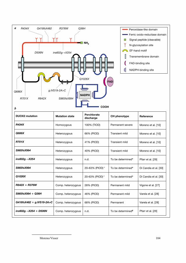
Moreno/Visser 104
Peroxidase-like domain
Ferric oxido-reductase domain
Signal peptide (cleavable)
N-glycosylation site
EF-hand motif
Transmembrane domain
FAD-binding site
NADPH-binding site
DUOX2 mutation Mutation state Perchlorate discharge
CH phenotype Reference
R434X Homozygous 100% (TIOD) Permanent severe Moreno et al. [10]
Q686X Heterozygous 66% (PIOD) Transient mild Moreno et al. [10]
R701X Heterozygous 41% (PIOD) Transient mild Moreno et al. [10]
S965fsX994 Heterozygous 40% (PIOD) Transient mild Moreno et al. [10]
ins602g X254 Heterozygous n.d. To be determined* Pfarr et al. [29]
S965fsX994 Heterozygous 20–63% (PIOD)� To be determined* Di Candia et al. [30]
Q1026X Heterozygous 20-63% (PIOD)� To be determined* Di Candia et al. [30]
R842X � R376W Comp. heterozygous 28% (PIOD) Permanent mild Vigone et al. [27]
S965fsX994 � Q36H Comp. heterozygous 46% (PIOD) Permanent mild Varela et al. [28]
G418fsX482 � g.IVS19-2A>C Comp. heterozygous 68% (PIOD) Permanent Varela et al. [28]
ins602g X254 � D506N Comp. heterozygous n.d. To be determined# Pfarr et al. [29]
COOH
NH2
NADPH
FAD
Q36HR376W
Q686X
R701X
R434X G418fsX482
S965fsX994
Q1026X
g.IVS19-2A>C
R842X
D506N ins602g X254
a
b

New Phenotypes in Thyroid Dyshormonogenesis: Hypothyroidism due to DUOX2 Mutations 105
in phagocytes, active in host defense. THOX2 turned out to be the full-length
version of the partial p138Tox sequence. Applying serial analysis of gene expres-
sion (SAGE) to human thyroid tissue, subtraction of tissue-specific SAGE tags
(individual mRNAs overrepresented in thyroid) and screening of a thyroid
cDNA library with SAGE tag-related ESTs (expressed sequence tags), a clone
containing THOX2 3�-sequences was obtained, showing preferential expression
of the human gene in thyroid tissue [15].
Human THOX genes are located on chromosome 15q15.3, only 16 kb
apart from each other and in opposite transcriptional orientations. THOX1
comprises around 36 kb and contains 35 exons (of which the first two are non-
coding), while THOX2 spans over 22 kb and has 34 exons (the first one being
noncoding). THOX1 and THOX2 proteins contain 1,551 and 1,548 amino
acids, respectively, showing 84% sequence similarity.
Edens et al. [16], who cloned the homologous sequences in Caenorhabditiselegans, suggested a change of nomenclature (DUOX, for dual oxidases) based
on the structural features of these proteins, since two distinct parts can be distin-
guished in DUOXes: a C-terminal segment with high homology to NADPH
oxidases and an N-terminal segment homologous to peroxidases (fig. 2a).
Supporting this change in nomenclature, expression of DUOXes was shown not
to be restricted to the thyroid [15, 17]. Immunohistochemistry localized DUOX
proteins in the apical membrane of the thyrocyte [14] with antibodies cross-react-
ing with the 2 proteins. Based upon the proposed topographic model, the amino-
terminal peroxidase domain would be extracellular, facing the follicular lumen,
and the carboxy-terminal tail of the oxidase domain, intracellular (fig. 2a).
DUOXes are glycoproteins with seven putative transmembrane domains,
between the first and second of which a large intracellular loop exists contain-
ing EF-hand motifs for calcium binding. The C-terminal, intracellular segment
contains typical oxidase domains: 1 FAD-binding site and 4 NADPH-binding
sites (fig. 2a). The presence of EF-hand motifs suggests that calcium ions
Fig. 2. Structural model for DUOX2 protein and mutations found in CH patients.
a Structural and functional domains of DUOX2. Location of genetic defects found in CH
patients at the amino acid level is indicated by dotted lines. The squared legend details the
symbolic figures assigned to each motif or functional feature in DUOX2. The 2007 release
of UniProtkB/Swiss-Prot for the functional topography of Q9NRD8 protein (DUOX2_
HUMAN) recognizes 3 EF-hand motifs in DUOX2, only the first two of which have poten-
tial for Ca2� binding. The g.IVS19-2A�C mutation is represented here as (putatively)
skipping exons 20 and 21 of DUOX2, based on exon-trapping experiments (minigene).
b Characteristics of known human DUOX2 mutations and essentials of human hypothyroid
phenotypes associated with them. n.d. � Not determined; � � range of discharge in a group
of patients; * � expected transient upon the genotype; # � expected permanent upon the
genotype.

Moreno/Visser 106
regulate the enzyme activity. In dog thyrocytes, but not so much in the human,
increasing cAMP levels by forskolin stimulate the expression of DUOXes [14].
Expression of either or of both DUOX proteins is currently documented in
more than 20 tissues [17], but a distinct expression profile for each of the
DUOXes among tissues is emerging that suggests differentially regulated
expression. DUOX2 is highly expressed in thyroid but also along the intestinal
tract, salivary glands and pancreas, while DUOX1 shows preferential expres-
sion over DUOX2 in airway epithelial tract and skin. Even when some tissues
coexpress both DUOX (e.g. thyroid and epithelial airways), their pattern of
expression strongly suggests 2 functionally independent proteins and separate
physiological functions for DUOX1 and DUOX2 [17].
Investigations of the function of DUOXes has been hampered by the lack
of a working cellular model where these proteins could be functionally
expressed in heterologous cell lines. When DUOX enzymes were expressed in
mammalian nonthyroidal cell systems, they remained in the endoplasmic reti-
culum (ER) and did not reach the orthotopic cellular location at the membrane
[18]. Whereas the postulated function of DUOXes in the thyroid was the genera-
tion of H2O2 for thyroid hormone synthesis, this could not be experimentally
confirmed by reconstitution of hydrogen peroxide in vitro. Recently,
Grasberger and Refetoff [19] identified an ER-resident factor, named
DUOXA2, which allows the transition of DUOX2 from ER to Golgi, its matu-
ration and its final translocation to the plasma membrane. By coexpression of
this new factor with DUOX2 in mammalian HeLa cells, they could reconstitute
in vitro H2O2 production. A protein paralog (DUOXA1) was also identified as
the maturation factor for DUOX1. Interestingly, genomic location of both
DUOXA genes lies within the 16 kb that separates DUOX1 from DUOX2 on
chromosome 15, and the ancient genomic rearrangement that evolutionarily
linked DUOX and DUOXA genes suggests their coupled activity as a sort of
eukaryotic operon-like functional unit.
Availability of this functional assay for DUOX2 has straightforward impli-
cations. First, it has now experimentally confirmed the involvement of DUOX2
in H2O2 generation (so far exclusively evidenced by finding DUOX2 defects in
severely hypothyroid patients), but also shows that DUOX2, independently
from DUOX1, suffices for the generation of hydrogen peroxide. Furthermore, it
opens the opportunity for structure-function analyses on DUOX2, including the
study of missense mutations and sequence variations with less obvious patho-
genic effects than the early truncating stop-codon (nonsense) mutations origi-
nally described [10, 20] (fig. 2a).
While the initial expectation was that DUOXes would work in close asso-
ciation with each other and/or with other (putatively cytosolic) proteins in a
coordinate system (as occurs with the gp91phox complex in phagocytes [21]),

New Phenotypes in Thyroid Dyshormonogenesis: Hypothyroidism due to DUOX2 Mutations 107
experimental evidence now supports that membrane-located DUOX2 alone suf-
fices for the generation of hydrogen peroxide. Thus, it seems now more doubtful
that other cytosolic/membranal adjuvant proteins might cooperate with DUOX2
or be necessary components of the functional H2O2-generating machinery in
the thyroid gland. This does not exclude physical interactions between DUOX2
and proteins like the thioredoxin-like EFP1 (identified by 2-hybrid screening
using DUOXes as bait [22]) or the maturation factor DUOXA2 (which stays in
the ER while DUOX2 proceeds towards insertion in the membrane [19]), which
would only represent physical interaction with chaperon-like factors with
the purpose of protein processing, but not a functional cooperation for H2O2
production.
Both DUOX isoforms are present in the thyroid gland, but we currently do
not understand the functional significance of the presence of these two highly
homologous proteins in the same tissue. No functional or structural differences
have been described between DUOX1 and DUOX2 but, again, genotype-phenotype
studies in CH patients suggest that DUOX1 is not involved in H2O2 generation in
the thyroid, since intact DUOX1 cannot functionally rescue the lack of activity
of DUOX2 in patients with either severe or milder hypothyroidism [10].
In the synthesis of thyroid hormone, TPO activity has been classically
regarded as responsible for both the oxidation of iodine prior to iodination of tyrosil
residues of Tg (iodide organification) and the linking of mono- or bi-iodinated
tyrosines to form T3 or T4 (coupling). However, the interesting findings of Edens
et al. [16] that DUOX homologues of C. elegans catalyze the crosslink of tyrosines
on the cuticular extracellular matrix of the worm, inducing its stabilization, sug-
gest the straightforward possibility that DUOXes could be involved, alone or in
cooperation with TPO, in the coupling of iodotyrosines in the Tg molecule. In
this respect, there is controversy as to whether the N-terminal peroxidase domain
of DUOX can be active or not, based on the absence in DUOX sequences of
highly conserved histidine residues that are necessary for heme binding in per-
oxidases [23]. However, recent evidence that the peroxidase-like domain of
DUOX is active when expressed in bacteria [16] and also in eukaryotes (human
respiratory tract epithelial cells [24]) further substantiates this hypothesis.
Again, the in vitro functional study of natural or artificial point mutations in the
peroxidase-like domain of DUOX2 (leaving the oxidase domain intact) might
be pivotal in the analysis of this question.
DUOX2 Phenotypes of CH
To validate the aforementioned findings and investigate the clinical impli-
cations of novel DUOXes, 9 patients diagnosed with CH by neonatal screening

Moreno/Visser 108
were selected for mutational analysis of DUOX1 and DUOX2 genes [10]. All of
them showed a thyroid gland in situ and all had a positive perchlorate discharge
test, indicating that their dyshormonogenesis was due to an iodide organifica-
tion defect, either total (TIOD, 100% discharge: in 1 patient) or partial (PIOD,
20–90% discharge, in 8 patients). These patients did not show clinical features,
biochemical signs or molecular findings suggesting defects in TPO, Tg or PDS
genes.
The patient with TIOD had a severe form of CH and harbored a homozy-
gous nonsense mutation in DUOX2 (R434X) which prematurely truncates the
protein before coding of the first transmembrane domain (fig. 2a). His clinical
phenotype is indistinguishable from the classical phenotype of homozygote
TPO defects. This biallelic mutation encoding a prematurely DUOX2 termina-
tion signal is the only in vivo evidence in humans establishing the role of
DUOX2 in thyroid hormonogenesis. The absence of DUOX2 activity in this
patient, which cannot be compensated by an intact DUOX1, is shown to com-
pletely block the synthesis of thyroid hormone. The exclusive thyroidal pheno-
type in this girl suggests that presence of DUOX2 in other tissues, like digestive
tract or pancreas, is not essential or can at least be compensated by other
sources of reactive oxygen species in these tissues.
Three CH patients with PIOD were found to have heterozygous nonsenseor frameshift mutations (Q686X, R701X and S965fsX994) also leading to pre-
mature truncation of most functional domains of the NADPH oxidase segment
of DUOX2 (fig. 2a). Interestingly, these 3 patients (as well as the additional 5
studied) showed a transient form of CH [10]. Their blood spot T4 and TSH lev-
els in the Dutch screening program showed a milder type of CH, confirmed
later through plasma thyroid function tests. A high uptake of 123I and washout of
40–65% indicated dyshormonogenesis due to PIOD. These three baby girls
were started on T4 substitution treatment, but T4 dosage requirements became
low (1.2–1.4 �g/kg/day) compared to most CH cases. At ages 3–5 years, T4
withdrawal trials showed they could all maintain euthyroidism for 2 months
without thyroxine. These findings for the first time showed that TCH can be a
genetic disease, and disclose that at least a proportion of the etiologically ‘idio-
pathic’ cases of TCH (table 1) correspond to monoallelic DUOX2 defects caus-
ing thyroid dyshormonogenesis.
The apparent paradox that an obviously permanent genetic defect could
cause a transient phenotype of disease deserved justification. We believe that
the most likely explanation relates to the existence of well-documented, high
requirements of thyroid hormone at the beginning of postnatal life (12 �g/kg/day),
which progressively decrease during the first 6 months (6 �g/kg/day) to finally
reach a plateau after the first year (at around 3 �g/kg/day) [25] (fig. 3). In this
setting, it is plausible to think that DUOX2 haploinsufficiency, reducing to 50%

New Phenotypes in Thyroid Dyshormonogenesis: Hypothyroidism due to DUOX2 Mutations 109
the capacity to generate H2O2 and thyroid hormone, could be detectable at
neonatal screening, but the severity of hypothyroidism might progressively
diminish and gradually allow a sufficient H2O2 production which would herald
the beginning of the euthyroid phase of the phenotype (fig. 3). However, a
decreased capacity for hydrogen peroxide and T3/T4 production will character-
ize these patients throughout their lives. This, in combination with the fact that
thyroid hormone requirements increase during pregnancy and maybe also dur-
ing puberty (fig. 3), supports the notion that patients with monoallelic DUOX2
mutations need to be followed up and closely monitored for possible develop-
ment of ‘subclinical’ or overt hypothyroidism after they have reached the euthy-
roid phase of the disease.
Timely detection of possible TSH elevations could prevent goitrogenesis
but, most important, close monitoring of thyroid status of these patients during
pregnancy might detect the development hypothyroxinemia, which would affect
the psychomotor development of the fetus [26]. Furthermore, the fetus might
also harbor the DUOX2 defect, narrowing its own chances for compensation of
thyroid hormone shortage during the 2nd and 3rd trimesters of gestation.
Speculation of a ‘transient-recurrent’ phenotype of hypothyroidism in DUOX2
defects might have to be proven in the future through long-term follow-up of
these (and other) reported patients with TCH. At this point, thyroid function
DUOX2 functional threshold
TCH
Early infancy Puberty Pregnancy
‘Euthyroid phase’
Rel
ativ
e T 4
req
uire
men
t
Fig. 3. Phenotypical features and possible outcome of hypothyroidism due to haploin-
sufficiency of DUOX2. In blue, high requirements for thyroid hormone at the beginning of
life decrease dramatically during the first year of age to reach a plateau, but relative T4 needs
could reincrease during accelerated growth in puberty and during pregnancy. Depending on
individual thresholds for H2O2 production (and T4, T3 synthesis), in red, neonatal-infantile
TCH could be followed by a ‘euthyroid’ phase of the disease, which could be interrupted in
circumstances such as increased thyroid hormone requirement, reduced iodine availability,
thyroid gland damage by inflammation and others. This speculative scheme might not be
exclusive for monoallelic DUOX2 defects, but could be also applicable to biallelic defects
with diminished but residual activity in each allele.

Moreno/Visser 110
monitoring of pregnant mothers of DUOX2 patients with TCH who themselves
carry the DUOX2 defect is clinically advisable and can shed light on the possi-
bility of recurrence of this type of hypothyroidism in adulthood.
Novel DUOX2 Variants, Increased Variability of Phenotypes
Recently, eight novel mutations and one that has already been described in
DUOX2 have been identified in 7 family pedigrees from different countries
(fig. 2) [27–30]. For the first time, some mutations are reported in compound
heterozygosity, while others are simple heterozygous. They comprise nonsense(2), frameshift (3) and missense (3) mutations, as well as one defect putatively
affecting exon splicing. The already described heterozygous S965fsX994
frameshift (due to insertion of 4 base pairs in exon 21 of DUOX2) is now present
in 2 additional index patients of a different genetic background, and emerges as
a DUOX2 sequence segment prone to mutation. While the pathogenic impact of
the 3 missense mutations and the spliced variant of DUOX2 was not formally
ascertained in vitro, the fact that mutations correspond to highly conserved
amino acids in vertebrate DUOX2 homologues and, mainly, because some per-
chlorate discharge tests were reportedly positive in family members harboring
these mutations in simple heterozygosity make these novel defects likely to be
causally linked to the hypothyroid phenotypes described below.
Vigone et al. [27] reported the first familial case of hypothyroidism asso-
ciated with DUOX2 defect. Two Italian siblings harboring the same genotype
(a nonsense R842X mutation and a missense R376W mutation in compound
heterozygosity) showed relevant phenotypic differences. The first sibling was
detected by TSH screening with a moderate but goitrous neonatal hypothy-
roidism (TSH: 173.2 mU/l). His brother was normal at the screening (TSH:
2.9 mU/l) but, because CH was detected in the first sibling, plasma TSH was
determined, showing abnormal increases at 11 days (9.6 mU/l) and 45 days of
life (18.4 mU/l). These important differences were attributed to the higher
iodine load in the second sibling, as determined by urinary iodine. Both broth-
ers were substituted with T4 and after its withdrawal, both showed a mild but
persistent elevation of TSH levels and PIOD (28 and 12% washout, respec-
tively), for which thyroxine substitution was reintroduced. Perchlorate dis-
charge in the euthyroid parents, respectively harboring each of the mutations,
showed very mild but detectable PIOD (13 and 8% washouts). It cannot be
excluded that the parents would have had a mild TCH described for monoallelic
DUOX2 defects. This pedigree is interesting because, first, it suggests that
DUOX2 phenotypes can be modified by environmental factors, such as individual
iodine supplies. Second, it is also interesting because the environmentally

New Phenotypes in Thyroid Dyshormonogenesis: Hypothyroidism due to DUOX2 Mutations 111
mediated phenotypic variability of this DUOX2 defect prevented, in the case of
the second sibling, the routine detection of the disease by neonatal screening,
while biochemical hypothyroidism became clearly expressed after 1 month of
age. The intellectual outcome of the brothers was assessed at T4 withdrawal,
with intellectual quotients (IQs) of 103 and 112, respectively.
Varela et al. [28] reported 2 families whose index patients had permanent
hypothyroidism and PIOD. In family 1, the index patient was detected by
screening. After re-evaluation at 5 years of age, hypothyroidism and goiter were
confirmed and perchlorate discharge indicated PIOD (46% washout). This boy
also had two DUOX2 mutations in compound heterozygosity: the described
S965fsX994 and a novel missense mutation in the peroxidase domain (Q36H)
(fig. 2a). While pathogenicity of the frameshift is clear, the impact of Q36H
(Q36 being well-conserved among DUOX2 homologues) on DUOX2 function
is less clear since 2 family members with the mutation in simple heterozygosity
were reported to be euthyroid and to have a negative discharge test.
In family 2, 2 index patients presented severe neonatal CH and PIOD with
60–68% discharge. Late diagnosis and treatment of the first sibling led to men-
tal retardation. Both are compound heterozygotes for two new DUOX2 vari-
ants, a clearly inactivating frameshift (G418fs482) with very early truncation of
the protein before the first transmembrane domain (fig. 2a), and a nucleotide
transversion in the acceptor site of intron 19 (g.IVS19-2A�C), which seems to
affect exon splicing in minigene studies [28]. In vitro, g.IVS19-2A�C splices
out exon 20, but authors also found skipping of exon 21 in both wild-type and
mutant constructs, possibly representing an undescribed alternatively spliced
mRNA of DUOX2. Worth noting is that if only exon 20 is skipped from
DUOX2 cDNA because of the mutation, exon 21 would enter the sequence out
of frame, leading to an early stop codon which would delete essential domains
of the NADPH oxidase segment of DUOX2 and predict complete inactivation
(fig. 2a). This severe pathogenicity neither seems in accordance with the PIOD
determined in both index patients with 68 and 60% discharge, respectively, nor
with the TIOD (as expected in individuals with biallelic and complete inactiva-
tion of DUOX2 [10]). On the other hand, g.IVS19-2A�C does have an impact
on DUOX2 function, as indicated by the mild euthyroid hyperthyrotropinemia
and PIOD (31% washout) identified in a child family member with this muta-
tion in simple heterozygosity. Besides, when both exons 20 and 21 are spliced
out, exon 22 enters the sequence in frame, encoding a protein with intact FAD-,
NADPH-binding sites and transmembrane domains. The pathogenic impact of
such a mutant would likely be less intense, and this would be more in agreement
with the phenotypes mentioned. Further analyses are needed, e.g. retrotran-
scription of mRNA from blood lymphocytes, to confirm the precise splicing
event taking place in these patients.

Moreno/Visser 112
These two pedigrees from Argentina further reflect the interindividual phe-
notypic variability observed with the same DUOX2 genotypes, within a similar
genetic background. Interestingly, the mild hypothyroidism present in an adult
female of the second pedigree harboring a monoallelic, fully inactivating
DUOX2 mutation (G418fs482) substantiates the notion of possible recurrence
of hypothyroidism during adulthood in children who suffered from neonatal-
infantile TCH.
Genotype-phenotype correlations of the 7 available familial pedigrees with
DUOX2 defects, 4 described in the Netherlands and these 3 novel pedigrees from
Italy and Argentina, suggest that permanency of hypothyroidism due to DUOX2
mutations seems to require alterations in both alleles. Patients with permanent
hypothyroidism and DUOX2 mutations in compound heterozygosity have full
inactivation of at least one DUOX2 allele by severe mutations (nonsense or
frameshifts with premature truncation of the protein) accompanied by counteral-
lele mutations that might allow some residual activity. TCH is so far associated
with monoallelic severe inactivation of DUOX2. More patients’ profiles might
clarify in the future whether biallelic but very subtle defects of DUOX2 might
also associate, under certain environmental conditions (e.g., iodine insufficiency)
or life time events (e.g. immediate postnatal life or pregnancy), with transient
hypothyroidism or whether, in contrast, monoallelic defects under e.g. (perma-
nent) iodine insufficiency could behave as permanent phenotypes. The follow-up
of 3 additional patients with DUOX2 mutations whose short evolution has not yet
allowed classification of their hypothyroidism as permanent or transient will be
helpful to analyze the effect of age on the expression of monoallelic DUOX2
defects in different geographical areas [29, 30].
While other patients with documented thyroid H2O2-generating defects are
found in the literature [31, 32], no DUOX mutations have been studied and/or
identified in these pedigrees. This, together with the absence of DUOX2,
DUOX1 or TPO defects in 5 additional patients with TCH and PIOD included in
the original study describing DUOX2 defects among Dutch patients, is compati-
ble with the existence of other genetic factors involved in hydrogen peroxide
production in the thyroid gland. This includes the novel maturation factor for
DUOX2, DUOXA2, alterations of which have not been linked to any phenotype.
Only one case is reported in the literature that associates alteration in thy-
roidal H2O2 production and thyroid neoplasia [33]. H2O2 generation capacity of
an excised ‘cold nodule’ tissue was found to be lost in comparison to surround-
ing tissue. This finding might not necessarily mean a direct involvement of H2O2
defects in the pathogenesis of the lesion of this patient (which was shown to be
benign), but represents a secondary event framed in the known downregulation
of markers of thyroid differentiation that occur in thyroid neoplasias. On the
other hand, as previously reported, untreated dyshormonogenesis of the thyroid

New Phenotypes in Thyroid Dyshormonogenesis: Hypothyroidism due to DUOX2 Mutations 113
can result in thyroid tumors and malignancies under continuous, long-standing
TSH stimulation [34–37]. This sequence of events is applicable to undiagnosed
DUOX2 defects, only adding to the possible benefits of timely diagnosis and
close follow-up of mild dyshormonogenetic defects of the thyroid.
No human disease associated with DUOX1 defects has been described.
Based on the pattern of expression of the gene and on studies of DUOX genes
in CH patients where DUOX1 was invariably found to be normal, it is tempting
to speculate that putative DUOX1 phenotypes might be rather related to defec-
tive host defense against infection in the airways and other mucosal surfaces
[17] than related to a thyroidal disorder.
Consequences of Transient Neonatal Hypothyroidism
As stated in the introduction, implementation of neonatal screening pro-
grams for CH has successfully eradicated the irreversible and severe neurologi-
cal and psychomotor sequelae that used to devastate the lives of affected
children with undiagnosed or late-diagnosed hypothyroidism in the prescreen-
ing era [2]. Despite this outstanding advance in the health care of the CH popu-
lation and also the continued efforts to optimize screening programs and reduce
to a minimum the time between birth, diagnosis and treatment of patients as
well as to find optimal initial T4 substitution dosages, studies on cognitive and
motor outcomes of hypothyroid children and adolescents reveal the existence of
persistent intellectual and motor deficits [38].
Because of its transitory nature, the consequences of TCH on psychomotor
and intellectual outcome of children may have been underestimated in the past
[39]. Even when TCH does not seem to affect parameters of physical develop-
ment and psychomotor performance [40], accumulating evidence suggests that
neonatal transient hypothyroidism and hyperthyrotropinemia are associated
with impaired intellectual outcome [39, 41, 42]. This impairment is reflected in
the loss of an average of 10 IQ points in children with TCH of 1–3 months’
duration at 9 years of age compared to a matched control population [10]. The
most affected IQ scores are the ones assessing the ‘verbal’ and the ‘perfor-
mance’ skills as well as the global IQ. Most studies showing adverse long-term
intellectual development of TCH children were performed in iodine deficiency
areas, but their conclusions are applicable to any etiology of TCH.
Another problem is posed by the possibility that mildly hypothyroid chil-
dren might ‘escape’ screening as false negatives, leaving us without the option
of follow-up during infancy and childhood. A survey by Calaciura et al. [43]
shows that ‘subclinical’ hypothyroidism in early childhood is a frequent out-
come among children who were catalogued at screening as ‘false negatives’

Moreno/Visser 114
because they had either completely normal (0.8–4.9 mU/l) or nearly normal/
borderline elevated (5–11.7 mU/l) TSH levels at confirmatory examination. The
consequences of persistent hyperthyrotropinemia in children have not been
studied, but in adults it is established that, even minimal abnormalities, may
lead over the course of years to important or irreversible problems [43]. Lipid
metabolism, myocardial function, linear growth and cognitive abilities are
among the functions that may be adversely affected by ‘subclinical hypothy-
roidism’, which should be treated [44–46].
Concluding Remarks
DUOX1 and DUOX2, which are the components of the DUOX system of
NADPH- and Ca2�-dependent oxidases, are the latest members incorporated in
the family of NOX proteins. While DUOX2 has been unequivocally associated
with thyroid physiology through the production of H2O2 in the thyroid gland,
indispensable for thyroid hormonogenesis, DUOX1 has not, and its precise role
in thyroid and other tissues remains obscure.
Biallelic inactivation of DUOX2 in a patient with a complete block of thy-
roid hormone production reflects the importance of this protein for the synthesis
of T3 and T4. Monoallelic inactivation of this gene has also been linked to milder
and transient cases of neonatal hypothyroidism. While a possible recurrence of
hypothyroidism coinciding with increases in T4 requirement during some peri-
ods of life remains a theoretical possibility, the adult mild hypothyroidism shown
in the newly described patients with the same genotype supports this idea.
Additionally, compound heterozygosity for severe DUOX2 mutation and muta-
tions with putative residual activity have been associated with permanent but
mild (‘subclinical’) hypothyroidism. To what extent the clinical expression of
this type of hypothyroidism through life depends on genetic determinants (num-
ber of alleles affected and severity of mutations), environmental factors (such as
individual iodine supplies), physiological (such as puberty or pregnancy, which
increase thyroid hormone demands) or pathological circumstances (such as thy-
roiditis, which reduces the capacity for thyroid hormone production) is a matter
for further study, which has practical implications.
TCH is a frequent clinical situation. Optimization of screening programs
to detect mild transient cases of hypothyroidism, sometimes taken as false
positives at recall [43], is important, both for the negative impact that TCH
has on the achievement of the full intellectual potential of children and to
prevent ‘subclinical hypothyroidism’ in infancy and early childhood, and its
consequences.

New Phenotypes in Thyroid Dyshormonogenesis: Hypothyroidism due to DUOX2 Mutations 115
The discovery of the molecular basis of TCH just started with the identifi-
cation of DUOX2 defects. A few reports suggest that a larger number of either
known [47] or unknown genes might be involved in its pathogenesis. Clinical
and molecular studies will now explore the possibility of genetic heterogeneity
of TCH and be of help in the proper identification, molecular classification and
management of patients with transient forms of hypothyroidism.
References
1 Vanderpump MPJ, Tunbridge WMG: The epidemiology of thyroid diseases; in Braverman LE,
Utiger RD (eds): Werner & Ingbar’s The Thyroid, a Fundamental and Clinical Text, ed 8.
Philadelphia, Lippincott Williams & Wilkins, 2000, pp 467–473.
2 New England Congenital Hypothyroidism Collaborative: Effects of neonatal screening for
hypothyroidism: prevention of mental retardation by treatment before clinical manifestations.
Lancet 1981;ii:1095–1098.
3 Fisher DA, Dussault JH, Foley TP, Klein AH, LaFranchi S, Larsen PR, Mitchell ML, Murphey WH,
Walfish PG: Screening for congenital hypothyroidism: results of screening one million North
American infants. J Pediatr 1996;128:548–554.
4 Coackley JC, Francis I, Gold H, Mathur K, Connelly JF: Transient primary hypothyroidism in the
newborn: experience of the Victorian Neonatal Thyroid Screening Program. Aust Paediatr J 1989;
25:25–30.
5 Vulsma T: Etiology and Pathogenesis of Congenital Hypothyroidism; thesis University of
Amsterdam, 1991.
6 Léger J, Czernichow P: Hyperthyrotropinémie néonatale transitoire. Arch Fr Pediatr 1988;45:
783–786.
7 Vulsma T, de Vijlder JJM: Thyroid disease in newborns, infants and children; in Wass JA,
Shalet SM (eds): Oxford Textbook of Endocrinology and Diabetes. New York, Oxford University
Press, 2002, pp 532–544.
8 De Felice M, Di Lauro R: Thyroid development and its disorders: genetics and molecular mecha-
nisms. Endocr Rev 2004;25:722–746.
9 Moreno JC, de Vijlder JJM, Vulsma T, Ris-Stalpers C: Genetic basis of hypothyroidism: recent
advances, gaps and strategies for future research. Trends Endocrinol Metab 2003;14:318–326.
10 Moreno JC, Bikker H, Kempers MJ, van Trotsenburg AS, Baas F, de Vijlder JJ, Vulsma T,
Ris-Stalpers C: Inactivating mutations in the gene for thyroid oxidase 2 (THOX2) and congenital
hypothyroidism. N Engl J Med 2002;347:95–102.
11 Nose O, Harada T, Miyai K, et al: Transient neonatal hypothyroidism probably related to immatu-
rity of thyroidal iodine organification. J Pediatr 1986;108:573–576.
12 Moreno JC, Keijser R, Gilhuijs-Pederson L, Gestel D, de Vijlder JJM, Ris-Stalpers C: Cloning and
characterization of the human thyroid dehalogenase. Horm Res 2003;60(suppl 2):2.
13 Dupuy C, Ohayon R, Valent A, Noel-Hudson MS, Deme D, Virion A: Purification of a novel flavo-
protein involved in the thyroid NADPH oxidase. Cloning of the porcine and human cDNAs. J Biol
Chem 1999;274:37265–37269.
14 De Deken X, Wang D, Many MC, Costagliola S, Libert F, Vassart G, Dumont JE, Miot F: Cloning
of two human thyroid cDNAs encoding new members of the NADPH oxidase family. J Biol Chem
2000;275:23227–23233.
15 Moreno JC, Pauws E, van Kampen AH, Jedlickova M, de Vijlder JJ, Ris-Stalpers C: Cloning of
tissue-specific genes using serial analysis of gene expression and a novel computational substrac-
tion approach. Genomics 2001;75:70–76.
16 Edens WA, Sharling L, Cheng G, Shapira R, Kinkade JM, Lee T, Heather A, Edens HA, Tang X,
Sullards C, Flaherty DB, Benian GM, Lambeth JD: Tyrosine cross-linking of extracellular matrix
Moreno/Visser 116
is catalyzed by Duox, a multidomain oxidase/peroxidase with homology to the phagocyte oxidase
subunit gp91phox. J Cell Biol 2001;154:879–891.
17 Ris-Stalpers C: Physiology and pathophysiology of the DUOXes. Antioxid Redox Signal
2006;8:1563–1572.
18 De Deken X, Wang D, Dumont JE, Miot F: Characterization of ThOX proteins as components of
the thyroid H2O2 generating system. Exp Cell Res 2002;273:187–196.
19 Grasberger H, Refetoff S: Identification of the maturation factor for dual oxidase. Evolution of an
eukaryotic operon equivalent. J Biol Chem 2006;281:18269–18272.
20 Grasberger H, De Deken X, Miot F, Pohlenz J, Refetoff S: Missense mutations of the dual oxidase
2 (DUOX2) implicated in congenital hypothyroidism have impaired trafficking in cells reconsti-
tuted with DUOX2 maturation factor. Mol Endocrinol DOI:10.1210/me.2007-0018.
21 Leusen JHW, Verhoeven AJ, Roos D: Interactions between the components of the human NADPH
oxidase: intrigues in the phox family. Front Biosci 1996;1:d72–d90.
22 Wang D, De Deken X, Milenkovic M, Song Y, Pirson I, Dumont JE, Miot F: Identification of a
novel partner of duox: EFP1, a thioredoxin-related protein. J Biol Chem 2005;280:3096–3103.
23 Donkó A, Péterfi Z, Sum A, Leto T, Geiszt M: Dual oxidases. Phil Trans R Soc B 2005;360:
2301–2308.
24 Harper RW, Xu C, McManus M, Heidersbach A, Eiserich JP: Duox2 exhibits potent heme peroxi-
dase activity in human respiratory tract epithelium. FEBS Lett 2006;580:5150–5154.
25 Delange F, Bourdoux P, Ketelbant-Balasse P, Van Humskerken A, Glinoer D, Ermans AM:
Transient primary hypothyroidism in the newborn; in Dussault JH, Walker P (eds): Congenital
Hypothyroidism. New York, Marcel Dekker, 1983, pp 275–301.
26 Haddow JE, Palomaki GE, Allan WC, Williams JR, Knight GJ, Gagnon J, O’Heir CE, Mitchell ML,
Hermos RJ, Waisbren SE, Faix JD, Klein RZ: Maternal thyroid deficiency during pregnancy and
subsequent neuropsychological development of the child. N Engl J Med 1999;341:549–555.
27 Vigone MC, Fugazzola L, Zamproni I, Passoni A, di Candia S, Chiumello G, Persani L, Weber G:
Persistent mild hypothyroidism associated with novel sequence variants of the DUOX2 gene in
two siblings. Hum Mutat 2005;26:395.
28 Varela V, Rivolta CM, Esperante SA, Grun2eiro-Papendieck L, Chiesa A, Targovnik HM: Three
mutations (p.Q36H, p.G418fsX482 and g.IVS19-2A�C) in the dual oxidase 2 gene responsible
for congenital goiter and iodide organification defect. Clin Chem 2006;52:182–191.
29 Pfarr N, Korsch E, Kaspers S, Herbst A, Stach A, Zimmer C, Pohlenz J: Congenital hypothy-
roidism caused by new mutations in the dual oxidase 2 (THOX2) gene. Clin Endocrinol 2006;65:
810–815.
30 Di Candia S, Zamproni I, Cortinovis F, Passoni A, Vigone MC, Fugazzola L, Persani L, Weber G:
Congenital hypothyroidism and partial iodide organification defects: two mutations in DUOX2
gene. Horm Res 2006;65(suppl 4):38.
31 Niepomniszcze H, Targovnik HM, Gluzman BE, Curuchet P: Abnormal H2O2 supply in the thyroid
of a patient with goiter and iodine organification defect. J Clin Endocrinol Metab 1987;65: 344–348.
32 Figueiredo MLD, Cardose LC, Ferreira ACF, Campos DVB, da Cruz Domingos M, Corbo R,
Nasciutti LE, Vaisman M, Carvalho DP: Goiter and hypothyroidism in two siblings due to
impaired Ca2�/NAD(P)H-dependent H2O2-generating activity. J Clin Endocrinol Metab 2001;94:
4843–4848.
33 Demester-Mikkine M, van Sande J, Corvilain J, Dumont JE: Benign thyroid nodule with normal
iodide trap and defective organification. J Clin Endocrinol Metab 1975;41:1169–1171.
34 Vickery AL: The diagnosis of malignancy in dyshormonogenetic goitre. Clin Endocrinol Metab
1981;10:317–335.
35 Alabbasy AJ, Delbridge L, Cowell C, Silink M: Microfollicular thyroid adenoma and congenital
goitrous hypothyroidism. Arch Dis Child 1992;67:1294–1295.
36 Camargo R, Limbert E, Gillam M, et al: Aggressive metastatic follicular thyroid carcinoma with
anaplastic transformation arising from a long-standing goiter in a patient with Pendred’s syn-
drome. Thyroid 2001;11:981–988.
37 Alzahrani AS, Baitei EY, Zou M, Shi Y: Clinical case seminar: metastatic follicular thyroid carci-
noma arising from congenital goiter as a result of a novel splice donor site mutation in the thy-
roglobulin gene. J Clin Endocrinol Metab 2006;91:740–746.

New Phenotypes in Thyroid Dyshormonogenesis: Hypothyroidism due to DUOX2 Mutations 117
38 Kempers MJE, van der Sluijs Veer L, Nijhuis-van der Sanden MWG, Kooistra L, Wiedijk BM,
Faber I, Last BF, de Vijlder JJM, Grootenhuis MA, Vulsma T: Intellectual and motor development
of young adults with congenital hypothyroidism diagnosed by neonatal screening. J Clin
Endocrinol Metab 2006;91:418–424.
39 Rapaport R: Congenital hypothyroidism: expanding the spectrum. J Pediatr 2000;136:10–12.
40 Gruters A, Kohler B, Schnabel D, Helge H: Follow-up of thyroid function, thyroid size and devel-
opment in children with transient neonatal hypothyroidism up to the age of 14 years. J Endocrinol
Invest 1994;17(suppl 1):59.
41 Calaciura F, Mendorla G, Distefano M, Castorina F, Fazio T, Motta RM, Sava L, Delange F,
Vigneri R: Childhood IQ measurements in infants with transient congenital hypothyroidism. Clin
Endocrinol (Oxf) 1995;43:473–477.
42 Azizi F, Afkhami M, Sarshar A, Nafarabadi M: Effects of transient neonatal hyperthyrotropinemia
on intellectual quotient and psychomotor performance. Int J Vitam Nutr Res 2001:71:70–73.
43 Calaciura F, Motta RM, Miscio G, Ficher G, Leonardi D, Carta A, Trischitta V, Tassi V, Sava L,
Vigneri R: Subclinical hypothyroidism in early childhood: a frequent outcome of transient neona-
tal hyperthyrotropinemia. J Clin Endocrinol Metab 2002;87:3209–3214.
44 Surks MI, Ocampo E: Subclinical thyroid disease. Am J Med 1996;100:217–223.
45 Hack AE, Pols HA, Visser TJ, Drexhage HA, Hofman A, Witterman JC: Subclinical hypothy-
roidism is an independent risk factor for atherosclerosis and myocardial infarction in elderly
women: The Rotterdam study. Ann Intern Med 2000;132:270–278.
46 McDermott MT, Ridgway EC: Subclinical hypothyroidism is mild thyroid failure and should be
treated. J Clin Endocrinol Metab 2001;86:4591–4599.
47 Niu D-M, Lin C-Y, Hwang B, Jap T-S, Liao J-S, Wu J-Y: Contribution of genetic factors to neona-
tal transient hypothyroidism. Arch Dis Child Fetal Neonatal Ed 2005;90:F69–F72.
José C. Moreno, MD, PhD
Department of Internal Medicine, Erasmus Medical Center, Erasmus University
Dr. Molewaterplein 50
NL–3015 GE Rotterdam (The Netherlands)
Tel. �31 10 463 3385, Fax �31 10 463 5430, E-Mail [email protected]

Van Vliet G, Polak M (eds): Thyroid Gland Development and Function.
Endocr Dev. Basel, Karger, 2007, vol 10, pp 118–126
Thyroid Hormone Transporter Defects
Annette Grüters
Institute for Experimental Pediatric Endocrinology, Charité Children’s Hospital,
Humboldt and Free University, Berlin, Germany
AbstractIn in vitro experiments, active transport of thyroid hormones had been repeatedly
demonstrated. The membrane transporters for thyroid hormones which have been identified
include the organic anion transporting polypeptide, heterodimeric amino acid transporters
and the monocarboxylate transporters (MCT) which are the focus of this chapter. The gene
encoding MCT8 which was identified as a specific thyroid hormone transporter is located on
chromosome Xq13.2. The expression pattern of MCT8 indicates that MCT8 plays an impor-
tant role in the development of the central nervous system by transporting thyroid hormone
into neurons as its main target cells. Mutational analysis of the MCT8 gene revealed muta-
tions or deletions in the MCT8 gene in unrelated male patients with severe psychomotor
retardation and biochemical findings consistent with thyroid hormone resistance. Indeed,
thyroid function tests in patients with MCT8 mutations demonstrated marked elevations of
serum T3 (in the thyrotoxic range), a significant decrease in serum T4 or fT4 and normal to
elevated TSH levels.
Copyright © 2007 S. Karger AG, Basel
Thyroid Hormone Transport into Cells
Thyroid hormones are important for the development of many tissues and
their metabolic function throughout life. Of outmost importance is the critical role
of thyroid hormones in brain and nervous system development [1, 2]. The biologi-
cal effects of thyroid hormones are mediated through the binding of T3 to nuclear
receptors, which leads to a change in the interaction of the receptors with
T3-responsive elements in regulatory regions of many genes [3]. The thyroid produces
mainly thyroxine (T4), which is converted to T3 through outer ring deiodination by
the type 1 (D1) or type 2 (D2) iodothyronine deiodinase. In contrast, inner ring
deiodination of T4 to reverse T3 (rT3) or of T3 to 3,3-diiodothyronine by the type 3
iodothyronine deiodinase (D3) leads to inactivation [4]. It has been assumed that
Disorders of Thyroid Hormone Metabolism
Thyroid Hormone Transporter Defects 119
hepatic D1 is the main source of serum T3, whereas in the central nervous system
(CNS) D2 is important for local regulation of thyroid hormone bioactivity. Since
the T3 receptors and the deiodinases are located in the cells, cellular entry is
required for conversion of the thyroid hormones by intracellular deiodinases. Until
recently it was assumed that the uptake of the lipophilic thyroid hormones into the
cells was achieved by passive diffusion through lipid membranes. However, in
in vitro experiments active transport of thyroid hormones had been repeatedly
demonstrated [5, 6]. The membrane transporters for thyroid hormones which have
been identified include the organic anion transporting polypeptide, heterodimeric
amino acid transporters and the monocarboxylate transporters (MCT).
Monocarboxylate Transporters
To date, 14 members of the MCT family have been identified in various
tissues from different species. The MCTs are proteins of 426–565 amino acids
with 12 predicted transmembrane domains with the N- and the C-terminal
domains of the proteins located inside the cells. Members of the MCT family
transport monocarboxylates such as lactate, pyruvate, and ketone bodies. For
expression at the plasma membrane, MCTs require ancillary proteins. CD147 is
a widely distributed cell surface glycoprotein, which enables the proper expres-
sion and function of MCTs at the cell surface [7]. To date 14 different genes
encoding MCTs are known, which are located on several human chromosomes
(see table 1). All MCTs have a widely distributed expression, some are ubiqui-
tously expressed and several MCTs show a neuronal expression.
The gene encoding MCT8 is located on chromosome Xq13.2. It consists
of 6 exons and codes for a protein of about 67 kDa [8]. The protein contains
12 predicted transmembrane domains and contains at the N-terminus, a PEST
domain rich in Pro (P), Glu (E), Ser (S), and Thr (T) residues (fig. 1). PEST
domains serve as proteolytic signals, which result in rapid degradation of the
protein and a reduced half-life. Expression of MCT8 was described in the
human heart, brain, placenta, lung, kidney, skeletal muscle and in the liver. No
biological function of MCT8 was known until Friesema et al. [9] identified
MCT8 as a specific thyroid hormone transporter.
It was shown that expression of MCT8 induced an approximate 10-fold
increase in uptake of T4 and T3 relative to control oocytes. MCT8 transports T4,
T3, rT3, and T2, but not sulfonated iodothyronines, the amino acids Tyr, Trp,
Leu, and Phe, and lactate. There is a high degree of homology in the amino
sequences of rat, mouse, and human MCT8.
MCT8 is expressed in numerous human tissues, including brain, heart,
placenta, lung, kidney, skeletal muscle and liver. Detailed studies of MCT8
Grüters 120
expression in the CNS have been performed in mice [10]. Highest transcript
levels for MCT8 were detected in the choroid plexus of the lateral, third, and
fourth ventricles. Significant MCT8 mRNA concentrations were also found in
the amygdala, in the pyramidal cell layer of the hippocampal formation and in
the granule cell layer of the dentate gyrus. Hybridization was also found
throughout striatal areas, with high transcript levels in the olfactory tubercle
and to a lesser extent in the caudate-putamen and nucleus accumbens. Lower
expression signals were detected in the striatum and cerebellum. MCT8 is
mainly expressed in neurons, in which coexpression of D2 could be demon-
strated [10]. This expression pattern of MCT8 indicates that MCT8 plays an
important role in the development of the CNS by transporting thyroid hormone
into neurons as its main target cells.
Table 1. Clinical and biochemical findings in patients with MCT8 mutations, patients with Allan-
Herndon-Dudley syndrome and in mice with targeted deletion of MCT8
Clinical symptoms Patients with MCT8 Patients with MCT8
mutations Allan-Herndon- knockout mice
Dudley syndrome
Generalized muscular hypotonia in infancy � � absent
Increasing spasticity with age � � absent
Spastic quadriplegia � � absent
Involuntary dystonic movements � � absent
Nystagmus � � not reported
Vision normal normal normal
Secondary microcephalus � � �Feeding problems � � absent
Sleeping disorder � not reported absent
Seizures � (�) absent
Mental retardation � � �Ability to walk absent � �Ataxia � � �Speech development absent � not applicable
Dysarthric speech � � not applicable
Facial abnormalities � � absent
(elongated face,
depressed nasal bridge)
Increased serum T3 � � �Low serum T4 � � �Normal/increased TSH � � �Increased D1/D2 activity � not reported �

Thyroid Hormone Transporter Defects 121
MCT8 mRNA was detected also in early first trimester placenta with a
significant increase with advancing gestation. MCT8 immunostaining was
demonstrated in villous cytotrophoblast, syncytiotrophoblast and extravillous
trophoblast cells with increasing intensity with advancing gestation. It has been
concluded that the expression of MCT8 in placenta from early gestation is
compatible with an important role in thyroid hormone transport during fetal
development and a specific role in placental development [11].
Human Phenotype of MCT8 Mutations
Since the MCT8 gene is located on the X chromosome, it was suggested
that mutations in MCT8 could cause an X-linked form of thyroid hormone
resistance and mental retardation. Subsequently, mutational analysis of the
MCT8 gene revealed mutations or deletions in the MCT8 gene in unrelated
male patients with severe psychomotor retardation and biochemical findings
consistent with thyroid hormone resistance [12–14]. Until the mutation was
identified in all these boys the cause of the retardation had been unknown. In
the mothers of all patients the mutations were detected as well, but as expected
Fig. 1. Putative structure of the MCT8 protein.

Grüters 122
for an X-linked disorder, none of the mothers had signs of mental retardation or
impairment of motor development.
More recently, MCT8 gene mutations have been found to be the cause of
Allan-Herndon-Dudley syndrome, an X-linked syndromic form of mental
retardation first described in 1944 [15] (Online Mendelian Inheritance in Man,
access No. OMIM 309600).
Clinical FindingsThe clinical findings in most patients with MCT8 mutations are uniform:
in the first year of life general hypotonia with a complete inability to hold the
head, lack of eye fixation and involuntary movements is observed. Later on, the
inability to sit and walk as well as involuntary dystonic movements of the arms
and spontaneous activity of facial muscles are observed. Primitive (glabella,
snout) reflexes are easily elicited or occur spontaneously as well as bilateral
asymmetric tonic neck reflex.
Nystagmus is frequently present, but hearing and vision are found to
be normal, as demonstrated by brainstem evoked response audiometry.
Microcephaly may occur, but it is possible that this is a secondary event.
Generalized epilepsy with tonic and tonic-clonic seizures leads to antiepileptic
drug treatment in many patients. In some patients, mental development may
improve with age, since smiling, laughing as well as voluntary grasping is present
in older patients. Most likely as a consequence of the neurological impairment,
severe feeding problems and failure to thrive are a frequent complication some-
times requiring a gastrostomy feeding tube. Sleeping disorders are also a frequent
finding and speech development is absent in all patients. Spastic quadriplegia
has developed in some of the patients.
Allan-Herndon-Dudley syndrome was one of the first described syndromes
of X-linked mental retardation. In six large families linkage studies have mapped
the gene locus to Xq13.2 and after the identification of the human MCT8 muta-
tions described above, mutations have been found in each of the six families.
There are some differences with the clinical findings described above. Some of
these patients learned to walk independently, but exhibited severe ataxia, which
ultimately made them wheelchair-bound. Patients who learned to speak exhib-
ited dysarthric speech. There is a transition in childhood to spasticity manifested
by hyperreflexia at the large joints, clonus, and a positive Babinski sign.
In addition to the neurological findings comparable to those described
above, facial manifestations with bifrontal narrowing, simply formed or cupped
ears, elongated and myopathic faces with midface hypoplasia, narrow high
palate, anteverted nares, spaced teeth and square faces were described.
The development of all 32 affected males who were evaluated was severely
impaired from birth. Compatibility with longevity was evident from the 8 affected

Thyroid Hormone Transporter Defects 123
males, of the 32 that were evaluated, who lived beyond 70 years. Since most
affected males were unable to meet the baseline of standardized cognitive tests,
the precise level of mental retardation could not be determined.
Prenatal and postnatal growth as well as pubertal development seem to
be grossly normal. Other clinical symptoms observed in congenital hypothy-
roidism, e.g. prolonged jaundice, constipation, bradycardia, delayed skeletal
maturation or skin manifestations such as myxedema, have never been reported.
Thyroid Function TestsThyroid function tests in the patients with MCT8 mutations demonstrated
marked elevations of serum T3 (in the thyrotoxic range), a significant decrease
in serum T4 or fT4 and normal to elevated TSH levels, compatible with a
diagnosis of apparent thyroid hormone resistance. rT3 levels are normal or
decreased.
When measured, serum sex hormone-binding globulin was found to be
significantly elevated, indicating a hyperthyroid state of the liver.
In some of the patients treatment with high doses of L-thyroxine sup-
pressed serum TSH, but had no clinical effects. In contrast, in 1 patient treat-
ment with T3 alone (20 �g/day) induced a decrease in serum TSH and fT4 and
treatment with T4 (100 �g/day) plus T3 (30 �g/day) resulted in impaired weight
gain and increased sweating, but did not improve neurological symptoms [16].
From these manifestations, it can be concluded that mutations in MCT8
predominantly result in a severe neurological phenotype, because MCT8 is
essential for the proper local supply of T3 to neurons of the CNS avoiding lack
or excess of thyroid hormones. However, it still cannot be excluded that MCT8
might also be involved in the transport of other unidentified substances.
Since typical clinical symptoms of hypothyroidism are lacking in other organ
systems impaired MCT8 function seems to cause tissue-specific hypothy-
roidism in the (developing) CNS. The lack of manifestations in other organs
therefore implies that there are other compensating thyroid hormone transport
mechanisms in these tissues. However, since besides the liver, no other organ,
e.g. the intestinal tract and the heart, exhibits hyperthyroid symptoms, whether
enhanced local degradation of thyroid hormones is present in these tissues
remains to be established.
Molecular FindingsIn figure 2, the mutations which have been identified so far are depicted in
red. In vitro functional studies with transfected mutant constructs of the mis-
sense mutations revealed the absence of T3 uptake for most of the mutants. Only
the transfection of the Arg271His mutation resulted in a residual T3 uptake,
which was however significantly reduced compared with transfection of the

Grüters 124
wild-type (WT) MCT8. Furthermore, with the Ala150Val mutant, a reduced
cell surface expression of the mutated transporter in comparison to WT MCT8
was demonstrated. The subcellular localization of the mutated transporter
revealed a more reticular expression pattern than expression on the cell mem-
brane, which could be clearly demonstrated for the WT MCT8. Moreover, it
was shown that the Ala150Val mutant MCT8 forms homodimers and that
dimerization is comparable to the WT MCT8 in vitro [16].
Studies of cultured skin fibroblasts isolated from the patients demonstrated
a decreased uptake of both T4 and T3 [17] as well as a significantly increased
D2 activity. The increased conversion of T4 to T3 by D2 together with a
decreased T3 uptake may lead to the decreased serum T4 and increased serum T3
concentrations.
Animal Model: Targeted Disruption of MCT8 in Mice
As described by Dumitrescu [17] and according to known observations in
a different knockout approach, deletion of the MCT8 gene in mice leads to bio-
chemical findings comparable to those observed in human patients. The MCT8
Fig. 2. Human mutations of the MCT8 protein.

Thyroid Hormone Transporter Defects 125
knockout mice generated by Dumitrescu et al., like the human patients, present
with low T4 and high T3 serum concentrations and pituitary resistance to thyroid
hormone. Also in the animal model increased D1 and D2 activities were observed
in several tissues including the CNS.
In spite of the fact that MCT8 deletion reproduced the biochemical pheno-
type in mice, no obvious signs of neurological disturbances were observed: the
mice did not present with abnormalities in motor development or behavior. The
crucial questions to be addressed in further research studies are the potential
compensating mechanisms of thyroid hormone transport in rodents, e.g. by
other transporters or different sensitivities of developing structures of the CNS
to hyper- or hypothyroidism. The lack of MCT8-mediated thyroid hormone
transport in MCT8 knockout mice is not comparable to the situation in Pax8�/�
mice, because residual thyroid hormone function may be present due to alterna-
tive transport mechanisms.
Concluding Remarks
Hemizygous MCT8-deficient males present a syndrome with two compo-
nents: a thyroid defect (increased total and free serum T3 and decreased total
and free T4 and rT3 concentrations) and severe psychomotor and developmental
delay (generalized dystonia combined with spasticity, mental retardation, lack
of verbal communication, poor head control and coordination). As is the case
for most X-linked diseases, males are more severely affected in terms of both
the neurological and thyroid defects, whereas female carriers have only mild
thyroid function test abnormalities. Since MCT8 is not the only thyroid hor-
mone transporter, different human tissues may express different transporters.
Mutations in MCT8 will cause local hypothyroidism in tissues depending on
MCT8, possibly in different regions of the developing CNS. Concomitantly, the
high serum T3 concentrations may cause a hyperthyroid state of tissues
independent of MCT8, e.g. the liver. Therefore, the differences between the
human and rodent phenotype may reflect differences in the effect of hypo- and
hyperthyroid states of various tissues, especially of the developing CNS.
References
1 Oppenheimer JH, Schwartz HL: Molecular basis of thyroid hormone-dependent brain develop-
ment. Endocr Rev 1997;18:462–475.
2 Morreale de Escobar G, Obregón MJ, Escobar del Rey F: Role of thyroid hormone during early
brain development. Eur J Endocrinol 2004;151:U25–U37.
3 Bernal J: Action of thyroid hormone in brain. J Endocrinol Invest 2002;25:268–288.

Grüters 126
4 Bianco AC, Salvatore D, Gereben B, Berry MJ, Larsen PR: Biochemistry, cellular and molecular
biology, and physiological roles of the iodothyronine selenodeiodinases. Endocr Rev 2002;23:
38–89.
5 Hennemann G, Docter R, Friesema EC, de Jong M, Krenning EP, Visser TJ: Plasma membrane
transport of thyroid hormones and its role in thyroid hormone metabolism and bioavailability.
Endocr Rev 2001;22:451–476.
6 Friesema EC, Jansen J, Milici C, Visser TJ: Thyroid hormone transporters. Vitam Horm 2005;70:
137–167.
7 Kirk P, Wilson MC, Heddle C, Brown MH, Barclay AN, Halestrap AP: CD147 is tightly associated
with lactate transporters MCT1 and MCT4 and facilitates their cell surface expression. EMBO J
2000;19:3896–3904.
8 Lafrenière RG, Carrel L, Willard HF: A novel transmembrane transporter encoded by the XPCT
gene in Xq13.2. Hum Mol Genet 1994;3:1133–1139.
9 Friesema EC, Ganguly S, Abdalla A, Manning Fox JE, Halestrap AP, Visser TJ: Identification of
monocarboxylate transporter 8 as a specific thyroid hormone transporter. J Biol Chem 2003;278:
40128–40135.
10 Heuer H, Maier MK, Iden S, Mittag J, Friesema EC, Visser TJ, Bauer K: The monocarboxylate
transporter 8 linked to human psychomotor retardation is highly expressed in thyroid hormone-
sensitive neuron populations. Endocrinology 2005;146:1701–1706.
11 Chan S-Y, Franklyn JA, Pemberton HN, Bulmer JN, Visser TJ, McCabe CJ, Kilby MD:
Monocarboxylate transporter 8 expression in the human placenta: the effects of severe intrauterine
growth restriction. J Endocrinol 2006;189:465–471.
12 Friesema EC, Grueters A, Biebermann H, Krude H, von Moers A, Reeser M, Barrett TG, Mancilla
EE, Svensson J, Kester MH, et al: Association between mutations in a thyroid hormone transporter
and severe X-linked psychomotor retardation. Lancet 2004;364:1435–1437.
13 Dumitrescu AM, Liao XH, Best TB, Brockmann K, Refetoff S: A novel syndrome combining thy-
roid and neurological abnormalities is associated with mutations in a monocarboxylate transporter
gene. Am J Hum Genet 2004;74:168–175.
14 Lenzner S, Rosenkranz MD, Grueters A, Biebermann H, Chelly J, Moraine C, Frijns J, van
Bokhoven H, Visser T, Ropers H: Severe X-linked mental retardation caused by mutations in
the gene for the thyroid hormone transporter MCT8. European Society of Human Genetics
Concurrent Symposia, Munich, 2004.
15 Schwartz CE, May MM, Carpenter NJ, Rogers RC, Martin J, Bialer MG, Ward J, Sanabria J,
Marsa S, Lewis JA, et al: Allan-Herndon-Dudley syndrome and the monocarboxylate transporter 8
(MCT8) gene. Am J Hum Genet 2005;77:41–53.
16 Biebermann H, Ambrugger P, Tarnow P, von Moers A, Schweizer U, Grueters A: Extended clinical
phenotype, endocrine investigations and functional studies of a loss-of-function mutation A150V
in the thyroid hormone specific transporter MCT8. Eur J Endocrinol 2005;153:359–366.
17 Dumitrescu AM, Liao XH, Weiss RE, Millen K, Refetoff S: Tissue-specific thyroid hormone
deprivation and excess in monocarboxylate transporter (Mct) 8-deficient mice. Endocrinology
2006;147:4036–4043.
Prof. Annette Grüters
Institute for Experimental Pediatric Endocrinology
Charité Children’s Hospital, Humboldt and Free University
Augustenburger Platz 1, DE–13353 Berlin (Germany)
Tel. �49 30 450 566 251, Fax �49 30 450 566 936, E-Mail [email protected]

Van Vliet G, Polak M (eds): Thyroid Gland Development and Function.
Endocr Dev. Basel, Karger, 2007, vol 10, pp 127–139
Novel Biological and Clinical Aspectsof Thyroid Hormone Metabolism
Alexandra M. Dumitrescua, Samuel Refetoff a–c
Departments of aMedicine and bPediatrics and cCommittee on Genetics,
University of Chicago, Chicago, Ill., USA
AbstractIntracellular metabolism of thyroid hormone (TH) and availability of the active hor-
mone T3 is regulated by three selenoprotein iodothyronine deiodinases (Ds). D1 and D2 con-
vert the precursor T4 into the active hormone, T3. D3 is the principal inactivator of T4 and T3
to their respective metabolites, rT3 and T2. While acquired changes in D activities are com-
mon, inherited defects in humans were not known. Recently, two families with abnormal thy-
roid function tests, high serum T4, high rT3, low T3 and slightly increased TSH, were
identified. Linkage analysis and sequencing excluded abnormalities in all 3 DIO genes, yet
clinical studies showed reduced responsiveness to T4 but not to T3. Extensive search for puta-
tive defects in genes involved in D2 metabolism led to the identification of mutations in the
Sec insertion sequence binding protein (SBP)2 gene, involved in the synthesis of selenopro-
teins, including Ds. Affected children were either homozygous or compound heterozygous
for these mutations. Other selenoproteins, including glutathione peroxidase, were also
reduced in affected subjects, confirming a generalized effect of the SBP2 defect. Opposite
thyroid test abnormalities are found in mutations of the TH transporter MCT8, and appear to
be caused by the resulting increases in D2 and D1 activities.
Copyright © 2007 S. Karger AG, Basel
Introduction and Physiologic Background
Thyroid hormones (TH) are iodinated compounds known to influence gene
expression and metabolism in tissues of all vertebrate animals. The effects of TH
are dependent on the quantity of the hormone that reaches peripheral tissues,
their intracellular availability and the presence of unaltered TH receptors. Entry
of TH into cells is an active process and several classes of TH membrane trans-
porters with different kinetics and substrate preferences have been identified [1].
Disorders of Thyroid Hormone Metabolism
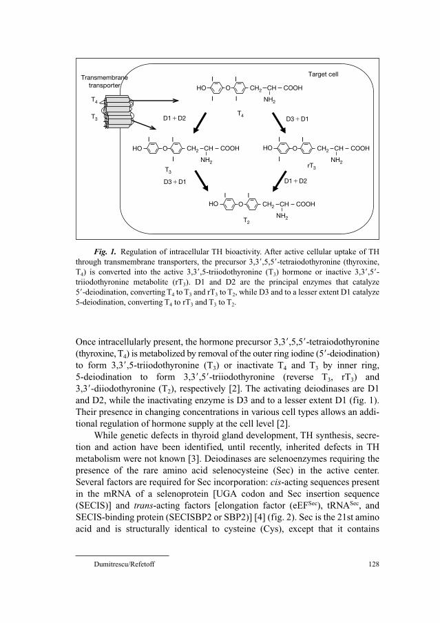
Dumitrescu/Refetoff 128
Once intracellularly present, the hormone precursor 3,3�,5,5�-tetraiodothyronine
(thyroxine, T4) is metabolized by removal of the outer ring iodine (5�-deiodination)
to form 3,3�,5-triiodothyronine (T3) or inactivate T4 and T3 by inner ring,
5-deiodination to form 3,3�,5�-triiodothyronine (reverse T3, rT3) and
3,3�-diiodothyronine (T2), respectively [2]. The activating deiodinases are D1
and D2, while the inactivating enzyme is D3 and to a lesser extent D1 (fig. 1).
Their presence in changing concentrations in various cell types allows an addi-
tional regulation of hormone supply at the cell level [2].
While genetic defects in thyroid gland development, TH synthesis, secre-
tion and action have been identified, until recently, inherited defects in TH
metabolism were not known [3]. Deiodinases are selenoenzymes requiring the
presence of the rare amino acid selenocysteine (Sec) in the active center.
Several factors are required for Sec incorporation: cis-acting sequences present
in the mRNA of a selenoprotein [UGA codon and Sec insertion sequence
(SECIS)] and trans-acting factors [elongation factor (eEFSec), tRNASec, and
SECIS-binding protein (SECISBP2 or SBP2)] [4] (fig. 2). Sec is the 21st amino
acid and is structurally identical to cysteine (Cys), except that it contains
Target cell
T3rT3
Transmembrane transporter HO O
I
I
I
I
HO O CH2 COOH
NH2
CH CH2 COOH
NH2
CH
CH2 COOH
NH2
CH
CH2 COOH
NH2
CH
I I
I
HO OI
I
I
T4
HO OI I
T2
T4
T3 D1�D2
D3�D1
D3�D1
D1�D2
Fig. 1. Regulation of intracellular TH bioactivity. After active cellular uptake of TH
through transmembrane transporters, the precursor 3,3�,5,5�-tetraiodothyronine (thyroxine,
T4) is converted into the active 3,3�,5-triiodothyronine (T3) hormone or inactive 3,3�,5�-triiodothyronine metabolite (rT3). D1 and D2 are the principal enzymes that catalyze
5�-deiodination, converting T4 to T3 and rT3 to T2, while D3 and to a lesser extent D1 catalyze
5-deiodination, converting T4 to rT3 and T3 to T2.

Novel Biological and Clinical Aspects of TH Metabolism 129
selenium instead of sulfur. Sec has a distinct functional advantage at physiolog-
ical pH. When Sec is replaced with Cys, the catalytic activity of a selenoen-
zyme is drastically reduced [5].
Sec is incorporated through recoding of a UGA codon present in the
mRNAs of a selenoprotein, dictated by the presence of SECIS, a characteristic
stem-loop RNA structure in the 3� untranslated region. Using the SECIS ele-
ment as bait, the rat SECIS-binding protein, SBP2, was purified and cloned in
2000 [6]. We have recently identified a genetic defect in SBP2 that results in
deficient deiodinase enzymatic activity perturbing the TH metabolism that
manifested as a novel thyroid phenotype.
Clinical Presentation
Several members of two families presented with similar thyroid function test
(TFT) abnormalities, namely, high T4, low T3, high rT3 and slightly elevated TSH.
One family of Bedouin origin from Saudi Arabia (family A) was referred
because of abnormal TFTs in three of seven siblings (fig. 3a). The propositus,
Cis-acting sequences
Start Stop
5’UTR 3’UTRCoding
UGA SECIS
Trans-acting factors
Start Stop
5’UTR
3’UTR
Coding
UGA
SBP2
EFSectRNASec
a
b
Fig. 2. Components involved in Sec incorporation. a Cis-acting sequences present in
the mRNA of selenoproteins: an in-frame UGA codon and Sec incorporation sequence
(SECIS) element, a stem loop structure located in the 3�UTR (untranslated region). b Binding
of the trans-acting factor SECIS-binding protein (SBP2) which recruits the Sec-specific
elongation factor (EFSec) and Sec-specific tRNA (tRNASec). The result is a recoding of the
UGA codon and Sec incorporation.

Dumitrescu/Refetoff 130
SBP2 R540Q mutation
.. E Q Q ..GA
.. E R Q ..
WT Het Mut
II
I2
TT4 (µg/dl) TT3 (ng/dl)
TrT3 (ng/dl) TSH (mU/l)
10.3 133 32.6 2.6
8.4 130 23.5 2.4
14.2 73
49.3 4.1
8.2 131 29.3 1.9
9.2 147 22.3 1.8
9.8 133 34.5 1.4
15.7 95
54.2 3.7
17.9 86
81.8 5.4
7.8 93
20.9 2.0
1
2
Normal range5.0–11.8 90–185*
14.5–35.0 0.4–3.6
6 71 3 4 5
16 14 13 11 9.5 7
3847
221133
121221
121221
332242
121221
332242
121221
221133
121221
121221
121221
121221
332242
221133
121221
121221
121221
121221
16 –18 cM
SBP2 locus
a
b
c
L-T4 treatment L-T3 treatment
T4 (µg/dl)
TSH
(mU
/l)
2016128
0.01
0.1
1
10
�0.005400300200100
�0.0050.01
0.1
1
10
T3 (ng/dl)
TSH
(mU
/l)
d
e
f
D2 enzymatic activity
Unaffected Affected0
200
400
600
Cha
nge
vs. u
ntre
ated
(%) DIO2 mRNA expression
Cha
nge
vs.
norm
al b
asel
ine
(%)
Unaffected Affected0
100
300
500
Serum SePP
Unaffected Affected0
40
80
120p�0.001
Inte
grat
ed d
ensi
ty (A
U)
0
40
80
120
Unaffected Affected
Cha
nge
vs. n
orm
al (%
)
Serum GPx activity
*
*
**
p�0.001
p�0.01
Affected Unaffected(II-6)(II-2)
(II-3)(II-5)
Baseline �1 mM db-cAMP
4
Fig. 3. Identification of a SBP2 gene mutation and the resulting in vivo and in vitro
abnormalities. a Pedigree and TFTs in members of family A. TFT values are aligned under
each subject symbol. TT4 � Total T4; TT3 � total T3; TrT3 � total reverse T3. Abnormal val-
ues are in bold characters, high values in red, low values in blue. * � The normal range for
children is 100–205 �g/dl. b Haplotyping with genetic markers overlapping the SBP2 locus.
Affected share homozygous haplotypes while the unaffected parents and siblings are het-
erozygous. c Sequences of the SBP2 region harboring the R540Q mutation. Arrow indicates
the location of the G → A transition in a CpG dinucleotide changing the codon CGG to
CAG. Sequences are those of an unaffected control (WT), a heterozygous parent (Het) and
an affected homozygous subject (Mut). Corresponding amino acids are written above the
nucleotide triplet. d Serum TSH and corresponding serum T4 and T3 levels, before and during
the oral administration of incremental doses of L-T4 and L-T3. Note the higher concentra-
tions of T4 required to reduce serum TSH in the affected subjects. e Deiodinase 2 enzymatic
activity and mRNA expression. Bars indicate � SEM. Baseline and stimulated D2 activity is
significantly lower in affected. There is a significant increase of DIO2 mRNA with db-
cAMP, in both normals and affected (*p � 0.001). There are no significant differences in
baseline and db-cAMP-stimulated DIO2 mRNA in affected versus the unaffected. f Effect of
SBP2 deficiency on other selenoproteins. Bars indicate � SEM. GPx enzymatic activity in

Novel Biological and Clinical Aspects of TH Metabolism 131
the second child born to unrelated parents from the same tribe, was 14 years old
when he presented with short stature. Since age 11 he had been growing below
the 3rd percentile and bone ages were 7 and 9 at chronological ages of 11 and 13,
respectively, while still prepubertal [7]. Somatomedin C and stimulated growth
hormone values were within the normal range. It is of note that, at neonatal
screening, his serum TSH was high but no treatment was given as serum T4 was
not low. TSH values remained above normal but development proceeded nor-
mally. There was no hearing impairment as assessed by an audiogram. The con-
centrations of total T4, free T4 and total rT3 were high while that of total and free
T3 were low. The same pattern of TFT abnormalities was found in a 7-year-old
brother and 4-year-old sister (fig. 3a), both clinically euthyroid. TFT of all other
family members, including parents and 4 siblings, were normal.
In the second family (family B) of mixed Irish and African origin, the sin-
gle affected child presented at age 6 years with growth retardation and thyroid
abnormalities similar to the children of family A. The mother mentioned fre-
quent fainting after exertion but clinical, biochemical and electrophysiological
investigations did not detect abnormalities.
In vivo and in vitro Studies
Defects in serum and transmembrane [8, 9] transport of TH were excluded.
The potency of L-T4 compared to L-T3 to suppress TSH was used to test the
hypothesis of a defect in deiodinase-mediated iodothyronine metabolism. When
incremental doses of L-T4 and L-T3 were given to 2 affected and 2 normal sib-
lings, higher doses and serum concentrations of T4, but not T3, were required to
reduce TSH in the affected siblings, suggesting a defect in T4 to T3 conversion
(fig. 3d). However linkage of the phenotype to loci of the three deiodinases was
excluded and their sequences were normal.
Cultured skin fibroblasts were used to test in vitro the hypothesis of abnor-
mal T4 to T3 conversion. Because D2, an integral membrane ER-resident sele-
noenzyme [2], but no D1 or D3 is expressed in these cells, all subsequent
studies measured D2 expression and function. As the DIO2 gene contains a
cAMP-responsive element (CRE), the synthetic analogue dibutyryl-cAMP (db-
cAMP) also stimulates D2 expression and activity. The baseline D2 activity in
serum from all 4 affected children and 10 unaffected individuals. Results are expressed as
percent of the average of unaffected subjects (*p � 0.001). SePP levels in serum from all 4
affected and available unaffected members of both families were quantified by scanning the
Western blot. AU � Arbitrary units.
Dumitrescu/Refetoff 132
fibroblasts from the affected subjects was reduced to near or below the limit of
detection (fig. 3e). In all fibroblasts from normal individuals, DIO2 mRNA and
enzymatic activity increased with db-cAMP stimulation, whereas in the
affected subjects, the increase in DIO2 mRNA was not accompanied by a cor-
responding increase in enzymatic activity (fig. 3e). These results further sup-
port the clinical findings of defective TH metabolism and provided direct
evidence for abnormal D2 function.
SBP2 Gene Mutations
A posttranscriptional defect through impaired D2 metabolism or synthesis
was considered as possibly responsible for the reduced enzymatic activity.
Supply of active D2 enzyme is regulated by degradation in proteosomes through
ubiquitination [10] and by deubiquitination [11]. Linkage was excluded for
genes encoding proteins involved in D2 ubiquitination (UbE2G1, UbE2G2,UbE2L3) [10] and in D2 de-ubiquitination (VDU1, VDU2) [11]. From the com-
ponents of the Sec incorporation machinery [4], a defect in cis sequences,
tRNASec and EFSec was excluded by linkage analysis or by sequencing.
However, affected subjects shared homozygous haplotypes at the SBP2 locus
(fig. 3b). Sequencing revealed a missense mutation in exon 12, R540Q.
Affected children were homozygous and the parents were heterozygous carriers
(fig. 3c). The haplotype harboring the mutation is most likely inherited identical
by descent in this Bedouin family. Extensive genotyping with markers flanking
the locus showed a 16- to 18-cM segment shared identical by descent by the
parents, suggestive of a recent common ancestor.
Mutations in the SBP2 gene were also found in family B, the affected child
being compound heterozygous. He inherited from his father a nonsense muta-
tion (K438X) and from the mother, an intronic mutation (IVS8ds�29 G → A).
The latter mutation creates an alternative donor splice site producing an alter-
native transcript that incorporates 26 bp into exon 8. The abnormally spliced
transcripts represented 52% of the transcripts generated from the mutant mater-
nal allele in lymphocytes and change the reading frame to produce a putative
truncated protein.
Population screens showed that these SBP2 gene sequence differences
were not polymorphic in the respective populations. The homozygous mutation
R540Q in family A is a nonsynonymous change located in a conserved amino
acid across species and likely creates a hypomorphic rather than a null allele. In
family B, IVS8ds�29 G → A results in partial alternative splicing and abnor-
mal transcripts. The total amount of normal transcripts in the affected child
was estimated to be 24%. In both instances the result is a partial, rather than a

Novel Biological and Clinical Aspects of TH Metabolism 133
complete defect predicting a reduced rather than complete deficiency of seleno-
protein synthesis and a mild phenotype.
Consequences for Other Selenoproteins
As SBP2 is epistatic to selenoprotein synthesis, identification of decreased
D2 activity due to recessive SBP2 defect was likely to affect other selenopro-
teins. As D1 and D3 are too low to measure or absent in skin fibroblasts and
lymphocytes, other selenoproteins were assessed. Glutathione peroxidase
(GPx) activity was 7.5-fold lower in serum (fig. 3f) and 3.3-fold lower in
fibroblasts of the affected as compared to normal subjects. Furthermore,
selenoprotein P (SePP) levels in serum were significantly lower in the affected
of both families compared with the unaffected (fig. 3f).
SBP2 is expressed at low levels in all tissues tested, with a high expression
and an additional transcript in testis [12]. Human SBP2, cloned in 2002 [12],
has 854 amino acids. The C-terminal domain is sufficient for all known func-
tions of SBP2 including SECIS binding, ribosome binding and Sec incorpora-
tion [13]. A unique N-terminal domain contains a strong nuclear localization
signal but its precise function is unknown [12].
How could a gene involved in the synthesis of an entire class of proteins
manifest only as abnormal TFT? There appears to be a hierarchical preservation
of selenoproteins during Se deprivation, conserving the enzymatic activity of
those with a more important function [13, 14]. This hierarchy is supposedly
produced by the rates of selenoprotein degradation and by the functional
demands of particular selenoproteins. Therefore, selenoproteins with short half-
life and high demand for their function, such as D2, might be the first to fail
when the Sec incorporation machinery becomes inefficient.
Other Inherited Defects That Affect Iodothyronine Deiodination in Humans
Mutations in the monocarboxylate transporter 8 (MCT8) gene, located on
the X chromosome, were first reported in 2004 [8, 9]. Subsequently, 26 fami-
lies, comprising more than 100 affected subjects have been identified (pers.
observations). The phenotype has a thyroid and a neuropsychiatric component
[see chapter by Grüters, pp. 118–126]. The thyroid phenotype consists of TFT
abnormalities that are the opposite of those found in patients with SBP2
defects, namely high T3, low T4, low rT3 but also slightly elevated TSH. These
TFT abnormalities are found in males and to a lesser degree in carrier females.

Dumitrescu/Refetoff 134
The neuropsychiatric component consisting of severe motor and developmental
delay, gait disturbance, dystonia, and poor head control is found only in males.
Understanding of the mechanism mediating the TFT abnormalities and the
involvement of TH metabolism was derived from investigations in vitro, using
the patients’ fibroblasts and in vivo, using a genetically engineered mouse,
deficient in Mct8 [15]. Cultured skin fibroblasts from the patients have
increased D2 activity and mRNA when cultured in the same hormonal milieu as
fibroblasts from normal individuals. In Mct8KO mice, which replicate the char-
acteristic thyroid phenotype observed in humans, the defect in cell uptake of
TH is not uniform in all tissues. Due to redundancy in transmembrane trans-
porters, T3 uptake in liver is not impaired while that in brain is substantially
reduced. The consequence of these opposite states of intracellular TH availabil-
ity is an increase in brain D2 and liver D1, adding to the consumptive effect on
T4 levels and resulting in increased T3 generation.
Acquired Deiodinase Defects in Humans
Until recently, known defects in TH metabolism in humans were all
acquired. A frequently encountered one is the ‘low T3’ syndrome in nonthyroidal
illness [16]. A decreased serum T3 level is the most common thyroid function
abnormality in patients with acute illness and can be detected within 2 h after the
onset of severe physical stress. As the severity of the illness progresses, there is a
gradual development of a more complex syndrome associated with low levels of
T3 and T4. Altered TH levels have been reported in starvation, acute and chronic
medical illnesses, bone marrow transplantation, surgery, trauma, and can be seen
in any severe systemic illness. Decreased 5�-monodeiodination reducing both
the conversion of T4 to T3 and the degradation of rT3 is the principal mechanism
responsible for the decrease in circulating T3 and increase in rT3 levels in severe
illness. With more prolonged illness, increased turnover of T3 and T4 and an
alteration in thyrotropin (TSH) secretion play secondary roles. Some inflamma-
tory cytokines such as TNF-�, IL-1 and IL-6 have been recently implicated at
both central and peripheral levels. Exogenous administration of TNF-� and IL-1
in humans and animals replicates the TFT changes reported in the syndrome
[16]. Certain pharmacologic agents (dopamine, amiodarone, corticosteroids)
may also alter the pattern of TFTs in a similar way and this should be taken into
consideration when evaluating patients with nonthyroidal illness and multiple
tests at different time points are recommended.
Another form of acquired abnormality in TH metabolism is that caused by
increased D3 content in hemangiomas [17]. The phenotype resolves with tumor
involution or resection. Nine cases in infants, and one in an adult, have been

Novel Biological and Clinical Aspects of TH Metabolism 135
reported. The phenotype is that of consumptive hypothyroidism, with increased
TSH and marked elevation of rT3 in the context of normal T3 and T4.
Hemangiomas are the most common tumors in infancy, with a prevalence of
5–10% among 1-year-olds. The high level of D3 produced by the tumor inacti-
vates T4 by conversion to rT3 at rates that exceed the synthetic capacity of the
thyroid gland. D3 activity of the tumor was found to be 3- to 7-fold higher than
that of term placenta, the human tissue with the highest D3 activity.
Mouse Models of Deiodinase Deficiencies
D1 has been recently inactivated in mice [18]. The general health and repro-
ductive capacity of the D1KO mouse were seemingly unimpaired. Serum levels
of T4 and rT3 were elevated, whereas those of TSH and T3 were unchanged.
However, D1 deficiency resulted in marked changes in TH metabolism and fecal
excretion of endogenous iodothyronines was greatly increased. Although D1 is
of questionable importance for the well-being of the euthyroid mouse, it may
play a major role in limiting the detrimental effects of conditions that alter nor-
mal thyroid function, including hyperthyroidism and iodine deficiency.
Another model for D1 deficiency is a naturally occurring mouse strain
with marked decrease in D1 activity. C3H mice, compared to those of the
C57BL6 strain, have a 5- to 10-fold lower D1 activity [19]. Sequence compari-
son revealed that the Dio1 gene of C3H mice has a 21-nucleotide insertion in
the promoter, containing 5 CTG repeats, and a 150-nucleotide expansion of
highly repetitive sequences in intron 2. This was associated with a decrease in
the Dio1 mRNA level, caused by a decreased rate of transcription. The thyroid
phenotype was that of increased T4 and rT3 with normal T3. Severe Se defi-
ciency caused a similar pattern of TFTs in rats through reduction in D1 activity.
D2 was the first deiodinase to be deleted in the mouse through homolo-
gous recombination [20]. Except for minimal (9%) growth retardation in males,
no gross abnormalities were observed in D2KO mice. No D2 activity has been
observed in these animals under basal conditions or under stimuli, such as cold
exposure or hypothyroidism. D2KO mice have defective auditory function,
retarded differentiation of the cochlear inner sulcus and sensory epithelium, and
deformity of the tectorial membrane [21]. This suggests that D2 is essential for
hearing, and this hormone-activating enzyme confers on the cochlea the ability
to generate its own T3 at a critical developmental period. Overall development
and reproductive function appeared normal with normal serum levels of T3.
However, T4 and TSH levels were significantly elevated, 40 and 100%, res-
pectively. This indicates that the pituitary gland of D2KO mice is resistant to
the feedback effect of plasma T4, caused by the reduction in the generation of

Dumitrescu/Refetoff 136
intracellular T3 through 5�-deiodination by D2. In fact, the elevated serum TSH
levels were corrected by the administration of T3.
Mice with targeted disruption of the Dio2 gene were backcrossed into C3H
strain with genetically low D1 expression to create the C3H-D2KO mouse [22].
Remarkably, these mice maintain normal serum T3 levels and no decrease in
expression of hepatic T3-responsive genes. However, serum T4 concentration is
increased 1.2-fold relative to the already elevated level in C3H mice, and serum
TSH is increased 1.4-fold. There is no further increase in rT3 in C3H-D2KO
mice compared to the already 2.5-fold higher rT3 in C3H mice. C3H-D2KO
mice have residual D1 activity in liver and kidney. A comparison of the C3H-
D2KO animal model with mice with no D1 and D2 will provide further insight
into the role of this small residual D1 present in the C3H-D2KO in maintaining
normal serum T3 levels.
D3 has been recently inactivated in mice, by replacing the Sec codon with
cysteine, thus losing the characteristic enzymatic activity [23]. Mice heterozy-
gous for the mutation showed decreased D3 activity when the null allele was
inherited from the father and no change when it was inherited from the mother.
This is due to imprinting, with the paternal Dio3 being predominantly
expressed. Dio3 is highly expressed during fetal life and in the placenta which
appears to be the basis for the occurrence of partial neonatal mortality and
growth retardation in D3-deficient mice.
Mouse Models of Sec Incorporation Defects
Since the report in 1973 that GPx is a selenoprotein, more than 20 other
Sec-containing proteins have been identified. Although novel selenoprotein genes
have been identified in the human genome using bioinformatic approaches,
their functions remain largely unknown. Selenoproteins with known function
play critical roles in a variety of biological processes, several being involved in
antioxidant defense [5, 24]. Their synthesis in vivo is highly selenium-depen-
dent, and a hierarchy of selenoprotein expression is believed to exist when sele-
nium is limiting. Sec incorporation has been initially studied in prokaryotes,
and subsequently the components of the eukaryotic machinery have also been
identified [13].
Knockout mice for the gene encoding tRNASec (Trsp) die early in embry-
onic development [25]. Different conditional knockout and transgenic lines
have been created to overcome this early lethality in order to study the implica-
tions for the Sec incorporation and selenoprotein function [26–29]. There are 2
major isoforms of mammalian tRNASec that differ by a methyl group on U34, the
first nucleotide of the anticodon. These isoforms are utilized differently in
Novel Biological and Clinical Aspects of TH Metabolism 137
selenoprotein biosynthesis, demonstrating a unique manner in which the Sec
machinery has evolved to express this class of proteins under conditions of
selenium deprivation. There is no known mouse model of a SBP2 defect and it
is possible that a complete deficiency might be lethal in embryonic stem cells.
Conclusions and Speculations
The phenotype produced by SBP2 mutations is characterized by low T3,
high T4 and high rT3 and variable growth delay without other obvious abnor-
malities resulting from a general deficiency in selenoprotein synthesis [7].
SBP2 is believed to be the major determinant of Sec incorporation as its
in vitro addition increases selenoprotein synthesis by 20-fold, whereas its immuno-
depletion eliminates Sec incorporation [13]. The absence of more prominent
and generalized symptoms in the patients described above is undoubtedly due
to the partial loss of SBP2 function.
It is remarkable that affected individuals have a similar thyroid phenotype
as mice with combined complete D1 and D2 targeted disruption, without hav-
ing a defect in these loci. As the subjects described herein, these mice have high
serum T4 and TSH concentration, low T3 and markedly elevated rT3 [V. A. Galton,
pers. commun]. In contrast to D2KO mice, the affected children had normal
hearing, probably due to partial deiodinase deficiency.
The global effect of SBP2 deficiency on the synthesis of selenoproteins
has been documented. Although the reduction in GPx and SePP is not trivial,
thyroid abnormalities resulting from decreased D2 activity and likely also D1
and D3 appear to dominate the clinical phenotype. Among the known seleno-
proteins, the UGA codon of D2 is most distant from the SECIS element and the
half-life of the protein is less than 45 min. These factors and the hierarchy
among selenoproteins might aggravate a deficit in Sec incorporation in D2,
producing this specific thyroid phenotype.
The connection between Se and fertility is known as is the role of seleno-
proteins in sperm maturation [28]. The propositus of the Saudi family had
delayed puberty, but we do not know if this is a characteristic of the syndrome or
a coincidental occurrence, as the other 3 patients are below 9 years of age. In
various mouse models of deficiencies of selenoproteins, such as GPx1 and GPx2
KOs, it was shown that exposure to oxidative stress, UV irradiation, administra-
tion of neurotoxic agents and exposure to viruses that infect the digestive tract
has more deleterious effects compared to their normal littermates. These result in
skin cancer, neuronal damage, and gastric cancer, respectively [30].
Finally, the consequences of the observed SBP2 defects might be under-
estimated in these young subjects that, with age, could present additional

Dumitrescu/Refetoff 138
manifestations, such as decreased fertility [28] and propensity to develop can-
cer due to impaired antioxidative protection [30]. Having this Sec incorporation
defect and the low levels of serum Se, it is possible that Se supplementation
would be beneficial to these patients. The finding of a predominant thyroid phe-
notype in SBP2 defects highlights the importance of selenoproteins for thyroid
feedback regulation. The identification of additional patients and their long-
term follow-up are important in further characterizing this recently described
defect.
Acknowledgment
This study was supported by grants DK17050, DK20595 and RR00055 from the
National Institutes of Health (S.R.) and Howard Hughes Medical Institute Predoctoral
Fellowship (A.M.D.).
References
1 Friesema EC, Jansen J, Milici C, Visser TJ: Thyroid hormone transporters. Vitam Horm 2005;70:
137–167.
2 Bianco AC, Salvatore D, Gereben B, Berry MJ, Larsen PR: Biochemistry, cellular and molecular
biology, and physiological roles of the iodothyronine selenodeiodinases. Endocr Rev 2002;23:
38–89.
3 Refetoff S, Dumont JE, Vassart G: Thyroid disorders; in Scriver CR (ed): The Metabolic and
Molecular Bases of Inherited Diseases. New York, McGraw Hill, 2001, chap 158.
4 Driscoll DM, Copeland PR: Mechanism and regulation of selenoprotein synthesis. Annu Rev Nutr
2003;23:17–40.
5 Hatfield DL, Gladyshev VN: How selenium has altered our understanding of the genetic code.
Mol Cell Biol 2002;22:3565–3576.
6 Copeland PR, Fletcher JE, Carlson BA, Hatfield DL, Driscoll DM: A novel RNA binding protein,
SBP2, is required for the translation of mammalian selenoprotein mRNAs. EMBO J 2000;19:
306–314.
7 Dumitrescu AM, Liao XH, Abdullah MS, et al: Mutations in SECISBP2 result in abnormal thy-
roid hormone metabolism. Nat Genet 2005;37:1247–1252.
8 Dumitrescu AM, Liao XH, Best TB, Brockmann K, Refetoff S: A novel syndrome combining thy-
roid and neurological abnormalities is associated with mutations in a monocarboxylate transporter
gene. Am J Hum Genet 2004;74:168–175.
9 Friesema EC, Grueters A, Biebermann H, et al: Association between mutations in a thyroid hor-
mone transporter and severe X-linked psychomotor retardation. Lancet 2004;364:1435–1437.
10 Botero D, Gereben B, Goncalves C, et al: Ubc6p and ubc7p are required for normal and substrate-
induced endoplasmic reticulum-associated degradation of the human selenoprotein type 2
iodothyronine monodeiodinase. Mol Endocrinol 2002;16:1999–2007.
11 Curcio-Morelli C, Zavacki AM, Christofollete M, et al: Deubiquitination of type 2 iodothyronine
deiodinase by von Hippel-Lindau protein-interacting deubiquitinating enzymes regulates thyroid
hormone activation. J Clin Invest 2003;112:189–196.
12 Lescure A, Allmang C, Yamada K, Carbon P, Krol A: cDNA cloning, expression pattern and RNA
binding analysis of human selenocysteine insertion sequence (SECIS) binding protein 2. Gene
2002;291:279–285.

Novel Biological and Clinical Aspects of TH Metabolism 139
13 Copeland PR: Regulation of gene expression by stop codon recoding: selenocysteine. Gene
2003;312:17–25.
14 Kohrle J: Selenium and the control of thyroid hormone metabolism. Thyroid 2005;15:841–853.
15 Dumitrescu AM, Liao XH, Weiss RE, Millen K, Refetoff S: Tissue-specific thyroid hormone
deprivation and excess in monocarboxylate transporter (mct) 8-deficient mice. Endocrinology
2006;147:4036–4043.
16 Umpierrez GE: Euthyroid sick syndrome. South Med J 2002;95:506–513.
17 Huang SA, Tu HM, Harney JW, et al: Severe hypothyroidism caused by type 3 iodothyronine deio-
dinase in infantile hemangiomas. N Engl J Med 2000;343:185–189.
18 Schneider MJ, Fiering SN, Thai B, et al: Targeted disruption of the type 1 selenodeiodinase gene
(Dio1) results in marked changes in thyroid hormone economy in mice. Endocrinology 2006;147:
580–589.
19 Maia AL, Berry MJ, Sabbag R, Harney JW, Larsen PR: Structural and functional differences in the
dio1 gene in mice with inherited type 1 deiodinase deficiency. Mol Endocrinol 1995;9:969–980.
20 Schneider MJ, Fiering SN, Pallud SE, et al: Targeted disruption of the type 2 selenodeiodinase
gene (DIO2) results in a phenotype of pituitary resistance to T4. Mol Endocrinol 2001;15:
2137–2148.
21 Ng L, Goodyear RJ, Woods CA, et al: Hearing loss and retarded cochlear development in mice
lacking type 2 iodothyronine deiodinase. Proc Natl Acad Sci USA 2004;101:3474–3479.
22 Christoffolete MA, Arrojo EDR, Gazoni F, et al: Mice with impaired extrathyroidal thyroxine to
3,5,3�-triiodothyronine (T3) conversion maintain normal serum T3 concentrations. Endocrinology
2007;148:954–960.
23 Hernandez A, Fiering S, Martinez E, Galton VA, St Germain D: The gene locus encoding iodothy-
ronine deiodinase type 3 (Dio3) is imprinted in the fetus and expresses antisense transcripts.
Endocrinology 2002;143:4483–4486.
24 Schomburg L, Schweizer U, Kohrle J: Selenium and selenoproteins in mammals: extraordinary,
essential, enigmatic. Cell Mol Life Sci 2004;61:1988–1995.
25 Bosl MR, Takaku K, Oshima M, Nishimura S, Taketo MM: Early embryonic lethality caused by
targeted disruption of the mouse selenocysteine tRNA gene (Trsp). Proc Natl Acad Sci USA
1997;94:5531–5534.
26 Schweizer U, Schomburg L, Savaskan NE: The neurobiology of selenium: lessons from transgenic
mice. J Nutr 2004;134:707–710.
27 Carlson BA, Novoselov SV, Kumaraswamy E, et al: Specific excision of the selenocysteine
tRNASerSec (Trsp) gene in mouse liver demonstrates an essential role of selenoproteins in liver
function. J Biol Chem 2004;279:8011–8017.
28 Carlson BA, Xu XM, Gladyshev VN, Hatfield DL: Selective rescue of selenoprotein expression in
mice lacking a highly specialized methyl group in selenocysteine tRNA. J Biol Chem 2005;
280:5542–5548.
29 Weiss Sachdev S, Sunde RA: Selenium regulation of transcript abundance and translational effi-
ciency of glutathione peroxidase-1 and -4 in rat liver. Biochem J 2001;357:851–858.
30 Chu FF, Esworthy RS, Chu PG, et al: Bacteria-induced intestinal cancer in mice with disrupted
Gpx1 and Gpx2 genes. Cancer Res 2004;64:962–968.
Prof. Samuel Refetoff
University of Chicago, MC 3090
5841 S. Maryland Ave.
Chicago, IL 60637 (USA)
Tel. �1 773 702 6939, Fax �1 773 702 6940, E-Mail [email protected]

Van Vliet G, Polak M (eds): Thyroid Gland Development and Function.
Endocr Dev. Basel, Karger, 2007, vol 10, pp 140–172
Papillary and Follicular Thyroid Cancers in Children
Vasyl Vaskoa, Andrew J. Bauera,b, R. Michael Tuttlec, Gary L. Francisd
aDepartment of Pediatrics, Uniformed Services University of the Health Sciences,
Bethesda, Md., bDepartment of Pediatrics, Walter Reed Army Medical Center,
Washington, D.C., cDepartment of Endocrinology, Memorial Sloan Kettering Cancer
Center, New York, N.Y., and dDepartment of Pediatrics, Medical College of Virginia,
Virginia Commonwealth University, Richmond, Va., USA
AbstractBenign and malignant neoplasms of the thyroid are uncommon during childhood, but
they create diagnostic problems for the clinician to identify malignant legions that must be
removed as well as medically important lesions that require treatment. Throughout the 20th
century there was a rapid increase in the incidence of thyroid neoplasms which we now know
were induced by ionizing radiation acquired from radiation therapy for benign medical con-
ditions or from environmental sources such as nuclear testing and accidents [Cancer
1961;14:734–743]. Over recent decades, there have been major advances in our understand-
ing of the molecular biology and clinical management of thyroid neoplasms. We hope that
the reader of this chapter will find this information of benefit in the clinical management of
children with thyroid neoplasms and will be encouraged to study remaining controversial
issues. We have divided this chapter into two major sections, the first of which pertains to
thyroid nodules and the second to well-differentiated thyroid cancers including papillary,
follicular and other variants.
Copyright © 2007 S. Karger AG, Basel
Thyroid Nodules in Children
The incidence and prevalence of thyroid nodules during childhood are not
well known. Estimates suggest that the incidence is about 1–1.5% [1, 2].
However most clinicians do not identify nodular disease of thyroid in such a
high proportion of children. It has also been estimated from postmortem and
ultrasound (US) studies that 13% of young adults have thyroid nodules and that
50% of adults will develop thyroid nodules by the age of 50 years [2]. From
Pediatric Thyroid Tumors

Thyroid Neoplasia in Children 141
these data it would appear that increasing age is a risk factor for nodular disease
of the thyroid. It is well known that iodine deficiency is another risk factor [3].
Whether or not these associations relate to increasing thyrotropin (TSH) levels
throughout life, an increased prevalence of iodine deficiency in older patients,
or cumulative exposure to environmental toxins is not at all clear.
Regardless of the cause, we now know that many thyroid nodules contain
mutations in either the stimulatory subunit of the guanine nucleotide triphosphate-
binding protein (Gs�) or a genetic recombination between the paired homeobox
gene (Pax 8) and the peroxisome proliferator gene (PPAR-�) [3]. Both of these
mutations have been directly linked to the induction and growth of follicular
neoplasms in adults.
One of the most important distinctions between nodular disease of the thy-
roid during childhood and adult life is the probability that a thyroid nodule will
be malignant. In adults the proportion of thyroid nodules that are proven to be
malignant is relatively low, on the order of 5–10% [4]. However, during child-
hood 30–50% of thyroid nodules are malignant [5]. For this reason, it is of crit-
ical importance to distinguish benign from malignant thyroid nodules in
children. The diagnostic tools available to accomplish this include thyroid
scintigraphy, thyroid US, and fine needle aspiration (FNA). A recent study by
Vierhapper et al. [6] showed that routine determination of serum calcitonin lev-
els in patients with thyroid nodules facilitates early diagnosis and treatment of
medullary thyroid carcinoma (MTC). The authors suggested that routine serum
calcitonin levels should be determined in all patients; however, the benefit of
this approach has not yet been specifically demonstrated for children.
Thyroid scintigraphy is not particularly beneficial in the distinction of
benign from malignant lesions [7]. Based on scintigraphy, thyroid nodules are
classified as either ‘cold’ when the tracer uptake is less than that of the sur-
rounding normal thyroid, ‘warm’ when uptake is seen in both the nodule and the
surrounding tissue, and ‘hot’ when the nodule shows avid tracer uptake and the
surrounding thyroid is fully suppressed. Only in the latter case, that of a ‘hot’
nodule, are the risks of malignant disease reduced. Despite the reassuring find-
ing of a ‘hot’ nodule, malignant thyroid neoplasms with ‘hot’ uptake on scintig-
raphy have been reported and have been linked to the presence of activating
mutations in the Gs� or the TSH receptor [8].
The increasing use of thyroid US has allowed several characteristics of
benign and malignant lesions to be identified. These are not used as the sole cri-
teria on which to base clinical management, but are used as additional factors to
support or refute surgery [9]. A translucent halo surrounding the lesion,
homogenous echotexture, and lack of internal calcification are more commonly
associated with benign lesions [10]. In contrast, indistinct margins, internal cal-
cifications particularly microcalcifications (indicative of psammoma bodies)
Vasko/Bauer/Tuttle/Francis 142
and variable echodensity are more commonly associated with malignant
lesions [10].
Blood flow to the nodule has been assessed with the newer technique of
power Doppler. DeNicola et al. [11] examined 86 thyroid nodules for flow pat-
tern and resistive index (RI). The flow pattern was ranked on a scale of 0–4, and
the RI was defined by the average of 1–3 values. The average RI in nonneoplas-
tic nodules was 0.588 compared to 0.763 in malignant nodules. Peripheral flow
or predominantly peripheral flow was seen in 93.5% of nonneoplastic nodules
but also 20% of malignant nodules. Predominantly central flow was seen in
70% of malignant nodules, but also in 28.6% of adenomas. Despite a statisti-
cally significant correlation between central blood flow and malignant disease,
power Doppler could not be used to exclude malignant disease because 20% of
malignant nodules had predominantly peripheral flow.
Perhaps the greatest strengths of US lie in the exquisite sensitivity of this
technique to identify thyroid nodules, to ascertain additional lesions that were
not evident on palpation of the gland, and to follow the size and characteristics
of small lesions over time [12].
FNA has been widely employed in the study of adult neoplasms to distin-
guish benign from malignant disorders. Reviews of multiple series such as
those of Mazzaferri [13, 14] (9,119 FNAs) and Gharib et al. [15, 16] (18,183
FNAs) confirm the overall value of FNA with sensitivity ranging from 68 to
98% and specificity from 72 to 100%. The major limitations of FNA are related
to the skill of the operator and the experience of the cytopathologist [17–19].
Due to the uncommon finding of thyroid nodules in children, not many
centers have sufficient experience in this age group to comment on the sensitivity
and specificity of FNA specifically for children. However, Gharib et al. [15, 16]
performed 10,971 FNAs, of which 57 were performed on patients younger than
17 years of age. Sixty-six percent of the FNAs were benign, 15% were malig-
nant, 6% were suspicious and 13% were nondiagnostic. There were no false-
positive results but there was one false-negative result (1/7 malignant lesions,
14%). Corrias et al. [9] used FNA along with several clinical features in the
examination of 41 children with thyroid nodules. There were no false-negative
results but all patients were at low risk of malignant disease. Smaller series by
Degnan et al. [20] and Raab et al. [21] also report individual false-negative
FNA leading some authors to suggest that all thyroid nodules in children should
be surgically removed.
In general, FNA is most valuable for identifying papillary thyroid cancer
(PTC), which can be distinguished by the nuclear characteristics on cytology.
Cells may be arranged in clusters with occasional papillary fragments.
Individual cells have enlarged nuclei, dense chromatin, and variable shape [22].
Nuclear inclusions and grooves are rarely seen if the preparation was stained
Thyroid Neoplasia in Children 143
with Diff-Quik, but are common with the Papanicolaou stain [22]. Psammoma
bodies may also be seen.
Unfortunately tissue architecture cannot be ascertained from FNA and for
that reason it is impossible to distinguish benign follicular neoplasms from
malignant follicular thyroid cancer (FTC) [23]. The latter diagnosis is based on
the presence of vascular or capsular invasion. Results of FNA are therefore
classified as (1) consistent with PTC, (2) suspicious for malignancy, (3) consis-
tent with a follicular neoplasm, (4) consistent with a benign lesion, or (5) inad-
equate/nondiagnostic findings. Cytology consistent with PTC, suspicious for
malignancy and all follicular neoplasms are removed surgically [18, 19].
An area of controversy is the management of the patient with an inade-
quate or nondiagnostic FNA. Orija et al. [24] surveyed 143 physicians who per-
form FNA. Most of them (57.5%) had fewer than 10% nondiagnostic FNA. The
survey results showed that the vast majority (82%) would repeat the FNA, while
17% would monitor the size of the nodule and 1% would refer for surgery. No
one was willing to repeat the FNA more than 3 times. Based on the higher prob-
ability of malignant disease in children with thyroid nodules, it is not clear if
this approach would be recommended by a majority of practitioners. However,
it is our personal opinion that nondiagnostic findings on FNA in children war-
rant either repeat FNA for adolescent low-risk patients or surgical removal for
younger children and for high-risk lesions.
Recent attempts to improve diagnosis from nondiagnostic specimens have
focused on the potential utility of molecular markers whose expression is
upregulated in thyroid cancer. Haugen et al. [25] examined telomerase activity
in 24 thyroid tumors using a telomeric repeat amplification protocol (TRAP).
TRAP activity was detected in 11 of 20 thyroid carcinomas (55%) but was not
detected in 4 benign adenomas, 3 FTC, 1 MTC or 1 anaplastic thyroid carci-
noma. Six of 7 invasive PTC (86%) had TRAP activity.
Based on these initial observations, Lerma and Mora [26] studied telom-
erase activity in patients with nonconclusive cytology on FNA. Telomerase
activity was detected in 6 of 18 thyroid neoplasms including 1 of 3 follicular
adenomas (FA), 3 of 11 PTC, 0 of 1 FTC, 1 of 2 MTC, and 1 of 1 lymphoma.
Detection of telomerase activity helped to confirm neoplasia in 6 of 23 (26%)
suspicious nodules.
Telomerase analyses have also been used to attempt to distinguish FTC
from benign FA. Kammori et al. [27] used TRAP assays of telomerase activity
and in situ hybridization to detect telomerase gene expression in paired FNA
and tissue samples from 6 FTC and 15 FA. Telomerase activity was detected in
all 6 FTC and 5 of the 15 (33%) FA. Telomerase gene expression was detected
in all of the FTC and 1 of the adenomas (7%) and in 4 of the 6 (67%) FTC biopsy
specimens obtained using FNA. Similar studies have not been performed in
Vasko/Bauer/Tuttle/Francis 144
childhood thyroid cancers; however, Straight et al. [28] did detect telomerase
expression in a number of childhood thyroid cancers where expression was
associated with a more aggressive clinical course. Future study will be required
to determine the utility of telomerase assays in the detection of malignant
lesions by FNA in this age group.
Cystic lesions also present a diagnostic challenge for FNA as cysts may
harbor occult PTC. Immunostaining for the tumor marker, galectin-3, has been
used to improve the detection of occult PTC by FNA of cystic lesions. Papotti
et al. [29] examined samples from 32 cystic PTC and 12 benign cysts obtained
by FNA. Almost all cystic PTC (29/30, 97%) were galectin-3 positive. In com-
parison, cytology made a correct diagnosis in only 25 of 32 (78%). All benign
cysts were negative for galectin-3.
Another controversial area is the follow-up of patients with benign lesions
on FNA, specifically regarding the value or requirement of repeating the FNA
over time. Orlandi et al. [30] performed annual FNA on 306 patients (aged
14–84 years) with benign initial FNA over 2–12 years. Of all patients, 97.7%
continued to have benign cytology, while only 3 patients (0.98%) developed
suspicious findings and 4 (1.30%) developed PTC. Of the 4 with PTC, 1 was
diagnosed on the second and 3 on the third repeat FNA. The authors suggest
that at least three annual FNA should be performed in order to reduce the risk
of missing a PTC. Despite these suggestions, most adult endocrinologists per-
form repeat FNA only for large nodules, nodules that increase in size, or nod-
ules that develop suspicious US findings on serial examination. Although
some patients in the study by Orlandi et al. were adolescents (14 years of
age and older) it is not clear if the data pertain to younger children (less than
10 years of age) who are generally believed to have a higher risk of malignant
disease [31].
Many clinicians prescribe thyroid hormone to reduce the size of benign
nodules. However, Gharib [32], Gharib and Mazzaferri [33] and Giuffrida and
Gharib [34] showed that only 10–20% of benign lesions in adults respond to
thyroid hormone suppression (defined as a decrease of �50% in size). A simi-
lar response rate was also seen in the placebo group. A recent meta-analysis by
Sdano et al. [35] reported outcomes for 609 subjects with thyroid nodules.
Patients treated with thyroid hormone suppression were almost twice as likely
to show reduction in nodule volume. Unfortunately, 8 patients would be
exposed to the risks of thyroid hormone suppression for every 1 patient who
benefits. Papini et al. [36] showed that thyroid hormone suppression reduced
the risk of additional nodule formation from 28.5 to 7.5% over 5 years. The
risks of thyroid hormone suppression are not well known for children and
adolescents and the data from these studies appear to favor thyroid hormone
suppression with goals of reducing nodule volume and the risk of additional

Thyroid Neoplasia in Children 145
nodule formation. For these reasons, many clinicians including the authors of
this chapter, prescribe thyroid hormone suppression for children with benign
thyroid nodules. Optimal TSH suppression is controversial since most benign
nodules remained stable and benign over long periods of time and since sup-
pression of TSH to levels below the normal range (�0.3 mIU/ml in most
assays) induces subclinical hyperthyroidism [37–39].
We use several clinical features in our decision of whether to follow or
remove an apparent benign nodule. These include (1) patient age, (2) past his-
tory of ionizing radiation, (3) family history of thyroid nodules or thyroid can-
cer, (4) US characteristics, and (5) FNA cytology [40]. We believe the probability
of malignant disease is greater in children under 10 years of age and are less
inclined to follow benign-appearing lesions in this age group. However, for
low-risk patients such as the adolescent with no history of ionizing radiation
exposure, benign US features and benign FNA cytology, we have been willing
to follow these lesions over time with serial US examination. Nodules that
increase in size are re-evaluated with FNA or surgically removed [32–34].
Differentiated Thyroid Carcinoma
Incidence and Radiation-Associated RisksThe first report of differentiated thyroid carcinoma in a child was that of
Ehrhardt [41] in 1902. During the 1950s there was a dramatic increase in the
incidence of childhood PTC that was directly linked to the use of external beam
radiation therapy for the treatment of benign medical conditions such as acne
and tinea capitis [42]. A landmark article published by Winship and Rosvall [1]
in 1961 clearly made the association between radiation exposure and the subse-
quent development of PTC often 1–2 decades later. As a result, ionizing radia-
tion therapy was discontinued as a treatment for these disorders and the
incidence of PTC declined. However, in recent decades the incidence of PTC
has increased worldwide perhaps as a result of contamination from the
Chernobyl nuclear accident [43]. Not only the incidence, but also the mortality
for thyroid cancers has increased over the last 20 years. Currently, differentiated
thyroid carcinomas including papillary (PTC) and follicular (FTC) variants
account for 1–2% of all childhood malignant disease [44]. The annual incidence
is approximately 1/1,000,000 prior to puberty but this increases to approxi-
mately 30/1,000,000 in girls and 6/1,000,000 in boys during puberty [44].
Reasons for the greater incidence in young women are not yet clear.
The powerful relationship between ionizing radiation and the development
of PTC has been a subject of great interest. Nikiforov [45] recently identified a
unique geometric packaging of chromosomes within the nucleus that predisposes

Vasko/Bauer/Tuttle/Francis 146
to rearrangements between the ret proto-oncogene and other genes that result in
unregulated ret transcription. Buckwalter et al. [46] have shown that these
rearranged ret proto-oncogenes (ret/PTC) are sufficient to induce thyroid can-
cers in transgenic mice.
Nikiforov [45] used two-color fluorescent in situ hybridization and three-
dimensional microscopy to map the locations in interphase nuclei of the ret
proto-oncogene and the other genes involved in ret/PTC recombinations. They
found a juxtaposition of the RET proto-oncogene and H4 that could facilitate
generation of ret/PTC-1 in 35% of normal human thyroid cells. They also found
that the RET/PTC-3 rearrangement, which is formed by fusion of the ELE1 and
RET genes, is highly prevalent in radiation-induced post-Chernobyl PTC [47].
They found juxtaposition and opposite alignment of ELE1 and RET in each
tumor suggesting that a single ionizing event could energize both genes and
allow for a recombinant event between the RET proto-oncogene and ELE1
[47]. These elegant studies provide the first molecular insights into the mecha-
nisms by which ionizing radiation could specifically induce thyroid cancer.
Studies by Ron et al. [48] showed that the risk of developing thyroid cancer
is related to the absorbed dose of radiation with a relative risk of 7.7 per
absorbed gray and studies by Shore [49] showed that children are at least
10-fold more sensitive than adults to the effects of ionizing radiation on the thy-
roid. Of major interest from a public health perspective were differences in the
management and outcome of children exposed to radiation following the
Chernobyl nuclear accident. Children in Belarus and Ukraine received no pro-
phylaxis and had almost a 200-fold increase in the incidence of PTC [50]. In
contrast, children in Poland received stable iodine at doses of 15 mg for new-
borns, 50 mg for children less than 5 years of age, and 70 mg to all others [51].
Overall, 10.5 million doses were administered to children and 90% of all chil-
dren in Poland received at least 1 dose of stable iodide. Nauman and Wolff [51]
reviewed the results of this widespread prophylaxis that revealed only minor
complications. A few (0.37%) newborns had a transient increase in TSH and a
concomitant reduction in thyroxine (T4) levels. There were no long-term effects
on thyroid function and 91.4% of the population had normal TSH values. Most
importantly, there was no increase in the incidence of thyroid nodules, thyroid
cancer, goiter, or hypothyroidism.
PresentationThe most common presentation of thyroid carcinoma in children is that of
a thyroid nodule [52]. As discussed in the preceding section, thyroid nodules
are uncommon in children (1–1.5%) but when present, they are frequently
malignant (30–50%) [53]. Children are also more likely than adults to present
with widely disseminated disease at diagnosis. Approximately 80% of children
Thyroid Neoplasia in Children 147
with PTC have disease involving the cervical lymph nodes, 20% have extrathy-
roidal extension, and distant pulmonary metastasis is present in 5–6% [52]. If
these were adults, mortality would be very high. Tubiana et al. [54] showed that
mortality for adult thyroid cancer increases with age from under 1% for adults
less than 50 years of age to almost 50% for adults greater than 70 years of age.
Likewise mortality increased with tumor size, from less than 1% in those with
small tumors (less than 2 cm in diameter) to 50% for those with tumors greater
than 7 cm. Finally, mortality increased in adults with distant metastasis reaching
approximately 70% for those with pulmonary metastasis and 40% for those
with recurrent disease.
In contrast, however, children with thyroid cancer rarely die of disease.
Studies by Powers et al. [55, 56] showed that mortality was less than 15% in all
series of childhood thyroid cancers and in most series was as low as 1–2%.
Even after recurrence, none of the 7 children in their study died of disease,
compared to 38.8% mortality in adults. They also found that mortality was
lower in children with distant metastasis than in adults.
Brink et al. [57] followed 14 children for up to 45 years, during which
time, none died even following recurrence. However, 7 of these children devel-
oped persistent disease. Similar data have been published by LaQuaglia et al.
[58]. They followed 83 children with distant metastasis for an average of 10 years.
Overall survival was 100% and progression-free survival was 65–70% over 5 years.
Again, this study found that 30–45% of children with pulmonary metastasis
develop stable but persistent disease. This propensity for PTC to develop stable,
persistent disease in children with pulmonary metastasis is another of the strik-
ing differences between PTC in children and adults.
In an attempt to determine if these differences could be related to a differ-
ent molecular signature, we and others have investigated mutation profiles in
pediatric and adult thyroid cancers. Mutations in the RAS proto-oncogene have
been identified in 12% of adult PTC, 29% of FTC, and 50% of anaplastic thy-
roid carcinoma [59–61, this study]. However, Nikiforov et al. [62] found a low
incidence of RAS mutations in a series of childhood thyroid cancers that arose
following the Chernobyl incident. Fenton et al. [63] examined a series of spon-
taneous PTC in United States children and found only 2 PTC with RAS muta-
tions for an overall prevalence of 6.5%.
The ret/PTC gene rearrangements are known to occur in adult PTC with an
overall incidence ranging from 23 to 76% [64–67]. In spontaneous PTC these
are predominantly ret/PTC1. However, in radiation-induced disease, ret/PTC3 is
most common [47]. Fenton et al. [68] showed that the prevalence of ret/PTC
rearrangements was much higher in childhood PTC, approximately 45%.
However, there was no correlation between the presence of a ret/PTC rearrange-
ment and patient age, tumor size, extent of disease, or the risk of recurrence.

Vasko/Bauer/Tuttle/Francis 148
More recently, BRAF mutations have been identified as the most common
in adult PTC [69–71]. Of these, the T1796A transversion has been demon-
strated in 42% of adult PTC [this study, 69, 72]. Penko et al. [73] amplified the
T1796A transversion site in 13 spontaneous PTC from children and found that
none of these tumors contained a T1796A transversion. The prevalence of the
T1796A transversion was significantly less than that reported in adults (42%).
Of interest, 58% of the tumors in her study contained a ret/PTC rearrangement
and none contained a RAS mutation. The tumors generally exhibited low-risk
features. The patients had a favorable age distribution (10–21 years) and the
tumors were not particularly large (0.7–2.9 cm in diameter). At diagnosis, 7 of
the PTC were DeGroot class 1 (confined to the gland), 5 were DeGroot class 2
(regional lymph node involvement), and 1 was DeGroot class 3 (extending
beyond the thyroid). None of the tumors were DeGroot class 4 (distant or pul-
monary metastases) and none recurred over a median follow-up of 66 months.
These data suggest the possibility that absence of a BRAF mutation might be
associated with more favorable outcome.
PTC and FTC from children are generally well-differentiated and one can
detect the thyroid-specific nuclear transcription factor [thyroid transcription
factor 1 (TTF-1)] by immunohistochemistry in 78% of thyroid cancers from
children [74]. TTF-1 is critical for maintaining expression of TSH receptor,
sodium iodide symporter (NIS) and thyroglobulin (Tg), all of which are mark-
ers of differentiated thyroid tumors [75, 76]. In addition, the paired homeobox
gene, Pax 8, is also important for thyroid differentiation and can be demon-
strated on immunohistochemistry in the nucleus of 36% of childhood PTC and
in the cytoplasm of 85% of these tumors [77]. The NIS is another marker of
well-differentiated tumors and is also important for treatment by promoting
uptake of radioactive iodine into thyroid cancers. Patel et al. [78] detected the
NIS by immunohistochemistry in 36% of childhood PTC. Despite the fact that
NIS was diffuse and cystoplasmic in location, there were important correlates
between the intensity of NIS staining and clinical outcome. Twelve tumors had
detectable NIS expression and 20 did not. Recurrent disease developed only in
those patients who had undetectable NIS (overall incidence of 20%). In addi-
tion, the patients with tumors in which NIS was detected were successfully
treated with lower cumulative doses of radioactive iodine.
In children, thyroid tumors also induce an immune response characterized
by lymphocytic infiltration and by the presence of proliferating lymphocytes.
Gupta et al. [79] showed that the risk of recurrence was directly correlated
with the absence of lymphocytic infiltration. None of the tumors with more
than 10 proliferating lymphocytes per high power field developed recurrent
disease. However, almost 60% of tumors from which proliferating lympho-
cytes were absent developed recurrent disease within the first 2 years. Modi

Thyroid Neoplasia in Children 149
et al. [80] also recently showed that these lymphocytes consist of a mixture
of CD4-, CD8- and CD19-positive cells suggesting involvement of both
humoral- and cellular-mediated immune responses. Of interest, tumors
that contained a combination of CD4-, CD8-, and CD19-positive lymphocytes
also contained a higher number of proliferating lymphocytes per high power
field (44.6 � 22.9/ high power field) when compared to tumors with any other
combination of lymphocytes (3.4 � 2.1 proliferating lymphocytes/high power
field).
TreatmentThe management of children with differentiated thyroid cancer has been
controversial. Many factors contribute to this dilemma. First, the disease is
uncommon. Second, life-long follow-up is required to demonstrate improve-
ment in outcome as mortality generally occurs in the second to third decades
after diagnosis. Finally, mortality rates are extremely low, on the order of 1%.
For these reasons, prospective studies are unlikely to be conducted. All the data
we currently possess regarding treatment of children with thyroid cancer are
derived from retrospective analyses that are subject to selection bias, stratifica-
tion of treatment plans based on the extent of disease at diagnosis, and short-
term follow-up of 5–10 years in the majority of reports. Fortunately, the last few
years have seen data beginning to appear from studies with 30–50 years of follow-
up and this has allowed several important observations to be made regarding the
success of our current treatment programs.
Hung and Sarlis [31] in a recent review conceptually stratify pediatric
patients into two age groups, those less than 10 years of age with a high risk of
recurrence and mortality, and those greater than 10 years of age in whom mor-
tality and recurrence risks are lower and more similar to those seen in young
adults. Most surgeons, according to Hung and Sarlis perform total or near-total
thyroidectomy. This preference is based on several features of childhood thy-
roid cancer. First, 40% of all children with PTC have multifocal disease that
could place them at higher risk of recurrence if less than total thyroidectomy is
performed [52]. From molecular signatures we now know that these multifocal
tumors represent individual clones and not intrathyroidal metastasis [81]. For
that reason, all cells in the thyroid are at potential risk of malignant transforma-
tion. Second, many children diagnosed with PTC will have disseminated dis-
ease at the time of diagnosis and these patients will require radioactive iodine
therapy [52]. This will be more effective in patients who have undergone total
thyroidectomy prior to radioactive iodine ablation. Third, newer and more sensi-
tive assays for serum Tg levels are now available and are used as a marker for
persistent and/or recurrent disease [82]. However, Tg is produced in large
quantities by any remaining normal thyroid tissue. For that reason Tg levels are

Vasko/Bauer/Tuttle/Francis 150
useful as a tumor marker predominantly in patients who have undergone total
thyroidectomy and radioactive iodine ablation [82].
Although retrospective studies have generally failed to document a sur-
vival advantage for patients managed with total thyroidectomy, the data do indi-
cate a lower risk of recurrence for patients who undergo total or near-total
thyroidectomy. Welch Dinauer et al. [52] published a series of 198 children with
thyroid cancer spanning more than 4 decades. Eighty-one percent of the tumors
were PTC and 19% were FTC. The presenting feature was a thyroid nodule in
84% of patients and palpable cervical lymph nodes in 23%. Almost 6% of the
patients had pulmonary metastases at diagnosis and these patients tended to be
younger (median age 10.5 years), to have larger tumors (4.5 cm in diameter),
and were more likely to have multifocal disease (71.4%). The vast majority
(82.5%) were treated with total or subtotal thyroidectomy. Forty-seven percent
had cervical lymph node dissection and 58.4% had radioactive iodine ablation.
Over time, 4.3% of patients developed recurrence. These also tended to be
younger with a median age of 13.5 years.
When stratified for risk factors that predict recurrence at any location,
tumor size �2 cm at diagnosis, the presence of metastatic disease at diagnosis,
palpable cervical lymph nodes at diagnosis, and the presence of multifocal dis-
ease were individually predictive of recurrence.
Similar findings have also been reported by Borson-Chazot et al. [83]. They
evaluated 74 patients less than 20 years of age. Forty-five percent of the patients
had total or subtotal thyroidectomy. They stratified patients according to the pres-
ence or absence of palpable cervical lymph nodes at diagnosis, and confirmed
that patients with palpable cervical lymph nodes at diagnosis were more likely to
recur (53% vs. no recurrence in patients without palpable nodes) and to develop
persistent disease (30% compared to none for those without palpable cervical
nodes). Patients with palpable cervical nodes were also more likely to have dif-
fuse sclerosing histology (63 compared to 4% for patients without nodal disease),
and more likely to have multifocal disease (89 compared to 16% for those without
nodal disease). They also had a higher incidence of pulmonary metastasis at diag-
nosis (20% compared to none for those without nodal disease). Of additional
interest, several of the patients who did not have nodal disease were treated with
lobectomy and 10% of them recurred in the contralateral lobe underscoring the
importance of bilateral surgery for reduction in the risk of recurrence.
Ian Hay [84] evaluated 189 patients ranging in age from 3 to 20 years, 81%
of whom had initial neck node involvement, and 5% had pulmonary metastasis
at diagnosis. The risk of recurrence was reduced by more than 50% if a bilateral
thyroid resection was performed compared to a unilateral lobe resection. Over
40 years of follow-up, the risk of recurrence following bilateral surgery was
approximately 25%. However following lobectomy alone, the risk of recurrence
Thyroid Neoplasia in Children 151
was 60% (p � 0.0049). Similar data on the value of bilateral thyroid surgery
were also published by Welch Dinauer et al. [85]. She examined only patients
who were believed to have disease confined to the thyroid gland. In that group
of low-risk patients, lobectomy alone was associated with a 57% recurrence
risk corresponding to an 8.7-fold increase when compared to recurrence rates
following subtotal or total thyroidectomy.
The value of lymph node dissection for the treatment of PTC in children has
not been as well studied. However, Demidchek and Kontratovich [86] published
follow-up data on 662 children who developed thyroid cancer after exposure to
radiation from the Chernobyl nuclear accident. One hundred and ten of them
(16.6%) required reintervention for disease. Seventy-five required reinterven-
tion for lymph node metastasis and 12 required reintervention for pulmonary
metastasis. When the primary intervention included no lymph node dissection,
20.6% required secondary intervention. However, when bilateral lymph node
dissection was performed, only 7.2% required secondary intervention.
The use of radioactive iodine ablation for the treatment of children with thy-
roid cancer continues to be debated. Hung and Sarlis [31] indicate that despite
surgery, significant uptake (�0.3%) is usually found in the thyroid bed. For that
reason, they suggest children should receive a 30-mCi ablative dose 6 weeks fol-
lowing initial surgery. This should be followed in 6 months by a thyroid hormone
withdrawal preparation whole body scan and repeat radioactive iodine therapy if
disease is detected. This should be repeated every 6 months until the serum thy-
roglobin level is �8 ng/ml and the whole body scan is negative. Until recently, it
was difficult to document the efficacy of such an approach.
Welch Dinauer et al. [52] found that the risk of recurrence after surgery
alone was 32% as compared to 34% following surgery plus radioactive iodine.
It should be remembered, however, that 82.5% of the patients in their study
were treated with total thyroidectomy and 58.4% received radioactive iodine
ablation. Ian Hay [84] compared the outcomes for 92 patients who were treated
by near-total or total thyroidectomy with that of 45 patients treated by near-total
or total thyroidectomy plus radioactive iodine ablation. His data showed that the
risk of recurrence was similar in both groups over a 40-year follow-up interval.
However, both of these studies are retrospective and it is difficult to discern
why individual patients were selected for radioactive iodine ablation. If the
patients who received radioactive iodine had more extensive disease than
patients who did not receive radioactive iodine, then similar recurrence rates
would actually support the use of radioactive iodine.
A recent study by Chow et al. [87] strongly supports the use of radioactive
iodine ablation in children with differentiated thyroid cancer. They treated
60 naïve and 14 recurrent patients who ranged in age from 8.6 to 20.9 years. They
were followed for an average of 14 years. Eighty-two percent of the tumors

Vasko/Bauer/Tuttle/Francis 152
were PTC and at diagnosis, 45% had cervical lymph node disease, 6.7% had
pulmonary metastasis, and 20% of the PTC were multifocal. In contrast to other
studies, the use of radioactive iodine ablation was standardized. Radioactive
iodine was given to patients if tumor diameter was �1 cm, or cervical lymph
node disease was present, or extrathyroidal extension was present, or residual
postoperative disease remained in situ, or distant metastasis was present. They
gave 80 mCi if no distant metastasis was identified and 150 mCi if distant
metastasis was present. Over time, the recurrence rate for patients who did not
receive radioactive iodine was 42%, but this was dramatically reduced to 6.3%
in patients who received radioactive iodine (p � 0.001). Of additional impor-
tance, 20.8% of patients who did not receive radioactive iodine developed pul-
monary metastasis. In contrast, none of the 32 patients who were given
radioactive iodine developed pulmonary metastasis. However, the numbers of
patients in both of these latter groups were too small to achieve statistical sig-
nificance (p � 0.1).
Another potentially important observation from their study can be extrap-
olated from the management of children with small (�1 cm) PTC. Over time,
42% of the patients with these small PTC developed recurrent disease. This is
another of the major differences between children and adults with PTC. Adults
with small PTC are commonly managed as low-risk patients, but children
appear to have a much higher recurrence risk from these small PTC.
When counseling families as to anticipated response to therapy, Powers
et al. [56] showed that the extent of disease at diagnosis predicts the response to
initial therapy. They reviewed outcomes for 47 patients, of whom 33 (70%)
went into remission with initial therapy (total thyroidectomy, lymph node dis-
section, and radioactive iodine ablation). When stratified by DeGroot classifi-
cation, 79% of class 1, 86% of class 2, and 100% of class 3 patients went into
remission. However, none of the patients with DeGroot class 4 disease (pul-
monary metastases) entered remission after initial therapy.
Powers et al. [55] also reported on the treatment of recurrent PTC in children.
Although the numbers of patients were small, 4 children had recurrent disease in
the thyroid bed, 1 in the local lymph nodes, 1 in the lungs, and 1 in the cervical
lymph nodes plus bone. Overall 5 out of 6 patients (83%) achieved remissions that
lasted from 10 to 99 months and none of the patients died of disease.
Selecting an optimal dose of radioactive iodine for thyroid remnant abla-
tion or for treatment of regional or distant metastasis is very difficult. Hung and
Sarlis [31] suggest that the doses of radioactive iodine should either be adjusted
for body size or should be adjusted based on dosimetry. They would adjust the
doses routinely given to adults (100–150 mCi for thyroid bed uptake, 150 mCi
for cervical node disease, and 200 mCi for pulmonary uptake) on a body weight
basis using a ratio between the child’s weight and an estimated adult weight of

Thyroid Neoplasia in Children 153
70 kg. Leboulleux et al. [88] suggested that the doses of radioactive iodine
should approximate 1 mCi/kg body weight and should be repeated every 6 months
until remission is achieved. In their experience, 80% of patients entered remis-
sion after 4–6 treatments. Reynolds [89] in treatment of thyroid cancer in child-
hood NIDDK, NIH suggested a dose of radioactive iodine of approximately
1.5 mCi/kg body weight.
Dosimetry might be useful in calculating doses of radioactive iodine, par-
ticularly for small children or for patients with persistent disease who have
already received multiple treatments. Dosimetry might also be helpful in miti-
gating against the risks of second malignancy and pulmonary fibrosis. Benua
and Leeper [90] reviewed the experience of the Memorial Sloan Kettering
Cancer Treatment Center and showed that doses which provided less than 2 Gy
of exposure to bone marrow and retention of �120 mCi at 48 h did not induce
permanent bone marrow suppression. However, Dorn et al. [91] found that the
bone marrow was the dose-limiting organ in 46% of cases while the lung was
the dose-limiting organ in almost 10% of cases. We do not yet know the fre-
quency with which the lung is actually the dose-limiting organ in children.
Occasional children have been treated using dosimetry-based radioactive
iodine doses and yet these children developed pulmonary fibrosis (fig. 1).
There could be several potential explanations for this. Computer-based pro-
grams for selecting the maximal-tolerable dose were derived from adult data. It
is possible that these programs may need to be adjusted for children. Another
potential explanation could be the production of growth factors by individual
tumors that might induce pulmonary fibrosis. Fenton et al. [92] identified high
levels of vascular endothelial growth factor (VEGF) expression in a group of
pediatric thyroid cancers, and showed that recurrence was more common in
those with the most intense VEGF expression. Lennard et al. [93] confirmed
these findings in adult PTC and also found higher blood levels of VEGF in
patients with the most extensive disease. Patel et al. [94] also found expression
of the matrix metalloproteinases (MMPs) and MMP inhibitors in childhood
thyroid cancers. Collectively these data support the hypothesis that some
tumors express high levels of growth factors that might stimulate pulmonary
fibrosis but a direct link between high levels of expression and pulmonary
fibrosis has not yet been examined.
Prescription of thyroid hormone to suppress the serum TSH level is a critical
element in all treatment protocols for differentiated thyroid cancer including those
for children. DeGroot et al. [95] showed that TSH is a powerful growth stimulus
for thyroid cancer and that thyroid hormone suppression reduces the risk of recur-
rence. Despite these important findings, the optimal level of TSH suppression has
not yet been defined for children. Undersuppression would predispose to recurrent
thyroid cancer while oversuppression would induce subclinical hyperthyroidism

Vasko/Bauer/Tuttle/Francis 154
that might have significant impact on growing children. Leboulleux et al. [88] rec-
ommended initial suppression of TSH to less than 0.1 �IU/ml followed by relax-
ation of TSH suppression to 0.5 �IU/ml once the patients enter remission.
However, the long-term outcome of this approach has not been examined.
Many studies have now confirmed the extremely low mortality associated
with this combined therapeutic approach (total thyroidectomy, lymph node dis-
section, radioactive iodine ablation and thyroid hormone suppression). Most
demonstrate disease-specific mortality of 1% or less [96]. For these reasons,
our expectation is that children with differentiated thyroid cancer will survive,
but they may be at increased risk of complications from therapy.
c
a b
Fig. 1. 131I-induced pulmonary fibrosis. Routine chest radiograph showing extensive
pulmonary fibrosis (a, b) and histological section from open lung biopsy showing extensive
fibrosis (solid arrow) and metastatic papillary thyroid cancer (open arrow) (c).

Thyroid Neoplasia in Children 155
Risks of TreatmentExtensive surgery (total or near-total thyroidectomy) carries increased
risks when compared to less extensive procedures such as lobectomy.
Although some have suggested that the risks of surgery are the same in chil-
dren as in adults, many pediatric surgeons have not had such encouraging
experience [88]. LaQuaglia and Telander [97] found that the risk for develop-
ing recurrent laryngeal nerve injury, hypoparathyroidism, infection,
hematoma, seroma, hemorrhage, or hypertrophic scar approached 90% in the
youngest patients (those �5 years of age). Other centers confirm these com-
plications. Robie et al. [98] presented data from 126 children and adolescents,
17% of whom developed postoperative hypocalcemia and 3% developed recur-
rent laryngeal nerve injury. The probability of complications was significantly
greater for those treated with total or subtotal thyroidectomy and approached
20%. None of the patients treated with lobectomy developed surgical compli-
cations. Patients with gross extension of disease beyond the thyroid had an
even higher (40%) risk of complications. Kowalski et al. [99] reported their
experience with 38 patients ranging in age from 4 to 18 years and followed for
over 9.4 years. Surgical complications included transient hypocalcemia in
24%, permanent hypocalcemia in 16%, vocal cord paralysis in 5%, and wound
infection in 5%. Van Santen et al. [100] presented a series of 26 children
followed for up to 40 years. All were treated with total thyroidectomy and 60%
received radioactive iodine ablation. Eighty-four percent had at least 1 adverse
event. Thirty-one percent developed permanent hypoparathryoidism, 23%
developed recurrent laryngeal nerve damage, and 8% developed Horner
syndrome.
Radioactive iodine is associated with both short- and long-term risks. In
the short term, many patients develop nausea and emesis. Antiemetics such as
ondansetron hydrochloride are beneficial and fluid intake must be maintained
to reduce radiation exposure to the bladder, colon and surrounding structures
[40]. Patients can be encouraged to suck on sour hard candies (such as lemon
drops) in an effort to stimulate salivary flow and reduce the risks of sialadenitis,
but prospective trials of this approach have not confirmed benefit. Sialadenitis
will generally develop within 2–4 days after radioactive iodine treatment, but
can also occur up to 6 months after therapy. Most patients treated with over
100 mCi radioactive iodine develop hypogeusia, and some develop xerostomia
[101]. Dorn et al. [91] showed that platelet counts reach a nadir of 50–100,000
and that white blood counts reach a nadir of 2,000–4,500 approximately 4–6
weeks after radioactive iodine therapy.
Over months to years, transient increases in follicle-stimulating hormone
have been reported in adolescent males [102–104] while ovarian function tends
to be preserved in women and pregnancy outcomes appear to be normal

Vasko/Bauer/Tuttle/Francis 156
[105–109]. However, a conservative recommendation is to avoid pregnancy for
at least 6–12 months after treatment [110].
Of more serious concern are long-term follow-up data presented by Ian
Hay [84]. He noted increased overall mortality in children who had thyroid can-
cer when compared to age-matched controls. Only 60% of childhood thyroid
cancer victims were alive at the age of 60 years. In contrast, 75% of the control
population was alive at the age of 60 years (p � 0.001). This excess mortality
correlated with use of either radioactive iodine or external beam radiation ther-
apy in all but 2 of the 13 cases (85%). Mortality in all 13 cases was from a vari-
ety of second malignancies (9 separate types) that were not thyroid cancers.
It is clear from these data that in order to improve on long-term outcomes
we need to develop an effective mechanism by which to stratify patients into
low- and high-risk groups. High-risk patients would presumably continue to
benefit from this aggressive approach, while low-risk patients might be effec-
tively treated with less aggressive measures that have a lower risk of life-long
complications. To date however, no simple strategy has been proven to predict
outcome in individual patients. We attempted to use immunohistochemical and
molecular markers to identify individual subjects who would develop recurrent
disease. We selected these markers based on their association with an increased
risk of recurrence across our entire study population. Despite their statistical sig-
nificance across groups, several patients with low-risk markers developed recur-
rent disease while several patients with high-risk markers did not [94, 111–113].
Among adult thyroid cancers, several histological features are associated
with a more aggressive clinical course. These have not usually been segregated
for independent analysis in studies of childhood thyroid cancer but we believe
their importance in adults warrants a discussion in this chapter with the hope
that future pediatric studies will identify individual subtypes of PTC and deter-
mine the risks for each individual subtype. This stratification might be useful
for selecting high- and low-risk subjects.
Thyroid Tumor Pathology
In the last edition of the classification of thyroid tumors from the World
Health Organization (2004), thyroid tumors are separated into 3 groups: thyroid
carcinomas, thyroid adenomas and other thyroid tumors (table 1) [114–116].
PTC is a malignant epithelial tumor with evidence of follicular cell differ-
entiation and distinctive nuclear features such as clear nuclei and intranuclear
inclusions [22]. In children, typical PTC comprise 40% of all cases, the follicu-
lar variant of PTC (FVPTC) accounts for 30% of cases, the solid variant is seen
in 10–35% of cases and the mixed variant is found in 20–30%. The solid variant

Thyroid Neoplasia in Children 157
is thought to occur more commonly in children particularly in radiation-induced
tumors. By immunohistochemical staining, the RET oncogene has been detected
in 80% of childhood PTC with solid patters of growth. The ret/PTC-3 transloca-
tion is most frequent in radiation-induced PTC in children, whereas ret/PTC-1 is
more frequently detected in sporadic PTC of children and adults [47, 68].
The mixed variant of PTC in children is most often defined by the presence
of equivalent areas of solid and follicular growth containing only occasional
loci of papillary structure. The columnar variant is uncommon but is more
aggressive in adults and possibly in children. Cribriform PTC and PTC with
focal, insular, squamous, or anaplastic components are also very uncommon.
The diffuse sclerosing, oxyphilic cell, clear cell, and encapsulated variants of
PTC are rare in children. The true combined papillary-medullary variant of
PTC is exceedingly rare and its significance is debated.
With few exceptions, correlations between molecular profiles and histol-
ogy have been inconsistent. The TRK rearrangement occurs in 10% of PTC,
while mutations at codon 61 of NRAS are found exclusively in the FVPTC
[71]. However, the ret/PTC rearrangements and BRAF mutations are the most
frequent molecular events found in PTC with variable prevalence between 10
and 80% among studies [68, 73]. Some of these variations are correlated with
the histology and epidemiology of the tumors (ret/PTC-3 and radiation-induced
disease for example), while others are probably due to differences in methodology
[64–66].
Table 1. Histological classification of thyroid tumors according to the World Health Organization (2004)
Thyroid carcinomas Thyroid adenomas and related tumorsPapillary carcinoma FA
Follicular carcinoma Hyalinizing trabecular tumor
Poorly differentiated carcinoma
Undifferentiated carcinoma Other thyroid tumorsSquamous cell carcinoma Teratoma
Mucoepidermoid carcinoma Primary lymphoma and plasmacytoma
Sclerosing mucoepidermoid carcinoma with eosinophilia Ectopic thymoma
Mucinous carcinoma Angiosarcoma
Medullary carcinoma Smooth muscle tumors
Mixed medullary and follicular carcinoma Peripheral nerve sheath tumors
Spindle cell tumor with thymus-like differentiation Paraganglioma
Carcinoma showing thymus-like differentiation Solitary fibrous tumor
Follicular dendritic cell tumor
Langherans cell histiocytosis
Secondary tumors

Vasko/Bauer/Tuttle/Francis 158
PTC with solid patterns of growth are more likely to develop metastases
(75% of cases). However, other features that are associated with low-risk dis-
ease in adults such as papillary microcarcinomas (PTC measuring �1 cm) are
frequently associated with invasive or metastatic disease in children [87].
Thyroid follicular carcinoma is characterized by the presence of invasion
that penetrates through the tumor capsule and/or directly into the blood vessels
[23]. In adults, thyroid FTC in nonendemic regions comprise 5–10% of malig-
nant thyroid tumors, but in areas of iodine deficiency the incidence of FTC
increases to 25–40%. In children, FTC are generally uncommon and have been
reported in as few as 3% to as many as 20% of all cases.
Microscopically, FTC are composed of follicles (microfollicles or occa-
sionally macrofollicles), or trabeculae. Solid areas may also be noted. These
lesions tend to be unifocal since they spread by vascular channels and do not
invade the lymphatics. They metastasize by a hematogenous route. The onco-
cytic and clear cell variants remain the two main variants of FTC. The oncocytic
variant of FTC accounts for 3–4% of thyroid malignancies. Some studies sug-
gest a primary role for mitochondrial abnormalities in their genesis.
FTC may be subdivided into two distinct pathological types. The first is
FTC in which gross invasion is noted at the time of surgery, and the second is
a macroscopically encapsulated variant. Another form, the ‘grossly encapsu-
lated angioinvasive’ form, has been recently introduced along with the classical
‘minimally’ and ‘widely invasive’ forms of FTC. This was done to distinguish
encapsulated FTC showing minimal capsular invasion from encapsulated FTC
showing vascular invasion. Among these encapsulated variants, the risk of
local recurrence or distant metastasis is higher when vascular invasion is
present.
Cytogenetic and comparative genomic hybridization studies suggest
involvement of genes located on chromosomes 2, 3p, 6, 7q, 8, 9, 10q, 11, 13q,
17p and 22 in the genesis of FTC [117,118]. Rearrangements of the peroxisome
proliferator-activated receptor gamma (PPAR�) are found in 25–50% of FTC
[119]. The most common recombinant gene gives rise to the PAX8-PPAR�rearrangement which is commonly found in low-stage angioinvasive FTC.
Mutations of the RAS genes are found in 20–50% of FTC [120]. The most com-
mon are activating mutations in codon 61 of H- and N-RAS.
Most thyroid cancers of follicular origin are categorized as well-differentiated
(papillary or follicular) tumors with excellent prognosis. A small number are
classified as anaplastic or undifferentiated and have poor prognosis [121]. It
seems reasonable that a tumor of intermediate grade should exist. These, poorly
differentiated thyroid carcinomas present limited evidence of follicular cell dif-
ferentiation and are classified as intermediate between differentiated and
anaplastic tumors. Several lesions may be candidates for this intermediate

Thyroid Neoplasia in Children 159
group. Poorly differentiated papillary and follicular carcinomas tend to show
solid, trabecular, or scirrhous areas usually found in tumors that are Tg positive
and therefore of follicular derivation. This class of tumors also includes solid,
insular, trabecular FTC and poorly differentiated PTC. They are thought to
derive from preexisting differentiated FTC and PTC, but may also develop
‘de novo’.
Alterations of TP53, PTEN and -catenin genes are implicated in the
progression from well-differentiated to poorly differentiated FTC [122–123].
Cytogenetic studies of poorly differentiated carcinomas show complex chromo-
somal aberrations [125]. Mutations of RAS occur in 50% of cases and princi-
pally involve N-RAS at codon 61 [59, 60, this study]. In contrast, ret/PTC or
TRK rearrangements are uncommon. Mutations of p53 are found in 50% of the
cases and -catenin in 0–30%. The 5-year survival (50%) is significantly lower
for poorly differentiated carcinomas than for differentiated follicular and papil-
lary carcinomas.
Although rare in children, metastatic tumors to the thyroid should always
be considered when an unusual histology pattern is encountered. Immunostaining
for Tg and/or calcitonin may be useful in distinguishing tumors of thyroidal ori-
gin from metastatic disease. Metastatic carcinomas to the thyroid usually repre-
sent carcinomas of the kidney, colon, lung, breast, and melanoma.
Anaplastic carcinomas tend to occur in elderly patients (especially women)
who have a long history of goiter [121]. The tumors present no evidence of fol-
licular cell differentiation and are categorized as small- or large-cell types. The
lesions tend to be rapidly growing and are histologically composed of spindled
and sometimes epithelial cells. Many histological variants have been described:
epithelial-resembling large-cell lung carcinoma and spindled with features of
hemangiopericytoma, malignant fibrous histiocytoma, rhabdomyosarcoma, and
osteoclastoma. Extensive pleomorphism and necrosis are common and extrathy-
roidal spread is usually seen.
Immunohistochemical analysis may help distinguish anaplastic carcinoma
from lymphoma. Leukocyte common antigen and/or immunoglobulin stains
may establish the lymphoid derivation of a neoplasm. Tumors classified as
anaplastic carcinomas often contain keratin or epithelial membrane antigen.
However, one third of such lesions are so undifferentiated that no markers can
be identified by immunohistochemistry.
MTC are malignant thyroid tumors showing C cell differentiation [126].
This category includes classical forms and numerous variants, i.e. papillary,
follicular, spindle cell, oncocytic variant, and others. In most of these cases, the
tumors are heterogeneous and careful search on multiple blocks will find some
more typical areas, allowing the diagnosis. Immunohistochemical staining of
chromogranin, calcitonin and carcinoembryonic antigen is useful in less typical

Vasko/Bauer/Tuttle/Francis 160
variants. Staining for Tg, Ki 67 (Mib1), and PS100 may also help to distinguish
MTC (TCT, TG�, PS100�) from poorly differentiated follicular carcinomas
(TCT�, TG), hyalinizing trabecular adenoma (TCT�, TG, Mib1), or
paragangliomas (TCT�, PS100 in sustentacular cells). Molecular genetic
typing in sporadic forms may show a loss of heterozygosity involving chromo-
somes 1, 3, 11, 13, 17 or 22 [127]. Somatic mutation of RET M918T is found in
20–80% of all cases. Heritable forms include familial MTC, and MTC associ-
ated with MEN2A and MEN2B and are discussed in detail in other chapters.
FA are benign encapsulated thyroid tumors consisting of follicular epithe-
lial cells [128]. The FA is the second most common benign nodular lesion in
children (second only to nodular goiter) accounting for up to 30% of all cases.
FA are more commonly seen in girls than boys (the female:male ratio is 4.5:1).
Grossly the adenoma or adenomatous follicular nodule is a solitary lesion
demarcated from the surrounding thyroid gland by a capsule of fibrous tissue.
Microscopically, these lesions are usually composed of follicles of varying
sizes with macrofollicles, microfollicles, or trabeculae being present. However,
some nodules are uniform in composition. Thyroid adenomas include macro-
and microfollicular, adenolipoma, oxyphilic, clear cell, mucin-producing, and
papillary subtypes. The surrounding thyroid may show compression atrophy or
may conversely appear normal.
Most adenomas represent monoclonal proliferations, i.e. they are true neo-
plasms. Isoenzyme histochemical studies in animals have shown monoclonal
derivation in solitary nodules and polyclonal proliferations in multinodular goi-
ters. Clonal cytogenetic aberrations are found in 45% of FA. The most common
are trisomy 7, translocations 19q13 and 2p21, and deletions on 3p, 10, 13 and
19 [129]. Toxic adenomas frequently contain mutations in the TSH receptor or
Gs� while Ras mutations are found in micro-FA [130]. In these tumors, the
presence of cellular atypia is correlated with mutation in codon 61 of NRAS,
and the presence of incomplete nuclear features of papillary cancers correlates
with the presence of ret/PTC rearrangements. PAX8/PPAR� rearrangements
are found in some micro-FA. Chromosomal DNA imbalance and mitochondrial
DNA polymorphism exist in all types of oncocytic tumors.
The actual nature of trabecular hyalinizing tumors is still debated, since a
high rate of ret/PTC rearrangements have been described in these tumors and
the nuclei possess some characteristics of papillary cancers. However, malig-
nant cases are so infrequent that their reality remains questionable.
According to the Post-Chernobyl Thyroid Pathologists group, use of the
highly controversial term ‘atypical adenoma’ is discouraged and should be
replaced by ‘tumor of uncertain malignant potential’ when a tumor exhibits
incomplete features of PTC or disputable capsular invasion [131]. The distinc-
tion of these tumors from FVPTC has triggered much debate. In part this is

Thyroid Neoplasia in Children 161
because recognition of the so-called ‘PAP nuclei’ and the distinction of FVPTC
from tumors possessing only limited nuclear features of PTC is poorly repro-
ducible and subject to great interobserver variation (nearly 60%) [132].
Immunohistochemical detection of malignant markers can improve the cytolog-
ical diagnosis of thyroid tumors on FNA, but has little utility for the final histo-
logical diagnosis of malignancy. Indeed, HBME1 staining and TPO alterations
are not fully specific for malignancy, whereas galectin is often absent from
malignant follicular tumors [133, 134]. It is recommended by most authors to
make a diagnosis of FVPTC when more than 50% of nuclei are typically papil-
lary or when some foci are entirely made of cells possessing typical papillary
nuclei.
Follow-UpThe optimal means and frequency with which to detect persistent or recur-
rent disease in children are not well defined. Hung and Sarlis [31] suggest that
radioactive iodine whole body scan (RAI-WBS) and serum Tg levels should be
determined at 6-month intervals for the first 18 months after surgery. Persistent
or recurrent disease should be treated at 6-month intervals. Once the patient is
free from disease, thyroid US and chest computerized tomography (CT) should
also be performed. If all studies are negative, serum Tg and RAI-WBS could be
performed at 3 and 5 years and if the patient is free from disease at 11 years and
6 months, annual unstimulated serum Tg could be used along with RAI-WBS
and stimulated Tg at intervals of every 5 years.
This approach is commonly used but might not take full advantage of thyroid
US. Furthermore, the absolute value of serum Tg levels that indicate disease of
sufficient quantity to benefit from treatment is not well defined. Antonelli et al.
[135] compared the results of RAI-WBS and thyroid US in 35 children and the
results of thyroid US and serum Tg in 29 children. Three out of 35 (8.6%) had
negative RAI-WBS but positive US and 6/35 (17.1%) had positive RAI-WBS but
negative US. Likewise, 5/29 (17.2%) had negative Tg but positive US while 10/29
(34.4%) had positive Tg but negative US. Overall, 8 (23%) patients had disease
not detected by Tg or RAI-WBS and 16 (46%) patients had disease not detected
by US. Their observations suggest that most patients will have disease that can be
detected by RAI-WBS, serum Tg, or US, but that a few will have disease detected
by only one of these tests. For that reason, patients might benefit from all three
procedures at regular intervals.
The value of chest CT in the detection of residual or recurrent disease is
not strongly supported. Bal et al. [71] evaluated 28 patients who were �20
years of age (range: 6–20 years, mean: 13.9 years) using whole body scintigra-
phy (2–3 mCi), chest radiograph and chest CT. Almost all (93%) had undergone
near-total thyroidectomy and cervical node dissection. The patients were then

Vasko/Bauer/Tuttle/Francis 162
treated with radioactive iodine and posttherapy images were obtained. All
28 patients had disease visualized on the posttherapy images but only 25–30%
of chest radiographs and chest CT identified disease and the standard 2–3 mCi
whole body scan found disease in only slightly more than half of the patients.
These data question the sensitivity of radiographs, chest CT and even routine
RAI-WBS for the detection of pulmonary metastases in children.
Thyroid hormone withdrawal or recombinant human thyrotropin (rhTSH)
stimulation with subsequent determination of the serum Tg level has been
increasingly important for the detection of residual or recurrent disease in
adults. As of this time, however, rhTSH is not yet approved for use in children
and the data pertaining to its utility in this age group are sparse.
However, Pacini et al. [82, 136] compared serum Tg determinations and RAI-
WBS for detecting residual disease in adults following thyroid hormone with-
drawal. Among the 315 patients with undetectable serum Tg levels, 225 (71.4%)
had negative RAI-WBS and 90 (28.6%) had uptake in the thyroid bed. No local
or distant metastases were discovered. Over time, 281 patients (89.2%) showed
complete remission and only 2 patients (0.6%) developed local recurrence.
Their data suggest that undetectable serum Tg levels stimulated by thyroid hor-
mone withdrawal are predictive of complete remission [82].
Cailleux et al. [137] found that serum Tg levels above 10 ng/ml (off thyroid
hormone suppression) are highly predictive of residual disease. Serum Tg levels
�10 ng/ml were found in 15/256 patients (6%). Three of these patients had dis-
ease outside the thyroid bed, 1 (11%) had pulmonary metastases, and 1 (11%)
had cervical lymph node involvement. Overall, 5 of these 15 patients (33%) had
disease outside the thyroid bed [137].
Mazzaferri and Kloos [138] showed that rhTSH-stimulated serum Tg was
a sensitive method by which to detect disease. Twenty out of 107 (19%) patients
had rhTSH-stimulated Tg �2 ng/ml. Eleven of them (10%) had disease in the
lung or lymph nodes. These findings have been confirmed by multiple studies.
Haugen et al. [139] found that rhTSH-stimulated Tg levels �4 ng/ml rarely
required additional evaluation while Robbins et al. [140] found that a rhTSH-
stimulated Tg �2 ng/ml had a 91.7% negative predictive value.
What remains somewhat ambiguous from these data is the significance of
a rhTSH-stimulated serum Tg value between 2 and 10 ng/ml in the setting of a
negative diagnostic whole body scan. Wang et al. [141] evaluated the response
to therapy of patients with rhTSH-stimulated serum Tg between 2 and 10 ng/ml.
Over time, the majority of patients had stable or decreasing rhTSH-stimulated
Tg values when followed without additional radioactive iodine or surgical inter-
vention. Empiric therapy with 150–200 mCi of radioactive iodine did not
appear to result in a more substantial decline in these low level serum Tg levels.
To date, however, no study has defined the absolute lower limit of serum Tg that

Thyroid Neoplasia in Children 163
would indicate disease of sufficient magnitude to warrant therapy in a child. It
is possible that these data pertain to children, but the possibility of illegitimate
Tg transcription and false-positive serum Tg values, particularly at the lower
limits of detection, remain a concern. This has led to the concept of treating to a
negative whole body scan and continual follow-up of serum Tg values [142].
Despite this controversy, we believe that serial Tg measurements are a sen-
sitive method by which to assess disease status [143]. Patients who have been
treated with total thyroidectomy and radioactive iodine ablation should have
undetectable serum Tg (�0.5 ng/ml) while on thyroid hormone suppression
[82]. In the absence of circulating Tg antibodies, recurrent disease should be
suspected in any child with detectable serum Tg [40]. Whether or not to treat
individual children based on serum Tg alone will remain an individual decision
based on risk of recurrence, magnitude of serum Tg, and the history of prior
response to therapy. We presume that the patients with stable but persistent dis-
ease reported by La Quaglia et al. [144] would have persistent elevations in
serum Tg but whether or not these remain stable over time remains unknown.
For children and adolescents, life-long follow-up is required. Use of
rhTSH to stimulate Tg production in lieu of thyroid hormone withdrawal could
be an ideal means by which to follow these patients. However, insufficient data
currently exist to allow us to determine if rhTSH-stimulated serum Tg levels are
as effective for detection of disease as are our current protocols. These are gen-
erally based on thyroid hormone withdrawal for the stimulation of serum Tg
and the preparation for a RAI-WBS. Thyroid US is also being increasingly used
for follow-up. Iorcansky et al. [145] have recently published initial data regard-
ing the safety of rhTSH stimulation in children. They examined the serum TSH
levels obtained following two injections of rhTSH (0.9 mg each injection which
is the adult dose) given 24 h apart to 19 children and adolescents. Their data
show that at the time of radioactive iodine administration, serum TSH levels
(134 � 75 mIU/l) were similar to those obtained in children following thyroid
hormone withdrawal (188 � 118 mIU/l, p � 0.07) and similar to those in adults
following rhTSH stimulation (105 � 43 and 124 � 59 mIU/l from two different
centers). There were no untoward effects from the adult dose of rhTSH in these
children.
The optimal frequency with which to perform stimulated serum Tg testing
(either in response to thyroid hormone withdrawal or rhTSH) is unknown for
children but it is our clinical practice to assess serum Tg responses on an annual
basis and to treat patients with persistent or recurrent disease based on the
results of these annual tests. We stratify our patients according to the risk of
detecting disease based on age (�10 years are high risk), tumor size (�2 cm are
high risk), previous disease in the cervical nodes (high risk), or pulmonary
metastases (high risk). Patients at high risk are prepared by conventional

Vasko/Bauer/Tuttle/Francis 164
thyroid hormone withdrawal in anticipation of a requirement to treat with
radioactive iodine. The latter will be expedited by preparing the patients with
conventional thyroid hormone withdrawal. Low-risk patients (when old
enough) could be prepared with rhTSH stimulation in anticipation of finding no
active disease.
The requirement for annual Tg stimulation testing, RAI-WBS and US will
be relaxed over time but the optimal time at which to lengthen the intervals
between examinations is not yet clear. Welch Dinauer et al. [52] found that 90%
of all her patients who developed recurrent disease did so within 7 years after
diagnosis. Other studies have shown equal probability of recurrence in the first
and second decades after diagnosis [146]. Again, one might revert to risk strat-
ification to help identify those patients at higher risk of recurrence. These might
benefit from longer annual surveillance.
One question commonly raised by parents is whether or not PTC are
hereditary and whether not other children in the family are at risk. Overall about
5% of PTC are inherited through a dominant mode of transmission [147]. PTC
can also be the presenting malignancy in familiar adenomatoid polyposis,
Gardner’s syndrome (an association of intestinal tumors, desmoid tumors, lipo-
mas, and epidermoid cysts) or Cowden’s syndrome (an association of multiple
hamartomas, breast cancer, colon cancer, and nodular goiter) [40]. Another
interesting and provocative report by Memon et al. [148] evaluated 313 cases
and case controls. Seventy-eight (24%) of the cases had a positive family his-
tory of benign thyroid disease compared to only 12.8% of case controls.
Overall, there was a 2-fold increased risk of thyroid cancer if the mother or
sister had benign thyroid disease, but there was no correlation between benign
thyroid disease and other cancers such as breast cancer. Overall, this suggests
a possible link or common association between benign and malignant thyroid
diseases.
Summary
The American Thyroid Association (ATA) Taskforce recently published
guidelines for treatment of patients with differentiated thyroid cancer [149]. We
believe children are sufficiently unique to warrant consideration of these treat-
ment protocols on an individual basis. Thyroid cancers are uncommon pediatric
tumors but an updated understanding of the molecular biology and treatment of
these conditions will assist the clinician in selecting specific treatment and
follow-up programs for individual patients. In general, children with thyroid
cancers may have a modest decline in life expectancy, but they continue to be at
risk of recurrent disease through many decades. Optimal surveillance techniques

Thyroid Neoplasia in Children 165
and intervals have yet to be determined and it is our hope that this chapter will
stimulate interest in collaborative studies designed to answer questions such as
the optimal level of TSH suppression that might be appropriate for children
with PTC or FTC, the optimal interval for serum Tg measurements and whole
body scans, and the use of rhTSH stimulation.
The opinions or assertions contained herein are the private views of the authors and are
not to be construed as official or to reflect the opinions of the Uniformed Services University
of the Health Sciences, Walter Reed Army Medical Center, the Department of the Army, or
the Department of Defense.
References
1 Winship T, Rosvall RV: Childhood thyroid carcinoma. Cancer 1961;14:734–743.
2 Wartofsky L: The thyroid nodule; in Wartofsky L (ed): Thyroid Cancer: a Comprehensive Guide to
Clinical Management. Totowa, Humana Press, 2000, pp 3–7.
3 Moretti F, Nanni S, Pontecorvi A: Molecular pathogenesis of thyroid nodules and cancer.
Baillieres Best Pract Res Clin Endocrinol Metab 2000;14:517–539.
4 Hopwood NJ, Kelch RP: Thyroid masses: approach to diagnosis and management in childhood
and adolescence. Pediatr Rev 1993;14:481–487.
5 Hung W: Nodular thyroid disease and thyroid carcinoma. Pediatr Ann 1992;21:50–57.
6 Vierhapper H, Niederle B, Bieglmayer C, Kaserer K, Baumgartner-Parzer S: Early diagnosis and
curative therapy of medullary thyroid carcinoma by routine measurement of serum calcitonin in
patients with thyroid disorders. Thyroid 2005;15:1267–1272.
7 Lugo-Vicente H, Ortiz VN: Pediatric thyroid nodules: insights in management. Bol Asoc Med P R
1998;90:74–78.
8 Mircescu H, Parma J, Huot C, Deal C, Oligny LL, Vassart G, Van Vliet G: Hyperfunctioning
malignant thyroid nodule in an 11-year-old girl: pathologic and molecular studies. J Pediatr
2000;137:585–587.
9 Corrias A, Einaudi S, Chiorboli E, Weber G, Crino A, Andreo M, Cesaretti G, de Sanctis L,
Messina MF, Segni M, Cicchetti M, Vigone M, Pasquino AM, Spera S, de Luca F, Mussa GC,
Bona G: Accuracy of fine needle aspiration biopsy of thyroid nodules in detecting malignancy in
childhood: comparison with conventional clinical, laboratory, and imaging approaches. J Clin
Endocrinol Metab 2001;86:4644–4648.
10 James EM, Charboneau JW, Hay ID: The thyroid; in Rumack CM, Wilson SR, Charboneau JW
(eds): Diagnostic Ultrasound. St Louis, Mosby Year Book, 1991, vol 1, pp 507–523.
11 De Nicola H, Szejnfeld J, Logullo AF, Wolosker AM, Souza LR, Chiferi V Jr: Flow pattern and
vascular resistive index as predictors of malignancy risk in thyroid follicular neoplasms. J Ultrasound
Med 2005;24:897–904.
12 Tan GH, Gharib H, Reading CC: Solitary thyroid nodule. Comparison between palpation and
ultrasonography. Arch Intern Med 1995;155:2418–2423.
13 Mazzaferri EL: Thyroid cancer in thyroid nodules: finding a needle in the haystack. Am J Med
1992;93:359–362.
14 Mazzaferri EL: Management of a solitary thyroid nodule. N Engl J Med 1993;328:553–559.
15 Gharib H: Fine-needle aspiration biopsy of thyroid nodules: advantages, limitations, and effect.
Mayo Clin Proc 1994;69:44–49.
16 Gharib H, Goellner JR: Fine-needle aspiration biopsy of the thyroid: an appraisal. Ann Intern Med
1993;118:282–289.

Vasko/Bauer/Tuttle/Francis 166
17 Burch HB, Burman KD, Reed HL, Buckner L, Raber T, Ownbey JL: Fine needle aspiration of thy-
roid nodules. Determinants of insufficiency rate and malignancy yield at thyroidectomy. Acta
Cytol 1996;40:1176–1183.
18 Oertel YC: Fine-needle aspiration and the diagnosis of thyroid cancer. Endocrinol Metab Clin
North Am 1996;25:69–91.
19 Oertel YC: The thyroid nodule: fine needle aspiration biopsy; in Wartofsky L (ed): Thyroid
Cancer: a Comprehensive Guide to Clinical Management. Totowa, Humana Press, 2000,
pp 35–38.
20 Degnan BM, McClellan DR, Francis GL: An analysis of fine-needle aspiration biopsy of the thy-
roid in children and adolescents. J Pediatr Surg 1996;31:903–907.
21 Raab SS, Silverman JF, Elsheikh TM, Thomas PA, Wakely PE: Pediatric thyroid nodules: disease
demographics and clinical management as determined by fine needle aspiration biopsy. Pediatrics
1995;95:46–49.
22 Oertel J, Oertel Y: Papillary carcinoma; in Wartofsky L (ed): Thyroid Cancer: a Comprehensive
Guide to Clinical Management. Totowa, Humana Press, 2000, pp 193–208.
23 Oertel J, Oertel Y: Pathology of follicular thyroid cancer; in Wartofsky L (ed): Thyroid Cancer:
a Comprehensive Guide to Clinical Management. Totowa, Humana Press, 2000, pp 289–296.
24 Orija IB, Hamrahian AH, Reddy SS: Management of nondiagnostic thyroid fine-needle aspiration
biopsy: survey of endocrinologists. Endocr Pract 2004;10:317–323.
25 Haugen BR, Nawaz S, Markham N, Hashizumi T, Shroyer AL, Werness B, Shroyer KR:
Telomerase activity in benign and malignant thyroid tumors. Thyroid 1997;7:337–342.
26 Lerma E, Mora J: Telomerase activity in ‘suspicious’ thyroid cytology. Cancer 2005;105:492–497.
27 Kammori M, Takubo K, Nakamura K, Furugouri E, Endo H, Kanauchi H, Mimura Y, Kaminishi
M: Telomerase activity and telomere length in benign and malignant human thyroid tissues.
Cancer Lett 2000;159:175–181.
28 Straight A, Patel A, Fenton C, Dinauer C, Tuttle RM, Francis G: Thyroid carcinomas that express
telomerase follow a more aggressive clinical course for children and adolescents. J Endocrinol
Invest 2002;25:302–308.
29 Papotti M, Volante M, Saggiorato E, Deandreis D, Veltri A, Orlandi F: Role of galectin-3 immuno-
detection in the cytological diagnosis of thyroid cystic papillary carcinoma. Eur J Endocrinol
2002;147:515–521.
30 Orlandi A, Puscar A, Capriata E, Fideleff H: Repeated fine-needle aspiration of the thyroid in benign
nodular thyroid disease: critical evaluation of long-term follow-up. Thyroid 2005;15:274–278.
31 Hung W, Sarlis NJ: Current controversies in the management of pediatric patients with well-
differentiated nonmedullary thyroid cancer: a review. Thyroid 2002;12:683–702.
32 Gharib H: Changing concepts in the diagnosis and management of thyroid nodules. Endocrinol
Metab Clin North Am 1997;26:777–800.
33 Gharib H, Mazzaferri EL: Thyroxine suppressive therapy in patients with nodular thyroid disease.
Ann Intern Med 1998;128:386–394.
34 Giuffrida D, Gharib H: Controversies in the management of cold, hot, and occult thyroid nodules.
Am J Med 1995;99:642–650.
35 Sdano MT, Falciglia M, Welge JA, Steward DL: Efficacy of thyroid hormone suppression for
benign thyroid nodules: meta-analysis of randomized trials. Otolaryngol Head Neck Surg 2005;133:
391–396.
36 Papini E, Petrucci L, Guglielmi R, Panunzi C, Rinaldi R, Bacci V, Crescenzi A, Nardi F, Fabbrini
R, Pacella CM: Long-term changes in nodular goiter: a 5-year prospective randomized trial of
levothyroxine suppressive therapy for benign cold thyroid nodules. J Clin Endocrinol Metab
1998;83:780–783.
37 Cooper DS: Clinical review 66: thyroxine suppression therapy for benign nodular disease. J Clin
Endocrinol Metab 1995;80:331–334.
38 Faber J, Galloe AM: Changes in bone mass during prolonged subclinical hyperthyroidism due to
L-thyroxine treatment: a meta-analysis. Eur J Endocrinol 1994;130:350–356.
39 Nuzzo V, Lupoli G, Esposito Del Puente A, Rampone E, Carpinelli A, Del Puente AE, Oriente P:
Bone mineral density in premenopausal women receiving levothyroxine suppressive therapy.
Gynecol Endocrinol 1998;12:333–337.

Thyroid Neoplasia in Children 167
40 Bauer AJ, Tuttle RM, Francis G: Thyroid nodules and thyroid carcinoma in children; in Pescovitz
O, Eugster E (eds): Pediatric Endocrinology: Mechanisms, Manifestations, and Management.
Philadelphia, Lippincott, 2004.
41 Ehrhardt O: Zur Anatomie und Klinik der Struma maligna. Beitr Klin Chir 1902;35:343–364.
42 DeGroot L, Paloyan E: Thyroid carcinoma and radiation. A Chicago endemic. JAMA 1973;225:
487–491.
43 Mangano JJ: A post-Chernobyl rise in thyroid cancer in Connecticut, USA. Eur J Cancer Prev
1996;5:75–81.
44 Ries LAG, Smith M, Gurney JG, Linet M, Tamra T, Young JL, Bunin GR: Cancer Incidence and
Survival among Children and Adolescents: United States SEER Program, 1975–1995. Bethesda,
National Cancer Institute, 1999, Publ No 99-4649.
45 Nikiforov Y: Spatial positioning of RET and H4 following radiation exposure leads to tumor
development. Scientific World J 2001;1:186–187.
46 Buckwalter TL, Venkateswaran A, Lavender M, La Perle KM, Cho JY, Robinson ML, Jhiang SM:
The roles of phosphotyrosines-294, -404, and -451 in RET/PTC1-induced thyroid tumor forma-
tion. Oncogene 2002;21:8166–8172.
47 Nikiforov YE, Koshoffer A, Nikiforova M, Stringer J, Fagin JA: Chromosomal breakpoint posi-
tions suggest a direct role for radiation in inducing illegitimate recombination between the ELE1
and RET genes in radiation-induced thyroid carcinomas. Oncogene 1999;18:6330–6334.
48 Ron E, Lubin JH, Shore RE, Mabuchi K, Modan B, Pottern LM, Schneider AB, Tucker MA, Boice
JD Jr: Thyroid cancer after exposure to external radiation: a pooled analysis of seven studies.
Radiat Res 1995;141:259–277.
49 Shore RE: Issues and epidemiological evidence regarding radiation-induced thyroid cancer.
Radiat Res 1992;131:98–111.
50 Becker DV, Robbins J, Beebe GW, Bouville AC, Wachholz BW: Childhood thyroid cancer follow-
ing the Chernobyl accident: a status report. Endocrinol Metab Clin North Am 1996;25:197–211.
51 Nauman J, Wolff J: Iodide prophylaxis in Poland after the Chernobyl reactor accident: benefits and
risks. Am J Med 1993;94:524–532.
52 Welch Dinauer CA, Tuttle RM, Robie DK, McClellan DR, Svec RL, Adair C, Francis GL: Clinical
features associated with metastasis and recurrence of differentiated thyroid cancer in children,
adolescents and young adults. Clin Endocrinol (Oxf) 1998;49:619–628.
53 Hung W, Anderson KD, Chandra RS, Kapur SP, Patterson K, Randolph JG, August GP: Solitary
thyroid nodules in 71 children and adolescents. J Pediatr Surg 1992;27:1407–1409.
54 Tubiana M, Schlumberger M, Rougier P, Laplanche A, Benhamou E, Gardet P, Caillou B, Travagli
JP, Parmentier C: Long-term results and prognostic factors in patients with differentiated thyroid
carcinoma. Cancer 1985;55:794–804.
55 Powers PA, Dinauer CA, Tuttle RM, Francis GL: Treatment of recurrent papillary thyroid carci-
noma in children and adolescents. J Pediatr Endocrinol Metab 2003;16:1033–1040.
56 Powers PA, Dinauer CA, Tuttle RM, Robie DK, McClellan DR, Francis GL: Tumor size and extent
of disease at diagnosis predict the response to initial therapy for papillary thyroid carcinoma in
children and adolescents. J Pediatr Endocrinol Metab 2003;16:693–703.
57 Brink JS, van Heerden JA, McIver B, Salomao DR, Farley DR, Grant CS, Thompson GB,
Zimmerman D, Hay ID: Papillary thyroid cancer with pulmonary metastases in children: long-
term prognosis. Surgery 2000;128:881–887.
58 LaQuaglia M, Black T, Holcomb G, Sklar C, Azizkhan R, Haase G, Newman K: Differentiated
thyroid cancer: clinical characteristics, treatment, and outcome in patients under 21 years of age
who present with distant metastases. A report from the Surgical Discipline Committee of the
Children’s Cancer Group. J Pediatr Surg 2000;35:955–959.
59 Bertherat J, Groussin L, Sandrini F, Matyakhina L, Bei T, Stergiopoulos S, Papageorgiou T,
Bourdeau I, Kirschner LS, Vincent-Dejean C, Perlemoine K, Gicquel C, Bertagna X, Stratakis
CA: Molecular and functional analysis of PRKAR1A and its locus (17q22-24) in sporadic
adrenocortical tumors: 17q losses, somatic mutations, and protein kinase A expression and activity.
Cancer Res 2003;63:5308–5319.
60 Nikiforova MN, Lynch RA, Biddinger PW, Alexander EK, Dorn GW 2nd, Tallini G, Kroll TG,
Nikiforov YE: RAS point mutations and PAX8-PPAR gamma rearrangement in thyroid tumors:

Vasko/Bauer/Tuttle/Francis 168
evidence for distinct molecular pathways in thyroid follicular carcinoma. J Clin Endocrinol Metab
2003;88:2318–2326.
61 Hara H, Fulton N, Yashiro T, Ito K, DeGroot LJ, Kaplan EL: N-ras mutation: an independent
prognostic factor for aggressiveness of papillary thyroid carcinoma. Surgery 1994;116:
1010–1016.
62 Nikiforov YE, Nikiforova MN, Gnepp DR, Fagin JA: Prevalence of mutations of ras and p53 in
benign and malignant thyroid tumors from children exposed to radiation after the Chernobyl
nuclear accident. Oncogene 1996;13:687–693.
63 Fenton C, Anderson J, Lukes Y, Dinauer CA, Tuttle RM, Francis GL: Ras mutations are uncommon
in sporadic thyroid cancer in children and young adults. J Endocrinol Invest 1999;22:781–789.
64 Pacini F, Agate L, Elisei R, Capezzone M, Ceccarelli C, Lippi F, Molinaro E, Pinchera A:
Outcome of differentiated thyroid cancer with detectable serum Tg and negative diagnostic (131)I
whole body scan: comparison of patients treated with high (131)I activities versus untreated
patients. J Clin Endocrinol Metab 2001;86:4092–4097.
65 Nikiforov Y, Gnepp DR, Fagin JA: Thyroid lesions in children and adolescents after the Chernobyl
disaster: implications for the study of radiation tumorigenesis. J Clin Endocrinol Metab 1996;81:
9–14.
66 Thomas GA, Bunnell H, Cook HA, Williams ED, Nerovnya A, Cherstvoy ED, Tronko ND,
Bogdanova TI, Chiappetta G, Viglietto G, Pentimalli F, Salvatore G, Fusco A, Santoro M, Vecchio
G: High prevalence of RET/PTC rearrangements in Ukrainian and Belarussian post-Chernobyl
thyroid papillary carcinomas: a strong correlation between RET/PTC3 and the solid-follicular
variant. J Clin Endocrinol Metab 1999;84:4232–4238.
67 Learoyd DL, Messina M, Zedenius J, Guinea AI, Delbridge LW, Robinson BG: RET/PTC and
RET tyrosine kinase expression in adult papillary thyroid carcinomas. J Clin Endocrinol Metab
1998;83:3631–3635.
68 Fenton CL, Lukes Y, Nicholson D, Dinauer CA, Francis GL, Tuttle RM: The ret/PTC mutations are
common in sporadic papillary thyroid carcinoma of children and young adults. J Clin Endocrinol
Metab 2000;85:1170–1175.
69 Kimura ET, Nikiforova MN, Zhu Z, Knauf JA, Nikiforov YE, Fagin JA: High prevalence of BRAF
mutations in thyroid cancer. Cancer Res 2003;63:1454–1457.
70 Soares P, Trovisco V, Rocha AS, Lima J, Castro P, Preto A, Maximo V, Botelho T, Seruca R,
Sobrinho-Simoes M: BRAF mutations and RET/PTC rearrangements are alternative events in the
etiopathogenesis of PTC. Oncogene 2003;22:4578–4580.
71 Bal CS, Padhy AK, Kumar A: Clinical features of differentiated thyroid carcinoma in children and
adolescents from a sub-Himalayan iodine-deficient endemic zone. Nucl Med Commun 2001;22:
881–887.
72 Xing M, Vasko V, Tallini G, Larin A, Wu G, Udelsman R, Ringel MD, Ladenson PW, Sidransky D:
BRAF T1796A transversion mutation in various thyroid neoplasms. J Clin Endocrinol Metab
2004;89:1365–1368.
73 Penko K, Livezey J, Fenton C, Patel A, Nicholson D, Flora M, Oakley K, Tuttle RM, Francis G:
BRAF mutations are uncommon in papillary thyroid cancer of young patients. Thyroid 2005;15:
320–325.
74 Fenton CL, Patel A, Burch HB, Tuttle RM, Francis GL: Nuclear localization of thyroid transcrip-
tion factor-1 correlates with serum thyrotropin activity and may be increased in differentiated thy-
roid carcinomas with aggressive clinical course. Ann Clin Lab Sci 2001;31:245–252.
75 Civitareale D, Lonigro R, Sinclair AJ, Di Lauro R: A thyroid-specific nuclear protein essential for
tissue-specific expression of the thyroglobulin promoter. EMBO J 1989;8:2537–2542.
76 Heldin NE, Westermark B: The molecular biology of the human anaplastic thyroid carcinoma cell.
Thyroidology 1991;3:127–131.
77 Scouten WT, Patel A, Terrell R, Burch HB, Bernet VJ, Tuttle RM, Francis GL: Cytoplasmic local-
ization of the paired box gene, Pax-8, is found in pediatric thyroid cancer and may be associated
with a greater risk of recurrence. Thyroid 2004;14:1037–1046.
78 Patel A, Jhiang S, Dogra S, Terrell R, Powers PA, Fenton C, Dinauer CA, Tuttle RM, Francis GL:
Differentiated thyroid carcinoma that express sodium-iodide symporter have a lower risk of recur-
rence for children and adolescents. Pediatr Res 2002;52:737–744.

Thyroid Neoplasia in Children 169
79 Gupta S, Patel A, Folstad A, Fenton C, Dinauer CA, Tuttle RM, Conran R, Francis GL: Infiltration
of differentiated thyroid carcinoma by proliferating lymphocytes is associated with improved
disease-free survival for children and young adults. J Clin Endocrinol Metab 2001;86:1346–1354.
80 Modi J, Patel A, Terrell R, Gupta S, Fenton C, Tuttle R, Francis G: Papillary thyroid carcinoma
from children and adolescents contain a mixture of lymphocytes. J Clin Endocrinol Metab 2003;88:
4418–4425.
81 Sugg SL, Ezzat S, Rosen IB, Freeman JL, Asa SL: Distinct multiple RET/PTC gene rearrange-
ments in multifocal papillary thyroid neoplasia. J Clin Endocrinol Metab 1998;83:4116–4122.
82 Pacini F, Capezzone M, Elisei R, Ceccarelli C, Taddei D, Pinchera A: Diagnostic 131-iodine
whole-body scan may be avoided in thyroid cancer patients who have undetectable stimulated
serum Tg levels after initial treatment. J Clin Endocrinol Metab 2002;87:1499–1501.
83 Borson-Chazot F, Causeret S, Lifante JC, Augros M, Berger N, Peix JL: Predictive factors for
recurrence from a series of 74 children and adolescents with differentiated thyroid cancer. World
J Surg 2004;28:1088–1092.
84 Brink JS, van Heerden JA, McIver B, Salomao DR, Farley DR, Grant CS, Thompson GB,
Zimmermann D, Hay ID: Papillary thyroid cancer with pulmonary metastases in children: long-
term prognosis. Surgery 2000;128:881–887.
85 Welch Dinauer CA, Tuttle RM, Robie DK, McClellan DR, Francis GL: Extensive surgery
improves recurrence-free survival for children and young patients with class I papillary thyroid
carcinoma. J Pediatr Surg 1999;34:1799–1804.
86 Demidchik IE, Kontratovich VA: Repeat surgery for recurrent thyroid cancer in children. Vopr
Onkol 2003;49:366–369.
87 Chow S, Law S, Mendenhall W, Au S, Yau S, Mang O, Lau W: Differentiated thyroid carcinoma
in childhood and adolescence – clinical course and role of radioiodine. Pediatr Blood Cancer
2004;42:176–183.
88 Leboulleux S, Baudin E, Hartl DW, Travagli JP, Schlumberger M: Follicular cell-derived thyroid
cancer in children. Horm Res 2005;63:145–151.
89 Reynolds JC: Comparison of I-131 absorbed radiation doses in children and adults: a tool for esti-
mating therapeutic I-131 doses in children; in Robbins JM (ed): Treatment of Thyroid Cancer in
Childhood. Bethesda, NIDDK, National Institutes of Health, 1992, pp 127–135.
90 Benua R, Leeper R: A method and rationale for treating thyroid carcinoma with the largest safe
dose of I-131; in Meideros-Neto G, Gaitan E (eds): Frontiers in Thyroidology. New York, Plenum,
1986, vol 2, pp 1317–1321.
91 Dorn R, Kopp J, Vogt H, Heidenreich P, Carroll R, Gulec S: Dosimetry-guided radioactive iodine
treatment in patients with metastatic differentiated thyroid cancer: largest safe dose using a risk-
adapted approach. J Nucl Med 2003;44:451–456.
92 Fenton C, Patel A, Dinauer C, Robie DK, Tuttle RM, Francis GL: The expression of vascular
endothelial growth factor and the type 1 vascular endothelial growth factor receptor correlate with
the size of papillary thyroid carcinoma in children and young adults. Thyroid 2000;10:349–357.
93 Lennard CM, Patel A, Wilson J, Reinhardt B, Tuman C, Fenton C, Blair E, Francis GL, Tuttle RM:
Intensity of vascular endothelial growth factor expression is associated with increased risk of recur-
rence and decreased disease-free survival in papillary thyroid cancer. Surgery 2001;129:552–558.
94 Patel A, Straight AM, Mann H, Duffy E, Fenton C, Dinauer C, Tuttle RM, Francis GL: Matrix met-
alloproteinase (MMP) expression by differentiated thyroid carcinoma of children and adolescents.
J Endocrinol Invest 2002;25:403–408.
95 DeGroot LJ, Kaplan EL, McCormick M, Straus FH: Natural history, treatment, and course of pap-
illary thyroid carcinoma. J Clin Endocrinol Metab 1990;71:414–424.
96 Causeret S, Lifante JC, Borson-Chazot F, Varcus F, Berger N, Peix JL: Differentiated thyroid car-
cinoma in children and adolescents: therapeutic strategy according to clinic presentation. Ann
Chir 2004;129:359–364.
97 LaQuaglia M, Telander R: Differentiated and medullary thyroid cancer in children and adoles-
cence. Semin Pediatr Surg 1997;6:42–49.
98 Robie DK, Dinauer CW, Tuttle RM, Ward DT, Parry R, McClellan D, Svec R, Adair C, Francis G:
The impact of initial surgical management on outcome in young patients with differentiated thy-
roid cancer. J Pediatr Surg 1998;33:1134–1140.

Vasko/Bauer/Tuttle/Francis 170
99 Kowalski LP, Goncalves Filho J, Pinto CA, Carvalho AL, de Camargo B: Long-term survival rates
in young patients with thyroid carcinoma. Arch Otolaryngol Head Neck Surg 2003;129:746–749.
100 van Santen HM, Aronson DC, Vulsma T, Tummers RF, Geenen MM, de Vijlder JJ, van den Bos C:
Frequent adverse events after treatment for childhood-onset differentiated thyroid carcinoma:
a single institute experience. Eur J Cancer 2004;40:1743–1751.
101 Sweeney D, Johnston G: Radioiodine treatment of thyroid cancer; in Wartofsky L (ed):
Thyroid Cancer: a Comprehensive Guide to Clinical Management. Totowa, Humana Press, 2000,
pp 155–162.
102 Handelsman DJ, Turtle JR: Testicular damage after radioactive iodine (I-131) therapy for thyroid
cancer. Clin Endocrinol (Oxf) 1983;18:465–472.
103 Ahmed SR, Shalet SM: Radioactive iodine and testicular damage. N Engl J Med 1984;311:1576.
104 Pacini F, Gasperi M, Fugazzola L, Ceccarelli C, Lippi F, Centoni R, Martino E, Pinchera A:
Testicular function in patients with differentiated thyroid carcinoma treated with radioiodine.
J Nucl Med 1994;35:1418–1422.
105 Casara D, Rubello D, Saladini G, Piotto A, Pelizzo MR, Girelli ME, Busnardo B: Pregnancy after
high therapeutic doses of iodine-131 in differentiated thyroid cancer: potential risks and recom-
mendations. Eur J Nucl Med 1993;20:192–194.
106 Balan KK, Critchley M: Outcome of pregnancy following treatment of well-differentiated thyroid
cancer with 131iodine. Br J Obstet Gynaecol 1992;99:1021–1022.
107 Kulakov VI, Sokur TN, Volobuev AI, Tzibulskaya IS, Malisheva VA, Zikin BI, Ezova LC,
Belyaeva LA, Bonartzev PD, Speranskaya NV, et al: Female reproductive function in areas
affected by radiation after the Chernobyl power station accident. Environ Health Perspect 1993;101
(suppl 2):117–123.
108 Lin JD, Wang HS, Weng HF, Kao PF: Outcome of pregnancy after radioactive iodine treatment for
well differentiated thyroid carcinomas. J Endocrinol Invest 1998;21:662–667.
109 Schlumberger M, De Vathaire F, Ceccarelli C, Francese C, Pinchera A, Parmentier C: Outcome of
pregnancy in women with thyroid carcinoma. J Endocrinol Invest 1995;18:150–151.
110 Smith MB, Xue H, Takahashi H, Cangir A, Andrassy RJ: Iodine 131 thyroid ablation in female chil-
dren and adolescents: long-term risk of infertility and birth defects. Ann Surg Oncol 1994;1: 128–131.
111 Patel A, Fenton C, Terrell R, Powers PA, Dinauer C, Tuttle RM, Francis GL: Nitrotyrosine,
inducible nitric oxide synthase (iNOS), and endothelial nitric oxide synthase (eNOS) are increased
in thyroid tumors from children and adolescents. J Endocrinol Invest 2002;25:675–683.
112 Gydee H, O’Neill JT, Patel A, Bauer AJ, Tuttle RM, Francis G: Differentiated thyroid carcinomas from
children and adolescents express insulin-like growth factor-1 (IGF-1) and the IGF-1 receptor (IGF-1-R).
Cancers with the most intense IGF-1-R expression may be more aggressive. Pediatr Res 2004;55:1–7.
113 Powers PA, Dinauer CA, Tuttle RM, Francis G: The MACIS score predicts the clinical course of pap-
illary thyroid carcinoma in children and adolescents. J Pediatr Endocrinol Metab 2004;17:339–343.
114 DeLellis R, Lloyd RV, Heitz PU, Eng C: World Health Organization Classification of Tumours.
Pathology and Genetics of Tumours of Endocrine Organs. Lyon, IARC Press, 2004.
115 Hedinger C, Williams ED, Sobin LH: The WHO histological classification of thyroid tumors: a
commentary on the second edition. Cancer 1989;63:908–911.
116 Wittekind C, Greene F, Hutter R, Klimpfinger M, Sobin L: TNM Atlas, ed 5. Berlin, Springer, 2005.
117 Snijders AM, Nowee ME, Fridlyand J, Piek JM, Dorsman JC, Jain AN, Pinkel D, van Diest PJ,
Verheijen RH, Albertson DG: Genome-wide-array-based comparative genomic hybridization
reveals genetic homogeneity and frequent copy number increases encompassing CCNE1 in fal-
lopian tube carcinoma. Oncogene 2003;22:4281–4286.
118 Wreesmann VB, Ghossein RA, Hezel M, Banerjee D, Shaha AR, Tuttle RM, Shah JP, Rao PH,
Singh B: Follicular variant of papillary thyroid carcinoma: genome-wide appraisal of a controver-
sial entity. Genes Chromosomes Cancer 2004;40:355–364.
119 McIver B, Grebe SK, Eberhardt NL: The PAX8/PPAR gamma fusion oncogene as a potential ther-
apeutic target in follicular thyroid carcinoma. Curr Drug Targets Immune Endocr Metabol Disord
2004;4:221–234.
120 Foukakis T, Au AY, Wallin G, Geli J, Forsberg L, Clifton-Bligh R, Robinson BG, Lui WO,
Zedenius J, Larsson C: The Ras effector NORE1A is suppressed in follicular thyroid carcinomas
with a PAX8-PPARgamma fusion. J Clin Endocrinol Metab 2006;91:1143–1149.
Thyroid Neoplasia in Children 171
121 Ain KB, Egorin MJ, DeSimone PA: Treatment of anaplastic thyroid carcinoma with paclitaxel:
phase 2 trial using ninety-six-hour infusion. Collaborative Anaplastic Thyroid Cancer Health
Intervention Trials (CATCHIT) Group. Thyroid 2000;10:587–594.
122 Zedenius J, Larsson C, Wallin G, Backdahl M, Aspenblad U, Hoog A, Borresen AL, Auer G:
Alterations of p53 and expression of WAF1/p21 in human thyroid tumors. Thyroid 1996;6:1–9.
123 Di Loreto C, Tell G, Pestrin M, Pandolfi M, Damante G, Puglisi F: PTEN and Egr-1 expression in
thyroid proliferative lesions. Cancer Lett 2005;224:105–109.
124 Kremenevskaja N, von Wasielewski R, Rao AS, Schofl C, Andersson T, Brabant G: Wnt-5a has
tumor suppressor activity in thyroid carcinoma. Oncogene 2005;24:2144–2154.
125 Nikiforov YE: Genetic alterations involved in the transition from well-differentiated to poorly dif-
ferentiated and anaplastic thyroid carcinomas. Endocr Pathol 2004;15:319–327.
126 Oertel J, Oertel Y: Medullary thyroid cancer; in Wartofsky L (ed): Thyroid Cancer: a Comprehensive
Guide to Clinical Management. Totowa, Humana Press, 2000, pp 383–388.
127 Marsh DJ, Theodosopoulos G, Martin-Schulte K, Richardson AL, Philips J, Roher HD, Delbridge
L, Robinson BG: Genome-wide copy number imbalances identified in familial and sporadic
medullary thyroid carcinoma. J Clin Endocrinol Metab 2003;88:1866–1872.
128 LiVolsi VA, Baloch ZW: Follicular neoplasms of the thyroid: view, biases, and experiences. Adv
Anat Pathol 2004;11:279–287.
129 Cornille F, Wecker K, Loffet A, Genet R, Roques B: Efficient solid-phase synthesis of Vpr from
HIV-1 using low quantities of uniformly 13C-, 15N-labeled amino acids for NMR structural studies.
J Pept Res 1999;54:427–435.
130 Younes N, Robinson B, Delbridge L: The aetiology, investigation and management of surgical dis-
orders of the thyroid gland. Aust NZ J Surg 1996;66:481–490.
131 Papotti M, Rodriguez J, De Pompa R, Bartolazzi A, Rosai J: Galectin-3 and HBME-1 expression
in well-differentiated thyroid tumors with follicular architecture of uncertain malignant potential.
Mod Pathol 2005;18:541–546.
132 Rigaud C: Papillary carcinoma of the thyroid: development of the histological criteria for diagno-
sis. Study of 29 cases and review of the literature. Ann Pathol 1988;8:211–219.
133 Herrmann ME, LiVolsi VA, Pasha TL, Roberts SA, Wojcik EM, Baloch ZW: Immunohistochemical
expression of galectin-3 in benign and malignant thyroid lesions. Arch Pathol Lab Med 2002;126:
710–713.
134 van Hoeven KH, Kovatich AJ, Miettinen M: Immunocytochemical evaluation of HBME-1,
CA 19-9, and CD-15 (Leu-M1) in fine-needle aspirates of thyroid nodules. Diagn Cytopathol
1998;18: 93–97.
135 Antonelli A, Miccoli P, Fallahi P, Grosso M, Nesti C, Spinelli C, Ferrannini E: Role of neck ultrasonog-
raphy in the follow-up of children operated on for thyroid papillary cancer. Thyroid 2003;13: 479–484.
136 Pacini F, Molinaro E, Castagna MG, Agate L, Elisei R, Ceccarelli C, Lippi F, Taddei D, Grasso L,
Pinchera A: Recombinant human thyrotropin-stimulated serum thyroglobulin combined with neck
ultrasonography has the highest sensitivity in monitoring differentiated thyroid carcinoma. J Clin
Endocrinol Metab 2003;88:3668–3673.
137 Cailleux AF, Baudin E, Travagli JP, Ricard M, Schlumberger M: Is diagnostic iodine-131 scanning
useful after total thyroid ablation for differentiated thyroid cancer? J Clin Endocrinol Metab
2000;85:175–178.
138 Mazzaferri EL, Kloos RT: Is diagnostic iodine-131 scanning with recombinant human TSH useful
in the follow-up of differentiated thyroid cancer after thyroid ablation? J Clin Endocrinol Metab
2002;87:1490–1498.
139 Haugen BR, Ridgway EC, McLaughlin BA, McDermott MT: Clinical comparison of whole-body
radioiodine scan and serum thyroglobulin after stimulation with recombinant human thyrotropin.
Thyroid 2002;12:37–43.
140 Robbins RJ, Chon JT, Fleisher M, Larson SM, Tuttle RM: Is the serum thyroglobulin response to
recombinant human thyrotropin sufficient, by itself, to monitor for residual thyroid carcinoma?
J Clin Endocrinol Metab 2002;87:3242–3247.
141 Wang L, Robbins R, Feldman E, Qualey R, Fleischer M, Tuttle R: Management of low measurable
thyroglobulin (Tg) levels in thyroid cancer survivors who have negative whole body scans. 76th
Annual Meeting of the American Thyroid Association, Vancouver, 2004.

Vasko/Bauer/Tuttle/Francis 172
142 Durante C, Haddy N, Baudin E, Leboulleux S, Hartl D, Travagli JP, Caillou B, Ricard M,
Lumbroso JD, De Vathaire F, Schlumberger M: Long-term outcome of 444 patients with distant
metastases from papillary and follicular thyroid carcinoma: benefits and limits of radioiodine
therapy. J Clin Endocrinol Metab 2006;91:2892–2899.
143 Ceccarelli C, Pacini F, Lippi F, Elisei R, Arganini M, Miccoli P, Pinchera A: Thyroid cancer in chil-
dren and adolescents. Surgery 1988;104:1143–1148.
144 La Quaglia MP, Black T, Holcomb GW 3rd, Sklar C, Azizkhan RG, Haase GM, Newman KD:
Differentiated thyroid cancer: clinical characteristics, treatment, and outcome in patients under
21 years of age who present with distant metastases. A report from the Surgical Discipline
Committee of the Children’s Cancer Group. J Pediatr Surg 2000;35:955–960.
145 Iorcansky S, Herzovich V, Qualey RR, Tuttle RM: Serum thyrotropin (TSH) levels after recombi-
nant human TSH injections in children and teenagers with papillary thyroid cancer. J Clin
Endocrinol Metab 2005;90:6553–6555.
146 Schlumberger M, De Vathaire F, Travagli JP, Vassal G, Lemerle J, Parmentier C, Tubiana M:
Differentiated thyroid carcinoma in childhood: long term follow-up of 72 patients. J Clin Endocrinol
Metab 1987;65:1088–1094.
147 Ozaki O, Ito K, Kobayashi K, Suzuki A, Manabe Y, Hosoda Y: Familial occurrence of differenti-
ated, nonmedullary thyroid carcinoma. World J Surg 1988;12:565–571.
148 Memon A, Berrington de Gonzalez A, Luqmani Y, Suresh A: Family history of benign thyroid dis-
ease and cancer and risk of thyroid cancer. Eur J Cancer 2004;40:754–760.
149 Cooper DS, Doherty GM, Haugen BR, Kloos RT, Lee SL, Mandel SJ, Mazzaferri EL, McIver B,
Sherman SI, Tuttle RM: Management guidelines for patients with thyroid nodules and differenti-
ated thyroid cancer. Thyroid 2006;16:109–142.
Prof. Gary L. Francis, MD, PhD
Division of Pediatric Endocrinology, Department of Pediatrics
Medical College of Virginia, Virginia Commonwealth University
PO Box 980140, Richmond, VA 23298-0140 (USA)
Tel. 1 804 828 6703, Fax 1 804 628 0267, E-Mail [email protected]

Van Vliet G, Polak M (eds): Thyroid Gland Development and Function.
Endocr Dev. Basel, Karger, 2007, vol 10, pp 173–187
Hereditary Medullary Thyroid Carcinoma:How Molecular Genetics Made MultipleEndocrine Neoplasia Type 2 a PaediatricDisease
Gabor Szinnaia,c, Sabine Sarnackib, Michel Polaka
aPaediatric Endocrinology and INSERM U845 and bPaediatric Surgery, Hôpital
Necker-Enfants Malades, Paris, France; cPaediatric Endocrinology, University
Children’s Hospital Basel, Basel, Switzerland
AbstractMultiple endocrine neoplasia type 2 (MEN 2) is a genetic disorder associated with
nearly 100% of lifetime risk of medullary thyroid carcinoma (MTC). MTC is the first tumour
of the syndrome to manifest, it shows a nearly 100% penetrance and is the most common
cause of death in patients with MEN 2. MEN 2A accounts for over 60–90% of patients with
hereditary MTC and is characterized by a combination of MTC, pheochromocytoma and
parathyroid adenoma. MEN 2B has a high risk of MTC, pheochromocytoma and includes
additional clinical features such as mucosal neuromas, ganglioneuromatosis of the gastro-
intestinal tract, and a marfanoid habitus. Familial MTC, the third subtype of MEN 2, is char-
acterized by MTC in the objective absence of adrenal and parathyroid gland involvement.
The identification of the RET proto-oncogene as the susceptibility gene for MEN 2 has
fundamentally changed diagnosis and treatment of the disease since 1993. Availability of
genetic screening of at-risk children in MEN 2 kindreds made prophylactic thyroidectomy in
asymptomatic mutation carriers possible and genotype-phenotype correlations led to codon-
oriented prophylactic surgery. In this context, MEN 2 has become a disease of the young child.
Copyright © 2007 S. Karger AG, Basel
Introduction
In the last 15 years, unprecedented advances in the diagnosis and treatment
of hereditary medullary thyroid carcinoma (MTC) have been made: (1) by the
identification of the RET proto-oncogene as the susceptibility gene for multiple
endocrine neoplasia type 2 (MEN 2) in 1993 [1, 2], (2) by the identification of
Pediatric Thyroid Tumors

Szinnai/Sarnacki/Polak 174
genotype-phenotype correlations in 1995 and 1996 [3, 4], and (3) by the demon-
stration of age-related progression of MTC in 2003 [5, 6]. Availability of
genetic screening of at-risk children in MEN 2 kindreds made prophylactic thy-
roidectomy in mutation carriers possible and genotype-phenotype correlations
led to codon-oriented prophylactic surgery.
Classification of MEN 2
MTC, a rare calcitonin-producing tumour, was first described in 1959.
This tumour was found to be a component of the MEN 2. MEN 2A is defined
as the association of MTC, pheochromocytoma and parathyroid hyperplasia/
adenoma, while MEN 2B represents the combination of MTC, pheochromocy-
toma, mucosal and intestinal ganglioneuromatosis, decreased upper/lower body
ratio, and marfanoid habitus. The third subgroup of MEN 2, the familial
medullary thyroid carcinoma (FMTC), is diagnosed in families with 4 or more
cases of MTC in the absence of pheochromocytoma or parathyroid hyperpla-
sia/adenoma. Families with 2 or 3 cases of MTC and incompletely documented
screening for pheochromocytoma and parathyroid disease may represent MEN
2A [7]. It has been suggested that these families should be considered ‘unclas-
sified’ until a definitive distinction between MEN 2A and FMTC can be made
[8]. MEN 2A may be associated with cutaneous lichen amyloidosis, and MEN
2A and FMTC with Hirschsprung’s disease in rare cases. Clinical findings in
the 3 MEN 2 subtypes are summarized in table 1.
All MEN 2 subtypes are inherited in an autosomal dominant manner. All 3
subtypes have a nearly 100% lifetime risk of developing MTC, 70% of which
become clinically apparent by the age of 70 years [9]. Penetrance of pheochro-
mocytoma and parathyroid disease are lower (table 1). MEN 2A is the most
common subtype and accounts for over 60–90% of MEN 2 cases. Up to 5% of
MEN 2 cases are of the MEN 2B subtype, while FMTC accounts for 5–35% of
all MEN 2 cases [10]. The prevalence of MEN 2 has been estimated to be
1:30,000, the incidence of MTC at 20–25 new cases/year among a population
of 55 million [8].
Clinical Features of MEN 2
MTC in MEN 2AMTC originates from the parafollicular C cells of the thyroid gland. C cells
derive embryologically from the neural crest. MTC represents only 3–4% of
all thyroid cancers. MTC is sporadic in 70–75% of cases or occurs as the first

Hereditary Medullary Thyroid Carcinoma 175
manifestation of the MEN 2 syndrome in 25–30% of cases. Sporadic MTC is
usually unifocal with a peak incidence in the 5th and 6th decades of life.
Hereditary MTC tumours are usually bilateral and multifocal occurring already
during the first decades of life [11].
Malignant transformation of C cells in MEN 2 is characterized first by a
premalignant diffuse C cell hyperplasia (CCH), followed by the appearance of
uni- or multifocal MTC, with or without metastases [12]. CCH is diagnosed his-
tologically by the presence of an increased number of diffusely scattered or clus-
tered C cells. MTC is diagnosed when nests of C cells appear to extend beyond
the basement membrane and to infiltrate and destroy thyroid follicles [11].
Metastatic spread of MTC to regional lymph nodes or distant sites such as liver
is common in patients who present with palpable thyroid mass and diarrhoea.
MTC represents an endocrinologically active calcitonin-producing tumour.
Elevated serum calcitonin is a well-established sensitive and specific marker for
CCH and MTC [13]. Before the advent of genetic testing, it was the standard
screening tool for members of MEN 2 kindreds [14]. Today, serum calcitonin
continues to be useful in the surveillance of patients after total thyroidectomy,
where elevated levels indicate residual or recurrent disease [13, 15]. Provocative
testing can be performed by use of intravenous calcium or pentagastrin. Plasma
calcitonin concentrations are measured before (unstimulated basal level) and at
2 and 5 min after pharmacological stimulation of the C cells. Normal values for
basal and peak stimulated levels (�10 pg/ml) have been established for an
immunoradiometric assay of calcitonin [13].
The three MEN 2 subtypes differ in the age of appearance of MTC, a direct
sign of tumour aggressiveness [7]. MTC in the setting of MEN 2B shows the
most aggressive course. MTC is often clinically manifest in the first years of
Table 1. Classification of hereditary MTC and penetrance of clinical features of MEN 2
by subtype
Subtype MTC Pheochromocytoma Parathyroid Number of affected
% % disease, % family members
MEN 2Aa 100 50 20–30 any
MEN 2Bb 100 50 0 any
FMTC 100 0 0 �4
Unclassified 100 ? ? �3
aDiagnosis of pheochromocytoma and/or parathyroid disease is required.bCharacteristic mucosal neuromas on the lips, tongue, and gastrointestinal tract are
required.
Szinnai/Sarnacki/Polak 176
life, and metastatic disease has been observed at the age of 2 years [16]. The
malignant transformation of C cells is slower in MEN 2A. In a review of the lit-
erature of 42 MEN 2A patients with total thyroidectomy before the age of
6 years, 5% had normal histology, 38% showed CCH, and 57% had MTC. No
metastatic disease was observed [5]. MTC in an FMTC setting represents the
least aggressive form of hereditary MTC and it has a corresponding older age at
onset compared to MEN 2A and MEN 2B.
Pheochromocytoma and Parathyroid Disease in MEN 2Pheochromocytoma has a lower penetrance than MTC in MEN 2.
Pheochromocytoma may be uni- or bilateral in MEN 2 and is suspected among
patients with refractory hypertension or hypertensive crises. Malignant transfor-
mation is rare [17]. Pheochromocytoma occurs usually after MTC, however its
presence should be excluded before thyroidectomy in any patient with hereditary
MTC [9]. In a large review of 260 paediatric MEN 2A patients, only 3 developed
pheochromocytoma at an age of 13–20 years [5]. Annual biochemical screening
is recommended in thyroidectomized patients. Appropriate biochemical screen-
ing consists of screening for elevated excretion of catecholamines and cate-
cholamine metabolites, such as norepinephrine, epinephrine, metanephrine,
vanillymandelic acid and metavanillic acid in 24-hour urine collections [7].
MEN 2A-related hyperparathyroidism is generally associated with mild,
often asymptomatic hypercalcaemia, although hypercalciuria and renal calculi
may occur. Annual biochemical screening is recommended for those patients
who had not had parathyroidectomy and autotransplantation, starting at the time
of diagnosis [7, 18].
Molecular Genetics of MEN 2
The RET Proto-OncogeneThe rearranged during transfection (RET) proto-oncogene is localized on
chromosome 10. It contains 21 exons and encodes a protein of approximately
1,100 amino acids. The protein is a transmembrane receptor kinase (RTK),
termed RET. Its extracellular portion contains four cadherin-like repeats, a
calcium-binding site, and a cysteine rich domain. The intracellular portion contains
a typical tyrosine kinase domain (fig. 1), which causes downstream signalling
events leading to stimulation of several intracellular regulatory pathways of cell
survival, differentiation, and proliferation, e.g. the phosphatidylinositol-3-
kinase (PI3K)/v-akt murine thymoma viral oncogene homolog 1 (AKT1) cas-
cade which regulates survival and cell cycle progression [19].
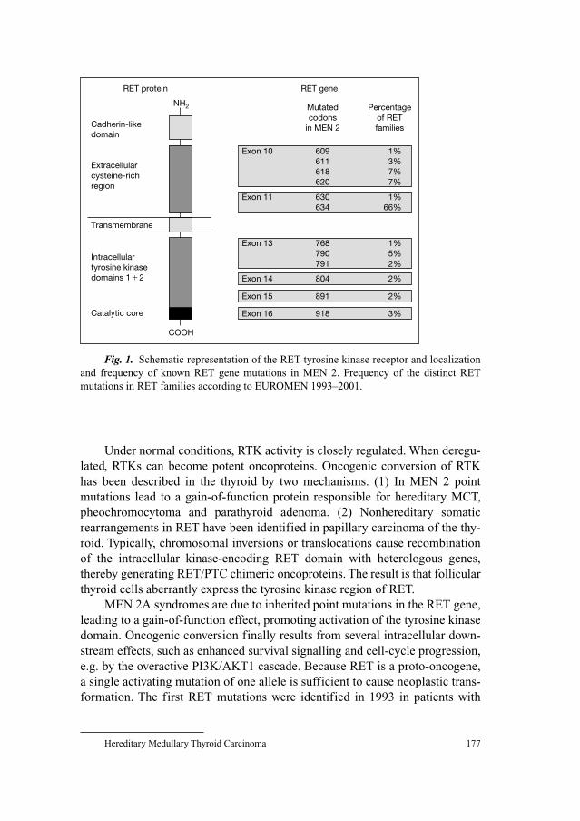
Hereditary Medullary Thyroid Carcinoma 177
Under normal conditions, RTK activity is closely regulated. When deregu-
lated, RTKs can become potent oncoproteins. Oncogenic conversion of RTK
has been described in the thyroid by two mechanisms. (1) In MEN 2 point
mutations lead to a gain-of-function protein responsible for hereditary MCT,
pheochromocytoma and parathyroid adenoma. (2) Nonhereditary somatic
rearrangements in RET have been identified in papillary carcinoma of the thy-
roid. Typically, chromosomal inversions or translocations cause recombination
of the intracellular kinase-encoding RET domain with heterologous genes,
thereby generating RET/PTC chimeric oncoproteins. The result is that follicular
thyroid cells aberrantly express the tyrosine kinase region of RET.
MEN 2A syndromes are due to inherited point mutations in the RET gene,
leading to a gain-of-function effect, promoting activation of the tyrosine kinase
domain. Oncogenic conversion finally results from several intracellular down-
stream effects, such as enhanced survival signalling and cell-cycle progression,
e.g. by the overactive PI3K/AKT1 cascade. Because RET is a proto-oncogene,
a single activating mutation of one allele is sufficient to cause neoplastic trans-
formation. The first RET mutations were identified in 1993 in patients with
RET protein
Cadherin-likedomain
NH2
Extracellularcysteine-richregion
Transmembrane
Catalytic core
COOH
Exon 16 918 3%
Exon 11 630 1% 634 66%
Exon 10 609 1% 611 3% 618 7% 620 7%
Exon 13 768 1% 790 5% 791 2%
Exon 14 804 2%
Exon 15 891 2%
Intracellulartyrosine kinasedomains 1�2
RET gene
Mutatedcodons
in MEN 2
Percentageof RETfamilies
Fig. 1. Schematic representation of the RET tyrosine kinase receptor and localization
and frequency of known RET gene mutations in MEN 2. Frequency of the distinct RET
mutations in RET families according to EUROMEN 1993–2001.

Szinnai/Sarnacki/Polak 178
MEN 2A and FMTC [1, 2]. The RET germline mutations of MEN 2 are local-
ized in only a small fraction of the open reading frame. Most are in the cysteine-
rich portion of the extracellular portion, while some are localized in the tyrosine
kinase domain of the intracellular portion.
Oncogenic mutations of RET show a striking and important correlation
with the MEN 2 subtype (fig. 2). Approximately 95% of MEN 2A families have
a RET mutation in exon 10 and 11. Mutations of codon 634 occur in about 85%
of families, mutation of cysteine codons at amino acid positions 609, 611, 618,
620 and 630 together accounts for the remainder of identifiable mutations.
Other rare mutations have been reported in single families. Approximately 85%
of FMTC families have an identifiable RET mutation. FMTC mutations are
evenly distributed among the various cysteine residues (609, 611, 618, 620 and
634) as those of MEN 2A. Why some families with the same mutation will
develop only FMTC and not MEN 2A remains to be clarified. Other mutations
seemingly specific for FMTC are found in codon 533 of the extracellular
cysteine-rich domain and codons 791–891 in the intracellular tyrosine kinase
domain of RET. Most MEN 2B patients (95%) carry the M918T mutation in
exon 16 in the RET kinase domain. A second mutation A883F has been identi-
fied in a small number of families [20].
The mechanisms leading to RET oncogenic conversion in MEN 2 depend
on the site of mutation: mutations in codons in the cysteine-rich extracellular
domain lead to ligand-independent RET receptor homodimerization and consti-
tutive activation of the tyrosine kinase domain. Different transforming activity
Phenotype ofMEN 2 subtypes
MTC
Pheochromocytoma
Neurinomas Parathyroid disease
MEN 2B
918 883 922
804�806 804�904
635 637
532 533 630 768 844 912
609 611 618 620 634 790 791 804 891
MEN 2A Overlap FMTC
Specific RETcodon mutations
Fig. 2. Phenotype-genotype correlation in MEN 2. All RET mutations described so far
correlated to MEN 2 phenotype. The frequent mutations are indicated in bold.

Hereditary Medullary Thyroid Carcinoma 179
between mutations in the cysteine residues has been demonstrated, while differ-
ent point mutations in the same codon seem not to differ in transforming activ-
ity [21]. In contrast, the intracellular mutation M918T responsible for MEN 2B
lies within the catalytic core of the tyrosine kinase. It produces constitutive acti-
vation of the RET tyrosine kinase by change of substrate specificity, indepen-
dent of RET dimerization [22]. In line with this model, MEN 2B mutants differ
from MEN 2A mutants in the stoichiometry of phosphorylation of tyrosine
residues and various intracellular proteins. These molecular differences explain
in part the more aggressive tumour biology of MEN 2B MTC.
Genetic Testing and Genetic CounsellingThe detection of germline mutations in the RET proto-oncogene had
important diagnostic impacts for the management of MEN 2 families: genetic
screening of patients at risk allowed the identification of RET mutation carriers
with very high specificity and sensitivity and, more importantly, even before
onset of disease. Based on these results, and the fact that MEN 2 is a well-
described endocrine tumour syndrome, the disease meets the criteria related to
indications for genetic testing for cancer susceptibility, outlined by the
American Society of Clinical Oncology (ASCO) in its genetic testing policy
statement [23]. Current recommendations state that RET mutational screening
is mandatory for all children at 50% risk. In consequence, genetic testing has
completely replaced biochemical measurements as first-line screening in MEN 2
families [7, 24].
Genetic counselling is essential for patients and their families who face the
risk of hereditary MTC. The autosomal dominant transmission mode implies a
50% risk for children of each family member. Ninety-five percent of MEN 2A
and FMTC patients have affected parents. Thus, it is appropriate to evaluate the
first-degree relatives of an individual MEN 2A patient for manifestations of the
disorder. In only 5%, does the disease originate from a de novo mutation or is
due to incomplete penetrance of the mutant allele. In contrast, as many as about
50% of MEN 2B patients have de novo RET mutations [25, 26].
The most accurate technique to detect RET mutations is DNA sequencing.
DNA sequencing can be done in selected exons with the most common muta-
tions (usually exons 10, 11, 13, 14, 15 and 16) or by sequencing all exons. This
is the only technique that allows also identification of new unreported muta-
tions in the RET gene.
Genotype-Phenotype CorrelationsSpecific RET mutations correlate not only with the expression of specific
MEN 2 subtypes, but also with the aggressiveness of MTC within the MEN 2
subtypes [5, 7, 27]. Our meta-analysis of children with MEN 2A provided
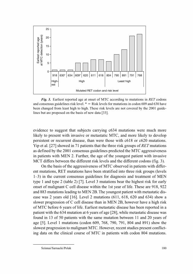
Szinnai/Sarnacki/Polak 180
evidence to suggest that subjects carrying c634 mutations were much more
likely to present with invasive or metastatic MTC, and more likely to develop
persistent or recurrent disease, than were those with c618 or c620 mutations.
Yip et al. [27] showed in 71 patients that the three risk groups of RET mutations
as defined by the 2001 consensus guidelines predicted the MTC aggressiveness
in patients with MEN 2. Further, the age of the youngest patient with invasive
MCT differs between the different risk levels and the different codons (fig. 3).
On the basis of the aggressiveness of MTC observed in patients with differ-
ent mutations, RET mutations have been stratified into three risk groups (levels
1–3) in the current consensus guidelines for diagnosis and treatment of MEN
type 1 and type 2 (table 2) [7]. Level 3 mutations bear the highest risk for early
onset of malignant C cell disease within the 1st year of life. These are 918, 922
and 883 mutations leading to MEN 2B. The youngest patient with metastatic dis-
ease was 2 years old [16]. Level 2 mutations (611, 618, 620 and 634) show a
slower progression of C cell disease than in MEN 2B, however have a high risk
of MTC before 6 years of life. Earliest metastatic disease has been reported in a
patient with the 634 mutation at 6 years of age [28], while metastatic disease was
found in 13 of 50 patients with the same mutation between 11 and 20 years of
age [5]. Level 1 mutations (codon 609, 768, 790, 791, 804 and 891) show the
slowest progression to malignant MTC. However, recent studies present conflict-
ing data on the clinical course of MTC in patients with codon 804 mutations.
0
5
10
15
20
25
918 630* 634 609* 620 611 618 804 790 891 791 768
High- est
High Least high
Mutated RET codon and risk level
Ear
liest
rep
orte
d a
ge
at M
TC d
etec
tion
(yea
rs)
Fig. 3. Earliest reported age at onset of MTC according to mutations in RET codons
and consensus guidelines risk level. * � Risk levels for mutations in codon 609 and 630 have
been changed from least high to high. These risk levels are not covered by the 2001 guide-
lines but are proposed on the basis of new data [33].

Hereditary Medullary Thyroid Carcinoma 181
Frohnauer et al. [29] reported one child with metastatic disease at the age of
6 years and in the same series an adult with normal histology in another family.
Gimm et al. [30] reported the most variable phenotype of MTC in 23 patients
with codon 804 mutations in a series of 140 patients with RET mutations.
Lesueur et al. [31] report a low penetrance of MTC in three homozygous and
6 heterozygous patients with V804L and V804M mutations. Both groups propose
that individuals heterozygous for weakly transforming mutations of RET require
a second germline or somatic mutation in RET or a gene of the RET pathway to
result in clinical expression of the disease and that this accounts for the wide
clinical variability associated with codon 804 mutations.
The knowledge on genotype-phenotype correlations for all MEN 2 muta-
tions is constantly increasing. They are the basis of current treatment guidelines
and for future adaptations.
Management of MEN 2
Total ThyroidectomyHereditary MTC is multicentric in 90% of patients, and nodal metastases
are present in more than 70% of patients with palpable disease. There is no
known effective systemic therapy for MTC. C cells do not concentrate radio-
active iodine, and MTC does not respond well to external radiation or conven-
tional cytotoxic chemotherapy. Total thyroidectomy is the only preventive or
curative therapeutic approach for MTC [32].
There are two approaches for preventive surgery for hereditary MTC: total
thyroidectomy with removal of the posterior capsule alone is the less aggressive
intervention. However, the risk of recurrence is higher because the lymph nodes
that may be the site of recurrence are not removed. Total thyroidectomy with
removal of the posterior capsule and central node dissection is the more com-
plete preventive surgical approach. It removes potential micro-metastasis in the
Table 2. Consensus guidelines for treatment of MTC in MEN 2
MTC risk Level RET genotype Phenotype Age at total Central node
mutation in codon thyroidectomy dissection
Highest 3 918, 922, 883 MEN 2B �6 months yes
High 2 611, 618, 620, 634 MEN 2A, FMTC �5 years no consensus
Least high 1 609, 630, 768, 790, MEN 2A, FMTC no consensus no consensus
791, 804, 891 5–10 years

Szinnai/Sarnacki/Polak 182
central lymph node compartment, and allows much easier surgical approach of
the parathyroids in the case of later development of hyperparathyroidism. In
case of advanced disease with palpable tumour further lymph node compart-
ments (jugular) should be resected [32, 33]. Identification of recurrent nerves
and parathyroid glands is challenging in very young children and total thy-
roidectomy requires thus experienced surgeons and an appropriate paediatric
environment.
Prophylactic and Genotype-Oriented ThyroidectomyThe advances in the understanding of the molecular basis of MEN 2 syn-
drome had major impact on management of MTC. First, the availability of
genetic screening of at-risk children in MEN 2 kindred made ‘prophylactic’ or
early total thyroidectomy at a preclinical and premalignant stage of disease pos-
sible. Second, knowledge on genotype-phenotype correlations for specific RET
mutations led to a codon-dependent timing of prophylactic thyroidectomy in
mutation carriers.
As outlined above, the current recommendations define three levels of risk
for MTC. Level 3 comprises children with MEN 2B phenotype and/or the RET
codon 883, 918 or 922 mutations with the highest risk for early development of
MTC. They should undergo total thyroidectomy with central node dissection
within the first 6 months of life, preferably during the 1st month of life.
Codon 634, 620, 618 and 611 are classified as level 2, with high risk for
early and aggressive MTC. Total thyroidectomy with removal of the posterior
capsule is recommended for patients with these mutations before the age of
5 years. There was no consensus in 2001 recommendations regarding the need
for prophylactic dissection of the central lymph nodes. This issue is still contro-
versial in 2006.
Codon 609, 768, 790, 791, 804 and 891 mutations are classified as level 1,
with the least high risk for MTC. There was no consensus in the 2001 recom-
mendations on the management of patients with these mutations with some
experts favouring thyroidectomy at 5 years, others at 10 years, and still others
on the basis of calcium and pentagastrin-stimulated calcitonin levels.
Based on new data, some authors recommend a change of risk level for
codon 609 mutations from the level 1 to the level 2 group and the classification
of the codon 630 in the level 2 group (fig. 3) [33]. However, combined genetic
and biochemical screening starting in the 1st year of life is mandatory for the
detection of unexpected early onset of MTC in all patients independent of the
genotype (fig. 4) [33].
Evidence for better long-term outcome after thyroidectomy in asympto-
matic RET mutation carriers at a young age is increasing. A meta-analysis
comparing the stage of disease and outcome after early (0–5 years) versus late
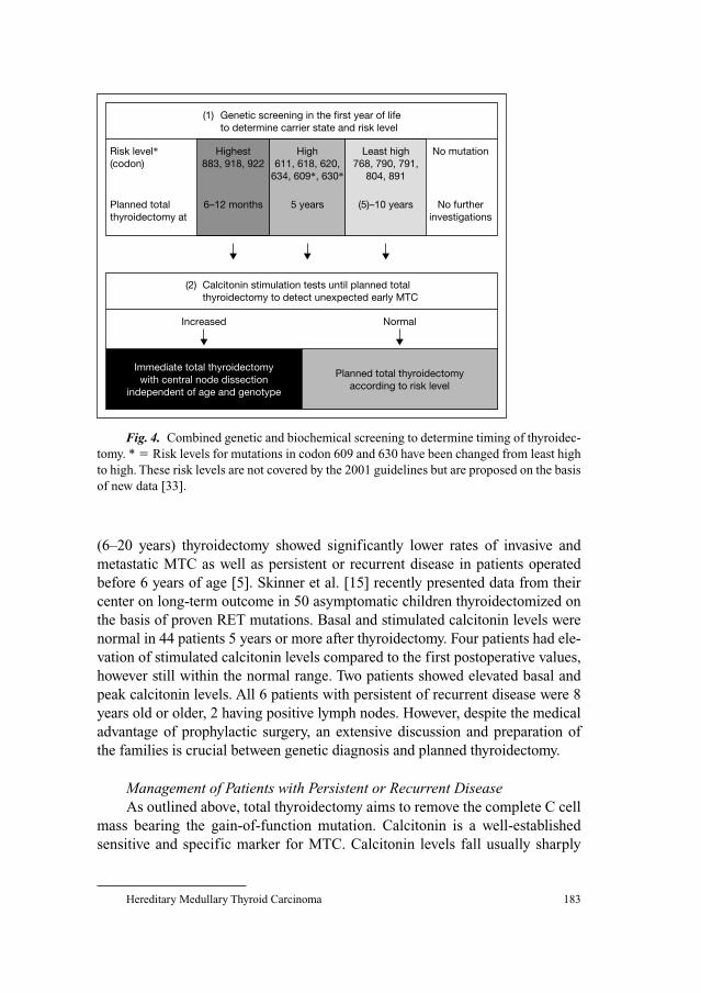
Hereditary Medullary Thyroid Carcinoma 183
(6–20 years) thyroidectomy showed significantly lower rates of invasive and
metastatic MTC as well as persistent or recurrent disease in patients operated
before 6 years of age [5]. Skinner et al. [15] recently presented data from their
center on long-term outcome in 50 asymptomatic children thyroidectomized on
the basis of proven RET mutations. Basal and stimulated calcitonin levels were
normal in 44 patients 5 years or more after thyroidectomy. Four patients had ele-
vation of stimulated calcitonin levels compared to the first postoperative values,
however still within the normal range. Two patients showed elevated basal and
peak calcitonin levels. All 6 patients with persistent of recurrent disease were 8
years old or older, 2 having positive lymph nodes. However, despite the medical
advantage of prophylactic surgery, an extensive discussion and preparation of
the families is crucial between genetic diagnosis and planned thyroidectomy.
Management of Patients with Persistent or Recurrent DiseaseAs outlined above, total thyroidectomy aims to remove the complete C cell
mass bearing the gain-of-function mutation. Calcitonin is a well-established
sensitive and specific marker for MTC. Calcitonin levels fall usually sharply
Risk level*(codon)
Highest883, 918, 922
6–12 months
High611, 618, 620,
634, 609*, 630*
Least high768, 790, 791,
804, 891
5 years (5)–10 years
No mutation
No furtherinvestigations
(1) Genetic screening in the first year of life to determine carrier state and risk level
(2) Calcitonin stimulation tests until planned total thyroidectomy to detect unexpected early MTC
Planned totalthyroidectomy at
Increased Normal
Planned total thyroidectomyaccording to risk level
Immediate total thyroidectomywith central node dissection
independent of age and genotype
Fig. 4. Combined genetic and biochemical screening to determine timing of thyroidec-
tomy. * � Risk levels for mutations in codon 609 and 630 have been changed from least high
to high. These risk levels are not covered by the 2001 guidelines but are proposed on the basis
of new data [33].

Szinnai/Sarnacki/Polak 184
after thyroidectomy, indicating ‘biochemical cure’ of an MTC patient. Per-
sistently abnormal calcitonin levels indicate residual disease, however delayed
reduction may occur. After postoperative evaluation, all patients should be fol-
lowed annually by serum calcitonin levels. For this, most authors recommend
stimulated calcitonin levels. Elevated basal or stimulated levels during follow-
up indicate recurrent disease. Patients with persistent or recurrent disease may
stay asymptomatic for many years. A re-operation with formal neck dissection
is necessary if nodal metastases are detectable. In the absence of localizable
disease by physical or radiological examination without previous neck dissec-
tion, re-operation with adequate central and lateral lymph node dissection or
observation of the patient by radiological surveillance might be an option [32].
Management of Pheochromocytoma and Parathyroid DiseaseUnilateral adrenalectomy appears to be a reasonable management strategy
for unilateral pheochromocytoma in patients with MEN 2. The long interval of
metachronous pheochromocytoma argues against prophylactic removal of the
contralateral normal gland. Patients however need periodic surveillance for the
development of the disease in the contralateral adrenal gland. Total adrenalec-
tomy and heterotopic autotransplantation of medulla-free cortex are currently
preferred to bilateral adrenalectomy as they may diminish the need for life-long
steroid substitution and seem not to expose to recurrence [34]. The coelioscopic
approach has rendered this surgical procedure less invasive. Nevertheless, it
requires adequate medical preparation, a skilful surgeon and an experienced
anaesthesiologist.
Parathyroid disease is not part of MEN 2B. The parathyroid glands may be
left in the neck. For MEN 2A and FMTC patients, it is suggested that glands be
implanted in the non-dominant forearm to minimize the need for further
surgery on the neck after thyroidectomy.
Psychological Impact of Genetic Testing and Prophylactic ThyroidectomyThe medical value of early screening and prophylactic treatment in
MEN 2 are contrasted with the psychosocial impact of the genetic diagnosis of
MEN 2 on parents and children. Disagreement about the value and the timing
of prophylactic surgery between parents of affected children and misconception
about genetic disease and the question of guilt are only some of the problems
that may cause emotional distress within families. The need for attention to the
potential psychological vulnerability of parents and children should not be
underestimated.

Hereditary Medullary Thyroid Carcinoma 185
Perspectives
More Experience with Rare RET Gene MutationsThe current consensus guidelines have been published in 2001 [7].
Meanwhile, more data are becoming available for genotype-phenotype correla-
tions for the rare RET mutations. This knowledge will allow a more precise risk
estimation, and more adapted guidelines for prophylactic thyroidectomy (e.g. as
proposed for 609 and 630 mutations) and node dissection for the specific risk
levels.
New Pharmacological Treatment Option: RET Tyrosine Kinase InhibitorsDifferent RET tyrosine kinase inhibitors have been developed in the last
years for treatment of malignant haematological disorders and solid tumours
caused by tyrosine kinase dysregulation. Preliminary results indicate that dif-
ferent tyrosine kinase inhibitors selectively inhibited cell growth and RET tyro-
sine kinase activity in MTC cells in vitro in a dose-dependent manner. These
results suggest that tyrosine kinase inhibitors might be useful for the treatment
of MTC and open a new pharmacological treatment option for MEN 2 patients
with persistent or recurrent disease after thyroidectomy [35, 36].
References
1 Mulligan LM, Kwok JB, Healey CS, Elsdon MJ, Eng C, Gardner E, Love DR, Mole SE, Moore JK,
Papi L, Ponder MA, Telenius H, Tunnacliffe A, Ponder B: Germ-line mutations of the RET proto-
oncogene in multiple endocrine neoplasia type 2A. Nature 1993;363:458–460.
2 Donis-Keller H, Dou S, Chi D, Carlson KM, Toshima K, Lairmore TG, Howe JR, Moley JF,
Goodfellow P, Wells SA Jr: Mutations in the RET proto-oncogene are associated with MEN 2A
and FMTC. Hum Mol Genet 1993;2:851–856.
3 Mulligan LM, Marsh DJ, Robinson BG, Schuffenecker I, Zedenius J, Lips CJM, Gagel RF, Takai SI,
Noll WW, Fink M, Raue F, Lacroix A, Thibodeau SN, Frilling A, Ponder BAJ, Eng C: Genotype-
phenotype correlation in multiple endocrine neoplasia type2: report of the International RETMutation Consortium. J Intern Med 1995;238:343–346.
4 Eng C, Clayton D, Schuffenecker I, Lenoir G, Cote G, Gagel RF, van Amstel HK, Lips CJ,
Nishisho I, Takai SI, Marsh DJ, Robinson BG, Frank-Raue K, Raue F, Xue F, Noll WW, Romei C,
Pacini F, Fink M, Niederle B, Zedenius J, Nordenskjöld M, Komminoth P, Hendy GN, Gharib H,
Thibodeau SN, Lacroix A, Frilling A, Ponder BAJ, Mulligan LM: The relationship between spe-
cific RET proto-oncogene mutations and disease phenotype in multiple endocrine neoplasia type 2.
International RET mutation consortium analysis. JAMA 1996;276:1575–1579.
5 Szinnai G, Meier C, Komminoth P, Zumsteg UW: Review of multiple endocrine neoplasia type 2A
in children: therapeutic results of early thyroidectomy and prognostic value of codon analysis.
Pediatrics 2003;111:E132–E139.
6 Machens A, Niccoli-Sire P, Hoegel J, Frank-Raue K, van Vroonhoven TJ, Roehrer HD, Wahl RA,
Lamesch P, Raue F, Conte-Delvolx B, Dralle H; European Multiple Endocrine Neoplasia
(EUROMEN) Study Group: Early malignant progression of hereditary medullary thyroid cancer.
N Engl J Med 2003;349:1517–1525.

Szinnai/Sarnacki/Polak 186
7 Brandi ML, Gagel RF, Angeli A, Bilezikian JP, Beck-Peccoz P, Bordi C, Conte-Delvolx B,
Falchetti A, Gheri RG, Libraia A, Lips CJM, Lombardi G, Mannelli M, Pacini F, Ponder BAJ,
Raue F, Skogseid B, Tamburano G, Thakker RV, Thompson NW, Tomassetti P, Tonelli F, Wells SA Jr,
Marx SJ: Guidelines for diagnosis and therapy of MEN type 1 and 2. J Clin Endocrinol Metab
2001;86:5658–5671.
8 Ponder BA: Multiple endocrine neoplasia type 2; in Vogelstein B, Kinzler KW (eds): The Genetic
Basis of Human Cancer. New York, McGraw-Hill, 2002, pp 501–513.
9 Ponder BAJ, Ponder MA, Coffey R, Pembrey ME, Gagel RF, Telenius-Berg M, Semple P, Easton DP:
Risk estimation and screening in families of patients with medullary thyroid carcinoma. Lancet
1988;i:397–401.
10 National Cancer Institute: Genetics of medullary thyroid cancer. http://www.cancer.gov/cancertopics/
pdq/genetics/medullarythyroid/HealthProfessional/page2.
11 Block MA, Jackson CE, Greenawald KA, Yott JB, Tashjian AH Jr: Clinical characteristics distin-
guishing hereditary from sporadic medullary thyroid carcinoma. Treatment implications. Arch
Surg 1980;115:142–148.
12 Wolfe HJ, Melvin KE, Cervin-Skinner SJ, Saadi AA, Juliar JF, Jackson CE, Tashjian AH Jr: C-cell
hyperplasia preceding medullary thyroid carcinoma. N Engl J Med 1973;289:437–441.
13 Niccoli P, Wion-Barbot N, Carron P, Henry JF, de Micco C, Saint Andre JP, Bigorgne JC,
Modigliani E, Conte-Delvolx B: Interest of routine measurement of serum calcitonin: study in a
large series of thyroidectomized patients. The French Medullary Study Group. J Clin Endocrinol
Metab 1997;82:338–341.
14 Gagel RF, Tashjian AH Jr, Cummings T, Papathanasopoulos N, Kaplan MM, DeLellis RA, Wolfe HJ,
Reichlin S: The clinical outcome of prospective screening for multiple endocrine neoplasia type
2A: an 18-year experience. N Engl J Med 1988;318:478–484.
15 Skinner MA, Moley JA, Dilley WG, Owzar K, DeBenedetti MK, Wells SA Jr: Prophylactic thy-
roidectomy in multiple endocrine neoplasia type 2A. N Engl J Med 2005;353:1105–1113.
16 Leboulleux S, Travagli JP, Caillou B, Laplanche A, Bidart JM, Schlumberger M, Baudin E:
Medullary thyroid carcinoma as part of a multiple endocrine neoplasia type 2B syndrome. Cancer
2002;94:44–50.
17 Modiglioni E, Vasen HM, Raue K, Dralle H, Frilling A, Gheri RG, Brandi ML, Limbert E,
Niederle B, Forgas L, Rosenberg-Bougin M, Calmettes C, et al: Pheochromocytoma in multiple
endocrine neoplasia type 2: European study. The EUROMEN Study Group. J Intern Med 1995;238:
363–367.
18 Kraimps JL, Denizot A, Carnaille B, Henry JF, Proye C, Bacourt F, Sarfati E, Dupond JL, Maes B,
Travagli JP, Boneu A, Roger P, Houdent C, Barbier J, Modigliani E: Primary hyperparathyroidism
in multiple endocrine neoplasia type IIa: retrospective French multicentric study. Groupe d’Etude
des Tumeurs à Calcitonine (GETC, French Calcitonin Tumors Study Group), French Association
of Endocrine Surgeons. World J Surg 1996;20:808–812.
19 Santoro M, Melillo RM, Carlomagno F, Vecchio G, Fusco A: Minireview: RET: normal and
abnormal functions. Endocrinology 2004;145:5448–5451.
20 Marx SJ: Molecular genetics of multiple endocrine neoplasia types 1 and 2. Nat Rev Cancer
2005;5:367–375.
21 Ito S, Iwashita T, Asai N, Muratami H, Iwata Y, Sobue G, Takahashi M: Biological properties of
RET with cysteine mutations correlate with multiple endocrine neoplasia type 2A, familial
medullary thyroid carcinoma, and Hirschsprung’s disease phenotype. Cancer Res 1997;57:
2870–2872.
22 Santoro M, Carlomagno F, Romano A, Bottaro DP, Dathan NA, Grieco M, Vecchio G, Fusco A,
Kraus M, Di Fiore PP: Activation of RET as a dominant transforming gene by germline mutations
of MEN2A and MEN2B. Science 1995;267:381–383.
23 American Society of Clinical Oncology policy statement update: Genetic testing for cancer sus-
ceptibility. J Clin Oncol 2003;21:2397–2406.
24 Lips CJM, Landsvater RM, Höppener JW, Geerdink RA, Blijham G, van Veen JM, van Gils APG,
De Wit M, Zewald RA, Berends MJH, Beemer FA, Brouwers-Smalbraak J, Jansen RPM, Ploos
van Amstel HK, van Vroonhoven TJMV, Vroom TM: Clinical screening as compared with DNA
analysis in families with multiple endocrine neoplasia type 2A. N Engl J Med 1994;331:828–835.
Hereditary Medullary Thyroid Carcinoma 187
25 Schuffenecker I, Ginet N, Godgar D, Eng C, Chambe B, Boneu A, Houdent C, Pallo D,
Schlumberger M, Thivolet C, Lenoir GM: Prevalence and parental origin of de novo RET muta-
tions in multiple endocrine neoplasia type 2A and familial medullary thyroid carcinoma. Le
Groupe d’Etude des Tumeurs à Calcitonin. Am J Hum Genet 1997;60:233–237.
26 Carlson KM, Bracamontes J, Jackson CE, Clark R, Lacroix A, Wells SA Jr, Goodfellow PJ:
Parent-of-origin effects in multiple endocrine neoplasia type 2B. Am J Hum Genet 1994;55:
1076–1082.
27 Yip L, Cote GJ, Shapiro SE, Ayers GD, Herzog CE, Sellin RV, Sherman SI, Gagel RF, Lee JE,
Evans DB: Multiple endocrine neoplasia type 2: evaluation of genotype-phenotype relationship.
Arch Surg 2003;138:409–416.
28 Gill JR, Reyes-Mugica M, Iyengar S, Kidd KK, Touloukian RJ, Smith C, Keller MS, Genel M:
Early presentation of metastatic medullary carcinoma in multiple endocrine neoplasia, type IIA:
implications for therapy. J Pediatr 1996;129:459–464.
29 Frohnauer MK, Decker RA: Update on the MEN 2A c804 RET mutation: is prophylactic thy-
roidectomy indicated? Surgery 2000;128:1052–1058.
30 Gimm O, Ukkat J, Niederle BE, Weber T, Tanh PN, Brauckhoff M, Niederle B, Dralle H: Timing
and extent of surgery in patients with familial medullary thyroid carcinoma/multiple endocrine
neoplasia 2A-related mutations not affecting codon 634. World J Surg 2004;28:1312–1316.
31 Lesueur F, Cebrian A, Cranston A, Leyland J, Faid TM, Clements MR, Robledo M, Whittaker J,
Ponder BAJ: Germline homozygous mutations at codon 804 in the RET proto-oncogene in
medullary thyroid carcinoma/multiple endocrine neoplasia type 2A patients. J Clin Endocrinol
Metab 2005;90:3454–3457.
32 Quayle FJ, Moley JF: Medullary thyroid carcinoma: including MEN 2A and MEN 2B syndromes.
J Surg Oncol 2005;89:122–129.
33 Machens A, Ukkat J, Brauckhoff M, Gimm O, Dralle H: Advances in the management of heredi-
tary medullary thyroid cancer. J Intern Med 2005;257:50–59.
34 Inabnet W, Caragliano P, Pertsemlidis D: Pheochromocytoma: inherited associations, bilaterality,
and cortex preservation. Surgery 2000;128:1007–1012.
35 Strock CJ, Park JI, Rosen M, Ruggeri B, Denmeade SR, Ball DW, Nelkin BD: Activity of irinote-
can and the tyrosine kinase inhibitor CEP-751 in medullary thyroid cancer. J Clin Endocrinol
Metab 2006;91:79–84.
36 de Groot JWB, Links TP, Plukker JTM, Lips CJM, Hofstra RMW: RET as a diagnostic and thera-
peutic target in sporadic and hereditary endocrine tumors. Endocr Rev 2006;27:535–560.
Dr. Gabor Szinnai
Paediatric Endocrinology, University Children’s Hospital Basel
Römergasse 8
CH–4005 Basel (Switzerland)
Tel. �41 61 685 65 65, Fax �41 61 685 65 66, E-Mail [email protected]

188
Bauer, A.J. 140
Calvo, R.M. 86
Castanet, M. 15
De Felice, M. 1
Deladoëy, J. 29
Di Lauro, R. 1
Dumitrescu, A.M. 127
Escobar del Rey, F. 86
Francis, G.L. 140
Garel, C. 43
Glinoer, D. 62
Grüters, A. 118
Léger, J. 15, 43
Moreno, J.C. 99
Morreale de Escobar, G. 86
Obregon, M.J. 86
Polak, M. IX, 15, 173
Refetoff, S. 127
Sarnacki, S. 173
Savage, M.O. VII
Szinnai, G. 173
Tuttle, R.M. 140
Van Vliet, G. IX, 29
Vasko, V. 140
Vassart, G. 29
Visser, T.J. 99
Author Index

189
Acute thyroiditis, ultrasonography findings
in children 53
Brain, thyroid hormone and development
88, 94, 95
Budding, see Thyroid primordium
Cell differentiation, genetic regulation 8, 9
Congenital hypothyroidism (CH), see alsoThyroid dysgenesis
classification 100
diagnosis 45, 46
epidemiology 45, 100
thyroid oxidase defects, see Dual
oxidases
transient disease
consequences 113, 114
dual oxidase defects, see Dual oxidases
etiology 100, 101
prospects for study 114, 115
Deiodinases
acquired defects 134, 135
knockout mice 135, 136
SBP2 mutations, see SECIS-binding
protein
DNA methylation, thyroid dysgenesis
36, 37
Dual oxidases
DUOX2
congenital hypothyroidism phenotypes
107–110
functional assay 106
genotype-phenotype correlations
110–113
genes 103, 105
hydrogen peroxide generation 106,
107
processing 106
structure 105, 106
thyroid hormone synthesis role 107
tissue distribution 106
DUOX2, see Dual oxidases
Expansion, genetic regulation in fetal
thyroid 9, 10
Fine needle aspiration (FNA)
thyroid cancer in children 142–145
thyroid nodules in children 142
Follicular thyroid cancer, see Thyroid
cancer
Foxe1
human thyroid disease 10, 11, 24
thyroid anlage specification role 2
transcription factor interactions in early
thyroid morphogenesis 7
Goiter
iodine deficiency in pregnancy and
gestational goiter
consequences 73
formation 72, 73
prevention 75, 76
ultrasonography in children 52
Graves’ disease, ultrasonography findings in
children 55, 56
Hashimoto’s disease, ultrasonography in
children 53
Subject Index
Subject Index 190
Hhex
functional overview 4
human thyroid disease 10, 11, 23
thyroid anlage specification role 2
thyroid primordium budding role 4
transcription factor interactions in early
thyroid morphogenesis 7
Hox genes, human thyroid diseases 24
Hyperparathyroidism, multiple endocrine
neoplasia type 2 association and
management 176, 184
Hyperthyroidism, see Graves’ disease
Iodine
deficiency in pregnancy
epidemiology 67, 68, 76
fetal and neonatal consequences
77–80
goiter
consequences 73
formation 72, 73
prevention 75, 76
hypothyroxinemia 71
intake adequacy monitoring 70
management 68, 69, 76, 77, 81
thyroid hormone response 70
thyroid-stimulating hormone changes 72
thyroxine-binding globulin response 70
metabolism 65–67
Lymph node dissection, thyroid cancer
management in children 151
Magnetic resonance imaging (MRI), ectopic
thyroid 48, 49
MCT8
developmental expression 120, 121
gene 119
knockout mouse 124, 125
mutations
clinical findings 122, 123, 133, 134
thyroid function tests 123
types 123, 124
structure 119, 121
tissue distribution 119, 120
Medullary thyroid carcinoma, see Multiple
endocrine neoplasia type 2; Thyroid
cancer
Monocarboxylate transporters, see MCT8
Mouse models, thyroid gland development
2–12
Multiple endocrine neoplasia type 2
(MEN2)
clinical features of MEN2A
hyperparathyroidism 176
medullary thyroid carcinoma 174–176
pheochromocytoma 176
genetics of MEN2A
genotype-phenotype correlations
179–181
RET mutations 176–179
testing and counseling 179
management of MEN2A
genetic testing and counseling
psychological impact 184
hyperparathyroidism 184
persistent or recurrent disease 183,
184
pheochromocytoma 184
thyroidectomy 181–183
prospects for study 185
subtypes 174
Nkx2.5, human thyroid disease mutations
24, 25
Papillary thyroid cancer, see Thyroid cancer
Pax8
human thyroid disease 6, 10, 11, 23,
24, 33
thyroid anlage specification role 2
thyroid primordium budding role 5
transcription factor interactions in early
thyroid morphogenesis 7
Pheochromocytoma, multiple endocrine
neoplasia type 2 association and
management 176, 184
Pregnancy
fetal thyroid hormone expression and
development role 87, 88, 92, 93
Graves’ disease and fetal risks 55
iodine deficiency
epidemiology 67, 68, 76
fetal and neonatal consequences
77–80
goiter

Subject Index 191
consequences 73
formation 72, 73
prevention 75, 76
hypothyroxinemia 71
intake adequacy monitoring 70
management 68, 69, 76, 77, 81
thyroid hormone response 70
thyroid-stimulating hormone changes
72
thyroxine-binding globulin response 70
iodine metabolism 65–67
maternal thyroid hormone and fetal
development
brain development 94, 95
conception to midgestation 90, 91
deficiency and cretinism 88
human observations 89, 90
midgestation to birth 92–95
premature infant considerations 94–96
rat studies 89
thyroid function 63, 64
Premature infant, thyroid hormone status
94–96
Radioactive iodine ablation
pulmonary fibrosis induction 153, 154
risks 155, 156
thyroid cancer management in children
151–153
Radionuclide thyroid scanning
children 53, 141
thyroid dysgenesis differential diagnosis
33
RET
function 176
gene 176
multiple endocrine neoplasia type 2
genetic testing and counseling 179
genotype-phenotype correlations
179–181
mutations 177–179
regulation 177
therapeutic targeting 185
SECIS-binding protein (SBP2)
discovery 129
function 128
global effects of deficiency 137, 138
mutation and thyroid hormone
dysfunction
clinical presentation 129, 131
fibroblast studies 131, 132
mutation types 132, 133
tissue distribution 133
Selenocysteine
incorporation in proteins 128, 129
knockout mouse models of incorporation
defects 136, 137
Sonic hedgehog (Shh), thyroid lobulation
role 8
Thyroglobulin, tumor marker specificity
149, 150
Thyroglossal duct cyst, ultrasonography 50
Thyroid anlage, specification 2
Thyroid cancer, pediatric
clinical presentation 146–149
epidemiology 145
fine needle aspiration 142–145
follow-up 161–165
guidelines 164
management 149–156
multiple endocrine neoplasia, seeMultiple endocrine neoplasia type 2
pathology
adenomas 160, 161
anaplastic carcinoma 159
follicular thyroid cancer 158, 159
medullary thyroid carcinoma 159,
160
papillary thyroid cancer 156–158
radiation induction 145, 146
treatment risks 154–156
ultrasonography findings 58, 60
Thyroid dysgenesis
classification
agenesis 31
differential diagnosis 33
ectopic thyroid 31
hemiagenesis 32, 33
hypoplasia 31, 32
overview 16–18, 46
early somatic mutations 33–35
epigenetic defects 36, 37
familial congenital hypothyroidism
19, 21

Subject Index 192
familial dysgenesis in first-degree
relatives of children with congenital
hypothyroidism 21–23
Mendelian versus non-Mendelian
mechanisms 30–38
molecular mechanisms 23
pathogenesis 15, 16
thyroid development 16
two-hit model of germline and somatic
changes 37, 38
Thyroidectomy
multiple endocrine neoplasia type 2
181–183
risks 155
thyroid cancer management in children
150, 151
Thyroid hormone
fetal expression and development role
87, 88, 92, 93
iodine deficiency in pregnancy response
70
maternal thyroid hormone and fetal
development
brain development 94, 95
conception to midgestation 90, 91
deficiency and cretinism 88
human observations 89, 90
midgestation to birth 92–95
premature infant considerations
94–96
rat studies 89
SBP2 mutations, see SECIS-binding
protein
synthesis 101, 103, 128
thyroid cancer management in children
153, 154
thyroid oxidases, see Dual oxidases
transport
MCT8, see MCT8
monocarboxylate transporters 119
overview 118, 119
Thyroid lobulation, genetic regulation 7, 8
Thyroid nodules, children
cancer, see Thyroid cancer
epidemiology 140, 141
fine needle aspiration 142
malignancy 141
scintigraphy 141
ultrasonography findings 56, 141, 142
Thyroid oxidases, see Dual oxidases
Thyroid primordium
budding and gene regulation 3–6
migration 6, 7
Thyroid-stimulating hormone (TSH)
iodine deficiency in pregnancy response
72
neonatal screening 35
Thyroxine-binding globulin (TBG), iodine
deficiency in pregnancy response 70
Titf1
human thyroid disease 5, 10, 11, 24
thyroid anlage specification role 2
thyroid primordium budding role 4, 5
transcription factor interactions in early
thyroid morphogenesis 7
TTF-1, see Titf1
TTF-2, human thyroid disease 33
Ultrasonography
child thyroid imaging
acute thyroiditis 53
ectopic thyroid 48
empty thyroid area 46–48
goiter 52
Graves’ disease 55, 56
Hashimoto’s disease 53
hemiagenesis 50
normal findings 44, 45
technique 44, 45
thyroglossal duct cyst 50
thyroid cancer 56, 58
thyroid nodules 56, 141, 142
echogenicity of thyroid 46
thyroid dysgenesis differential diagnosis
33
Thyroid dysgenesis(continued)