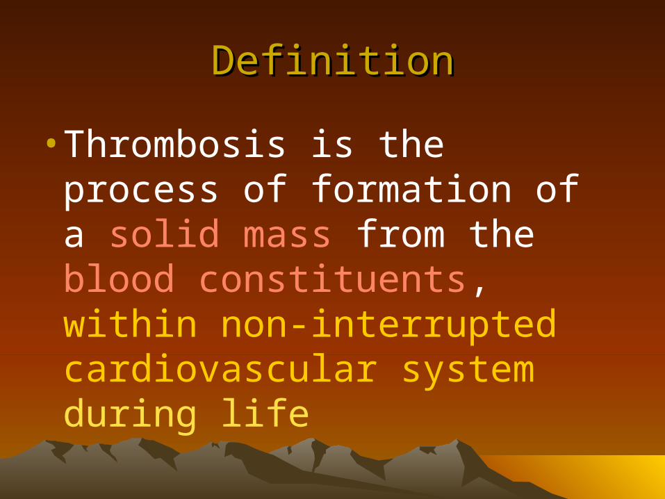
Thrombosis. Definition Thrombosis is the process of formation of a solid mass from the blood...
43
Thrombos Thrombos is is
-
Upload
brianna-edwina-dean -
Category
Documents
-
view
214 -
download
0
Transcript of Thrombosis. Definition Thrombosis is the process of formation of a solid mass from the blood...

ThrombosisThrombosis
DefinitionDefinition
• Thrombosis is the process of formation of a solid mass from the blood constituents, within non-interrupted cardiovascular system during life
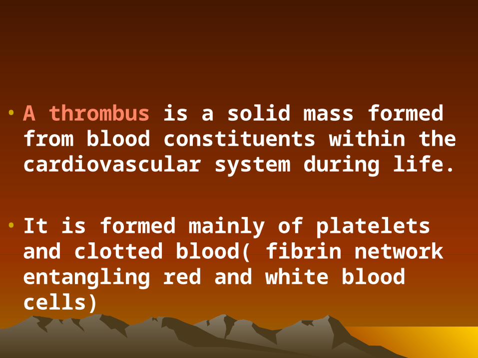
• A thrombus is a solid mass formed from blood constituents within the cardiovascular system during life.
• It is formed mainly of platelets and clotted blood( fibrin network entangling red and white blood cells)
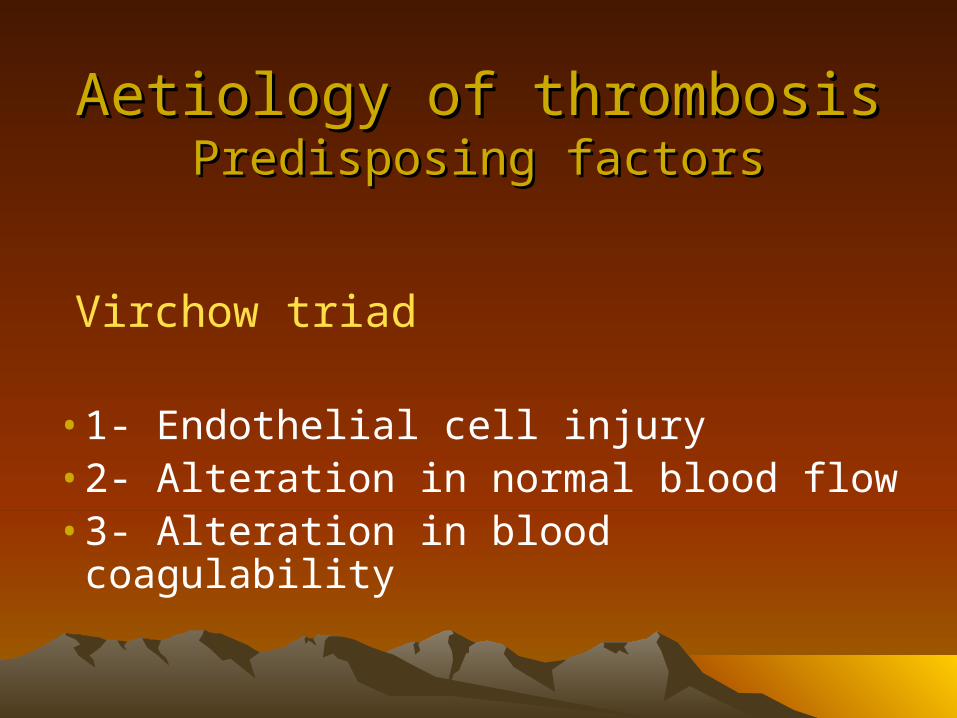
Aetiology of thrombosisAetiology of thrombosisPredisposing factorsPredisposing factors
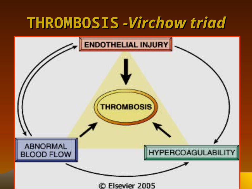
Virchow triad
• 1- Endothelial cell injury• 2- Alteration in normal blood flow• 3- Alteration in blood coagulability
THROMBOSISTHROMBOSIS -Virchow triad -Virchow triad
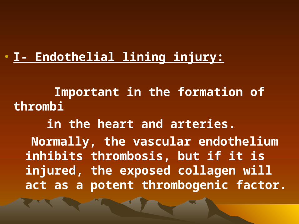
• I- Endothelial lining injury:
Important in the formation of thrombi
in the heart and arteries.
Normally, the vascular endothelium inhibits thrombosis, but if it is injured, the exposed collagen will act as a potent thrombogenic factor.
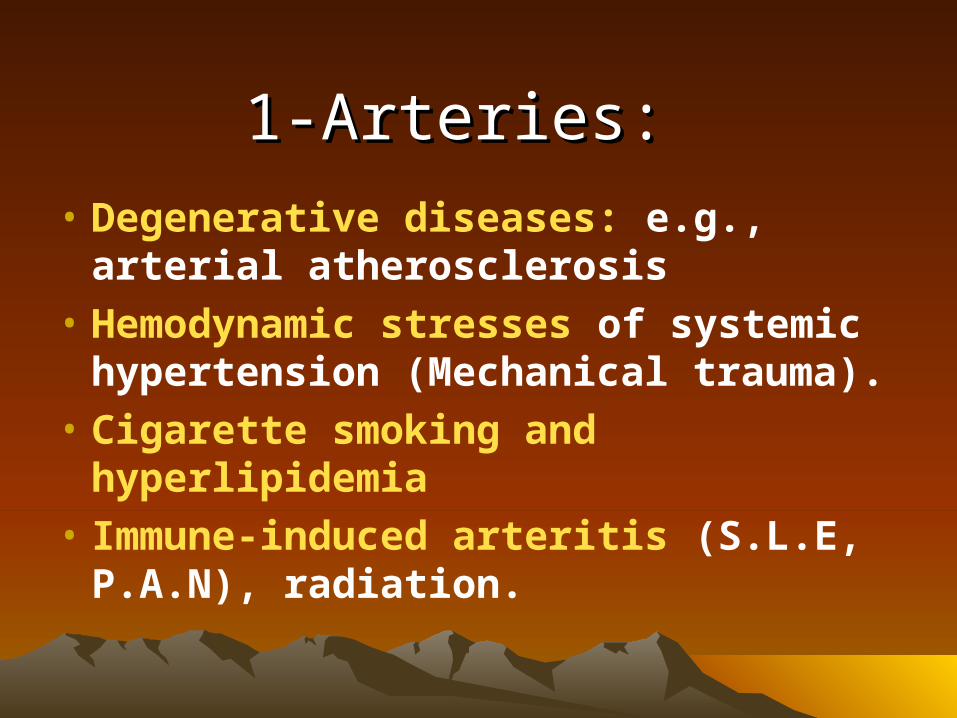
1-Arteries:1-Arteries: • Degenerative diseases: e.g., arterial
atherosclerosis
• Hemodynamic stresses of systemic hypertension (Mechanical trauma).
• Cigarette smoking and hyperlipidemia
• Immune-induced arteritis (S.L.E, P.A.N), radiation.
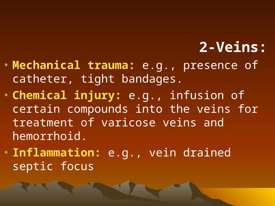
2-Veins:• Mechanical trauma: e.g., presence of
catheter, tight bandages.
• Chemical injury: e.g., infusion of certain compounds into the veins for treatment of varicose veins and hemorrhoid.
• Inflammation: e.g., vein drained septic focus
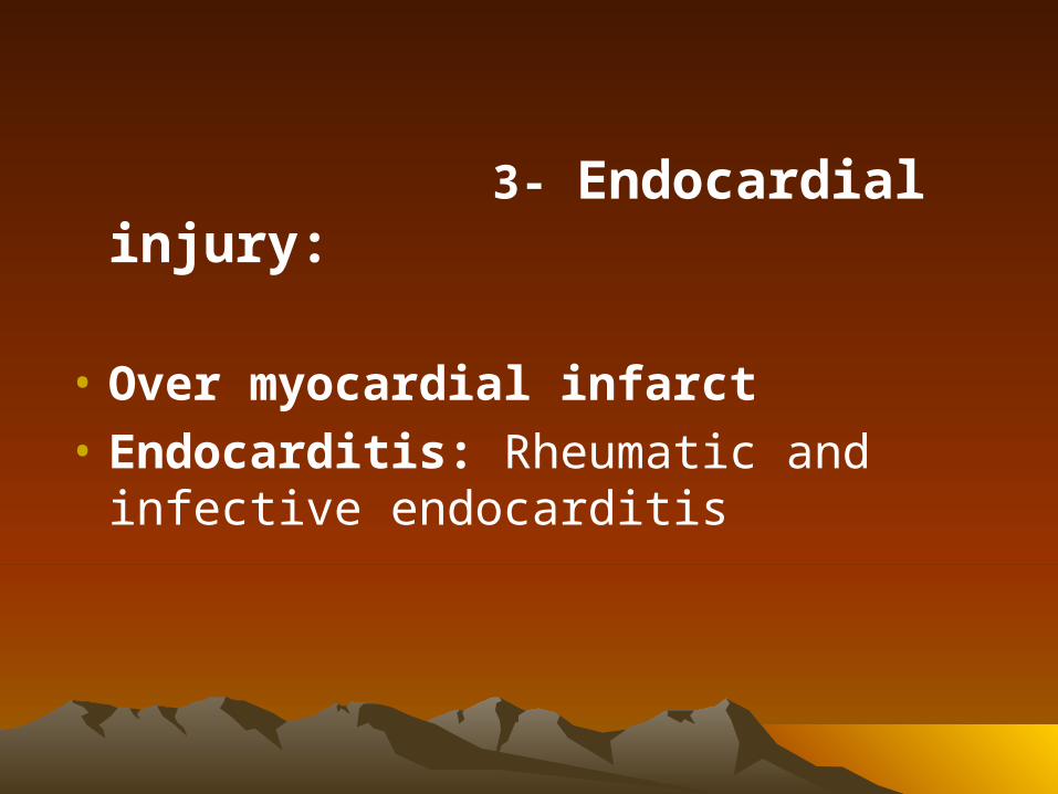
3- Endocardial injury:
• Over myocardial infarct
• Endocarditis: Rheumatic and infective endocarditis
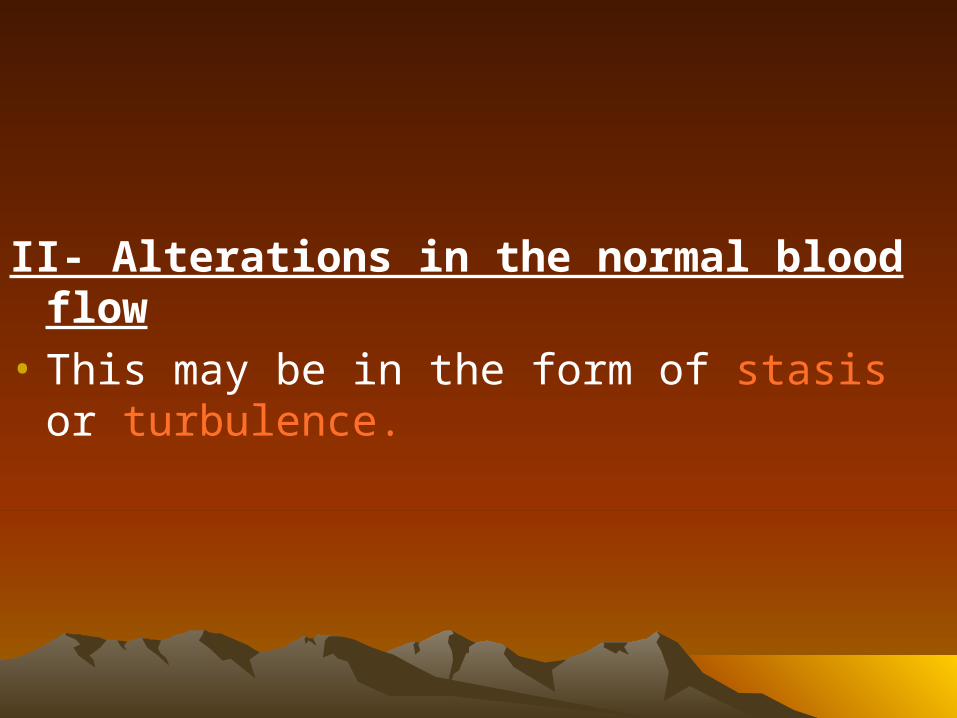
II- Alterations in the normal blood flow
• This may be in the form of stasis or turbulence.

Slowing of the speed of blood flow (Stasis)• It is the major factor in the development of
venous thrombosis & caused by:• Local causes:
• Pressure on the veins by bandages, tumours , gravid uterus.
• Lack of muscular pumping action as in bedridden patients, in postoperative period or after delivery.
• Dilatation of veins and vulvular incompetence e.g., in varicose veins.
• General factors: e.g., congestive heart failure
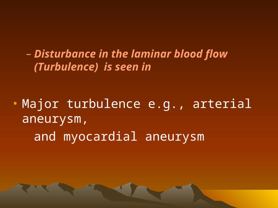
– Disturbance in the laminar blood flow (Turbulence) is seen in
• Major turbulence e.g., arterial aneurysm,
and myocardial aneurysm
Effects of Alterations in the normal blood flow:
• Endothelial injury or dysfunction
• Disrupting the laminar blood flow and bring platelets into contact with endothelium
III- Alterations in blood coagulability (Hypercoagulability):
• might be primary or genetic; and secondary or acquired.
• Primary or genetic deficiency of protein “C” or antithrombin III
• B-acquired hypercoagulablility :• Increased blood viscosity as in severe dehydration• Postoperative and postpartum patients• Oral contraceptives• Tissue damage e.g., massive burn, fracture, surgery (
release of tissue thromboplastin).• In association with some tumours as carcinoma of pancreas• After splenectomy • Hyperlipidemia ( platelet adhesiveness and aggregation).
•
Morphology of thrombi
a-Gross appearance:
• 1-Site and shape:– Anywhere in the cardiovascular system: within the
cardiac chambers; on valve cusps; or in arteries, veins, or capillaries.
– Cardiac or arterial thrombi begin at the site of endothelial injury (atherosclerotic plaque) or turbulence (aneurysms & vessel bifurcation).
– On the other hand, venous thrombi usually occur at sites of stasis.
– All thrombi are attached to the vessel wall or cardiac chamber at the point of their origin.
– Arterial thrombi tend to grow in a retrograde direction from the point of attachment (that is away from the heart), while venous thrombi extend in, the direction of blood flow (i.e. towards the heart).
– The propagating tail may detach leading to emboli especially found in veins.
• 2-The colour:– Characteristic laminations (lines of Zahn) due to
alternating layers of platelets (pale) and fibrin containing RBCS & leukocytes (red)are especially noticed in thrombi of the heart and large arteries as the aorta.
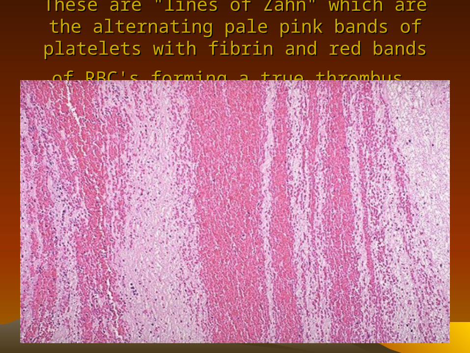
These are "lines of Zahn" which are the alternating These are "lines of Zahn" which are the alternating pale pink bands of platelets with fibrin and red pale pink bands of platelets with fibrin and red
bands of RBC's forming a true thrombusbands of RBC's forming a true thrombus
– Red thrombus: It occurs when stasis plays a prominent role in its pathogenesis e.g., in veins. It is formed of platelets and fibrin entangling a large number of R.B.Cs.
– Pale thrombus: When there is no stasis and alteration of blood flow is prominent. It is composed predominantly of platelets and fibrin with few entrapped R.B.Cs
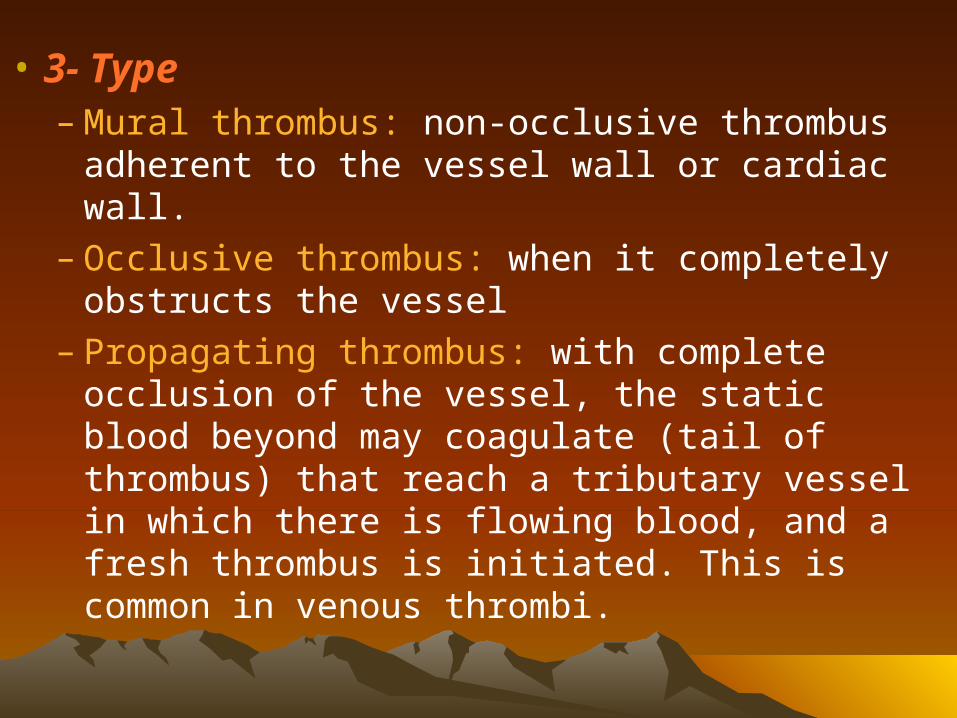
• 3- Type– Mural thrombus: non-occlusive thrombus adherent
to the vessel wall or cardiac wall.– Occlusive thrombus: when it completely obstructs
the vessel – Propagating thrombus: with complete occlusion of
the vessel, the static blood beyond may coagulate (tail of thrombus) that reach a tributary vessel in which there is flowing blood, and a fresh thrombus is initiated. This is common in venous thrombi.
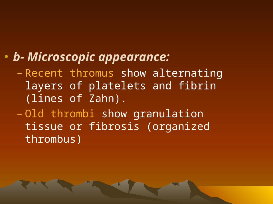
• b- Microscopic appearance:– Recent thromus show alternating layers of
platelets and fibrin (lines of Zahn).– Old thrombi show granulation tissue or
fibrosis (organized thrombus)
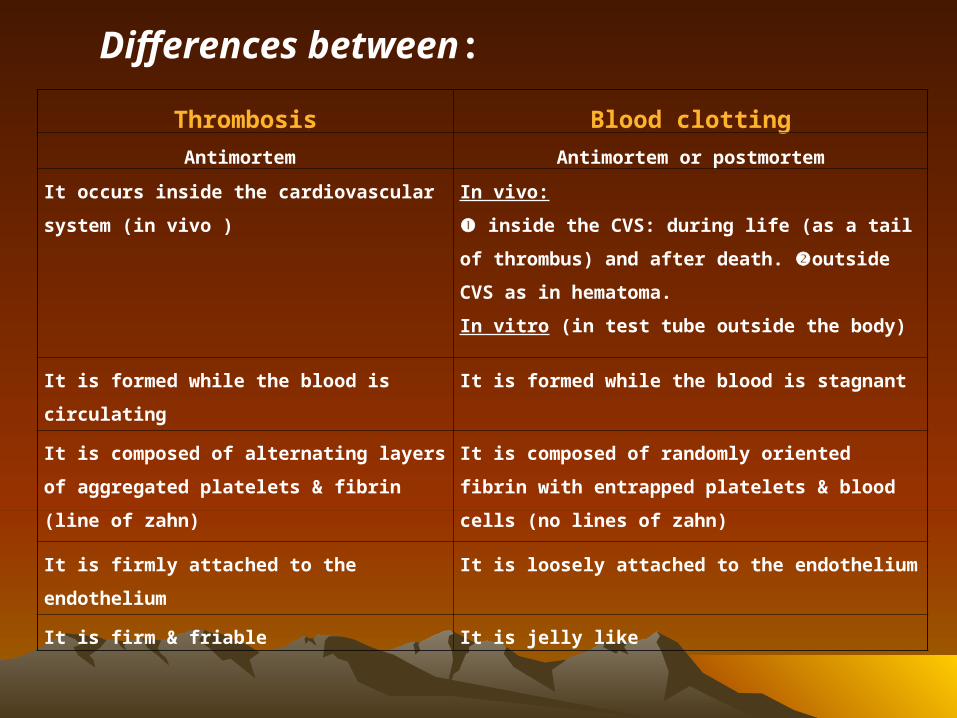
Thrombosis Blood clotting
Antimortem Antimortem or postmortem
It occurs inside the cardiovascular
system (in vivo )
In vivo:
inside the CVS: during life (as a tail of
thrombus) and after death. outside CVS
as in hematoma.
In vitro (in test tube outside the body)
It is formed while the blood is
circulating
It is formed while the blood is stagnant
It is composed of alternating layers of
aggregated platelets & fibrin (line of
zahn)
It is composed of randomly oriented fibrin
with entrapped platelets & blood cells (no
lines of zahn)
It is firmly attached to the
endothelium
It is loosely attached to the endothelium
It is firm & friable It is jelly like
Differences between:
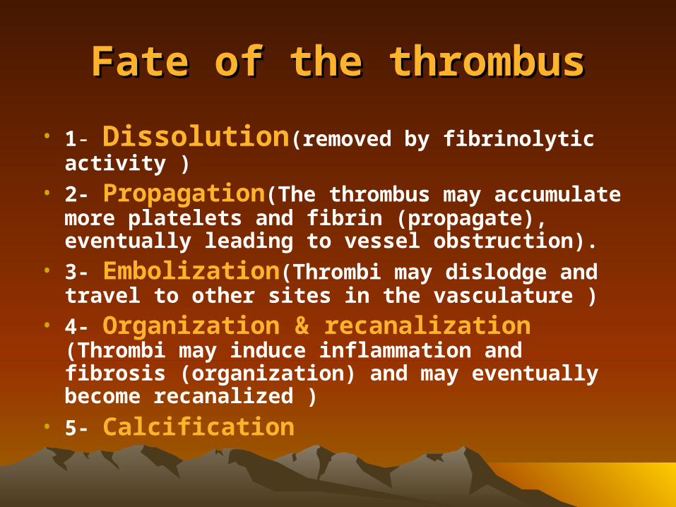
Fate of the thrombusFate of the thrombus
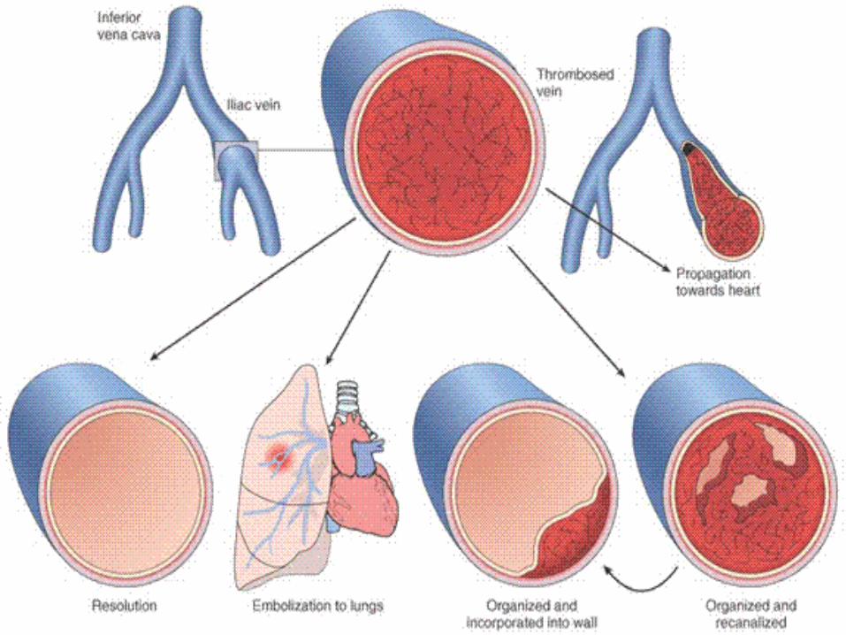
• 1- Dissolution(removed by fibrinolytic activity )
• 2- Propagation(The thrombus may accumulate more platelets and fibrin (propagate), eventually leading to vessel obstruction).
• 3- Embolization(Thrombi may dislodge and travel to other sites in the vasculature )
• 4- Organization & recanalization (Thrombi may induce inflammation and fibrosis (organization) and may eventually become recanalized )
• 5- Calcification
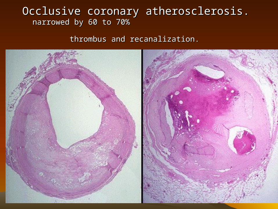
Occlusive coronary atherosclerosis. Occlusive coronary atherosclerosis.
narrowed by 60 to 70% thrombus and recanalization.narrowed by 60 to 70% thrombus and recanalization.
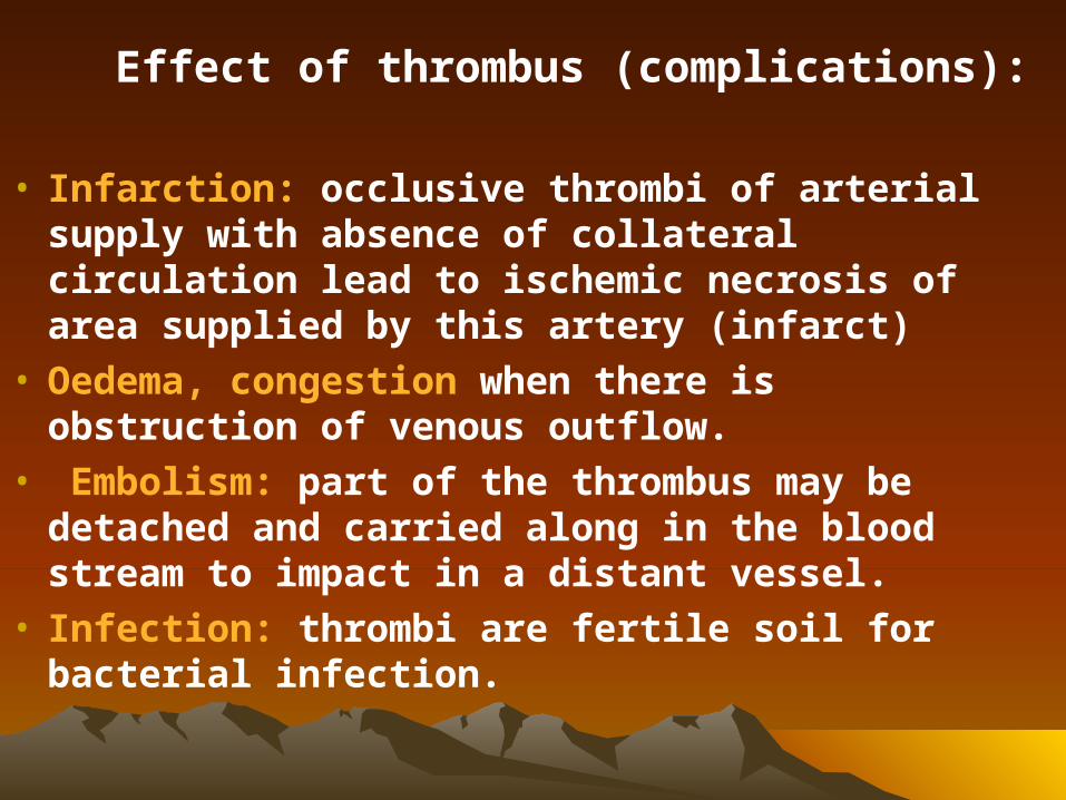
Effect of thrombus (complications):
• Infarction: occlusive thrombi of arterial supply with absence of collateral circulation lead to ischemic necrosis of area supplied by this artery (infarct)
• Oedema, congestion when there is obstruction of venous outflow.
• Embolism: part of the thrombus may be detached and carried along in the blood stream to impact in a distant vessel.
• Infection: thrombi are fertile soil for bacterial infection.
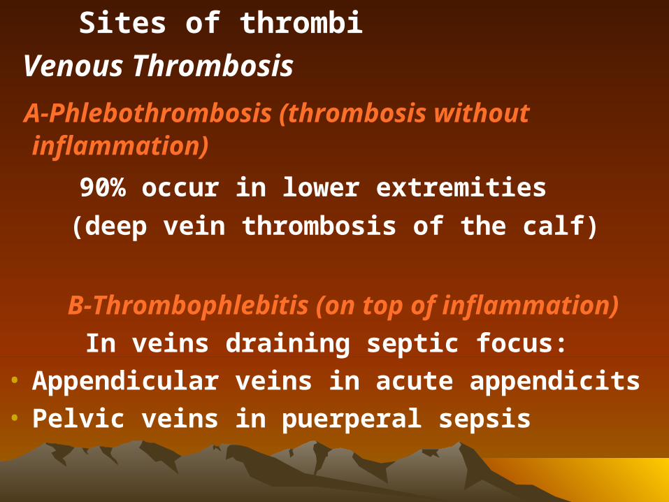
Sites of thrombi
Venous Thrombosis
A-Phlebothrombosis (thrombosis without inflammation)
90% occur in lower extremities
(deep vein thrombosis of the calf)
B-Thrombophlebitis (on top of inflammation)
In veins draining septic focus:• Appendicular veins in acute appendicits• Pelvic veins in puerperal sepsis

Points of differences
Phlebothrombosis Thrombophlebitis
Cause StasisInflammation of vein wall.
Size SmallLarger, depending on the extent of phlebitis.
Size of propagated clot
Long & is poorly anchored to the vessel wall.
Usually none & if present is short & well anchored to the vessel wall
EmboliCommon, massive & sterile → infarct.
Rare except in infective cases. Septic & → pyemic abscesses.
Site Usually calf veins Any where
Clinical pictureSilent or there are few signs & symptoms
Pain & signs of acute inflammation
Thrombosis In Arteries– Less common than veins because of the rapid flow,
and the thick elastic arterial wall which resists injury.
– Thrombosis in arteries is caused by: (endothelial injury & blood turbulence).
Types of arterial thrombosis are: - • Occluding thrombus in medium sized and small
arteries, frequently in association with atherosclerosis.• Mural thrombus: occurring in a large artery or aorta.
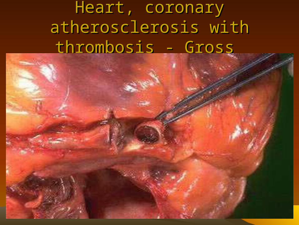
Heart, coronary atherosclerosis Heart, coronary atherosclerosis with thrombosis - Gross with thrombosis - Gross
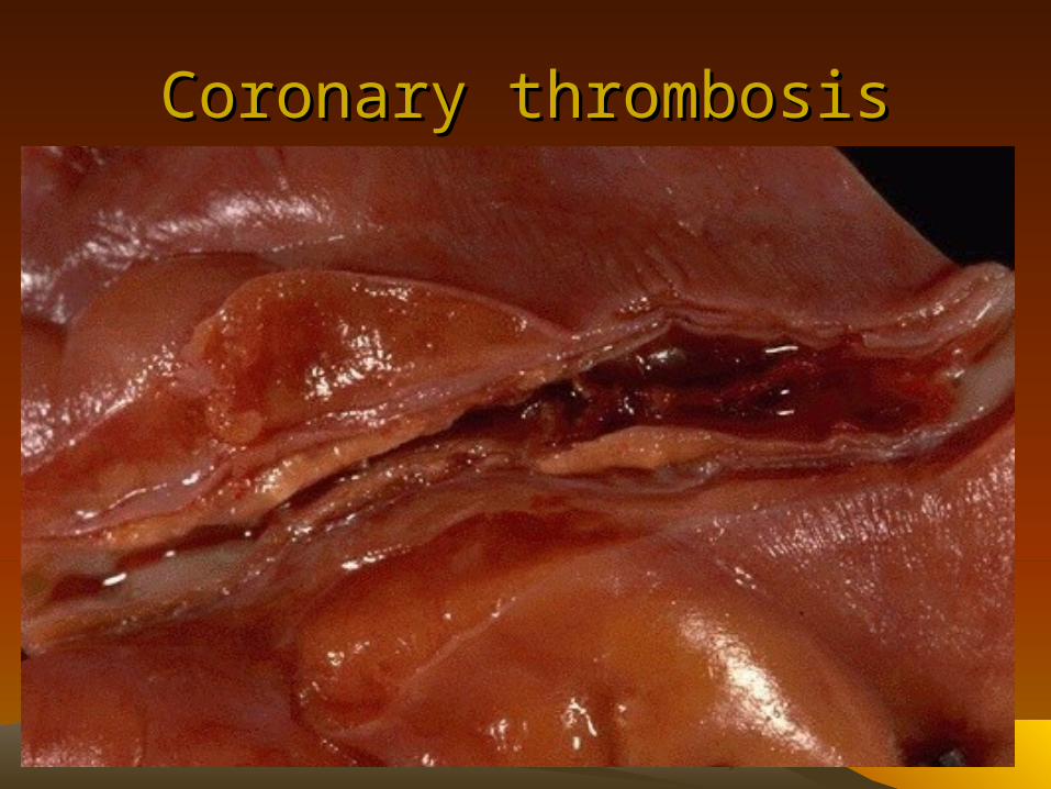
Coronary thrombosisCoronary thrombosis
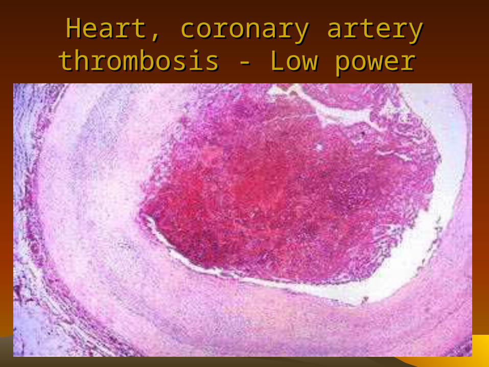
Heart, coronary artery thrombosis - Heart, coronary artery thrombosis - Low power Low power
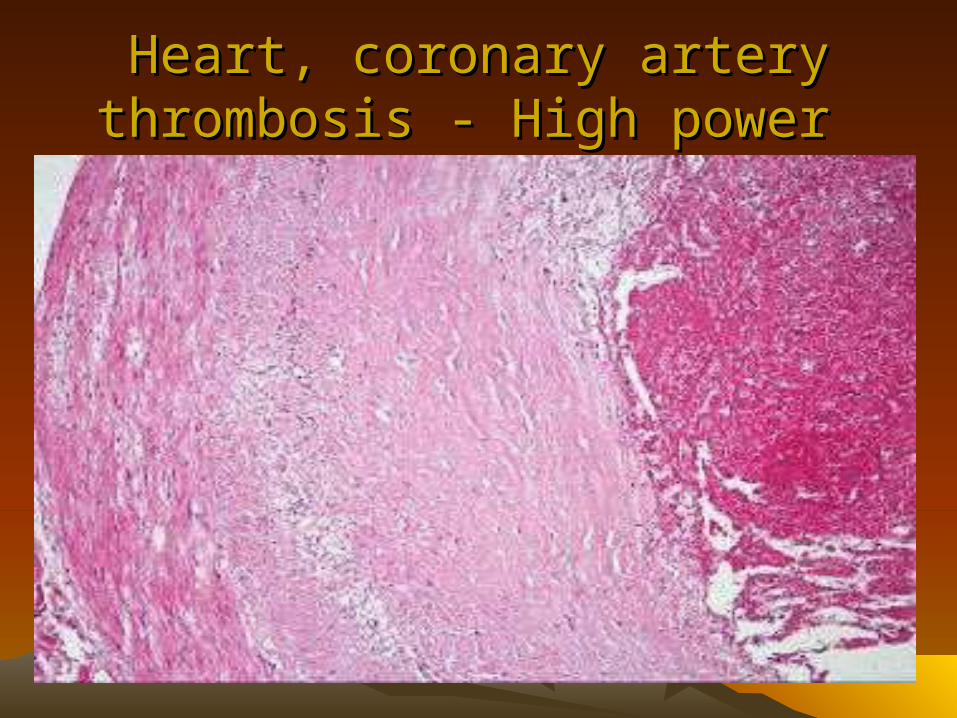
Heart, coronary artery thrombosis - Heart, coronary artery thrombosis - High power High power
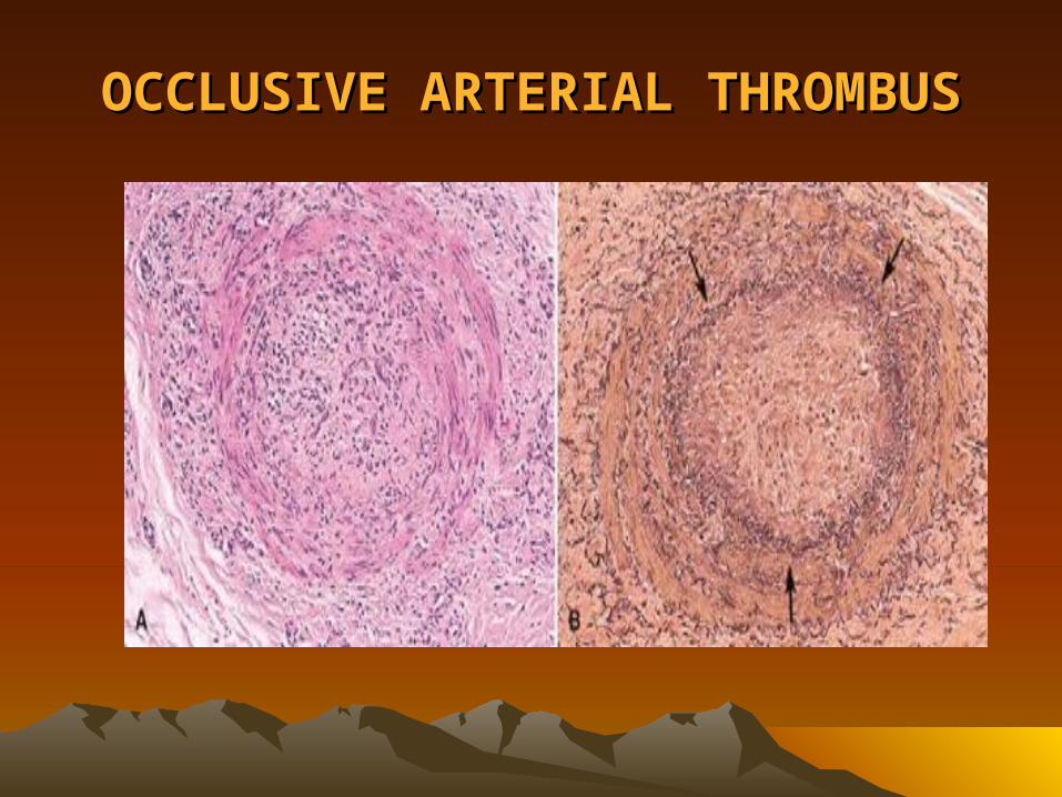
OCCLUSIVE ARTERIAL THROMBUSOCCLUSIVE ARTERIAL THROMBUS
– Effects: occlusive thrombi of arterial supply with absence of collateral circulation lead to ischemic necrosis of area supplied by this artery (infarct)
• Thrombi In The Heart– Mural thrombi occur in the heart chambers
especially in the left ventricle at sites of myocardial infarction, and, in the left atrium in cases of mitral stenosis. It is usually associated with atrial fibrillation.
– Valvular thrombi or "vegetations" are found in rheumatic valvulitis and infective endocarditis.
– Agonal thrombi, which occur during the last hours of life when death occurs slowly, are composed mainly of yellow fibrin commonly attached to the apex of the right ventricle and forming a mass as in the pulmonary artery.

MURAL THROMBI, HEARTMURAL THROMBI, HEART
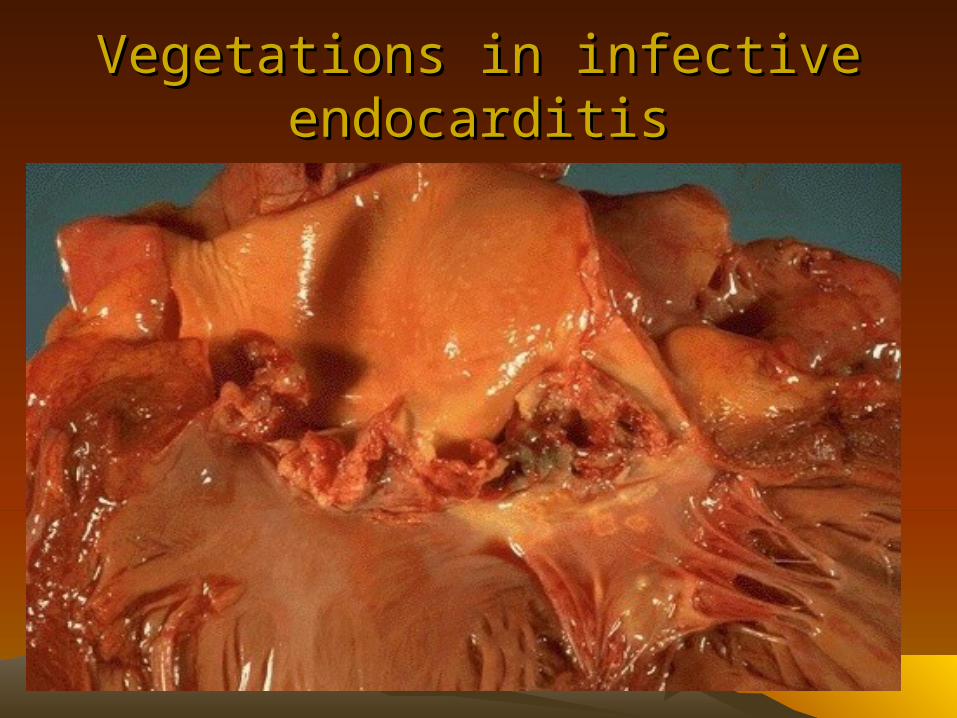
Vegetations in infective Vegetations in infective endocarditisendocarditis
• Thrombosis In Capillaries– It is seen in a variety of disorders, where there are
sudden or insidious onset of widespread fibrin thrombi in the microcirculation. It is known as disseminated intravascular coagulation (DIC).
– The thrombi are not visible by the naked eye, but are apparent microscopically.
Disseminated Intravascular Coagulation (DIC)
• A variety of disorders ranging from obstetric complications to advanced malignancy may be complicated by DIC, the sudden or insidious onset of widespread fibrin thrombi in the microcirculation.
• These thrombi can cause diffuse circulatory insufficiency, particularly in the brain, lungs, heart, and kidneys
DIC
• With the development of the multiple thrombi, there is a rapid consumption of platelets and coagulation proteins ; the same time, fibrinolytic mechanisms are activated, and as a result an initially thrombotic disorder can evolve into a serious bleeding disorder.

DIC ( fibrin thrombus in the lung)DIC ( fibrin thrombus in the lung)