Thoracoscopy and Massive Hemothorax in Hemodynamically Stable ...
4
Volume 2 • Issue 1 • 1000161 J Trauma Treat ISSN: 2167-1222 JTM, an open access journal Research Article Open Access Rodolfo Valentin et al., J Trauma Treat 2013, 2.1 http://dx.doi.org/10.4172/2167-1222.1000161 Research Article Open Access Trauma & Treatment Thoracoscopy and Massive Hemothorax in Hemodynamically Stable Patients Rodolfo Valentin B 1# , Bernardo Alfonso B 2 * and Sergio G 3# 1 General Surgeon and Thoracic Surgeon, El Bosque University, Bogotá, DC, Colombia 2 General Surgeon El Bosque University, Bogotá, DC, Colombia 3 Epidemiologist El Bosque University, Bogotá, DC, Colombia # Equal contribution *Corresponding author: Bernardo Alfonso B, General Surgeon El Bosque University, Bogotá, DC, Colombia, Tel: 011-571-8118463; E-mail: [email protected] Received March 09, 2013; Accepted March 29, 2013; Published April 01, 2013 Citation: Rodolfo Valentin B, Bernardo Alfonso B, Sergio G (2013) Thoracoscopy and Massive Hemothorax in Hemodynamically Stable Patients. J Trauma Treat 2: 161. doi:10.4172/2167-1222.1000161 Copyright: © 2013 Rodolfo Valentin B, et al. This is an open-access article distributed under the terms of the Creative Commons Attribution License, which permits unrestricted use, distribution, and reproduction in any medium, provided the original author and source are credited. Keywords: oracoscopy; Massive Hemothorax; Hemodynamically stable patients; Surgery Introduction Trauma is the leading cause of death and disability in the labor force in our country, causing economic losses and prolonged disability. e cardiothoracic trauma is responsible for about 75% of deaths attributed to trauma, penetrating injuries by bladed weapon and firearm projectile generate mortality ranging between 3 and 20% depending on the mechanism of trauma being the most lethal last. In Colombia, the mortality rate is from 3 to 10% inchest wounds stab and from 14 to 20% by firearms, with an incidence of thoracic trauma by blunt trauma 4% and 96% penetrating injuries [1-8]. oracoscopic procedures in a trained allow alternate access to the thoracic cavity for the treatment of multiple traumatic pathologies. e use of thoracoscopy in thoracic trauma is well supported in the literature as long as the patient is hemodynamically stable, allowing not only the diagnosis and treatment of patients with diaphragmatic injury thoracoabdominal trauma, but also the diagnosis and treatment of lesions in the chest wall, lung, heart, mediastinum and hemothorax drain, providing a low rate of complications and morbidity of minimally invasive surgery [9-14]. Materials and Methods Retrospective study Cross sectional, type III level of evidence. We included patients with massive hemothorax secondary to penetrating trauma, hemodynamically stable, managed by thoracoscopy or thoracotomy during the period from September 1, 2008 until September 30, 2011. Information was collected retrospectively by investigators and was obtained from a review of medical records of patients who met the selection criteria. is information was documented in the data collection format validated by the oracic Surgery Service of Hospital Santa Clara. During the revision of history were extracted data that were relevant to the analysis of this study, based on a data collection form designed for that purpose. e information was entered into a database in Excel which was revised and exported to SPSS (Statistical Package for Social Sciences) for information processing and statistical analysis. Statistical analyzes were performed descriptive type proportion for categorical variables and calculated the mean, standard deviation, median and range for numeric variables. For bivariate analyzes used Fisher’s test or chi- square tests for categorical variables and Student’s t according to the numerical distribution. e value determined for the statistical significance of all tests was 0.05. Results 45 patients were included in the study. 24 patients approached by thoracoscopy and thoracotomy 21. We found that 42 patients (93.3%) were male and three patients (6.7%) women with an age range between 13 and 55 years with a mean of 26,6 years. e mechanism of trauma in 43 patients (95.6%) were injured by sharp weapon and in two patients (4.4%) injured by gun fire to which Abstract Background: This study compares patient operated by thoracoscopy and thoracotomy in patients with penetrating chest wounds with massive hemothorax, hemodynamically stable. Materials and methods: Retrospective study Cross sectional, type III level of evidence.45 patients were included in the study. In 24 of them were approached by thoracoscopy and the remaining 21 by thoracotomy and compared morbidity, hospital stay, operative time, complications and conversion rate in patients with penetrating trauma massive hemothorax managed by thoracotomy hemodynamically stable with those managed by thoracoscopy during the period from September 1, 2008 until September 30, 2011 operated by the Department of General Surgery and Thoracic Surgery Hospital Santa Clara, Bogota, Colombia. Results: Thoracoscopy is associated with reduced blood loss, less operative time, shorter hospital stay by filing a conversion rate of 12.5% video assisted surgery procedures and a lower rate of complications. Conclusions: Thoracoscopy represents a therapeutic option and a safe approach in the right hands to the chest injured patients with massive hemothorax, hemodynamically stable.
Transcript of Thoracoscopy and Massive Hemothorax in Hemodynamically Stable ...
Volume 2 • Issue 1 • 1000161J Trauma TreatISSN: 2167-1222 JTM, an open access journal
Research Article Open Access
Rodolfo Valentin et al., J Trauma Treat 2013, 2.1http://dx.doi.org/10.4172/2167-1222.1000161
Research Article Open Access
Trauma & Treatment
Thoracoscopy and Massive Hemothorax in Hemodynamically Stable PatientsRodolfo Valentin B1#, Bernardo Alfonso B2* and Sergio G3#
1General Surgeon and Thoracic Surgeon, El Bosque University, Bogotá, DC, Colombia2General Surgeon El Bosque University, Bogotá, DC, Colombia3Epidemiologist El Bosque University, Bogotá, DC, Colombia#Equal contribution
*Corresponding author: Bernardo Alfonso B, General Surgeon El Bosque University, Bogotá, DC, Colombia, Tel: 011-571-8118463; E-mail: [email protected]
Received March 09, 2013; Accepted March 29, 2013; Published April 01, 2013
Citation: Rodolfo Valentin B, Bernardo Alfonso B, Sergio G (2013) Thoracoscopy and Massive Hemothorax in Hemodynamically Stable Patients. J Trauma Treat 2: 161. doi:10.4172/2167-1222.1000161
Copyright: © 2013 Rodolfo Valentin B, et al. This is an open-access article distributed under the terms of the Creative Commons Attribution License, which permits unrestricted use, distribution, and reproduction in any medium, provided the original author and source are credited.
Keywords: Thoracoscopy; Massive Hemothorax; Hemodynamically stable patients; Surgery
IntroductionTrauma is the leading cause of death and disability in the labor
force in our country, causing economic losses and prolonged disability. The cardiothoracic trauma is responsible for about 75% of deaths attributed to trauma, penetrating injuries by bladed weapon and firearm projectile generate mortality ranging between 3 and 20% depending on the mechanism of trauma being the most lethal last. In Colombia, the mortality rate is from 3 to 10% inchest wounds stab and from 14 to 20% by firearms, with an incidence of thoracic trauma by blunt trauma 4% and 96% penetrating injuries [1-8].
Thoracoscopic procedures in a trained allow alternate access to the thoracic cavity for the treatment of multiple traumatic pathologies. The use of thoracoscopy in thoracic trauma is well supported in the literature as long as the patient is hemodynamically stable, allowing not only the diagnosis and treatment of patients with diaphragmatic injury thoracoabdominal trauma, but also the diagnosis and treatment of lesions in the chest wall, lung, heart, mediastinum and hemothorax drain, providing a low rate of complications and morbidity of minimally invasive surgery [9-14].
Materials and MethodsRetrospective study Cross sectional, type III level of evidence. We
included patients with massive hemothorax secondary to penetrating trauma, hemodynamically stable, managed by thoracoscopy or thoracotomy during the period from September 1, 2008 until September 30, 2011.
Information was collected retrospectively by investigators and was obtained from a review of medical records of patients who met the selection criteria. This information was documented in the data collection format validated by the Thoracic Surgery Service of Hospital
Santa Clara. During the revision of history were extracted data that were relevant to the analysis of this study, based on a data collection form designed for that purpose.
The information was entered into a database in Excel which was revised and exported to SPSS (Statistical Package for Social Sciences) for information processing and statistical analysis. Statistical analyzes were performed descriptive type proportion for categorical variables and calculated the mean, standard deviation, median and range for numeric variables. For bivariate analyzes used Fisher’s test or chi-square tests for categorical variables and Student’s t according to the numerical distribution.
The value determined for the statistical significance of all tests was 0.05.
Results45 patients were included in the study. 24 patients approached by
thoracoscopy and thoracotomy 21. We found that 42 patients (93.3%) were male and three patients (6.7%) women with an age range between 13 and 55 years with a mean of 26,6 years.
The mechanism of trauma in 43 patients (95.6%) were injured by sharp weapon and in two patients (4.4%) injured by gun fire to which
AbstractBackground: This study compares patient operated by thoracoscopy and thoracotomy in patients with
penetrating chest wounds with massive hemothorax, hemodynamically stable.
Materials and methods: Retrospective study Cross sectional, type III level of evidence.45 patients were included in the study. In 24 of them were approached by thoracoscopy and the remaining 21 by thoracotomy and compared morbidity, hospital stay, operative time, complications and conversion rate in patients with penetrating trauma massive hemothorax managed by thoracotomy hemodynamically stable with those managed by thoracoscopy during the period from September 1, 2008 until September 30, 2011 operated by the Department of General Surgery and Thoracic Surgery Hospital Santa Clara, Bogota, Colombia.
Results: Thoracoscopy is associated with reduced blood loss, less operative time, shorter hospital stay by filing a conversion rate of 12.5% video assisted surgery procedures and a lower rate of complications.
Conclusions: Thoracoscopy represents a therapeutic option and a safe approach in the right hands to the chest injured patients with massive hemothorax, hemodynamically stable.
Citation: Rodolfo Valentin B, Bernardo Alfonso B, Sergio G (2013) Thoracoscopy and Massive Hemothorax in Hemodynamically Stable Patients. J Trauma Treat 2: 161. doi:10.4172/2167-1222.1000161
Page 2 of 4
Volume 2 • Issue 1 • 1000161J Trauma TreatISSN: 2167-1222 JTM, an open access journal
they underwent thoracotomy. the rest of the patients was performed thoracoscopy.
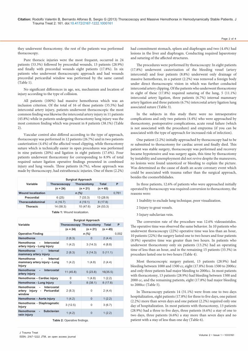
Pure thoracic injuries were the most frequent, occurred in 24 patients (53.3%) followed by precordial wounds, 13 patients (28.9%) and finally with precordial wounds eight patients (17.8%). In six patients who underwent thoracoscopic approach and had wounds precordial pericardial window was performed by the same cannel (Table 1).
No significant differences in age, sex, mechanism and location of injury according to the type of collision.
All patients (100%) had massive hemothorax which was an inclusion criterion. Of the total of 16 of these patients (35.5%) had intercostal artery injury, patients underwent thoracoscopic the most common finding was likewise the intercostal artery injury in 11 patients (45.8%) while in patients undergoing thoracotomy lung injury was the most common finding which was present in 8 patients (38.1%) (Table 2).
Vascular control also differed according to the type of approach, thoracoscopy was performed in 12 patients (26.7%) and in two patients cauterization (4.4%) of the affected vessel clipping, while thoracotomy suture which is technically easier in open procedures was performed in nine patients (20%) and ligation in eight patients (17.6%). Four patients underwent thoracotomy for corresponding to 8.9% of total required suture ligation operative findings presented in combined injury and lung vessels. Three patients (6.7%), whose approach was made by thoracoscopy, had extrathoracic injuries. One of them (2.2%)
had commitment stomach, spleen and diaphragm and two (4.4%) had lesions in the liver and diaphragm. Conducting required laparotomy and suturing of the affected structures.
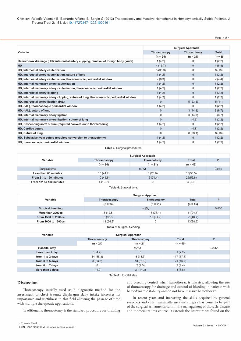
The procedures were performed by thoracoscopy: In eight patients (17.8%) underwent cauterization of the bleeding vessel (artery intercostal) and four patients (8.8%) underwent only drainage of massive hemothorax, in a patient (2.2%) was removed a foreign body under direct thoracoscopic vision in which was further conducted intercostal artery clipping. Of the patients who underwent thoracotomy in eight of these (17.8%) required suturing of the lung, 5 (11.1%) intercostal artery ligation, three patients (6.7%) internal mammary artery ligation and three patients (6.7%) intercostal artery ligation lung associated suture (Table 3).
In the subjects in this study there were no intraoperative complications and only two patients (4.4%) who were approached by thoracotomy, postoperative complications, acute renal failure (which is not associated with the procedure) and empyema (if you can be associated with the type of approach for increased risk of infection).
One patient (2.2%) initially approached by thoracoscopy had to be re submitted to thoracotomy for cardiac arrest and finally died. This patient was stable surgery, thoracoscopy was performed and recovery became hypotensive and was surgery again, this time by thoracotomy by instability and unemployment did not revive despite the maneuvers, no lesions were found unnoticed or bleeding to explain the picture. Was determined as the cause of death an acute coronary event which could be associated with trauma rather than the surgical approach, besides the coomorbilidades.
In three patients, 12.6% of patients who were approached initially operated by thoracoscopy was required conversion to thoracotomy, the reasons were:
1 Inability to exclude lung technique, poor visualization.
2 Injury to great vessels.
3 Injury subclavian vein.
The conversion rate of the procedure was 12.6% videoasistidos. The operative time was observed the same behavior. In 10 patients who underwent thoracoscopy (22%) operative time was less than an hour, 10 patients (22%) the surgery lasted one to two hours and four patients (8.9%) operative time was greater than two hours. In patients who underwent thoracotomy only six patients (13.2%) had an operating time of less than an hour, and in the remaining 15 patients (33.3%) in procedure lasted one to two hours (Table 4).
Most thoracoscopic surgery patient, 13 patients (28.9%) had bleeding between 1000 and 1500 cc, eight (17.8%) from 1500 to 2000cc and only three patients had major bleeding to 2000cc. In most patients with thoracotomy, 13 patients (28.9%) had bleeding between 1500 and 2000 cc, and the remaining patients, eight (17.8%) had major bleeding to 2000cc (Table 5).
In Thoracoscopy patients 14 (31.1%) were from one to two days hospitalization, eight patients (17.8%) for three to five days, one patient (2.2%) more than seven days and one patient (2.2%) required only one day of hospitalization. In most patients with thoracotomy, 13 patients (28.9%) had a three to five days, three patients (6.6%) a stay of one to two days, three patients (6.6%) a stay more than seven days and no patient with a stay of less than one day (Table 6).
VariableSurgical Approach
Thoracoscopy Thoracotomy Total P(n = 24) (n = 21) (n = 45)
Wound localization n (%) 0,761 Precordial 6 (25) 7 (33.3) 13 (28.9)
Thoracoabdominal 4 (16.7) 4 (19.1) 8 (17.8) Thoracic 14 (58.3) 10 (47.6) 24 (53.3)
Table 1: Wound localization.
VariableSurgical Approach
Thoracoscopy Thoracotomy Total P(n = 24) (n = 21) (n = 45)
Operative Finding n (%) 0,002 Hemothorax 2 (8.3) 0 2 (4.4) Hemothorax - Intercostal artery injury - Lung Injury 1 (4.2) 3 (14.3) 4 (8.8)
Hemothorax - Internal mammary artery injury 2 (8.3) 3 (14.3) 5 (11.1)
Hemothorax - Internal mammary artery injury - Lung Injury
1 (4.2) 1 (4.8) 2 (4.4)
Hemothorax - Intercostal artery injury 11 (45.8) 5 (23.8) 16(35.5)
Hemothorax – Cardiac injury 0 1 (4.8) 1 (2.2) Hemothorax - Lung Injury 0 8 (38.1) 8 (17.8) Hemothorax - Intercostal artery injury - Pericardial window
2 (8.3) 0 2 (4,4)
Hemothorax – Aorta injury 1 (4.2) 0 1 (2.2) Hemothorax - Diaphragmatic Injury 3 (12.5) 0 3 (6.7)
Hemothorax - Subclavian vein injury 1 (4.2) 0 1 (2.2)
Table 2: Operative findings.
Citation: Rodolfo Valentin B, Bernardo Alfonso B, Sergio G (2013) Thoracoscopy and Massive Hemothorax in Hemodynamically Stable Patients. J Trauma Treat 2: 161. doi:10.4172/2167-1222.1000161
Page 3 of 4
Volume 2 • Issue 1 • 1000161J Trauma TreatISSN: 2167-1222 JTM, an open access journal
DiscussionThoracoscopy initially used as a diagnostic method for the
assessment of chest trauma diaphragm daily intake increases its importance and usefulness in this field allowing the passage of time with multiple therapeutic applications.
Traditionally, thoracotomy is the standard procedure for draining
and bleeding control when hemothorax is massive, allowing the use of thoracoscopy for drainage and control of bleeding in patients with hemodynamic stability and do not have massive hemothorax.
In recent years and increasing the skills acquired by general surgeons and chest, minimally invasive surgery has come to be part of the surgical armamentarium in the management of thoracic disease and thoracic trauma course. It extends the literature we found on the
VariableSurgical Approach
Thoracoscopy Thoracotomy Total(n = 24) (n = 21) (n=45)
Hemothorax drainage (HD), intercostal artery clipping, removal of foreign body (knife) 1 (4.2) 0 1 (2.2)HD 4 (16.7) 0 4 (8.8)HD, Intercostal artery cauterization 8 (33.3) 0 8 (18)HD, Intercostal artery cauterization, suture of lung 1 (4.2) 0 1 (2.2)HD, Intercostal artery cauterization, thoracoscopic pericardial window 2 (8.3) 0 2 (4.4)HD, Internal mammary artery cauterization 1 (4.2) 0 1 (2.2)HD, Internal mammary artery cauterization, thoracoscopic pericardial window 1 (4.2) 0 1 (2.2)HD, Intercostal artery clipping 1 (4.2) 0 1 (2.2)HD, Internal mammary artery clipping, suture of lung, thoracoscopic pericardial window 1 (4.2) 0 1 (2.2)HD, Intercostal artery ligation (IAL) 0 5 (23.8) 5 (11)HD, (IAL), thoracoscopic pericardial window 1 (4.2) 0 1 (2.2)HD, (IAL), suture of lung 0 3 (14.3) 3 (6.7)HD, Internal mammary artery ligation 0 3 (14.3) 3 (6.7)HD, Internal mammary artery ligation, suture of lung 0 1 (4.8) 1 (2.2)HD, Descending aorta suture (required conversion to thoracotomy) 1 (4.2) 0 1 (2.2)HD, Cardiac suture 0 1 (4.8) 1 (2.2)HD, Suture of lung 0 8 (38.1) 8 (18)HD, Subclavian vein suture (required conversion to thoracotomy) 1 (4.2) 0 1 (2.2)HD, thoracoscopic pericardial window 1 (4.2) 0 1 (2.2)
Table 3: Surgical procedures.
VariableSurgical Approach
Thoracoscopy Thoracotomy Total P(n = 24) (n = 21) (n = 45)
Surgical time n (%) 0,054 Less than 60 minutes 10 (41.7) 6 (28.6) 16(35.5)
From 61 to 120 minutes 10 (41.6) 15 (71.4) 25(55.6) From 121 to 180 minutes 4 (16.7) 0 4 (8.9)
Tabla 4: Surgical time.
VariableSurgical Approach
Thoracoscopy Thoracotomy Total P(n = 24) (n = 21) (n = 45)
Surgical bleeding n (%) 0,000 More than 2000cc 3 (12.5) 8 (38.1) 11(24.4)
From 1500 to 2000cc 8 (33.3) 13 (61.9) 21(46.7) From 1000 to 1500cc 13 (54.2) 0 13(28.9)
Tabla 5: Surgical bleeding.
Variable Surgical ApproachThoracoscopy Thoracotomy Total P
(n = 24) (n = 21) (n = 45) Hospital stay n (%) 0,005*
Less than 1 day 1 (4.2) 0 1 (2.2) from 1 to 2 days 14 (58.3) 3 (14.3) 17 (37.8) from 3 to 5 days 8 (33.3) 13 (61.9) 21 (46.7) from 6 to 7 days 0 2 (9.5) 2 (4.4) More than 7 days 1 (4.2) 3 ( 14.3) 4 (8.8)
Table 6: Hospital stay.
Citation: Rodolfo Valentin B, Bernardo Alfonso B, Sergio G (2013) Thoracoscopy and Massive Hemothorax in Hemodynamically Stable Patients. J Trauma Treat 2: 161. doi:10.4172/2167-1222.1000161
Page 4 of 4
Volume 2 • Issue 1 • 1000161J Trauma TreatISSN: 2167-1222 JTM, an open access journal
different uses of this technique, including early handling trauma and posttraumatic hemothorax particularly with lower morbidity and costs given by reduced hospital stay demonstrated in this study.
In three patients who were initially approached by thoracoscopy was required conversion to thoracotomy due to poor visualization, large vessel injury and subclavian vein injury, with a conversion rate of 12.5%.
Regarding operative findings, all patients had massive hemothorax which was an inclusion criteria of the study. Regarding the differences associated lesions were evident: the majority of patients who were underwent thoracoscopy intercostal artery injury, whereas patients who underwent thoracotomy injury was most frequently found pulmonary which in turn may have determined the type of collision.
Three patients, whose approach was made by thoracoscopy, had extrathoracic injuries. One of them had committed in stomach, spleen and diaphragm and the other two had lesions in the liver and diaphragm. Conducting required laparotomy and suturing of the affected structures.
Vascular control also differ according to the type of approach, in thoracoscopy by cauterization and by thoracotomy suture and ligation.
Significant differences regarding intraoperative bleeding according to the type of approach, finding less bleeding in patients who underwent thoracoscopy. Although this may be related to the process may also be related to the severity of the lesions.
The operative time was observed the same behavior, being lower in thoracoscopy.
Where significant differences were found according to the type of collision, was in the hospital stay. In most patients who had undergone approach for thoracoscopy, thoracostomy duration was less than two days, while patients who underwent thoracotomy duration was three to five days in the majority and 14.3% greater than 7 days.
In the subjects in this study there were no intraoperative complications and only two patients, who were approached by thoracotomy, postoperative complications, acute renal failure (which is not associated with the procedure) and empyema (if can be associated with the type of approach at increased risk of infection).
One of the patients initially approached by thoracoscopy had to be re submitted to thoracotomy for cardiac arrest and finally died. It was considered that the cause of death was not related to the initial procedure.
In this study we found that all hemodynamically stable patients whose bleeding met criteria for massive hemothorax and were taken to thoracoscopy were handled successfully by this technique with statistically significant differences in terms of morbidity and shorter hospital stay compared to patients undergoing thoracotomy, and a similarity surgery time.
ConclusionsIn the group of patients analyzed in this study we can conclude
that thoracoscopy compared with thoracotomy was a safe approach
for the management of acute chest trauma in hemodynamically stable patients with massive hemothorax in terms of hospital stay and fewer complications with surgical times like.
No statistically significant difference was found regarding advantages or socio-demographic variables finding that patients affected are young men without excluding extreme age or gender. Occurred more frequently sharp weapon wounds which affected mostly the pure thoracic lesions with precordial and thoracoabdominal negligible.
In patients undergoing thoracoscopic presents a shorter hospital stay, given the degree of surgical trauma and possibly lower rate of complications associated with the type of approach and reduced exposure to hospital environment.
No statistical difference versus time comparing surgical procedures and open procedures video asistidos and conversely if thoracoscopy decreases hospital stay in a major way.
The conversion rate video asistidos procedures is presented in a low rate and was due mostly to the high complexity of the lesions and the impossibility of performing highly complex procedures by thoracoscopy.
References
1. Meredith JW, Hoth JJ (2007) Thoracic trauma: when and how to intervene. Surg Clin North Am 87: 95-118.
2. Meyer DM (2007) Hemothorax related to trauma. Thorac Surg Clin 17: 47-55.
3. He J (2011) History and current status of mini-invasive thoracic surgery. J Thorac Dis 3: 115-121.
4. Murray JA, Demetriades D, Cornwell EE 3rd, Asensio JA, Velmahos G, et al. (1997) Penetrating left thoracoabdominal trauma: the incidence and clinical presentation of diaphragm injuries. J Trauma 43: 624-626.
5. Mattox KL, Wall MJ, Pickard LR (2001) Lesiones De La Pared Torácica. Trauma. (4th edn), 507 – 516.
6. Petrone P, Asensio JA (2009) Surgical management of penetrating pulmonary injuries. Scand J Trauma Resusc Emerg Med 17: 8.
7. Programa Avanzado De Apoyo Vital En Trauma Para Médicos. ATLS. (7thedn).
8. Ivatury RR (2010)Thoracoscopy for Trauma. European Journal of Trauma and Emergency Surgery 36: 15–18.
9. Jacobaeus HC (1992) The practical importance of thoracoscopy in surgery of the chest.Surg Gynecol Obstet 34: 289-293
10. Martins Castello Branco J (1946) Thoracoscopy as a method of exploration in penetrating injuries of the thorax. Dis Chest 12: 330-335.
11. Ahmad J, Beattie GC, Kennedy R, Kennedy JA, Clements WD (2007) Penetrating trauma to the junctional zone needs aggressive management. BMJ 334: 257-258.
12. Parreira JG, Rasslan S, Utiyama EM (2008) Controversies in the management of asymptomatic patients sustaining penetrating thoracoabdominal wounds. Clinics (Sao Paulo) 63: 695-700.
13. Al-Koudmani I, Darwish B, Al-Kateb K, Taifour Y (2012) Chest trauma experience over eleven-year period at al-mouassat university teaching hospital-Damascus: a retrospective review of 888 cases. J Cardiothorac Surg 7: 35.
14. Kaewlai R, de Moya MA, Santos A, Asrani AV, Avery LL, et al. (2011) Blunt cardiac injury in trauma patients with thoracic aortic injury. Emerg Med Int 2011: 848013.
Citation: Rodolfo Valentin B, Bernardo Alfonso B, Sergio G (2013) Thoracoscopy and Massive Hemothorax in Hemodynamically Stable Patients. J Trauma Treat 2: 161. doi:10.4172/2167-1222.1000161