Thoracic outlet syndrome Part 2: Conservative management...
10
Masterclass Thoracic outlet syndrome Part 2: Conservative management of thoracic outlet L.A. Watson a, b , T. Pizzari b, * , S. Balster a a LifeCare Prahran Sports Medicine Centre, 316 Malvern Road, Prahran, VIC 3181, Australia b Musculoskeletal Research Centre, La Trobe University, Bundoora VIC 3086, Australia article info Article history: Received 10 March 2009 Received in revised form 23 September 2009 Accepted 1 March 2010 Keywords: Thoracic outlet syndrome Conservative management TOS Physical therapy abstract Thoracic outlet syndrome (TOS) is a symptom complex attributed to compression of the nerves and vessels as they exit the thoracic outlet. Classified into several sub-types, conservative management is generally recommended as the first stage treatment in favor of surgical intervention. In cases where postural deviations contribute substantially to compression of the thoracic outlet, the rehabilitation approach outlined in this masterclass will provide the clinician with appropriate management strategies to help decompress the outlet. The main component of the rehabilitation program is the graded restoration of scapula control, movement, and positioning at rest and through movement. Adjunctive strategies include restoration of humeral head control, isolated strengthening of weak shoulder muscles, taping, and other manual therapy techniques. The rehabilitation outlined in this paper also serves as a model for the management of any shoulder condition where scapula dysfunction is a major contributing factor. Ó 2010 Elsevier Ltd. All rights reserved. 1. Introduction Thoracic outlet syndrome (TOS) is a symptom complex char- acterized by pain, paresthesia, weakness and discomfort in the upper limb which is aggravated by elevation of the arms or by exaggerated movements of the head and neck (Lindgren and Oksala, 1995). Part one of this two-part article series outlined the classification, aetiology, varying clinical presentations, subjective and physical assessment, differential diagnoses and management principles of TOS. The purpose of this paper is to comprehensively describe one treatment approach for the conservative manage- ment of TOS. 2. Classification, aetiology, & management As noted in Part one, TOS is often categorized into two specific clinical entities: Vascular TOS and Neurological TOS (Atasoy, 1996; Rayan, 1998; Sharp et al., 2001) and further sub-divided into arte- rial and venous TOS under the vascular umbrella and true neuro- logical TOS and symptomatic TOS (sTOS) under the neurological heading (Fig. 1). The majority of patients presenting with TOS will fall under the sTOS classification (Wilbourn, 1990; Rayan, 1998; Davidovic et al., 2003). sTOS may be aggravated by postural or occupational stressors with repetitive overuse and associated soft tissue adaptations causing intermittent compression of the neurovascular complex (Mackinnon and Novak, 2002). In the absence of any acute or progressive neurological or vascular lesion, conservative treatment is often recommended as the first stage of management for all sub-types of TOS and surgery is only considered if this fails (Sharp et al., 2001; Mackinnon and Novak, 2002). Conservative management may involve medication, injection therapy, rest, modification of aggravating activities, physiotherapy or a combination of all strategies. The majority of the studies published on TOS highlight physiotherapy strengthening exercises and postural re-educational drills as being the mainstay of any conservative management programme for TOS (Aligne and Barral, 1992; Kenny et al., 1993; Jamieson and Chinnick, 1996; Urschel and Razzuk, 1997; Molina, 1998; Athanassiadi et al., 2001; Mackinnon and Novak, 2002; Wright and Jennings, 2005; Cagli et al., 2006). However specifics of the programmes are rarely given and outcome measures and expected time frames for potential recovery are not included. The conservative physiotherapy regimen outlined in this article will be suitable for patients presenting with TOS where there is a strong postural contribution to their symptoms. In particular, in cases of TOS where the scapula mechanics are poor and the patient presents with the dropped shoulder condition (scapula depressed and/or downwardly rotated, and/or anteriorly tilted) (Ranney,1996). 3. Dropped shoulder condition Many forms of scapula asymmetry may well exist in TOS pop- ulations, but in the limited research that has been done, scapula or * Corresponding author. Tel.: þ61 3 94795872. E-mail address: [email protected] (T. Pizzari). Contents lists available at ScienceDirect Manual Therapy journal homepage: www.elsevier.com/math 1356-689X/$ e see front matter Ó 2010 Elsevier Ltd. All rights reserved. doi:10.1016/j.math.2010.03.002 Manual Therapy 15 (2010) 305e314
Transcript of Thoracic outlet syndrome Part 2: Conservative management...

lable at ScienceDirect
Manual Therapy 15 (2010) 305e314
Contents lists avai
Manual Therapy
journal homepage: www.elsevier .com/math
Masterclass
Thoracic outlet syndrome Part 2: Conservative management of thoracic outlet
L.A. Watson a,b, T. Pizzari b,*, S. Balster a
a LifeCare Prahran Sports Medicine Centre, 316 Malvern Road, Prahran, VIC 3181, AustraliabMusculoskeletal Research Centre, La Trobe University, Bundoora VIC 3086, Australia
a r t i c l e i n f o
Article history:Received 10 March 2009Received in revised form23 September 2009Accepted 1 March 2010
Keywords:Thoracic outlet syndromeConservative managementTOSPhysical therapy
* Corresponding author. Tel.: þ61 3 94795872.E-mail address: [email protected] (T. Pizzar
1356-689X/$ e see front matter � 2010 Elsevier Ltd.doi:10.1016/j.math.2010.03.002
a b s t r a c t
Thoracic outlet syndrome (TOS) is a symptom complex attributed to compression of the nerves and vesselsas they exit the thoracic outlet. Classified into several sub-types, conservative management is generallyrecommended as the first stage treatment in favor of surgical intervention. In cases where posturaldeviations contribute substantially to compression of the thoracic outlet, the rehabilitation approachoutlined in this masterclass will provide the clinician with appropriate management strategies to helpdecompress the outlet. The main component of the rehabilitation program is the graded restoration ofscapula control, movement, and positioning at rest and through movement. Adjunctive strategies includerestoration of humeral head control, isolated strengthening of weak shoulder muscles, taping, and othermanual therapy techniques. The rehabilitation outlined in this paper also serves as a model for themanagement of any shoulder condition where scapula dysfunction is a major contributing factor.
� 2010 Elsevier Ltd. All rights reserved.
1. Introduction
Thoracic outlet syndrome (TOS) is a symptom complex char-acterized by pain, paresthesia, weakness and discomfort in theupper limb which is aggravated by elevation of the arms or byexaggerated movements of the head and neck (Lindgren andOksala, 1995). Part one of this two-part article series outlined theclassification, aetiology, varying clinical presentations, subjectiveand physical assessment, differential diagnoses and managementprinciples of TOS. The purpose of this paper is to comprehensivelydescribe one treatment approach for the conservative manage-ment of TOS.
2. Classification, aetiology, & management
As noted in Part one, TOS is often categorized into two specificclinical entities: Vascular TOS and Neurological TOS (Atasoy, 1996;Rayan, 1998; Sharp et al., 2001) and further sub-divided into arte-rial and venous TOS under the vascular umbrella and true neuro-logical TOS and symptomatic TOS (sTOS) under the neurologicalheading (Fig. 1).
The majority of patients presenting with TOS will fall under thesTOS classification (Wilbourn, 1990; Rayan, 1998; Davidovic et al.,2003). sTOS may be aggravated by postural or occupationalstressors with repetitive overuse and associated soft tissue
i).
All rights reserved.
adaptations causing intermittent compression of the neurovascularcomplex (Mackinnon and Novak, 2002).
In the absence of any acute or progressive neurological orvascular lesion, conservative treatment is often recommended asthe first stage of management for all sub-types of TOS and surgeryis only considered if this fails (Sharp et al., 2001; Mackinnon andNovak, 2002). Conservative management may involve medication,injection therapy, rest, modification of aggravating activities,physiotherapy or a combination of all strategies. Themajority of thestudies published on TOS highlight physiotherapy strengtheningexercises and postural re-educational drills as being themainstay ofany conservative management programme for TOS (Aligne andBarral, 1992; Kenny et al., 1993; Jamieson and Chinnick, 1996;Urschel and Razzuk, 1997; Molina, 1998; Athanassiadi et al., 2001;Mackinnon and Novak, 2002; Wright and Jennings, 2005; Cagliet al., 2006). However specifics of the programmes are rarelygiven and outcome measures and expected time frames forpotential recovery are not included.
The conservative physiotherapy regimen outlined in this articlewill be suitable for patients presenting with TOS where there isa strong postural contribution to their symptoms. In particular, incases of TOS where the scapula mechanics are poor and the patientpresents with the dropped shoulder condition (scapula depressedand/ordownwardly rotated, and/or anteriorly tilted) (Ranney,1996).
3. Dropped shoulder condition
Many forms of scapula asymmetry may well exist in TOS pop-ulations, but in the limited research that has been done, scapula or

Thoracic Outlet Syndrome
(TOS)
Vascular thoracic outlet syndrome
(vTOS)
Neurological thoracic outlet syndrome
(nTOS)
True neurological thoracic outlet syndrome
(tnTOS)
Symptomatic thoracic outlet syndrome
(sTOS)
Arterial thoracic outlet syndrome (aTOS)
Venous thoracic outlet syndrome (vTOS)
Fig. 1. Sub-types of thoracic outlet syndrome.
L.A. Watson et al. / Manual Therapy 15 (2010) 305e314306
shoulder girdle depression or “drooping” has been consistentlyobserved (Kenny et al., 1993;Walsh, 1994; Pascarelli and Hsu, 2001;Skandalakis and Mirilas, 2001).
Scapula depression will lead to an alteration of the anatomicalalignment of the structures in both the cervical and thoracic outlet(Telford and Mottershead, 1948; Kai et al., 2001; Skandalakis andMirilas, 2001) (Fig. 2). It may potentially lead to tractional stressbeing placed on the nerve, vascular and muscular elements as wellas compression as the clavicle descends closer towards either thefirst rib or any other bony element present. Elevation of theshoulder girdle can alleviate these stressors and potentially lead to“decompressing” the thoracic outlet (Kitamura et al., 1995). Themanagement approach described in this article is designed toelevate the shoulder girdle and restore scapula control.
4. Clinical presentation: scapula position
One of the consistent objective findings that we have observedand measured in cases of sTOS is that the scapula can be depressedat rest (Fig. 3) on the symptomatic side compared to the other side(in unilateral TOS) and to the normative data in cases of bilateralTOS (Kai et al., 2001).
Importantly it is not only at rest that the scapula demon-strates dysfunction but also through elevation motions such as
Fig. 2. Thoracic outlet anatomy. A: Normal position of the shoulder girdle. B: Scapula deprcervical and thoracic outlet.
abduction and flexion. In abduction, patients with droppedshoulder TOS (dsTOS) frequently demonstrate late and insuffi-cient upward rotation of the scapula compared to the other sideand/or to normal (Figs. 4 and 5). This can often lead to anapparent restriction of abduction range but the deficit is due toinadequate shoulder girdle muscle control and reduced upwardrotation of the scapula. Abduction is usually the most provoc-ative motion in dsTOS and it often reproduces the patient’spain, neurological or vascular symptoms, especially if sustainedas part of a provocation test (refer to article 1). In flexion, thesame tendency for depression and downward rotation is seenbut is often over-shadowed by an obvious winging of thescapula due to serratus anterior insufficiency (Mackinnon andNovak, 2002).
Increased anterior tilt of the scapula is also commonly identifiedin sTOS (Sucher, 1990; Aligne and Barral, 1992; Press and Young,1994; Walsh, 1994) and it is frequently coupled clinically withincreased downward rotation of the scapula.
5. Clinical presentation: scapula muscle control
Although no EMG research exists examining muscle charac-teristics in a TOS patient population, it is conceivable that alter-ations in recruitment, intensity and force of shoulder girdle
ession causing an alteration of the anatomical alignment of the structures in both the

Fig. 3. Measurement of scapula elevation/depression. The patient is positioned inrelaxed standing posture. A tape measure is attached (via piece of tape) to the C7spinous process. It is then let to drop down the length of the thoracic spine. A secondpiece of tape may be utilized to attach the tape measure to the spine distally (plumbline). With a second tape measure the therapist may measure across from the inferiorangle of the scapula, the end of the spine of the scapula and the acromioclavicular jointto the plumb line. The therapist can then make a note of 1) the centimetre referencepoint that correlates to the vertical level of each of the three bony landmarks. 2) thedistance of each of the bony landmarks to the plumb line. The therapist will be able tojudge if the scapula is either elevated or depressed, medially or laterally placed. Oneside is compared to the other to determine if any asymmetry is present. Anything morethan 1 cm of asymmetry is thought to be significant (Kibler, 1991).
Fig. 4. Measurement of scapula upward and downward rotation at rest. The patient ispositioned in relaxed standing posture. One inclinometer is attached to the patient’shumerus with two Velcro straps. A second inclinometer is placed on the spine of thepatient’s scapula e midway between the postero-lateral corner of the acromion andthe end of the spine of the scapula. A measurement of the patient’s resting humeralabduction angle is taken and a measurement of the scapula’s upward/downwardrotation at rest.
Fig. 5. Measurement of the scapula’s position through range of motion. The patientmoves their arm into abduction or flexion. The patient may pause at any positionthrough range that the therapist wants to examine (e.g. symptom onset). Ameasurement of upward/downward rotation of the scapula is taken and the humeralangle. This may be performed all the way through range so that a pattern of scapulamotion through range can be determined.
L.A. Watson et al. / Manual Therapy 15 (2010) 305e314 307
musculature may lead to the aberrant scapula control that hasbeen observed and measured clinically in our patient populationwith dsTOS.
Clinical manual muscle strength tests performed by theseauthors with a hand-held strength dynamometer (Power Track II�,USA) have consistently demonstrated decreased strength in manyshoulder girdle muscles in patients with dsTOS. There is oftena general weakness in shoulder girdle function in the symptomaticcompared to the asymptomatic side, in particular in upper andmiddle trapezius. Substitution or increased consistency of recruit-ment by other muscle groups such as rhomboids, levator scapulaeand pectoralis minor may also occur, leading to the scapula asym-metries commonly observed: downward rotation, depression andanterior tilt of the scapula.
Scapula dysfunction is not only seen in dsTOS with activemotions but in response to loading strategies as well. When resis-ted humeral motion tests (such as resisted external rotation) (Fig. 6)are performed, scapula dyskinesia often becomes more apparent asthe scapula is forced to stabilize itself and hold form against thethorax while humeral force is generated.
6. Clinical presentation: scapula correction
The final clinical assessment finding that may indicate thataberrance in scapula mechanics is implicated in either the devel-opment or progression of some forms of TOS, is the observationthat correction of the scapula asymmetry will frequently alleviateor improve the patients’ symptoms (Refer to Part 1). The amountand direction of correction required depends on the position of thescapula but usually the manoeuvre involves elevation and upwardrotation of the scapula, trying to maintain the scapula at a similarlevel to the unaffected side or in the position that achieves the bestsymptom relief.
Correction manoeuvres can be applied to any assessmentposition, including loaded tests such as glenohumeral externalrotation. If correction of the scapula improves symptoms (pain,weakness, distal symptoms, range of motion) due to betterbiomechanical performance of the shoulder girdle then logicallycorrection of the scapula position should be addressed in thepatient’s rehabilitation.

Fig. 6. Dynamic External Rotation Scapula Stability Test. Patient is positioned inrelaxed standing posture, examiner behind patient. External rotation of the humerus isresisted with the patient in 0� of abduction. In the normal shoulder the patient usuallymaintains their resting scapula posture (A). If scapula muscle weakness is present thenthe scapula will go into a position that reflects the weakness and highlights anydominant strategies (B). For example if upper trapezius is weak and levator scapulaeþ/� rhomboids are dominant then the scapula will pull into downward rotation. Ifserratus anterior is weak and pectoralis minor is dominant then the scapula will wingand pull into anterior tilt. Varying combinations of scapula asymmetry may be seen.
L.A. Watson et al. / Manual Therapy 15 (2010) 305e314308
7. Rehabilitation overview
The cornerstone of this exercise program is firstly to focus onestablishing normal scapula muscle recruitment and control in theresting position. Once this is achieved then the program is pro-gressed to maintaining scapula control while both motion and loadare applied. The programme begins in lower ranges of abductionand is gradually progressed further up into abduction and flexionrange until muscles are being re-trained in functional movementpatterns at higher ranges of elevation (Fig. 7).
7.1. Scapula setting and control
The aim of the scapula “setting” drills is to have the patientactively place their scapula into a “normalized position” and holdthis position while humeral motion is superimposed. The scapulamotion can be measured clinically using an inclinometer, allowingthe therapist to determine if the patient is getting insufficient orexcessive scapula motion occurring at any stage through range ofmotion (Fig. 5). Comparison can be made to the other side or
normative “ideals”. The same can occur for medial/lateral shift andelevation/depression of the scapula.
Using three reference points (Fig. 3) the scapula position can bemapped. From the normative data we have collated so far for totalshoulder abduction, the inferior angle of the scapula typicallymoveslaterally 8 cm and superiorly by 3 cm, the medial end of the spine ofthe scapula moves inferiorly 3 cm and laterally by 1.5 cm and theacromion moves superiorly 4 cm andmedially by 5 cm. The scapulashould tilt posteriorly 30� and rotate externally 25� although clini-cally this is very difficult to measure objectively. In flexion a similarscapula motion pattern is seen (McClure et al., 2001; Bourne et al.,2007). Small deviations in scapula control are expected, it is thelarger obvious asymmetries that need to be assessed.
The overall aim of the scapula control work is to achievea “neutral” scapula position without over elevation or depressionandmaintaining control against the thoraxwith sufficient posteriortilt of the scapula to keep themedial border of the scapula stabilized(Fig. 8). This will be achieved by contribution from all of the scapulastabilizers, but in particular serratus anterior, upper, middle andlower trapezius. The aim of the rehabilitation drills is to have all themuscles activating synchronously such that a net smooth upwardrotation of the scapula occurs through range of motion. There is norequirement to “stop” somemusclesworking (such as rhomboids orlevator scapulae) but rather the emphasis is on facilitating andencouraging sufficient firing in any muscles that may be weak,inhibited or slow to switch on in the normal movement strategies.
Themusclesmost commonly requiring facilitation are upper andmiddle trapezius, serratus anterior or lower trapezius. These are alltonic stabilizers and direct the use of endurance repetitions in theinitial phase of rehabilitation (Mackinnon and Novak, 2002).Patientswith TOS have been found to develop fatigue earlier in theirupper extremities than healthy individuals (Ozcakar et al., 2004).
7.2. Dosage and progression
The therapist assesses how many repetitions of “setting” thescapula the patient can achieve with good form. The aim is toachieve the “neutral” scapula position without substitution fromother scapula stabilizers to the extent that aberrant motions such asincreased anterior tilt or downward rotation of the scapula areoccurring. If repetitions of 20 can be achieved without fatigue(either physically feeling fatigue in muscles or inability of thepatient to control/activate scapula muscles correctly) then this isprescribed three times per day. Once this is achieved the drill needsto be progressed, either by increasing the resistance or by progr-essing the movement patterns or both if the patient can maintaincontrol. If progression is via motion pattern then usually endurancerepetitions are still utilized with low resistance. If progression is viaadding a weight then this starts off very low (usually 0.5 kg) as thescapula stabilizers are usually still working in primarily a stabilizingrole. The weight is incrementally increased by 0.5 kg until anendurance base is achieved (for most muscle groups two sets of 20with 2 kg is sufficient). Once any form of resistance is applied theregime is dropped to performing twice a day. Movement controland strength work is always commenced initially through abduc-tion as this is the most commonly affected and symptom producingmotion for patients with sTOS (Mackinnon and Novak, 2002).
Once recruitment patterns are achieved then isolatedstrengthening drills may be given for any specific individual musclegroups that test weak. These are only given if the patient cancontrol the scapula and humeral head position during the range ofmotion utilized in these loaded positions. While emphasis has beenplaced here on scapula control, humeral head control is equallyimportant at all stages of rehabilitation. It is important that thetherapist assesses humeral head position during any loaded

Fig. 7. Rehabilitation overview.
L.A. Watson et al. / Manual Therapy 15 (2010) 305e314 309
exercise to ensure control is maintained (Fig. 9). Initially lightresistance (of either theraband or weights) is utilized with endur-ance repetitions. For most muscle groups 2 kg max is sufficient forwomen and 3 kg for men once per day. Hypertrophy drills may
Fig. 8. “Setting” of the scapula involves small isometric contractions. Patientmoves or “sets” thMay be a small upward rotation elevation (C) medial glide (B) posterior tilt or scapula depress
eventually be added only if a patients’ job or sport requires strengthcapacity (such as heavy lifting). This is commenced only if a patientis using 3 kg or above in any particular drill and can be progressedas required. Usually the dosage needs to be altered to facilitate
eir scapula into the corrected position using their fingers if possible to feel the contraction.ion (A) depending on the asymmetry of the scapula and corrective mechanism required.

Fig. 9. Dynamic Humeral Head Assessment. Patient positioned in relaxed standing.Patient moves arm out into abduction/external rotation combination, usually againstresistance (although any motion may be performed). Therapist palpates the humeralhead position. Right shoulder e therapist utilizes left hand. Middle finger on thecoracoid, index finger on the humeral head and thumb posteriorly on the humeralhead. Therapist right hand resists the patient's active motion. Therapist looks for anyaberrant humeral head movement or loss of ability to centralize or stabilize thehumeral head (usually the humeral head goes anteriorly).
Fig. 10. Upward rotation shrug e performed in 20e30� of abduction in standing.Facilitates upper trapezius. Can be utilized for assessment or rehabilitation.
L.A. Watson et al. / Manual Therapy 15 (2010) 305e314310
more hypertrophy of the muscle, therefore repetitions of six oreight are selected by four sets (if fatigue allows) only once a day. Itmay also be beneficial to have “rest” days formuscles to recover andto ensure exercise induced pain is not being created.
Patients are usually discharged on a home program consisting ofexercises 3e4 times per week.
8. Rehabilitation program e initial phase
8.1. Aim of this phase
Control of scapula position at rest and in lower ranges ofabduction (30� and below).
Commonly in dsTOS the patient will require facilitation ofupward rotation of the scapula with a slight amount of elevation.This is achieved by performing an upward rotation shrug in 20e30�
of abduction in standing (Fig. 10). Common errors include anteriortilt of scapula or over-retraction by rhomboids. Facilitation tech-niques include therapist positioning of the scapula with the patientgradually taking over, slight therapist resistance being applied overthe acromioclavicular joint, patient self palpation of upper trape-zius, visual feedback via mirrors, video monitoring or biofeedback.The patient should perform approximately 80% of their maximumrepetition without fatigue or substitution. Once endurance repeti-tions are achieved then progression can occur by adding a weightinto the hand (0.5 kg increments) or resistance of theraband. Theamount of resistance is determined by the patients’ ability tocontrol the drill without fatigue. If the patient encounters signifi-cant cervical pain, an exacerbation of distal symptoms or just findsthe exercises too hard then they should be performed in sidelyingwith the arm supported on a pillow into slight abduction.
Progression can also occur by progressing functional movementpatterns. Usually abduction in external rotation is selected first asthis is the most functional movement pattern (Fig. 11). If the patientcannot maintain scapula control throughout the whole movementarc, then the movement is broken down so that the patient onlyperforms that part of the arc that they can control or the resistancemay need to be reduced. Sometimes the combination motion maybe too difficult because there is a co-existing glenohumeral jointweakness (such as external rotation). In this case then themotion isbroken down into just that component the patient can control (forexample extension against theraband with scapula setting) whilespecific isolated drills for the weak glenohumeral component aregiven (such as sidelying external rotation for external rotationdeficits). Once sufficient strength is achieved then the combineddrill is performed.
As well as ensuring scapula control is maintained during themotion, humeral head position must also be assessed. If there isinsufficient control of the humeral head position then specific drillsmay need to be given to facilitate humeral head control. The most

Fig. 11. Abduction external rotation patterning. Patient “sets” their scapula and thenmoves their arm into functional motion pattern maintaining both scapula and humeralhead control.
L.A. Watson et al. / Manual Therapy 15 (2010) 305e314 311
common aberrant humeral head position is an increase in anteriorplacement of the humeral head. A useful strategy to help facilitateco-contraction of the rotator cuff to help stabilize and centralize thehumeral head is to facilitate a mid level isometric contraction of therotator cuff by applying resistance to the humeral head (Dark et al.,2007). The best place to apply resistance is posteriorly over thehumeral head via a rubber band (Fig. 12) as this resistance has theadded benefit of facilitating the posterior scapula stabilizers. Thismay be integrated further into movement patterns. Initiallyemphasis is on slow controlled concentric/eccentric motion drills.
Isolated muscle strengthening drills in these lower ranges ofabduction that can be given safely, if required, are; upper trapezius(upward rotation shrugs), external rotation (usually sidelying),posterior deltoid (extension standing), subscapularis (supineinternal rotation) and anterior deltoid (supine flexion).
9. Rehabilitation program e exercises in 45e90� of abduction
Aim of this phase:
Control of scapula position above 45� of elevation.Centralize and control humeral head position.Load individual muscles that display weaknesses.
Once control is achieved in the lower ranges of abduction,movement control work should be performed in higher ranges ofabduction commencing at 45� and progressing towards 70� then90� of abduction. This can be performed against varying grades ofresistance (usually theraband).
Once 90� control is developing, hypertrophy drills for trapeziuscan be performed in prone horizontal extension if more strengthis required. Traditionally these positions have been described fordeveloping middle and lower trapezius strength (Kendall et al.,1971; Ekstrom et al., 2003), but in reality all of the scapula
stabilizers will fire in these positions producing a combined effortto stabilize the scapula against the thorax. Precisely which muscleis working is probably not as important as making sure that thescapula is being “set” into a normal stabilizing pattern withupward rotation and stabilization of the medial border of thescapula occurring. Initially the drill is commenced with the elbowbent (short lever) and neutral rotation of the shoulder to avoidany compression to the rotator cuff. If it is required, then longlever strength work can be performed in either internal orexternal rotation. This would usually only be for those patientswho require overhead strength for their occupation or sport (suchas painters and swimmers) and only in patients where there is nopathology (such as supraspinatus tendinopathy) that might getaggravated by this position. Prone horizontal extension drills arealso very good drills for developing posterior deltoid, supra-spinatus, infraspinatus and teres minor. Always ensure that thepatient can maintain both scapula and humeral head controlthroughout the drill.
Isolated muscle strengthening drills in these mid ranges ofabduction that can be given safely, if required, are; posterior deltoid(extension in standing against theraband in higher ranges ofabduction, elbow bent), subscapularis (supine internal rotation orstanding internal rotation in varying degrees of abduction). Middledeltoid performed in sitting to 60� abduction maximum (to avoidany cuff impingement) with elbow flexion can also be performed ifrequired.
10. Rehabilitation program e flexion control
Once reasonable abduction control is established then thetherapist needs to re-assess flexion control. Frequently in flexionthere is winging of the scapula reflecting weakness and inhibitionof serratus anterior. It is tempting to give serratus anterior drillsearly on in the rehabilitation programme that involve flexion orweight bearing strategies since these have traditionally beenprescribed for serratus anterior deficit (Moseley et al., 1992;Ekstrom et al., 2004), however one complication of using thesestrategies is that pectoralis minor is also recruited early on in theflexion movement. These drills can over-stimulate pectoralis minorrecruitment which may potentially propagate the symptoms ofsTOS. Abduction external rotation strategies described above areoften sufficient to trigger serratus recruitment and control withoutthe risk of over-activating pectoralis minor (Wickham et al., 2009)(Fig. 11). If there is persisting serratus anterior deficit in the flexionmovement, especially in the eccentric lowering phase of themotion, the same principles of scapula facilitation and movementcontrol can be applied to a graduated programme emphasising thecontrol of the flexion motion against theraband resistance. Thetherapist may utilize hand placement to facilitate posterior tilt ofthe scapula (Fig. 13).
Once flexion control is achieved isolated muscle drills that canbe added, as required, are; anterior deltoid (sitting flexion) andserratus anterior (military raise in above 120� of flexion or plane ofthe scapula elevation).
11. Rehabilitation program e exercises in 90� of abductionand above/functional progressions
The same strategies are employed as for the lower ranges butthis is only performed in patients who functionally require thesehigher ranges of strength, usually an athlete or overhead manualworker. The same principles of rehabilitation can also be incorpo-rated into functional movement patterns such as reproducinga swimming stroke, or emulating the repeated arm movement oftypingwhilemaintaining scapula and humeral head control against

Fig. 12. Facilitation of humeral head control. Therapist utilizes their fingers to apply pressure to the posterior humeral head (or further distally down the arm) and asks the patientto push back against their fingers. Only moderate resistance is applied. This will facilitate co-contraction of the rotator cuff muscles and help resist any aberrant humeral headmotion (such as anterior glide). Patient then attaches one theraband proximally to mimic the therapist pressure and another distally in the hand. Patient “sets” their scapula, pushesback into the top rubber band and then pulls out into the functional motion pattern required maintaining both scapula and humeral head control. Endurance repetitions utilized.
L.A. Watson et al. / Manual Therapy 15 (2010) 305e314312
light resistance. However, caremust be takenwith strengthwork inthe overhead position with many cases of TOS since if performedtoo early many patients’ symptoms may be provoked. Strengthwork in this position is never performed in any patient with any
form of vascular TOS as damage to the vascular structures mayoccur. Initially focus will be on slow control of concentric andeccentric motion but eventually ballistic/plyometric type contrac-tions are encouraged as functionally required.
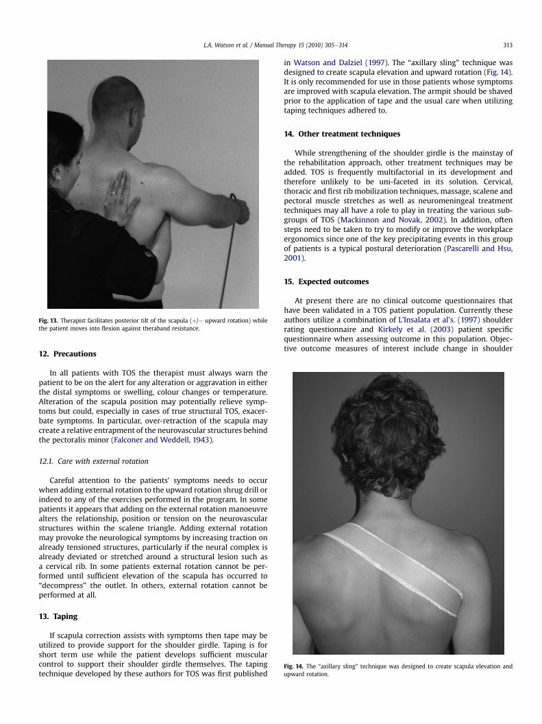
Fig. 13. Therapist facilitates posterior tilt of the scapula (þ/� upward rotation) whilethe patient moves into flexion against theraband resistance.
Fig. 14. The “axillary sling” technique was designed to create scapula elevation andupward rotation.
L.A. Watson et al. / Manual Therapy 15 (2010) 305e314 313
12. Precautions
In all patients with TOS the therapist must always warn thepatient to be on the alert for any alteration or aggravation in eitherthe distal symptoms or swelling, colour changes or temperature.Alteration of the scapula position may potentially relieve symp-toms but could, especially in cases of true structural TOS, exacer-bate symptoms. In particular, over-retraction of the scapula maycreate a relative entrapment of the neurovascular structures behindthe pectoralis minor (Falconer and Weddell, 1943).
12.1. Care with external rotation
Careful attention to the patients’ symptoms needs to occurwhen adding external rotation to the upward rotation shrug drill orindeed to any of the exercises performed in the program. In somepatients it appears that adding on the external rotation manoeuvrealters the relationship, position or tension on the neurovascularstructures within the scalene triangle. Adding external rotationmay provoke the neurological symptoms by increasing traction onalready tensioned structures, particularly if the neural complex isalready deviated or stretched around a structural lesion such asa cervical rib. In some patients external rotation cannot be per-formed until sufficient elevation of the scapula has occurred to“decompress” the outlet. In others, external rotation cannot beperformed at all.
13. Taping
If scapula correction assists with symptoms then tape may beutilized to provide support for the shoulder girdle. Taping is forshort term use while the patient develops sufficient muscularcontrol to support their shoulder girdle themselves. The tapingtechnique developed by these authors for TOS was first published
in Watson and Dalziel (1997). The “axillary sling” technique wasdesigned to create scapula elevation and upward rotation (Fig. 14).It is only recommended for use in those patients whose symptomsare improved with scapula elevation. The armpit should be shavedprior to the application of tape and the usual care when utilizingtaping techniques adhered to.
14. Other treatment techniques
While strengthening of the shoulder girdle is the mainstay ofthe rehabilitation approach, other treatment techniques may beadded. TOS is frequently multifactorial in its development andtherefore unlikely to be uni-faceted in its solution. Cervical,thoracic and first rib mobilization techniques, massage, scalene andpectoral muscle stretches as well as neuromeningeal treatmenttechniques may all have a role to play in treating the various sub-groups of TOS (Mackinnon and Novak, 2002). In addition, oftensteps need to be taken to try to modify or improve the workplaceergonomics since one of the key precipitating events in this groupof patients is a typical postural deterioration (Pascarelli and Hsu,2001).
15. Expected outcomes
At present there are no clinical outcome questionnaires thathave been validated in a TOS patient population. Currently theseauthors utilize a combination of L’Insalata et al’s. (1997) shoulderrating questionnaire and Kirkely et al. (2003) patient specificquestionnaire when assessing outcome in this population. Objec-tive outcome measures of interest include change in shoulder

L.A. Watson et al. / Manual Therapy 15 (2010) 305e314314
girdle strength, scapula position at rest and in motion and time toonset of symptoms during provocation testing. Clinical data hasshown us that patients can reliably achieve a change in theirscapula resting position by 6 weeks of rehabilitation and an alter-ation in both their shoulder girdle strength and scapula motionthrough range by 12weeks of rehabilitation. Concurrent with this isusually a gradual improvement in the patients’ signs and symptomsand an improvement in their functional state. Some patients havecompleted their program and have achieved normal strength andscapula measurements by 12 weeks, others take 6 months andsome never progress past a plateau or fail to complete the program.It is reasonable that if this program is going to benefit your patient,then there should be some subjective and objective improvementin symptoms by six to eight weeks.
16. Conclusion
The rehabilitation approach outlined in this article is based onour clinical experience of working with patients with TOS and atthe current time we can only describe anecdotal evidence ofsuccess. There is a need for good quality research in this area todetermine the most appropriate treatment path for the differentTOS sub-types and the optimal composition of conservativerehabilitation.
References
Aligne C, Barral X. Rehabilitation of patients with thoracic outlet syndrome. Annalsof Vascular Surgery 1992;6(4):381e9.
Atasoy E. Thoracic outlet compression syndrome. Orthopedic Clinics of NorthAmerica 1996;27(2):265e303.
Athanassiadi K, Kalavrouziotis G, Karydakis K, Bellenis I. Treatment of thoracicoutlet syndrome: long-term results. World Journal of Surgery 2001;25(5):553e7.
Bourne DA, Choo AM, Regan WD, MacIntyre DL, Oxland TR. Three-dimensionalrotation of the scapula during functional movements: an in vivo study inhealthy volunteers. Journal of Bone and Joint Surgery 2007;16(2):150e62.
Cagli K, Ozcakar L, Beyazit M, Sirmali M. Thoracic outlet syndrome in an adolescentwith bilateral bifid ribs. Clinical Anatomy 2006;19(6):558e60.
Dark A, Ginn KA, Halaki M. Shoulder muscle recruitment patterns during commonlyused rotator cuff exercises: an electromyographic study. Physical Therapy2007;87(8):1039e46.
Davidovic LB, Kostic DM, Jakovljevic NS, Kuzmanovic IL, Simic TM. Vascular thoracicoutlet syndrome. World Journal of Surgery 2003;27(5):545e50.
Ekstrom RA, Bifulco KM, Lopau CJ, Anderson CF, Gough JR. Comparing the functionof the upper and lower parts of serratus anterior muscle using surface elec-tromyography. Journal of Orthopaedic & Sports Physical Therapy 2004;34:235e43.
Ekstrom RA, Donatelli RA, Soderberg GL. Surface electromyographic analysis ofexercises for the trapezius and serratus anterior muscles. Journal of Ortho-paedic & Sports Physical Therapy 2003;33(5):247e58.
Falconer MA, Weddell G. Costoclavicular compression of the subclavian artery andvein. Lancet 1943;2:539e43.
Jamieson WG, Chinnick B. Thoracic outlet syndrome: fact or fancy? A review of 409consecutive patients who underwent operation. Canadian Journal of Surgery1996;39(4):321e6.
Kai Y, Oyama M, Kurose S, Inadome T, Oketani Y, Masuda Y. Neurogenic thoracicoutlet syndrome in whiplash injury. Journal of Spinal Disorders 2001;14(6):487e93.
Kendall HO, Kendall FP, Wadsworth GE. Muscles, testing and function. 2nd ed.Baltimore: Williams and Wilkins; 1971.
Kenny RA, Traynor GB, Withington D, Keegan DJ. Thoracic outlet syndrome: a usefulexercise treatment option. American Journal of Surgery 1993;165(2):282e4.
Kibler BW. The role of the scapula in the overhead throwing motion. ContemporaryOrthopaedics 1991;22(5):525e32.
Kirkely A, Griffin S, Dainty K. Scoring systems for the functional assessment of theshoulder. Arthroscopy: The Journal of Arthroscopic and Related Surgery2003;19(10):1109e20.
Kitamura T, Takagi K, Yamaga M, Morisawa K. Brachial plexus stretching injuries:microcirculation of the brachial plexus. Journal of Shoulder and Elbow Surgery1995;4(2):118e23.
L’Insalata J, Warren R, Cohen S, Altchek D, Peterson M. A self-administered ques-tionnaire for assessment of the symptoms and function of the shoulder. Journalof Bone and Joint Surgery 1997;79-A(5):738e48.
Lindgren KA, Oksala I. Long-term outcome of surgery for thoracic outlet syndrome.American Journal of Surgery 1995;169(3):358e60.
Mackinnon SE, Novak CB. Thoracic outlet syndrome. Current Problems in Surgery2002;39(11):1070e145.
McClure PW, Michener LA, Sennett BJ, Karduna AR. Direct 3-dimensionalmeasurement of scapular kinematics during dynamic movements in vivo.Journal of Shoulder and Elbow Surgery 2001;10(3):269e77.
Molina JE. Combined posterior and transaxillary approach for neurogenic thoracicoutlet syndrome. Journal of the American College of Surgeons 1998;187(1):39e45.
Moseley Jr JB, Jobe FW, Pink M, Perry J, Tibone J. EMG analysis of the scapularmuscles during a shoulder rehabilitation program. American Journal of SportsMedicine 1992;20(2):128e34.
Ozcakar L, Dincer F, Atalay A, Kaymak B, Aksu AE, Akyurek M. Compressive injury ofthe brachial plexus after axillary arteriography and its further consequences.Joint Bone Spine 2004;71(4):349e51.
Pascarelli EF, Hsu YP. Understanding work-related upper extremity disorders:clinical findings in 485 computer users, musicians, and others. Journal ofOccupational Rehabilitation 2001;11(1):1e21.
Press JM, Young JL. Vague upper-extremity symptoms? The Physician and Sports-medicine 1994;22(7):57e64.
Ranney D. Thoracic outlet: an anatomical redefinition that makes clinical sense.Clinical Anatomy 1996;9(1):50e2.
Rayan GM. Thoracic outlet syndrome. Journal of Shoulder and Elbow Surgery1998;7(4):440e51.
Sharp WJ, Nowak LR, Zamani T, Kresowik TF, Hoballah JJ, Ballinger BA, et al. Long-term follow-up and patient satisfaction after surgery for thoracic outletsyndrome. Annals of Vascular Surgery 2001;15(1):32e6.
Skandalakis JE, Mirilas P. Benign anatomical mistakes: the thoracic outlet syndrome.American Surgeon 2001;67(10):1007e10.
Sucher BM. Thoracic outlet syndromeea myofascial variant: part 2. Treatment.Journal of the American Osteopathic Association 1990;90(9):810e2. 817e823.
Telford ED, Mottershead S. Pressure at the cervico-brachial junction; an operativeand anatomical study. Journal of Bone and Joint Surgery 1948;30A(2):249e65.
Urschel Jr HC, Razzuk MA. Upper plexus thoracic outlet syndrome: optimal therapy.Annals of Thoracic Surgery 1997;63(4):935e9.
Walsh MT. Therapist management of thoracic outlet syndrome. Journal of HandTherapy 1994;7(2):131e44.
Watson L, Dalziel R. Conservative treatment of thoracic outlet syndrome by scapulastrengthening techniques. In: Skirving AP, editor. Shoulder surgery: the Asianperspective; 1997. p. 219e21. Perth.
Wickham J, Pizzari T, Stansfeld K, Burnside A, Watson L. Quantifying ‘norma’shoulder muscle activity during abduction. Journal of Electromyography andKinesiology 2009;20(2):212e22.
Wilbourn AJ. The thoracic outlet syndrome is overdiagnosed. Archives of Neurology1990;47(3):328e30.
Wright D, Jennings PR. Thoracic outlet syndrome. Journal of the American Academyof Physician Assistants 2005;18(5):57e8.