Thoracic Cavity and Tumors of Lung and Pleuraand...
34
Tutorial Module 6 Tutorial Module 6 Thoracic Cavity and Tumors of Lung Thoracic Cavity and Tumors of Lung and Pleura and Pleura and Pleura and Pleura Alfonso López Alfonso López Atlantic Veterinary College Atlantic Veterinary College University of Prince Edward Island University of Prince Edward Island Canada Canada ©2009 ©2009 Enero Enero 3
-
Upload
nguyenlien -
Category
Documents
-
view
225 -
download
0
Transcript of Thoracic Cavity and Tumors of Lung and Pleuraand...

Tutorial Module 6Tutorial Module 6
Thoracic Cavity and Tumors of Lung Thoracic Cavity and Tumors of Lung and Pleuraand Pleuraand Pleuraand Pleura
Alfonso LópezAlfonso LópezAtlantic Veterinary CollegeAtlantic Veterinary Collegey gy g
University of Prince Edward IslandUniversity of Prince Edward Island
CanadaCanada
©2009©2009 EneroEnero 33

Thoracic CavityThoracic Cavity
There are significant anatomical differences in the mediastinum of domestic animals: BovinesThere are significant anatomical differences in the mediastinum of domestic animals: Bovines, like humans, have well-developed mediastinal separation between the left and right hemithorax, thus unilateral changes can occur. On the other hand, horses have poorly developed central mediastinum and a lesion in one side generally affects the contralateral hemithorax (bilateral l i ) Oth i h i th iddllesions). Other species are somewhere in the middle.
This is a normal This is a normal canine canine lung lung that collapsed that collapsed when when the thora as opened andthe thora as opened andthe thorax was opened and the thorax was opened and the negative the negative thoracic thoracic pressure pressure was was lost. lost.
Note that the Note that the lungs appear lungs appear smaller than the thoracic smaller than the thoracic cavity cavity as illustrated by the as illustrated by the HeartHeart ca tyca ty as ust ated by t eas ust ated by t ered red arrowsarrows
HeartHeart

The thoracic cavity normally contains small amounts of fluid sufficient to lubricate the l d id f i ti b t i l
Thoracic CavityThoracic Cavity
The The most most common abnormalities common abnormalities
lungs and avoid friction between visceral and parietal pleurae.
Normal pleura is thin and transparent.in the thoracic cavity are: in the thoracic cavity are:
•• PneumothoraxPneumothorax (Air(Air))
p p
• Visceral pleura lines the lungs.• Parietal pleura lines ribs and intercostal
muscles PneumothoraxPneumothorax (Air(Air))
•• Hydrothorax Hydrothorax (Fluid(Fluid))
•• HemothoraHemothora (Blood(Blood))
muscles.
Normal swine thoracic cavityNormal swine thoracic cavity HemothoraHemothora (Blood(Blood))
•• Chylothorax Chylothorax (Chyle(Chyle))
•• PyothoraxPyothorax (Pus(Pus))PyothoraxPyothorax (Pus(Pus))
Note very thin pleural whichNote very thin pleural whichNote very thin pleural which Note very thin pleural which makes the lung parenchyma makes the lung parenchyma
clearly visibleclearly visible
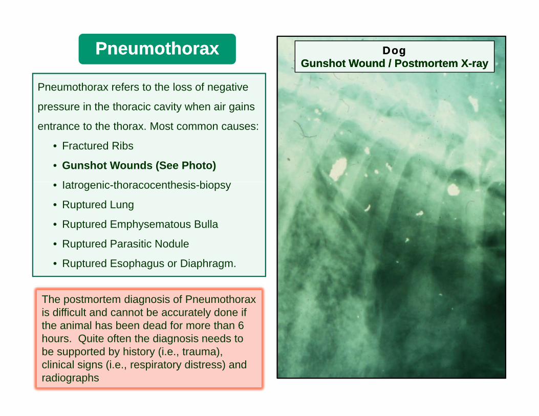
PneumothoraxPneumothorax DogDogGunshot Wound / Postmortem XGunshot Wound / Postmortem X--rayray
Pneumothorax refers to the loss of negative
pressure in the thoracic cavity when air gains
entrance to the thorax. Most common causes:entrance to the thorax. Most common causes:
• Fractured Ribs
• Gunshot Wounds (See Photo)
I t i th th i bi• Iatrogenic-thoracocenthesis-biopsy
• Ruptured Lung
• Ruptured Emphysematous Bulla
• Ruptured Parasitic Nodule
• Ruptured Esophagus or Diaphragm.
The postmortem diagnosis of Pneumothorax is difficult and cannot be accurately done if the animal has been dead for more than 6 hours Quite often the diagnosis needs tohours. Quite often the diagnosis needs to be supported by history (i.e., trauma), clinical signs (i.e., respiratory distress) and radiographs
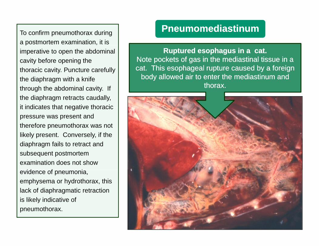
To confirm pneumothorax during a postmortem examination, it is
PneumomediastinumPneumomediastinump
imperative to open the abdominal cavity before opening the thoracic cavity. Puncture carefully
Ruptured esophagus in a cat. Ruptured esophagus in a cat. Note pockets of gas in the mediastinal tissue in a Note pockets of gas in the mediastinal tissue in a cat. This esophageal rupture caused by a foreign cat. This esophageal rupture caused by a foreign
body allowed air to enter the mediastinum andbody allowed air to enter the mediastinum andthe diaphragm with a knife through the abdominal cavity. If the diaphragm retracts caudally, it indicates that negative thoracic
body allowed air to enter the mediastinum and body allowed air to enter the mediastinum and thorax. thorax.
it indicates that negative thoracic pressure was present and therefore pneumothorax was not likely present. Conversely, if the diaphragm fails to retract and subsequent postmortem examination does not show
id f ievidence of pneumonia, emphysema or hydrothorax, this lack of diaphragmatic retraction is likely indicative ofis likely indicative of pneumothorax.

T d t i th
HydrothoraxHydrothorax
• Transudate in thorax
• Etiology:• Congestive heart failure• Congestive heart failure• Hypoproteinemia:
• Starvation• Renal disease• Intestinal disease• Lymphatic obstruction
(neoplasia)• Sequels:
• Atelectasis• Chronic pleural irritation
Note Note thoracic cavity and syringe filled with transudate. thoracic cavity and syringe filled with transudate. In severe cases, hydrothorax results in compressive In severe cases, hydrothorax results in compressive t l t i d i t di t N t th t th lt l t i d i t di t N t th t th latelectasis and respiratory distress. Note that the lungs atelectasis and respiratory distress. Note that the lungs
of this cat are collapsed (dark) because of the of this cat are collapsed (dark) because of the compression exerted by the thoracic fluid. compression exerted by the thoracic fluid.

Canine. These organs belong to a dog with chronic heart failure that developed severe ascites and hydrothorax.
Thoracic Cavity: Note the fluid filling the thoracic cavity. Compressive atelectasis was also present which appears as dark, collapsed lungs (arrows).
Abdominal Cavity: Note abdomen filled with transudate.
Heart: Note endocarditis involving the aorticHeart: Note endocarditis involving the aortic valve (arrow). This dog also had congestive hear failure. R= right; L= left.

HemothoraxHemothorax
Note a thoracic cavity filled with clotted Note a thoracic cavity filled with clotted blood (blood (arrowarrow). The thoracic cage of this ). The thoracic cage of this animal was severely traumatized, causinganimal was severely traumatized, causinganimal was severely traumatized, causing animal was severely traumatized, causing the rupture of a major the rupture of a major blood blood vessel. vessel.
Careful Careful dissection and postmortem dissection and postmortem examination are often required to locate the examination are often required to locate the source of hemorrhage.source of hemorrhage.
Common causes of hemothorax are:Common causes of hemothorax are:
•• TraumaTrauma
•• Ruptured aneurysm (dilation and Ruptured aneurysm (dilation and weakening of a major vessel)weakening of a major vessel)
N l iN l i•• NeoplasiaNeoplasia
•• CoagulopathiesCoagulopathies

Hemothorax / Parasitic Aortitis / CanineHemothorax / Parasitic Aortitis / Canine
KKKKKK
AortaAortaLiverLiver LungLung
FMVZFMVZ--UNAMUNAM
HeartHeart
This dog This dog died suddenly and died suddenly and the owner suspected malicious poisoning. Howeverthe owner suspected malicious poisoning. However, , postmortem postmortem examination examination revealed pale mucous revealed pale mucous membranes membranes ((anemia) and anemia) and the the thorax thorax filled filled with blood. with blood. Examination of great vessels revealed inflammation and a large aneurysm Examination of great vessels revealed inflammation and a large aneurysm in thoracic aorta (arrow).in thoracic aorta (arrow).
Aorta:Aorta: Note the aortic wall notable corrugated Note the aortic wall notable corrugated (white arrow(white arrow) as compared to the normal segment of ) as compared to the normal segment of the aorta (black arrows). the aorta (black arrows). K= kidneysK= kidneys. The aortitis and aneurysm in this dog was caused by . The aortitis and aneurysm in this dog was caused by Spirocerca lupiSpirocerca lupi which migrates through an aortic wall. which migrates through an aortic wall.

ChylothoraxChylothorax
• Chylothorax is the accumulation of chylous fluid (free lymph) in a thoracic cavity.
• This chylous fluid is rich in:• Lymphocytes• TriglyceridesTriglycerides
• Rupture of a major lymphatic vessel as a result of:
• Trauma• Iatrogenic (surgery)• Neoplasia• Idiopathic
Note thorax filled with milky fluid. Note thorax filled with milky fluid. H= heart H= heart
Sometimes it is impossible to identify or Sometimes it is impossible to identify or locate the ruptured lymphatic vessels. locate the ruptured lymphatic vessels.

This is not my milk… it is This is not my milk… it is chylouschylous fluid fluid removed from my chest by my Vet.removed from my chest by my Vet.
ChylothoraxChylothorax

More images of chylothoraxMore images of chylothorax

Acute: Largely lymphocytesAcute: Largely lymphocytes
ChylothoraxChylothorax
Acute: Largely lymphocytesAcute: Largely lymphocytes
Fluid obtained by Fluid obtained by thoracocenthesisthoracocenthesisthoracocenthesisthoracocenthesis
Chronic: Lymphocytes Chronic: Lymphocytes and some neutrophilsand some neutrophils

•• PleuritisPleuritis can occur alone or incan occur alone or in
Pleuritis and PleuresyPleuritis and Pleuresy
•• Pleuritis Pleuritis can occur alone or in can occur alone or in combination with combination with pneumonia.pneumonia.
•• According to exudateAccording to exudate::•• According to exudateAccording to exudate::•• FibrinousFibrinous•• Purulent Purulent (suppurative)(suppurative)•• EmpyemaEmpyema•• EmpyemaEmpyema•• GranulomatousGranulomatous
•• Chronic pleuritis typically results in Chronic pleuritis typically results in •• Chronic pleuritis typically results in Chronic pleuritis typically results in pleural pleural adhesions.adhesions.
•• Etiology: Most cases are Etiology: Most cases are •• Etiology: Most cases are Etiology: Most cases are infectious, although isolation is not infectious, although isolation is not always always possible.possible.
Fibrinous pleuritis characterized by Fibrinous pleuritis characterized by extensive deposition of fibrin on pleural extensive deposition of fibrin on pleural
membranesmembranes

Pleuritis (Pleurisy) as part of PleuropneumoniaPleuritis (Pleurisy) as part of Pleuropneumonia
Lung consolidationLung consolidation

Fibrinous Pleuritis (Pleurisy) / HorseFibrinous Pleuritis (Pleurisy) / Horse
Note pleural surface covered by a thick layer of fibrinNote pleural surface covered by a thick layer of fibrin
LiverLiver
There was no lung consolidation in this horse
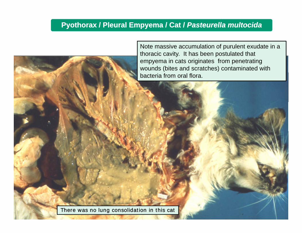
Pyothorax / Pleural Empyema / Cat / Pyothorax / Pleural Empyema / Cat / Pasteurella multocidaPasteurella multocida
Note massive accumulation of purulent exudate in a thoracic cavity. It has been postulated that empyema in cats originates from penetrating wounds (bites and scratches) contaminated with bacteria from oral flora.
There was no There was no lung lung consolidation in this catconsolidation in this cat

PyothoraxPyothorax
Thoracocenthesis: DogThoracocenthesis: Dog
Rebar AH Handbook of VeterinaryRebar AH Handbook of Veterinary
Cytology: Cytology: Predominant Predominant population of population of neutrophilsneutrophils
Rebar AH Handbook of Veterinary Rebar AH Handbook of Veterinary Cytology. Ralston Purina 1978Cytology. Ralston Purina 1978

Pleural Empyema (Pyothorax)Pleural Empyema (Pyothorax)
Note purulent exudate filling the Note purulent exudate filling the entire thoracic entire thoracic cavity. cavity.

Canine / Chronic Pleuritis and Pyothorax / Canine / Chronic Pleuritis and Pyothorax / NocardiaNocardia((“Tomato Soup”)“Tomato Soup”)
This "tomato soup" appearance is highly suggesting Nocardia infection. Histological lesions are those of a pyogranulomatous inflammation with many capillaries i th l ti ti f hi h t din the granulation tissue some of which rupture and leak RBCs, hence the hemorrhagic appearance.
As in all cases of pleuritis, bacteriologic analysis is required

Lung and Pleural TumorsLung and Pleural Tumors
•• Relatively rare in animals compared to human beings. Relatively rare in animals compared to human beings.
•• More common in dogs and catsMore common in dogs and cats•• More common in dogs and cats.More common in dogs and cats.
•• According to cell line:According to cell line:
•• Epithelial (adenoma or carcinoma)Epithelial (adenoma or carcinoma)Epithelial (adenoma or carcinoma).Epithelial (adenoma or carcinoma).
•• Mesenchymal (fibroma or fibrosarcoma / hemangioma or Mesenchymal (fibroma or fibrosarcoma / hemangioma or hemangiosarcoma).hemangiosarcoma).
•• Most common Most common malignant tumors malignant tumors in domestic animals:in domestic animals:
•• Adenocarcinoma.Adenocarcinoma.
B hi lB hi l l l il l i•• BronchioloBronchiolo--alveolar carcinoma.alveolar carcinoma.
•• Mesothelioma (pleura).Mesothelioma (pleura).
•• Histopathology: Lung biopsy is the last diagnostic resourceHistopathology: Lung biopsy is the last diagnostic resourceHistopathology: Lung biopsy is the last diagnostic resource.Histopathology: Lung biopsy is the last diagnostic resource.
•• Secondary tumors (metastatic) are Secondary tumors (metastatic) are very common in the lung.very common in the lung.

Pulmonary Carcinoma in a DogPulmonary Carcinoma in a Dog
Note large number of tumoral nodules infiltrating the lung. Based on gross appearance Note large number of tumoral nodules infiltrating the lung. Based on gross appearance alone, it is not possible to determine whether this is a primary lung cancer or a alone, it is not possible to determine whether this is a primary lung cancer or a secondary metastatic tumor originating elsewhere. Histopathology is always required.secondary metastatic tumor originating elsewhere. Histopathology is always required.
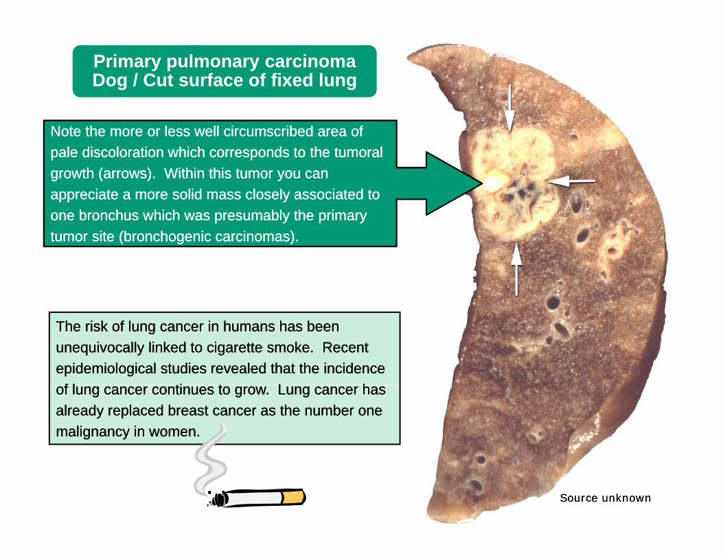
Primary pulmonary carcinoma Dog / Cut surface of fixed lung
Note the more or less well circumscribed area of Note the more or less well circumscribed area of pale discoloration which corresponds to the tumoral pale discoloration which corresponds to the tumoral growth (arrows) Within this tumor you cangrowth (arrows) Within this tumor you cangrowth (arrows). Within this tumor you can growth (arrows). Within this tumor you can appreciate a more solid mass closely associated to appreciate a more solid mass closely associated to one bronchus which was presumably the primary one bronchus which was presumably the primary tumor site (bronchogenic carcinomas). tumor site (bronchogenic carcinomas). ( g )( g )
The The risk of lung cancer in humans has been risk of lung cancer in humans has been unequivocally linked to cigarette smoke. Recent unequivocally linked to cigarette smoke. Recent epidemiological studies revealed that the incidence epidemiological studies revealed that the incidence f l ti t L hf l ti t L hof lung cancer continues to grow. Lung cancer has of lung cancer continues to grow. Lung cancer has
already replaced breast cancer as the number one already replaced breast cancer as the number one malignancy in women. malignancy in women.
Source unknownSource unknown

Pulmonary MetastasisPulmonary Metastasis
Insert: close-up
Note metastatic tumoral nodules scattered in the pulmonary parenchymascattered in the pulmonary parenchyma.

Metastatic Mammary Carcinoma / CanineMetastatic Mammary Carcinoma / Canine
Note lung Note lung filled filled with neoplastic nodules most of which have a depressed center giving with neoplastic nodules most of which have a depressed center giving the tumors an umbilicated appearance. This umbilicated appearance is mainly found in the tumors an umbilicated appearance. This umbilicated appearance is mainly found in carcinomas and results from rapid tumoral growth and ischemic necrosis of the center. carcinomas and results from rapid tumoral growth and ischemic necrosis of the center.
The most common secondary (metastatic) tumors in the lung are renal, ovarian and mammary carcinoma, osteosarcoma, hemangiosarcoma and melanoma. Histopathology is required for confirmatory purposes.

OsteosarcomaOsteosarcoma
Osteosarcoma often metastasizes to the lung (See next slide)
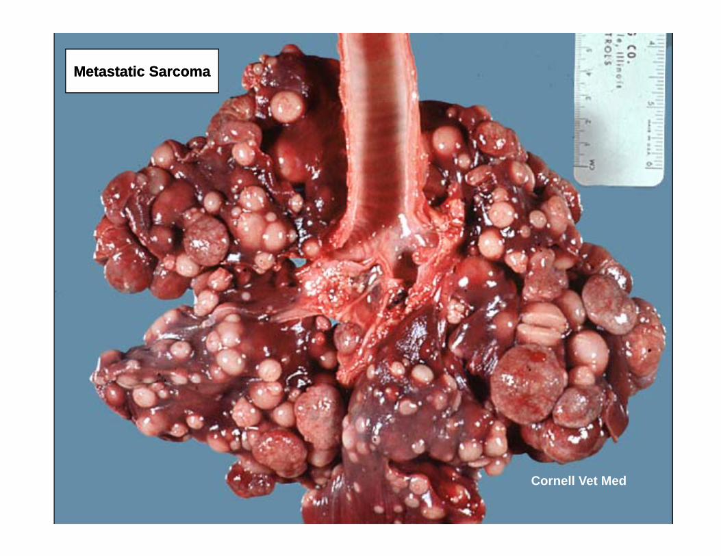
Metastatic SarcomaMetastatic Sarcoma
Cornell Vet Med

HemangiosarcomaHemangiosarcoma(lung metastasis(lung metastasis))
Malignant MelanomaMalignant Melanoma(lung metastasis(lung metastasis))
Thyroid CarcinomaThyroid Carcinoma(lung metastasis(lung metastasis))(lung metastasis(lung metastasis)) (lung metastasis(lung metastasis))

Lymphoma (Lymphosarcoma) / DogLymphoma (Lymphosarcoma) / DogLymphoma (Lymphosarcoma) / DogLymphoma (Lymphosarcoma) / Dog
Note enlarge tracheobronchial Note enlarge tracheobronchial lymph nodes (arrows).lymph nodes (arrows).

MesotheliomaMesotheliomaMesotheliomaMesotheliomaNote visceral and parietal pleura extensively infiltrated by tumoral masses
Mesothelioma is a rare tumor arising from the mesothelium of the peritoneal, pericardial or pleural serosal membranes.
Although metastasis to distal organs are rare mesothelioma isorgans are rare, mesothelioma is easily implanted on contact between serosal surfaces, thus disseminating very rapidly within g y p ythe cavity.
Inhalation of asbestos fibers has been linked to mesothelioma in human beings. Experimental exposure of laboratory animals to asbestos also results in theasbestos also results in the formation of mesothelioma.

I hate I hate I hate I hate PathologyPathology
Irrational thoughts commonly observed in students undergoing examIrrational thoughts commonly observed in students undergoing exam--i d d ti d d tinduced stress.induced stress.
Following graduation in Following graduation in the the rapidly approaching rapidly approaching summer of 2011, summer of 2011, these uncontrollable these uncontrollable feelings feelings maymay progressively change into a more positive view of Pathology Contrary to what has beenprogressively change into a more positive view of Pathology Contrary to what has beenmay may progressively change into a more positive view of Pathology. Contrary to what has been progressively change into a more positive view of Pathology. Contrary to what has been said by some contemporary philosophers, you do not said by some contemporary philosophers, you do not need to need to be insane to become a be insane to become a Veterinary PathologistVeterinary Pathologist.. Have a nice day!!Have a nice day!!

Some images were acquired from veterinary colleges of Some images were acquired from veterinary colleges of Canada, United States and Mexico and the names ofCanada, United States and Mexico and the names ofCanada, United States and Mexico and the names of Canada, United States and Mexico and the names of pathologists who contributed with some slides are known. pathologists who contributed with some slides are known. Their valuable contribution is sincerely acknowledged.Their valuable contribution is sincerely acknowledged.
I would like to thank Adriana Lopez, University of Western I would like to thank Adriana Lopez, University of Western Ontario, and Eileen Kinch for editorial assistance; Dr. Ontario, and Eileen Kinch for editorial assistance; Dr. María Forzán, Atlantic Veterinary College, for critically María Forzán, Atlantic Veterinary College, for critically reviewing these modules.reviewing these modules.

Module 6Module 6Module 6Module 6
If you have any comments, criticisms or suggestions about these If you have any comments, criticisms or suggestions about these
tutorial modules please let me know. tutorial modules please let me know.
Also, if you find any errors or typos please let me know too Also, if you find any errors or typos please let me know too
lopez@upeicalopez@upeica