Thomas Madden and Stanley Rafalko Articles of Interest · excerpts from the IU Bloomington Faculty...
17
2014 Volume 17, Numbers 1/2 To Honor Former Faculty: Thomas Madden and Stanley Rafalko Associated Phorias at Four Near Test Distances and Reading Eyestrain Current Clinical Vision Science Articles of Interest
Transcript of Thomas Madden and Stanley Rafalko Articles of Interest · excerpts from the IU Bloomington Faculty...

2014Volume 17, Numbers 1/2
To Honor Former Faculty: Thomas Madden and Stanley Rafalko
Associated Phorias at Four Near Test Distances and Reading Eyestrain
Current Clinical Vision Science Articles of Interest

In This Issue
The optometry school at Indiana University has now been in existence for over 60 years.
Because of that, most current faculty, staff, and students have probably never heard of
many faculty members who made significant contributions to the school in its early
years. To honor the memory of two of those early faculty members, this issue features
excerpts from the IU Bloomington Faculty Council Memorial Resolutions for Thomas
Madden and J. Stanley Rafalko. This issue also includes reviews of some notable
papers from the clinical vision science literature and a research paper on associated
phoria testing distance.
David A. Goss
Editor
In This Issue
Correspondence and manuscripts submitted for publication should be sent to the Editor: David A.
Goss, School of Optometry, Indiana University, Bloomington, IN 47405 USA (or
ON THE COVER: Group photo in 1953 of the first Optometry entering class at IndianaUniversity and members of the faculty

Indiana University School ofOptometry Administration:
Joseph A. Bonanno, O.D., Ph.D.,Dean
Clifford W. Brooks, O.D., Executive Associate Dean ofAcademic Affairs
Stephen Burns, Ph.D.,Associate Dean for GraduatePrograms
Peter S. Kollbaum, O.D., Ph.D., Associate Dean for Research
Kimberly Kohne, O.D.,Associate Dean for Students
Richard E. Meetz, O.D.,Associate Dean for FiscalAffairs
Neil Pence, O.D., Chief of ClinicalServices
Indiana Journal of Optometry
Editor:David A. Goss, O.D., Ph.D.
Editorial Board:Arthur Bradley, Ph.D.Clifford W. Brooks, O.D.Daniel R. Gerstman, O.D., M.S.Victor E. Malinovsky, O.D.Neil A. Pence, O.D.
Production and LayoutJ. Craig Combs, M.H.A.
2014 Volume 17, Numbers 1/2
Table of Contents
TABLE OF CONTENTS
To Honor Former Faculty:
Thomas Madden and Stanley Rafalko………………..... 1
Associated Phorias at Four Near Test
Distances and Reading Eyestrain………...............….... 5
Current Clinical Vision Science
Articles of Interest………...........................……………. 8
Statement of Purpose: The Indiana Journal of Optometry is published by the
Indiana University School of Optometry to provide members of the Indiana
Optometric Association, Alumni of the Indiana University School of Optometry, and
other interested persons with information on the research and clinical expertise at
the Indiana University School of Optometry, and on new developments in
optometry/vision care.
The Indiana Journal of Optometry and Indiana University are not responsible for
the opinions and statements of the contributors to this journal. The authors and
Indiana University have taken care that the information and recommendations
contained herein are accurate and compatible with the standards generally
accepted at the time of publication. Nevertheless, it is impossible to ensure that all
the information given is entirely applicable for all circumstances. Indiana
University disclaims any liability, loss, or damage incurred as a consequence,
directly or indirectly, of the use and application of any of the contents of this
journal. This journal is also available on the world wide web at:
http://www.opt.indiana.edu/IndJOpt/home.html
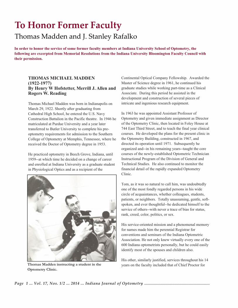
THOMAS MICHAEL MADDEN (1922-1977)By Henry W Hofstetter, Merrill J. Allen andRogers W. Reading
Thomas Michael Madden was born in Indianapolis on
March 29, 1922. Shortly after graduating from
Cathedral High School, he entered the U.S. Navy
Construction Battalion in the Pacific theatre. In 1946 he
matriculated at Purdue University and a year later
transferred to Butler University to complete his pre-
optometry requirements for admission to the Southern
College of Optometry at Memphis, Tennessee, where he
received the Doctor of Optometry degree in 1953.
He practiced optometry in Beech Grove, Indiana, until
1959--at which time he decided on a change of career
and enrolled at Indiana University as a graduate student
in Physiological Optics and as a recipient of the
Continental Optical Company Fellowship. Awarded the
Master of Science degree in 1961, he continued his
graduate studies while working part-time as a Clinical
Associate. During this period he assisted in the
development and construction of several pieces of
intricate and ingenious research equipment.
In 1963 he was appointed Assistant Professor of
Optometry and given immediate assignment as Director
of the Optometry Clinic, then located in Foley House at
744 East Third Street, and to teach the final year clinical
courses. He developed the plans for the present clinic in
the Optometry Building, constructed in 1967, and
directed its operation until 1971. Subsequently he
organized and--in his remaining years--taught the core
courses of the newly-established Optometric Technician
Instructional Program of the Division of General and
Technical Studies. He also continued to monitor the
financial detail of the rapidly expanded Optometry
Clinic.
Tom, as it was so natural to call him, was undoubtedly
one of the most fondly regarded persons in his wide
circle of acquaintances, whether colleagues, students,
patients, or neighbors. Totally unassuming, gentle, soft-
spoken, and ever thoughtful--he dedicated himself to the
service of others--with never a trace of bias for status,
rank, creed, color, politics, or sex.
His service-oriented mission and a phenomenal memory
for names made him the perennial Registrar for
conventions and seminars of the Indiana Optometric
Association. He not only knew virtually every one of the
600 Indiana optometrists personally, but he could easily
identify most of the spouses and children also.
His other, similarly justified, services throughout his 14
years on the faculty included that of Chief Proctor for
Page 1 ... Vol. 17, Nos. 1/2 ... 2014 ... Indiana Journal of Optometry ........................................................
To Honor Former Faculty Thomas Madden and J. Stanley RafalkoIn order to honor the service of some former faculty members at Indiana University School of Optometry, thefollowing are excerpted from Memorial Resolutions from the Indiana University Bloomington Faculty Council withtheir permission.
Thomas Madden instructing a student in theOptometry Clinic.

.................................................... Indiana Journal of Optometry ... 2014 ... Vol. 17, Nos. 1/2... page 2
the National Board of
Examiners in Optometry,
Treasurer of the Indiana
University Optometry
Alumni Association,
Manager of the Public
School Vision Screening
Program of the School of
Optometry, and Optometry
Liaison Officer to the
Indiana State Agency for
the Blind. For 7 years he
was Editor of the quarterly
Indiana Journal of
Optometry, and for 6 years
he was the chief I.U.
delegate at the meetings of the Association of School
and Colleges of Optometry. There was probably never a
time during his professional and academic career when
he was not serving on 2 or more committees at the
campus, community, state, or national level.
In no sense a joiner, but rather with a genuine sense of
duty, he maintained active membership in the Omega
Epsilon Phi fraternity, the American Optometric
Association, the Association of Optometric Editors, the
American Academy of Optometry (fellow), and the
American Association for the Advancement of Science
(fellow). His honors included membership in Sigma Xi
(scientific) and Beta Sigma Kappa (optometric)
honorary societies.
While most of Tom's acquaintances will long remember
him for his unfailing service to humanity, his utter
fairness and sense of justice, his meticulous concern for
orderliness, organization, and personal responsibility,
and his creative designs of systems and procedures--
those of us who worked with him daily year in and year
out will also recall again and again such things as his
gentle--always victimless--wit, his fascination for
ingenious and intricate mechanisms, his quiet hobby of
collecting foreign postmarked envelopes, and--yes--his
delight in repairing umbrellas, especially for friends.
His most relaxing recreation was driving an automobile,
undoubtedly the remaining trace of his earlier
motorcycling interests. For example, on trips with
colleagues to conferences even in another's car he never
hesitated to solicit, however meekly, the privilege of
driving-- and he drove extremely well.
Dr. Madden, Tom, died on July 18, 1977 following a
lingering illness, the terminal nature of which he was
well aware, but which he faced so gracefully as to win
the deepest admiration of simply everyone who attended
or visited him. His survivors include his wife Mary Rita
(nee Murphy), two daughters at home, Lisa Marie and
Susan Mary, and two brothers, Richard C. and Harry E.
Madden. Funeral services were held at the St. Charles
Catholic Church and interment was in the Valhalla
Memory Gardens.
J. STANLEY RAFALKO (1905-1988)By Gordon G. Heath
Joseph Stanley
Rafalko was born
December 6, 1905 in
Scranton,
Pennsylvania, and
raised there. He
received his A.B.
degree in biology with
high honors from
Villanova College in
Philadelphia in 1929.
Special accreditation
enabled him to immediately obtain a teaching permit, so
he was able to teach part time in West Philadelphia high
schools for the next two years while also completing his
M.S. degree in zoology at the University of
Pennsylvania. He accepted a faculty position at LaSalle
College in Philadelphia where he served as Professor of
Comparative Anatomy and Embryology from 1931 to
1943. He then gathered his courage and his meager
savings and returned to graduate school at the University
of Pennsylvania for three years in order to earn his Ph.D.
degree. It was there that he met and married Margaret
Miskell, a fellow graduate student in zoology.
After a one-year appointment as assistant professor of
zoology at Syracuse University's "Triple Cities" branch
in Endicott, New York, he accepted appointment as
associate professor of zoology at Southern Illinois
University in Carbondale from 1947 to 1950 where, in
addition to his teaching, he also co-authored a textbook
of general zoology. From there he went to Memphis,
Tennessee, to become Chairman of the Department of
Anatomy at the Southern College of Optometry.
A quiet, gentle, and ever-courteous man of high
J. Stanely Rafalko
Thomas Madden, on theright, with M. EmersonWoodruff, who received aPh.D. from IU in 1969.

Page 3 ... Vol. 17, Nos. 1/2 ... 2014 ... Indiana Journal of Optometry .....................................................
principles who willingly went out of his way to help
others, he was never able to comprehend, much less
condone, rudeness or cruelty to others. Segregation of
public facilities in Memphis offended him and he often
spoke out against these practices.
It was just at this time that Indiana University was
initiating a new program in Optometry and seeking
faculty to develop courses in the anatomy and
physiology of the eye. The late Professor Richard
Webb, then Chairman of the Department of Anatomy in
the IU School of Medicine, learned of Professor
Rafalko's reputation as an outstanding teacher and at
once proceeded to recruit him for this purpose. From
1953 until his retirement in 1976 his nearly total
dedication was to his teaching. At various times during
this period, he taught histology to both medical and
dental students, histological techniques to medical
technology students, and both histology and gross
anatomy to optometry students. He published a few
papers on cytological observations. With his heavy
teaching loads he had little time for personal research;
therefore he long ago had decided anyway to devote his
career to teaching and advising his students, activities he
thoroughly enjoyed and in which he excelled.
It was often said by his students that the notes for Dr.
Rafalko's class contained all the information needed for
the subject, for they were indeed meticulously detailed.
It was also frequently supposed that the subject of
anatomy, and hence his notes, did not change from year
to year; but that simply was not true. He carefully
studied the other courses in the optometry curriculum to
be sure his courses remained relevant and met the needs
of the students. The changes he introduced were usually
small, but important, in keeping his teaching up to date.
Professor Rafalko's excellence as a teacher was
demonstrated by the high ratings he received in student
evaluations each year, by the high rankings consistently
achieved by his students on National Boards and other
professional examinations, and by the many dozens of
unsolicited letters of gratitude and praise that he
received from former students over the years.
Group photo in 1953 of the first Optometry entering class at Indiana University and members of the faculty. J.Stanley Rafalko is third from the left in the front row.

Previously, at the Southern College of Optometry, he
had received the L.A. Farmer Award for Distinguished
Teaching, and at IU he was three times nominated
jointly by the Division of Optometry and the
Department of Anatomy and Physiology for the Frederic
Bachman Lieber Award for Excellence in Teaching. On
the first of these three occasions, he withdrew his own
name from nomination because he was a member of the
University Committee for Improvement of Teaching,
which selected the winners.
Professor Rafalko retired from the teaching he loved in
1976, thereafter devoting himself to his many
community interests. He was a permanent member of
the board of directors of the Hoosier Hills Art Guild, of
which he had served as president for two years. He was
Deputy Grand Chancellor of the State of Indiana
Domain of the Knights of the Order of Pythias, having
earlier served the Bloomington Lodge as its Chancellor
Commander. He served on the board of directors of the
Bloomington Boys' Club and was a long-term member
of Torch International, serving at various times in all the
offices of the Bloomington chapter.
Stanley Rafalko, although not an optometrist, dedicated
much of his career to the profession and was a Special
Member of the American Optometric Association and a
Fellow of the American Academy of Optometry, as well
as holding honorary membership in the two leading
optometric fraternities, Omega Delta and Omega
Epsilon Phi. As a final gesture of gratitude and
allegiance to the professional school he had come to
regard as his true home base, he bequeathed his entire
estate--a substantial sum accumulated through the years
of frugal living after he and Margaret were divorced--to
the IU Foundation for use by the IU School of
Optometry in support of optometric education.
Professor Rafalko died September 19, 1988. The J.
Stanley Rafalko Memorial Trust Fund will serve as a
perpetual reminder of the dedicated life and work of this
good man who often proudly declared, "I live a life of
service--service to my students."
................................................... Indiana Journal of Optometry ... 2014 ... Vol. 17, Nos. 1/2... page 4

Page 5 ... Vol. 17, Nos. 1/2 ... 2014 ... Indiana Journal of Optometry ...................................................
INTRODUCTION
Associated phoria is the amount of prism required to
reduce fixation disparity to zero.1 The presence of
fixation disparity, and thus a non-zero value for
associated phoria, is related to nearpoint vision
symptoms.2,3
Various instruments are available for the measurement
of associated phorias at near, including the Saladin card,
Mallett unit, Borish card, and Bernell lantern.4,5
Nearpoint associated phoria testing is typically
conducted at 40 cm.6 Because fixation disparity varies
as a function of viewing distance,7 we could ask
whether other test distances for associated phorias would
be as effective as 40 cm for distinguishing between
symptomatic and asymptomatic individuals.
Accordingly, we compared symptom questionnaire
scores in subjects with zero associated phoria to those
with non-zero associated phorias for each of four test
distances (25, 33, 40, 50 cm).
METHODSA total of 99 subjects, ages 20 to 30 years, all of whom
were optometry students, participated in this study.
Subjects had a best-corrected near visual acuity of 20/20
or better in both eyes and had no self-reported history of
strabismus, amblyopia, suppression, extraocular muscle
disorders, nerve palsies, or ocular disease. The study
protocols were approved by the Institutional Review
Board at Indiana University for Human Subjects.
Written informed consent was obtained from all the
subjects before enrollment in the study.
The subjects attended scheduled appointments on
Mondays and Wednesdays during the hours of 1, 2, 4,
and 6 pm. Prior to any testing procedures, subjects
completed a Reading Eyestrain Symptom Questionnaire.
Subjects responded never, infrequently, sometimes,
fairly often, or always to each of the following ten
questions:
1. Do your eyes feel tired when reading or doing close
work?
2. Do your eyes feel uncomfortable when reading or
doing close work?
3. Do you have headaches when reading or doing close
work?
4. Do you feel sleepy when reading or doing close
work?
5. Do you have double vision when reading or doing
close work?
6. Do the words seem to move or jump when reading or
doing close work?
7. Do your eyes ever hurt when reading or doing close
work?
8. Do your eyes ever feel sore when reading or doing
close work?
9. Do you feel a ‘pulling” around your eyes when
reading or doing close work?
10. Do you notice the words blurring or going in and out
of focus when reading or doing close work?
Symptom scores were determined by adding points as
follows for each of the 10 questions’ answers: never-0,
infrequently-1, sometimes-2, fairly often-3, always-4.
Potential scores could thus range from 0 to 40 points.
This questionnaire, which is very similar to the
Associated Phorias at Four Near Test Distancesand Reading EyestrainMEREDITH ROGERS, O.D., AIMEE STASKAL-BRECHT, O.D.,AND DAVID A. GOSS, O.D., PH.D.
ABSTRACTBackground: Nearpoint visual fatigue is more likely to occur in patients with a fixation disparity at near. Nearassociated phorias are typically done with a test distance of 40 cm. This study was conducted to compare symptomlevels in subjects with zero associated phoria to those with non-zero associated phoria at 40 cm and at three other neartest distances.
Methods: Saladin card associated phorias were measured at four different near distances (25 cm, 33 cm, 40 cm, and50 cm), and a ten question symptom survey was administered.
Results: The mean symptom scores showed statistically significant differences between those with zero associatedphoria and those with non-zero associated phoria for testing at 33 and 40 cm, but not for testing at 25 or 50 cm.
Conclusion: For associated phoria testing with the Saladin card, 33 and 40 cm are more appropriate testing distancesthan 25 or 50 cm.

............................................Indiana Journal of Optometry ... 2014 ... Vol. 17, Nos. 1/2... page 6
Convergence Insufficiency Symptom Survey,8,9 was
used in one previous publication.10
Each subject was instructed to place his or her head in a
chin rest and forehead rest that was clamped to a table.
Lateral dissociated phorias were taken using the
modified Thorington test target on the Saladin Near
Point card.11,12 Lateral associated phorias for each
subject were measured using the Saladin Near Point
Card.Subjects placed polarized glasses over their current
spectacle correction. A Saladin card was placed on the
subject’s eye level at a distance 50 cm from the
forehead rest. Participants were asked if they noticed a
misalignment of the vertical lines on the associated
phoria circle while focusing on the central fusion lock
dot on the Saladin card. If no misalignment was
observed, zero associated phoria was recorded. If
misalignment was reported, a horizontal prism bar was
used over the subject’s right eye to measure his or her
associated phoria. The minimum prism that resulted in
alignment was recorded as the associated phoria. After
testing at 50 cm, associated phorias were determined at
40 cm, 33 cm, 25 cm, in that order.
Mean symptom scores were calculated for subjects with
zero and non-zero (base-in or base-out) associated
phorias at each of the four test distances. Statistical
significance of the difference between subjects with
zero associated phoria and subjects with non-zero
associated phoria in mean symptoms scores was tested
using a one-tailed t-test. A p value of 0.05 was used to
establish statistical significance.
RESULTS
Mean symptom scores are given in Table 1. The mean
symptom score was 1.4 lower in those with zero
associated phoria at 25 cm than in those with non-zero
associated phoria at that distance, but that difference
was not statistically significant by t-test (t=1.302,
p=0.099). The difference between the two groups at 50
cm was only 1.0 and was also not significant(t=0.848,
p=0.199). The symptom score differences between the
two groups were 2.0 and 2.1 for 40 cm and 33 cm,
respectively, with statistical significance of the
differences for both distances (t=1.814, p=0.036 for 40
cm testing and t=1.864, p=0.033 for 33 cm testing).
Modified Thorington dissociated phorias at 40 cm were
examined as a secondary analysis. The mean symptom
scores were: for subjects with more than 6 prism
diopters of exo, 8.5 (n=23, SD=6.6); for subjects with
phorias in the range of ortho to 6 prism diopters exo, 7.1
(n=55, SD=4.8); for subjects with eso at 40 cm, 8.6
(n=21, SD=6.5). The difference between the three
groups was not statistically significant by one-way
ANOVA (F=0.773, p=0.464). Dissociated phoria at 40
cm showed a statistically significant Pearson coefficient
of correlation with associated phoria at 40 cm (r=0.56,
p<0.001). A scatterplot of dissociated phoria and
associated phoria is shown in Figure 1.
DISCUSSION
The results of the present study are consistent with
previous work showing that near fixation disparity
measurements are affected by viewing distance.7,13
The results of this study are also consistent withstudies
showing that fixation disparity parameters are good
Test distance Zero associated phoria Non-zero associated phoria Stat. sig.25 cm 7.0 (n=44, SD=5.1) 8.4 (n=55, SD=6.0) 0.09933 cm 6.5 (n=39, SD=5.3) 8.6 (n=60, SD=5.8) 0.03340 cm 6.5 (n=37, SD=5.1) 8.5 (n=62, SD=5.8) 0.03650 cm 7.2 (n=41, SD=5.3) 8.2 (n=58, SD=5.8) 0.199
Table 1. Mean symptom scores in subjects with zero associated phoria and subjects with non-zero associatedphoria with statistical significance of difference by t-test. (n=number of subjects; SD=standard deviation)
Figure 1.Scatterplot of associated phoria at 40 cmwith dissociated phoria at 40 cm. Points representingmultiple subjects have the number of subjects givenjust above each point.

Page 7 ... Vol. 17, Nos. 1/2 ... 2014 ... Indiana Journal of Optometry ......................................................
discriminators between symptomatic and asymptomatic
individuals.2,3
The results suggest that 33 cm or 40 cm are better
associated phoria testing distances than are 25 cm or 50
cm with the Saladin card. Further study might be
advisable to determine whether this is also true with
other associated phoria testing devices and whether
similar results would be obtained with subjects less
aware of testing procedures or the nature of asthenopic
symptoms.
One limitation of this study realized in hindsight is that
no inquiry was made of the subjects concerning usual
reading distance. The better discrimination of the 40
cm and 33 cm testing distances may reflect the fact that
these are typical distances for reading and near work.
With that fact in mind, it may be noted that Carlson and
Kurtz14 recommended that associated phoria be tested
at either 40 cm or the patient’s customary nearpoint
working distance.
CONCLUSIONFor associated phoria testing with the Saladin card, 33
and 40 cm are more appropriate testing distances than
25 or 50 cm.
REFERENCES 1. Hofstetter HW, Griffin JR, Berman MS, Everson
RW. Dictionary of Visual Science and Related Clinical
Terms, 5th ed. Boston: Butterworth Heinemann,
2000:387.
2. Sheedy JE, Saladin JJ. Validity of diagnostic criteria
and case analysis in binocular vision disorders. In
Schor CM, Ciuffreda KJ, eds. Vergence Eye
Movements: Basic and Clinical Aspects. Boston:
Butterworths, 1983:517-540.
3. Pickwell LD, Kaye NA, Jenkins TC. Distance and
near readings of associated heterophoria taken on 500
patients. Ophthal Physiol Opt 1991;11:291-296.
4. Grosvenor T. Primary Care Optometry, 5th ed. St.
Louis: Butterworth Heinemann Elsevier, 2007:236-240.
5. Goss DA. Ocular Accommodation, Convergence, and
Fixation Disparity, 3rd ed. Santa Ana, CA: Optometric
Extension Program Foundation, 2009:89-93.
6. Saladin JJ. Phorometry and stereopsis. In: Benjamin
WJ, ed. Borish’s Clinical Refraction, 2nd ed. St. Louis;
Butterworth Heinemann Elsevier, 2006:899-960.
7. Jainta S, Jaschinski W. Fixation disparity: binocular
vergence accuracy for a visual display at different
positions relative to the eyes. Human Factors
2002;44:443-450.
8. Rouse MW, Borsting EJ, Mitchell GL, Scheiman M,
Cotter SA, Cooper J, Kulp MT, London R, Wensveen J,
et al. Validity and reliability of the revised convergence
insufficiency symptom survey in adults. Ophthal
Physiol Opt 2004;24:384-390.
9. Rouse MW, Borsting EJ, Mitchell GL, Cotter SA,
Kulp M, Scheiman M, Barnhardt C, Bade A, Yamada T,
et al. Validity of the convergence insufficiency
symptom survey: a confirmatory study. Optom Vis Sci
2009;86:357-363.
10. Goss DA, Reynolds JL, Todd RE. Comparison of
four dissociated phoria tests; reliability and correlation
with symptom survey scores. J Behav Optom
2010;21:99-104.
11. Goss DA. A versatile new nearpoint test card: the
Saladin card. Indiana J Optom 2005;8:17-19.
12. Corbett A, Maples WC. Test-retest reliability of the
Saladin card. Optom – J Am Optom Assoc
2004;75:629-639.
13. Jaschinski W. The proximity-fixation-disparity
curve and the preferred viewing distance at a visual
display as an indicator of near vision fatigue. Optom
Vis Sci 2002;79;158-169.
14. Carlson NB, Kurtz D. Clinical Procedures for
Ocular Examination, 3rded. New York: McGraw-Hill,
2004:204-207.
Meredith Rogers and Aimee Staskal-Brecht were
members of the Indiana University School of
Optometry Class of 2011.

...............................................................Indiana Journal of Optometry ... 2014 ... Vol. 17, Nos. 1/2 ... page 8
Current Clinical Vision Science Articles of InterestDAVID A. GOSS, O.D., PH.D.
Cacho-Martínez P, García-Munoz Á, Ruiz-CanteroMT. Diagnostic validity of clinical signs associatedwith a large exophoria at near. Journal ofOphthalmology. 2013:article 549435.http://dx.doi.org/10.1155/2013/549435.
This study sought to find which binocular vision tests
best distinguished between symptomatic subjects with
large exophorias at near and asymptomatic subjects with
normal phorias at distance and near. Subjects were
recruited from among 18 to 35 year old patients at the
Optometry Clinic of the University of Alicante in Spain.
Subjects in both groups did not have strabismus by
cover test and had distance and near visual acuity of at
least 20/20, no ocular motility disorders, no vertical
deviation, and no ocular disease.
The symptomatic high exo group (n=33) had moderate
to severe symptoms on the Conlon symptom survey and
more than 6 prism diopters exophoria at 40 cm by prism
neutralized cover test. The asymptomatic group (n=33)
had a low symptom level on the Conlon symptom
survey.
The tests performed were: (1) monocular and binocular
accommodative facility with +2/-2 D flippers, 40 cm
test distance, and a target with suppression control, (2)
MEM dynamic retinoscopy at 40 cm, (3) negative and
positive relative accommodation (NRA and PRA) with
20/30 letters at 40 cm, (4) base-out fusional vergence
range at 40 cm with Risley prism and a 20/30 letter
target, (5) near point of convergence (NPC) break and
recovery with 20/30 target, (6) vergence facility at 40
cm with 20/30 target using 3 Δ BI and 12 Δ BO loose
prisms, (7) gradient AC/A ratio with -1.00 D lenses
using cover test, (8) monocular push-up amplitude of
accommodation, and (9) stereopsis with graded circles
on the Randot SO-002 test.
Tests which showed a statistically significant difference
between the two groups by Mann-Whitney U test were:
monocular accommodative facility (p<0.001), binocular
accommodative facility (p<0.001), MEM (p=0.002),
NRA (p=0.02), NPC break (p<0.001), NPC recovery
(p<0.001), and vergence facility (p<0.001). The mean
base-out blur findings of 19.7 Δ (SD=6.1) in the normal
group and 16.7 Δ (SD=6.5) in the high exo group were
not significantly different (p=0.09), perhaps due to the
test’s large variability. Base-out fusional vergence
breaks, averaging 25.6 Δ (SD=7.1) in the normal group
and 22.9 Δ (SD=8.4) in the high exo group, likewise did
not show a significant difference between the two
groups (p=0.10). Other tests not showing a significant
difference between groups were gradient AC/A ratio
(p=0.25), amplitude of accommodation (p=0.83), PRA
(p=0.10), base-out recovery (p=0.90), and stereopsis
(p=0.31).
The authors ranked the diagnostic accuracy of the tests
which were significantly different between groups by
the area under the receiver characteristic (ROC) curve.
The ROC curve is a plot of sensitivity versus one minus
specificity for various test cut-off values. The ranking
of the tests was: NPC recovery, binocular
accommodative facility, NPC break, monocular
accommodative facility, vergence facility, MEM, and
NRA.
By balancing sensitivity and specificity, the authors
came up with the following cut-offs to best distinguish
the symptomatic high exo group from the asymptomatic
group: NPC recovery, at least 8.25 cm; binocular
accommodative facility, 8.25 cycles per minute or less;
NPC break, at least 5.35 cm; monocular accommodative
facility, 8.25 cycles per minute or less; vergence facility,
14.75 cycles per minute or less; MEM, 0.63 D or less;
and NRA, 2.38 D or less. The cut-off for MEM seems
unusual because high values for MEM are considered
abnormal, but their result may be explained by the fact
that the study involved separating subjects with high
exophoria from normal. The mean MEM findings in
this study were 0.62 D (SD=0.23) in the normal group
and 0.34 D (SD=0.37) in the high exo group. As a
general rule, exophores tend to have lower lags and
esophores tend to have higher lags than normals under
binocular conditions.
The authors suggested that their study showed that the
tests having the best diagnostic accuracy for
symptomatic high exophoria at near are the near point of
convergence and binocular accommodative facility.
However, they did acknowledge that this study was
“based on a limited number of subjects and should be
confirmed in forthcoming studies.” (p. 8)

Page 9 ... Vol. 17, Nos. 1/2 ... 2014 ... Indiana Journal of Optometry ........................................................
Sanker N, Prabhu A, Ray A. A comparison of near-dissociated heterophoria tests in free space. Clinicaland Experimental Optometry 2012;95:638-642.
Noting that the modified Thorington dissociated phoria
test is a highly repeatable test and that it has gained
acceptance among clinicians, the authors of this study
compared it to three other lateral dissociated phoria tests.
All four tests were done in free space without a
phoropter, using a 40 cm target distance. Subjects were
49 students, ages 18 to 26 years, at Manipal University
in India. All subjects had distance visual acuity of at
least 6/6 in each eye and they did not have ocular
disease, strabismus as determined by cover test, or
history of eye surgery.
The tests which were compared were the modified
Thorington, Maddox rod, von Graefe, and alternating
cover test. The Bernell Muscle Imbalance measure card
for near was used for the modified Thorington test. On
the Maddox rod test loose prisms were used to align the
red streak with the white light. On the von Graefe test,
the eyes were dissociated with a 6 Δ base-up prism while
subjects viewed an N8 line on a nearpoint chart;
alignment was achieved with horizontal loose prisms in
one prism diopter steps. For the alternating cover test,
subjects viewed a 6/9 letter on a nearpoint stick and
prism neutralization was done using a prism bar with
two prism diopter steps.
The authors determined the mean difference between the
modified Thorington and each of the other three tests.
They also calculated the standard deviations of the
differences for each of the comparisons.
The modified Thorington averaged more exo than the
Maddox rod and less exo than the von Graefe test and
the cover test. The mean differences were: modified
Thorington minus Maddox rod, -0.42 Δ (SD=1.49);
modified Thorington minus von Graefe, +0.53 Δ
(SD=1.88); modified Thorington minus cover test, +0.88
Δ (SD=1.86). Expressed as mean differences plus or
minus the 95% limits of agreement (standard deviation
of the differences X 1.96), the differences become,
modified Thorington minus Maddox rod, -0.42 Δ +/-
2.92; modified Thorington minus von Graefe, +0.53 Δ
+/-3.68; modified Thorington minus cover test, +0.88 Δ
+/-3.64. Bland-Altman plots suggested that the
magnitudes of the differences were not dependent on the
magnitude of the averages of the two phorias being
compared for any of the three comparisons.
The authors suggested that the modified Thorington
might have had the best agreement with the Maddox rod
because they use the same method of dissociation.
Because of the relatively high limits of agreement, the
authors concluded that “the Maddox rod, the von Graefe
and the alternate cover tests cannot be interchanged with
the modified Thorington test for near phoria
measurements in free space.” (p. 642)
Cebrian JL, Antona B, Barrio A, Gonzalez E,Gutierrez A, Sanchez I. Repeatability of the modifiedThorington card used to measure far heterophoria.Optometry and Vision Science 2014;91:786-792.
The authors of this study recognized that several studies
have shown the modified Thorington test to be the most
repeatable of commonly used dissociated phoria tests at
near, but that it had not been studied as much for
distance testing. Therefore they examined the
repeatability of, and the agreement among, four
dissociated phoria tests at distance. The four tests were
prism neutralized cover test, Maddox rod, von Graefe,
and modified Thorington.
Their subjects were first year students in the optics and
optometry program at the Universidad Complutense de
Madrid in Madrid, Spain. Subjects had visual acuity of
at least 0.9 at distance and near in each eye; no history of
eye disease, strabismus, nystagmus, amblyopia, or
refractive surgery; no medication or disease that could
affect accommodation, fusional vergence ranges, or
ocular motility; and no accommodation or vergence
anomalies from testing amplitude of accommodation,
near point of convergence, 4 Δ base-out test, prism bar
vergence ranges, and Randot near stereopsis test. The
94 subjects had an age range of 18 to 32 years.
The Bernell Muscle Imbalance Measure distance card
was used for the modified Thorington test. For the cover
test, a single row of 0.8 visual acuity letters was used as
a target, and a prism bar was used to neutralize
movement on the alternating cover test. On the von
Graefe test, the fixation target was single column of 0.8
visual acuity letters. A 6 Δ base-up prism over one eye
eliminated fusion and a prism bar was used to align the
two images. For the Maddox rod test, subjects reported
when the prism in a prism bar made the red line seen by
one eye pass through the white spot of light seen by the
other eye. At one testing session, two optometrists did
each of the four tests. At a second testing session,
averaging a week later, the same two optometrists again
performed each of the four tests.
Interexaminer and intraexaminer coefficients of
repeatability were calculated by finding the standard
deviations of the differences between examiners and
between sessions, respectively, and multiplying those
standard deviations by 1.96. The coefficient of
repeatability (COR) gives an estimate of the range of
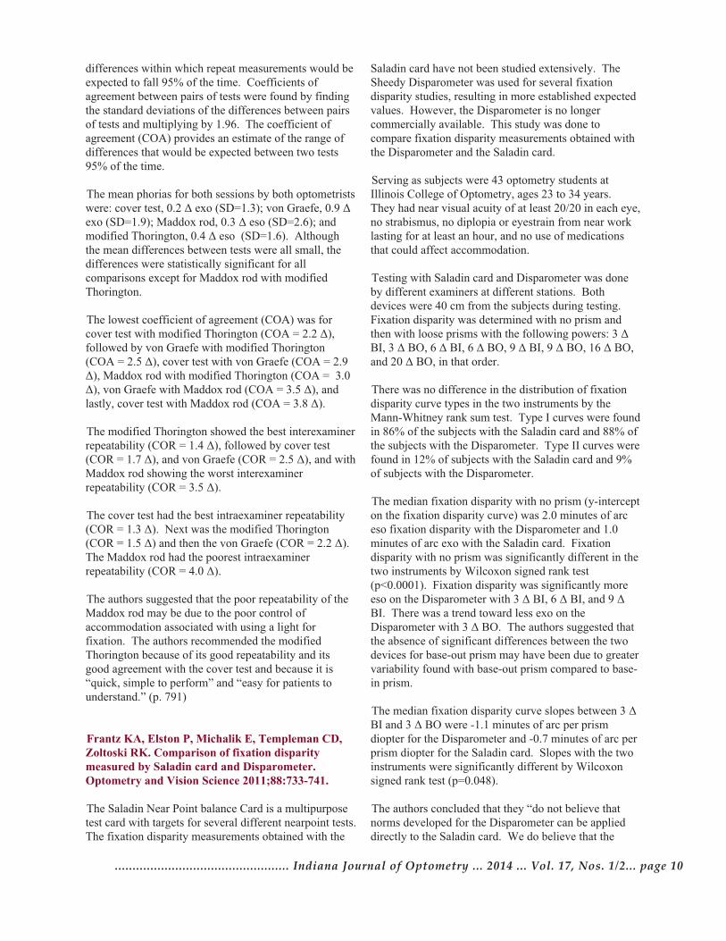
................................................. Indiana Journal of Optometry ... 2014 ... Vol. 17, Nos. 1/2... page 10
differences within which repeat measurements would be
expected to fall 95% of the time. Coefficients of
agreement between pairs of tests were found by finding
the standard deviations of the differences between pairs
of tests and multiplying by 1.96. The coefficient of
agreement (COA) provides an estimate of the range of
differences that would be expected between two tests
95% of the time.
The mean phorias for both sessions by both optometrists
were: cover test, 0.2 Δ exo (SD=1.3); von Graefe, 0.9 Δ
exo (SD=1.9); Maddox rod, 0.3 Δ eso (SD=2.6); and
modified Thorington, 0.4 Δ eso (SD=1.6). Although
the mean differences between tests were all small, the
differences were statistically significant for all
comparisons except for Maddox rod with modified
Thorington.
The lowest coefficient of agreement (COA) was for
cover test with modified Thorington (COA = 2.2 Δ),
followed by von Graefe with modified Thorington
(COA = 2.5 Δ), cover test with von Graefe (COA = 2.9
Δ), Maddox rod with modified Thorington (COA = 3.0
Δ), von Graefe with Maddox rod (COA = 3.5 Δ), and
lastly, cover test with Maddox rod (COA = 3.8 Δ).
The modified Thorington showed the best interexaminer
repeatability (COR = 1.4 Δ), followed by cover test
(COR = 1.7 Δ), and von Graefe (COR = 2.5 Δ), and with
Maddox rod showing the worst interexaminer
repeatability (COR = 3.5 Δ).
The cover test had the best intraexaminer repeatability
(COR = 1.3 Δ). Next was the modified Thorington
(COR = 1.5 Δ) and then the von Graefe (COR = 2.2 Δ).
The Maddox rod had the poorest intraexaminer
repeatability (COR = 4.0 Δ).
The authors suggested that the poor repeatability of the
Maddox rod may be due to the poor control of
accommodation associated with using a light for
fixation. The authors recommended the modified
Thorington because of its good repeatability and its
good agreement with the cover test and because it is
“quick, simple to perform” and “easy for patients to
understand.” (p. 791)
Frantz KA, Elston P, Michalik E, Templeman CD,Zoltoski RK. Comparison of fixation disparitymeasured by Saladin card and Disparometer.Optometry and Vision Science 2011;88:733-741.
The Saladin Near Point balance Card is a multipurpose
test card with targets for several different nearpoint tests.
The fixation disparity measurements obtained with the
Saladin card have not been studied extensively. The
Sheedy Disparometer was used for several fixation
disparity studies, resulting in more established expected
values. However, the Disparometer is no longer
commercially available. This study was done to
compare fixation disparity measurements obtained with
the Disparometer and the Saladin card.
Serving as subjects were 43 optometry students at
Illinois College of Optometry, ages 23 to 34 years.
They had near visual acuity of at least 20/20 in each eye,
no strabismus, no diplopia or eyestrain from near work
lasting for at least an hour, and no use of medications
that could affect accommodation.
Testing with Saladin card and Disparometer was done
by different examiners at different stations. Both
devices were 40 cm from the subjects during testing.
Fixation disparity was determined with no prism and
then with loose prisms with the following powers: 3 Δ
BI, 3 Δ BO, 6 Δ BI, 6 Δ BO, 9 Δ BI, 9 Δ BO, 16 Δ BO,
and 20 Δ BO, in that order.
There was no difference in the distribution of fixation
disparity curve types in the two instruments by the
Mann-Whitney rank sum test. Type I curves were found
in 86% of the subjects with the Saladin card and 88% of
the subjects with the Disparometer. Type II curves were
found in 12% of subjects with the Saladin card and 9%
of subjects with the Disparometer.
The median fixation disparity with no prism (y-intercept
on the fixation disparity curve) was 2.0 minutes of arc
eso fixation disparity with the Disparometer and 1.0
minutes of arc exo with the Saladin card. Fixation
disparity with no prism was significantly different in the
two instruments by Wilcoxon signed rank test
(p<0.0001). Fixation disparity was significantly more
eso on the Disparometer with 3 Δ BI, 6 Δ BI, and 9 Δ
BI. There was a trend toward less exo on the
Disparometer with 3 Δ BO. The authors suggested that
the absence of significant differences between the two
devices for base-out prism may have been due to greater
variability found with base-out prism compared to base-
in prism.
The median fixation disparity curve slopes between 3 Δ
BI and 3 Δ BO were -1.1 minutes of arc per prism
diopter for the Disparometer and -0.7 minutes of arc per
prism diopter for the Saladin card. Slopes with the two
instruments were significantly different by Wilcoxon
signed rank test (p=0.048).
The authors concluded that they “do not believe that
norms developed for the Disparometer can be applied
directly to the Saladin card. We do believe that the
Saladin card is a useful clinical instrument, but further
study is needed to establish appropriate norms for
interpretation of its findings.” (p. 740)
Zhang L, Zhang Y-Q, Zhang J-S, Xu L, Jonas JB.Visual fatigue and discomfort after stereoscopicdisplay viewing. Acta Ophthalmologica2013;91:e149-e153.
This study compared visual fatigue from 3D movie
viewing using a polaroid system and a liquid crystal
shutter system. The 30 subjects were 20 to 30 years of
age and had no more than 3 D of myopia, less than 1 D
of anisometropia, stereopsis of at least 60 seconds of
arc, and no strabismus.
On one morning subjects watched a 3D movie for 20
minutes with polaroid glasses and on another morning
they watched the same 3D movie for 20 minutes with
liquid crystal shutter glasses. Half of the subjects used
the polaroid glasses on the first day and half used the
shutter glasses on the first day.
Before and after movie viewing study participants were
interviewed concerning visual discomfort, including
questions about dizziness or nausea, headache, blur,
ghost images, eye pain, eye dryness, eye burning,
eyestrain, and heavy eyelids. Subjects were also asked
to rate their visual fatigue on a scale from 0 to 5. The
following tests were performed before and after viewing
the movie: monocular amplitude of accommodation,
monocular and binocular accommodative facility,
negative and positive relative accommodation, near
point of convergence break and recovery, base-in and
base-out fusional vergence breaks and recoveries at
distance and near, and AC/A ratio.
The mean visual fatigue score was significantly greater
(p=0.02) after viewing with the shutter glasses (1.7)
than after viewing with polaroid glasses (1.2). The
results on the separate visual discomfort questions were
not significantly different between the two different
modes of 3D viewing.
Several accommodation and vergence tests suggested
more fatigue with the liquid crystal shutter system than
with the polaroid system. There was a significantly
greater reduction in amplitude of accommodation with
the shutter glasses (right eye, p<0.001; left eye, p=0.01).
The near point of convergence break receded more with
the shutter glasses (p=0.007). The near point of
convergence recovery did not change with polaroid
glasses, but receded with shutter glasses. Binocular
accommodative facility increased with polaroid glasses,
but decreased with shutter glasses. Relative
accommodation findings did not change significantly
with polaroid glasses, but showed small but significant
reductions with shutter glasses (NRA, p=0.008; PRA,
p=0.007).
The authors noted that the results may have been
limited by the fact that the movie was viewed for only
20 minutes. They concluded that the liquid crystal
shutter system led to greater visual fatigue than the
polaroid system, as shown by the subject’s subjective
rating of fatigue as well as by the results of
accommodation and vergence tests.
Kim S-H, Suh Y-W, Yun C, Yoo E-J, Yeom J-H,Cho YA. Influence of stereopsis and abnormalbinocular vision on ocular and systemic discomfortwhile watching 3D television. Eye 2013;27:1243-1248.
This study was conducted at an ophthalmology
department in Korea to evaluate discomfort from 3D
television viewing in persons with abnormal binocular
vision compared to normal. Ninety-eight patients who
were at least nine years of age and had strabismus,
amblyopia, or anisometropia were recruited for the
abnormal binocular vision group. Of these 98, 49 were
classified in the strabismus subgroup as determined by
cover test, 22 were in the amblyopia subgroup (visual
acuity less than 16/20 or more than two lines difference
between the eyes), and 27 were in the anisometropia
subgroup (difference of more than 2 D between the
spherical equivalents of the two eyes). Subjects were
considered to be in the amblyopia subgroup whether
they had strabismic amblyopia or anisometropic
amblyopia. The abnormal binocular vision group was
compared to a control group of 32 subjects. The phoria
and accommodation status of members of the control
group was not stated.
Stereoacuity with both the Frisby-Davis distance
stereotest and the Stereo Optical Stereo Fly test was
significantly better in the control group than in the
abnormal binocular vision group (p<0.001).
Subjects viewed a 3D video on a high definition
television for 20 minutes. A survey with 13 items, each
of which was answered on a five category scale from 0
(no symptom) to 4 (extremely severe symptom) was
completed by the subjects after viewing the video. The
13 symptom items were dizziness, headache, nausea,
eye fatigue, eye pain, tearing, eye dryness, blurred
vision, difficulty in focusing, double vision, transient
dimness after watching TV, lack of perception of
Page 11 ... Vol. 17, Nos. 1/2 ... 2014 ... Indiana Journal of Optometry ......................................................

................................................. Indiana Journal of Optometry ... 2014 ... Vol. 17, Nos. 1/2... page 12
stereoscopic vision, and difficulty in tracking motion on
the TV.
Symptom levels did not differ significantly between the
abnormal binocular vision group and the control group
on any of the 13 individual items. However, interesting
results were found in additional analyses. Symptom
levels differed between the three subgroups of those with
abnormal binocular vision only for not perceiving
stereoscopic vision, with the symptom level for lack of
stereopscopic perception being greatest in subjects with
amblyopia and the lowest symptom level in subjects in
the anisometropia subgroup.
The authors divided subjects into those with good
stereopsis (which they defined as stereoacuity of 60
arcsec of better on the Stereo Fly test) and those with
moderate to poor stereopsis (defined as worse than 60
arcsec). Those with good stereopsis had significantly
higher symptom levels for dizziness, headache, eye
fatigue, and eye pain. The symptom level for not being
able to perceive stereoscopic vision during 3D television
viewing was greater for the moderate to poor stereopsis
group.
The authors also compared symptoms in the subjects
with abnormal binocular vision but good stereopsis to
symptoms in the control subjects. Symptom levels were
significantly higher in the abnormal binocular vision
group for eye fatigue and nearly significantly greater for
headache.
The authors suggested that their study showed that visual
discomfort from viewing 3D television is more likely in
persons with abnormal binocular vision but good
stereopsis and that inability to observe stereo effects is
related to abnormal binocular vision, such as in
strabismus, amblyopia, or anisometropia. They did not
evaluate the relation of 3D visual discomfort to other
binocular vision characteristics, such as phorias,
vergence ability, or accommodative characteristics.

Page 13... Vol. 17, Nos. 1/2 ... 2014 ... Indiana Journal of Optometry ......................................................
The first 42 volumes (1970-2011) of the
Optometric Historical Society’s publication
Hindsight: Journal of Optometry History are now
online. Over 2,500 pages of high-quality, OCR
enhanced, searchable, digital documents are now freely
accessible to researchers at IUScholarWorks
(http://scholarworks.iu.edu/journals/index.php/hindsight
/issue/archive). This was made possible by a joint effort
of the Optometric Historical Society and
IUScholarWorks. IUScholarWorks is a service of the
Indiana University Libraries with additional technology
support from Indiana University Information
Technology Services.
Examples of the scholarship found in Hindsight include:
• Biographical sketches of influential figures such as
John Eberhardt (1857-1927), who championed the term
“optometrist” to unite those who identified themselves
as “refracting opticians”, “sight-testers” or other related
terms (volume 18, pages 43-44) and Frederic Woll
(1874-1955), whose tours and reviews of optometry
schools in the 1920s served to strengthen the standards
of optometric education (volume 42, pages 63-66);
• Personal memoirs and reflections of educators,
innovators, practitioners, advocates and leaders in the
profession;
• Historical research on individuals, events and trends in
vision care, the profession and legislation;
• Book reviews and historiographies.
OHS founder, Dr. Henry W Hofstetter, noted that
studying optometry history can lead to an appreciation
of its “centuries-long existence and emergence from a
prestigious and sophisticated handicraft to its present
academic stature, a truly proud history which includes
many prominent and accomplished personalities.”
(Hindsight, volume 27, pages 17-18).
Hindsight is a publication of the Optometric Historical
Society (OHS). OHS was formed in 1969 through the
efforts of Dr. Hofstetter, then outgoing president of the
American Optometric Association (AOA), and Maria
Dablemont, librarian and archivist for the AOA.
Presidents of OHS have included six Deans of
optometry schools, two AOA presidents, an editor of the
Journal of the American Optometric Association, and
other noted educators and practitioners. The current
president of OHS is Dr. John F. Amos.
The first publication of OHS, the Newsletter of the
Optometric Historical Society, appeared in January,
1970. OHS has produced a quarterly publication ever
since. Starting with volume 23 (1992), the title of the
publication became Hindsight: Newsletter of the
Optometric Historical Society. Beginning with volume
38 (2007) and continuing to the present, it has been
titled Hindsight: Journal of Optometry History.
The Optometric Historical Society has always had a
strong IU and Indiana connection. Dr. Hofstetter, the
first director of the IU’s optometry school, was editor of
the OHS publication for most of the first 25 years.
Taking over for Hofstetter at various intervals during
that time or serving as co-editor were John R. Levene,
an IU faculty member, and Douglas Penisten, an
alumnus of the IU O.D. and Ph.D. programs. David
Goss, a current IU faculty member, has been editor since
1995. Only four of the thirteen OHS presidents over its
43 year history have not either been an IU faculty
member, IU alumnus, or Indiana practicing optometrist.
IU faculty or alumni who have been OHS presidents
include Hofstetter, Levene, Penisten, T. David Williams,
Charles Haine, Walter W. Chase, and John F. Amos.
Indiana practicing optometrists Jerry Abrams and James
Leeds are among the former presidents of OHS.
In 2009, members of the Board of Directors of OHS and
Optometry Cares – The AOA Foundation signed a
Memorandum of Understanding that places OHS under
the umbrella of Optometry Cares. The AOA Foundation
is also in charge of the AOA Archives and Museum of
Optometry (http://www.aoafoundation.org/archives-
museum-of-optometry/).
New issues of Hindsight can be obtained in print form
as they are published by joining the Optometric
Historical Society. One year membership in the
Optometric Historical Society and subscription to
Hindsight is $25 for regular membership and $50 for
patron membership. A lifetime membership is $250.
Membership can be obtained by sending name and
address and check made out to the Optometric Historical
Society to Kirsten Hébert, Optometric Historical
Society, Archives and Museum of Optometry, 243 N.
Lindbergh Boulevard, St. Louis, MO 63141.

Non-Profit Org. U.S. Postage
PAIDBloomington, IN
Permit #2Indiana Journal of OptometryIndiana University School of Optometry800 East Atwater AvenueBloomington, IN 47405