This lecture was conducted during the Nephrology Unit Grand Ground by a Sub-intern under Nephrology...
26
This lecture was conducted during the Nephrology Unit Grand Ground by a Sub-intern under Nephrology Division, Department of Medicine in King Saud University. Nephrology Division is NOT responsible for the content of the presentation for it is
-
Upload
gabriella-mcbride -
Category
Documents
-
view
214 -
download
1
Transcript of This lecture was conducted during the Nephrology Unit Grand Ground by a Sub-intern under Nephrology...

This lecture was conducted during the Nephrology Unit Grand Ground by a
Sub-intern under Nephrology Division,
Department of Medicine in King Saud University.
Nephrology Division is NOT responsible for the content of the presentation for it is
intended for learning and /or education purpose
only.

Nephrotic syndrome
Done by : Nora AlMahmoud

Objectives:
0Definition0Pathophysiology0Causes0Clinical manifestations 0Complication 0Diagnosis0Treatment

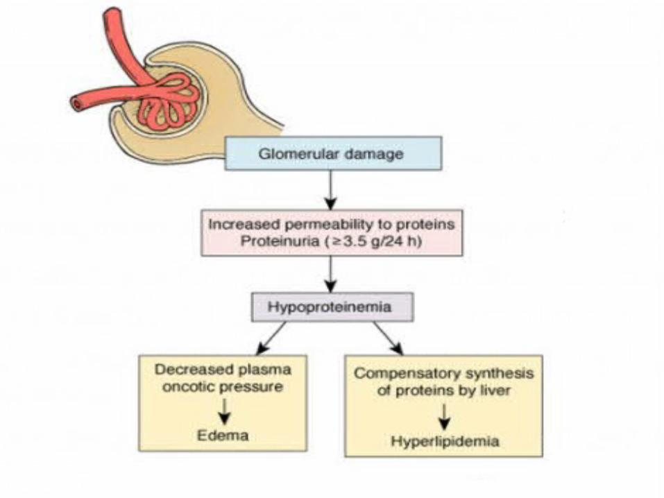
Definition
Clinical complex that include the following :1)Massive proteinuria (3.5g or more in adult).2)Hypoalbuminemia (<3mg/dl)3)Generalized edema4)Hyperlipidemia and lipidurea

0 Urinary loss of anticoagulants proteins, i.e.: protein C , protein S , and antithrombin
Hypercoagulable state
0Urinary loss of transport proteins
Iron , Copper , and Zinc deficiency

Pathophysiology Damage to Podocytes (glomerulus epithelial cells)
allows proteins to pass through a 'leaky' glomerulus into the urine.
Normally molecules of <20kDa will pass into the urine In Nephrotic syndrome molecules of >100kDa can
pass into the urine

Causes
0Causes of nephrotic syndrome vary according to age .
0In children : always caused by lesion primary to the kidney .
0In adult often due to renal manifestation of a systemic disease .

Primary causes
0Membranous GN0Minimal change disease 0Focal segmental glumerulosclerosis0Membranoproliferative GN
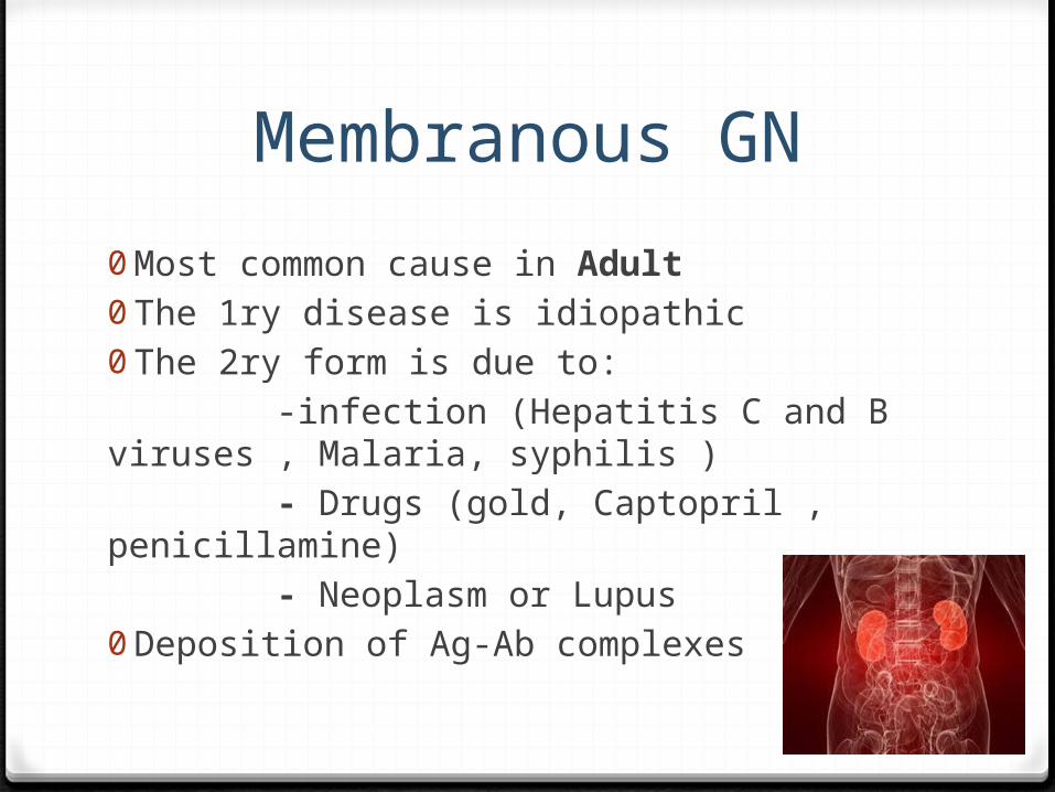
Membranous GN
0Most common cause in Adult0The 1ry disease is idiopathic 0The 2ry form is due to: -infection (Hepatitis C and B viruses , Malaria, syphilis ) - Drugs (gold, Captopril , penicillamine) - Neoplasm or Lupus 0Deposition of Ag-Ab complexes


Minimal change disease
0Most common cause in Children0Associated with Hodgkin’s and non-Hodgkin’s
lymphoma0No histological abnormality in light microscopy ,
Fusion of foot processes on Electron microscopy.


FSGN0Account for 25% of cases of nephrotic syndrome in adult
0Associated with: HIV , Heroin , sickle cell and obesity. 0 It has fair to poor prognosis – resistant to steroid therapy
– patient develop renal insufficiency within 5-10 years of diagnosis , the course is progressive.

Membranoproliferative GN
0Can be idiopathic or 2ry to chronic immune disease (Hepatitis C , Alpha1 antitrypsin , HIV , Malignancy )
0GBM alteration , sub endothelial leukocytes infiltration , predominant masengial involvement .

Secondary causes
0Systemic lupus erythematosus (SLE).0Amyloidosis.0Diabetes Mellitus.0Allergy.0 Infections.(HBV, HCV, HIV, Malria, syphilis).0Drugs(Gold)

Clinical Pictureedema first presents periorbitally Ankle edema Severe proteinuria increase BPMore severe features occur late or if untreated and may
include pulmonary effusion, genital edema and anasarca (severe generalised edema).


Complications
1. Venous thromboembolism 2. Extreme hypercholesterolaemia (>10mmol/L) 3. Infection 4. Renal Failure 5. Malnutrition
How to approach pt. with Neprotic syndrome?
0 Patient history - Identify medication or toxin exposure; risk factors for HIV or viral hepatitis; and symptoms suggesting other causes of edemaObtain history of diabetes, systemic lupus erythematosus, or other systemic illness0 Urine dipstick : Confirm proteinuria0 Random urine protein/creatinine ratio : Quantify degree of
proteinuria (ratio greater than 3 to 3.5)0 Serum creatinine : Rule out acute renal failure, assess glomerular
filtration rate0 Serum albumin : Assess degree of hypoalbuminemia0 Lipid panel0 Assess degree of hyperlipidemia

0 Additional studies suggested by patient factors: -HIV screening test - Identify HIV -Hepatitis serology panel -Identify hepatitis B or C -Serum or urine protein electrophoresis -Suggests amyloidosis or multiple myeloma -Rapid plasma reagin -Identify syphilis - Antinuclear antibodies or complement (C3 and C4) level -Identify systemic lupus erythematosus; complement levels may also be reduced in membranoproliferative disease

Diagnosis
0Urin analysis shows >3.5 g/24h : - Most often used: single spot urine for albumin and creatinine.0Most accurate test to determine etiology is : Renal Biopsy
Treatment3 Aims:
1) Treat fluid retention: Restrict salt and water intake loop diuretic +/- thiazide diuretic ACE inhibitor +/- Angiotensin 2 receptor antagonist
2) Avoid complications: prophylactic heparin prompt treatment of infection treat hyperlipidaemia (statins) target BP 125/75

3) Treat the underlying cause 0 steroids in all idiopathic 1ry renal disease0If steroids ineffective : -add Cyclophosphamide or Mycophenolate
0 ACE inhibitor used in all patients but does not reverse the disease

Nephrotic Vs. Nephritic syndromes
Nephrotic s. Nephritic s.
Pathogenesis Abnormal glomerular permeability due to number of conditions
Inflammation of the Glomeruli due to any of the causes of Glumerulonephritis
Causes 1ry and 2ry causes Poststrept. Glomerulonephritis is the most common cause
Lab finding Proteinuria >3.5g/24hHypoalbuminemia Hyperlipidemia , Fatty cast in the urine
HematuriaARF-Azotemia , OliguriaProteinuria if present mild
Clinical finding
Generalized EdemaHypercoagulable state Increase risk of infection
HTNEdema