This article was originally published in the Encyclopedia of ... · ganglia territories, and these...
8
This article was originally published in the Encyclopedia of Neuroscience published by Elsevier, and the attached copy is provided by Elsevier for the author's benefit and for the benefit of the author's institution, for non- commercial research and educational use including without limitation use in instruction at your institution, sending it to specific colleagues who you know, and providing a copy to your institution’s administrator. All other uses, reproduction and distribution, including without limitation commercial reprints, selling or licensing copies or access, or posting on open internet sites, your personal or institution’s website or repository, are prohibited. For exceptions, permission may be sought for such use through Elsevier's permissions site at: http://www.elsevier.com/locate/permissionusematerial Jarvis E D (2009) Bird Brain: Evolution. In: Squire LR (ed.) Encyclopedia of Neuroscience, volume 2, pp. 209-215. Oxford: Academic Press.
Transcript of This article was originally published in the Encyclopedia of ... · ganglia territories, and these...

This article was originally published in the Encyclopedia of Neuroscience published by Elsevier, and the attached copy is provided by Elsevier for the
author's benefit and for the benefit of the author's institution, for non-commercial research and educational use including without limitation use in
instruction at your institution, sending it to specific colleagues who you know, and providing a copy to your institution’s administrator.
All other uses, reproduction and distribution, including without limitation commercial reprints, selling or licensing copies or access, or posting on open
internet sites, your personal or institution’s website or repository, are prohibited. For exceptions, permission may be sought for such use through
Elsevier's permissions site at:
http://www.elsevier.com/locate/permissionusematerial
Jarvis E D (2009) Bird Brain: Evolution. In: Squire LR (ed.) Encyclopedia of Neuroscience, volume 2, pp. 209-215. Oxford: Academic Press.

Bird Brain: Evolution 209
Author's personal copy
Bird Brain: Evolution
E D Jarvis, Duke University Medical Center, Durham,NC, USA
ã 2009 Elsevier Ltd. All rights reserved.
The classical century-old view of bird brain evolutionwas based on a theory of linear and progressive evo-lution, where the vertebrate cerebrum was argued tohave evolved in lower to higher anatomical stages,with birds at an intermediate stage. As such, the aviancerebrum was thought to consist mostly of basalganglia territories, and these were thought to controlmostly primitive behaviors. This view, althoughslowly challenged, was not formally changed until2004 and 2005, when a forum of neuroscientiststhat reevaluated brain terminologies and homologiesfor birds and other vertebrates published a newnomenclature that reflects a modern understandingof bird brain evolution. This view proposes that theavian cerebrum was inherited as a package with pal-lial (or cortical-like), striatal, and pallidal regionsfrom its recent reptilian and distant stem amnioteancestors. The avian pallial domain is organized dif-ferently from the mammalian pallium or cortex inthat the avian is nuclear and the mammalian is lay-ered. However, the pallia of both groups performsimilar functions and have some similar connectivity.The avian striatal and pallidal domains are organizedmore similar to those of mammals and, likewise,perform similar functions in both groups. This articlereviews these classical and modern views for birdbrain evolution in the context of vertebrate brainevolution.
The Classical View
The classical view of telencephalic evolution had itsorigin in the late nineteenth and early twentieth cen-tury following the impact of Darwin’s theory of evo-lution. Inspired by Darwin’s theory, Ludwig Edingerformulated an evolution-based model of brain orga-nization from 1885 to 1908. Edinger and others com-bined Darwin’s concept of ‘evolution’ with thenineteenth-century version of Aristotle’s ‘scala nat-urae,’ resulting in the view that evolution was unilin-ear and progressive, from fish to amphibians, reptiles,birds and mammals, primates, and, finally, humans,ascending from ‘lower’ to ‘higher’ intelligence in achronological series. They believed that the brainsof extant vertebrates retained the ancestral structuresand, therefore, that the origin of specific human brainsubdivisions could be traced back in time by examin-ing the brains of extant nonhuman vertebrates.
Encyclopedia of Neuroscien
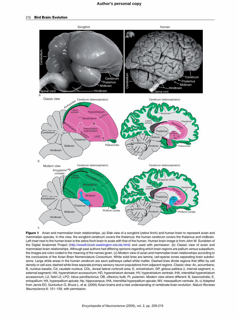
Although the spinal cord, hindbrain, midbrain, thala-mus, and cerebellum appeared to be well conservedacross vertebrates, Edinger noted that the internalorganization of the telencephala or cerebrum exhibitedthe most pronounced differences across vertebrates.In mammals, the outer part of the telencephalon hadprominently layered gray matter (Figure 1(b), green),whereas the inner part had nuclear gray matter(Figure 1(b), purple).The inner partwas located ventralto the lateral ventricle. In nonmammals, the outer andinner parts of the telencephala had mostly nuclear graymatter, and most of it was located ventral to the lateralventricle in birds and reptiles (Figure 1(b), purple).
On the basis of these considerations, Edinger pro-posed that telencephalic evolution occurred in pro-gressive stages of increasing complexity and size,culminating with the human cerebrum. He arguedthat the stages proceeded in a ventral-to-dorsal direc-tion, with each new vertebrate group acquiring amoreadvanced cerebral subdivision. He argued that firstthere was the old brain, the paleoencephalon (alsocalled the basal ganglia or subpallium), which con-trolled instinctive behaviors, followed by the additionof a new brain, the neoencephalon (also called thepallium or mantle at the top), which controlledlearned and intelligent behaviors. The telencephalicsubdivisions within each vertebrate group were fur-ther named by him, Ariens-Kappers, and others withthe prefixes ‘paleo’ (oldest), ‘archi’ (archaic), and‘neo’ (new) to designate the presumed relative orderof evolutionary appearance of each subdivision. Tothese prefixes the root word ‘striatum’ was addedfor the presumed paleoencephalic subdivisions and‘pallium’ (meaning mantle) or ‘cortex’ (meaningbark) for the presumed neoencephalic subdivisions.The term striatum was used because a large part ofthe basal ganglia (i.e., paleoencephalon) in mammals,which is now commonly call caudate putamen, con-tains fiber bundles coursing through it, giving it astriated appearance.
The classical view that became dominant in thecontext of bird brain was that the bird subpalliumconsisted of ‘paleostriatum’ (old striatum) inheritedoriginally from fish, said to be the antecedent of thehuman globus pallidus, and devoted to olfactoryinformation processing. Above the paleostriatum,birds were said to have inherited an ‘archistriatum’(archaic striatum) originally from amphibians, said tobe the antecedent of the human amygdala. Above andadjacent to the archistriatum, birds were said to haveinherited a ‘neostriatum’ (new striatum) from theirclosest relative, reptiles, said to be the antecedentof the human caudate and putamen. Reptiles were
ce (2009), vol. 2, pp. 209-215
Cerebellum
Cerebellum
Cerebellum
Amygdaloid
Nidopallium
Mesopallium
Hyperpallium
E
Piriform cortex Piriform cortex
AcOB
OB
B
Striatum
Hp CDL
Lateralventricle
L2
Pallidum
MV
IHAHA
complex
Cerebellum
Archistriatum
Arcopallium
Hindbrain
Hindbrain Paleocortex
Archicortex
Archi-
striatum
Paleostriatum
primitivum
Midbrain
Midbrain Ac
OB
Ptei
Thalamus
Hindbrain
Hindbrain
AmygdalaMidbrain
Midbrain
Hp
HpThalamus
Thalamus
Thalamus
Striatum
GPPaleostriatum
AcPtei
GP
Cd
Pallidum
Cd
LPO
PaleocortexOB
E
B
L2 Neostriatum
Neostriatum
Neocortex
Claustrum
Claustrum
Six-layered cortex
Cerebrum (telencephalon)Classic view
Songbird
a
Human
b
c
Modern view
Cerebrum (telencephalon)
Spinal cordHindbrain
MidbrainThalamus
Cerebrum
Hindbrain
MidbrainThalamus
Cerebrum
Cer
ebel
lum
Spinal cord
Cer
ebel
lum
Cerebrum (telencephalon) Cerebrum (telencephalon)
Hyperstriatum
HVHD
IHAHA
CDL
Paleostriatumaugmentatum
Lateralventricle
Lateralventricle
Lateralventricle
Archicortex
Figure 1 Avian and mammalian brain relationships. (a) Side view of a songbird (zebra finch) and human brain to represent avian and
mammalian species. In this view, the songbird cerebrum covers the thalamus; the human cerebrum covers the thalamus and midbrain.
Left inset next to the human brain is the zebra finch brain to scale with that of the human. Human brain image is from JohnW. Sundsten of
the Digital Anatomist Project (http://www9.biostr.washington.edu/da.html) and used with permission. (b) Classic view of avian and
mammalian brain relationships. Although past authors had differing opinions regarding which brain regions are pallium versus subpallium,
the images are color-coded to the meaning of the names given. (c) Modern view of avian and mammalian brain relationships according to
the conclusions of the Avian Brain Nomenclature Consortium. White solid lines are lamina, cell-sparse zones separating brain subdivi-
sions. Large white areas in the human cerebrum are axon pathways called white matter. Dashed lines divide regions that differ by cell
density or cell size; dashed white lines separate primary sensory neuron populations from adjacent regions. Classic view: Ac, accumbens;
B, nucleus basalis; Cd, caudate nucleus; CDL, dorsal lateral corticoid area; E, ectostriatum; GP, globus pallidus (i, internal segment; e,
external segment); HA, hyperstriatum accessorium; HD, hyperstriatum dorsale; HV, hyperstriatum ventrale; IHA, interstitial hyperstriatum
accessorium; L2, field L2; LPO, lobus parolfactorius; OB, olfactory bulb; Pt, putamen. Modern view where different: B, basorostralis; E,
entopallium; HA, hyperpallium apicale; Hp, hippocampus; IHA, interstitial hyperpallium apicale; MV, mesopallium ventrale. (b, c) Adapted
from Jarvis ED, Gunturkun O, Bruce L, et al. (2005) Avian brains and a new understanding of vertebrate brain evolution. Nature Reviews
Neuroscience 6: 151–159, with permission.
210 Bird Brain: Evolution
Encyclopedia of Neuroscience (2009), vol. 2, pp. 209-215
Author's personal copy
Bird Brain: Evolution 211
Author's personal copy
additionally thought to have elaborated the paleos-triatum into an older part (the primitivum) and anewer part (the augmentatum), which was passedonto birds. Birds were then thought to have evolveda large additional basal ganglia subdivision, the‘hyperstriatum’ (hypertrophied striatum), consideredunique to birds.The bird pallium was said to consist of a ‘paleocor-
tex’ inherited from fish pallium and believed to be theantecedent of the human olfactory cortex. Birds weresaid to have inherited an ‘archicortex,’ also thought tobe olfactory and primitive, from amphibians andbelieved to be the antecedent of the human hippocam-pus. Birdswere thought not to have evolved any furtherpallial regions. In contrast, mammals were thought tohave evolved from the paleocortex and/or archicortex,a ‘neocortex.’ The archicortex and/or paleocortexwiththeir two or three cell layers were assumed to beprimitive; the neocortex with its six layers wasassumed to be more recently evolved and a substratefor more sophisticated behavior. This classical viewwas codified in the major comparative neuroanatomytext by Ariens-Kappers, Huber, and Crosby in 1936and became pervasive throughout neuroscience. Thisview is now known to be incorrect.
The Modern View
In 2004 and 2005, an international consortiumof specialists in avian, mammalian, reptilian, andfish neurobiology – the Avian Brain NomenclatureConsortium – published several reports on a new nom-enclature that represents the current understandingof avian telencephalic organization and homologieswith mammals (Figure 1(c)). The consortium con-cluded that the avian telencephalon is organized intothree main, developmentally distinct domains thatare homologous in fish, amphibians, reptiles, birds,and mammals: pallial, striatal, and pallidal domains(Figure 1(c)). For birds, they renamed these domainsand their respective subdivisions with homology-based terms or roots that allow reference to namedregions in mammals. In doing so, they eliminated allphylogeny-based prefixes (paleo-, archi-, and neo-)that erroneously implied the relative age of eachsubdivision.The avian paleostriatum was simply renamed stri-
atum after its mammalian homolog (Figure 1(c)). Thepaleostriatum primitivum and adjacent ventral regionwas renamed pallidum also after its mammalianhomolog. Similar to the mammalian pallidum, theavian pallidum has a sparse distribution of cells, giv-ing the region its pale appearance and its name. Thedorsal part of the avian pallidum was determined tobe homologous to the mammalian globus pallidus
Encyclopedia of Neuroscien
and renamed as such, whereas the ventral part wasdetermined to be homologous to the mammalianventral pallidum. The dorsal pallidum, however, dif-fers between mammals and birds. In mammals, itconsists of two segments with distinct connectivity,the internal and external globus pallidus, whereas inbirds neurons with both phenotypes are intermingled(Figure 1(c)).
The avian pallium was determined to be organizednot into three striatal subdivisions (hyperstriatum,neostriatum, and archistriatum) but, rather, intofour pallial subdivisions renamed as hyperpallium(hypertrophied pallium; upper part of old hyperstria-tum), mesopallium (middle pallium; lower part of oldhyperstriatum), nidopallium (nest pallium; old neos-triatum), and arcopallium (arched pallium; most ofold archistriatum) (Figure 1(c)). Several neuronalpopulations adjacent to the arcopallium and the pos-terior part of what had been regarded as archistria-tum were considered homologous to pallial andsubpallial parts of the mammalian amygdala, andthey were renamed as the avian amygdaloid complex.Other regions that were widely recognized to behomologous among vertebrates – the hippocampus,olfactory (piriform) cortex, and olfactory bulb – didnot require name changes.
Some Anatomical and Molecular Parallels
Some key similarities between avian and mammaliansubpallium include a high enrichment of dopaminer-gic axon terminals in the striatum that originate fromthe substantia nigra pars compacta and ventral teg-mental area neurons of the midbrain. Both avian andmammalian striatum contain two major classes ofspiny neuron types, those with the neuropeptide sub-stance P (SP) and those with the neuropeptide enkeph-alin (ENK), which project to two different neuronpopulations in the pallidum. In both birds and mam-mals, the SP neurons seem to be involved in promotingplanned movement, whereas the ENK neurons seemto be involved in inhibiting unwanted movements.Both the avian and the mammalian striatum partici-pate not only in instinctive behavior and movementbut also in motor learning. Developmental studiesindicate that the avian and mammalian subpalliumconsists of two separate histogenetic zones thatexpress different sets of transcription factors: (1) adorsal zone that corresponds to the lateral ganglioniceminence and that selectively expresses the transcrip-tion factors Dlx1 and Dlx2 but not Nkx2.1 and (2) aventral zone that corresponds to themedial ganglioniceminence and selectively expresses all three transcrip-tion factors. The lateral ganglionic eminence gives riseto the striatum and the medial ganglionic eminence
ce (2009), vol. 2, pp. 209-215

212 Bird Brain: Evolution
Author's personal copy
gives rise to the pallidum. Similar striatal and pallidalterritories have been found in reptiles.Some similarities between avian and mammalian
pallium (i.e., mammalian cortex) include direct pro-jections of sensory visual, auditory, and somatosen-sory input from the thalamus. The correspondingavian brain regions subserve the same type of sensoryinformation processing as is performed by the mam-malian cortex. Likewise, the avian hyperpallium andarcopallium give rise to major descending projectionsto premotor and motor neurons of the brain stem andspinal cord, as do mammalian corticobulbar and cor-ticospinal pathways. These avian brain regions sub-serve critical roles in motor control and sensorimotorlearning, similar to the mammalian cortex. The pal-lial relationships between these avian and mamma-lian brain regions are also supported by molecularembryology studies. During development, the avianhyperpallium, mesopallium, and nidopallium andthe mammalian pallium selectively express the pal-lium-specific transcription factors Emx1, Pax6, andTbr1. In adult birds and mammals, comparativeexpression profiles of the neurotrophic factor brain-derived neurotrophic factor and the glutamate recep-tor mGluR2 indicate that the avian arcopalliumexpress these pallial-specific mRNAs at high levels,as does the mammalian cortex.
AModern View of Telencephalic Evolution
The organization of the true basal ganglia of birdsrelative to mammals and other vertebrates (i.e., dis-tinct nuclear striatal and pallidal domains) is quiteconserved. In contrast, the organization of their pallial
MYAEraperiod
408 360 320 286 248
TriassPermianDevonianSilurian
Ancestral bony fish
Ancestral amphibians
Stem
Ver
tebr
ate
grou
p
Amniotes Therapsids
Sauropsids
Pennsyl-Paleozoic
vanianMississ-
ipian
Figure 2 Simplified modern view of vertebrate evolution. The diagram
to land vertebrates. This view differs from the classic view in that inst
have given rise to stem amniotes. Stem amniotes then split into at lea
reptiles as we know them today, and the therapsids, which, through a
Many sauropsids (reptiles) are currently living. Solid horizontal lines i
ancestral links from other types of data. MYA, millions of years ago. Ba
Paleontology and Evolution, pp. 1–13. New York: Freeman; and Evan
and Cardew G (eds.) Evolutionary Developmental Biology of the Cere
Encyclopedia of Neuroscienc
domains differs to a greater degree. The nuclear typeof organization of the pallium found in birds is alsopresent in reptiles. The avian hyperpallium possessesa unique organization so far found only in birds; itconsists of semilayered subdivisions, and it mighthave evolved more recently than the mammaliansix-layered cortex since birds evolved well after mam-mals (by �50–100 million years) (Figure 2). Theregion below the ventricle, called the dorsal ventricu-lar ridge (DVR), contains mesopallium, nidopallium,and arcopallium, and it is a nuclear pallial organiza-tion that is unique to birds and reptiles. The six-layered cortex is a pallial organization unique tomammals. Because all major groups of living mam-mals (monotremes, marsupials, and placentals) havea six-layered cortex, it was presumably inheritedfrom their common therapsid ancestor more than200 million years ago (Figure 2). Furthermore, mam-mals may not have arisen from reptiles but, rather,from therapsids, because reptile and mammallineages apparently had their last common ancestorin stem amniotes. Because all nonmammalian therap-sids are extinct (Figure 2), it is difficult to trace fromstem amniotes to mammals the evolutionary historyof mammalian telencephalic organization – layered,nuclear, or otherwise. Thus, the reptilian nuclear pal-lial organization cannot be assumed to represent theancestral condition for mammals, as it is for birds.
In general, the evidence suggests that pallial, stria-tal, and pallidal domains exist in most or all verte-brates. Thus, it has been hypothesized that thetelencephalon of early fishes possessed all threedomains, which were then inherited as a package bylater vertebrates, including birds, and independently
213Mesozoic Cenozoic
Tertiary
Qua
tern
ary
CretaceousJurassicic
Mammals
Reptiles
Birds
144 65 2
begins with the fish group that contains the most recent ancestor
ead of giving rise to reptiles, ancestral amphibians are thought to
st two major groups: the sauropsids, which gave rise to all modern
series of now-extinct intermediate forms, evolved into mammals.
ndicate temporal fossil evidence. Dashed lines indicate proposed
sed on Carroll RL (1987) Fossils and relationships. In: Vertebrate
s SE (2000) General discussion II: Amniote evolution. In: Bock GR
bral Cortex, no. 228, pp. 109–113. Chichester, UK: Wiley.
e (2009), vol. 2, pp. 209-215

Bird Brain: Evolution 213
Author's personal copy
modified in them. The conserved organization ofstriatal and pallidal domains indicates that theremight be constraints on how the basal ganglia canbe organized. The diverse organizations of the pallialdomains suggest that there are fewer constraints onhow the pallium can be organized.
Detailed Views That Need Further Testing
When the modern view of bird brain evolution wasdeveloping, it was accompanied by several newhypotheses about one-to-one homologies betweenspecific avian and mammalian pallial subdivisions.These hypotheses are under active investigation andmake up a large area of research on bird brain evolu-tion. They are classified into two main categories:nuclear-to-layered hypotheses and nuclear-to-claus-trum/amygdala nuclei hypotheses.
Nuclear-to-Layered Hypotheses
First proposed by Karten, these hypotheses (Figure 3(a)) state that the similarities in connectivity betweenthe avian hyperpallium, nidopallium and arcopalliumof birds, and the six-layered cortex of mammals stemfrom a common origin of these structures – that is,they are homologous. Karten proposed that the com-mon ancestor of birds, reptiles, and mammals pos-sessed a nuclear pallium that was transformed into alaminar pallium early in the mammalian lineage,maintaining connectivity of the ancestral nuclear net-work. In this regard, he argued that the avian palliumis divided into three sets of serially connected types ofneurons – thalamorecipient neurons (field L2, ento-pallium, and basorostralis), pallio-pallial neurons(nidopallium), and extratelencephalic projection neu-rons (arcopallium) – with cell types and interconnec-tivity similar to those of mammalian cortical layers IV,II and III, and V and VI, respectively (Figure 3(a)).Similar arguments were made by others for the avianupper hyperpallium (also known as the Wulst), whichalso has serially connected neuron types that resemblethose found in the mammalian cortex. In this view,avian L2 neurons are homologous to layer IV neuronsof mammalian primary auditory cortex, basorostralisneurons to layer IVof primary somatosensory cortex,entopallium neurons to layer IV of extrastriate visualcortex, and the interstitial hyperpallium apicale neu-rons to layer IVof striate visual cortex. Supporting thishypothesis, gene expression studies have shown thatavian thalamorecipient nuclear fields (L2, entopal-lium, basorostralis, and interstitial hyperpallium)and the mammalian thalamorecipient layer IV of cor-tex selectively express some of the same genes. Avianextratelencephalic projection neurons (in the arcopal-lium but not in the hyperpallium) and mammalian
Encyclopedia of Neuroscien
extratelencephalic projection neurons (layer Vneurons of cortex) both selectively express some ofthe same genes. Thus, although the avian pallium isnot organized cytoarchitectonically into layers, itsnuclear subdivisions bear marked similarities in con-nectivity and molecular profiles to different layers ofthe mammalian cortex.
Nuclear-to-Claustrum/Amygdala Hypotheses
These hypotheses differ from the nuclear-to-layeredhypotheses with regard to mammalian homologieswith the avian DVR (mesopallium, nidopallium, andarcopallium; Figure 3(b)). In an early formulation ofthe nuclear-to-claustrum/amygdala hypotheses, Bruceand Neary proposed that the avian DVR represents anelaboration of parts of the mammalian amygdala. Sub-sequently, Striedter proposed that the avian DVRrepresents an elaboration of the mammalian amygdalaand claustrum, and that the connectivity that the DVRshares with the neocortex evolved independently. Sup-port for this view is based on findings that both theavian DVR and mammalian claustrum/amygdala arenuclear in organization, that both the avian DVR andpart of the mammalian amygdala have similar connec-tions, and that both have conserved developmentalexpression patterns of regulatory genes that play keyroles in brain regionalization and morphogenesis.Puelles and colleagues proposed that the commontopographic expression patterns of the transcriptionfactors Emx1 and Pax6 in the avian mesopallium andin the mammalian dorsal claustrum and basolateralamygdala indicate that these structures commonlyarose from the lateral pallium (Figure 3(b)). Theyargued that the absence of Emx1 but the presence ofother pallial genes in the avian nidopallium and in themammalian ventral claustrum and lateral anterioramygdala indicate that these structures commonlyarose from the ventral pallium. They further arguedthat the avian arcopallium and mammalian amygdalaconsist of subpallial parts derived from striatal andpallidal cell groups and, by this association, that theavian arcopallium is homologous to the mammalianamygdala, as originally proposed by Edinger.
Both categories of hypotheses have their limita-tions. For the nuclear-to-layer hypotheses, develop-mental studies have not been conducted to testwhether the three types of serially connected neuronsin birds arise from cell types similar to those that giverise to the cortical layers in mammals. Furthermore,not all gene expression patterns support one-to-onemolecular relationships between avian pallial subdi-visions and mammalian cortical layers. Neither do allfindings support the nuclear-to-claustrum/amygdalanuclei hypotheses. More investigation is required.This is an active area of research and debate, but the
ce (2009), vol. 2, pp. 209-215

Hp
L2
E=IV
Lateralventricle
Pallidum
Acropallium
= v and VI
Nidopallium= II and III
Thalamus
RtMidbrain
Hindbrain
Eye retina
a
b
Telencephalon
Hp = MP
Lateralventricle
Nidopallium = VPL2
Tn
Thalamus
Midbrain
Songbird Rodent
Hindbrain
CerebellumStraitum
B
OB
Piriform cortex
Pallidum
EArcopallium
= amygdala
HA = DP
Telencephalon
Hp = MP
Cerebellum
Hindbrain
Midbrain Thalamus
Piriform cortex
CI-
v
Lateralventricle
Striatum
CI-d
OB
Pallidum
Am
ygdala
Cortex = DP
Corpus callosum
Dorsal mesopallium or HD
Ventral mesopallium= LP
Cerebellum
TeO
Striatum
BOB
Piriform cortex
Cerebellum
Hindbrain
Eye retina
Midbrain Thalamus
Pallidum
Lateralventricle
Striatum
CI-d
CI-v
OB
Amyg
dala
Sc Pul
Telencephalon
Cortex
Corpus callosum
I
IIIIIIIVVVI
IIIIIIIVVVI
Hp
Telencephalon
MV
Hyperpallium
HA=V and VI
IHA = IV
MD or HD= II and III
Figure 3 Working hypotheses on avian and mammalian pallial homologies in the context of the new avian brain nomenclature. (a) An
example of a nuclear-to-layered hypothesis. Connectivity of tectofugal visual pathways in avian (songbird, left) and mammalian (rat, right)
brains is shown. Color coding indicates proposed homologies between birds and mammals. (b) An example of a nuclear-to-claustrum/
amygdala hypothesis. B, basorostralis; Cl-d, claustrum, dorsal part; Cl-v, claustrum, ventral part; DP, dorsal pallium; E, entopallium;
HA, hyperpallium apicale; HD, hyperpallium densocellulare; Hp, hippocampus; L2, field L2; LP, lateral pallium; MD, dorsal mesopallium;
MP, medial pallium; MV, verntral mesopallium; OB, olfactory bulb; Pul, pulvinar nucleus; Rt, nucleus rotundus; Sc, superior colliculus;
TeO, optic tectum; Tn, nucleus taenia; VP, ventral pallium. Adapted from Jarvis ED, Gunturkun O, Bruce L, et al. (2005) Avian brains and
a new understanding of vertebrate brain evolution. Nature Reviews Neuroscience 6: 151–159, with permission.
214 Bird Brain: Evolution
Author's personal copy
new avian terminology is compatible with adoptionof any one-to-one homology hypothesis should futureevidence be more convincing.
Avian Cognition and Brain Function
Based on the modern view, the adult avian pallium, asin mammals, comprises approximately 75% of thetelencephalic volume. This realization of a relativelylarge and well-developed avian pallium that processesinformation in a similar manner to mammalian sen-sory and motor cortices may help explain some of the
Encyclopedia of Neuroscienc
cognitive abilities of birds. As summarized by theAvian Brain Nomenclature Consortium, pigeons canmemorize up to 725 different visual patterns, learn tocategorically discriminate objects as ‘human-made’versus ‘natural,’ discriminate cubistic and impression-istic styles of painting, communicate using visual sym-bols, rank patterns using transitive inferential logic,and occasionally ‘lie.’ New Caledonian crows maketools out of leaves or novel human-made material, usethem appropriately to retrieve food, and are believedto pass this knowledge on to other crows throughsocial learning. Magpies develop an understanding
e (2009), vol. 2, pp. 209-215
Bird Brain: Evolution 215
Author's personal copy
of object constancy at an earlier relative age in theirlife span than any other organism tested and can usethis skill to the same extent as humans. Scrub-jaysshow episodic memory, recall for events that takeplace at a specific time or place, once thought to beunique to humans. This same species modifies itsfood-storing strategy according to the possibility offuture stealing by other birds and therefore displays abehavior that would qualify as theory of mind. Owlshave a highly sophisticated capacity for sound locali-zation, used for nocturnal hunting, that rivals that ofhumans and that is developed through learning. Par-rots, hummingbirds, and oscine songbirds possess therare skill of vocal learning. This trait is a criticalsubstrate in humans for spoken language, and withthe exceptions of cetaceans and possibly bats andelephants, it is not found in any other mammal. Par-rots, in addition, can learn humanwords and use themto communicate reciprocally with humans. Africangray parrots in particular can use human words innumerical and relational concepts – abilities oncethought unique to humans. These vocal behaviorsare controlled by vocal learning pathways throughboth pallial and subpallial regions.In general, these cognitive functions are carried out
by the telencephalon, including the six-layered cortexin mammals but by nuclear pallial areas in birds. Thatis, the mammalian six-layered cortical architecture isnot the only neuroarchitectural solution for the gener-ation of complex cognitive behaviors. Pallial-corticalfolding is also not required. Birds apparently cannotuse cortical folding because of the nuclear organizationof their telencephalon; among mammals, such foldingis related more to absolute brain size than to behav-ioral complexity. Rather, the presence of specific brainsubdivisions and connections is a more important fac-tor for the generation of behavioral complexity. Thissuggests that there is an avian method to performcomplex behaviors and a mammalian method.
Conclusion
The avian telencephalon is thought to have beeninherited from its common ancestors with reptilesand mammals as a package containing pallidal, stria-tal, and pallial domains. It conserved the pallidal andstriatal domains but modified the pallial domains.The pallial domain still performs similar functionsas the pallial regions of mammals, such as the six-layered cortex and amygdala. To determine morespecific homologies between avian and mammalianpallia, additional investigations are necessary, andsuch investigations should help determine the basicfunctions of specific cell types in the vertebrate brain.
Encyclopedia of Neuroscien
See also: Basal Ganglia: Evolution; Bird Song Systems:
Evolution; Brain Asymmetry: Evolution; Brain Connectivity
and Brain Size; Brain Evolution: Developmental
Constraints and Relative Developmental Growth.
Further Reading
Ariens Kappers CU, Huber CG, and Crosby EC (1936) Compara-tive Anatomy of the Nervous System of Vertebrates, includingMan. New York: Hafner (Reprinted 1960).
Bruce LL and Neary TJ (1995) The limbic system of tretopods:
A comparative analysis of cortical and amygdalar populations.
Brain, Behavior and Evolution 46: 224–234.Butler AB and Cotterill RM (2006) Mammalian and avian neuro-
anatomy and the question of consciousness in birds. BiologyBulletin 211: 106–127.
Carroll RL (1987) Fossils and relationships. In: Vertebrate Paleon-tology and Evolution, pp. 1–13. New York: Freeman.
Doupe AJ, Perkel DJ, Reiner A, and Stern EA (2005) Birdbrains
could teach basal ganglia research a new song. Trends in Neu-rosciences 28: 353–363.
Evans SE (2000) General discussion II: Amniote evolution. In: Bock
GR and Cardew G (eds.) Evolutionary Developmental Biologyof the Cerebral Cortex, no. 228, pp. 109–113. Chichester, UK:Wiley.
Gunturkun O (2006) Avian cerebral asymmetries: The view from
the inside. Cortex 42: 104–106.Jarvis ED, Gunturkun O, Bruce L, et al. The Avian, Brain Nomen-
clature, Consortium (2005) Avian brains and a new understand-
ing of vertebrate brain evolution.Nature Reviews Neuroscience6: 151–159.
Karten HJ (1991) Homology and evolutionary origins of the ‘neo-
cortex.’ Brain, Behavior and Evolution 38: 264–272.
Medina L and Reiner A (2000) Do birds possess homologues of
mammalian primary visual, somatosensory and motor cortices?Trends in Neurosciences 23: 1–12.
Molnar Z and Butler AB (2002) Neuronal changes during forebrain
evolution in amniotes: An evolutionary developmental perspec-tive. Progress in Brain Research 136: 21–38.
Northcutt RG (2001) Changing views of brain evolution. BrainResearch Bulletin 55: 663–674.
Puelles L, Kuwana E, Puelles E, et al. (2000) Pallial and subpallialderivatives in the embryonic chick and mouse telencephalon,
traced by the expression of the genes Dlx-2, Emx-1, Nkx-
2.1, Pax-6, and Tbr-1. Journal of Comparative Neurology424: 409–438.
Reiner A, Perkel DJ, Bruce LL, et al. (2004) Revised nomenclature
for avian telencephalon and some related brainstem nuclei.
Journal of Comparative Neurology 473: 377–414.
Reiner A, Perkel DJ, Bruce L, et al. (2004) The Avian Brain Nomen-clature Forum: A new century in comparative neuroanatomy.
Journal of Comparative Neurology 473: E1–E6.
Reiner A, Perkel DJ,Mello CV, and Jarvis ED (2004) Songbirds andthe revised avian brain nomenclature. Annals of the New YorkAcademy of Sciences 1016: 77–108.
Striedter GF (1997) The telencephalon of tetrapods in evolution.
Brain, Behavior and Evolution 49: 179–213.
Relevant Websites
http://avianbrain.org – AvianBrain.org.
http://www9.biostr.washington.edu – University of Washington,
Digital Anatomist Project.
ce (2009), vol. 2, pp. 209-215