Third Brazilian Consensus for autoantibodies screening … · 3º Consenso Brasileiro para pesquisa...
11
99 Rev Bras Reumatol 2009;49(2):89-109 ORIGINAL ARTICLE Received on 11/17/2008. Approved 01/14/09. Third Brazilian Consensus received grant from Rheumatology Aid Fund, of Brazilian Society of Rheumatology. 1. Doctorate student in Rheumatology at Universidade Federal de São Paulo (UNIFESP); Scientific advisor of the Immunology Department at Fleury Medicina Diagnóstica 2. Postdoctorate at Scripps Medical Center La Jolla La USA; Medical Coordinator of AFIP – Medicina Laboratorial e Centro Imuno-Reumatológico de São Paulo 3. Biomedical researcher at Padrão Laboratório Clínico 4. Postdoctorate at Reumaklinik Aachen-Germany; Full Professor of Clinical Medicine at Universidade Federal de Uberlândia; Coordinator of the Rheumatology Department at HCUFU 5. Professor of Rheumatology at PUCRS Medical School; Doctorate in Medicine at Rheinisch-Westfalische Technische Hochschule/Aachen Medical School, Germany; Postdoctorate at The Scripps Research Institute, La Jolla, USA; Technical Director of Metanalysis Centro de Diagnósticos Médicos, Porto Alegre 6. Doctor and Biomedical Researcher; Expert in Clinical Pathology; Expert in Public Health; Master’s student in Biology of Infectious and Parasite Agents/UFPA; Technical Supervisor at Laboratório Amaral Costa; Professor of Clinical Pathology at CESEP/PA 7. Scientific adviser at NewLife Comércio de Produtos Laboratoriais 8. Medical Research Laboratories at Hospital das Clínicas/FMUSP 9. Master in Tropical Medicine at Universidade Federal de Goiás; Assistant Professor of Immunology at Universidade Católica de Goiás 10. Clinical Pathologist and Rheumatologist, Doctorate in Pathology at Universidade de São Paulo Medical School (FMUSP); Doctor at the Immunology Sec- tion at the Central Laboratory Division (LIM 03) at Hospital das Clínicas, FMUSP; Medical Manager of the Department of Clinical Pathology (Preventive and Diagnostic Medicine) at Hospital Israelita Albert Einstein 11. Clinical Pathologist – Immunology at Instituto Hermes Pardini 12. Cellular and Molecular Biology, Scientific and Technical Support at Biometrix Diagnóstica 13. Professor of Rheumatology Medical School, University of São Paulo Universidade Católica de Goiás; Coordenadora do Laboratório de Apoio Didático do Departamento de Biomedicina UCG 14. Master in Environmental Science and Health at Universidade Católica de Goiás (UCG); Assistant Professor I of Immunology in the Biomedicine course at Universidade Católica de Goiás; Coordinator of Laboratório de Apoio Didático (Educational Support Laboratory) at the Department of Biomedicine (UCG) 15. Clinical Pathologist at Laboratório Atalaia 16. Exame Medicina Laboratorial, Distrito Federal 17. Clinical Pathologist and Virologist; Postgraduate in Parasitary Biology at FIOCRUZ; Scientific Consultant of Immunoassay Sector at Sérgio Franco Me- ird Brazilian Consensus for autoantibodies screening in HEp-2 cells (ANA). Recommendations for standardization of autoantibodies screening trial in HEp-2 cells, quality control and clinical associations Alessandra Dellavance (1) , Alexandre Gabriel Júnior (2) , Barbara Nuccitelli (3) , Ben Hur Taliberti (4) , Carlos Alberto von Mühlen (5) , Carlos David Araújo Bichara (6) , Cláudio Henrique Ramos dos Santos (7) , Cleonice Bueno (8) , Cristiane Martinez Yano (9) , Cristóvão Luis Pitangueira Mangueira (10) , Darlene Gonçalves Carvalho (11) , Elizângela Cardoso (12) , Eloísa Bonfá (13) , Flávia Ikeda e Araújo (14) , Gustavo Gabriel Rassi (15) , Hugo Mendonça Mundim (16) , Izidro Bendet (17) , Jozélia Rego (18) , Lisiane Maria Enriconi dos Anjos Vieira (19) , Luis Eduardo Coelho Andrade (20) , Maria Ordália Ferro Barbosa (21) , Mitiko Sugiyama (22) , Mittermayer Barreto Santiago (23) , Natasha Slhessarenko (24) , Nilzio Antônio da Silva (25) , Paulo Luiz Carvalho Francescantonio (26) , Renata Jarach (27) , Roberto Suda (28) , Roger Abramino Levy (29) , Silvia Oliveira Sampaio (30) , Suzane Pretti Figueiredo Neves (31) , Wilson de Melo Cruvinel (32) , Wilton Silva dos Santos (33) , Yanna K. de M. Nóbrega (34)
Transcript of Third Brazilian Consensus for autoantibodies screening … · 3º Consenso Brasileiro para pesquisa...

99Rev Bras Reumatol 2009;49(2):89-109
ORIGINAL ARTICLE
Received on 11/17/2008. Approved 01/14/09. Third Brazilian Consensus received grant from Rheumatology Aid Fund, of Brazilian Society of Rheumatology.1. Doctorate student in Rheumatology at Universidade Federal de São Paulo (UNIFESP); Scientific advisor of the Immunology Department at Fleury Medicina Diagnóstica2. Postdoctorate at Scripps Medical Center La Jolla La USA; Medical Coordinator of AFIP – Medicina Laboratorial e Centro Imuno-Reumatológico de São Paulo3. Biomedical researcher at Padrão Laboratório Clínico4. Postdoctorate at Reumaklinik Aachen-Germany; Full Professor of Clinical Medicine at Universidade Federal de Uberlândia; Coordinator of the Rheumatology Department at HCUFU5. Professor of Rheumatology at PUCRS Medical School; Doctorate in Medicine at Rheinisch-Westfalische Technische Hochschule/Aachen Medical School, Germany; Postdoctorate at The Scripps Research Institute, La Jolla, USA; Technical Director of Metanalysis Centro de Diagnósticos Médicos, Porto Alegre6. Doctor and Biomedical Researcher; Expert in Clinical Pathology; Expert in Public Health; Master’s student in Biology of Infectious and Parasite Agents/UFPA; Technical Supervisor at Laboratório Amaral Costa; Professor of Clinical Pathology at CESEP/PA7. Scientific adviser at NewLife Comércio de Produtos Laboratoriais8. Medical Research Laboratories at Hospital das Clínicas/FMUSP9. Master in Tropical Medicine at Universidade Federal de Goiás; Assistant Professor of Immunology at Universidade Católica de Goiás10. Clinical Pathologist and Rheumatologist, Doctorate in Pathology at Universidade de São Paulo Medical School (FMUSP); Doctor at the Immunology Sec-tion at the Central Laboratory Division (LIM 03) at Hospital das Clínicas, FMUSP; Medical Manager of the Department of Clinical Pathology (Preventive and Diagnostic Medicine) at Hospital Israelita Albert Einstein11. Clinical Pathologist – Immunology at Instituto Hermes Pardini12. Cellular and Molecular Biology, Scientific and Technical Support at Biometrix Diagnóstica13. Professor of Rheumatology Medical School, University of São PauloUniversidade Católica de Goiás; Coordenadora do Laboratório de Apoio Didático do Departamento de Biomedicina UCG14. Master in Environmental Science and Health at Universidade Católica de Goiás (UCG); Assistant Professor I of Immunology in the Biomedicine course at Universidade Católica de Goiás; Coordinator of Laboratório de Apoio Didático (Educational Support Laboratory) at the Department of Biomedicine (UCG)15. Clinical Pathologist at Laboratório Atalaia16. Exame Medicina Laboratorial, Distrito Federal17. Clinical Pathologist and Virologist; Postgraduate in Parasitary Biology at FIOCRUZ; Scientific Consultant of Immunoassay Sector at Sérgio Franco Me-
Third Brazilian Consensus for autoantibodies screening in HEp-2 cells (ANA).
Recommendations for standardization of autoantibodies screening trial in HEp-2 cells,
quality control and clinical associationsAlessandra dellavance(1), Alexandre Gabriel Júnior(2), Barbara Nuccitelli(3), Ben Hur Taliberti(4),
Carlos Alberto von Mühlen(5), Carlos david Araújo Bichara(6), Cláudio Henrique Ramos dos Santos(7), Cleonice Bueno(8), Cristiane Martinez Yano(9), Cristóvão Luis Pitangueira Mangueira(10),
darlene Gonçalves Carvalho(11), Elizângela Cardoso(12), Eloísa Bonfá(13), Flávia Ikeda e Araújo(14), Gustavo Gabriel Rassi(15), Hugo Mendonça Mundim(16), Izidro Bendet(17), Jozélia Rego(18),
Lisiane Maria Enriconi dos Anjos vieira(19), Luis Eduardo Coelho Andrade(20), Maria Ordália Ferro Barbosa(21), Mitiko Sugiyama(22), Mittermayer Barreto Santiago(23),
Natasha Slhessarenko(24), Nilzio Antônio da Silva(25), Paulo Luiz Carvalho Francescantonio(26), Renata Jarach(27), Roberto Suda(28), Roger Abramino Levy(29), Silvia Oliveira Sampaio(30),
Suzane Pretti Figueiredo Neves(31), Wilson de Melo Cruvinel(32), Wilton Silva dos Santos(33), Yanna K. de M. Nóbrega(34)
EM0000 Rev Bras Reumat 49(2).indd 99 24/3/2009 22:46:48

Dellavance et al.
100 Rev Bras Reumatol 2009;49(2):89-109
INTRODUCTION
Historic outlook of autoantibodies screening using HEp-2 in Brazil
Technological and scientific development has promoted chan-ges in laboratory tests that affect important parameters for clinical interpretation, such as positive and negative predictive values, sensibility and specificity. An excellent example of this situation is the antinuclear antibody (ANA) screening using the indirect immunofluorescent (IIF) technique, also known as antinuclear factor, today called “screening for antibody
against cell antigens” in serum of patients under autoimmune disease suspicion. It is an excellent test to track autoantibodies that, along the last decades, has been technically changed to provide a progressively higher sensitivity. As a consequence, the antibody screening against cell antigens also started to present lower specificity.1
However, the increment of ANA-IIF test sensitivity using HEp-2 cells also brought up a loss in specificity, as some individuals without clinical or laboratory evidence of autoim-mune disease also presented positive results in HEp-2 cells, requiring a strict interpretation of serological findings.2 The high frequency of positive results in healthy individuals or
dicina Diagnóstica18. Rheumatologist and Coordinator of the Immunorheumatology Laboratory at Hospital das Clínicas, Universidade Federal de Goiás Medical School19. Master in Medical Sciences at Universidade Federal de Santa Catarina; Professor at Universidade do Sul de Santa Catarina; Professor at Universidade do Vale do Itajaí; Clinical Pathologist at Laboratório Médico Santa Luzia20. Associate Professor of Rheumatology at UNIFESP, Coordinator of Postgraduate Course of Rheumatology of Universidade Federal de São Paulo; Medical Adviser of the Immunology Section at Fleury Medicina e Saúde21. Master in Biomedical Science at San Francisco State University, California; Biochemist at Saúde and Hospital and Maternidade and Jardim América labo-ratories22. Scientific Adviser at Hemagen Diagnósticos23. Postdoctorate at University of Calgary; Rheumatologist at Universidade Federal da Bahia; Associate Professor at Fundação Bahiana para Desenvolvimento das Ciências (Science Development Foundation) and Head of the Rheumatology Department at Hospital Santa Izabel24. Clinical Pathologist, Master in Medicine (Pediatrics) at University of São Paulo; Professor at Universidade Federal de Mato Grosso25. Doctor in Medicine (Rheumatology) at Universidade de São Paulo, Full Professor of Rheumatology at Universidade Federal de Goiás Medical School26. Master in Environmental Science and Health at Universidade Católica de Goiás (UCG); Assistant Professor I of Immunology in the Biomedicine Course at Universidade Católica de Goiás; Coordinator of the 3rd Brazilian Consensus for autoantibodies screening.27. Master in Tropical Medicine at Universidade Federal de Goiás; Biomedicine Researcher at Padrão Laboratório Clínico28. Scientific Adviser at Alka.29. Doctor in Biological Sciences (Biophysics) of Universidade Federal do Rio de Janeiro; Employee at Universidade do Estado do Rio de Janeiro; Assistant Professor of Rheumatology at UERJ; Coordinator of Autoimmunity Center at Hospital Pró-cardíaco RJ and scientific consultant at Diagnósticos da América SA30. Doctorate in Immunology at Institute of Biomedical Science at Universidade de São Paulo, Graduate at Universidade de São Paulo Medical School (FMUSP), Specialist in Rheumatology (FMUSP); Doctor at the Immunology Section at Laboratório Diagnósticos da América SA31. Clinical Pathologist; Doctor in Science at Fundação Oswaldo Cruz; Associate Professor of Universidade Federal de Minas Gerais Medical School; Coordina-tor of the Serum Immunology Section of the Laboratory Medicine Department at Hospital das Clínicas – UFMG32. Doctorate student in Rheumatology at Universidade Federal de São Paulo (UNIFESP); Assistant Professor I of Immunology in the Biomedicine and Medicine Courses at Universidade Católica de Goiás; Secretary of 3rd Brazilian Consensus for autoantibodies screening33. Rheumatologist, Coordinator of the Rheumatology Laboratory at Hospital Universitário de Brasília; Doctorate in Rheumatology (UNIFESP)34. Master in Molecular Pathology/Immunology at Universidade de Brasília (UnB); Professor at União Educacional do Planalto Central and Centro Universitário Unieuro; Scientific Adviser at Imunotech Sistemas Diagnósticos Importação e ExportaçãoCorrespondence to: Laboratório de Apoio Didático. Departamento de Biomedicina - Universidade Católica de Goiás. Avenida Universitária, 1069. Setor Universitário, Zip Code:74605-010, Goiânia - Goiás. Phone.: 55 62 3946-1393. E-mail: [email protected]
ABSTRACTObjective: The Third Brazilian Consensus for autoantibodies Screening in HEp-2 cells had as purpose the evaluation of difficulties in the accomplishment of the 2nd Consensus recommendations that took place in the year of 2002, the discussion of strategies for quality control of the assay and the promotion of an update of the clinical associations of the several immunofluorescent patterns. Methods: Several ANA experts from university centers and private laboratories in different areas in Brazil joined the workshop in Goiânia on 2008 April 13 and 14 with the purpose of discussing and approving the recommendations for standardization, interpretation and use of the test by physicians. Commercial representatives of different ANA slide brands were also invited as listeners to the workshop. Results and Conclusions: The 3rd Consensus emphasized the need for quality control in indirect immunofluorescent since there is a considerable heterogeneity of available microscopes and reagents. It also promoted adaptations in the previously approved terminol-ogy used to classify the different patterns and finally updated the clinical associations of the several patterns with the purpose of providing guidance for interpretation of the assay by clinical pathologists and assistant physicians. Keywords: autoantibodies, HEp-2 cells, Antinuclear antibodies, immunofluorescent.
EM0000 Rev Bras Reumat 49(2).indd 100 24/3/2009 22:46:48
3º Consenso Brasileiro para pesquisa de autoanticorpos em Células HEp-2 (FAN)Third Brazilian Consensus for autoantibodies screening in HEp-2 cells (ANA)
101Rev Bras Reumatol 2009;49(2):89-109
individuals with vague clinical manifestations has revealed a situation called “syndrome of idiopathic antinuclear antibody”.1 Loss of specificity in this test has also been aggravated due to the fact that a wide range of expert physicians started to use it. Initially, rheumatologists and nephrologists were the main users of this exam and, due to a familiarity with it and their patients’ characteristics, they had more chances to request this test for those who really had an autoimmune history.
Today, ANA-IIF using HEp-2 is a test requested with fewer criteria by a wide range of specialists, who obviously see to different patients, in which autoimmune rheumatic disease diagnosis is less prevalent. Thus, the chance of positive results in healthy individuals, or with low expressive clinical presenta-tions, became higher. Some elements are important to suitable value the ANA-IIF in HEp-2 test. Firstly, this test should be requested only under convincing suspicion of autoimmune disease. Requesting this test for a patient presenting uncertain complaints frequently will bring more confusion to the clini-cal judgment, as a positive result does not necessary mean autoimmunity. A second point to be considered is ANA-IIF in HEp-2 titer: in general, autoimmune patients tend to present moderate (1/160 and 1/320) and high (≥ 1/640) titers, while healthy individuals tend to present low titers (1/80).3 However, both situations may present exceptions.4 Another important point is the fluorescence pattern that shows the identity of autoantibody(ies) in consideration;5 it should be carefully analyzed taking in account the experience and expertise of observer, as well as the capacity to reproduce the pattern using kits by different manufacturers.
Autoantibodies against some antigens have specific asso-ciation with certain autoimmune diseases or to autoimmunity state while others occur indiscriminately in autoimmune and non-autoimmune individuals. So, certain fluorescence patterns are more specific to autoimmune disease while others happen frequently in healthy individuals or in patients with other non-autoimmune diseases.1
Another point to be considered is that the physiological autoimmunity level, or basal level, may fluctuate depending on the overcharges the immune system is exposed to. The autoantibody presence broken out by infections, drugs or neoplasia is well demonstrated. High prevalence of autoanti-bodies in patients with HIV (human immunodeficiency virus) and other lymphotropic virus6 has been clearly demonstrated. Therefore, an important point to take into consideration when evaluating a positive finding of ANA-IIF using HEp-2 cells refers to the possibility of recent viral infections, drug use and neoplasic processes.
Several evidences demonstrate that autoantibodies fre-quently precede clinical diagnosis of autoimmune diseases.7 A positive ANA-IIF using HEp-2 test may precede clinical systemic lupus erythematosus (SLE) in up to nine years. Almost 80% of patients with SLE present positive ANA-IIF in HEp-2 before the first symptoms. Although in a lower per-centage, it is also valid for several autoantibodies specific for this disease, such as double stranded anti-dNA and anti-Sm. Consequently, another possibility to be considered facing a clinically inconsistent finding of positive ANA-IIF in HEp-2 is that the patient may develop an autoimmune disease in the future years. However, some individuals may continue for decades with circulating autoantibodies without developing any signal of autoimmune disease.8
It is essential to better characterize a positive ANA-IIF in HEp-2 cell by searching for hallmark antibodies proper to autoimmune pathologies by specific techniques. This evalua-tion should be supported by clinical or laboratorial evidence of a systemic autoimmune disease. Additionally to clinical examination, it is important to check possible changes in blood count, urine, C-reactive protein and erythrocyte sedimentation rate (vHS), which may be considered extensions of the clinical exam. In some cases, analyzing hepatic and muscular enzymes may be valid. vague symptoms, like arthralgias and asthenia, with normal general laboratory tests are not sufficient to pro-vide support for a laboratorial finding of ANA-IIF in HEp-2 in low titer and with low specific fluorescence pattern. In such cases, common sense and follow-up of the patients with regular visits may be the best decision.
The first two Consensuses contributed to improve readings and interpretation of ANA-IIF using HEp-2 patterns by the definition of morphological criteria to be followed during the reading of test, and by the establishment of a combination cri-teria including the main groups (nucleus, nucleolus, cytoplasm, mitotic apparatus and mixed). Information on main clinical associations of different patterns was approached and a new terminology was suggested so that the test would express its diagnostic dimension.
After the second Consensus, some questions arose requiring more discussion and better orientation. One of the problems observed concerns the classification of homogeneous nuclear pattern regarding the nucleolus reactivity and the mixed pattern classification, approaching multiple reactivities in the same group, e.g., a pattern with two or more autoantibodies against nuclear antigens. Another relevant aspect was the need to ad-vice about substract heterogeneity, beginning with conjugate titer. Since some laboratories are not familiarized with titering the conjugate against an absolute or consensual pattern, the
EM0000 Rev Bras Reumat 49(2).indd 101 24/3/2009 22:46:48

Dellavance et al.
102 Rev Bras Reumatol 2009;49(2):89-109
same test carried out in kits by different brands may present different titers. These problems, as a whole, are responsible for heterogeneity in results among several laboratories and should be deeply discussed.
Finally, along these two years, there was a need to review clinical associations of different patterns established in the 2nd Consensus. Thus, the Third Brazilian Consensus for autoanti-bodies Screening in HEp-2 Cells (ANA) intended to evaluate the difficulties in the accomplishment of the 2nd Consensus recommendations that took place in the year of 2002, to discuss strategies for quality control of the assay and to promote an update for the clinical associations of the several immunoflu-orescent patterns.
METHOD OF WORk
Several ANA experts from university centers and private labo-ratories from different areas in Brazil joined the workshop in Goiânia on April 13 and 14, 2008, with the purpose of discus-sing and approving the recommendations for standardization, interpretation and use of the test by physicians. Commercial representatives of different ANA slide brands were also invited as listeners to ANA-IIF in HEp-2 test.
The group approached problems like the need for test quality control, definition of some controversial aspects in the classification proposed in the 2nd Consensus, report of new fluorescence patterns and review of clinical associations. These problems were presented to members and widely dis-cussed in order to get a consensus among several participants. discussions were based on previous review of the literature concerning different subjects, as well as presentation of their own data.
GENERAL RECOMMENDATION
Nucleolus classification according to homogeneous nuclear pattern becomes negative
The 3rd Consensus reaffirms the current classification of fluores-cence patterns in four cell structures (cytoplasm, nucleus, nu-cleolus and mitotic apparatus). Additionally, some definitions were done for some possible ambiguous or vague situations. In cases where nucleus is uniformly stained and nucleolus region is not highlighted, the Consensus members understand that there is no essential reactivity against nucleolus; therefore, it should be described as “negative”. Obviously, nucleolus will also be described as “negative” in cases where it is not stained. When there is a nuclear pattern, nucleolus will only
be described as “positive” when its stain becomes visible over the nucleus stain. The example below showing homogeneous nuclear pattern report is recorded:• Patient: F.C.O.F.• Assay: antibody screening against cell antigens (ANA)• Nucleus: Positive.• Nucleolus: Negative.• Cytoplasm: Negative.• Mitotic apparatus: Negative.• Chromosomal metaphase plate: Positive• Pattern: Homogeneous nuclear
Mixed patterns
Mixed Patterns definition was corrected. Every case where different staining for cellular structures (nucleus, nucleolus, cytoplasm or mitotic apparatus) or different fluorescence patterns in one cell structure was classified as mixed pattern. Thus, for example, NuMA-1 pattern is considered a mixed pattern, because the nucleus and mitotic apparatus are stained. Another example is represented by a serum with an autoanti-body mixture that simultaneously stain the nucleus with a fine speckled pattern and centromere speckled pattern.
Speckled nuclear pattern with separate dots
The 3rd Consensus changed the decision of the 2nd Consensus about the need for subclassification of the number of dots in speckled nuclear pattern with isolate dots in >10 and <10. Pattern terminology has been established as speckled with isolated dots. This change happens because the number of nuclear stained bodies by anti-p80-coilin and anti-sp-100 antibodies varies according cell substrate in use. Although, in most cases, expert observer may safely suggest the most probable autoantibody, the number of dots per nucleus is not an absolute criterion.
Non-characterized patterns or patterns with new features
Consensus members acknowledge that there are non-charac-terized patterns or patterns with no defined characteristics in the existing classification. In these cases, recommendation is to describe morphologically the pattern observed and to add a note specifying that it is not part of the Consensus terminology and that its immunologic and clinical associations have not been defined yet. In such cases, it is essential that the labora-tory tests this new proposed pattern in commercial kit under a different brand than the one where the pattern was originally
EM0000 Rev Bras Reumat 49(2).indd 102 24/3/2009 22:46:48

3º Consenso Brasileiro para pesquisa de autoanticorpos em Células HEp-2 (FAN)Third Brazilian Consensus for autoantibodies screening in HEp-2 cells (ANA)
103Rev Bras Reumatol 2009;49(2):89-109
observed, avoiding human conditions that could result a false interpretation.
Two new fluorescence patterns were communicated by few Consensus members. Many other members declared they had already observed such patterns.
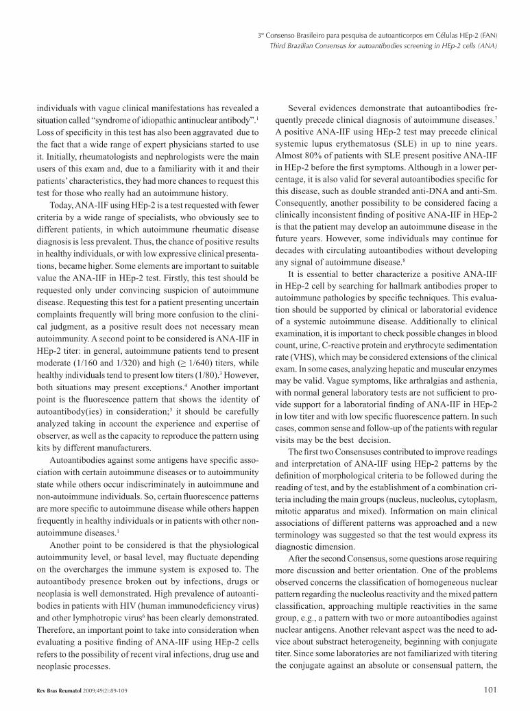
The first one is a nuclear fine speckled immunofluorescent pattern, with an almost homogeneous texture and with homo-geneously stained metaphase plate. Its clinical association and immunological identity are not defined. Its importance comes from the fact that it may be easily confused with a dense fine speckled pattern and a homogeneous pattern (Figure 1).
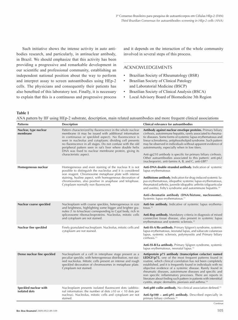
The second is a cytoplasmatic rod- and ring-shaped pat-tern which is apparently associated to HCv infection. There are ongoing studies to establish its immunological identity (Figure 2).
As they are not definitely characterized, these two new patterns have been considered preliminary and the 3rd Con-sensus recommended that both patterns have to be completely characterized and presented in the next meeting.
Genetically modified cell-based substracts
The 3rd Consensus did not discuss any systematic study with genetically modified cell substracts; this could be a possible topic for the next meetings.
qUALITy CONTROL
The Third Consensus promotes and recommends strategies for quality control. This recommendation aims at facing difficulties to assure test quality due to specialized trained professionals needs, heterogeneity of commercial kits and non-standardization of optical equipment at laboratories. Among institutional quality programs, the following programs were mentioned: College of American Pathologists (CAP) and edu-cational program for ANA-IIF in HEp-2 at Controlab. Other programs and reagents for quality control were also mentioned, such as PCQAUTO by GMK diagnósticos, Conexão HEp-2 by Hemagen diagnósticos and the blade FITC-QC® by ALKA Tecnologia em diagnósticos.
It was also recommended the titration of conjugate as a fundamental measure to adjust the amount of fluorochrome according to the lamp power, matching the different depart-ments. This procedure should also be performed for every new kit in a different lot. This titration maintenance in kits from the same lot may be performed using low intensity controls. In this item, it is useful to use commercial slides with pre-calibrated
Continue
Figure 1. (A) Nuclear fine speckled tending to be homogeneous. Cells present nucleoplasm with finely speckled texture with homogeneous trend and stained metaphase plate in the same texture. This pattern should not be confused with homogeneous nuclear pattern and dense fine speckled pattern, because there is no reactivity against native DNA and LEDGF/75Kda protein, respectively. In figures B and C may be observed Homogeneus nuclear pattern and dense nuclear fine speckled pattern, respectively.
C - Dense nuclear fine speckled
B - Homogeneous nuclear
A - Nuclear fine speckled tending to be homogeneous
EM0000 Rev Bras Reumat 49(2).indd 103 24/3/2009 22:46:49

Dellavance et al.
104 Rev Bras Reumatol 2009;49(2):89-109
microsphere for different fluorescence intensity and that may be used for training and internal calibration of reading.
In order to stimulate the achievement of authentic results, the 3rd Consensus recommends the laboratories to take part at quality control programs and systematically perform technical quality control. It is fundamental to remember that IIF reaction depends on five factors: Optical system (microscope), light power (20, 50 or 100W), conjugate concentration, control serum of minimum reactivity (1/80) and observer.
Conjugate concentration allows matching different optical systems, different power lights and observer’s reading. For example, if the light power is low, a more concentrate conjugate use is recommended in order to get the same fluorescence of a light with more power. In order to establish the ideal concen-tration of conjugate (conjugate’s titer), one should use titration technique in blocks where several dilutions of conjugate are crossed with several concentrations of a reference serum, searching the higher dilution of conjugate that is capable to reproduce nominal titer of reference serum.
This reference serum may be included with the kit and the laboratory will begin to be based on the optical, light and reader system according to patterns established by the manufacturer. This reference serum may be commercially acquired from quality control institutions or the laboratory will be able to send its serum to a laboratory of its reference and, then, with the results, start to consider this laboratory as reference. Once the first reference is done, the laboratory may store weekly aliquots of serum of titers established in this laboratory.
The laboratory should keep in its serum bank samples for controlling with minimum reactivity titer (1/80), to be diluted
Figure 2. Cytoplasmatic rod- and ring-shaped pattern Apparently associated with HCv infection and interferon treatment. Target antigens of these antibodies are under definition.
Cytoplasmatic in rods and rings
in 1/40, 1/80, 1/160 and 1/320. At each daily set, the labora-tory should process the low control and consider this set valid if titer variation is more or less one dilution. If any control inconformity is seen, for example, if a sample with average titer of 1/80 is negative, this set should be considered invalid. Using the minimum reactivity control (1/80), a set of tests will only be considered valid after reading one titer at more or less 80. In case of a decrease over 1 titer, the system is probably unstable. It is recommended to first verify if there was any problem in the aliquot stored; in this case, repeat the test with the aliquot stored the week before. If in the end of repetition, the titer found was the expected, we may conclude that the control aliquot used was deteriorated. If the new aliquot also presented a titer decrease of more than one dilution, we should check the optical system (UV filter deterioration, buffered glycerin in the objective lens and lamp performance). If no change is found in these components and in the number of hours of the lamp use, the most probable cause is conjugate degradation; if so, conjugate should go through a new titration process, as described before.
Notes
1. It is recommended to perform conjugate control every 15 days.
2. When a new kit is opened, a new titration of conjugate should be performed.
3. Microscope’s objective lens and filters interfere in the definition of conjugate titer and should be periodically checked.
4. Additionally to checking the time of the lamp, it is necessary to check if the lamp is in the center. It can be performed by placing a white paper sheet on the microscope table and using the objective lens 10 to see if the lamp is in the center. If you notice any dark part of the field, adjust it by using the following bottoms: left-right center, up and down center, and lamp focus.
CLINICAL ASSOCIATIONS AND PATTERN DESCRIPTION
Members of the 3rd Consensus proceeded to a wide discussion for validation and reevaluation of clinical and immunological associations about ANA-IIF patterns in HEp-2. Recommen-dations are listed in Table 1.
Most of the associations above had been accepted by general agreement by the Consensus members. In rare non-unanimous cases, the majority opinion prevailed.
EM0000 Rev Bras Reumat 49(2).indd 104 24/3/2009 22:46:49
3º Consenso Brasileiro para pesquisa de autoanticorpos em Células HEp-2 (FAN)Third Brazilian Consensus for autoantibodies screening in HEp-2 cells (ANA)
105Rev Bras Reumatol 2009;49(2):89-109
Continue
Table 1ANA pattern by IIF using HEp-2 substrate, description, main related autoantibodies and more frequent clinical associationsPatterns Description Clinical relevance for autoantibodies
Nuclear, type nuclear membrane
Pattern characterized by fluorescence in the whole nuclear membrane (it may be issued with additional information in continuous or speckled aspect). No fluorescence is seen in nucleolus and cytoplasm; dividing cell presents no fluorescence in all stages. Do not confuse with the old peripheral pattern seen in rat’s liver where double helix DNA was fixed to nuclear membrane protein, giving its characteristic aspect.
Antibody against nuclear envelope proteins. Primary biliary cirrhosis, autoimmune hepatitis, rarely associated to rheuma-tic diseases. Some forms of systemic lupus erythematosus and linear scleroderma, antiphosoholipid syndrome. Such pattern may be observed in individuals without apparent evidence of autoimmunity, especially when in low titers.
Anti-gp210 antibody is specific for primary biliary cirrhosis.Other autoantibodies associated to this pattern: anti-p62 (nucleoporin), anti-lamins A, B, and C, anti-LBP.9
Homogeneous nuclear Homogeneous and even staining of the nucleus It is not possible to distinguish the nucleolus and it is considered non reagent. Chromosome metaphase plate with intense staining, hyaline aspect, with homogeneous decoration of chromosomes, also positive in anaphase and telophase. Cytoplasm normally non fluorescent.
Anti-DNA double stranded antibody. Indication of systemic lupus erythematosus
Antihistone antibody. Indication for drug-induced systemic lu-pus erythematosus, idiopathic systemic lupus erythematosus, rheumatoid arthritis, juvenile idiopathic arthritis (oligoarticular and uveitis), Felty’s syndrome and autoimmune hepatitis.10
Anti-chromatin antibody (DNA/histone, nucleosome). Systemic lupus erythematosus.11
Nuclear coarse speckled Nucleoplasm with coarse speckles, heterogeneous in size and brightness, highlighting some bigger and brighter gra-nules (1 to 6/nucleus) corresponding to Cajal body, rich in spliceosome ribonucleoproteins. Nucleolus, mitotic cells and cytoplasm are not stained.
Anti-Sm antibody. Indicative of systemic lupus erythema-tosus.12
Anti-Rnp antibody. Mandatory criteria in diagnosis of mixed connective tissue disease, also present in systemic lupus erythematosus and systemic sclerosis.12
Nuclear fine speckled Finely granulated nucleoplasm. Nucleolus, mitotic cells and cytoplasm are not stained.
Anti-SS-A/Ro antibody. Primary Sjögren’s syndrome, systemic lupus erythematosus, neonatal lupus, and subacute cutaneous lupus, systemic sclerosis, polymyositis and Primary biliary cirrhosis.13
Anti-SS-B/La antibody. Primary Sjögren syndrome, systemic lupus erythematosus, neonatal lupus.13
Dense nuclear fine speckled Nucleoplasm of a cell in interphase stage present as a peculiar speckle, with heterogeneous distribution, not stai-ned nucleolus. Mitotic cells present an intense and rough speckled decoration of chromosomes in metaphase plate. Cytoplasm not stained.
Antiprotein p75 antibody (transcription cofactor) named LEDGF/p75. one of the most frequent patterns found in routine, which clinical correlation has not been completely established, and it is frequently found in individuals with no objective evidence of a systemic disease. Rarely found in rheumatic diseases, autoimmune diseases and specific and non specific inflammatory processes. There are reports in literature about finding such pattern in patients with interstitial cystitis, atopic dermatitis, psoriasis and asthma.14
Speckled nuclear with isolated dots
Nucleoplasm presents isolated fluorescent dots (additio-nal information: the number of dots ≥10 or < 10 dots per nucleus). Nucleolus, mitotic cells and cytoplasm are not stained.
Anti-p80 coilin antibody. No clinical association defined.15
Anti-Sp100 – anti-p95 antibody. Described especially in primary biliary cirrhosis.16
Such initiative shows the intense activity in auto anti-bodies research, and particularly, in antinuclear antibody, in Brazil. We should emphasize that this activity has been providing a progressive and remarkable development in our scientific and professional community, establishing an independent national position about the way to perform and interpret assay to screen autoantibodies using HEp-2 cells. The physicians and consequently their patients has also benefited of this laboratory test. Finally, it is necessary to explain that this is a continuous and progressive process
and it depends on the interaction of the whole community involved in several steps of this process.
ACkNOWLEDGEMENTS
• Brazilian Society of Rheumatology (BSR)• Brazilian Society of Clinical Patology
and Laboratorial Medicine (BSCP)• Brazilian Society of Clinical Analysis (BSCA)• Local Advisory Board of Biomedicine 3th Region
EM0000 Rev Bras Reumat 49(2).indd 105 24/3/2009 22:46:49

Dellavance et al.
106 Rev Bras Reumatol 2009;49(2):89-109
Nuclear speckled centromere 46 bright speckles spread over the nucleus of interphase cells. Nucleolus and cytoplasm not stained. Mitotic cells presents dots concentration in metaphase plate.
Anticentromere antibody (CENP-A, CENP-B and CENP-C proteins). CREST -systemic sclerosis (calcinosis, Raynaud’s phenomenon, esophageal dysmotility, sclerodactyly and te-langectasia), primary biliary cirrhosis, and Sjögren’s syndrome. Rarely observed in other autoimmune diseases. It may precede CREST for many years.17,18
Pleomorphic nuclear speckled Nucleoplasm is totally not stained in G1 cell interphase, becoming speckled with grains ranging from coarse, fine to dense fine, at the time the cell passes to phases S and G2. Nucleolus and cytoplasm not stained.
Proliferating cell nuclear antigen (anti-PCNA). Specifically found in patients with systemic lupus erythematosus.19
Homogenous nucleolar evenly bright fluorescence nucleolus, mitotic cells and cytoplasm not stained.
Antihistone anti-To/Th. Occur s in systemic sclerosis.20
Antinucleolin antibody. Very rare, describe in systemic lu-pus erythematosus, graft-versus-host disease and infectious mononucleosis.
Anti-B23 antibody (nucleophosmin). Described in systemic sclerosis, some types of cancer, antiphospholipid syndrome and graft-versus-host disease.
Clumpy nucleolar Nucleolus presents tight clusters of fluorescent granules (such as a bunch of grapes). Cytoplasm and nucleus not stained, mitotic cell is amorphous, with soft color around metaphase plate chromosomes.
Antifibrillarin antibody (U3-nRNP). Related to systemic sclerosis, especially with severe visceral damage, such as pulmonary hypertension.21
Speckled nucleolar
Fine and discrete speckles in the nucleolus; 5-10 different and brilliant dots along chromosomal metaphase plate. Cytoplasm and nucleus not stained.
Anti-NOR 90 antibody. Initially described in systemic scle-rosis. Today described in other autoimune diseases, however without clinical relevance defined.21
Polymerase I anti-RNA antibody. Diffuse systemic sclerosis with trend to visceral damage, more frequent and severe.21
Anti-ASE antibody (ERCC-1 antisense). Frequently found associated to anti-NOR-90 antibodies. The most frequent association seems to be systemic lupus erythematosus.
Linear cytoplasmatic fibrilar
Stressed fibers forming cytoskeleton decorated, crossing the whole extension of the cell, not respecting nuclear limits. Nucleus and nucleolus not stained.
Anti-actin antibody. Found in liver diseases: autoimmune hepatitis, cirrhosis.
Antimyosin antibody. Hepatitis C, hepatocarcinoma, myas-thenia gravis. When in low or moderate titers, may have no clinical relevance.22,23
Filamentar cytoplasmatic fibrillar
Decoration with filaments with unipolar or bipolar accentu-ation related to nuclear membrane. Nucleus and nucleolus not stained.
Anti-vimentin and anti-keratin antibody. Anti-keratin antibody is the most important antibody in alcohol-related hepatic disease. Described in several inflammatory and infectious diseases. When in low or moderate titers, may have no cli-nical relevance.24
Segmentary cytoplasmatic fibrilar
Only short stressed fiber segments are fluorescent. Nucleus and nucleolus not stained. In mitotic cells, we can eventu-ally observe multiple fluorescent grains corresponding to globular form of cytoplasm protein.
Anti alpha-actinin, anti-vinculin and anti-tropomyosin. Antibodies found in myasthenia gravis, Crohn’s disease and ulcerative colitis. When in low or moderate titers, may have no clinical relevance.24
Polar speckled cytoplasmatic
This is also a mandatory report, because it makes evident Golgi apparatus. Decoration is only cytoplasmatic in grou-ped dots in perinuclear situation on one pole of the nucleus. Nucleolus, mitotic cells and nucleus are not stained.
Anti-golgin antibody (Golgi apparatus cistern). Rare in sys-temic lupus erythematosus, primary Sjögren syndrome and other systemic autoimmune diseases. Reported in idiopathic cerebellar ataxia, paraneoplasic cerebellar degeneration and viral infection by Epstein Barr virus (EBV) and human immunodeficiency virus (HIV). When in low or moderate titers, may have no clinical relevance.25,26
Speckled cytoplasmatic with isolated dots
Defined dots with variable number for the whole extension of cytoplasm. Nucleolus, mitotic cells and nucleus are not stained.
Anti-EEA1 antibody and anti-phosphatidylserine. No clinical associations well defined.
Anti-GWB antibody. Related to primary Sjögren syndrome, al-though also observed in several other clinical conditions.27
Continue
EM0000 Rev Bras Reumat 49(2).indd 106 24/3/2009 22:46:50

3º Consenso Brasileiro para pesquisa de autoanticorpos em Células HEp-2 (FAN)Third Brazilian Consensus for autoantibodies screening in HEp-2 cells (ANA)
107Rev Bras Reumatol 2009;49(2):89-109
Fine dense speckled cytoplasmatic
Fine dot, dense and confluent fluorescence, almost homo-geneous. Nucleus is not stained, but may present a slight homogeneous decoration in nucleolus area. Mitotic cell is not stained. In case of concomitant fluorescence of cyto-plasm and nucleolus, pattern is classified as mixed.
Anti-PL7/PL12 antibody. This pattern may be rarely associated with antibodies found in polymyositis.20
Antiribosomal P protein antibody. This pattern is found in systemic lupus erythematosus.28
Fine speckled cytoplasmatic Fine speckles covering the whole cytoplasm. Mitotic cell and nucleolus not stained.
Histidil anti tRNA synthetase antibody (Jo1). Marker antibody of polymtositis in adults. Rarely described in dermatomyositis. Other anti tRNA synthetases antibodies may generate the same pattern.20
Reticular speckled cytoplasmatic
Large and irregular speckles concentrated in the perinu-clear region and extended in lower density to the rest of the cytoplasm. Nucleolus, mitotic cells and nucleus are not stained.
Antimitochondrial antibody. Primary biliary cirrhosis ma-rker. Rarely found in systemic sclerosis. Due to relatively common finding of a similar pattern and not related to antimitochondrial antibodies, specific test confirmation is fundamental.29,30
Mitotic apparatus type centriole
In interphase cells, fluorescence is confined to a single sphe-re close to the nucleus, dividing in two and migrating to the opposite pole of nucleus as the cell starts to split.
Anti-alpha-enolase antibody. In low titers, there is no clinical association. In high titers, it may be associated with systemic sclerosis.
Mitotic apparatus type intercellular bridge
Antigens forming the union between mother/daughter cells in the end of telophase. They may be observed with intense fluorescence in cytoplasmatic tip that will suffer cleavage in the end of cell division.
Anti-beta tubulin antibody. May be found in systemic lupus erythematosus and Mixed Connective Tissue Disease. Other not well defined antibodies may generate the same pattern. Associated with several auto immune conditions with low specificity, with clinical relevance only in high titers.22
Mitotic apparatus type mitotic spindle (NuMa-2)
Interphase cells showed no nuclear or cytoplasmic staining, but mitotic cells had brightly stained poles and spindles. At telophase, staining shifted to the midbody and the in-tercellular bridge.
Anti-HsEg5/NuMA-2 antibody. Associated with several auto immune conditions with low specificity, with clinical rele-vance only in high titers.36
Mixed, Nuclear fine speckled type with fluorescence in Mitotic apparatus
Cells in interphase present a stained nucleus with a very fine speckled, generally, in high titer. Miotic cells in metaphase and anaphase present well define and delicate location of pericentromeric region and proximal parts of mitotic spindle. In telophase, the staining shifted from the centrosomes to the reforming nuclei and no stain in intercellular bridge.
Anti-NuMa1 antibody. Associated to Sjögren’s Syndrome and may occur also in other auto immune or chronic in-flammatory conditions. When in low or moderate titers, may not be associated with the objective evidence of systemic inflammatory disease.32
Mixed, type homogeneous nucleolar and coarse speckled
Interphase cells present a stained nucleus with a coarse spe-ckled and homogeneously reddish nucleolus. In metaphase, there is stain around metaphase plate.
Anti-KU antibody. Marker of polymtositis and systemic scle-rosis overlap. May occur in systemic lupus erythematosus and scleroderma.33
Mixed, type fine nuclear and nucleolar fine speckled with stained metaphase plate
Interphase cells present a stained nucleus with a fine speckled form and nucleolus is also highlighted with fine speckled pattern. In metaphase, metaphase plate presents fine speckled pattern.
Anti-DNA Topoisomerase I antibody (Scl-70). Associated with systemic sclerosis, diffuse form, presenting with more visceral damage. More rarely in CREST syndrome and overlap.34
Mixed, type speckled nucleolar and nuclear fine speckled
Interphase cells present a stained nucleus with a fine speckled form and nucleolus is also highlighted with fine speckled pattern (individual dots). Cytoplasm is not stai-ned. In metaphase, 5 to 10 isolated bright dots are seen in metaphase plate, corresponding to nucleolus organizing regions (NOR).
Anti-RNA Polymerase I and II antibodies. These two autoanti-bodies usually appear together, and RNA pol. I is responsible for nucleolar distribution and in NOR, while RNA pol. II res-ponds for nuclear distribution. Anti-RNA pol. I is considered a systemic sclerosis marker and anti-RNA pol. II appears in several autoimmune conditions.34
Mixed, type fine dense speckled cytoplasmatic nucleolar to homogeneous nucleolar
Nucleus is totally not stained and nucleolus is poorly stained. Cytoplasm presents strong stain with very fine and very dense speckled, almost homogeneous. Mitotic cells are not stained.
Anti-rRNP antibody (anti-ribosomal P protein). systemic lupus erythematosus marker and more frequently related to lupus psychosis. Also seems to be related to the disease’s activity.28,35
EM0000 Rev Bras Reumat 49(2).indd 107 24/3/2009 22:46:50

Dellavance et al.
108 Rev Bras Reumatol 2009;49(2):89-109
REFERÊNCIASREFERENCES
1. dellavance A, Andrade LEC. Como interpretar e valorizar adequadamente o teste de anticorpos antinúcleo. J Bras Patol Med Lab 2007;43(3):157-68.
2. Forslid J, Heigl z, Jonsson J, Scheynius A. The prevalence of antinuclear antibodies in healthy young persons and adults, comparing rat liver tissue sections with HEp-2 cells as antigen substrate. Clin Exp Rheumatol 1994;12(2):137-41.
3. Tan EM, Feltkamp TE, Smolen JS, Butcher B, dawkins R, Fritzler MJ, et al. Range of antinuclear antibodies in "healthy" individuals. Arthritis Rheum1997;40(9):1601-11.
4. Leser PG, dellavance A, Barbosa SH, Guis G, Rodrigues SH, Sato EI. distinctive features of antinuclear antibodies observed in health and in subjects with autoimmune rheumatic disease. In: Conrad K, Bachmann MO, Chan EKL, Fritzler MJ, Humble RL, Sack U. editors. From animal models to human genetics: research on the induction and pathogenicity of autoantibodies. dresden: Pabst 2004. 493-510.
5. Fritzler MJ, Pauls Jd, Kinsella Td, Bowen TJ. Antinuclear, anticytoplasmic and anti-Sjögren’s syndrome antigen A (SS-A/Ro) antibodies in female blood donors. Clin Immunol Immunopathol 1985;36:120-8.
6. Massabki PS, Accetturi C, Nishie IA, da Silva NP, Sato EI, Andrade LE. Clinical implications of autoantibodies in HIv infection. Aids 1997;11(15):1845-50.
7. Arbuckle MR, McClain MT, Rubertone MV, Scofield RH, Dennis GJ, James JA, et al. development of autoantibodies before the clinical onset of systemic lupus erythematosus. N Engl J Med 2003;349(16):1526-33.
8. deane PMG, Liard G, Siegel dM, Baum J. The outcome of children referred to a pediatric rheumatology clinic with a positive antinuclear antibody test but without an autoimmune disease. Pediatrics 1995;95(6):892-5.
9. Borg AA, dawes PT, Mattey dL. Autoantibodies to nuclear lamins and to intermediate filament proteins. J Rheumatol 1993;20:1988-90.
10. Rubin RL. Autoimmune reactions induced by procainamide and hydralazine. In Kammuller M, Bloksma M, Siemen W (eds): Autoimmunity and Toxicology: Immune dysregulation Induced by drugs and Chemicals. Amsterdam, Elsevier 1988.
11. Amoura z, Koutouzov S, Chabre H, Cacoub P, Amoura I, Musset L, et al. Presence of antinucleosome autoantibodies in a restricted set of connective tissue diseases: antinucleosome antibodies of the IgG3 subclass are markers of renal pathogenicity in systemic lupus erythematosus. Arthritis Rheum 2000;43(1):76-84.
12. Notman DD, Kurata N, Tan EM. Profiles of antinuclear antibodies in systemic rheumatic diseases. Ann Intern Med 1975;83(4):464-9.
13. Barcellos KS, Nonogaki S, Enokihara MM, Teixeira MS, Andrade LE. differential expression of Ro/SSA 60 kda and La/SSB, but not Ro/SSA 52 kda, mRNA and protein in minor salivary glands from patients with primary Sjögren's syndrome. J Rheumatol 2007;34(6):1283-92.
14. Ochs RL, Muro Y, Si Y, Ge H, Chan EK, Tan EM. Autoantibodies to dFS 70 kd/transcription coactivator p75 in atopic dermatitis and other conditions. J Allergy Clin Immunol 2000;105(6):1211-20.
15. Andrade LEC, Chan EKL, Raska I, Peebles CL, Roos G, Tan EM. Human autoantibody to a novel protein of the nuclear coiled body: immunological characterization and cdNA cloning of p80-coilin. J Exp Med 1991;173:1407-19.
16. Lohse AW, zum Büschenfelde KH, Franz B, Kanzler S, Gerken G, dienes HP. Characterization of the overlap syndrome of Primary Biliary Cirrhosis (PBC) and Autoimmune Hepatitis: evidence for it being a hepatitic form of PBC in genetically susceptible individuals. Hepatology 1999;29(4):1078-84.
17. Fritzler MJ, Kinsella Td. The CREST syndrome: a distinct serologic entity with anticentromere antibodies. Am J Med 1980;69(4):520-6.
18. Göring Hd, Panzner M, Lakota W, zeiner A. Association of Scleroderma and Primary Biliary Cirrhosis - results of a systematic study on a dermatological clientele. Hautarzt 1998;49:361-66.
19. Takasaki Y, deng JS, Tan EM. A nuclear antigen associated with cell proliferation and blast transformation. J Exp Med 1981;154:1899-909.
20. Targoff IN. Autoantibodies in polymyositis. Rheum dis Clin North Am 1992;18:455-82.
21. Reimer G, Raska I, Tan EM, Scheer U. Human autoantibodies: probes for nucleolus structure and function. virchows Arch B Cell Pathol Incl Mol Pathol 1987;54:131-43.
22. Agarwal N, Handa R, Acharya SK, Wali JP, dinda AK, Aggarwal P. A study of autoimmune markers in hepatitis C infection. Indian J Med Res 2001;113:170-4.
23. Leibovitch L, George J, Levi Y, Bakimer R, Shoenfeld Y. Anti-actin antibodies in sera from patients with autoimmune liver diseases and patients with carcinomas by ELISA. Immunol Lett 1995;48(2):129-32.
24. Krapf AR, von Mühlen CA, Krapf FE, Nakamura RM, Tan EM. Atlas of immunofluorescent autoantibodies. Munich, Urban & Schwarzenberg, 1996.
25. Massabki PS, Accetturi C, Nishie IA, Silva NP, Sato EI, Andrade LE: Clinical implications of autoantibodies in HIv infection. AIdS (archive) 11:1845-50, 1997.
26. Yang Y, Fujita J, Tokuda M, Bandoh S, Ishida T. Clinical features of several connective tissue diseases with anti-Golgi antibody. Ann Rheum dis 2001;60:986-987.
27. Eystathioy T, Chan EK, Tenenbaum SA, Keene JD, Griffith K, Fritzler MJ. A phosphorylated cytoplasmic autoantigen, GW182, associates with a unique population of human mRNAs within novel cytoplasmic speckles. Mol Biol Cell 2002;13:1338-51.
28. Bonfa E, Golombek SJ, Kaufman Ld, Skelly S, Weissbach H, Brot N, et al. Association between lupus psychosis and anti-ribosomal P protein antibodies. N Engl J Med 1987;317(5):265-71.
29. Alderuccio F, Toh BH, Barnett AJ, Pedersen JS. Identification and characterization of mitochondria autoantigens in progressive systemic sclerosis: identily with the 72,000 dalton autoantigen in primary biliary cirrhosis. J Immunol 1986;137(6):1855-9.
30. Chou MJ, LaI MY, Lee SL. Reactivity of anti-mitochondrial antibodies in primary biliary cirrhosis and systemic sclerosis. J From Med Ass 1992;91(11):1075-80.
EM0000 Rev Bras Reumat 49(2).indd 108 24/3/2009 22:46:50

3º Consenso Brasileiro para pesquisa de autoanticorpos em Células HEp-2 (FAN)Third Brazilian Consensus for autoantibodies screening in HEp-2 cells (ANA)
109Rev Bras Reumatol 2009;49(2):89-109
31. Krapf A, von Mühlen CA, Krapf F, Nakamura RMM, Tan EM. Human autoimmune diseases and autoantibodies. Bol Com Íbero-Americano Reumatol 1999;9:55-8.
32. Andrade LEC, Chan EKL, Peebles CL, Tan EM. Two major autoantigen-antibody systems of the mitotic spindle apparatus. Arthritis Rheum 1996;39:1643-53.
33. Francoeur AM, Peebles CL, Gompper PT, Tan EM. Identification of Ki (Ku, p70/p80) autoantigens and analysis of anti-Ki autoantibody reactivity. J Immunol 1986;136(5):1648-53.
34. Jarzabek-Chorzelska M, Balszczyk M, Jablonska S, Chorzelski T, Kumar V, Beutner EH. Scl -70 antibody, a specific marker of systemic sclerosis. Br J dermatol 1986;115(4):393-401.
35. Isshi K, Hirohata S. differential roles of the anti-ribosomal P antibody and antineuronal antibody in the pathogenesis of central nervous system involvement in systemic lupus erythematosus. Arthritis Rheum 1998;41(10):1819-27.
38. Carballo OG, von Mühlen CA, Nakamura R, de la Torre IG, Francescatonio PLC. Atlas Anti-Nucleocitoplasmaticos (HEp-2). Talleres Gráficos TBS, Buenos Aires, 2006.
36. von Mühlen CA, Nakamura RM. Clinical and laboratory evaluation of systemic rheumatic diseases. In McPherson & Pincus (eds): Clinical diagnosis and management by laboratory methods. Philadelphia, W.B. Saunders, 2006.
37. dellavance A, Gabriel Jr A, Nuccitelli B, Taliberti BH, von Mühlen CA, Bichara CdA, et al. Pesquisa de auto-anticorpos em células HEp-2. Editora UCG, Goiânia, 2008.
EM0000 Rev Bras Reumat 49(2).indd 109 24/3/2009 22:46:50