
TheUseofFractalDimensionAnalysisin ...downloads.hindawi.com/journals/cmmm/2012/793291.pdf ·...
7
Hindawi Publishing Corporation Computational and Mathematical Methods in Medicine Volume 2012, Article ID 793291, 6 pages doi:10.1155/2012/793291 Research Article The Use of Fractal Dimension Analysis in Estimation of Blood Vessels Shape in Transplantable Mammary Adenocarcinoma in Wistar Rats after Photodynamic Therapy Combined with Cysteine Protease Inhibitors Kamil Jurczyszyn, 1 Beata J. Osiecka, 2 and Piotr Zi ´ olkowski 2 1 Department of Dental Surgery, Wroclaw Medical University, Krakowska 26, 50-425 Wroclaw, Poland 2 Department of Pathomorphology, Wroclaw Medical University, Marcinkowskiego 1, 50-368 Wroclaw, Poland Correspondence should be addressed to Kamil Jurczyszyn, [email protected] Received 26 June 2012; Accepted 1 August 2012 Academic Editor: Facundo Ballester Copyright © 2012 Kamil Jurczyszyn et al. This is an open access article distributed under the Creative Commons Attribution License, which permits unrestricted use, distribution, and reproduction in any medium, provided the original work is properly cited. Fractal dimension analysis (FDA) is modern mathematical method widely used to describing of complex and chaotic shapes when classic methods fail. The main aim of this study was evaluating the influence of photodynamic therapy (PDT) with cystein proteases inhibitors (CPI) on the number and morphology of blood vessels inside tumor and on increase of effectiveness of combined therapy in contrast to PDT and CPI used separately. Animals were divided into four groups: control, treated using only PDT, treated using only CPI and treated using combined therapy, PDT and CPI. Results showed that time of animal survival and depth of necrosis inside tumor were significantly higher in CPI+PDT group in contrast to other groups. The higher value of fractal dimension (FD) was observed in control group, while the lowest value was found in the group which was treated by cystein protease inhibitors. The differences between FD were observed in CPI group and PDT+CPI group in comparison to control group. Our results revealed that fractal dimension analysis is a very useful tool in estimating differences between irregular shapes like blood vessels in PDT treated tumors. Thus, the implementation of FDA algorithms could be useful method in evaluating the efficacy of PDT. 1. Introduction One of the important factors responsible for the tumor growth is blood supply. Many studies revealed that the anatomic and spatial structure of tumor blood vessels is more chaotic than that of the normal tissues. One of the classic ways of analyzing the blood vessels is direct count in the light microscope. Immunohistochemical staining is very useful against specific proteins within blood vessel wall, for example vascular endothelial growth factor (VEGF), basic fibroblast growth factor (bFGF), cluster of differentiation such as CD34, and others [1, 2]. Many studies show that PDT is responsible for overexpression of VEGF in vivo and in vitro [3–5]. However, in the case of such complex and chaotic shapes of tumor blood vessels classic methods of evaluation may fail. Fractal dimension analysis may be very helpful in these cases. Fractal is a shape which is described by potentially simple mathematic formulas. If these formulas are iterated into infinity they may create shapes which we are able of magnifying without the end and each time we can see infinity numbers of shape details—it is a self similarity feature. Classic examples of fractals are Cantor’s set, Koch’s snowflake, and Sierpinski triangle (see Figure 1). Many natural shapes are very similar to fractals, for example, coastline, trees, clouds, bronchial tree, and blood vessels network. Any above-mentioned structures poses main feature of fractal—self-similarity. Ideal tool to describe a fractal is the fractal dimension (FD). Fractal dimension is a shape index and particularly it can be defined as a measure of irregularly shaped objects. Irregularity in shape and behavior is shared by all of the biological, including anatomical, systems. In Euclidian geometry a number of dimension is
Transcript of TheUseofFractalDimensionAnalysisin ...downloads.hindawi.com/journals/cmmm/2012/793291.pdf ·...

Hindawi Publishing CorporationComputational and Mathematical Methods in MedicineVolume 2012, Article ID 793291, 6 pagesdoi:10.1155/2012/793291
Research Article
The Use of Fractal Dimension Analysis inEstimation of Blood Vessels Shape in Transplantable MammaryAdenocarcinoma in Wistar Rats after Photodynamic TherapyCombined with Cysteine Protease Inhibitors
Kamil Jurczyszyn,1 Beata J. Osiecka,2 and Piotr Ziołkowski2
1 Department of Dental Surgery, Wroclaw Medical University, Krakowska 26, 50-425 Wroclaw, Poland2 Department of Pathomorphology, Wroclaw Medical University, Marcinkowskiego 1, 50-368 Wrocław, Poland
Correspondence should be addressed to Kamil Jurczyszyn, [email protected]
Received 26 June 2012; Accepted 1 August 2012
Academic Editor: Facundo Ballester
Copyright © 2012 Kamil Jurczyszyn et al. This is an open access article distributed under the Creative Commons AttributionLicense, which permits unrestricted use, distribution, and reproduction in any medium, provided the original work is properlycited.
Fractal dimension analysis (FDA) is modern mathematical method widely used to describing of complex and chaotic shapes whenclassic methods fail. The main aim of this study was evaluating the influence of photodynamic therapy (PDT) with cystein proteasesinhibitors (CPI) on the number and morphology of blood vessels inside tumor and on increase of effectiveness of combinedtherapy in contrast to PDT and CPI used separately. Animals were divided into four groups: control, treated using only PDT,treated using only CPI and treated using combined therapy, PDT and CPI. Results showed that time of animal survival anddepth of necrosis inside tumor were significantly higher in CPI+PDT group in contrast to other groups. The higher value of fractaldimension (FD) was observed in control group, while the lowest value was found in the group which was treated by cystein proteaseinhibitors. The differences between FD were observed in CPI group and PDT+CPI group in comparison to control group. Ourresults revealed that fractal dimension analysis is a very useful tool in estimating differences between irregular shapes like bloodvessels in PDT treated tumors. Thus, the implementation of FDA algorithms could be useful method in evaluating the efficacy ofPDT.
1. Introduction
One of the important factors responsible for the tumorgrowth is blood supply. Many studies revealed that theanatomic and spatial structure of tumor blood vessels ismore chaotic than that of the normal tissues. One of theclassic ways of analyzing the blood vessels is direct count inthe light microscope. Immunohistochemical staining is veryuseful against specific proteins within blood vessel wall, forexample vascular endothelial growth factor (VEGF), basicfibroblast growth factor (bFGF), cluster of differentiationsuch as CD34, and others [1, 2]. Many studies show thatPDT is responsible for overexpression of VEGF in vivo andin vitro [3–5]. However, in the case of such complex andchaotic shapes of tumor blood vessels classic methods ofevaluation may fail. Fractal dimension analysis may be very
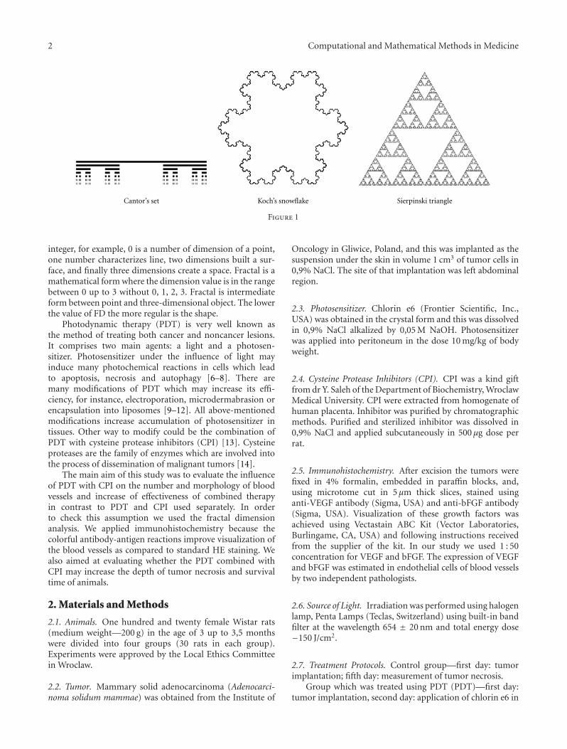
helpful in these cases. Fractal is a shape which is describedby potentially simple mathematic formulas. If these formulasare iterated into infinity they may create shapes which we areable of magnifying without the end and each time we cansee infinity numbers of shape details—it is a self similarityfeature. Classic examples of fractals are Cantor’s set, Koch’ssnowflake, and Sierpinski triangle (see Figure 1).
Many natural shapes are very similar to fractals, forexample, coastline, trees, clouds, bronchial tree, and bloodvessels network. Any above-mentioned structures posesmain feature of fractal—self-similarity. Ideal tool to describea fractal is the fractal dimension (FD). Fractal dimension is ashape index and particularly it can be defined as a measure ofirregularly shaped objects. Irregularity in shape and behavioris shared by all of the biological, including anatomical,systems. In Euclidian geometry a number of dimension is
2 Computational and Mathematical Methods in Medicine
Cantor’s set Koch’s snowflake Sierpinski triangle
Figure 1
integer, for example, 0 is a number of dimension of a point,one number characterizes line, two dimensions built a sur-face, and finally three dimensions create a space. Fractal is amathematical form where the dimension value is in the rangebetween 0 up to 3 without 0, 1, 2, 3. Fractal is intermediateform between point and three-dimensional object. The lowerthe value of FD the more regular is the shape.
Photodynamic therapy (PDT) is very well known asthe method of treating both cancer and noncancer lesions.It comprises two main agents: a light and a photosen-sitizer. Photosensitizer under the influence of light mayinduce many photochemical reactions in cells which leadto apoptosis, necrosis and autophagy [6–8]. There aremany modifications of PDT which may increase its effi-ciency, for instance, electroporation, microdermabrasion orencapsulation into liposomes [9–12]. All above-mentionedmodifications increase accumulation of photosensitizer intissues. Other way to modify could be the combination ofPDT with cysteine protease inhibitors (CPI) [13]. Cysteineproteases are the family of enzymes which are involved intothe process of dissemination of malignant tumors [14].
The main aim of this study was to evaluate the influenceof PDT with CPI on the number and morphology of bloodvessels and increase of effectiveness of combined therapyin contrast to PDT and CPI used separately. In orderto check this assumption we used the fractal dimensionanalysis. We applied immunohistochemistry because thecolorful antibody-antigen reactions improve visualization ofthe blood vessels as compared to standard HE staining. Wealso aimed at evaluating whether the PDT combined withCPI may increase the depth of tumor necrosis and survivaltime of animals.
2. Materials and Methods
2.1. Animals. One hundred and twenty female Wistar rats(medium weight—200 g) in the age of 3 up to 3,5 monthswere divided into four groups (30 rats in each group).Experiments were approved by the Local Ethics Committeein Wroclaw.
2.2. Tumor. Mammary solid adenocarcinoma (Adenocarci-noma solidum mammae) was obtained from the Institute of
Oncology in Gliwice, Poland, and this was implanted as thesuspension under the skin in volume 1 cm3 of tumor cells in0,9% NaCl. The site of that implantation was left abdominalregion.
2.3. Photosensitizer. Chlorin e6 (Frontier Scientific, Inc.,USA) was obtained in the crystal form and this was dissolvedin 0,9% NaCl alkalized by 0,05 M NaOH. Photosensitizerwas applied into peritoneum in the dose 10 mg/kg of bodyweight.
2.4. Cysteine Protease Inhibitors (CPI). CPI was a kind giftfrom dr Y. Saleh of the Department of Biochemistry, WroclawMedical University. CPI were extracted from homogenate ofhuman placenta. Inhibitor was purified by chromatographicmethods. Purified and sterilized inhibitor was dissolved in0,9% NaCl and applied subcutaneously in 500 μg dose perrat.
2.5. Immunohistochemistry. After excision the tumors werefixed in 4% formalin, embedded in paraffin blocks, and,using microtome cut in 5 μm thick slices, stained usinganti-VEGF antibody (Sigma, USA) and anti-bFGF antibody(Sigma, USA). Visualization of these growth factors wasachieved using Vectastain ABC Kit (Vector Laboratories,Burlingame, CA, USA) and following instructions receivedfrom the supplier of the kit. In our study we used 1 : 50concentration for VEGF and bFGF. The expression of VEGFand bFGF was estimated in endothelial cells of blood vesselsby two independent pathologists.
2.6. Source of Light. Irradiation was performed using halogenlamp, Penta Lamps (Teclas, Switzerland) using built-in bandfilter at the wavelength 654 ± 20 nm and total energy dose−150 J/cm2.
2.7. Treatment Protocols. Control group—first day: tumorimplantation; fifth day: measurement of tumor necrosis.
Group which was treated using PDT (PDT)—first day:tumor implantation, second day: application of chlorin e6 in

Computational and Mathematical Methods in Medicine 3
the dose of 10 mg/kg of body weight, third day: irradiation,fifth day: measurement of tumor necrosis.
Group which was treated using CPI (CPI)—first day:tumor implantation, second day: application of cysteinprotease inhibitors in 500 μg dose per animal, and fifth day:measurement of tumor necrosis.
Group which was treated using PDT and CPI together(PDT+CPI)—first day: tumor implantation, second day:application of cystein protease inhibitors in 500 μg dose peranimal, third day: application of chlorin e6 in the dose of10 mg/kg of body weight, fourth day: irradiation, and fifthday: measurement of tumor necrosis.
On the fifth day, each group was divided into two equalsubgroups. In the first subgroup animals were sacrificedusing the lethal dose of bioketan. During the autopsy thesamples from the tumor, lungs, liver, and kidneys were takento microscopic examination.
Animals from the second subgroup were kept in order toestimate the time of survival. In case of death of any animalthe autopsy was performed using the same protocol as in thefirst subgroup.
2.8. Measurement of Necrosis Depth. Microscopic slides werestained using standard hematoxylin-eosin method and nextthey were evaluated in light microscope (Olympus BX40) in200x magnification. Depth of necrosis was measured usingan eye-piece graticule.
2.9. Evaluation of the Number of Blood Vessels. Number ofblood vessels was counted on the microscopic glass slidesstained using anti-bFGF and anti-VEGF antibodies. Wechose randomly slides with 10 tumors (from 10 animals)from each group. We counted the number of blood vesselsin five fields of view at the magnification 400x and next wecalculated the average number from these five fields.
2.10. Statistical Analysis. Every statistical analysis was per-formed using Statistica version 6.0 (StatSoft Inc.). Hypothe-ses were verified on 0,05 significance level. Due to lack ofnormal distribution we used nonparametric Kolmogorov-Smirnov test.
2.11. Fractal Dimension Analysis (FDA). We used computerprogram Fractalyse version 2.4 (http://www.fractalyse.org/).Any graphic operation was performed in GIMP version 2.4.7.Fractalyse software enables measuring fractal dimensionusing box-counting method. Fractal dimension (DS) iscounted using following formula [15]:
DS = limε→ 0
logN(ε)log(1/ε)
, (1)
whereDS—fractal dimension, ε—length of box which createsmesh covering surface with examining pattern, N(ε)—minimal number of boxes which are required to coverexamining pattern.
Microscopic slides which were stained using anti-VEGFor anti-bFGF antibodies were examined using 100x magni-fication and the micrographs of blood vessels were taken in
65
60
55
50
45
40
35
30
25
20
15Controlgroup
PDT CPI
Method
Tim
e of
su
rviv
al (
days
)
Average
±Standard error
±SD
CPI+PDT
Figure 2: Influence of various treatment methods on time ofsurvival (SD—standard deviation).
1280 × 1024 points resolution. Next, the micrographs wereconverted into the binary white and black format. We appliedLaplace filter using matrix shown below:
∣∣∣∣∣∣∣
−1 −1 −1−1 8 −1−1 −1 −1
∣∣∣∣∣∣∣
. (2)
It was necessary to use the Laplace filter because this filteris able to generate very precise and thin outlines of bloodvessels [16]. After all these procedures the micrographsof blood vessels were entered to Fractalyse program forestimating the fractal dimension.
3. Results
3.1. Influence of Various Treatment Methods on Time ofSurvival. Figure 2 shows the average time of survival ineach group. Average survival time in control group was 24days. The longest survival time was observed in the groupwhich was treated by PDT and CPI together, that is, 53days. The groups of animals which were treated using onlyone method (PDT or CPI separately) were characterized bysimilar survival time, that is, 43 days. The average survivaltime in every group was statistically different in comparisonto control group.
Table 1 shows percentage of completely cured (completeresponses) animals in each group. The animals were consid-ered as completely cured when the survival time was 65 daysor longer and no palpable tumor was found. The highestpercentage of cure rate was observed in PDT+CPI group,that is, 33,33%. In the PDT group it reached 13,33%, in CPIgroup that percentage was only 6,67%. In control group noneof animal survived 65 days or more.

4 Computational and Mathematical Methods in Medicine
Table 1: Percentage of complete responses in animals treated withdifferent methods and in the control group.
Control group PDT CPI CPI+PDT
Percentage ofcomplete responses
0,00% 13,33% 6,67% 33,33%
Controlgroup
PDT CPI
Method
Average
±Standard error
±SD
20
18
16
14
12
10
8
6
4
2
Nec
rosi
s de
pth
(m
m)
CPI+PDT
Figure 3: Influence of various treatment methods on necrosis depth(SD—standard deviation).
3.2. Influence of Various Treatment Methods on NecrosisDepth. Figure 3 shows the average depth of necrosis withinthe tumor. The lowest value was observed in control group(5 mm). Separate application of PDT and CPI causedincrease of necrosis depth (for PDT 9,20 mm and 9,80 mmfor CPI). These values were significantly different in compar-ison to the control group. There was no statistical differencebetween the groups in which PDT and CPI were appliedseparately. The highest value (15,87 mm) of necrosis depthwas noted in the group which was treated by PDT and CPItogether.
3.3. Influence of Various Treatment Methods on the Value ofVessels’ Fractal Dimension. Figure 4 shows value of FD ofblood vessels. The higher value was noted in control group,that is, 1,135 while the lowest value was observed in the groupwhich was treated by cystein protease inhibitors (0,933). Thedifferences between FD in comparison to control group wereobserved in CPI group and PDT+CPI group (0,982). Thefractal dimension in PDT group was 1,037.
Table 2 shows the average number of vessels versus fractaldimension analysis. The lowest number of vessels was seenin control group (10 for VEGF and 8 for bFGF). Highernumbers of vessels were observed in the PDT group (17 forVEGF and 15 for bFGF) and in group PDT+CPI (18 forVEGF and 12 for bFGF).
Average
±Standard error
±SD
1.2
1.15
1.1
1.05
1
0.95
0.9
0.85
0.8
Val
ue
of fr
acta
l dim
ensi
on
Controlgroup
PDT CPI CPI+PDT
Figure 4: Influence of various treatment methods on the value ofvessels’ fractal dimension (SD—standard deviation).
Table 2: The average number of blood vessels versus fractaldimension analysis in groups of animals treated with differentmethods and in the control group.
Average number of vessels
Control group PDT CPI PDT+CPI
VEGF 10 17 11 18
bFGF 8 15 8 12
Fractal dimension 1,135 1,037 0,933 0,982
4. Discussion
Our results revealed that photodynamic therapy combinedwith cystein protease inhibitors causes increase of effective-ness of treatment in comparison to other methods. Highereffectiveness of PDT with CPI results in longer survival timeof animals and greater depth of necrosis in the tumors.The attempts of combining the photodynamic therapy withcystein protease inhibitors seem to be reasonable and thepositive synergistic effects were observed in our previousexperiments [13].
The whole mechanism of photodynamic therapy isnot clear. Next to destruction of cells due to phototoxicreactions, PDT influences blood vessels. Blood vessels insidethe tumor are very important target on the way to tumordestruction. Many studies revealed that the time betweenapplication of photosensitizer and irradiation plays a key rolein that damage. In case of rat chondrosarcoma, significantchanges within vessels were achieved in the group which wasirradiated 5 and 30 minutes after intravenous application ofbenzoporphyrin derivative. In a group which was irradiatedafter 180 minutes pathological lesions within vessels wereless intensive [17]. Blood vessel network is characterizedby highly complex spatial arrangement. Euclidean geometrybecause of its applicability to linear and regular forms,should not be applied to biological objects that one of

Computational and Mathematical Methods in Medicine 5
their most important feature is the irregularity in shape.Vascular network may be roughly considered as fractal.Most of the natural shapes are similar to fractals. Trees,clouds and neuronal network are good examples. Fractaldimension is a very useful parameter in describing suchshapes. Many studies revealed significant differences in valueof FD between normal and tumor tissue or low grade versushigh grade tumors. The example of such dependence is thedifference between vascular average FD of low-grade renalcarcinoma (Ds = 1, 55) and highgrade (Ds = 1, 45) [18].In case of renal carcinoma characterized by higher intensityof necrosis Ds = 1, 38 versus Ds = 1, 52 in tumors withoutnecrosis. In case of liver tumors an average value of FD is1,62 in comparison to surrounding tissue in which Ds = 1, 47[18]. The results showed above indicate that the tumor vesselnetwork is more chaotic than in surrounding tissue.
Our results showed that the highest value of FD wasfound in control group, which may suggest that the networkof blood vessels is here more complex than in other groups.Application of PDT, CPI, and both methods together leadsto decrease of fractal dimension. The lowest value of FDwas observed in PDT+CPI group, thus showing that thecombined therapy results in less chaotic arrangement ofthe blood vessels. The comparison of FD with quantity ofblood vessels revealed that the shape of blood vessels waschanged into more regular but amount of vessels was similar.The similar conclusions appeared after the comparison ofnumber of vessels in PDT and PDT+CPI group. In bothmentioned groups the number of vessels was increased andaccompanied by the reduction of FD value. The generalincrease of blood vessel number in PDT+CPI group maysuggest intensification of angiogenesis. Angiogenesis maybe a result of the feedback as a response to destruction ofblood vessels during PDT and to the increase in secretion ofVEGF and bFGF [3–5]. The increase of vessel number nextto reduction of fractal dimension proves that diameter ofvessels decreases (the lower fractal dimension—near to 0—the shape more similar to point which may be interpreted asa reduction of diameter).
Fractal dimension analysis of blood vessels may bea very efficient tool which enables comparison of shapeand distribution of vessels on the surface. FD is widelyused analysis of shapes in various visualization techniquesfor instance X-rays photos, ultrasonography and computedtomography [19–21]. Analysis of fractal dimension requiresprocedures which enable separation of examined shape andbackground. In case of microscopic slides stained using theroutine hematoxylin-eosin method the dynamics of colors ispoor which may make difficulties in automatic separation ofexamined shapes. Computer systems require strong contrastimages (very useful is the staining against CD34 in case ofanalysis of blood vessels) and special algorithms to search outexamined shapes are also needed [2].
5. Conclusions
Our results revealed that fractal dimension analysis is avery useful tool in estimating differences between irregular
shapes like blood vessels in PDT-treated tumors. Thus, theimplementation of FDA algorithms could be useful methodin evaluating the efficacy of PDT of tumors.
References
[1] P. Birner, A. Obermair, M. Schindl, H. Kowalski, G. Breite-necker, and G. Oberhuber, “Selective immunohistochemicalstaining of blood and lymphatic vessels reveals independentprognostic influence of blood and lymphatic vessel invasionin early-stage cervical cancer,” Clinical Cancer Research, vol. 7,no. 1, pp. 93–97, 2001.
[2] A. di Leva, F. Grizzi, G. Ceva-Grimaldi et al., “Fractaldimension as a quantitator of the microvasculature of normaland adenomatous pituitary tissue,” Journal of Anatomy, vol.211, no. 5, pp. 673–680, 2007.
[3] D. Dabkeviciene, A. Sasnauskiene, E. Leman et al., “MTHPC-mediated photodynamic treatment up-regulates the cytokinesVEGF and IL-1alpha,” Photochemistry and Photobiology, vol.88, no. 2, pp. 432–439, 2012.
[4] H. Nakagawa, T. Matsumiya, H. Sakaki et al., “Expression ofvascular endothelial growth factor by photodynamic therapywith mono-l-aspartyl chlorin e6 (NPe6) in oral squamous cellcarcinoma,” Oral Oncology, vol. 43, no. 6, pp. 544–550, 2007.
[5] J. Yang, A. C. Chen, Q. Wu et al., “The influence oftemperature on 5-aminolevulinic acid-based photodynamicreaction in keratinocytes in vitro,” Photodermatology, Photoim-munology & Photomedicine, vol. 26, no. 2, pp. 83–88, 2010.
[6] C. M. N. Yow, C. K. Wong, Z. Huang, and R. J. Ho,“Study of the efficacy and mechanism of ALA-mediatedphotodynamic therapy on human hepatocellular carcinomacell,” Liver International, vol. 27, no. 2, pp. 201–208, 2007.
[7] A. Radestock, P. Elsner, B. Gitter, and U. C. Hipler, “Inductionof apoptosis in HaCaT cells by photodynamic therapy withchlorin e6 or pheophorbide a,” Skin Pharmacology andPhysiology, vol. 20, no. 1, pp. 3–9, 2006.
[8] P. Harrod-Kim, “Tumor ablation with photodynamic therapy:introduction to mechanism and clinical applications,” Journalof Vascular and Interventional Radiology, vol. 17, no. 9, pp.1441–1448, 2006.
[9] J. Saczko, M. Nowak, N. Skolucka, J. Kulbacka, and M.Kotulska, “The effects of the electro-photodynamic in vitrotreatment on human lung adenocarcinoma cells,” Bioelectro-chemistry, vol. 79, no. 1, pp. 90–94, 2010.
[10] M. J. P. Gerritsen, T. Smits, M. M. Kleinpenning, P. C. M. VanDe Kerkhof, and P. E. J. Van Erp, “Pretreatment to enhanceprotoporphyrin IX accumulation in photodynamic therapy,”Dermatology, vol. 218, no. 3, pp. 193–202, 2009.
[11] B. Osiecka, K. Jurczyszyn, K. Symonowicz et al., “In vitro andin vivo matrix metalloproteinase expression after photody-namic therapy with a liposomal formulation of aminolevulinicacid and its methyl ester,” Cellular and Molecular BiologyLetters, vol. 15, no. 4, pp. 630–650, 2010.
[12] M. S. T. Rocha, C. M. Lucci, J. P. F. Longo et al., “Aluminum-chloride-phthalocyanine encapsulated in liposomes: activityagainst naturally occurring dog breast cancer cells,” Journal ofBiomedical Nanotechnology, vol. 8, no. 2, pp. 251–257, 2012.
[13] Y. Saleh, P. Ziolkowski, M. Siewinski, J. Milach, P. Marszalik,and J. Rybka, “The combined treatment of transplantable solidmammary carcinoma in wistar rats by use of photodynamictherapy and cysteine proteinase inhibitor,” In Vivo, vol. 15, no.4, pp. 351–357, 2001.

6 Computational and Mathematical Methods in Medicine
[14] K. Sheahan, S. Shuja, and M. J. Murnane, “Cysteine proteaseactivities and tumor development in human colorectal carci-noma,” Cancer Research, vol. 49, no. 14, pp. 3809–3814, 1989.
[15] F. Grizzi, C. Russo, P. Colombo et al., “Quantitative evalua-tion and modeling of two-dimensional neovascular networkcomplexity: the surface fractal dimension,” BMC Cancer, vol.5, article 14, 2005.
[16] H. Ahammer and T. T. J. DeVaney, “The influence ofedge detection algorithms on the estimation of the fractaldimension of binary digital images,” Chaos, vol. 14, no. 1, pp.183–188, 2004.
[17] V. H. Fingar, P. K. Kik, P. S. Haydon et al., “Analysis ofacute vascular damage after photodynamic therapy usingbenzoporphyrin derivative (BPD),” British Journal of Cancer,vol. 79, no. 11-12, pp. 1702–1708, 1999.
[18] F. Grizzi, B. Franceschini, B. Fiamengo, C. Russo, and N. Dio-guardi, “Vascular architecture: is it a helpful histopathologicalbiomarker for hepatocellular carcinoma?” Journal of ZhejiangUniversity. Science. B, vol. 8, no. 4, pp. 217–220, 2007.
[19] M. E. Mavroforakis, H. V. Georgiou, N. Dimitropoulos,D. Cavouras, and S. Theodoridis, “Mammographic massescharacterization based on localized texture and dataset fractalanalysis using linear, neural and support vector machineclassifiers,” Artificial Intelligence in Medicine, vol. 37, no. 2, pp.145–162, 2006.
[20] R. M. Rangayyan and T. M. Nguyen, “Fractal analysis ofcontours of breast masses in mammograms,” Journal of DigitalImaging, vol. 20, no. 3, pp. 223–237, 2007.
[21] T. Chikui, K. Tokumori, K. Yoshiura, K. Oobu, S. Nakamura,and K. Nakamura, “Sonographic texture characterization ofsalivary gland tumors by fractal analyses,” Ultrasound inMedicine and Biology, vol. 31, no. 10, pp. 1297–1304, 2005.

Submit your manuscripts athttp://www.hindawi.com
Stem CellsInternational
Hindawi Publishing Corporationhttp://www.hindawi.com Volume 2014
Hindawi Publishing Corporationhttp://www.hindawi.com Volume 2014
MEDIATORSINFLAMMATION
of
Hindawi Publishing Corporationhttp://www.hindawi.com Volume 2014
Behavioural Neurology
EndocrinologyInternational Journal of
Hindawi Publishing Corporationhttp://www.hindawi.com Volume 2014
Hindawi Publishing Corporationhttp://www.hindawi.com Volume 2014
Disease Markers
Hindawi Publishing Corporationhttp://www.hindawi.com Volume 2014
BioMed Research International
OncologyJournal of
Hindawi Publishing Corporationhttp://www.hindawi.com Volume 2014
Hindawi Publishing Corporationhttp://www.hindawi.com Volume 2014
Oxidative Medicine and Cellular Longevity
Hindawi Publishing Corporationhttp://www.hindawi.com Volume 2014
PPAR Research
The Scientific World JournalHindawi Publishing Corporation http://www.hindawi.com Volume 2014
Immunology ResearchHindawi Publishing Corporationhttp://www.hindawi.com Volume 2014
Journal of
ObesityJournal of
Hindawi Publishing Corporationhttp://www.hindawi.com Volume 2014
Hindawi Publishing Corporationhttp://www.hindawi.com Volume 2014
Computational and Mathematical Methods in Medicine
OphthalmologyJournal of
Hindawi Publishing Corporationhttp://www.hindawi.com Volume 2014
Diabetes ResearchJournal of
Hindawi Publishing Corporationhttp://www.hindawi.com Volume 2014
Hindawi Publishing Corporationhttp://www.hindawi.com Volume 2014
Research and TreatmentAIDS
Hindawi Publishing Corporationhttp://www.hindawi.com Volume 2014
Gastroenterology Research and Practice
Hindawi Publishing Corporationhttp://www.hindawi.com Volume 2014
Parkinson’s Disease
Evidence-Based Complementary and Alternative Medicine
Volume 2014Hindawi Publishing Corporationhttp://www.hindawi.com


![InferringFunctionalNeuralConnectivitywithPhase ...downloads.hindawi.com/journals/cmmm/2012/239210.pdf · such as the Hilbert transform [17] and the wavelet transform [31]. But actually,](https://static.fdocuments.net/doc/165x107/5e7bc62587afca5589077668/inferringfunctionalneuralconnectivitywithphase-such-as-the-hilbert-transform.jpg)


![InsightsintotheMolecularMechanismsofProtein-Ligand ...downloads.hindawi.com/journals/cmmm/2018/3502514.pdf · 2019. 7. 30. · OppA structure (PDB code 1B58 [14]) consists of three](https://static.fdocuments.net/doc/165x107/601aaeda3c0f2e40831790e7/insightsintothemolecularmechanismsofprotein-ligand-2019-7-30-oppa-structure.jpg)


