
Thetamingoftheneural crest:adevelopmental - Open...
12
rsos.royalsocietypublishing.org Review Cite this article: Sánchez-Villagra MR, Geiger M, Schneider RA. 2016 The taming of the neural crest: a developmental perspective on the origins of morphological covariation in domesticated mammals. R. Soc. open sci. 3: 160107. http://dx.doi.org/10.1098/rsos.160107 Received: 15 February 2016 Accepted: 3 May 2016 Subject Category: Biology (whole organism) Subject Areas: evolution/palaeontology/ developmental biology Keywords: ontogeny, modularity, dog, pleiotropy, island, evolutionary developmental biology Authors for correspondence: Marcelo R. Sánchez-Villagra e-mail: [email protected] Richard A. Schneider e-mail: [email protected] The taming of the neural crest: a developmental perspective on the origins of morphological covariation in domesticated mammals Marcelo R. Sánchez-Villagra 1 , Madeleine Geiger 1 and Richard A. Schneider 2 1 Palaeontological Institute and Museum, University of Zurich, Karl-Schmid-Street 4, 8006 Zurich, Switzerland 2 Department of Orthopaedic Surgery, University of California at San Francisco, 513 Parnassus Avenue, S-1161, San Francisco, CA, USA Studies on domestication are blooming, but the developmental bases for the generation of domestication traits and breed diversity remain largely unexplored. Some phenotypic patterns of human neurocristopathies are suggestive of those reported for domesticated mammals and disrupting neural crest developmental programmes have been argued to be the source of traits deemed the ‘domestication syndrome’. These character changes span multiple organ systems and morphological structures. But an in-depth examination within the phylogenetic framework of mammals including domesticated forms reveals that the distribution of such traits is not universal, with canids being the only group showing a large set of predicted features. Modularity of traits tied to phylogeny characterizes domesticated mammals: through selective breeding, individual behavioural and morphological traits can be reordered, truncated, augmented or deleted. Similarly, mammalian evolution on islands has resulted in suites of phenotypic changes like those of some domesticated forms. Many domesticated mammals can serve as valuable models for conducting comparative studies on the evolutionary developmental biology of the neural crest, given that series of their embryos are readily available and that their phylogenetic histories and genomes are well characterized. While the evolutionary origin of neural crest has attracted much attention, its subsequent evolution has received almost no attention and yet it is more readily open to experimental investigation and has greater relevance to understanding vertebrate evolution. — Donoghue et al. [1, p. 530] 2016 The Authors. Published by the Royal Society under the terms of the Creative Commons Attribution License http://creativecommons.org/licenses/by/4.0/, which permits unrestricted use, provided the original author and source are credited. on June 8, 2018 http://rsos.royalsocietypublishing.org/ Downloaded from
Transcript of Thetamingoftheneural crest:adevelopmental - Open...

rsos.royalsocietypublishing.org
ReviewCite this article: Sánchez-Villagra MR, GeigerM, Schneider RA. 2016 The taming of theneural crest: a developmental perspective onthe origins of morphological covariation indomesticated mammals. R. Soc. open sci.3: 160107.http://dx.doi.org/10.1098/rsos.160107
Received: 15 February 2016Accepted: 3 May 2016
Subject Category:Biology (whole organism)
Subject Areas:evolution/palaeontology/developmental biology
Keywords:ontogeny, modularity, dog, pleiotropy, island,evolutionary developmental biology
Authors for correspondence:Marcelo R. Sánchez-Villagrae-mail: [email protected] A. Schneidere-mail: [email protected]
The taming of the neuralcrest: a developmentalperspective on the origins ofmorphological covariationin domesticated mammalsMarcelo R. Sánchez-Villagra1, Madeleine Geiger1 and
Richard A. Schneider2
1Palaeontological Institute and Museum, University of Zurich, Karl-Schmid-Street 4,8006 Zurich, Switzerland2Department of Orthopaedic Surgery, University of California at San Francisco, 513Parnassus Avenue, S-1161, San Francisco, CA, USA
Studies on domestication are blooming, but the developmentalbases for the generation of domestication traits and breeddiversity remain largely unexplored. Some phenotypicpatterns of human neurocristopathies are suggestive of thosereported for domesticated mammals and disrupting neuralcrest developmental programmes have been argued to bethe source of traits deemed the ‘domestication syndrome’.These character changes span multiple organ systems andmorphological structures. But an in-depth examinationwithin the phylogenetic framework of mammals includingdomesticated forms reveals that the distribution of such traitsis not universal, with canids being the only group showinga large set of predicted features. Modularity of traits tied tophylogeny characterizes domesticated mammals: throughselective breeding, individual behavioural and morphologicaltraits can be reordered, truncated, augmented or deleted.Similarly, mammalian evolution on islands has resulted insuites of phenotypic changes like those of some domesticatedforms. Many domesticated mammals can serve as valuablemodels for conducting comparative studies on the evolutionarydevelopmental biology of the neural crest, given that series oftheir embryos are readily available and that their phylogenetichistories and genomes are well characterized.
While the evolutionary origin of neural crest hasattracted much attention, its subsequent evolutionhas received almost no attention and yet it ismore readily open to experimental investigation andhas greater relevance to understanding vertebrateevolution.
— Donoghue et al. [1, p. 530]
2016 The Authors. Published by the Royal Society under the terms of the Creative CommonsAttribution License http://creativecommons.org/licenses/by/4.0/, which permits unrestricteduse, provided the original author and source are credited.
on June 8, 2018http://rsos.royalsocietypublishing.org/Downloaded from

2
rsos.royalsocietypublishing.orgR.Soc.opensci.3:160107
................................................1. IntroductionNew appraisals of molecular and archaeological data are illuminating the origins of domestication [2,3]and genomic data are providing insights into diverse subjects [4], including the relationships amongwild progenitors and subsequent breeds [5–16] and the mechanisms for adaptations to different dietsand locomotory patterns [17,18]. Domesticated forms are experiments in evolution, as selective breedinghas produced rapid phenotypic changes that otherwise would occur in geological time. The interest indomestication has not waned since Darwin (1868) devoted so much effort to the subject [19–22]; thisis true for plants and animals, the latter the subject of this review. But a developmental perspectivehas largely been lacking, in spite of its centrality to current evolutionary theory [23]. Informationfrom development is necessary to understand the disparity among breeds [24–27] and the basis fordomestication in different species [28]. For this reason, the provocative hypothesis most recentlyarticulated by Wilkins et al. [29] on a potential common developmental mechanism underlying alldomestication in mammals deserves a closer look and a critical discussion.
Domestication has led to increased phenotypic variation and phenotypic novelty not observed inwild forebears [30]. Despite the different paths that may lead to domestication ([31,32]; figure 1), theoccurrence of phenotypic alterations associated with domestication in animals is often similar in diverseand unrelated groups [21]. In mammals, this has been called the ‘domestication syndrome’ [31,47],although the concept of a ‘domestication syndrome’ has also long been widely used to describe a similarphenomenon in crops and other cultivated plants [48–51].
Because the term ‘syndrome’ in this context does not refer to a specific pathological condition,one might prefer the use of a word such as ‘complex’. But the concept of characterizing this type ofevolutionary phenomenon as a ‘syndrome’ has a long history in the literature, especially concerning thedomestication of plants, where humans have selected ‘for interrelated syndromes of characteristics’ [52,p. 314]. Similarly, Faegri & Van der Pijl [53, p. 23], when explaining the coevolution of various partswithin a blossom in relation to pollination mechanisms, stated that ‘the constant occurrence together innature shows that the combinations of characters involved in a syndrome are far from being accidentalor redundant’. Thus, we will follow the convention of using the term ‘syndrome’ despite its somewhatnegative connotation in the context of neurocristopathies.
Among other characters, the following features are associated with domestication in mammals:increased docility, increased skillfulness in using human cues (gestures and glances), increased fecundity(including non-seasonality of oestrus cycles, hormonal changes, multiple breeding cycles per year andearlier sexual maturity), reduction of tooth size, shortening of the rostrum, reduction of brain size,floppiness of the ears, curliness of the tail and depigmentation of skin and fur [30]. The coupling ofcharacters indeed was at the core interest in the famous and on-going studies initiated in 1959 byD. K. Belyaev in Novosibirik, Siberia. In breeding experiments on silver foxes (a colour phase of the redfox), mink and rats, Belyaev showed that selection for specific behaviours such as tameness leads to theexpression of characteristics typical of the ‘domestication syndrome’ [33,54–58]. Already, after relativelyfew generations, the foxes were increasingly tame and docile, expressed aberrant pigmentation, floppyears, rolled and shortened tails, shorter limbs, shortened and widened rostra, smaller brains, earliersexual maturity and ability to perceive human gestures.
Some authors have pointed out the potential connection of these features to neural crest developmentand that neural crest cells have served as a conduit for the simultaneous evolution of multiple phenotypictraits [59–62]. Crockford [60,63] and Wilkins et al. [29] discussed this subject explicitly and in detail,so much so that the ‘domestication syndrome’ in mammals is now being referenced as indicative ofthe presence of the ‘syndrome’ and neural crest involvement [13,29]. According to Wilkins et al. [29],the selection for tameness leads to mild neural crest cell developmental deficits during embryonicdevelopment, which either directly or indirectly, cause most the characteristics of the ‘domesticationsyndrome.’
We recognize two fundamental aspects in the ideas on the domestication syndrome: (i) the frequencyand covariation of the traits and (ii) the role of the neural crest. These deserve critical treatment as theyconcern the developmental morphology of animals that interact very closely with humans, and theyillustrate the generative and regulatory role of development in evolution (sensu Alberch [64]).
2. On the occurrence of morphological features in domesticated formsNot all domesticated mammals present the totality of features predicted by the ‘domestication syndrome’and in most cases, just a subset of them occurs (figure 1). The full ‘syndrome’ is found in dogs
on June 8, 2018http://rsos.royalsocietypublishing.org/Downloaded from

3
rsos.royalsocietypublishing.orgR.Soc.opensci.3:160107
................................................
dog
silver fox
ferret
mink
cat
donkey
horse
buffalo
cattle
zebu
yak
goat
sheep
reindeer
pig
camel
llama
alpaca
rabbit
guinea pig
chinchilla
hamster
mouse
rat
gerbil
C
C
D
D
D
D
D
P
P
P
P
P
P
P
C
D
D
P
P
D
D
C
C
D
C/D
C
incr
ease
d ta
men
ess
decr
ease
d br
ain
size
decr
ease
d he
art w
eigh
t
shor
ter
muz
zle
redu
ced
toot
h si
ze
incr
ease
d va
riab
ility
of
vert
ebra
e co
unt*
chan
ge in
cau
dal v
erte
brae
cou
nt**
mor
e fr
eque
nt o
estr
us c
ycle
s
flop
py e
ars
curl
y ta
il
supe
rnum
erar
y to
es
curl
y ha
ir
woo
l
hair
less
ness
incr
ease
d sk
in a
rea;
ski
n fo
lds
path
way
of
dom
estic
atio
n
depi
gmen
tatio
n
disp
ropo
rtio
nate
dw
arfi
sm**
*
occur in all individuals/haveoccurred in early domesticatedforms of a species
occur in some varieties/breeds of a species
dromedary
Figure 1. Occurrence of features of the ‘domestication syndrome’ in domesticatedmammals ([29,30] and references therein) [33–36] andthe hypothesized mode of domestication for them [2,31]. The mode of domestication can be of different kinds: along the (i) ‘commensal’pathway, animals are attracted to and taking advantage of elements of the human niche and subsequently develop social and/oreconomic bonds with humans. The ‘prey’ and ‘directed’ pathways, on the other hand, are initiated by humans. The species undergoingthe (ii) ‘prey’ pathway are usually prey species which are domesticated following continuous stages of game management strategies,herd management strategies, and controlled breeding. The (iii) ‘directed’ pathway is an immediate and fast way of domestication, usingestablished knowledge about previous domestication processes (reviewed in [37]).We refer here to traits hypothesized to have been fixedin the initial process of domestication, and not to ‘improvement traits’ [2,38] present only in a proportion of domesticates. The lengthof the branches is proportional to time of divergence, based on conservative estimates for the divergence among species from differentsources [39–43]. A test for the presence of a phylogenetic signal [44,45] for each feature was performed using theMESQUITE software [46].Of the characters hypothesized to have occurred in early domesticated forms, only ‘more frequent oestrus cycles’ shows phylogeneticsignal which is statistically significant. C, commensal; D, directed; P, prey pathways; asterisks indicate: ‘*’, thoracic, lumbar; ‘**’, increaseor decrease; ‘***’, relatively short limbs.
and includes remarkable variation in features such as rostrum length (i.e. jaw size), coat colour andbehavioural sequences [54,58,65–67]. Interestingly, another group with a somewhat similar patternare foxes, also a canid—underscoring the potential relevance of considering phylogeny [39,68] whensearching for mechanisms underlying the genetic integration of developmental programmes or modules.However, the frequency and covariation of the non-variant main features in the domestication syndromeare not significantly tied to phylogeny in six out of seven cases (figure 1).
We have compiled the information on the distribution of features to the best of our knowledge,based on a critical and exhaustive review of the literature. However, clearly some patterns may warrantrevision, as knowledge of the wild populations for many species is either deficient or at best onlyindirectly available. The silver fox is a good example. Although this case has been well documented and
on June 8, 2018http://rsos.royalsocietypublishing.org/Downloaded from

4
rsos.royalsocietypublishing.orgR.Soc.opensci.3:160107
................................................is broadly cited, having greatly influenced ideas on the ‘domestication syndrome’ and its bases [29], theexperiment was conducted on farmed (not-wild) foxes (D. Kruska 2016, personal communication; [55]).The extent to which this fact has affected this classic and important experiment as a model fordomestication has to our knowledge never been explored.
There are many differences in the modes and extent of selective breeding in the history of the speciesdepicted in figure 1. One of the differences is the antiquity of the domestication process. Here, wepurposely avoid the term ‘event’, as the integration of genomic and archaeological data is demonstratingthe complexity of the history of domestication for each species, with potentially multiple and parallel‘events’ and population admixtures [69]. Ongoing work is revising and refining the estimates of thebeginnings of domestication for mammalian species [2,70].
3. On the potential role of neural crest in the concerted occurrenceof ‘domestication syndrome’ features
Wilkins et al. [29] suggested that a ‘mild neurocristopathy’ leads, as a by-product, to all the observedcomponents of the ‘domestication syndrome’. Neurocristopathies are complex and often severelypathological syndromes that span multiple organ systems and morphological structures and are unitedby abnormal migration, differentiation, division and/or survival of neural crest cells [71,72]. Neural crestcells emerge from the dorsal margins of the neural tube during early development and migrate alongstereotypical pathways to a number of sites. They differentiate into a wide range of ectomesenchymal(e.g. bone, cartilage and dentine) and non-ectomesenchymal (e.g. neurons, glia, pericytes andmelanocytes) derivatives [1,73,74]. The neural crest is the source of secretory cells and connectivetissues in glands such as the adrenal that produces epinephrine, norepinephrine and dopamine, andin the pituitary, thymus, thyroid and parathyroids [60,75–79]. Neural crest cells are also the source ofthe pigmented dopaminergic neurons in the substantia nigra, a brain region associated with learningand reward [80,81]. Neural crest-derived melanocytes are the source of pigmentation throughout thehead and body, and neural crest-derived dermis in the head provides species-specific pattern to theintegument and its various appendages, such as hair, feathers, beaks and horns [61,82]. All the bones,cartilages and muscle connective tissues (e.g. tendons) in the head originate from neural crest cells. Giventhe broad range of neural crest derivatives across multiple systems, regulatory changes to the neural crestcan be a major source of evolutionary transformations in behaviour, the integument and the skeleton [61].
What predictions does the neural crest hypothesis of the ‘domestication syndrome’ make aboutthe development of this population of progenitor cells? To answer this, we need to consider theembryological parameters that could be agents of change in neural crest evolution. These include:(i) timing of emigration, (ii) overall size of the progenitor pool, (iii) allocation and/or regionaldistribution of sub-populations (e.g. midbrain versus hindbrain), (iv) specification of lineages (e.g. celltypes and derivatives), (v) growth parameters (i.e. proliferation rates and timing of differentiation),(vi) signalling interactions with adjacent and non-neural crest-derived tissues, and (vii) regulatorychanges affecting spatial and temporal domains, and levels of gene expression. Details on theseparameters for different species of vertebrates are very limited, and this much needed fundamental andcomparative work is especially lacking for mammals.
The relative timing of neural crest development to other events and its relation to adult anatomy hasbeen studied in only a handful of species. In frogs, the relation is far from straightforward. Mitgutschet al. [83, p. 255] studied the development of the cranial neural crest, neural tube differentiation andsomite formation in discoglossid frogs and found that they ‘ . . . could not identify any obvious relationof our embryonic data with peculiarities of post-embryonic stages. Cell populations contributing tohead mesenchyme interplay in a highly integrated yet developmentally plastic manner’. On the otherhand, the comparison of marsupials and placentals within mammals suggested a coupling of relativetiming of neural crest cell migration and development of adult structures: cranial neural crest inMonodelphis domestica develops relatively early compared with other embryogenesis events among theplacentals investigated to date: mice, rats and macaque monkeys [84–86]. Neural crest-derived oraland facial structures develop very early in marsupials, in close association with their lactation aftera short gestation.
More than any other experimental model systems, those involving birds have provided criticalinsights into how changes to the neural crest during development can affect morphological evolution.Studies in birds have illuminated key morphogenetic events that probably generate phenotypic variationin many of the traits associated with the domestication syndrome, especially in the craniofacial complex
on June 8, 2018http://rsos.royalsocietypublishing.org/Downloaded from

5
rsos.royalsocietypublishing.orgR.Soc.opensci.3:160107
................................................and the integument. In particular, the use of a quail–duck chimeric system [59,87–89] has revealed how aseries of developmental mechanisms control the size of the neural crest-derived jaw skeleton during threemain phases of embryogenesis [90,91]. First, when the anterior neural tube becomes subdivided, duckshave a much broader midbrain from which the neural crest-derived jaw progenitors migrate, and thisprovides them with a 15% larger initial pool of cells to generate the jaw skeleton [92]. Second, when thispopulation of jaw progenitors expands, there is species-specific control over the cell cycle, which is neuralcrest-mediated and quickly doubles the size of the duck jaw primordia relative to stage-matched quail.Neural crest cells accomplish this task by differentially regulating and responding to various signallingpathways that are known to affect proliferation and exit from the cell cycle, and by executing autonomousmolecular and cellular programmes for cartilage and bone that are intrinsic to each species [59,89,93–98]. For example, by the time the jaw skeleton becomes mineralized in quail, expression levels for thetranscription factor Runx2 are more than double those of ducks [96]. Experimentally increasing levelsof Runx2 in chick embryos markedly decreases the size of the beak skeleton [96,99], which supports thepostulated relationship between Runx2 tandem repeat length and facial length reported for adult dogs([100–102], but see Pointer et al. [102] for mammals). Thus, another mechanism that affects jaw lengthis the way neural crest cells establish tight control over both the expression levels of key transcriptionfactors and the timing of skeletal differentiation [90,91].
In addition to controlling the deposition of bone and cartilage, neural crest cells also executeautonomous molecular and cellular programmes for matrix resorption through patterns and processesthat are intrinsic to each species. In fact, the amount of bone resorption in quail and duck embryos isinversely proportional to jaw length, bone resorption is neural crest-mediated and modulating boneresorption can lengthen or shorten the jaw skeleton [90]. Overall, the special ability of neural crest cellsto maintain spatio-temporal control over the induction, differentiation, deposition, mineralization andalso the resorption of skeletal tissues is what links the determinants of craniofacial size and shape acrossmultiple embryonic stages and is what gives neural crest its unique potential to generate craniofacialvariation throughout development and evolution.
4. On the segregation of features within species: the case of variationacross breeds in dogs
Dogs are well studied and canids are at the centre of the neural crest hypothesis, so a focus on theirbehaviour and morphology is particularly relevant to discuss ions on the domestication syndrome. Weconsider here the fact, to paraphrase George Orwell, ‘all dogs are tame, but some dogs are more tamethan others’, and examine the coupling or segregation of traits. In dogs, the occurrence of features of thedomestication syndrome cannot be related to different degrees of tameness among the breeds, as shownin the examples as follows.
Brachycephalic breeds, those with a relatively short and broad rostrum and broad skull, havebeen argued to be less fearful towards strangers than breeds with relatively longer rostra [103].Assuming a coupling of the neural crest to features of the domestication syndrome, a short facemight be expected to be linked to increased tameness and docility. However, the short rostrum ofbrachycephalic dog breeds is a result of secondary breeding efforts, not associated with the changesduring initial domestication, and thus not related to the domestication syndrome [25,29,54,58]. Solidcolour English cocker spaniels are significantly more likely to exhibit aggressive behaviour towardshumans than parti-colour breed members [104], but there is likely no causal link between coat colour andaggressiveness [104].
Domestic dog breeds that are characterized as especially dominant over the owner and snapping atchildren (e.g. miniature schnauzer, chow chow and Scottish terrier) and breeds that are not dominantover the owner and do not snap at children (e.g. golden retriever, collie and bloodhound) exhibita mixture of traits that are associated with the domestication syndrome and no clear association oftemperament and appearance in the sense of the domestic syndrome can be found [105]. In manycountries, domestic dogs are categorized according to their potential of danger to humans owing to theirpotential breed-specific aggressiveness. In the canton of Zurich, Switzerland, for example, some breedshave been classified as constituting an increased potential of danger, and thus, importing, breeding orkeeping them is prohibited. These are American Staffordshire terrier (pit bull terrier), Staffordshire bullterrier, (American) bull terrier, American pit bull terrier and crosses thereof (e.g. Bandong). Accordingto the breeding standards of the Fédération Cynologique Internationale, the Staffordshire bull terrier hasbeen described to exhibit a short rostrum, half pricked ears (erect ear that slightly folds over at the tip)
on June 8, 2018http://rsos.royalsocietypublishing.org/Downloaded from

6
rsos.royalsocietypublishing.orgR.Soc.opensci.3:160107
................................................or rose ears (backwards drooping ear), and a wide variety of coat colours, including white. A similarappearance is also found in the American Staffordshire terrier. If mild neurocristophathy would be themajor driver of the characteristics of the domestication syndrome, and no segregation of features wouldoccur, one would not expect to observe a shortened rostrum, non-erect ears and depigmentation of furin these, apparently, aggressive breeds.
However, segregation of behavioural traits among breeds associated with phenotypical peculiaritiesthat fit the neural crest hypothesis of the domestication syndrome can be found among working dogs.Coppinger et al. [54,65] suggested that herding dogs (breeds developed to herd livestock, e.g. bordercollie) have been selected for a characteristic behavioural pattern which is different from livestockguarding dogs (livestock protecting dogs, e.g. Maremma). Herding dogs retain certain segments of thepredatory sequence, which in wild canids can be described as ‘orient—eye—stalk—chase—grab-bite—crush-bite—eating behaviour’. Livestock guarding dogs, on the other hand, generally do not exhibiteven one component of this predatory sequence but rather display social play behaviour, which is acomponent of the juvenile behavioural repertoire that precedes the onset of the adult predatory sequence.Livestock guarding dogs can thus be described as behaviourally juvenilized concerning predatorybehaviour. Interestingly, this behavioural pattern is, at least partially, mirrored in the outer appearanceof these dogs: herding dogs often exhibit erect ears and a relatively long and narrow head, whereaslivestock guarding dogs often have a relatively short and broad head with a pronounced stop andfloppy ears; traits which are characteristic for neonates of wild canids. In a study on neurochemicaland behavioural correlates in the same groups of working dogs, low levels of predatory behaviourwere correlated with low neurotransmitter levels (dopamine and norepinephrine) in herding dogs (i.e.border collie) and livestock guarding dogs (i.e. Shar Planinetz) [80]. While regulatory changes withinthe neural crest-derived neurosecretory cells of the neuroendocrine system could affect neurotransmitterlevels and breed-specific behaviours, the underlying molecular, developmental and genetic mechanismsremain obscure.
While an argument can be made that all dogs regardless of breed are more tame than their wildancestors, the lack of clear associations among morphological traits and degrees of tameness amongbreeds or strains of domestic dogs exemplifies the complexity of the genetic mechanisms underlying thegeneration of the domestication syndrome and a decoupling of its single features from one another inthe course of domestication and artificial selection and breeding. Such uncoupling is best exemplified bythe radical work of Stockard [106], which reveals the dire morphological consequences of crossing breedsthat are extremely disparate in size and shape.
Similarly, experiments in other model organisms can provide insights into the segregation of traits.Two lines of rats (Rattus norvegicus) which have been selected from more than 60 generations forincreased tameness or aggression towards humans were intercrossed and quantitative trait loci (QTL)for tameness were identified [107]. Additional QTL for white colour patches of the fur were detected,but they showed no linkage to tameness. Therefore, the authors found no evidence for white patches offur being caused by the same loci that contribute to tameness at least in these strains. White spottingin dogs is caused by mutations in MITF, a transcription factor with a critical role in several cell typesoriginating from the neural crest [108] but no link between coat colour and behaviour has yet beenestablished [109]. Moreover, in the growing literature on the genomics of domestication, there has yetto emerge compelling evidence that any component of the domestication syndrome is a by-product ofselection for tameness. In fact, a recent and comprehensive study demonstrated a highly polygenic basisfor tameness in rabbits [110].
5. Single traits of the domestication syndromeSome of the features of the domestication syndrome deserve further investigation as little is knownabout them. Detailed anatomical surveys of domesticated forms remain incomplete and are still neededto evaluate fully the developmental integration resulting from domestication. Even classically studiedfeatures, such as teeth, have received a thorough quantification of the effects of domestication on shapeonly in pigs [111]; examinations on changes in tooth size exist for dogs [24,112] and the frequency ofanomalies in numbers and crowding of teeth has been documented for few species [30]. Other featuresare also minimally characterized. There are derivatives of the neural crest cells, such as in the gut, whichhave not been systematically evaluated in domesticated forms as compared with wild counterparts. Thesame is true of many specific structures, such as the footplate of the stapes [113], which can greatly varyin shape among wild species [114].
on June 8, 2018http://rsos.royalsocietypublishing.org/Downloaded from

7
rsos.royalsocietypublishing.orgR.Soc.opensci.3:160107
................................................Recent work has shown that the pinna in mice is derived at least partially from neural crest
cells [113,115]. Given that the origins of drooping ears are not entirely understood, comparativeanatomical studies of the cartilage of the pinna in domestic mammals would be worthwhile. Whethera deficiency of the cartilage or an increase in relative size of the pinna leads to a non-erect state of theexternal ear remains unclear.
The proximate causes of curly tails are not well investigated either and how this trait relates toanatomical peculiarities of domestic animals and also to neural crest cell development, if at all remainsunclear. This, like others, may represent structures not derived from the neural crest, but ultimatelyaltered by neural crest-mediated endocrine effects on morphology [106].
6. On domestication and heterochronyHeterochrony, in particular, paedomorphosis has been suggested to underlie certain behavioural andmorphological changes during domestication. The case of silver foxes has been explicitly discussed inthis context: the features are reportedly increased docility, depigmentation, floppy ears, curly tails andshortening and widening of the facial part of the skull [33,55,57]. First, the exploratory activity and fearresponse in silver fox pups selected for tameness lasts longer than in foxes from control strains. Second,melanoblast migration into depigmented areas is delayed in foxes selected for tameness—althoughto our knowledge no genetic evidence for co-segregation has been provided. Third, apparently, thetail is curled and the ears droop in cubs [57]. Fourth, the alterations in skull shape in some foxes ofthe tame strain mirror the morphological changes in early domestic dogs, which were characterizedby relatively short and broad rostra. This peculiarity has repeatedly been hypothesized to be theresult of a juvenilization associated with dog domestication [24,54,112,116–120]. As far as the skull isconcerned though, work in domestic dogs has repeatedly shown that the short and wide skulls in certainbreeds are not the result of global neotenic (paedomorphic) growth but are instead neomorphic, novelfeatures [24,25,54,58,121,122].
While some morphological traits seem to be neotenic, reproductive traits are not. Most domesticatedmammals, including the foxes which have been selected for tameability, exhibit a non-seasonalreproduction pattern, a bi-annual oestrus cycle, and reach sexual maturity earlier [30,33,55,57]. Asopposed to the morphological traits, these changes are apparently more strongly affected by socialand environmental factors, at least in domestic dogs [58]. Overall, the ‘domestication syndrome’ couldappear to imply that domesticated forms are paedomorphic, looking like more juvenilized versions oftheir ancestors. This may be true for some morphological features, but not for all. Furthermore, manylife-history traits do not follow the same pattern of timing change (absolute or relative).
7. Parallels between domestication and island evolution: an ‘islandsyndrome’?
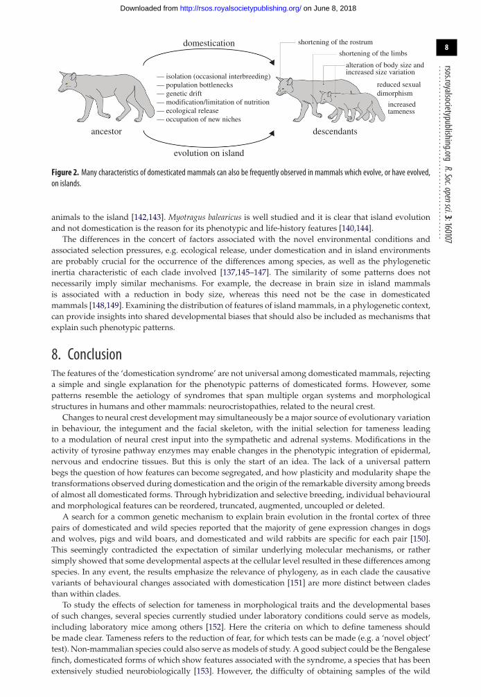
Many island mammals [123] exhibit phenotypic features also found in several domesticated mammals(figure 2), as in the alteration of body size, shortening of the rostrum and of the limbs, reduced sexualdimorphism [124] and reduction of brain size in some species, as well as tameness. In both domesticationand in island evolution, morphological changes tend to occur relatively fast [125,126]; there arepopulation bottlenecks [19,123,127–129], genetic drift [19,123], the occupation of new niches [30,123,130]and altered selection pressures. While on some islands there can be total isolation from the ancestralpopulation [123], in other cases, post-divergence gene flow between wild and domestic populations iscommon [69], as reported for dogs [16,127,131], pigs [132], cattle [133] and horses [134]. Selective breeding(i.e. artificial selection) occurs under domestication, whereas in island environments populations areexposed to novel selection regimes. In islands, there can be a modification/limitation of nutritionand ecological release, in some cases owing to decreased interspecific competition and/or absence ofpredators [123,135–139], which can lead to tameness.
The similarity of domestication and island evolution phenotypic patterns has resulted in extinct islandforms being interpreted as the result of domestication. This is the case of the Balearian ‘mouse goat’Myotragus balearicus [19,140] and the Falkland island fox Dusicyon australis [141]. The Falkland islandfox has been considered a feral domestic canid because of its white tail tip, rostrum and lower limbs,broad skull, bulbous forehead and its tameness [141]. This hypothesis has been rejected on the basisof divergence times, which are too old as to support the hypothesis of human transportation of these
on June 8, 2018http://rsos.royalsocietypublishing.org/Downloaded from
8
rsos.royalsocietypublishing.orgR.Soc.opensci.3:160107
................................................
— isolation (occasional interbreeding)
shortening of the rostrum
alteration of body size andincreased size variation
reduced sexualdimorphism
increasedtameness
shortening of the limbs
— population bottlenecks— genetic drift
— occupation of new niches— ecological release— modification/limitation of nutrition
domestication
descendantsancestor
evolution on island
Figure 2. Many characteristics of domesticated mammals can also be frequently observed in mammals which evolve, or have evolved,on islands.
animals to the island [142,143]. Myotragus balearicus is well studied and it is clear that island evolutionand not domestication is the reason for its phenotypic and life-history features [140,144].
The differences in the concert of factors associated with the novel environmental conditions andassociated selection pressures, e.g. ecological release, under domestication and in island environmentsare probably crucial for the occurrence of the differences among species, as well as the phylogeneticinertia characteristic of each clade involved [137,145–147]. The similarity of some patterns does notnecessarily imply similar mechanisms. For example, the decrease in brain size in island mammalsis associated with a reduction in body size, whereas this need not be the case in domesticatedmammals [148,149]. Examining the distribution of features of island mammals, in a phylogenetic context,can provide insights into shared developmental biases that should also be included as mechanisms thatexplain such phenotypic patterns.
8. ConclusionThe features of the ‘domestication syndrome’ are not universal among domesticated mammals, rejectinga simple and single explanation for the phenotypic patterns of domesticated forms. However, somepatterns resemble the aetiology of syndromes that span multiple organ systems and morphologicalstructures in humans and other mammals: neurocristopathies, related to the neural crest.
Changes to neural crest development may simultaneously be a major source of evolutionary variationin behaviour, the integument and the facial skeleton, with the initial selection for tameness leadingto a modulation of neural crest input into the sympathetic and adrenal systems. Modifications in theactivity of tyrosine pathway enzymes may enable changes in the phenotypic integration of epidermal,nervous and endocrine tissues. But this is only the start of an idea. The lack of a universal patternbegs the question of how features can become segregated, and how plasticity and modularity shape thetransformations observed during domestication and the origin of the remarkable diversity among breedsof almost all domesticated forms. Through hybridization and selective breeding, individual behaviouraland morphological features can be reordered, truncated, augmented, uncoupled or deleted.
A search for a common genetic mechanism to explain brain evolution in the frontal cortex of threepairs of domesticated and wild species reported that the majority of gene expression changes in dogsand wolves, pigs and wild boars, and domesticated and wild rabbits are specific for each pair [150].This seemingly contradicted the expectation of similar underlying molecular mechanisms, or rathersimply showed that some developmental aspects at the cellular level resulted in these differences amongspecies. In any event, the results emphasize the relevance of phylogeny, as in each clade the causativevariants of behavioural changes associated with domestication [151] are more distinct between cladesthan within clades.
To study the effects of selection for tameness in morphological traits and the developmental basesof such changes, several species currently studied under laboratory conditions could serve as models,including laboratory mice among others [152]. Here the criteria on which to define tameness shouldbe made clear. Tameness refers to the reduction of fear, for which tests can be made (e.g. a ‘novel object’test). Non-mammalian species could also serve as models of study. A good subject could be the Bengalesefinch, domesticated forms of which show features associated with the syndrome, a species that has beenextensively studied neurobiologically [153]. However, the difficulty of obtaining samples of the wild
on June 8, 2018http://rsos.royalsocietypublishing.org/Downloaded from

9
rsos.royalsocietypublishing.orgR.Soc.opensci.3:160107
................................................form for comparison would be an issue. Another model could be found among some species of cichlidfishes which have been raised in experimental conditions and have become tamed [154].
Some morphological features of domesticated mammals, which were considered to be the result ofjuvenilization, have proved not to be so. This does not exclude the potential relevance of heterochronyin the evolution of domesticated forms. The neoteny hypothesis for human evolution attempted toprovide a simple and universal explanation, which proved to be too simplistic to work, but neverthelessstimulated much empirical and conceptual advances in the field [155–157].
Research on domesticated animals, which on the one hand could be considered weird experimentsof artificial selection and not the result of true evolution in nature with its manifold ‘naturalmutants’ [158,159], might be construed as misguided or a waste of time. However, on the otherhand, domesticated animals do offer subjects of study that are potentially more accessible thanwild species, and the possibilities of integrating evo–devo with other approaches such as populationgenetics, archaeology and genomics are seemingly endless. Even though domestication processes arelike experiments in artificial selection, they have been underused in organismal biology studies ofmorphology and development, as well as in the investigation of physiological variables [160,161].Moreover, the developmental and organismal approaches needed to understand domestication are tiedto major concepts in evo–devo and evolutionary biology at large, (e.g. evolvability, modularity anddevelopmental plasticity), and thus many testable hypotheses and intriguing opportunities remain.
Authors’ contributions. All authors conceived of the study, participated in its design, conducted data collection, draftedthe manuscript, and read and approved the final manuscript.Competing interests. The authors declare that they have no competing interests.Funding. M.R.S.-V. and M.G. were funded by SNF 31003A-149605. R.A.S. was funded by NIH/NIDCR R01 DE016402.Acknowledgements. We thank M. K. Richardson (Leiden), L. Heck and M. Clauss (Zurich), R. Asher (Cambridge),D. Kruska (Kiel) and W. Salzburger (Basel) for discussion of ideas, E. Amson (Berlin) and L. Hautier (Montpellier)for advice on phylogenetic issues, T. Scheyer and A. Wegmann for technical support, and L. Andersson (Uppsala) andtwo anonymous reviewers for valuable suggestions to improve the manuscript.
References1. Donoghue PCJ, Graham A, Kelsh RN. 2008 The
origin and evolution of the neural crest. BioEssays30, 530–541. (doi:10.1002/bies.20767)
2. Larson G, Fuller DQ. 2014 The evolution of animaldomestication. Annu. Rev. Ecol. Evol. Syst. 45,115–136. (doi:10.1146/annurev-ecolsys-110512-135813)
3. Zeder MA. 2015 Core questions in domesticationresearch. Proc. Natl Acad. Sci. USA 112, 3191–3198.(doi:10.1073/pnas.1501711112)
4. Cagan A, Blass T. 2016 Identification of genomicvariants putatively targeted by selection duringdog domestication. BMC Evol. Biol. 16, 1.(doi:10.1186/s12862-015-0579-7)
5. Ostrander EA, Wayne RK. 2005 The caninegenome. Genome Res. 15, 1706–1716. (doi:10.1101/gr.3736605)
6. Tapio M. 2006 Sheep mitochondrial DNA variationin European, Caucasian, and Central Asian areas.Mol. Biol. Evol. 23, 1776–1783. (doi:10.1093/molbev/msl043)
7. Lipinski MJ et al. 2008 The ascent of cat breeds:genetic evaluations of breeds and worldwiderandom-bred populations. Genomics 91, 12–21.(doi:10.1016/j.ygeno.2007.10.009)
8. Cieslak M, Pruvost M, Benecke N, Hofreiter M,Morales A, Reissmann M, Ludwig A. 2010 Originand history of mitochondrial DNA lineages indomestic horses. PLoS ONE 5, e15311. (doi:10.1371/journal.pone.0015311)
9. Pollinger JP et al. 2010 Genome-wide SNP andhaplotype analyses reveal a rich historyunderlying dog domestication. Nature 464,898–902. (doi:10.1038/nature08837)
10. Thalmann O et al. 2013 Complete mitochondrialgenomes of ancient canids suggest a Europeanorigin of domestic dogs. Science 342, 871–874.(doi:10.1126/science.1243650)
11. Niemi M, Bläuer A, Iso-Touru T, Nyström V, HarjulaJ, Taavitsainen J-P, Storå J, Lidén K, Kantanen J.2013 Mitochondrial DNA and Y-chromosomaldiversity in ancient populations of domestic sheep(Ovis aries) in Finland: comparison withcontemporary sheep breeds. Genet. Sel. Evol. 45, 2.(doi:10.1186/1297-9686-45-2)
12. Ottoni C et al. 2012 Pig domestication andhuman-mediated dispersal in western Eurasiarevealed through ancient DNA and geometricmorphometrics.Mol. Biol. Evol. 30, 824–832.(doi:10.1093/molbev/mss261)
13. Montague MJ et al. 2014 Comparative analysis ofthe domestic cat genome reveals geneticsignatures underlying feline biology anddomestication. Proc. Natl Acad. Sci. USA 111,17 230–17 235. (doi:10.1073/pnas.1410083111)
14. Pilot M et al. 2015 On the origin of mongrels:evolutionary history of free-breeding dogs inEurasia. Proc. R. Soc. B 282, 20152189. (doi:10.1098/rspb.2015.2189)
15. Edea Z, Bhuiyan MSA, Dessie T, Rothschild MF,Dadi H, Kim KS. 2015 Genome-wide geneticdiversity, population structure and admixtureanalysis in African and Asian cattle breeds. Animal9, 218–226. (doi:10.1017/S1751731114002560)
16. Wang G-D et al. 2016 Out of southern East Asia: thenatural history of domestic dogs across the world.Cell Res. 26, 21–33. (doi:10.1038/cr.2015.147)
17. Andersson LS et al. 2012 Mutations in DMRT3 affectlocomotion in horses and spinal circuit function inmice. Nature 488, 642–646. (doi:10.1038/nature11399)
18. Axelsson E et al. 2013 The genomic signature of dogdomestication reveals adaptation to a starch-richdiet. Nature 495, 360–364. (doi:10.1038/nature11837)
19. Clutton-Brock J. 1999 A natural history ofdomesticated mammals, 2nd edn, p. 238.Cambridge, UK: Cambridge University Press.
20. Diamond J. 2002 Evolution, consequences andfuture of plant and animal domestication.Nature 418, 700–707. (doi:10.1038/nature01019)
21. Darwin C. 1868 The variation of animals and plantsunder domestication. London, UK: John Murray.
22. Andersson L. 2010 Studying phenotypic evolutionin domestic animals: a walk in the footsteps ofCharles Darwin. Cold Spring Harbor Symp. Quant.Biology 74, 319–325. (doi:10.1101/sqb.2009.74.039)
23. Laland K et al. 2014 Does evolutionary theory needa rethink? Nature 514, 161–164.(doi:10.1038/514161a)
24. Wayne RK. 1986 Cranial morphology of domesticand wild canids: the influence of development onmorphological change. Evolution 40, 243–261.(doi:10.2307/2408805)
25. Drake AG. 2011 Dispelling dog dogma: aninvestigation of heterochrony in dogs using 3Dgeometric morphometric analysis of skull shape.Evol. Dev. 13, 204–213. (doi:10.1111/j.1525-142X.2011.00470.x)
on June 8, 2018http://rsos.royalsocietypublishing.org/Downloaded from

10
rsos.royalsocietypublishing.orgR.Soc.opensci.3:160107
................................................26. Geiger M. 2015 Skeletal growth and life history
evolution in wild and domesticated mammals.Zurich, Switzerland: University of Zurich.
27. Geiger M, Haussman S. 2016 Cranial suture closurein domestic dog breeds and relations to skullmorphology. Anat. Rec. 299, 412–420.(doi:10.1002/ar.23313)
28. Francis RC. 2015 Domesticated: evolution in aman-made world. New York, NY: W. W. Norton &Company.
29. Wilkins AS, Wrangham RW, Fitch WT. 2014 The‘domestication syndrome’ in mammals: a unifiedexplanation based on neural crest cell behaviorand genetics. Genetics 197, 795–808. (doi:10.1534/genetics.114.165423)
30. Herre W, Röhrs M. 1990 Haustiere—zoologischgesehen, i–xiii, pp. 1–412. Stuttgart, Germany:Gustav Fischer Verlag.
31. Zeder MA. 2012 Pathways to animal domestication.In Biodiversity in agriculture: domestication,evolution, and sustainability (eds P Gepts et al.),pp. 227–259. New York, NY: Cambridge UniversityPress.
32. Vigne JD. 2011 The origins of animal domesticationand husbandry: a major change in the history ofhumanity and the biosphere. C. R. Biol. 334,171–181. (doi:10.1016/j.crvi.2010.12.009)
33. Trut LN, Plyusnina IZ, Oskina IN. 2004 Anexperiment on fox domestication and debatableissues of evolution of the dog. Russ. J. Genet. 40,644–655. (doi:10.1023/B:RUGE.0000033312.92773.c1)
34. Hoffman RA, Robinson PF, Magalhaes H. 1968 Thegolden hamster: its biology and use in medicalresearch, 1st edn. Ames. IA: Iowa State UniversityPress.
35. Feldhamer GA, Thompson BC, Chapman JA. 2003Wild mammals of North America: biology,management, and conservation. Baltimore, MD:Johns Hopkins University Press.
36. Zhang MQ, Xu X, Luo SJ. 2014 The genetics ofbrown coat color and white spotting in domesticyaks (Bos grunniens). Anim. Genet. 45, 652–659.(doi:10.1111/age.12191)
37. Driscoll CA, Macdonald DW, O’Brien SJ. 2009 Fromwild animals to domestic pets, an evolutionaryview of domestication. Proc. Natl Acad. Sci. USA106(Suppl. 1), 9971–9978. (doi:10.1073/pnas.0901586106)
38. Olsen KM, Wendel JF. 2013 A bountiful harvest:genomic insights into crop domesticationphenotypes. Annu. Rev. Plant Biol. 64, 47–70.(doi:10.1146/annurev-arplant-050312-120048)
39. O’Leary MA et al. 2013 The placental mammalancestor and the post–K-Pg radiation ofplacentals. Science 339, 662–667. (doi:10.1126/science.1229237)
40. Fabre PH, Hautier L, Dimitrov D, Douzery EJP. 2012A glimpse on the pattern of rodent diversification:a phylogenetic approach. BMC Evol. Biol. 12, 88.(doi:10.1186/1471-2148-12-88)
41. Bibi F. 2013 A multi-calibrated mitochondrialphylogeny of extant Bovidae (Artiodactyla,Ruminantia) and the importance of the fossilrecord to systematics. BMC Evol. Biol. 13, 166.(doi:10.1186/1471-2148-13-166)
42. Wu HG et al. 2014 Camelid genomes revealevolution and adaptation to desert environments.
Nat. Commun. 5, 5188. (doi:10.1038/ncomms6188)
43. Benton MJ et al. 2015 Constraints on the timescaleof animal evolutionary history. Palaeontol.Electron. 18. 1.1FC, 1–106.
44. Laurin M. 2004 The evolution of body size, Cope’srule and the origin of amniotes. Syst. Biol. 53,594–622. (doi:10.1080/10635150490445706)
45. Quemeneur S, Buffrénil V, Laurin M. 2013Microanatomy of the amniote femur and inferenceof lifestyle in limbed vertebrates. Biol. J. Linn. Soc.109, 644–655. (doi:10.1111/bij.12066)
46. Maddison WP, Maddison DR. 2001 Mesquite:a modular system for evolutionary analysis.Version 0.98. See http://mesquiteproject.org.
47. Zeder MA. 2012 The domestication of animals.J. Anthropol. Res. 68, 161–190. (doi:10.3998/jar.0521004.0068.201)
48. Hammer K. 1984 The domestication syndrome. DieKulturpflanze 32, 11–34. (doi:10.1007/BF02098682)
49. Koinange EM, Singh SP, Gepts P. 1996 Geneticcontrol of the domestication syndrome in commonbean. Crop Sci. 36, 1037–1045. (doi:10.2135/cropsci1996.0011183X003600040037x)
50. Motley TJ. 2006 Crop evolution: past, present, andfuture. In Darwin’s harvest: new approaches to theorigins, evolution, and conservation of crops (eds TJMotley, N Zerega, HB Cross), pp. 1–28. New York,NY: Columbia University Press.
51. Ross-Ibarra J, Morrell PL, Gaut BS. 2007 Plantdomestication, a unique opportunity to identifythe genetic basis of adaptation. Proc. Natl Acad. Sci.USA 104(suppl 1), 8641–8648. (doi:10.1073/pnas.0700643104)
52. Harlan JR, de Wet JMJ, Price EG. 1973 Comparativeevolution of cereals. Evolution 27, 311–325.(doi:10.2307/2406971)
53. Faegri K, Van der Pijl L. 1979 Principles ofpollination ecology. Philadelphia, PA: Elsevier.
54. Coppinger R, Schneider R. 1995 Evolution ofworking dogs. In The domestic dog: its evolution,behaviour, and interactions with people (ed. JSerpell), pp. 21–47. Cambridge, UK: CambridgeUniversity Press.
55. Trut LN. 1999 Early canid domestication: thefarm-fox experiment. Am. Sci. 161, 160–169.(doi:10.1511/1999.2.160)
56. Hare B, Plyusnina I, Ignacio N, Schepina O, StepikaA, Wrangham R, Trut L. 2005 Social cognitiveevolution in captive foxes is a correlatedby-product of experimental domestication. Curr.Biol. 15, 226–230. (doi:10.1016/j.cub.2005.01.040)
57. Trut L, Oskina I, Kharlamova A. 2009 Animalevolution during domestication: the domesticatedfox as a model. Bioessays 31, 349–360.(doi:10.1002/bies.200800070)
58. Lord K, Schneider RA, Coppinger R. In press.Evolution of working dogs. In The domestic dog(ed. J Serpell). Cambridge, UK: CambridgeUniversity Press.
59. Schneider RA, Helms JA. 2003 The cellular andmolecular origins of beak morphology. Science299, 565–568. (doi:10.1126/science.1077827)
60. Crockford SJ. 2002 Animal domestication andheterochronic speciation. In Human evolutionthrough developmental change (eds,N Minugh-Purvis, KJ McNamara), pp. 122–153.Baltimore, MD: Johns Hopkins University Press.
61. Schneider RA. 2005 Developmental mechanismsfacilitating the evolution of bills and quills. J. Anat.207, 563–573. (doi:10.1111/j.1469-7580.2005.00471.x)
62. Zrzavý J, Burda H, Storch D, Begall S, Mihulka S.2009 Evolution: Ein Lese-Lehrbuch. Wiesbaden,Germany: Springer Spektrum.
63. Crockford SJ. 2004 Animal domestication andvertebrate speciation: a paradigm for the originof species. Victoria, Canada: University ofVictoria.
64. Alberch P. 1982 The generative and regulatoryroles of development in evolution. InEnvironmental adaptation and evolution: atheoretical and empirical approach (edsD Mossakowski, G Roth), pp. 19–36. Stuttgart,Germany: G. Fischer-Verlag.
65. Coppinger R, Glendinning J, Torop E, Matthay C,Sutherland M, Smith C. 1987 Degree of behavioralneoteny differentiates canid polymorphs. Ethology75, 89–108. (doi:10.1111/j.1439-0310.1987.tb00645.x)
66. Coppinger R, Feinstein M. 1991 Hark, hark! Thedogs do bark . . . and bark and bark. Smithsonian21, 119–129.
67. Coppinger R, Coppinger L. 2001 Dogs: a startlingnew understanding of canine origin, behavior &evolution. NewYork, NY: Simon and Schuster.
68. Meredith RW et al. 2011 Impacts of the Cretaceousterrestrial revolution and KPg extinction onmammal diversification. Science 334, 521–524.(doi:10.1126/science.1211028)
69. Larson G, Burger J. 2013 A population geneticsview of animal domestication. Trends Genet. 29,197–205. (doi:10.1016/j.tig.2013.01.003)
70. Zeder MA, Bradley D, Emshwiller E, Smith BD(eds). 2006 Documenting domestication: newgenetic and archaeological paradigms. Oakland,CA: University of California Press.
71. Kissel P, André JM, Jacquier A. 1981 Theneurocristopathies. New York, NY: Masson.
72. Etchevers H, Amiel J, Lyonnet S. 2006Molecularbases of human neurocristopathies. In Neural crestinduction and differentiation (ed.J-P Saint-Jeannet), pp. 213–234. New York, NY:Springer.
73. Noden DM, Schneider RA. 2006 Neural crest cellsand the community of plan for craniofacialdevelopment. In Neural crest induction anddifferentiation (ed. J-P Saint-Jeannet), pp. 1–23.New York, NY: Springer.
74. Vickaryous MK, Hall BK. 2006 Human cell typediversity, evolution, development, andclassification with special reference to cells derivedfrom the neural crest. Biol. Rev. 81, 425–455.(doi:10.1017/S1464793106007068)
75. Le Lièvre CS, Le Douarin N. 1975 Mesenchymalderivatives of the neural crest: analysis ofchimaeric quail and chick embryos. J. Embryol. Exp.Morphol. 34, 125–154.
76. Bockman D, Kirby M. 1984 Dependence of thymusdevelopment on derivatives of the neural crest.Science 223, 498–500. (doi:10.1126/science.6606851)
77. Kuratani S, Bockman DE. 1990 The participation ofneural crest derived mesenchymal cells indevelopment of the epithelial primordium of thethymus. Arch. Histol. Cytol. 53, 267–273.(doi:10.1679/aohc.53.267)
on June 8, 2018http://rsos.royalsocietypublishing.org/Downloaded from
11
rsos.royalsocietypublishing.orgR.Soc.opensci.3:160107
................................................78. Varga I et al. 2008 The phylogenesis and
ontogenesis of the human pharyngeal regionfocused on the thymus, parathyroid, and thyroidglands. Neuroendocrinol. Lett. 29, 837–845.
79. Maeda K, Asai R, Maruyama K, Kurihara Y,Nakanishi T, Kurihara H, Miyagawa-Tomita S. 2016Postotic and preotic cranial neural crest cellsdifferently contribute to thyroid development. Dev.Biol. 409, 72–83. (doi:10.1016/j.ydbio.2015.10.026)
80. Arons CD, Shoemaker WJ. 1992 The distribution ofcatecholamines andβ-endorphin in the brains ofthree behaviorally distinct breeds of dogs andtheir F 1 hybrids. Brain Res. 594, 31–39.(doi:10.1016/0006-8993(92)91026-B)
81. Richardson MK, Sieber-BlumM. 1993 Pluripotentneural crest cells in the developing skin of the quailembryo. Dev. Biol. 157, 348–358. (doi:10.1006/dbio.1993.1140)
82. Eames BF, Schneider RA. 2005 Quail-duckchimeras reveal spatiotemporal plasticity inmolecular and histogenic programs of cranialfeather development. Development 132,1499–1509. (doi:10.1242/dev.01719)
83. Mitgutsch C, Olsson L, Haas A. 2009 Earlyembryogenesis in discoglossoid frogs: a study ofheterochrony at different taxonomic levels. J. Zool.Syst. Evol. Res. 47, 248–257. (doi:10.1111/j.1439-0469.2008.00502.x)
84. Smith KK. 2001 Early development of the neuralplate, neural crest and facial region of marsupials.J. Anat. 199, 121–131. (doi:10.1046/j.1469-7580.2001.19910121.x)
85. Vaglia JL, Smith KK. 2003 Early differentiation andmigration of cranial neural crest in the opossum,Monodelphis domestica. Evol. Dev. 5, 121–135.(doi:10.1046/j.1525-142X.2003.03019.x)
86. Smith KK. 2006 Craniofacial development inmarsupial mammals: developmental origins ofevolutionary change. Dev. Dyn. 235, 1181–1193.(doi:10.1002/dvdy.20676)
87. Tucker AS, Lumsden A. 2004 Neural crest cellsprovide species-specific patterning information inthe developing branchial skeleton. Evol. Dev. 6,32–40. (doi:10.1111/j.1525-142X.2004.04004.x)
88. Lwigale PY, Schneider RA. 2008 Other chimeras:quail–duck and mouse–chick.Methods Cell Biol.87, 59–74. (doi:10.1016/S0091-679X(08)00203-3)
89. Fish JL, Schneider RA. 2014 Chapter 6—Neuralcrest-mediated tissue interactions duringcraniofacial development: the origins ofspecies-specific pattern. In Neural crest cells (ed. PATrainor), pp. 101–124. Boston, MA: Academic Press.
90. Ealba EL, Jheon AH, Hall J, Curantz C, Butcher KD,Schneider RA. 2015 Neural crest-mediated boneresorption is a determinant of species-specific jawlength. Dev. Biol. 408, 151–163. (doi:10.1016/j.ydbio.2015.10.001)
91. Schneider RA. 2015 Regulation of jaw lengthduring development, disease, and evolution. Curr.Top. Dev. Biol. 115, 271–298. (doi:10.1016/bs.ctdb.2015.08.002)
92. Fish JL, Sklar RS, Woronowicz KC, Schneider RA.2014 Multiple developmental mechanismsregulate species-specific jaw size. Development141, 674–684. (doi:10.1242/dev.100107)
93. Eames BF, Schneider RA. 2008 The genesis ofcartilage size and shape during development andevolution. Development 135, 3947–3958.(doi:10.1242/dev.023309)
94. Merrill AE, Eames BF, Weston SJ, Heath T,Schneider RA. 2008 Mesenchyme-dependent BMPsignaling directs the timing of mandibularosteogenesis. Development 135, 1223–1234.(doi:10.1242/dev.015933)
95. Mitgutsch C, Wimmer C, Sánchez-Villagra MR,Hahnloser R, Schneider RA. 2011 Timing ofossification in duck, quail, and zebra finch:intraspecific variation, heterochronies, and lifehistory evolution. Zool. Sci. 28, 491–500.(doi:10.2108/zsj.28.491)
96. Hall J, Jheon AH, Ealba EL, Eames BF, Butcher KD,Mak S-S, Ladher R, Alliston T, Schneider RA. 2014Evolution of a developmental mechanism:species-specific regulation of the cell cycle and thetiming of events during craniofacial osteogenesis.Dev. Biol. 385, 380–395. (doi:10.1016/j.ydbio.2013.11.011)
97. Ealba EL, Schneider RA. 2013 A simple PCR-basedstrategy for estimating species-specificcontributions in chimeras and xenografts.Development 140, 3062–3068. (doi:10.1242/dev.092676)
98. Jheon AH, Schneider RA. 2009 The cells that fill thebill: neural crest and the evolution of craniofacialdevelopment. J. Dent. Res. 88, 12–21. (doi:10.1177/0022034508327757)
99. Eames BF, Sharpe PT, Helms JA. 2004 Hierarchyrevealed in the specification of three skeletal fatesby Sox9 and Runx2. Dev. Biol. 274, 188–200.(doi:10.1016/j.ydbio.2004.07.006)
100. Fondon III JW, Garner HR. 2004 Molecular originsof rapid and continuous morphological evolution.Proc. Natl Acad. Sci. USA 101, 18 058–18 063.(doi:10.1073/pnas.0408118101)
101. Sears KE, Goswami A, Flynn JJ, Niswander LA. 2007The correlated evolution of Runx2 tandem repeats,transcriptional activity, and facial length incarnivora. Evol. Dev. 9, 555–565. (doi:10.1111/j.1525-142X.2007.00196.x)
102. Pointer MA, Kamilar JM, Warmuth V, Chester SGB,Delsuc F, Mundy NI, Asher RJ, Bradley BJ. 2012RUNX2 tandem repeats and the evolution of faciallength in placental mammals. BMC Evol. Biol. 12,103. (doi:10.1186/1471-2148-12-103)
103. McGreevy PD, Georgevsky D, Carrasco J,Valenzuela M, Duffy DL, Serpell JA. 2013 Dogbehavior co-varies with height, bodyweight andskull shape. PLoS ONE 8, e80529. (doi:10.1371/journal.pone.0080529)
104. Podberscek AL, Serpell JA. 1996 The English cockerspaniel: preliminary findings on aggressivebehaviour. Appl. Anim. Behav. Sci. 47, 75–89.(doi:10.1016/0168-1591(95)01012-2)
105. Hart BL. 1996 Analysing breed and genderdifferences in behaviour. In The domestic dog: itsevolution, behaviour and interactions with people(ed. J Serpell), pp. 65–77. Cambridge, UK:Cambridge University Press.
106. Stockard CR. 1941 The genetic and endocrine basisfor differences in form and behaviour as elucidatedby studies of contrasted pure-line dog breeds andtheir hybrids. With special contributions onbehaviour by OD Anderson andWT James. Amer.Anat. Mem.19.
107. Albert FW et al. 2009 Genetic architecture oftameness in a rat model of animal domestication.Genetics 182, 541–554. (doi:10.1534/genetics.109.102186)
108. Baranowska Korberg I et al. 2014 A simple repeatpolymorphism in the MITF-M promoter is a keyregulator of white spotting in dogs. PLoS ONE 9,e104363. (doi:10.1371/journal.pone.0104363)
109. Linderholm A, Larson G. 2013 The role of humansin facilitating and sustaining coat colour variationin domestic animals. Seminars Cell Develop. Biol.24, 587–593. (doi:10.1016/j.semcdb.2013.03.015)
110. Carneiro M et al. 2014 Rabbit genome analysisreveals a polygenic basis for phenotypic changeduring domestication. Science 345, 1074–1079.(doi:10.1126/science.1253714)
111. Evin A, Dobney K, Schafberg R, Owen J,Vidarsdottir U, Larson G, Cucchi T. 2015 Phenotypeand animal domestication: a study of dentalvariation between domestic, wild, captive, hybridand insular Sus scrofa. BMC Evol. Biol. 15, 6.(doi:10.1186/s12862-014-0269-x)
112. Morey DF. 1992 Size, shape and development inthe evolution of the domestic dog. J. Archaeol. Sci.19, 181–204. (doi:10.1016/0305-4403(92)90049-9)
113. Anthwal N, Thompson H. 2016 The development ofthe mammalian outer and middle ear. J. Anat.228, 217–232. (doi:10.1111/joa.12344)
114. Horovitz I et al. 2008 The anatomy ofHerpetotherium cf. fugax Cope, 1873, ametatherian from the Oligocene of North America.Palaeontogr. Abteilung A 284, 109–141.
115. Minoux M, Kratochwil CF, Ducret S, Amin S,Kitazawa T, Kurihara H, Bobola N, Vilain N, Rijli FM.2013 Mouse Hoxa2 mutations provide a model formicrotia and auricle duplication. Development140, 4386–4397. (doi:10.1242/dev.098046)
116. Bolk L. 1926 Das Problem der Menschwerdung.Jena, Germany: Fischer Verlag.
117. Dechambre E. 1949 La theorie de la foetalisationet la formation des races de chiens et de porcs.Mammalia 13, 129–137. (doi:10.1515/mamm.1949.13.3.129)
118. Zeuner FE. 1963 A history of domesticated animals.New York, NY: Harper & Row.
119. Morey DF. 1994 The early evolution of thedomestic dog. Am. Sci. 82, 336–347.
120. Price EO. 1999 Behavioral development in animalsundergoing domestication. Appl. Anim. Behav. Sci.65, 245–271. (doi:10.1016/S0168-1591(99)00087-8)
121. Starck D. 1962 Der heutige Stand desFetalisationsproblems. Hamburg, Germany: VerlagPaul Parey.
122. Rosenberg K. 1966 Die postnataleProportionsänderung der Schädel zweier extremerWuchsformen des Haushundes. J. Anim. Breed.Genet. 82, 1–36. (doi:10.1111/j.1439-0388.1966.tb01499.x)
123. van der Geer A et al. 2010 Evolution of islandmammals: adaptation and extinction of placentalmammals on islands. Oxford, UK: Wiley-Blackwell.
124. Polák J, Frynta D. 2009 Sexual size dimorphism indomestic goats, sheep, and their wild relatives. Biol.J. Linn. Soc. 98, 872–883. (doi:10.1111/j.1095-8312.2009.01294.x)
125. Lister AM. 1989 Rapid dwarfing of red deer onJersey in the last interglacial. Nature 342,539–542. (doi:10.1038/342539a0)
126. Millien V. 2006 Morphological evolution isaccelerated among island mammals. PLoS Biol. 4,e321. (doi:10.1371/journal.pbio.0040321)
127. Freedman AH et al. 2014 Genome sequencinghighlights the dynamic early history of dogs. PLoS
on June 8, 2018http://rsos.royalsocietypublishing.org/Downloaded from

12
rsos.royalsocietypublishing.orgR.Soc.opensci.3:160107
................................................Genet. 10, e1004016. (doi:10.1371/journal.pgen.1004016)
128. Lindblad-Toh K et al. 2005 Genome sequence,comparative analysis and haplotype structure ofthe domestic dog. Nature 438, 803–819.(doi:10.1038/nature04338)
129. Wang GD et al. 2013 The genomics of selection indogs and the parallel evolution between dogs andhumans. Nat. Commun. 4, 1860. (doi:10.1038/ncomms2814)
130. Larson G et al. 2014 Current perspectives and thefuture of domestication studies. Proc. Natl Acad.Sci. USA 111, 6139–6146. (doi:10.1073/pnas.1323964111)
131. vonHoldt BM, Pollinger JP, Earl DA, Parker HG,Ostrander EA, Wayne RK. 2013 Identification ofrecent hybridization between gray wolves anddomesticated dogs by SNP genotyping.Mamm.Genome 24, 80–88. (doi:10.1007/s00335-012-9432-0)
132. Frantz LAF et al. 2015 Evidence of long-term geneflow and selection during domestication fromanalyses of Eurasian wild and domestic piggenomes. Nat. Genet. 47, 1141–1148. (doi:10.1038/ng.3394)
133. Park SDE et al. 2015 Genome sequencing of theextinct Eurasian wild aurochs, Bos primigenius,illuminates the phylogeography and evolution ofcattle. Genome Biol. 16, 234. (doi:10.1186/s13059-015-0790-2)
134. Warmuth V et al. 2012 Reconstructing the originand spread of horse domestication in the Eurasiansteppe. Proc. Natl Acad. Sci. USA 109, 8202–8206.(doi:10.1073/pnas.1111122109)
135. Tchernov E, Horwitz LK. 1991 Body size diminutionunder domestication: unconscious selection inprimeval domesticates. J. Anthropol. Archaeol.10, 54–75. (doi:10.1016/0278-4165(91)90021-O)
136. Parker HG, Shearin AL, Ostrander EA. 2010 Man’sbest friend becomes biology’s best in show:genome analyses in the domestic dog. Annu. Rev.Genet. 44, 303–336. (doi:10.1146/annurev-genet-102808-115200)
137. Lomolino MV, Sax DF, Palombo MR, van der GeerAA. 2012 Of mice and mammoths: evaluations ofcausal explanations for body size evolution ininsular mammals. J. Biogeogr. 39, 842–854.(doi:10.1111/j.1365-2699.2011.02656.x)
138. Roth V. 1992 Inferences from allometry and fossils:dwarfing of elephants on islands. In Oxford surveys
in evolutionary biology (eds D Futuyma,J Antonovics), pp. 259–288. Oxford, UK: OxfordUniversity Press.
139. van der Geer AA, Lyras GA, Lomolino MV, PalomboMR, Sax DF. 2013 Body size evolution ofpalaeo-insular mammals: temporal variations andinterspecific interactions. J. Biogeogr. 40,1440–1450. (doi:10.1111/jbi.12119)
140. Ramis D, Bover P. 2001 A review of the evidence fordomestication ofMyotragus balearicus Bate 1909(Artiodactyla, Caprinae) in the Balearic Islands.J. Archaeol. Sci. 28, 265–282. (doi:10.1006/jasc.2000.0548)
141. Clutton-Brock J. 1977 Man-made dogs. Science197, 1340–1342. (doi:10.1126/science.197.4311.1340)
142. Slater GJ et al. 2009 Evolutionary history of theFalklands wolf. Curr. Biol. 19, R937–R938.(doi:10.1016/j.cub.2009.09.018)
143. Austin JJ, Soubrier J, Prevosti FJ, Prates L, Trejo V,Mena F, Cooper A. 2013 The origins of theenigmatic Falkland Islands wolf. Nat. Commun. 4,1552. (doi:10.1038/ncomms2570)
144. Köhler M, Moyà-Solà S. 2009 Physiological and lifehistory strategies of a fossil large mammal in aresource-limited environment. Proc. Natl Acad. Sci.USA 106, 20 354–20 358. (doi:10.1073/pnas.0813385106)
145. Roth VL. 1993 Dwarfism and variability in theSanta Rosa Island mammoth: an interspecificcomparison of limb bone sizes and shapes inelephants. In Third California Islands symposium:recent advances in research on the California Islands(ed. FG Hochberg), pp. 433–442. Santa Barbara,CA: Santa Barbara Museum of Natural History.
146. Meiri S, Cooper N, Purvis A. 2008 The island rule:made to be broken? Proc. R. Soc. B 275, 141–148.(doi:10.1098/rspb.2007.1056)
147. Kolb C et al. 2015 Mammalian bonepalaeohistology: a survey and new data withemphasis on island forms. PeerJ 3, e1358.(doi:10.7717/peerj.1358)
148. Kruska DC. 2005 On the evolutionary significanceof encephalization in some eutherian mammals:effects of adaptive radiation, domestication, andferalization. Brain Behav. Evol. 65, 73–108.(doi:10.1159/000082979)
149. Weston EM, Lister AM. 2009 Insular dwarfism inhippos and a model for brain size reduction inHomo floresiensis. Nature 459, 85–88.(doi:10.1038/nature07922)
150. Albert FW et al. 2012 A comparison of brain geneexpression levels in domesticated and wildanimals. PLoS Genet. 8, e1002962. (doi:10.1371/journal.pgen.1002962)
151. Li Y, vonHoldt BM, Reynolds A, Boyko AR, WayneRK, Wu D-D, Zhang Y-P. 2013 Artificial selection onbrain-expressed genes during the domestication ofdog.Mol. Biol. Evol. 30, 1867–1876. (doi:10.1093/molbev/mst088)
152. Książek A, Konarzewski M, Łapo IB. 2004 Anatomicand energetic correlates of divergent selection forbasal metabolic rate in laboratory mice. Physiol.Biochem. Zool. 77, 890–899. (doi:10.1086/425190)
153. Okanoya K. 2004 The Bengalese finch: a windowon the behavioral neurobiology of birdsongsyntax. Ann. NY Acad. Sci. 1016, 724–735.(doi:10.1196/annals.1298.026)
154. Theis A, Ronco F, Indermaur A, Salzburger W,Egger B. 2014 Adaptive divergence between lakeand stream populations of an East African cichlidfish.Mol. Ecol. 23, 5304–5322. (doi:10.1111/mec.12939)
155. Shea BT. 1989 Heterochrony in human evolution:the case for neoteny reconsidered. Am. J. Phy.Anthropol. 32, 69–101. (doi:10.1002/ajpa.1330320505)
156. Godfrey LR, Sutherland MR. 1995 Flawedinference: why size-based tests of heterochronicprocesses do not work. J. Theor. Biol. 172, 43–61.(doi:10.1006/jtbi.1995.0004)
157. Zollikofer CP, de León MSP. 2010 The evolution ofhominin ontogenies. Seminars Cell Develop. Biol.21, 441–452. (doi:10.1016/j.semcdb.2009.10.012)
158. Milinkovitch MC, Tzika A. 2007 Escaping the mousetrap: the selection of new evo-devo model species.J. Exp. Zool. B 308, 337–346. (doi:10.1002/jez.b.21180)
159. Sears KE. 2011 Novel insights into the regulation oflimb development from ‘natural’ mammalianmutants. BioEssays 33, 327–331. (doi:10.1002/bies.201100005)
160. Konarzewski M, Książek A, Łapo IB. 2005 Artificialselection on metabolic rates and related traits inrodents. Integr. Comp. Biol. 45, 416–425.(doi:10.1093/icb/45.3.416)
161. Swallow JG, Garland T. 2005 Selectionexperiments as a tool in evolutionary andcomparative physiology: insights into complextraits—an introduction to the symposium. Integr.Comp. Biol. 45, 387–390. (doi:10.1093/icb/45.3.387)
on June 8, 2018http://rsos.royalsocietypublishing.org/Downloaded from


![Fossilevidenceandstages ofelongationofthe Giraffarsos.royalsocietypublishing.org/content/royopensci/2/10/... · females supports the sexual selection theory [4]. ... not apply the](https://static.fdocuments.net/doc/165x107/5afcc2d57f8b9a323490c0f3/fossilevidenceandstages-ofelongationofthe-supports-the-sexual-selection-theory-4.jpg)
![NovelFibonacciand non-Fibonaccistructureinrsos.royalsocietypublishing.org/content/royopensci/3/5/160091.full.pdf · understanding of plant organ positioning [12–14], ... Fibonacci](https://static.fdocuments.net/doc/165x107/5b05d0747f8b9a58148bf291/novelfibonacciand-non-fibo-of-plant-organ-positioning-1214-fibonacci-numbers.jpg)


![Investigatingthe associationbetweensocialrsos.royalsocietypublishing.org/content/royopensci/4/9/170194.full.pdfleadership [10], general job performance [11], sales ability [12] and](https://static.fdocuments.net/doc/165x107/5aabd2ef7f8b9a59658c5729/investigatingthe-associat-10-general-job-performance-11-sales-ability-12.jpg)
