Sequencing the Maize (B73) Genome Genome Sequencing Center Maize Genome Sequencing Consortium.

THESIS OF PhD DISSERTATION
WHOLE GENOME MOLECULAR ANALYSIS OF POTATO VIRUS S (PVS)
ISOLATES
ÉVA PÁJTLI
Budapest
2015


1
1. INTRODUCTION AND THE AIMS OF THE RESEARCH
Potato (Solanum tuberosum L.) is one of the most widely grown food crops in the world,
representing a staple food in many countries. Based on the harvested yield, it ranks fourth
among food crops after wheat, rice and maize. In Hungary there is a long tradition of potato
cultivation. The main breeding centre for potato is in Keszthely, where excellent new cultivars
are developed.
Potato is attacked by numerous phytopathogenic viruses, including potato virus S (PVS),
which is a member of the Carlavirus genus (Matthews, 1979), one of the least investigated
groups of plant viruses. According to Wetter (1971), PVS is one of the most widespread potato
viruses in the world. Its chief host plant is potato and nowadays it has spread to all potato-
growing countries (de Bruyn Ouboter, 1952; de Bokx, 1970). The ordinary strain of PVS causes
no visible symptoms on large numbers of potato cultivars, while on many others only very mild
symptoms appear (Vaughan and van Slogteren, 1956). The virus is of economic importance, as
it may reduce yields by 10–20% (Wetter, 1971). For this reason, stopping the spread of the virus
and developing virus-free reproduction material will be cardinal areas of research for the future
of potato production.
Between 2009 and 2013 our team participated as a consortium member in the project
entitled ‘Potato production technologies and the development of trademarks’ (NKTH-TECH-
09-A3-2009-0210), which provided scientific and financial assistance for the molecular
analysis of potato virus S.
The aim of the work was to elaborate a reliable PVS diagnostic technique to detect even
low concentrations of the virus. It was then hoped to use the technique to determine the
nucleotide sequences of the coat protein genes of collected isolates, which could then be
compared with isolates from other parts of the world in order to map the relationships between
them.
A further aim was to elaborate a way of determining the sequence of the whole PVS
genome, and to apply this method to identify the nucleotide sequences of the whole hereditary
material of our own PVS isolates, which would then be compared with those of other PVS
isolates in the international database and with those of related species, to obtain information on
their origin. Recombination analysis was planned to identify any intermolecular rearrangements
that may have taken place, while it was expected that conserved domain analysis would provide
new information on the possible functions of the PVS genes.

2
2. MATERIALS AND METHODS
2.1 Location and duration
Researches were done at Corvinus University of Budapest, Faculty of Horticultural
Sciences, Department of Plant Pathology, between 2009 and 2015.
2.2 Materials
In the course of the work 22 PVS isolates were collected from four countries. The Nested
PCR method developed in our laboratory proved suitable for the detection of PVS (1. table).
1. table Characteristics of the PVS isolates
Isolate Origin Host plant
Ewa POL Solanum tuberosum cv. Leona
Bonita HUN Solanum tuberosum cv. Bonita ojo (de) perdiz
Ditta HUN Solanum tuberosum cv. Ditta
FabiloaA HUN Solanum tuberosum cv. Fabiola
FabilolaB HUN Solanum tuberosum cv. Fabiola
FabiolaC HUN Solanum tuberosum cv. Fabiola
Lady Rosetta HUN Solanum tuberosum cv. Lady Rosetta
Mayan Twilight HUN Solanum tuberosum cv. Mayan Twilight
Papa negra HUN Solanum tuberosum cv. Papa negra
Desiré HUN, Keszthely Solanum tuberosum cv. Desiré
06.62 HUN, Keszthely Solanum sp. 06.62 klón
09.369 HUN, Keszthely Solanum sp. 09.369 klón
09.539 HUN, Keszthely Solanum sp. 09.539 klón
89.216 HUN, Keszthely Solanum sp. 89.216 klón
89.217 HUN, Keszthely Solanum sp. 89.217 klón
89.243 HUN, Keszthely Solanum sp. 89.243 klón
89.249 (PVS-HU1) HUN, Keszthely Solanum sp. 89.249 klón
Boglarka HUN, Nyírtelek Solanum tuberosum cv. Boglárka
Kilimanjaro TAN, Kilimandzsáró Solanum sp.
Alex UKR Solanum tuberosum cv. Finka
Irena UKR Solanum tuberosum cv. Finka
Valery UKR Solanum tuberosum cv. Finka
2.3 Methods
The extraction of total ribonucleic acid was carried out using a SpectrumTM Plant Total
RNA Kit (Sigma Aldrich, St. Louis, USA) according to the manufacturer’s instructions.
Products generated by Thermo Fisher Scientific (Waltham, USA) were used for RT-PCR.
Reverse transcription (RT) using antisense primers was applied to produce the first strand of
cDNA complementary to the nucleic acid of PVS. The reaction mixture had a final volume of
3
10 µl, consisting of 4 µl total nucleic acid, 1 µl (100 µM) antisense primer, 2 µl RT buffer (5×),
1 µl dNTP Mix (5 mM), 0.5 µl RevertAidTM Premium Reverse Transcriptase (200 u/µl), 0.25
µl RiboLockTM RNase Inhibitor (40 u/µl), and 1.25 µl distilled water. The total nucleic acid
was incubated at 65°C for 5 min in presence of the antisense primer, then the mixture was
cooled on ice for 5 min. The remaining components of the reaction mixture were then added,
prior to reverse transcription at 50°C for 30 min, after which the enzyme was inactivated by 5
min at 85°C. Following cDNA synthesis, the virus genome was amplified in six overlapping
regions by optimising PCR with the help of the antisense and sense primers. The reaction
mixture for the PCR analysis had a final volume of 50 µl consisting of 3 µl cDNA, 5 µl Taq
buffer (10×), 3 µl MgCl2 (25 mM), 2 µl dNTP Mix (5 mM), 1 µl each of antisense and sense
primer (20 µM), 0.5 µl Taq DNA polymerase (5 u/µl), and 34.5 µl distilled water. The PCR
products were purified using a High Pure Purification Kit (Roche, Basel, Switzerland) followed
by ligation into pGEM-T Easy Vectors. The recombinant plasmid was then transformed into E.
coli DH5α, TG90 or JM 109 competent cells using the heat shock method (Orkin, 1990).
2.4 Programs used for the bioinformatic analysis
2.4.1 Sequence analysis
The CLCSequence Viewer 7.6 and CLC Main Workbench (QIAGEN, Aarhus, Denmark)
software packages were used to align and analyse the sequences, while the phylogenetic
dendrograms were prepared by means of Neighbour Joining (NJ) and Unweighted Pair Group
Method Analysis (UPGMA), with Jukes–Cantor distance correction (Jukes, 1969). The
confidence interval for the phylogenetic analyses was based on bootstrap analysis with 1000
replications. Hydrophobicity values were calculated by the program using the Kyte–Doolittle
scale with a window 9 amino acids in width (Kyte and Doolittle, 1982).
2.4.2 Recombination analysis
The RDP4.39 Beta program was employed to detect potential recombination events
(Martin et al., 2010). The algorithms used by the program (RDP, Chimaera, BootScan, 3Seq,
GENECONV, MaxChi and SiScan) were applied using the default parameters (window size =
200 nt, sliding window size = 20 nt) at the 95% significance level (Atallah et al., 2012; Boni et
al., 2007; Gibbs et al., 2000; Martin and Rybicki, 2000; Martin et al., 2005; Padidam et al.,
1999; Posada and Crandall, 2001).
2.4.3 Detection of conserved domains
Conserved domains were detected using the CDD v3.13 program available at
http://www.ncbi.nlm.nih.gov/Structure/cdd/wrpsb.cgi. The CDD program makes use of RPS-

4
BLAST (Reverse Position-Specific Blast), which was applied using the default settings
(Marchler-Bauer et al., 2015).
3. Results
3.1 Molecular analysis of PVS isolates
3.1.1 Molecular analysis of CP region
With the help of this method the segment containing the coat protein region was amplified
and the nucleotide sequence was established. In all the isolates the ORF5 region coding for the
coat protein was found to consist of 885 nucleotides, including the stop codon. The coat protein
translated from this region was made up of 294 amino acids and measured approximately 33
kDa. The proteins were only variable for the first 38 amino acids at the N terminal end, while
differences in amino acids were only observed in five positions in the middle of the sequence,
and the C terminal end was completely homologous.
3.1.2 Molecular analysis of complete genomes
The whole genomes of three PVS isolates from Hungary, three from Ukraine and one
from Poland were amplified in six overlapping regions (PVS1–PVS6) using the PCR technique.
The method elaborated in this study makes it possible for the sequence of the PVS genome to
be determined rapidly and simply.
All the isolates with the exception of Ewa had a nucleotide length of 8485 nt (Ewa: 8482
nt). At the 5’ end of the genome there was a 62 nt untranslated region (5’ UTR) in all the
isolates, followed by six open reading frames. ORF1 was 5928 nucleotides in length (63–5990
nt) except in the Ewa isolate, where it contained only 5925 nucleotides (63–5987 nt). ORF2
consisted of 732 nucleotides, with a 14-base overlap with ORF1. ORF3 had 327 nucleotides,
23 of which overlapped with ORF2, while ORF4 was the shortest open reading frame, being
198 nucleotides in length, with a 37-base overlap with ORF3. Two possible start codons were
observed for ORF5, the first at position 6969 on the genome (Ewa: 6966) and the second 249
nucleotides away in the direction of the 3’ end at position 7218 (Ewa: 7215). ORF6 was 285
nucleotides in length and also overlapped with the the previous ORF (by 4 nt). The isolates all
had a poly(A) tail downstream of 3’ UTR (102 nt).
The complete genomes of the seven isolates were compared with the whole-genome
sequences of PVS isolates in the NCBI database. Phylogenetic analysis revealed that the isolates
were located on two branches of the dendrogram, with those belonging to the Andean strain
(PVSA) in the group marked in blue and those classified in the ordinary strain (PVSO) in that

5
marked in yellow. The Vltava isolate was located between the two groups, exhibiting a closer
relationship with the ordinary strain. A separate group designated PVSREC was established for
this isolate. In the case of two isolates in the PVSA group it was concluded from the length of
the branch on the phylogenetic tree that although they belonged to the same species the
relationship between them was very distant. Two subgroups could be distinguished within the
PVSO group. In group ‘A’, which includes Bonita, isolate 09.369 was found to be closely related
to the two American isolates. In group ‘B’, the Ukrainian isolates exhibited a close relationship
to the Polish isolate Ewa, and these appear to be mutually related to isolate 89.249 and the
ancestors of Leona.
ORF1, from which the replicase protein is translated, was the longest open reading frame
in the PVS genome. For the isolates examined, the protein translated from this region consisted
of 1975 amino acids and was approximately 223 kDa in size (222.769–223.435 kDa). This
protein had outstandingly high leucine content, in excess of 10%. The second most frequent
amino acid was alanine (~7.75%). A nucleotide triplet deletion was detected downstream of
432 nt in the isolate Ewa, which was not observed in any of the other isolates examined in the
present work or published/deposited in the NCBI database. Characteristic amino acid motifs
could be detected in the Ukrainian and Polish isolates (S475I619T688L794N862), suggesting a
common origin. A comparison with the NCBI GenBank conserved domain database (CDD)
revealed six domains on the 223K protein, including three specific and three non-specific hits.
If a protein sequence gives a specific hit, there is a high probability that the protein sequence in
question is a member of the protein family represented by the domain model and performs the
function described for the given domain. The three specific hits were located on the 223K
protein in the following domains: viral methyltransferase (43–352 aa), OTU-like cysteine
protease (900–994 aa) and AAA (ATPases associated with diverse cellular activities) (1171–
1285 aa). The three non-specific hits were located in the following positions: carlavirus
endopeptidase (999–1087 aa), viral (superfamily 1) RNA helicase (1181–1423 aa) and RNA-
dependent RNA polymerase (1766–1854 aa). A further AAA in the helicase multidomain
(1175–1263 aa) and an SSL2 (Suppressor of Stem-Loop) domain (1181–1275 aa) were also
detected for isolates Alex and 09.369.
There was no difference between the isolates with respect to the length of the ORF2
region, from which a protein containing 243 amino acids was translated. Data in the literature
suggest that this protein is 25 kDa in size, but for all the isolates the CLC Main Workbench
program calculated the molecular weight as ~27 kDa. Strain-specific amino acid motifs were
observed in this region, with a D170I172G212 motif in the ordinary strain and an E170V172S212
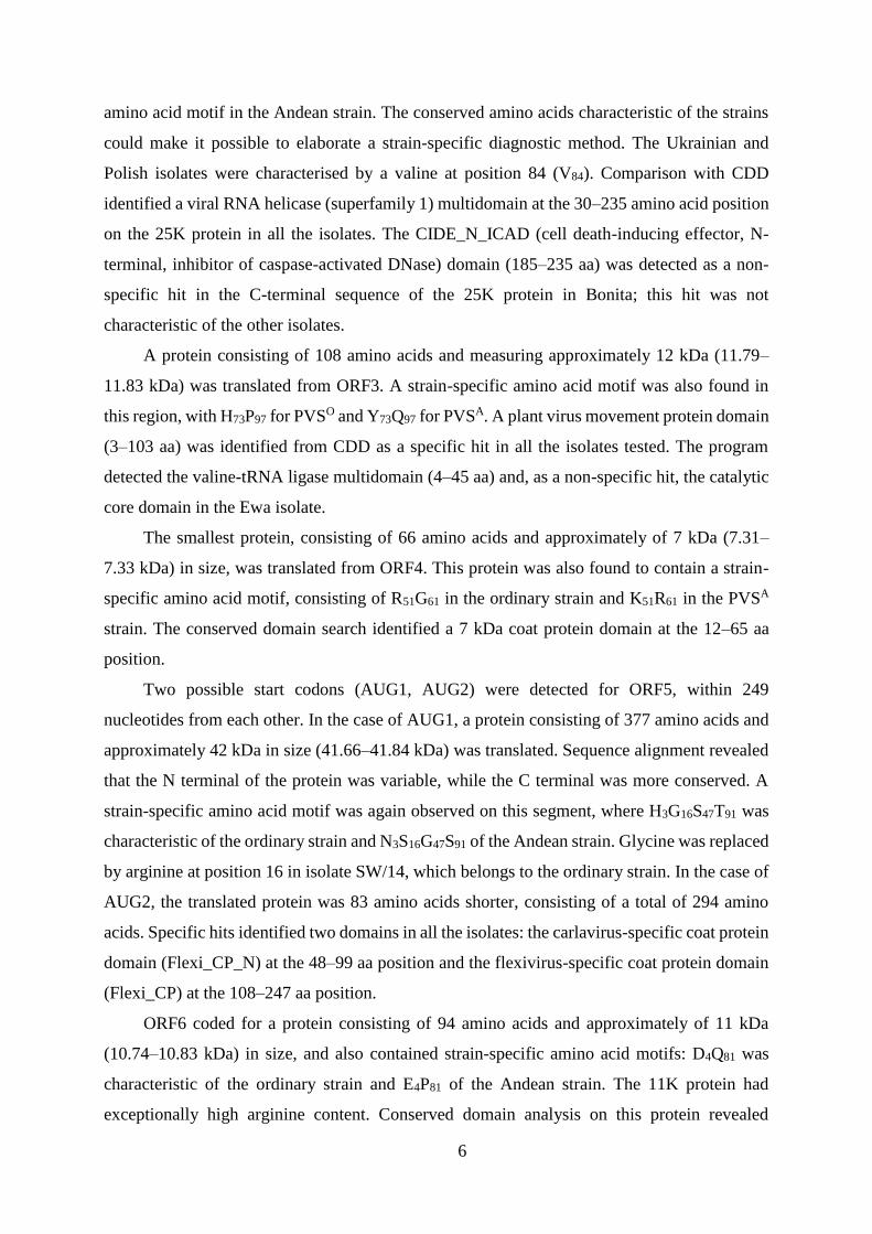
6
amino acid motif in the Andean strain. The conserved amino acids characteristic of the strains
could make it possible to elaborate a strain-specific diagnostic method. The Ukrainian and
Polish isolates were characterised by a valine at position 84 (V84). Comparison with CDD
identified a viral RNA helicase (superfamily 1) multidomain at the 30–235 amino acid position
on the 25K protein in all the isolates. The CIDE_N_ICAD (cell death-inducing effector, N-
terminal, inhibitor of caspase-activated DNase) domain (185–235 aa) was detected as a non-
specific hit in the C-terminal sequence of the 25K protein in Bonita; this hit was not
characteristic of the other isolates.
A protein consisting of 108 amino acids and measuring approximately 12 kDa (11.79–
11.83 kDa) was translated from ORF3. A strain-specific amino acid motif was also found in
this region, with H73P97 for PVSO and Y73Q97 for PVSA. A plant virus movement protein domain
(3–103 aa) was identified from CDD as a specific hit in all the isolates tested. The program
detected the valine-tRNA ligase multidomain (4–45 aa) and, as a non-specific hit, the catalytic
core domain in the Ewa isolate.
The smallest protein, consisting of 66 amino acids and approximately of 7 kDa (7.31–
7.33 kDa) in size, was translated from ORF4. This protein was also found to contain a strain-
specific amino acid motif, consisting of R51G61 in the ordinary strain and K51R61 in the PVSA
strain. The conserved domain search identified a 7 kDa coat protein domain at the 12–65 aa
position.
Two possible start codons (AUG1, AUG2) were detected for ORF5, within 249
nucleotides from each other. In the case of AUG1, a protein consisting of 377 amino acids and
approximately 42 kDa in size (41.66–41.84 kDa) was translated. Sequence alignment revealed
that the N terminal of the protein was variable, while the C terminal was more conserved. A
strain-specific amino acid motif was again observed on this segment, where H3G16S47T91 was
characteristic of the ordinary strain and N3S16G47S91 of the Andean strain. Glycine was replaced
by arginine at position 16 in isolate SW/14, which belongs to the ordinary strain. In the case of
AUG2, the translated protein was 83 amino acids shorter, consisting of a total of 294 amino
acids. Specific hits identified two domains in all the isolates: the carlavirus-specific coat protein
domain (Flexi_CP_N) at the 48–99 aa position and the flexivirus-specific coat protein domain
(Flexi_CP) at the 108–247 aa position.
ORF6 coded for a protein consisting of 94 amino acids and approximately of 11 kDa
(10.74–10.83 kDa) in size, and also contained strain-specific amino acid motifs: D4Q81 was
characteristic of the ordinary strain and E4P81 of the Andean strain. The 11K protein had
exceptionally high arginine content. Conserved domain analysis on this protein revealed

7
specific hits at the 1–89 aa position on all the isolates for a putative nucleic acid-binding protein
motif associated with the Carla_C4 superfamily and characteristic of the carlaviruses.
Six potential recombination events were detected on the Potato virus S isolates using the
RDP4.39 Beta program package, while no clear results were obtained for any of the isolates
using the PhylPro and LARD methods. In the course of evaluation, the isolate from which the
larger part of the recombinant isolate appears to originate is known as the major parent, while
that from which the smaller sequence originates is known as the minor parent. The breakpoints
are designated on the basis of sequence alignment. The dendrograms prepared for the
recombination analysis are not presented here. The first potential recombination event
(recombination event 1) was observed in the 6116–8518 nucleotide segment on the Vltava
isolate. The major parent was found to be Ewa, which belongs to the ordinary strain, while the
minor parent was BB-AND, a member of the Andean strain. The region derived from the major
parent in the recombinant Vltava isolate was found on the dendrogram in the neighbourhood of
Leona and Ewa among the members of the ordinary strain, while the minor parent region was
placed in the Andean strain, in company with the BB-AND isolate. In the case of recombination
event 2 isolate 89.249 was the potential recombinant, the genome of which originated mostly
from Valery (2805–8475 nt), while the minor part (1–2804 nt and 8476–8627 nt) was derived
from Ewa. The rearrangement of the dendrogram prepared for these regions confirmed this
hypothesis (not shown). The Polish isolate Ewa was the recombinant (recombination event 3),
where the major part of the genome (2795–8518 nt) originated from Vltava and the minor part
(1–2794 nt and 8519–8627 nt) from Valery. In the recombined region, Ewa was located on the
same branch of the dendrogram as the Ukrainian isolates. Previous studies confirmed the close
relationship between the Polish and Ukrainian isolates. Ewa was again the potential
recombinant isolate in the case of recombination event 4, where the analysis indicated that the
segments 1–5971 nt and 7231–8627 nt were derived from Leona and the minor part (5972–
7230 nt) from Valery. The recombined region contained the three TGB proteins. It was
concluded that this was further proof of the close relationship between the Polish and Ukrainian
isolates, which was also confirmed by the dendrogram of the recombination regions. All three
Ukrainian isolates could be considered as recombinants in the case of recombination event 5.
The 2761–8150 nt region appeared to have originated from isolate 09.369, and the 1–2760 and
8151–8627 nt regions from Leona. On the dendrogram of the major region, the Ukrainian
isolates formed a separate branch, while in the minor region Leona was also located on this
branch, suggesting the close relationship of the sequence segment. In Yunnan YN, the
recombinant produced by recombination event 6, the 1–4445 and 4878–8627 nt regions
originated from isolate WaDef-US, while the 4446–4877 nt region was derived from the
Id4106-US isolate. The analysis raised the possibility that the Chinese isolate had arisen from

8
the recombination of the two American isolates. The recombination event can also be read from
the phylogenetic dendrograms. Only three of the nine algorithms (RED, GENECONV,
Bootscan) detected this recombination event.
3.2 New scientific results
1. A diagnostic method was elaborated based on the Nested PCR technique, and this reliably
detected PVS even at low virus concentrations. The segment amplified using this method
contained the whole of the coat protein gene.
2. With the help of this diagnostic method the sequences of coat protein genes from 1 Polish,
1 Tanzanian, 3 Ukrainian and 17 Hungarian isolates were determined and the relationships
between them were pinpointed. The sequences have been uploaded to the international
databank.
3. A PCR technique has also been elaborated with which the whole PVS genome can be
amplified in 6 overlapping regions, making it possible to identify the hereditary material of
the virus simply and rapidly.
4. The whole genome sequences of 3 Hungarian, 3 Ukrainian and 1 Polish PVS isolates were
determined using the method elaborated. The sequences have been added to the
international database, thus considerably expanding the number of complete PVS genome
sequences in the database.
5. The conserved domain analysis provided new information on the possible functions of the
PVS genes.
6. Numerous amino acid motifs were identified, which allowed the isolates to be classified
into strains with little further analysis.
7. The recombination analysis proved that intermolecular rearrangements are characteristic of
the PVS genome. Six potential recombination events were detected, five of which have not
previously been described in the international literature.
8. It was recommended that a new strain, to be known as the recombinant strain, should be
created.
4. Conclusions
The whole genomes of three Hungarian, three Ukrainian and one Polish PVS isolates were
determined, and their structure was found to agree with that of the PVS isolate described by
Matoušek et al. (Matousek et al., 2005). A phylogenetic dendrogram was prepared using the
complete genomes of the seven isolates and whole-genome sequences from the NCBI database.
The two strains could be clearly distinguished on this dendrogram, but Vltava formed a separate
branch between the other two groups. According to Duarte et al. (Duarte et al., 2012), Vltava

9
is a recombinant originating from both strains. This was confirmed by the present study. On
this basis it is suggested that a new strain, to be designated as PVSREC should be established.
The analysis revealed that the Ukrainian isolates, which only differ from each other at the amino
acid level in the case of replicase, are also closely related to the Polish isolates. A possible
common origin was confirmed by the results of the recombination analysis. The authors are of
the opinion that the Hungarian isolate 09.369 originated from American ancestors, as
demonstrated by the analysis of all the regions.
ORF1 region
Numerous protein domains were detected on the protein coded by the ORF1 region in the
isolates tested. At the N-terminal end the methyltransferase domain was identified as a specific
hit. Other authors have reported that methyltransferase is involved in the formation of the cap
structure, which increases the stability of the virus RNA and is also essential for the initiation
of translation (Ahola et al., 2000; Ahola et al., 1997; Kong et al., 1999; Rozanov et al., 1992).
In this connection, Potato virus S was compared with 34 other species that belong to the
Carlavirus genus and whose whole genome sequences are available in the NCBI database. PVS
was represented in this analysis by isolate 89.249. All the other species from the genus were
also represented by a single isolate. Numerous conserved amino acid motifs were detected in
the methyltransferase domain, including YLSP, SHP and LEN, which are almost certainly
essential for the carlaviruses.
The second specific hit was OTU-like cysteine protease, a protein family containing
proteins that are homologous to the gene for ovarian tumours (OTU) in Drosophila species.
This family includes proteins originating from eukaryotes, viruses and pathogen bacteria. The
conserved cysteine and histidine, and possibly aspartic acid, represent the catalytic amino acids
in the course of the hypothesised protease function (Makarova et al., 2000). The homology
observed for the amino acid sequence suggests that this protein section also has protease activity
in the PVS isolates. This is supported by the fact that a carlavirus endopeptidase playing a
protease role was detected as the immediate continuation of this domain. It seems likely that
the two domains carry out this function jointly. Lawrence et al. (Lawrence et al., 1995)
identified a papain-like proteinase domain on the 223K protein of Blueberry scorch virus
(BBScV) in the carlavirus endopeptidase family (family C23 in the Merops peptidase database).
These authors suggested that C994H1075 or C895H984 were the catalytic amino acids of
autoproteolysis. During the multiple sequence alignment of the PVS genomes and the
Carlavirus genus it was observed that the C994H1075 amino acids of BBScV were characteristic
of all carlaviruses, without any alteration. It can thus be concluded that the presence of these
amino acids in this position is essential for the functioning of the viruses. The C895H984 amino
acids of BBScV were also conserved in the carlaviruses with a few exceptions (Cowpea mild

10
mottle virus, Aconitum latent virus, Potato latent virus, Sweet potato chlorotic fleck virus), in
which the change in nucleic acids was also manifested as a change in amino acids, but without
influencing the functioning of the virus. The same conclusion was reached previously by
Lawrence et al. in deletion analysis on BBScV (Lawrence et al., 1995). In this connection
Potato virus S was compared with 34 other species from the Carlavirus genus whose whole
genome sequences are available in the NCBI database. PVS was represented in the analysis by
isolate 89.249, and all the other species from the genus were also represented by a single isolate
(62). Based on the analysis it appears that, like the C994H1075 amino acids in BBScV, in the case
of PVS the catalytic amino acids of autoproteolysis are C1003H1084 (in Ewa: C1002H1083). In the
course of multiple sequence alignment a glycine (G1040) and an arginine (R1228) were observed
on the domain, which may also have an important function in all the carlaviruses tested. The
amino acid position was determined using the PVS isolate 89.249.
The third specific hit was the AAA_22 domain. The program identified this hit as
belonging to the ABC transporter protein superfamily. ABC transporters (ATP-binding cassette
transporters) belong to one of the largest and most ancient protein superfamilies, members of
which are to be found in all existing taxa, from prokaryotes to humans. The greatest similarity
is exhibited by the nucleotide-binding domain in all members of the family. These
transmembrane proteins are responsible for transporting a vast range of materials through both
the cell membranes and the internal membranes of the cell (Dean et al., 2001). The AAA family
is a relatively new family among the ATPases. The AAA motif is strongly conserved and
consists of ~230 amino acids. It includes a Walker motif with ATPase activity. In addition to
ATPase activity the AAA family may also perform numerous other functions at cell level, such
as cell cycle regulation, proteolysis, cytoskeleton regulation, or vesicle-mediated protein
transport (Patel and Latterich, 1998; Walker et al., 1982). A viral RNA helicase multidomain
was detected overlapping the AAA domain. Domains belonging to the viral RNA helicase
(superfamily 1) group have already been proved to have helicase and NTPase activity (de
Cedron et al., 1999). In isolates Alex and 09.369 a further AAA domain and an SSL2 domain
were also identified in the multidomain. It has been demonstrated that the SSL2 gene may have
ATPase or helicase activity in addition to its nucleic acid-repairing mechanism (de Cedron et
al., 1999; Gulyas and Donahue, 1992). As the existence of a hit indicates that the domain has
NTPase activity, with which the proteins in the tested isolates exhibit sequence homology, it
can be stated that this protein region probably has a similar role in PVS replication. All the
isolates contained the conserved motif GAGKS (1181–1185 aa, Ewa: 1180–1184 aa) at the N-
terminal end of the viral (superfamily 1) RNA helicase domain. This conserved motif can be
characterised as GXGKS in the case of carlaviruses, confirming the results of earlier studies

11
(Gorbalenya et al., 1988; Zimmern, 1987). A conserved motif that has a constant TFGESTG
sequence in all carlaviruses was identified in this domain.
The RdRp domain was identified at the C-terminal end of the protein translated from
ORF1. RdRp catalyses the synthesis of a complementary RNA strand from the given RNA
template, with the help of which the negative strands, positive strands and subgenomic RNAs
are all replicated (O'Reilly and Kao, 1998). It can thus be hypothesised that the C-terminal end
of the 223K protein performs an RNA polymerase function during virus replication.
It can be concluded from the results that ORF1 codes for a protein with replicase function.
Both the location of the methyltransferase, helicase and RNA-dependent RNA-polymerase
domains on the PVS replicase and their characteristics agree with those reported for other
members of the Carlavirus genus (Matousek et al., 2005). Two further domains with protease
activity were also detected, immediately next to each other. It is thought that the 187 aa segment
containing these two domains may be responsible for autoproteolysis.
ORF2 region
Like the ORF1 protein, the 25 kDa protein coded by ORF2 contains the NTPase/helicase
domain in which the conserved G-GKSS/T motif is to be found (Gorbalenya et al., 1988; Lin
et al., 2009b; Zimmern, 1987). In the isolates tested in the present work this motif has the
sequence GAGKS, as in the ORF1 region, and is located near the N-terminal end of the 25K
protein (47–51 aa). The viral RNA helicase function was confirmed by conserved domain
analysis, in the form of a multidomain detected in the 40–235 aa position.
The CIDE_N_ICAD domain (185–235 aa) was detected as a non-specific hit in the C-
terminal sequence of the 25K protein in Bonita. Although, despite its being a non-specific hit,
CDD classified the identification of this domain as a mathematically reliable result, it appears
more likely to be a chance sequence homology in the Bonita isolate.
ORF3 region
The protein coded by ORF3 is TGBp2, which is 12 kDa in size and has two hydrophobic
regions, as confirmed in the present study (Lin et al., 2009b). This property enables the virus to
spread from cell to cell. This was supported by the results of CDD analysis, where a plant virus
movement protein domain (3–103 aa) was identified as a specific hit in all the isolates. The
plant virus movement protein superfamily contains numerous known plant virus movement
proteins belonging to various different ssRNA plant virus families, including members of the
Potexvirus, Hordeivirus and Carlavirus genus (Scott et al., 1994).
A small domain detected at the N-terminal end of the protein was found to be the core
domain of valyl-tRNA synthetase, an aminoacyl-tRNA synthetase. Scientists have shown that
aminoacyl-tRNA synthetase is essential for all living organisms. This enzyme is a monomer
that aminoacetylates the 2’-OH group of nucleotides at the 3’ end of tRNAs during translation,
12
and is also known to have ligase and dinucleotide-binding properties. The core domain is based
on a glycine-rich Rossmann motif (GxGxxG), a characteristic ATP-binding site (Szymanski et
al., 2000; Venkatachalam et al., 1999). Although the characteristic Rossmann motif does not
contain the virus protein, it is nevertheless thought that the domain detected may have a similar
function.
ORF4 region
The ORF4 region is the shortest coding segment on the genome of the PVS isolates, from
which the smallest protein, 7 kDa in size, is translated. At the N-terminal end this protein
contains a strongly hydrophobic part that plays a role in intercellular movement (30–32).
According to Morozov et al., this hydrophobic segment functions as a signal for penetration
into the endoplasmic reticulum (Morozov et al., 1991).
ORF5 region
Two possible start codons (AUG1, AUG2) were detected for ORF5, 249 nucleotides
apart. An approx. 42 kDa protein is translated in the case of AUG1 and an approx. 33 kDa
protein in that of AUG2. The significance of the two start codons has already been investigated
in potexviruses. Scientists are of the opinion that the N-terminal end of CP, immediately
following AUG1, is important for the cell-to-cell movement of the virus, together with TGBp1,
but this segment is not essential for virion formation. The coat protein subunits, on the other
hand, are translated from the segment following AUG2 (Ozeki et al., 2009; Verchot-Lubicz et
al., 2007). This property of the protein was confirmed by conserved domain analysis. The
carlavirus-specific coat protein domain and the flexivirus-specific coat protein domain were
detected on the protein following AUG2.
The isolates tested in the present work also contained the conserved hydrophobic amino
acid motif (AGFDFFDGLL), which is characteristic of all filoviruses (Foster and Mills, 1991;
Koonin and Gorbalenya, 1989).
ORF6 region
According to the literature, the ORF6 region codes for the cysteine-rich nucleic acid-
binding protein (NABP). This protein is responsible for transmission by leaf aphids and for the
suppression of gene silencing (Chiba et al., 2006; Foster, 1991; Foster and Mills, 1992;
Gramstat et al., 1990). Conserved domain analysis identified a putative nucleic acid-binding
protein motif characteristic of carlaviruses as a specific hit. The carlavirus nucleic acid-binding
protein family contains a potential C4 zinc finger based on four conserved cysteines (Foster and
Mills, 1990a). In the PVS isolates tested here the zinc finger motif had the sequence
RCWRCYRVYPPICNSKCDNRTC and was located in the 54–75 amino acid position on the
protein.

13
The 11 kDa protein probably regulates virus transcription, and the zinc finger protein in
Chrysanthemum virus B has been shown to have a direct interaction with chromatin and plant
promoters, thus functioning as an eukaryotic transcription factor (TF) (Gramstat et al., 1990;
Lukhovitskaya et al., 2013). Based on the amino acid sequence homology it seems likely that
the 11 kDa protein also performs similar functions in PVS. Using the zinc finger model reported
by Gramstatt et al. (Gramstat et al., 1990), a model was designed for the isolates examined here,
on which the nuclear localisation signal (NLS) identified by Lukhovitskaya et al.
(Lukhovitskaya et al., 2013) is also marked.
3’ UTR
Foster et al. (Foster et al., 1992) identified a putative polyadenylation signal (AATAAA)
at the 3’ end of the genomes of Helenium virus S (HelVS) and PVM. This motif was found with
an AAGAAA sequence 24 nucleotides from the 3’ end in the isolates used in the present work.
Within the Carlavirus genus this hexamer is only characteristic of PVS isolates. Other authors
have reported the presence of another hexamer (ACTTAA) in the 3’ UTR region of Potato virus
X, which is essential for RNA synthesis (Batten et al., 2003).This motif was contained in an
unchanged form in all the PVS isolates belonging to the ordinary strain, while the GCTTAA
sequence was characteristic of the Andean strain.
Recombination analysis
The recombination analysis proved that the PVS genome is characterised by
intermolecular rearrangements. The analysis of the PVS isolates detected six potential
recombination events, five of which have not yet been described in the literature. All the isolates
tested here were involved in one or other of the potential recombination events. The first of
these (recombination event 1), which indicated that the Vltava isolate was recombinant and that
the parental sequences belonged to different strains, was previously reported by Duarte et al.
(Duarte et al., 2012) and suggests that regions responsible for aphid transmission and the
development of more severe symptoms were inherited by Vltava from the Andean strain, while
the replicase gene and the 5’ end of the TGBp1 gene originated from the ordinary strain. This
recombination event is of importance from a practical point of view, as it is a direct proof of
the possibility that the traits of the Andean strain, which has better adaptability and
competitiveness, may be transferred to members of the ordinary strain, which was not originally
transmitted by aphids and caused milder symptoms. This observation could explain differences
in the biological traits of isolates classified into strains on the basis of coat protein sequences.
It is therefore essential when studying the pathology and transmission of the virus to include an
analysis of the whole-genome sequences, due to the possibility of intermolecular
rearrangements within the genome. Recombination event 3 indicated that the major part of the
Polish isolate Ewa was derived from Vltava, and the minor part from Valery. This suggests that

14
the Vltava isolate, which is recombinant itself and originated from both PVS strains, may have
participated as a parent sequence in a recombination event with yet another isolate belonging
to the ordinary strain. The other three potential recombination events took place between
members of the ordinary strain. Great attention should be paid in future to the molecular
analysis of PVS and to resistance breeding, in order to prevent the development and spread of
more dangerous strains.
5. References
Ábrahám É. B. (2009). Fajta és öntözés hatása a burgonya termésmennyiségének és minőségének
alakulására mezőségi talajon. Doktori értekezés. Debrecen. 6-10, 34-38.
Ahola, T., den Boon, J. A., and Ahlquist, P. (2000). Helicase and capping enzyme active site
mutations in brome mosaic virus protein 1a cause defects in template recruitment, negative-
strand RNA synthesis, and viral RNA capping. Journal of Virology 74, 8803-8811.
Ahola, T., Laakkonen, P., Vihinen, H., and Kaariainen, L. (1997). Critical residues of Semliki
Forest virus RNA capping enzyme involved in methyltransferase and guanylyltransferase-like
activities. Journal of Virology 71, 392-397.
Batten, J. S., Yoshinari, S., and Hemenway, C. (2003). Potato virus X: a model system for virus
replication, movement and gene expression. Molecular Plant Pathology 4, 125-131.
Boni, M. F., Posada, D., and Feldman, M. W. (2007). An exact nonparametric method for inferring
mosaic structure in sequence triplets. Genetics 176, 1035-1047.
Chiba, M., Reed, J. C., Prokhnevsky, A. I., Chapman, E. J., Mawassi, M., Koonin, E. V.,
Carrington, J. C., and Dolja, V. V. (2006). Diverse suppressors of RNA silencing enhance
agroinfection by a viral replicon. Virology 346, 7-14.
de Bokx, J. A. (1969). Particle length of various isolates of Potato virus S. Netherlands Journal of
Plant Pathology 75, 144-146.
de Bruyn Oubuter, M. P. (1952). A new potato virus. In "Proceedings of the Conference on Potato
Virus Diseases", pp. 83, Wageningen-Lisse
de Cedron, M. G., Ehsani, N., Mikkola, M. L., Garcia, J. A., and Kaariainen, L. (1999). RNA
helicase activity of Semliki Forest virus replicase protein NSP2. Febs Letters 448, 19-22.
Dean, M., Rzhetsky, A., and Allikmets, R. (2001). The human ATP-binding cassette (ABC)
transporter superfamily. Genome Research 11, 1156-1166.
Duarte, P. D. G., Galvino-Costa, S. B. F., Ribeiro, S. R. R. D., and Figueira, A. D. (2012). Complete
genome sequence of the first Andean strain of potato virus S from Brazil and evidence of
recombination between PVS strains. Archives of Virology 157, 1357-1364.
Foster, G. D. (1991). Molecular Variation between Ordinary and Andean Strains of Potato Virus-
S. Research in Virology 142, 413-416.
Foster, G. D., and Mills, P. R. (1990). Detection of Strains of Potato Virus-S by Nucleic-Acid Spot
Hybridization (Nash). Potato Research 33, 487-495.
Foster, G. D., and Mills, P. R. (1991). Evidence for Subgenomic Rnas in Leaves Infected with an
Andean Strain of Potato Virus-S. Acta Virologica 35, 260-267.
Foster, G. D., and Mills, P. R. (1992). The 3'-Nucleotide Sequence of an Ordinary Strain of Potato
Virus-S. Virus Genes 6, 213-220.
Foster, G. D., Scott, R., Draper, J., and Mills, P. R. (1992). Expression of Helenium Virus-S Coat
Protein in Escherichia-Coli, Invitro in Rabbit Reticulocyte Lysate and Transgenic Tobacco.
Acta Virologica 36, 567-575.
Gibbs, M. J., Armstrong, J. S., and Gibbs, A. J. (2000). Sister-Scanning: a Monte Carlo procedure
for assessing signals in recombinant sequences. Bioinformatics 16, 573-582.
Gorbalenya, A. E., Koonin, E. V., Donchenko, A. P., and Blinov, V. M. (1988). A Novel
Superfamily of Nucleoside Triphosphate-Binding Motif Containing Proteins Which Are
Probably Involved in Duplex Unwinding in DNA and Rna Replication and Recombination.
Febs Letters 235, 16-24.

15
Gramstat, A., Courtpozanis, A., and Rohde, W. (1990). The 12-Kda Protein of Potato Virus-M
Displays Properties of a Nucleic Acid-Binding Regulatory Protein. Febs Letters 276, 34-38.
Gulyas, K. D., and Donahue, T. F. (1992). Ssl2, a Suppressor of a Stem-Loop Mutation in the His4
Leader Encodes the Yeast Homolog of Human Ercc-3. Cell 69, 1031-1042.
Ju, H. J., Samuels, T. D., Wang, Y. S., Blancaflor, E., Payton, M., Mitra, R., Krishnamurthy, K.,
Nelson, R. S., and Verchot-Lubicz, J. (2005). The potato virus X TGBp2 movement protein
associates with endoplasmic reticulum-derived vesicles during virus infection. Plant Physiology
138, 1877-1895.
Jukes, T. H. C., C.R. (1969). Evolution of protein molecules. In "Mammalian protein metabolism".
Academic Press, New York, US. .
Kong, F., Sivakumaran, K., and Kao, C. (1999). The N-terminal half of the brome mosaic virus 1a
protein has RNA capping-associated activities: Specificity for GTP and S-adenosylmethionine.
Virology 259, 200-210.
Koonin, E. V., and Gorbalenya, A. E. (1989). Evolution of Rna Genomes - Does the High Mutation-
Rate Necessitate High-Rate of Evolution of Viral-Proteins. Journal of Molecular Evolution 28,
524-527.
Kyte, J., and Doolittle, R. F. (1982). A Simple Method for Displaying the Hydropathic Character
of a Protein. Journal of Molecular Biology 157, 105-132.
Lawrence, D. M., Rozanov, M. N., and Bradley, I. H. (1995). Autocatalytic Processing of the 223-
Kda Protein of Blueberry Scorch Carlavirus by a Papain-Like Proteinase. Virology 207, 127-
135.
Lin, Y., Druffel, K., Whitworth, J. L., Pavek, M. J., and Pappu, H. (2009). Biological and molecular
properties of Potato virus S from late blight resistant potato. Phytopathology 99, S74.
Lukhovitskaya, N. I., Solovieva, A. D., Boddeti, S. K., Thaduri, S., Solovyev, A. G., and Savenkov,
E. I. (2013). An RNA Virus-Encoded Zinc-Finger Protein Acts as a Plant Transcription Factor
and Induces a Regulator of Cell Size and Proliferation in Two Tobacco Species. Plant Cell 25,
960-973.
Makarova, K. S., Aravind, L., and Koonin, E. V. (2000). A novel superfamily of predicted cysteine
proteases from eukaryotes, viruses and Chlamydia pneumoniae. Trends in Biochemical Sciences
25, 50-52.
Marchler-Bauer, A., Derbyshire, M. K., Gonzales, N. R., Lu, S. N., Chitsaz, F., Geer, L. Y., Geer,
R. C., He, J., Gwadz, M., Hurwitz, D. I., Lanczycki, C. J., Lu, F., Marchler, G. H., Song, J. S.,
Thanki, N., Wang, Z. X., Yamashita, R. A., Zhang, D. C., Zheng, C. J., and Bryant, S. H. (2015).
CDD: NCBI's conserved domain database. Nucleic Acids Research 43, D222-D226.
Martin, D., and Rybicki, E. (2000). RDP: detection of recombination amongst aligned sequences.
Bioinformatics 16, 562-563.
Martin, D. P., Posada, D., Crandall, K. A., and Williamson, C. (2005). A modified bootscan
algorithm for automated identification of recombinant sequences and recombination
breakpoints. Aids Research and Human Retroviruses 21, 98-102.
Matousek, J., Schubert, J., Ptacek, J., Kozlova, P., and Dedic, P. (2005). Complete nucleotide
sequence and molecular probing of Potato virus S genome. Acta Virologica 49, 195-205.
Matthews R. E. (1979). Classification and nomenclature of viruses. Intervirology. 12, 129-296.
Morozov, S. Y., Lukasheva, L. I., Chernov, B. K., Skryabin, K. G., and Atabekov, J. G. (1987).
Nucleotide-Sequence of the Open Reading Frames Adjacent to the Coat Protein Cistron in
Potato Virus-X Genome. Febs Letters 213, 438-442.
Morozov, S. Y., Miroshnichenko, N. A., Solovyev, A. G., Zelenina, D. A., Fedorkin, O. N.,
Lukasheva, L. I., Grachev, S. A., and Chernov, B. K. (1991). Invitro Membrane-Binding of the
Translation Products of the Carlavirus 7-Kda Protein Genes. Virology 183, 782-785.
O'Reilly, E. K., and Kao, C. C. (1998). Analysis of RNA-dependent RNA polymerase structure and
function as guided by known polymerase structures and computer predictions of secondary
structure. Virology 252, 287-303.
Orkin, S. (1990). Molecular-Cloning - a Laboratory Manual, 2nd Edition - Sambrook,J, Fritsch,Ef,
Maniatis,T. Nature 343, 604-605.
Ozeki, J., Hashimoto, M., Komatsu, K., Maejima, K., Himeno, M., Senshu, H., Kawanishi, T.,
Kagiwada, S., Yamaji, Y., and Namba, S. (2009). The N-terminal Region of the Plantago

16
asiatica mosaic virus Coat Protein Is Required for Cell-to-Cell Movement but Is Dispensable
for Virion Assembly. Molecular Plant-Microbe Interactions 22, 677-685.
Padidam, M., Sawyer, S., and Fauquet, C. M. (1999). Possible emergence of new geminiviruses by
frequent recombination. Virology 265, 218-225.
Patel, S., and Latterich, M. (1998). The AAA team: related ATPases with diverse functions. Trends
in Cell Biology 8, 65-71.
Posada, D., and Crandall, K. A. (2001). Evaluation of methods for detecting recombination from
DNA sequences: Computer simulations. Proceedings of the National Academy of Sciences of
the United States of America 98, 13757-13762.
Rozanov, M. N., Koonin, E. V., and Gorbalenya, A. E. (1992). Conservation of the Putative
Methyltransferase Domain - a Hallmark of the Sindbis-Like Supergroup of Positive-Strand Rna
Viruses. Journal of General Virology 73, 2129-2134.
Schepetilnikov, M. V., Manske, U., Solovyev, A. G., Zamyatnin, A. A., Schiemann, J., and
Morozov, S. Y. (2005). The hydrophobic segment of Potato virus X TGBp3 is a major
determinant of the protein intracellular trafficking. Journal of General Virology 86, 2379-2391.
Scott, K. P., Kashiwazaki, S., Reavy, B., and Harrison, B. D. (1994). The Nucleotide-Sequence of
Potato Mop-Top Virus-Rna-2 - a Novel Type of Genome Organization for a Furovirus. Journal
of General Virology 75, 3561-3568.
Smith, J. M. (1992). Analyzing the Mosaic Structure of Genes. Journal of Molecular Evolution 34,
126-129.
Szymanski, M., Deniziak, M., and Barciszewski, J. (2000). The new aspects of aminoacyl-tRNA
synthetases. Acta Biochimica Polonica 47, 821-834.
Vaughan E. K., van Slogteren D. H. M. (1956). Potato virus S in Oregon. American Potato Journal.
33, 218-219.
Venkatachalam, K. V., Fuda, H., Koonin, E. V., and Strott, C. A. (1999). Site-selected mutagenesis
of a conserved nucleotide binding HXGH motif located in the ATP sulfurylase domain of human
bifunctional 3 '-phosphoadenosine 5 '-phosphosulfate synthase. Journal of Biological Chemistry
274, 2601-2604.
Verchot-Lubicz, J., Ye, C. M., and Bamunusinghe, D. (2007). Molecular biology of potexviruses:
recent advances. Journal of General Virology 88, 1643-1655.
Walker, J. E., Saraste, M., Runswick, M. J., and Gay, N. J. (1982). Distantly Related Sequences in
the Alpha-Subunits and Beta-Subunits of Atp Synthase, Myosin, Kinases and Other Atp-
Requiring Enzymes and a Common Nucleotide Binding Fold. Embo Journal 1, 945-951.
Wetter C. (1971). Potato virus S. In: Descriptions of plant viruses. Commonwealth Mycological
Institute, Association of Applied Biologist, Kew, England. 60, 1-3.
Zimmern, D. (1987). Evolution of RNA viruses. In "RNA genetics" (J. Holland, Domingo, E,
Ahlquist, P ed.), pp. 211-240. Boca Raton, CRC Press.
This research would not have been possible without funding from the NKTH-
YECH-09-A3-2009-0210 grant.
6. Publications of the author in the topic of the thesis
Journals without IF
Pájtli É., Palkovics L. (2015): A burgonya S vírus (Potato virus S, PVS) lehetséges
rekombinációi. Növényvédelem, „in press”
International conferences (full papers)
Pájtli É., Zámbó Á., és Palkovics L. (2013): Recombination studies on a Hungarian
Potato virus S (PVS) isolate. Episteme czasopismo naukowo-kulturalne 3(18), 357-366.
International conferences (abstracts)

17
Pájtli É., Zámbó Á., Polgár Zs., Wolf István, Cernák I. és Palkovics L. (2012):Hazai
burgonya S vírus (Potato virus S, PVS) izolátum komplett genomjának molekuláris vizsgálata
és rokonsági viszonyainak feltérképezése 58. Növényvédelmi Tudományos Napok, 2012.
február 21-22, p 48.
Pájtli É., Zámbó Á., Polgár Zs., Wolf István, Cernák I. és Palkovics L. (2013):
Molecular analysis of the complete genome of Hungarian Potato virus S isolate and the mapping
of its genetic relationships. 12th IPVE Symposium on Plant Virus Epidemiology, Arusha,
Tanzania, 28 January-1 February, p 150.
Pájtli É., Zámbó Á., és Palkovics L. (2013): Egy hazai burgonya S vírus (Potato virus
S, PVS) izolátum rekombinációs vizsgálata. 59. Növényvédelmi Tudományos Napok, 2013.
február 19-20., p 56.
Pájtli É., Zámbó Á., Polgár Zs., Wolf István és Palkovics L. (2013): Phylogenetic
relationship among Potato virus S isolates. 17th Joint Meeting of EAPR Breeding and Varietal
Assessment Section and EUCARPIA Section Potatoes, Hévíz, Hungary, June 30- July 4, p 39.
7. Other publications of the author
Journals with IF
Petróczy M., Csejk Gy., Pájtli É. and Palkovics L. (2012): Plasmopara obducens
occuring on Impatiens walleriana hybrids and species in Hungary. Morphological and
molecular characterizatioin of the pathogen. Acta Alimentaria, 41 (Supplement), 171-179. IF
0.475
Palkovics L., Pájtli É. and Salamon P. (2015): First report on natural infection of
Colombian datura virus (CDV) in chinese lantern (Physalis alkekengi L.). Plant Disease, 99(6),
898-898, IF (2014): 2.742
Pájtli É., Eke S. and Palkovics L. (2015): First report of the Plantago asiatica mosaic
virus (PlAMV) incidence on Lilium sp. in Hungary. Plant Disease, 99(9),1288., IF (2014):
2.742
Journals without IF
Tóbiás I., Kiss B., Pájtli É., Tholt G., és Salánki K. (2008): A búza törpülés vírus (wheat
dwarf virus) árpa törzsének jellemzése és az átviteli kísérletek. Növényvédelem 44(11), 545-
552.
Pájtli É., Nagy G., és Pájtli J. (2011): A mák védelme. Növényvédelem 47(4), 145-159.
International conferences (abstracts)
Tóbiás I., Pájtli É., Tholt G., Zsiros L. R., Palkovics L. (2008): A búzatörpülés vírus
(wheat dwarf virus) koncentráció változása árpában. XVIII. Keszthelyi Növényvédelmi Fórum,
Keszthely, 2008. január 30.-február 1., p 19.
Tóbiás I., Kiss B., Pájtli É., Tholt G., és Salánki K. (2009): A búzatörpülés vírus (wheat
dwarf virus) árpáról izolált törzseinek jellemzése és átviteli kísérletek, 55. Növényvédelmi
Tudományos Napok, Budapest, 2009. február 23-24., p 32.
Pájtli É. (2009): A búza törpülés vírus (wheat dwarf virus ) etiológiai vizsgálata, XXIX.
Országos Tudományos Diákköri Konferencia, Agrártudományi szekció, Gödöll, 2009. április
6-8., p 245.
Salamon P., Wolf I., Pájtli É., Palkovics L. (2011): PVX-M3- Egy paprikáról (Capsicum
annuum L.) származó deviáns burgonya X-vírus (Potato virus X) izolátum, 57. Növényvédelmi
Tudományos Napok, 2011. február 21-22., p 28.
Palkovics L., Wolf I, Pájtli É. and Salamon P. (2011): PVX-M3 – A deviant pepper
isolate of Potato virus X. American Phytopathological Society-International Congress of Plant
Pathology (APS-ICPP) Joint Meeting. Honolulu, Hawaii USA, 6-10 August. p 136

18
Palkovics L., Wolf I., Pájtli É. and Salamon P. (2011): PVX-M3 – A deviant pepper
isolate of Potato virus X. Phytopathology 101(6, Supplement), p 136.
Pájtli É., Balotai B., Kiss E. és Palkovics L. (2012): Az őszirózsa sárgaság fitoplazma
(’Ca. Phytoplasma asteris’) járványos fellépése a salátatermesztésben, 2011-ben 58.
Növényvédelmi Tudományos Napok, 2012. február 21-22., p 96.
Pájtli É., Salamon P., Balotai B., Kopp A. és Palkovics L. (2013): A Turnip mosaic virus
(TuMV) új gazdanövényei Magyarországon. 59. Növényvédelmi Tudományos Napok, 2013.
február 19-20., p 57.
Pájtli É., Eke S., Halász B. és Palkovics L. (2014): Plantago asiatica mosaic virus
megjelenése Magyarországon liliomon. 60. Növényvédelmi Tudományos Napok, 2014. február
18-19. p 55.
Salamon P., Pájtli É., Nemes K., Kis A., Salánki K., Palkovics L. (2014): Újabb adatok
a Physalis fajokat spontán fertőző vírusokról Magyarországon. 60. Növényvédelmi
Tudományos Napok, 2014. február 18-19. p 70.
Végh A., Hevesi M., Pájtli É., Petrik K., Palkovics L. (2014): Hazai Erwinia amylovora
izolátumok összehasonlítása molekuláris vizsgálat alapján. 60. Növényvédelmi Tudományos
Napok, 2014. február 18-19. p 118.
Sojnóczki A., Pájtli É., Reiter D., Farkas P., Fail J. (2014): Review of Thrips tabaci
(Lindeman) Cytochrome C oxidase gene subunit I (Coi) sequences data. 4th Symposium on
Palaearctic Thysanoptera, Vienna, Austria. Book of abstracts, 8th – 11th September 2014, p 39.
Pájtli É., Eke S. és Palkovics L. (2015): Egy hazai Plantago asiatica mosaic virus
izolátum teljes genomjának jellemzése. 61. Növényvédelmi Tudományos Napok, 2014. február
17-18. p 54.
Koncz L. S., Pájtli É. és Nagy G. (2015): Előzetes felvételezési adatok a kajszi
gutaütésszerű elhalásáról Budapest környéki ültetvényekben. 61. Növényvédelmi Tudományos
Napok, 2014. február 17-18. p 61.
Varga T., Czotter N., Pájtli É., Burgyán J. és Várallyay É. (2015): RNS alapú
diagnosztikai módszer kidolgozása és felhasználása alma ültetvények virológiai felmérésben.
61. Növényvédelmi Tudományos Napok, 2014. február 17-18. p 97.