Thermo Scientific MAbPac HIC Columns · 5 Separation of Proteins Both MAbPac HIC-10 and MAbPac...
8
Thermo Scientific MAbPac HIC Columns Designed for Monoclonal Antibody Analysis Novel Hydrophobic Interaction HPLC Columns
Transcript of Thermo Scientific MAbPac HIC Columns · 5 Separation of Proteins Both MAbPac HIC-10 and MAbPac...
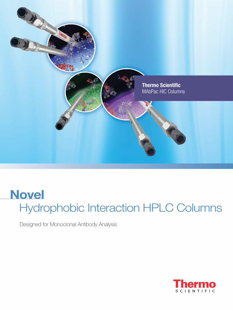
Thermo ScientificMAbPac HIC Columns
Designed for Monoclonal Antibody Analysis
Novel Hydrophobic Interaction HPLC Columns
2
Introduction
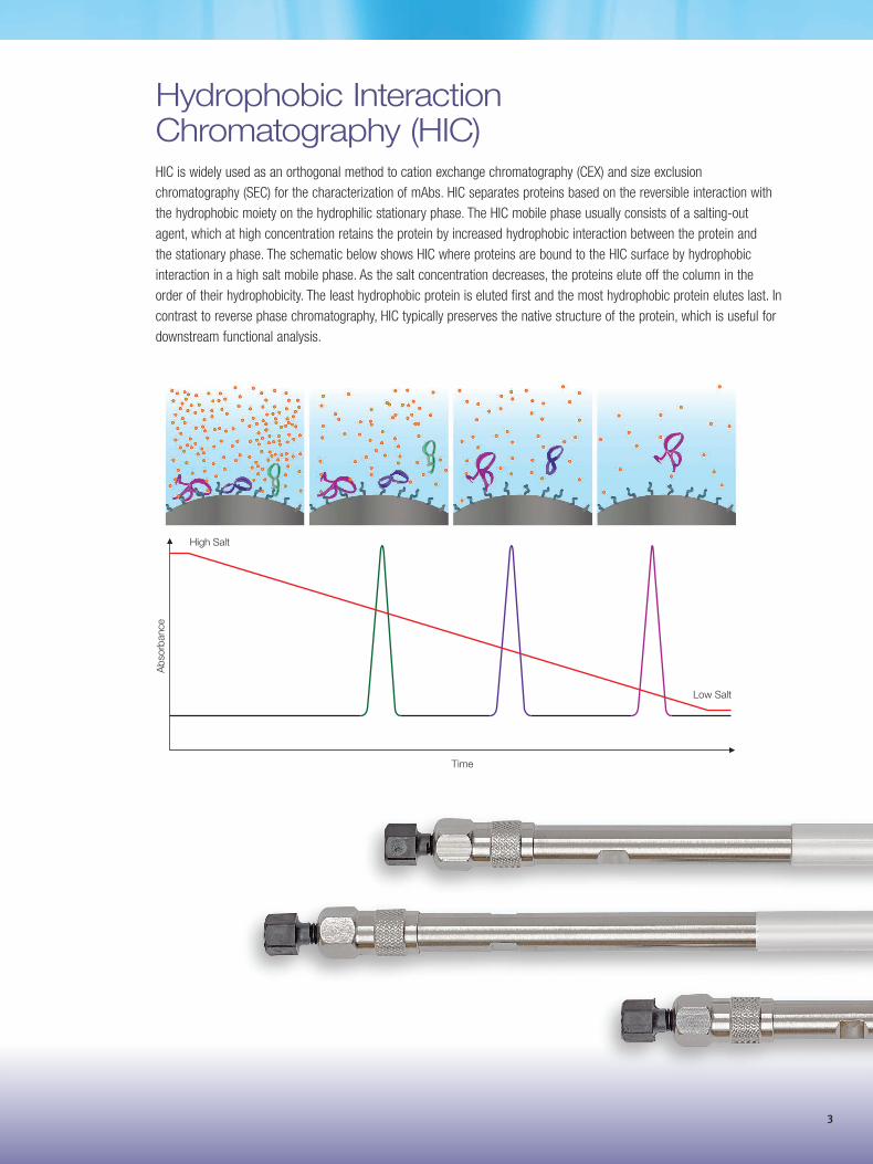
Methionine oxidation
Glycosylation
Reduction of disulfide bondor disulfide shuffling
CH2
VL
VH
CH1
VH
CH1
Antigen-bindingsite
CH3
Fc
Fab
CH2
CH3
Pyroglutamate formationfrom glutamine or glutamic acid
Asparagine deamidation
Aspartic acid isomerization
Heavy Chain
Light Chain
Lysine truncation
CL
VL
CL
K K
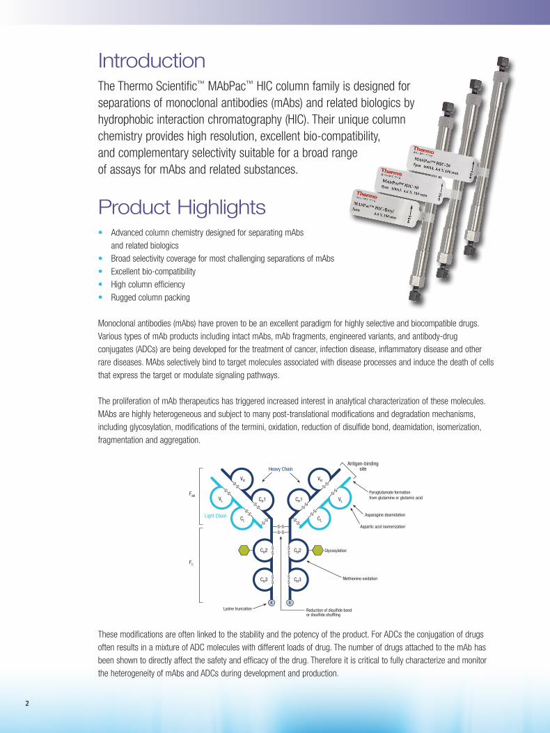
Product Highlights • Advanced column chemistry designed for separating mAbs and related biologics• Broad selectivity coverage for most challenging separations of mAbs• Excellent bio-compatibility• High column efficiency• Rugged column packing Monoclonal antibodies (mAbs) have proven to be an excellent paradigm for highly selective and biocompatible drugs. Various types of mAb products including intact mAbs, mAb fragments, engineered variants, and antibody-drug conjugates (ADCs) are being developed for the treatment of cancer, infection disease, inflammatory disease and other rare diseases. MAbs selectively bind to target molecules associated with disease processes and induce the death of cells that express the target or modulate signaling pathways.
The proliferation of mAb therapeutics has triggered increased interest in analytical characterization of these molecules. MAbs are highly heterogeneous and subject to many post-translational modifications and degradation mechanisms, including glycosylation, modifications of the termini, oxidation, reduction of disulfide bond, deamidation, isomerization, fragmentation and aggregation.
These modifications are often linked to the stability and the potency of the product. For ADCs the conjugation of drugs often results in a mixture of ADC molecules with different loads of drug. The number of drugs attached to the mAb has been shown to directly affect the safety and efficacy of the drug. Therefore it is critical to fully characterize and monitor the heterogeneity of mAbs and ADCs during development and production.
The Thermo Scientific™ MAbPac™ HIC column family is designed for separations of monoclonal antibodies (mAbs) and related biologics by hydrophobic interaction chromatography (HIC). Their unique column chemistry provides high resolution, excellent bio-compatibility, and complementary selectivity suitable for a broad range of assays for mAbs and related substances.
3
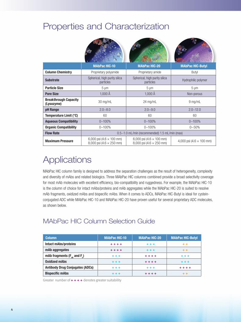
Hydrophobic Interaction Chromatography (HIC) HIC is widely used as an orthogonal method to cation exchange chromatography (CEX) and size exclusion chromatography (SEC) for the characterization of mAbs. HIC separates proteins based on the reversible interaction with the hydrophobic moiety on the hydrophilic stationary phase. The HIC mobile phase usually consists of a salting-out agent, which at high concentration retains the protein by increased hydrophobic interaction between the protein and the stationary phase. The schematic below shows HIC where proteins are bound to the HIC surface by hydrophobic interaction in a high salt mobile phase. As the salt concentration decreases, the proteins elute off the column in the order of their hydrophobicity. The least hydrophobic protein is eluted first and the most hydrophobic protein elutes last. In contrast to reverse phase chromatography, HIC typically preserves the native structure of the protein, which is useful for downstream functional analysis.
Low Salt
High Salt
Time
Abs
orba
nce
4
Applications MAbPac HIC column family is designed to address the separation challenges as the result of heterogeneity, complexity and diversity of mAbs and related biologics. Three MAbPac HIC columns combined provide a broad selectivity coverage for most mAb molecules with excellent efficiency, bio-compatibility and ruggedness. For example, the MAbPac HIC-10 is the column of choice for intact mAbs/proteins and mAb aggregates while the MAbPac HIC-20 is suited to resolve mAb fragments, oxidized mAbs and bispecific mAbs. When it comes to ADCs, MAbPac HIC-Butyl is ideal for cystein-conjugated ADC while MAbPac HIC-10 and MAbPac HIC-20 have proven useful for several proprietary ADC molecules, as shown below.
MAbPac HIC Column Selection Guide
Column MAbPac HIC-10 MAbPac HIC-20 MAbPac HIC-Butyl
Intact mAbs/proteins + + + + + + + + +
mAb aggregates + + + + + + + + +
mAb fragments (Fab and Fc) + + + + + + + + + +
Oxidized mAbs + + + + + + + + + +
Antibody Drug Conjugates (ADCs) + + + + + + + + + +
Bispecific mAbs + + + + + + + + +
Properties and Characterization
MAbPac HIC-10 MAbPac HIC-20 MAbPac HIC-Butyl
Column Chemistry Proprietary polyamide Proprietary amide Butyl
Substrate Spherical, high purity silica particles
Spherical, high purity silica particles Hydrophilic polymer
Particle Size 5 µm 5 µm 5 µm
Pore Size 1,000 Å 1,000 Å Non-porous
Breakthrough Capacity (Lysozyme) 30 mg/mL 24 mg/mL 9 mg/mL
pH Range 2.0−8.0 2.0−9.0 2.0−12.0
Temperature Limit (°C) 60 60 60
Aqueous Compatibility 0−100% 0−100% 0−100%
Organic Compatibility 0−100% 0−100% 0−50%
Flow Rate 0.5−1.0 mL/min (recommended) 1.5 mL/min (max)
Maximum Pressure 6,000 psi (4.6 × 100 mm) 8,000 psi (4.6 × 250 mm)
6,000 psi (4.6 × 100 mm) 8,000 psi (4.6 × 250 mm) 4,000 psi (4.6 × 100 mm)
Greater number of + + + + denotes greater suitability
5
Separation of Proteins Both MAbPac HIC-10 and MAbPac HIC-20 provide high efficiency, high resolution protein separation. The unique chemistry provides excellent bio-compatibility and extremely low carryover. Unlike conventional butyl columns, MAbPac HIC-10 and MAbPac HIC-20 are suitable for hydrophilic proteins as well as hydrophobic proteins.
Column: MAbPac HIC-10, 5 µm
Format: 4.6 × 100 mm
Mobile phase A: 2 M ammonium sulfate, 100 mM sodium phosphate, pH 7.0
Mobile phase B: 100 mM sodium phosphate, pH 7.0
Gradient: Time (min) %A %B -5.0 100 0 0.0 100 0 1.0 100 0 7.0 0 100 10.0 0 100
Flow Rate: 1.0 mL/min
Inj. Volume: 10 µL
Temp.: 30 ºC
Detection: UV (280 nm)
Sample: Protein mixture
Peaks: 1. Myoglobin (2.2 mg/mL) 2. Ribonuclease A (4.4 mg/mL) 3. Lysozyme (1.1 mg/mL) 4. α-Chymotrypsinogen A (2.2 mg/mL)
Abso
rban
ce (m
AU)
Retention Time (min)
0
40
80
120
160
0 2 4 6 8 10
1 2
3
4
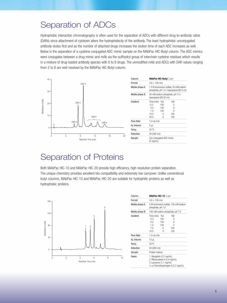
Separation of ADCsHydrophobic interaction chromatography is often used for the separation of ADCs with different drug-to-antibody ratios (DARs) since attachment of cytotoxin alters the hydrophobicity of the antibody. The least hydrophobic unconjugated antibody elutes first and as the number of attached drugs increases the elution time of each ADC increases as well. Below is the separation of a cysteine-conjugated ADC mimic sample on the MAbPac HIC-Butyl column. The ADC mimics were conjugates between a drug mimic and mAb via the sulfhydryl group of interchain cysteine residues which results in a mixture of drug-loaded antibody species with 0 to 8 drugs. The unmodified mAb and ADCs with DAR values ranging from 2 to 8 are well resolved by the MAbPac HIC-Butyl column.
Column: MAbPac HIC-Butyl, 5 µm
Format: 4.6 × 100 mm
Mobile phase A: 1.5 M ammonium sulfate, 50 mM sodium phosphate, pH 7.0 / isopropanol (95:5 v/v)
Mobile phase B: 50 mM sodium phosphate, pH 7.0 / isopropanol (80:20 v/v)
Gradient: Time (min) %A %B -5.0 100 0 0.0 100 0 1.0 100 0 15.0 0 100 20.0 0 100
Flow Rate: 1.0 mL/min
Inj. Volume: 5 µL
Temp.: 25 ºC
Detection: UV (280 nm)
Sample: Cys-conjugated ADC mimic (5 mg/mL)
Abso
rban
ce (m
AU)
Retention Time (min)
40 8 12 16 20
0
10
20
30
40
DAR 0
DAR 2
DAR 4
DAR 8
DAR 6
6
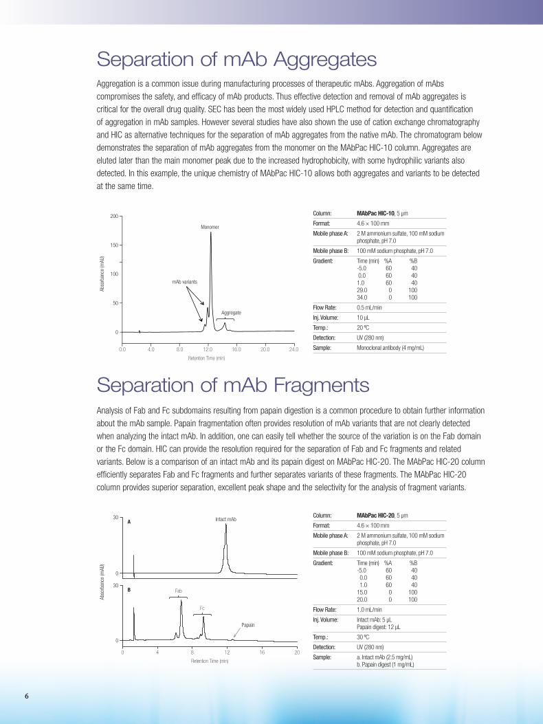
Separation of mAb Aggregates Aggregation is a common issue during manufacturing processes of therapeutic mAbs. Aggregation of mAbs compromises the safety, and efficacy of mAb products. Thus effective detection and removal of mAb aggregates is critical for the overall drug quality. SEC has been the most widely used HPLC method for detection and quantification of aggregation in mAb samples. However several studies have also shown the use of cation exchange chromatography and HIC as alternative techniques for the separation of mAb aggregates from the native mAb. The chromatogram below demonstrates the separation of mAb aggregates from the monomer on the MAbPac HIC-10 column. Aggregates are eluted later than the main monomer peak due to the increased hydrophobicity, with some hydrophilic variants also detected. In this example, the unique chemistry of MAbPac HIC-10 allows both aggregates and variants to be detected at the same time.
Separation of mAb FragmentsAnalysis of Fab and Fc subdomains resulting from papain digestion is a common procedure to obtain further information about the mAb sample. Papain fragmentation often provides resolution of mAb variants that are not clearly detected when analyzing the intact mAb. In addition, one can easily tell whether the source of the variation is on the Fab domain or the Fc domain. HIC can provide the resolution required for the separation of Fab and Fc fragments and related variants. Below is a comparison of an intact mAb and its papain digest on MAbPac HIC-20. The MAbPac HIC-20 column efficiently separates Fab and Fc fragments and further separates variants of these fragments. The MAbPac HIC-20 column provides superior separation, excellent peak shape and the selectivity for the analysis of fragment variants.
Column: MAbPac HIC-10, 5 µm
Format: 4.6 × 100 mm
Mobile phase A: 2 M ammonium sulfate, 100 mM sodium phosphate, pH 7.0
Mobile phase B: 100 mM sodium phosphate, pH 7.0
Gradient: Time (min) %A %B -5.0 60 40 0.0 60 40 1.0 60 40 29.0 0 100 34.0 0 100
Flow Rate: 0.5 mL/min
Inj. Volume: 10 µL
Temp.: 20 ºC
Detection: UV (280 nm)
Sample: Monoclonal antibody (4 mg/mL)
Column: MAbPac HIC-20, 5 µm
Format: 4.6 × 100 mm
Mobile phase A: 2 M ammonium sulfate, 100 mM sodium phosphate, pH 7.0
Mobile phase B: 100 mM sodium phosphate, pH 7.0
Gradient: Time (min) %A %B -5.0 60 40 0.0 60 40 1.0 60 40 15.0 0 100 20.0 0 100
Flow Rate: 1.0 mL/min
Inj. Volume: Intact mAb: 5 µL Papain digest: 12 µL
Temp.: 30 ºC
Detection: UV (280 nm)
Sample: a. Intact mAb (2.5 mg/mL) b. Papain digest (1 mg/mL)
Abso
rban
ce (m
AU)
Retention Time (min)
40 8 12 16 20
0
0
30
30
Fc
A
B
Intact mAb
Fab
Papain
0.0 4.0 8.0 12.0 16.0 20.0 24.0
50
100
150
200
Retention Time (min)
0
mAb variants
Aggregate
Abso
rban
ce (m
AU)
Monomer
7
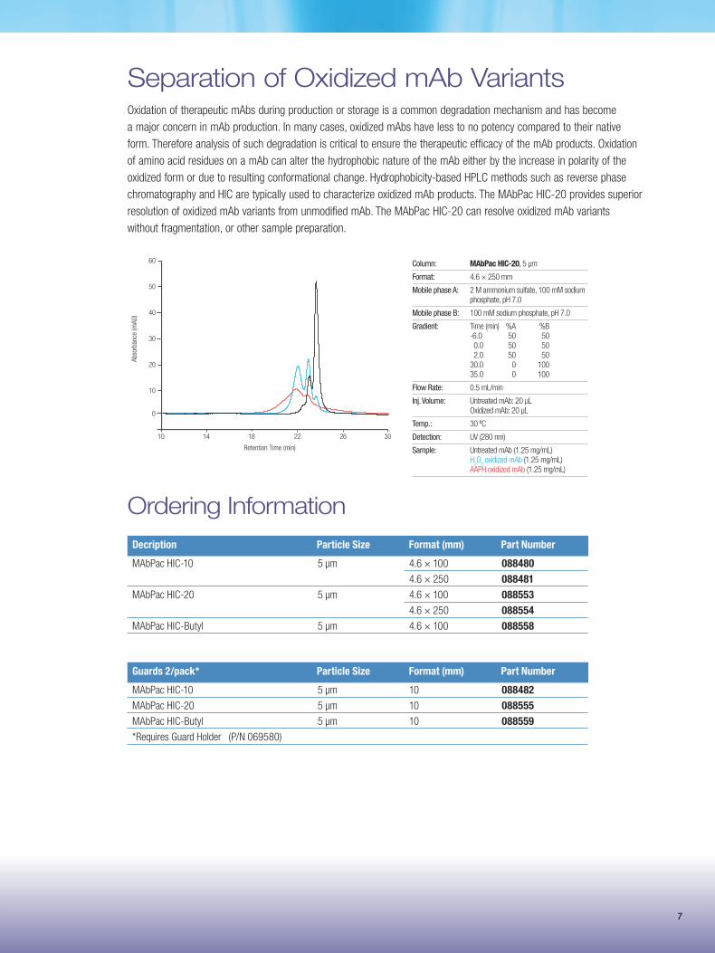
Separation of Oxidized mAb VariantsOxidation of therapeutic mAbs during production or storage is a common degradation mechanism and has become a major concern in mAb production. In many cases, oxidized mAbs have less to no potency compared to their native form. Therefore analysis of such degradation is critical to ensure the therapeutic efficacy of the mAb products. Oxidation of amino acid residues on a mAb can alter the hydrophobic nature of the mAb either by the increase in polarity of the oxidized form or due to resulting conformational change. Hydrophobicity-based HPLC methods such as reverse phase chromatography and HIC are typically used to characterize oxidized mAb products. The MAbPac HIC-20 provides superior resolution of oxidized mAb variants from unmodified mAb. The MAbPac HIC-20 can resolve oxidized mAb variants without fragmentation, or other sample preparation.
Column: MAbPac HIC-20, 5 µm
Format: 4.6 × 250 mm
Mobile phase A: 2 M ammonium sulfate, 100 mM sodium phosphate, pH 7.0
Mobile phase B: 100 mM sodium phosphate, pH 7.0
Gradient: Time (min) %A %B -6.0 50 50 0.0 50 50 2.0 50 50 30.0 0 100 35.0 0 100
Flow Rate: 0.5 mL/min
Inj. Volume: Untreated mAb: 20 µL Oxidized mAb: 20 µL
Temp.: 30 ºC
Detection: UV (280 nm)
Sample: Untreated mAb (1.25 mg/mL) H
2O
2 oxidized mAb (1.25 mg/mL)
AAPH oxidized mAb (1.25 mg/mL)
10 14 18 22 26 30
10
20
30
40
50
60
Retention Time (min)
0
Ab
sorb
ance
(mAU
)
Decription Particle Size Format (mm) Part Number
MAbPac HIC-10 5 µm 4.6 × 100 088480 4.6 × 250 088481 MAbPac HIC-20 5 µm 4.6 × 100 088553
4.6 × 250 088554
MAbPac HIC-Butyl 5 µm 4.6 × 100 088558
Ordering Information
Guards 2/pack* Particle Size Format (mm) Part Number
MAbPac HIC-10 5 µm 10 088482 MAbPac HIC-20 5 µm 10 088555
MAbPac HIC-Butyl 5 µm 10 088559 *Requires Guard Holder (P/N 069580)
A comprehensive product offering for your complete chromatography workflow
Sample Preparation
Sample Handling
LC Columns and Accessories
Separation
Analysis
DataProcessing
BR21089-EN 0616S
www.thermofisher.com/bioLC© 2016 Thermo Fisher Scientific Inc. All rights reserved. All trademarks are the property of Thermo Fisher Scientific and its subsidiaries. Specifications, terms and pricing are subject to change. Not all products are available in all countries. Please consult your local sales representative for details.