management of patient with structural infection and inflamatory cardiac desorder
Upload
jaime-giovanni-ballesteros-munozCategory
view
89download
0
insight review articles
Survival is impossible without vigilant defenceagainst attack and injury. The innate immunesystem continuously surveys the body for thepresence of invaders. When it encounters anattack, it involuntarily sets in motion a
discrete, localized inflammatory response to thwart mostpathogenic threats. The magnitude of the inflammatoryresponse is crucial: insufficient responses result inimmunodeficiency, which can lead to infection andcancer; excessive responses cause morbidity and mortalityin diseases such as rheumatoid arthritis, Crohn’s disease,atherosclerosis, diabetes, Alzheimer’s disease, multiplesclerosis, and cerebral and myocardial ischaemia. Ifinflammation spreads into the bloodstream, as occurs inseptic shock syndrome, sepsis, meningitis and severetrauma, the inflammatory responses can be moredangerous than the original inciting stimulus.Homeostasis and health are restored when inflammationis limited by anti-inflammatory responses that areredundant, rapid, reversible, localized, adaptive to changesin input and integrated by the nervous system.
The nervous system is composed of sensory systems(which detect the state of the body and organs) and motorsystems (which transmit signals to the body and organs).Whereas the somatic motor system controls voluntarymovements, the autonomic motor system controls visceralbody functions and innervates glands (involuntary). Theautonomic nervous system has two principal divisions, theparasympathetic pathway and the sympathetic pathway,which act either in synergy or in opposition to mediate basicphysiological responses in real time. The autonomic systemcontinuously controls heart rate and blood pressure, respiratory rate, gastrointestinal motility, body temperatureand other constantly changing, essential life functions. The autonomic nervous system interacts with the primitive brain, including the limbic system (servingimportant memory functions), brain stem and hypothala-mus. Hypothalamic neural output is relayed to sympatheticand parasympathetic nuclei in the brain stem and spinal cord. Hormonal input also controls the release ofpituitary hormones, which in turn regulate basic functionsof the endocrine organs. Autonomic nervous functions are normally subconscious, but essential basic autonomic functions can be placed under conscious control from signals originating in the higher brain (cerebral cortex). Forexample, subjects can be trained through biofeedback to
decrease their heart rate by increasing parasympathetic outflow.
Recent insights have identified a basic neural pathwaythat reflexively monitors and adjusts the inflammatoryresponse. Inflammatory stimuli activate sensory pathwaysthat relay information to the hypothalamus. Like a knee-jerk reflex, in which the stretching of a patellar tendonelicits a rapidly opposing motor action, inflammatory inputactivates an anti-inflammatory response that is fast and sub-conscious. This prevents spillage of inflammatory productsinto the circulation. The nervous system integrates theinflammatory response: it gathers information about inva-sive events from several local sites, mobilizes defences andcreates memory to improve chances for survival.
Here I review evidence showing that the neural control ofacute inflammation is reflexive, directly interconnected andcontrollable. Special emphasis is placed on cholinergic anti-inflammatory mechanisms that inhibit the activation ofmacrophages and the release of cytokines (Fig. 1). I also discuss evidence indicating that stimulation of the vagusnerve, by either electrical or pharmacological means, prevents inflammation and inhibits the release of cytokinesthat are clinically relevant drug targets for treating inflammatory disease.
Inflammation mediated by TNFTumour-necrosis factor (TNF), a cytokine with a relativemolecular mass of 17,000 (Mr17K), is produced by activatedmacrophages in response to pathogens and other injuriousstimuli, and is a necessary and sufficient mediator of localand systemic inflammation1,2. Local increases in TNF causethe cardinal clinical signs of inflammation, including heat,swelling, pain and redness. Systemic increases in TNF mediate tissue injury by depressing cardiac output, inducing microvascular thrombosis and mediating systemic capillary leakage syndrome. TNF amplifies andprolongs the inflammatory response by activating othercells to release both cytokines such as interleukin 1 (IL-1)and high mobility group B1 (HMGB1), and mediators suchas eicosanoids, nitric oxide and reactive oxygen species,which promote further inflammation and tissue injury3.TNF is essential for the complete expression of inflamma-tion during invasion, and self-limited inflammation is normally characterized by decreasing TNF activity.
Low amounts of TNF can contribute to host defence by limiting the spread of pathogenic organisms into the
The inflammatory reflexKevin J. Tracey
Laboratory of Biomedical Science, North Shore-LIJ Research Institute, 350 Community Drive, Manhasset, New York 11030, USA (e-mail: [email protected])
Inflammation is a local, protective response to microbial invasion or injury. It must be fine-tuned andregulated precisely, because deficiencies or excesses of the inflammatory response cause morbidity andshorten lifespan. The discovery that cholinergic neurons inhibit acute inflammation has qualitatively expandedour understanding of how the nervous system modulates immune responses. The nervous system reflexivelyregulates the inflammatory response in real time, just as it controls heart rate and other vital functions. Theopportunity now exists to apply this insight to the treatment of inflammation through selective and reversible‘hard-wired’ neural systems.
“The mind has great influence over the body, and maladies often have their origin there.” Molière (1622–1673).
NATURE | VOL 420 | 19/26 DECEMBER 2002 | www.nature.com/nature 853© 2002 Nature Publishing Group
anti-inflammatory mechanisms also inhibit the release of IL-1, IL-18 and HMGB1.
Anti-inflammatory responses normally inhibit inflammationHighly conserved, counter-regulatory mechanisms normally limitthe acute inflammatory response and prevent the spread of inflam-matory mediators into the bloodstream (Table 1). Activatedimmunologically competent cells release TNF receptor fragmentsthat bind and neutralize its inflammatory and potentially toxicactions9. Anti-inflammatory cytokines, such as IL-10 and transform-ing growth factor-! (TGF-!), specifically inhibit the release of TNFand other proinflammatory mediators10. Adrenal glucocorticoids,adrenaline, "-melanocyte-stimulating hormone ("-MSH) andother ‘classical’ stress hormones inhibit cytokine synthesis and intracellular signal transduction11–14. Spermine accumulates at sitesof tissue injury and infection and inhibits macrophage activation andcytokine synthesis15.
The importance of these endogenous anti-inflammatory pathways has been shown by experimentally impairing isolated pathways. For example, animals subjected to hypophysectomy oradrenalectomy are significantly sensitized to the lethal effects ofendotoxin16. In the absence of an adequate adrenocorticotropic hor-mone (ACTH) and glucocorticoid response, TNF is significantlyoverexpressed during endotoxaemia16–18. Functional deficiencies inthe release of corticotropin-releasing factor (CRF) predispose Lewisrats to developing experimental arthritis induced by streptococcalantigens because of an insufficient glucocorticoid response13,19,20.Animals deficient in IL-10 develop a chronic inflammatory boweldisease that predominately affects the colon21 and are susceptible to amore severe form of collagen-induced arthritis22. Administration ofspecific pharmacological spermine antagonists significantly increas-es local TNF activity and carrageenan-induced oedema formation,and amplifies the inflammatory response15. Together, these findingsshow that loss of endogenous anti-inflammatory mechanisms converts a normally protective, self-limited inflammatory responseinto an excessive, potentially deleterious response.
Communication between immune and nervous systemsThe activation of pituitary-dependent adrenal responses after endo-toxin administration23 provided early evidence that inflammatorystimuli can activate anti-inflammatory signals from the central nervous system (CNS). Subsequently, Besedovsky et al.24 showeddirectly that inflammation in peripheral tissues alters neuronal sig-nalling in the hypothalamus. Extensive work has identified a commonmolecular basis for communication, with cells from each systemexpressing signalling ligands and receptors from the other25. Forexample, neurons in the CNS can synthesize and express TNF and IL-1, and these cytokines may participate in neuronal communica-tion26,27. This communication is bi-directional, because cytokines canactivate hypothalamic-pituitary release of glucocorticoids and, inturn, glucocorticoids suppress further cytokine synthesis28. In addition, cells of the immune system can produce neuropeptides(including endorphins), acetylcholine and other neurotransmitters.
The importance of the interaction between the nervous systemand immune system signalling has been demonstrated recently in thedevelopment of pathological pain. Watkins and Maier29 have pro-posed that cytokines produced by inflammatory and glial cellschange neuronal excitability and that this link contributes directly tothe development of intractable pain.
Cholinergic anti-inflammatory pathwayOur understanding of the basic mechanisms that regulate inflamma-tion has been advanced by the identification of a neural mechanismthat inhibits macrophage activation through parasympathetic outflow30. Called the ‘cholinergic anti-inflammatory pathway’because acetylcholine is the principle parasympathetic neurotrans-mitter, macrophages that are exposed to acetylcholine are effectively
insight review articles
854 NATURE | VOL 420 | 19/26 DECEMBER 2002 | www.nature.com/nature
circulation, promoting coagulation to localize the invader, and stim-ulating the growth of damaged tissues4. In a typical ‘successful’inflammatory response, the duration and magnitude of TNF release is limited, its beneficial and protective activities predominate,and it is not released systemically. Studies of the inflammatory action of TNF in non-malignant disease have led to widespreadinvestigation of both the ‘normal’ mechanisms that regulate inflammation and the therapeutic potential of monoclonal antibodies specific for TNF.
Monoclonal antibodies against TNF Early studies using monoclonal antibodies against TNF showed thatthis approach effectively prevents lethal tissue injury during bacterialinvasion1. Subsequent clinical trials led to the registration of both mon-oclonal antibodies against TNF, and TNF-binding proteins for treatingrheumatoid arthritis and Crohn’s disease. Many individuals with thesedebilitating inflammatory illnesses have enjoyed disease remissionsand an improved quality of life. Crippling joint pain has been alleviatedin children with rheumatoid arthritis treated with TNF antibodies;some of the youngest patients have even experienced ‘catch-up growth’and normalization of development (U. Andersson, personal commu-nication). These and other clinical successes with TNF monoclonalantibodies have proved that cytokine responses can be manipulated tospecific therapeutic advantage for inflammatory disease.
But strategies using TNF antibodies have not been translated successfully into treatments for bacterial invasion or sepsis, for reasonsthat have been reviewed extensively elsewhere5. Most notably, serumconcentrations of TNF were undetectable in most of the individualsrecruited into clinical sepsis trials, because the study group comprised aheterogeneous population with diverse diseases at varying stages of illness. In early experiments of the use of TNF monoclonal antibodiesin bacteraemia, it became clear that TNF is an early mediator of inflam-mation and that TNF antibodies are ineffective if therapy is initiatedafter serum TNF has been cleared1. Continued interest in understand-ing the use of TNF antibodies for individuals with sepsis is now focusedon identifying a homogenous study population with increased serumTNF for treatment early in the course of illness.
An alternative therapeutic strategy is to target ‘late’ mediators oflethality that are produced after TNF in the inflammatory pathway3.HMGB1 has been implicated as an experimental therapeutic targetthat is produced relatively late in the course of endotoxaemia3,6. Antibodies specific for HMGB1 confer significant protection againstthe lethality of endotoxaemia, even when the first antibody doses areadministered after the early TNF concentrations have been cleared.Reducing serum concentrations of HMGB1 by administering ethylpyruvate as late as 24 h after the onset of sepsis rescues animals fromlethality, indicating that it may now be possible to develop therapiesfor sepsis that cover a significantly wider, clinically relevant treatment window7,8.
Other cytokines have been implicated as therapeutic targets in thepathogenesis of inflammatory diseases, and it is likely that futuretreatment plans will target mediators in addition to TNF. Although Ifocus the discussion of neural regulation of inflammation primarilyon cholinergic inhibition of TNF, evidence indicates that these neural
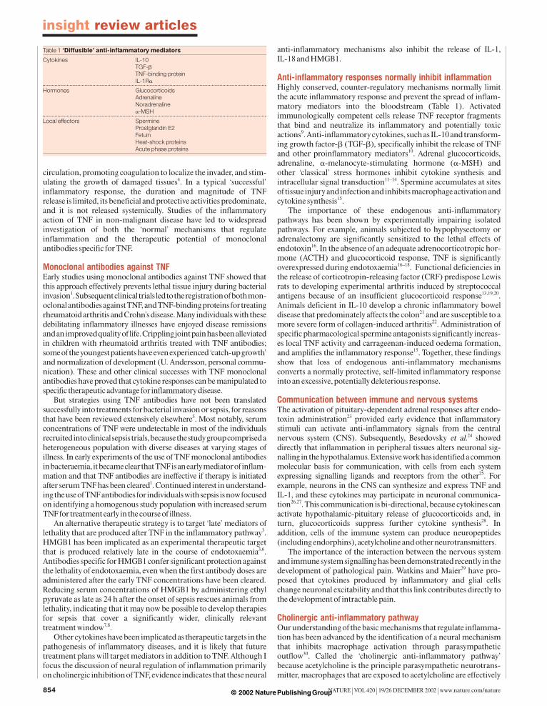
Table 1 ‘Diffusible’ anti-inflammatory mediators
Cytokines IL-10TGF-!TNF-binding proteinIL-1R"
Hormones GlucocorticoidsAdrenalineNoradrenaline"-MSH
Local effectors SpermineProstglandin E2FetuinHeat-shock proteinsAcute phase proteins
© 2002 Nature Publishing Group

insight review articles
NATURE | VOL 420 | 19/26 DECEMBER 2002 | www.nature.com/nature 855
deactivated (Fig. 1). The vagus nerve (which was named for its wan-dering course) innervates the principal organs, including those thatcontain the reticuloendothelial system (liver, lung, spleen, kidneysand gut)31. Experimental activation of the cholinergic anti-inflam-matory pathway by direct electrical stimulation of the efferent vagusnerve inhibits the synthesis of TNF in liver, spleen and heart, andattenuates serum concentrations of TNF during endotoxaemia30,32.Vagotomy significantly exacerbates TNF responses to inflammatorystimuli and sensitizes animals to the lethal effects of endotoxin.
This ‘hard-wired’ connection between the nervous and immunesystems functions as an anti-inflammatory mechanism in othermodels of systemic and local inflammation. Direct stimulation of thevagus nerve in situ inhibits proinflammatory cytokine synthesis inliver and cardiac tissue obtained from animals subjected toischaemia-reperfusion by transient aortic clamping. Stimulation ofeither the right or the left cervical vagus nerves protects against thedevelopment of hypotension and inhibits serum TNF responses toischaemia reperfusion32. The protection conferred by stimulation ofthe vagus nerve is dependent on the applied voltage and is associatedwith normalization of tachycardia during the reperfusion-inducedhypotensive phase32. In a standardized model of experimentalmurine arthritis induced by the application of carrageenan, vagusnerve stimulation inhibits the inflammatory response and suppress-es the development of paw swelling, indicating that the cholinergicanti-inflammatory pathway can inhibit localized inflammationspecifically33.
The molecular dovetail between the cholinergic nervous systemand the innate immune system is a nicotinic, "-bungarotoxin-sensitive macrophage acetylcholine receptor30. Exposure of humanmacrophages, but not peripheral blood monocytes, to nicotine oracetylcholine inhibits the release of TNF, IL-1 and IL-18 in responseto endotoxin. Tissue macrophages, but not circulating monocytes,produce most of the TNF that appears systemically during an excessive inflammatory response. Interaction between themacrophage cholinergic receptor and its ligand inhibits the synthesisof proinflammatory cytokines (TNF, IL-1 and IL-18) but not anti-inflammatory cytokines (such as IL-10)30. Acetylcholineinhibits the expression of TNF protein in macrophages, but not theinduction of TNF messenger RNA levels, indicating that activation of
the cholinergic receptor transduces intracellular signals that inhibitcytokine synthesis at a post-transcriptional stage.
As compared with macrophages, monocytes are refractory to thecytokine-inhibiting effects of acetylcholine: only supraphysiologicalconcentrations of cholinergic agonists inhibit cytokine synthesis inmonocytes30. The macrophage acetylcholine receptor is distinct fromthe muscarinic receptor activities identified on lymphocytes, periph-eral blood mononuclear cells and alveolar macrophages34,35. Theexquisite sensitivity of macrophages to acetylcholine suggests thatother non-neuronal cells that produce acetylcholine (such as epithe-lial cells, T lymphocytes and endothelial cells) might also participatein modulating the function of adjacent tissue macrophages36,37.
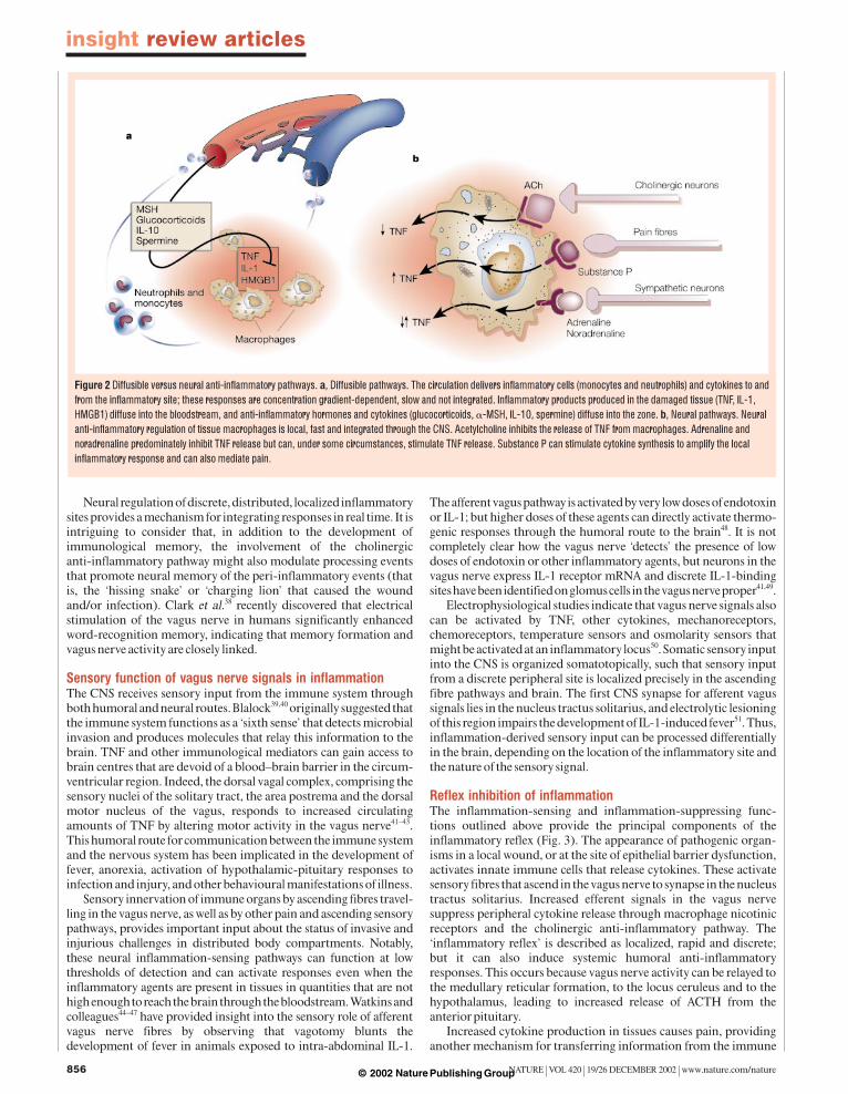
Vagus nerve stimulation suppresses inflammationStimulation of efferent vagus nerve activity has been associated classically with slowing heart rate, induction of gastric motility, dilation of arterioles and constriction of pupils. Inhibition of theinflammatory response can now be added to this list (Fig. 1). From anoversimplified, teleological engineering perspective, there are manyreasons why a neural-based anti-inflammatory pathway is advanta-geous. The diffusible anti-inflammatory network, which includesglucocorticoids, anti-inflammatory cytokines and other humoralmediators, is slow, distributed, non-integrated and dependent onconcentration gradients. By contrast, the cholinergic anti-inflamma-tory pathway is discrete and localized in tissues where invasion andinjury typically originate (Fig. 2).
As compared with the routine, biological pace of a typical, diffusible inflammatory response (hours to days), neural signalling islike lightning. This regulatory attribute is highly advantageous forcontaining immune activation at the crucial stages of a nascentresponse. Neural control of biological functions is short-lived: after abrief refractory period, responding cells can resume function asrequired in the absence of further neural input. Recovery of immunefunction after transient inhibition enables necessary local inflamma-tory responses to be mobilized during persisting threat or infection.The impact of sensitization or desensitization developing after,respectively, denervation or repeated neural firing to an inflammato-ry site has not been explored, but it would be predicted to influenceanti-inflammatory function.
Figure 1 The cholinergic anti-inflammatory pathway.Efferent activity in the vagus nerve leads toacetylcholine (ACh) release in organs of thereticuloendothelial system, including the liver, heart,spleen and gastrointestinal tract. Acetylcholineinteracts with "-bungarotoxin-sensitive nicotinicreceptors (ACh receptor) on tissue macrophages,which inhibit the release of TNF, IL-1, HMGB1 andother cytokines.
© 2002 Nature Publishing Group
The afferent vagus pathway is activated by very low doses of endotoxinor IL-1; but higher doses of these agents can directly activate thermo-genic responses through the humoral route to the brain48. It is notcompletely clear how the vagus nerve ‘detects’ the presence of lowdoses of endotoxin or other inflammatory agents, but neurons in thevagus nerve express IL-1 receptor mRNA and discrete IL-1-bindingsites have been identified on glomus cells in the vagus nerve proper41,49.
Electrophysiological studies indicate that vagus nerve signals alsocan be activated by TNF, other cytokines, mechanoreceptors,chemoreceptors, temperature sensors and osmolarity sensors thatmight be activated at an inflammatory locus50. Somatic sensory inputinto the CNS is organized somatotopically, such that sensory inputfrom a discrete peripheral site is localized precisely in the ascendingfibre pathways and brain. The first CNS synapse for afferent vagussignals lies in the nucleus tractus solitarius, and electrolytic lesioningof this region impairs the development of IL-1-induced fever51. Thus,inflammation-derived sensory input can be processed differentiallyin the brain, depending on the location of the inflammatory site andthe nature of the sensory signal.
Reflex inhibition of inflammationThe inflammation-sensing and inflammation-suppressing func-tions outlined above provide the principal components of theinflammatory reflex (Fig. 3). The appearance of pathogenic organ-isms in a local wound, or at the site of epithelial barrier dysfunction,activates innate immune cells that release cytokines. These activatesensory fibres that ascend in the vagus nerve to synapse in the nucleustractus solitarius. Increased efferent signals in the vagus nerve suppress peripheral cytokine release through macrophage nicotinicreceptors and the cholinergic anti-inflammatory pathway. The‘inflammatory reflex’ is described as localized, rapid and discrete; but it can also induce systemic humoral anti-inflammatory responses. This occurs because vagus nerve activity can be relayed tothe medullary reticular formation, to the locus ceruleus and to thehypothalamus, leading to increased release of ACTH from the anterior pituitary.
Increased cytokine production in tissues causes pain, providinganother mechanism for transferring information from the immune
insight review articles
856 NATURE | VOL 420 | 19/26 DECEMBER 2002 | www.nature.com/nature
Neural regulation of discrete, distributed, localized inflammatorysites provides a mechanism for integrating responses in real time. It isintriguing to consider that, in addition to the development ofimmunological memory, the involvement of the cholinergic anti-inflammatory pathway might also modulate processing eventsthat promote neural memory of the peri-inflammatory events (thatis, the ‘hissing snake’ or ‘charging lion’ that caused the wound and/or infection). Clark et al.38 recently discovered that electricalstimulation of the vagus nerve in humans significantly enhancedword-recognition memory, indicating that memory formation andvagus nerve activity are closely linked.
Sensory function of vagus nerve signals in inflammationThe CNS receives sensory input from the immune system throughboth humoral and neural routes. Blalock39,40 originally suggested thatthe immune system functions as a ‘sixth sense’ that detects microbialinvasion and produces molecules that relay this information to thebrain. TNF and other immunological mediators can gain access tobrain centres that are devoid of a blood–brain barrier in the circum-ventricular region. Indeed, the dorsal vagal complex, comprising thesensory nuclei of the solitary tract, the area postrema and the dorsalmotor nucleus of the vagus, responds to increased circulatingamounts of TNF by altering motor activity in the vagus nerve41–43.This humoral route for communication between the immune systemand the nervous system has been implicated in the development offever, anorexia, activation of hypothalamic-pituitary responses toinfection and injury, and other behavioural manifestations of illness.
Sensory innervation of immune organs by ascending fibres travel-ling in the vagus nerve, as well as by other pain and ascending sensorypathways, provides important input about the status of invasive andinjurious challenges in distributed body compartments. Notably,these neural inflammation-sensing pathways can function at lowthresholds of detection and can activate responses even when theinflammatory agents are present in tissues in quantities that are nothigh enough to reach the brain through the bloodstream. Watkins andcolleagues44–47 have provided insight into the sensory role of afferentvagus nerve fibres by observing that vagotomy blunts the development of fever in animals exposed to intra-abdominal IL-1.
Figure 2 Diffusible versus neural anti-inflammatory pathways. a, Diffusible pathways. The circulation delivers inflammatory cells (monocytes and neutrophils) and cytokines to andfrom the inflammatory site; these responses are concentration gradient-dependent, slow and not integrated. Inflammatory products produced in the damaged tissue (TNF, IL-1,HMGB1) diffuse into the bloodstream, and anti-inflammatory hormones and cytokines (glucocorticoids, "-MSH, IL-10, spermine) diffuse into the zone. b, Neural pathways. Neuralanti-inflammatory regulation of tissue macrophages is local, fast and integrated through the CNS. Acetylcholine inhibits the release of TNF from macrophages. Adrenaline andnoradrenaline predominately inhibit TNF release but can, under some circumstances, stimulate TNF release. Substance P can stimulate cytokine synthesis to amplify the localinflammatory response and can also mediate pain.
© 2002 Nature Publishing Group

insight review articles
NATURE | VOL 420 | 19/26 DECEMBER 2002 | www.nature.com/nature 857
system to the brain. This information can be relayed to other braincentres that influence motor output in the vagus nerve. Pain andstress can activate the flight-or-fight responses, and the resultantincrease of adrenaline and noradrenaline also can inhibitmacrophage activation and suppress synthesis of TNF and othercytokines13,52,53. High sympathetic activity and resultant increases incatecholamines stimulate the !-adrenergic-receptor-dependentrelease of IL-10, a potent anti-inflammatory cytokine, from mono-cytes11,54. Thus, the anti-inflammatory effects of the sympathetic andparasympathetic nervous systems seem to be synergistic in this setting.
Classical teaching stresses that actions of the sympathetic andparasympathetic nervous systems are usually in opposition. But inmany situations the two systems function synergistically. For exam-ple, simultaneous stimulation of both sympathetic and vagus nervesproduces a higher increase in cardiac output than does isolated stim-ulation of either nerve alone55. Flight-or-fight activation of sympa-thetic responses also stimulates increased vagus nerve output. Thecombined action of these neural systems is significantly anti-inflammatory and is positioned anatomically to constrain local inflammation by preventing spillover of potentially lethal toxins intothe circulation through both local (neural) and systemic (humoral)anti-inflammatory mechanisms.
Implications of the inflammatory reflex Knowledge of the inflammatory reflex and the cholinergic anti-inflammatory pathway is yielding insight into both physiologicalpathways and therapeutic strategies (Fig. 4). For example, it may bepossible to activate neural anti-inflammatory mechanisms usingsmall molecules that initiate signals in proximal components of thepathway in the CNS. One such molecule is CNI-1493, a tetravalentguanylhydrazone that was originally described as an inhibitor ofmacrophage activation and TNF release56,57.
CNI-1493 inhibits TNF synthesis and inflammatory responses inanimal models of local and systemic inflammation58. It also signifi-cantly reduced disease severity in a small clinical trial of severeCrohn’s disease and is currently being evaluated in a large phase IItrial of Crohn’s disease59. Unexpectedly, recent evidence has shownthat the TNF-suppressing activities of CNI-1493 in vivo are depen-dent on the cholinergic anti-inflammatory pathway, and that CNI-1493 functions as a pharmacological stimulator of the vagusnerve32,60: intracerebral application of small doses of CNI-1493 significantly inhibited peripheral TNF synthesis, and intact vagusnerves were required to prevent increases in serum TNF. The mecha-nism through which CNI-1493 activates the vagus nerve is unknown,but increased vagus nerve firing has been observed after either intrac-erebral or intravenous administration of CNI-1493 — an effect thatseems to be dependent on specific CNS receptors33.
It is likely that other experimental and clinically approved therapeutic agents suppress peripheral inflammation by activatingpathways in the CNS. Small doses of "-MSH applied intracerebrallyinhibited pulmonary myeloperoxidase activity in mice exposed toendotoxin61 and suppressed the development of intradermal oedemainduced by exposure to TNF or IL-1 (ref. 62). Specific anti-inflam-matory responses have been observed in response to intracerebralapplication of salicylates, but not dexamethasone63. The cardiac anti-arrhythmic drug amiodarone has been identified as an inhibitor ofTNF synthesis in monocytes in vitro64, but it also functions as a potentstimulator of vagus nerve activity65. Systemic administration of thenon-steroidal anti-inflammatory drugs aspirin, indomethacin and
Figure 3 Wiring of the inflammatory reflex. Inflammatory products produced indamaged tissues activate afferent signals that are relayed to the nucleus tractussolitarius; subsequent activation of vagus efferent activity inhibits cytokine synthesisthrough the cholinergic anti-inflammatory pathway (‘the inflammatory reflex’).Information can also be relayed to the hypothalamus and the dorsal vagal complex tostimulate the release of ACTH, thereby activating the humoral anti-inflammatorypathway. Activation of the sympathetic outflow by flight-or-fight responses or pain, orthrough direct signalling, can increase local concentrations of adrenaline andnoradrenaline, which can suppress inflammation further.
Figure 4 Targeting therapies to the cholinergic anti-inflammatory pathway. Thephysiological basis of the cholinergic anti-inflammatory pathway could guide thedevelopment of therapies based on either modulating the activity of the vagus nerve ortargeting specific components of the pathway. For example, biofeedback, conditioning,meditation, hypnosis or acupuncture could be potentially used to modulate vagus output,‘psychoactive’ drugs could be tailor-made to increase vagus output (‘pharmacologicalvagus nerve stimulators’; NSAIDs, non-steroidal anti-inflammatory drugs), and otheragents could be used to target macrophage cholinergic receptors in the periphery.Unbroken lines represent known vagus nerve pathways; dotted lines are hypothetical.
© 2002 Nature Publishing Group

necrosis factor-" and potentiates interleukin 10 production during human endotoxemia. J. Clin.Invest 97, 713–719 (1996).
12.Scheinman, R. I., Cogswell, P. C., Lofquist, A. K. & Baldwin, A. S. Jr Role of transcriptional activationof I#B" in mediation of immunosuppression by glucocorticoids. Science 270, 283–286 (1995).
13.Chrousos, G. P. The stress response and immune function: clinical implications. The 1999 Novera H.Spector Lecture. Ann. NY Acad. Sci. 917, 38–67 (2000).
14.Madden, K. S., Sanders, V. M. & Felten, D. L. Catecholamine influences and sympathetic neuralmodulation of immune responsiveness. Annu. Rev. Pharmacol. Toxicol. 35, 417–448 (1995).
15.Zhang, M., Borovikova, L. V., Wang, H., Metz, C. & Tracey, K. J. Spermine inhibition of monocyteactivation and inflammation. Mol. Med. 5, 595–605 (1999).
16.Bertini, R., Bianchi, M. & Ghezzi, P. Adrenalectomy sensitizes mice to the lethal effects of interleukin1 and tumor necrosis factor. J. Exp. Med. 167, 1708–1712 (1988).
17. Butler, L. D. et al. Neuroendocrine regulation of in vivo cytokine production and effects: I. In vivoregulatory networks involving the neuroendocrine system, interleukin-1 and tumor necrosis factor-".J. Neuroimmunol. 24, 143–153 (1989).
18.Bloom, O. et al. Hypophysectomy, high tumor necrosis factor levels, and hemoglobinemia in lethalendotoxemic shock. Shock 10, 395–400 (1998).
19.Sternberg, E. M. et al. Inflammatory mediator-induced hypothalamic-pituitary-adrenal axisactivation is defective in streptococcal cell wall arthritis-susceptible Lewis rats. Proc. Natl Acad. Sci.USA 86, 2374–2378 (1989).
20.Webster, J. I., Tonelli, L. & Sternberg, E. M. Neuroendocrine regulation of immunity. Annu. Rev.Immunol. 20, 125–163 (2002).
21.Davidson, N. J. et al. T helper cell 1-type CD4+ T cells, but not B cells, mediate colitis in interleukin10-deficient mice. J. Exp. Med. 184, 241–251 (1996).
22. Johansson, A. C., Hansson, A. S., Nandakumar, K. S., Backlund, J. & Holmdahl, R. IL-10-deficientB10. Q mice develop more severe collagen-induced arthritis, but are protected from arthritis inducedwith anti-type II collagen antibodies. J. Immunol. 167, 3505–3512 (2001).
23.Wexler BC, Dolgin AE & Tryczynski EW. Effects of a bacterial polysaccharide (Piromen) on thepituitary-adrenal axis: adrenal ascorbic acid, cholesterol and histologic alterations. Endocrinology 61,300–308 (1957).
24.Besedovsky, H., Sorkin, E., Felix, D. & Haas, H. Hypothalamic changes during the immune response.Eur. J. Immunol. 7, 323–325 (1977).
25.Blalock, J. E. A molecular basis for bidirectional communication between the immune andneuroendocrine systems. Physiol Rev. 69, 1–32 (1989).
26.Breder, C. D., Dinarello, C. A. & Saper, C. B. Interleukin-1 immunoreactive innervation of the humanhypothalamus. Science 240, 321–324 (1988).
27.Breder, C. D. et al. Regional induction of tumor necrosis factor " expression in the mouse brain aftersystemic lipopolysaccharide administration. Proc. Natl Acad. Sci. USA 91, 11393–11397 (1994).
28.Besedovsky, H., del Rey, A., Sorkin, E. & Dinarello, C. A. Immunoregulatory feedback betweeninterleukin-1 and glucocorticoid hormones. Science 233, 652–654 (1986).
29.Watkins, L. R. & Maier, S. F. Beyond neurons: evidence that immune and glial cells contribute topathological pain states. Physiol Rev. 82, 981–1011 (2002).
30.Borovikova, L. V. et al. Vagus nerve stimulation attenuates the systemic inflammatory response toendotoxin. Nature 405, 458–462 (2000).
31.Bellinger, D. L., Lorton, D., Lubahn, C. & Felten, D. L. in Psychoneuroimmunology (eds Ader R.,Felten, D. L. & Cohen, N) 55–112 (Academic, San Diego, 2001).
32.Bernik, T. R. et al. Pharmacological stimulation of the cholinergic antiinflammatory pathway. J. Exp.Med. 195, 781–788 (2002).
33.Borovikova, L. V. et al. Role of vagus nerve signaling in CNI-1493-mediated suppression of acuteinflammation. Auton. Neurosci. 85, 141–147 (2000).
34.Sato, K. Z. et al. Diversity of mRNA expression for muscarinic acetylcholine receptor subtypes andneuronal nicotinic acetylcholine receptor subunits in human mononuclear leukocytes and leukemiccell lines. Neurosci. Lett. 266, 17–20 (1999).
35. Sato, E., Koyama, S., Okubo, Y., Kubo, K. & Sekiguchi, M. Acetylcholine stimulates alveolar macrophagesto release inflammatory cell chemotactic activity. Am. J. Physiol. 274,L970–L979 (1998).
36.Wessler, I., Kirkpatrick, C. J. & Racke, K. Non-neuronal acetylcholine, a locally acting molecule,widely distributed in biological systems: expression and function in humans. Pharmacol. Ther. 77,59–79 (1998).
37.Kawashima, K. & Fujii, T. Extraneuronal cholinergic system in lymphocytes. Pharmacol. Ther. 86,29–48 (2000).
38.Clark, K. B., Naritoku, D. K., Smith, D. C., Browning, R. A. & Jensen, R. A. Enhanced recognitionmemory following vagus nerve stimulation in human subjects. Nature Neurosci. 2, 94–98 (1999).
39.Blalock, J. E. The immune system as a sensory organ. J. Immunol. 132, 1067–1070 (1984).40.Blalock, J. E. Shared ligands and receptors as a molecular mechanism for communication between the
immune and neuroendocrine systems. Ann. NY Acad. Sci. 741, 292–298 (1994).41.Goehler, L. E. et al. Vagal immune-to-brain communication: a visceral chemosensory pathway.
Auton. Neurosci. 85, 49–59 (2000).42.Hermann, G. E., Emch, G. S., Tovar, C. A. & Rogers, R. C. c-Fos generation in the dorsal vagal
complex after systemic endotoxin is not dependent on the vagus nerve. Am. J. Physiol. Regul. Integr.Comp Physiol. 280, R289–R299 (2001).
43.Emch, G. S., Hermann, G. E. & Rogers, R. C. TNF-" activates solitary nucleus neurons responsive togastric distension. Am. J. Physiol. Gastrointest. Liver Physiol. 279, G582–G586 (2000).
44.Watkins, L. R. & Maier, S. F. Implications of immune-to-brain communication for sickness and pain.Proc. Natl Acad. Sci. USA 96, 7710–7713 (1999).
45.Watkins, L. R. et al. Blockade of interleukin-1 induced hyperthermia by subdiaphragmatic vagotomy:evidence for vagal mediation of immune-brain communication. Neurosci. Lett. 183, 27–31 (1995).
46.Hansen, M. K. et al. Effects of vagotomy on lipopolysaccharide-induced brain interleukin-1! proteinin rats. Auton. Neurosci. 85, 119–126 (2000).
47.Hansen, M. K., O’Connor, K. A., Goehler, L. E., Watkins, L. R. & Maier, S. F. The contribution of thevagus nerve in interleukin-1!-induced fever is dependent on dose. Am. J. Physiol. Regul. Integr. Comp.Physiol. 280, R929–R934 (2001).
48.Romanovsky, A. A. Thermoregulatory manifestations of systemic inflammation: lessons fromvagotomy. Auton. Neurosci. Basic Clin. 85, 39–48 (2000).
49.Goehler, L. E. et al. Vagal paraganglia bind biotinylated interleukin-1 receptor antagonist: a possiblemechanism for immune-to-brain communication. Brain Res. Bull. 43, 357–364 (1997).
insight review articles
858 NATURE | VOL 420 | 19/26 DECEMBER 2002 | www.nature.com/nature
ibuprofen substantially increases vagus nerve activity66. Althoughthis vagus nerve response had been studied in the context of increasing gastric acidity and ulcer formation, knowledge of thecholinergic anti-inflammatory pathways raises the possibility thatthe vagus-nerve-stimulating activity of these agents may also contribute to their anti-inflammatory action. A better understand-ing of the CNS receptors, pathways and neural mechanisms that activate the vagus nerve to inhibit production of TNF should facilitate development of this pharmacological ‘vagus-nerve-stimulating’ approach.
Another experimental therapeutic approach is based on directelectrical stimulation of the vagus nerve. So far, more than 10,000individuals have received implantable vagus nerve stimulators for thetreatment of epilepsy67,68. Vagus nerve stimulation in humans withsmall, pacemaker-like devices is safe, well tolerated and not associat-ed with increased rates of infection. But the immunological effects ofthis approach have not been reported and, indeed, it will interestingto assess whether stimulating the vagus nerve in humans modulatesTNF synthesis and inflammation. In place of implantable devices, itshould be possible to develop pharmacological approaches that tar-get the peripheral macrophage receptor to inhibit TNF synthesis. Aprecedent for this approach has been already achieved in the clinic,because nicotine administration is significantly efficacious in reduc-ing the severity of ulcerative colitis69. Other preclinical studies usingstandard murine models of diabetes have shown that nicotinereduces the incidence of diabetes by reducing pancreatic concentra-tions of TNF and other cytokines70. Unanticipated activities of thecholinergic anti-inflammatory pathway in inflammatory disease andin non-immune cells might be determined by further studies.
Some of the earliest studies of the nervous system and inflammationexamined the effects of pavlovian conditioning on intra-abdominalinflammatory responses71. Behavioural conditioning using models oflearned association can reproducibly influence acute inflammatoryresponses and alter the course of experimental inflammatory diseases inanimals and humans72–74. Hypnosis and meditation can significantlyincrease vagus nerve output and have been observed to inhibit immedi-ate-type and delayed-type hypersensitivity responses75,76. Biofeedbackand acupuncture have been used to modulate vagus nerve activity toalter bowel function, gastric acidity and heart rate77,78. Each of theseapproaches has been used to reduce experimental inflammation, butthe relationships between vagus nerve activity and anti-inflammatoryaction had not been defined previously. Autonomic dysfunction occursas a classical complication of rheumatoid arthritis, diabetes and otherautoimmune disorders79–81. It is now intriguing to consider whethervagus nerve dysfunction underlies the progression of inflammation,owing to impairment of the cholinergic anti-inflammatory pathway. Itis reasonable to propose that, one day, the rational modulation of vagusnerve activity using these or other approaches may provide a therapeu-tic advantage for inflammatory disease. ■■
doi:10.1038/nature01321
1. Tracey, K. J. et al. Anti-cachectin/TNF monoclonal antibodies prevent septic shock during lethalbacteraemia. Nature 330, 662–664 (1987).
2. Tracey, K. J. et al. Shock and tissue injury induced by recombinant human cachectin. Science 234,470–474 (1986).
3. Wang, H. et al. HMG-1 as a late mediator of endotoxin lethality in mice. Science 285, 248–251 (1999).4. Tracey, K. J., Vlassara, H. & Cerami, A. Cachectin/tumour necrosis factor. Lancet i, 1122–1126 (1989).5. Tracey, K. J. & Abraham, E. From mouse to man: or what have we learned about cytokine-based anti-
inflammatory therapies? Shock 11, 224–225 (1999).6. Andersson, U. et al. High mobility group 1 protein (HMG-1) stimulates proinflammatory cytokine
synthesis in human monocytes. J. Exp. Med. 192, 565–570 (2000).7. Ulloa, L. et al. Ethyl pyruvate prevents lethality in mice with established lethal sepsis and systemic
inflammation. Proc. Natl Acad. Sci. USA 99, 12351–12356 (2002).8. Wang, H., Yang, H., Czura, C. J., Sama, A. E. & Tracey, K. J. HMGB1 as a late mediator of lethal
systemic inflammation. Am. J. Respir. Crit Care Med. 164, 1768–1773 (2001).9. Lantz, M., Gullberg, U., Nilsson, E. & Olsson, I. Characterization in vitro of a human tumor necrosis
factor-binding protein. A soluble form of a tumor necrosis factor receptor. J. Clin. Invest. 86, 1396(1990).
10.Tsunawaki, S., Sporn, M., Ding, A. & Nathan, C. Deactivation of macrophages by transforminggrowth factor-!. Nature 334, 260–262 (1988).
11.Van der, P. T., Coyle, S. M., Barbosa, K., Braxton, C. C. & Lowry, S. F. Epinephrine inhibits tumor
© 2002 Nature Publishing Group

insight review articles
NATURE | VOL 420 | 19/26 DECEMBER 2002 | www.nature.com/nature 859
50.Berthoud, H. R. & Neuhuber, W. L. Functional and chemical anatomy of the afferent vagal system.Auton. Neurosci. 85, 1–17 (2000).
51.Gordon, F. J. Effect of nucleus tractus solitarius lesions on fever produced by interleukin-1!. Auton.Neurosci. 85, 102–110 (2000).
52.Molina, P. E., Bagby, G. J. & Stahls, P. Hemorrhage alters neuroendocrine, hemodynamic, andcompartment-specific TNF responses to LPS. Shock 16, 459–465 (2001).
53.Molina, P. E. Noradrenergic inhibition of TNF upregulation in hemorrhagic shock.Neuroimmunomodulation 9, 125–133 (2001).
54.Woiciechowsky, C. et al. Sympathetic activation triggers systemic interleukin-10 release inimmunodepression induced by brain injury. Nature Med. 4, 808–813 (1998).
55.Koizumi, K., Terui, N., Kollai, M. & Brooks, C. M. Functional significance of coactivation of vagal andsympathetic cardiac nerves. Proc. Natl Acad. Sci. USA 79, 2116–2120 (1982).
56.Bianchi, M. et al. Suppression of proinflammatory cytokines in monocytes by a tetravalentguanylhydrazone. J. Exp. Med. 183, 927–936 (1996).
57.Bianchi, M. et al. An inhibitor of macrophage arginine transport and nitric oxide production (CNI-1493) prevents acute inflammation and endotoxin lethality. Mol. Med. 1, 254–266 (1995).
58.Tracey, K. J. Suppression of TNF and other proinflammatory cytokines by the tetravalentguanylhydrazone CNI-1493. Prog. Clin. Biol. Res. 397, 335–343 (1998).
59.Hommes, D. et al. Inhibition of stress-activated MAP kinases induces clinical improvement inmoderate to severe Crohn’s disease. Gastroenterology 122, 7–14 (2002).
60.Tracey, K. J., Czura, C. J. & Ivanova, S. Mind over immunity. FASEB J. 15, 1575–1576 (2001).61.Delgado, H. R. et al. Inhibition of systemic inflammation by central action of the neuropeptide "-
melanocyte-stimulating hormone. Neuroimmunomodulation 6, 187–192 (1999).62.Ceriani, G., Macaluso, A., Catania, A. & Lipton, J. M. Central neurogenic antiinflammatory action of
"-MSH: modulation of peripheral inflammation induced by cytokines and other mediators ofinflammation. Neuroendocrinology 59, 138–143 (1994).
63.Catania, A., Arnold, J., Macaluso, A., Hiltz, M. E. & Lipton, J. M. Inhibition of acute inflammation inthe periphery by central action of salicylates. Proc. Natl Acad. Sci. USA 88, 8544–8547 (1991).
64.Matsumori, A., Ono, K., Nishio, R., Nose, Y. & Sasayama, S. Amiodarone inhibits production oftumor necrosis factor-" by human mononuclear cells: a possible mechanism for its effect in heartfailure. Circulation 96, 1386–1389 (1997).
65.Dias, D. S. et al. Opposite effects of iv amiodarone on cardiovascular vagal and sympathetic efferentactivities in rats. Am. J. Physiol. Regul. Integr. Comp. Physiol. 283, R543–R548 (2002).
66.Arai, I., Hirose, H., Muramatsu, M., Okuyama, S. & Aihara, H. Possible involvement of non-steroidalanti-inflammatory drugs in vagal-mediated gastric acid secretion in rats. Jpn. J. Pharmacol. 37, 91–99(1985).
67.Ben Menachem, E. Vagus nerve stimulation, side effects, and long-term safety. J. Clin. Neurophysiol.18, 415–418 (2001).
68.Schachter, S. C. Vagus nerve stimulation: where are we? Curr. Opin. Neurol. 15, 201–206 (2002).69.Pullan, R. D. et al. Transdermal nicotine for active ulcerative colitis. N. Engl. J. Med. 330, 811–815
(1994).70.Mabley, J. G., Pacher, P., Southan, G. J., Salzman, A. L. & Szabo, C. Nicotine reduces the incidence of
type I diabetes in mice. J. Pharmacol. Exp. Ther. 300, 876–881 (2002).71.Metal’nikov, S. a. V. C. Role des reflexes conditionnels dans l’immunite. Ann. Inst. Pasteur 40,
893–900 (1926).72.Madden, K. S. & Felten, D. L. Experimental basis for neural-immune interactions. Physiol Rev. 75,
77–106 (1995).73.Ader R. & Cohen, N. in Psychoneuroimmunology (eds Ader R., Felten, D. L. & Cohen, N) 3–34
(Academic, San Diego, 2001).74.Exton, M. S. et al. Pavlovian conditioning of immune function: animal investigation and the
challenge of human application. Behav. Brain Res. 110, 129–141 (2000).75.Black, S. Inhibition of immediate-type hypersensitivity response by direct suggestion under
hypnosis. Br. Med. J. 1, 925–929 (1963).76.Zachariae, R. in Psychoneuroimmunology (eds Ader R., Felten, D. L. & Cohen, N.) 133–160
(Academic, San Diego, 2001).77.Noguchi, E. & Hayashi, H. Increases in gastric acidity in response to electroacupuncture stimulation
of the hindlimb of anesthetized rats. Jpn. J. Physiol 46, 53–58 (1996).78.Lux, G. et al. Acupuncture inhibits vagal gastric acid secretion stimulated by sham feeding in healthy
subjects. Gut 35, 1026–1029 (1994).79.Toussirot, E., Serratrice, G. & Valentin, P. Autonomic nervous system involvement in rheumatoid
arthritis. 50 cases. J. Rheumatol. 20, 1508–1514 (1993).80.Tan, J., Akin, S., Beyazova, M., Sepici, V. & Tan, E. Sympathetic skin response and R-R interval
variation in rheumatoid arthritis. Two simple tests for the assessment of autonomic function. Am. J.Phys. Med. Rehabil. 72, 196–203 (1993).
81.Edmonds, M. E., Jones, T. C., Saunders, W. A. & Sturrock, R. D. Autonomic neuropathy inrheumatoid arthritis. Br. Med. J. 2, 173–175 (1979).
AcknowledgementsSupported in part by grants from the National Institutes of Health (National Institute ofGeneral Medical Sciences) and the Defense Advanced Research Projects Agency(DARPA). The author is grateful for the thoughtful suggestions from C. Czura, M. Fink,S. Friedman, C. Nathan and B. Sherry.
© 2002 Nature Publishing Group