Vertebrate Body Structure. Vertebrate Body Vertebrate Tissue Types.
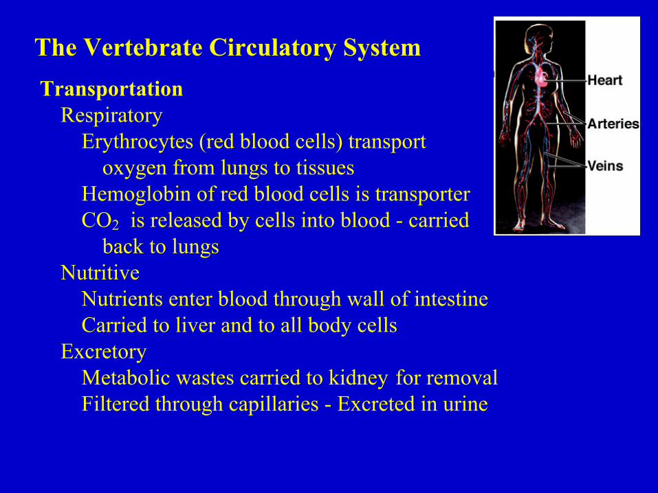
The Vertebrate Circulatory SystemTransportation
RespiratoryErythrocytes (red blood cells) transport
oxygen from lungs to tissuesHemoglobin of red blood cells is transporterCO2 is released by cells into blood - carried
back to lungsNutritive
Nutrients enter blood through wall of intestineCarried to liver and to all body cells
ExcretoryMetabolic wastes carried to kidney for removalFiltered through capillaries - Excreted in urine
RegulationHormone transport
Hormones produced in endocrine glands - transported to targettissues throughout body
Temperature regulationWarm-blooded vertebrates are homeothermsHeat distributed by circulating bloodTemperature adjusted by directing flow to or from extremities
ProtectionBlood clotting
Protects against blood loss when vessels are damagedInvolves proteins in plasma and platelets
Immune defenseLeukocytes, white blood cells, provide immunity against disease agentsAre phagocytic, produce antibodies or have other actions
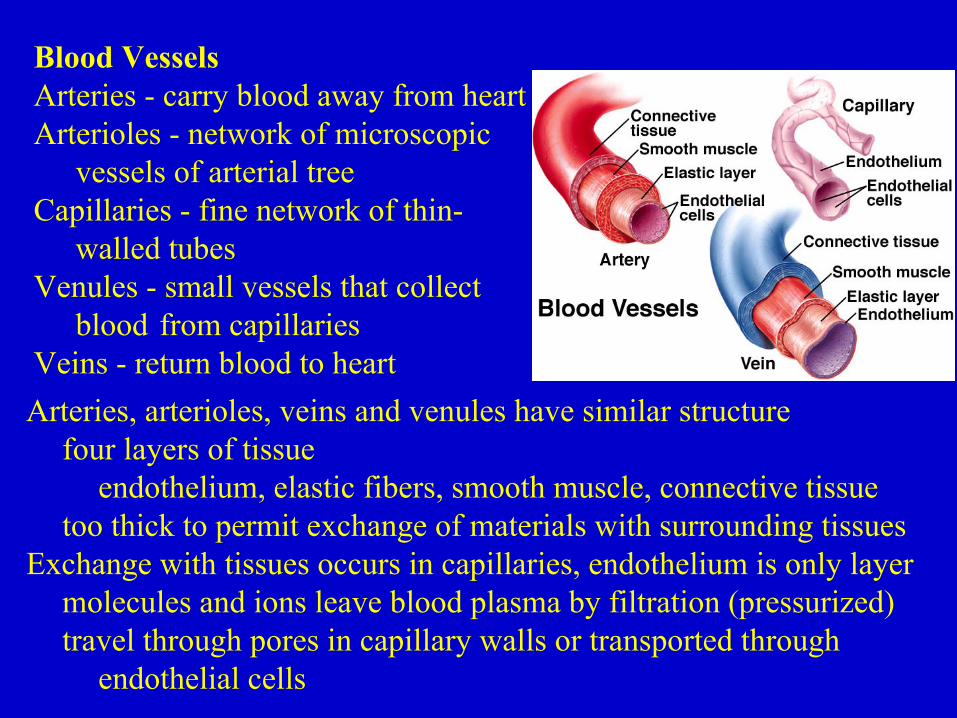
Blood VesselsArteries - carry blood away from heartArterioles - network of microscopic
vessels of arterial treeCapillaries - fine network of thin-
walled tubesVenules - small vessels that collect
blood from capillariesVeins - return blood to heartArteries, arterioles, veins and venules have similar structure
four layers of tissueendothelium, elastic fibers, smooth muscle, connective tissue
too thick to permit exchange of materials with surrounding tissuesExchange with tissues occurs in capillaries, endothelium is only layer
molecules and ions leave blood plasma by filtration (pressurized)travel through pores in capillary walls or transported through
endothelial cells

Arteries and Arterioleselastic fibers allow large arteries to
expand and recoil when receivingblood from heart - helps to buffereffect of pulsing on capillary beds
smaller arteries and arterioles are less elastic, but have thickersmooth muscle - allows change in diametersmall diameter arteries and arterioles cause greatest resistance
to blood flow
Vasoconstriction - through contraction of smooth muscleincreases resistance, decreases flow volume
Vasodilation - through relaxation of smooth muscledecreases resistance, increases flow volume
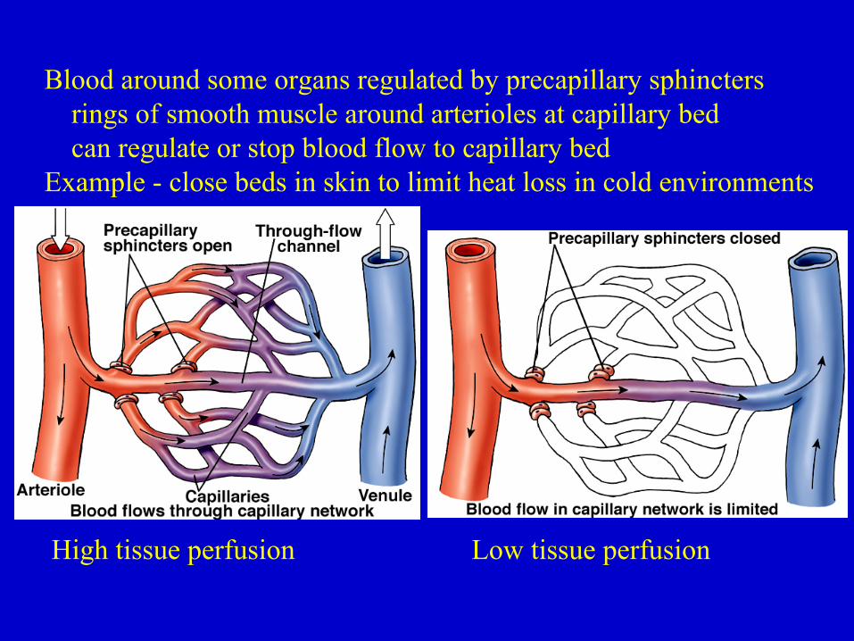
Blood around some organs regulated by precapillary sphinctersrings of smooth muscle around arterioles at capillary bedcan regulate or stop blood flow to capillary bed
Example - close beds in skin to limit heat loss in cold environments
High tissue perfusion Low tissue perfusion

Capillary ExchangeHeart provides sufficient pressure to pump against resistance of
arterial tree and into capillariesEvery cell is within 100 µm of a capillaryAverage capillary 1 mm long, 8 µm diameter, slightly larger than a
red blood cell
Capillaries have greatestcross-sectional area
Blood velocity decreases in capillary beds
Provides greater time for exchange of materials with tissues
Blood pressure is greatly reduced when blood enters veins

Venules and VeinsVeins and venules have thinner layer of smooth muscle than arteriespressure one-tenth that of arteriescan expand to hold greater quantities - most blood in body is in veins
Venous pressure is insufficient toreturn blood to heart from feet- aided by contraction of
skeletal musclesOne-way venous valves direct
flow toward heart
Varicose veins - caused by blood pooling in veins when valves fail
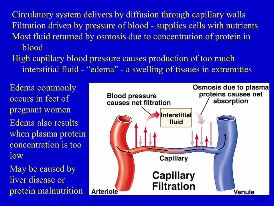
Circulatory system delivers by diffusion through capillary wallsFiltration driven by pressure of blood - supplies cells with nutrientsMost fluid returned by osmosis due to concentration of protein in
bloodHigh capillary blood pressure causes production of too much
interstitial fluid - “edema” - a swelling of tissues in extremities
Edema commonlyoccurs in feet ofpregnant womenEdema also resultswhen plasma proteinconcentration is toolowMay be caused byliver disease orprotein malnutrition

The lymphatic system recovers lost fluid and returns it to bloodComposed of lymphatic capillaries, lymphatic vessels, lymph nodes
and lymphatic organs like spleen and thymusFluid in tissues diffuses into blind-end lymph capillaries
Lymph passes into larger vesselsLymphatic vessels also contain one-way valves

Major lymphatic ducts drain into veins on sides of neckLymph fluid movement assisted by movement of musclesSome lymph vessels contract rhythmicallyLymph modified by phagocytic cells in lymph nodes and
lymphatic organs
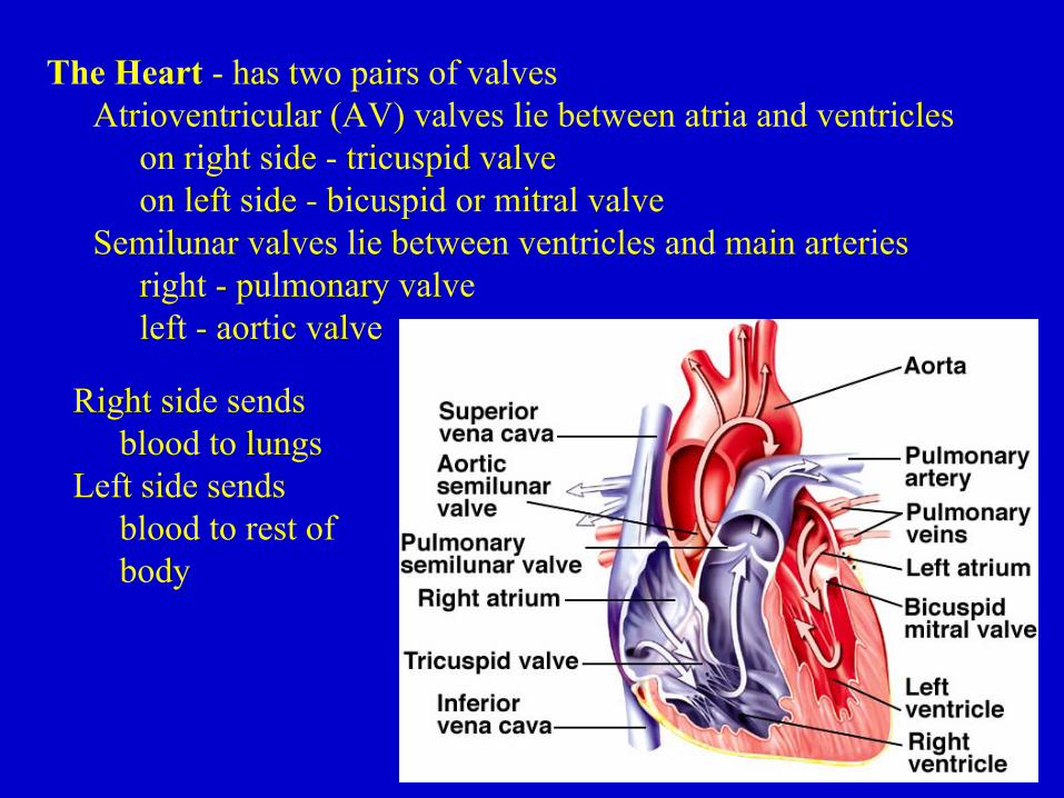
The Heart - has two pairs of valvesAtrioventricular (AV) valves lie between atria and ventricles
on right side - tricuspid valveon left side - bicuspid or mitral valve
Semilunar valves lie between ventricles and main arteriesright - pulmonary valveleft - aortic valve
Right side sends blood to lungs
Left side sends blood to rest of body

How the Heart Is Stimulated to ContractCaused by transmission of membrane depolarization
triggered by sinoatrial (SA) node - the “pacemaker”SA cells depolarize spontaneously with regular rhythm
depolarization passes from one cardiac muscle cell to anothercardiac cells are “electrically” coupled by gap junctions
Atria contract first - ventricular depolarization delayed by ~ 0.1 secSeparated by nonconductive connective tissueWave passes via atrioventricular (AV) nodeDelay permits atria to empty before ventricles contract
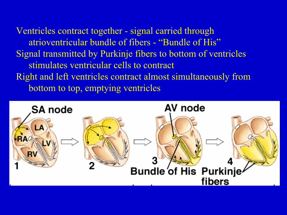
Ventricles contract together - signal carried through atrioventricular bundle of fibers - “Bundle of His”
Signal transmitted by Purkinje fibers to bottom of ventricles stimulates ventricular cells to contract
Right and left ventricles contract almost simultaneously from bottom to top, emptying ventricles

ECG readings and contraction
T =Ventricularrepolarization

Blood Pressure and the Baroreceptor ReflexArterial blood pressure depends on two factors
Cardiac output - how much ventricles pumpResistance to flow
Increased blood pressure caused byIncreased heart rate or blood volume or resistance
Vasoconstriction - produces increased resistance to flowBlood pressure will fall if
Heart rate slows or blood volume reduced or vasodilationBaroreceptors are sensitive to changes in arterial blood pressure
Located in walls of aortic arch and carotid arteriesConnected to cardiovascular control center in medulla
When baroreceptors detect decrease in blood pressureStimulates an increased heart rate and vasoconstriction of
vessels in skin and visceraRaises blood pressure

Baroreceptors act to maintain blood flow to brain with rapid standing
Rapid standing changes venous pressure in lower body, reduces pressure above the heart
Increases volume of blood in lower bodyReduced return of blood to heart and reduced cardiac outputLow blood flow to brain can cause light-headedness or fainting
Reflex rapidly increases heart rate, constricts arteriolesMaintains normal blood pressure


![ERYTHROCYTES [RBCs]](https://static.fdocuments.net/doc/165x107/56812e48550346895d93dd1e/erythrocytes-rbcs.jpg)
![ERYTHROCYTES [RBCs]](https://static.fdocuments.net/doc/165x107/56813dc0550346895da78963/erythrocytes-rbcs-56ea22b2e2743.jpg)


