The Vertebral Column, Pelvis, & Posture
17
7/28/2013 1 The Spine, Spinal Column, and Vertebral Column are synonymous terms referring to the bony components housing the spinal cord Spinal Cord = made of nervous tissue Facet = a small, smooth, flat surface on a bone Facet Joint = the articulation between the superior articular process of the vertebra below with the inferior articular process of the vertebra above Clarification of Terms Lippert, p211-212 Occipital Bone Temporal Bone Vertebrae Intervertebral Disk Atlas Axis C7 Osteology Lippert, p213-216
Transcript of The Vertebral Column, Pelvis, & Posture

7/28/2013
1
The Spine, Spinal Column, and Vertebral Column
are synonymous terms referring to the bony components housing the spinal cord
Spinal Cord = made of nervous tissue
Facet = a small, smooth, flat surface on a bone
Facet Joint = the articulation between the superior articular process of the vertebra below with the inferior articular process of the vertebra above
Clarification of Terms
Lippert, p211-212
Occipital Bone
Temporal Bone
Vertebrae
Intervertebral Disk
Atlas
Axis
C7
Osteology
Lippert, p213-216

7/28/2013
2
Occipital Bone:
Osteology…cont
Temporal Bone:
Osteology…cont
Osteology continued
Parts of a Vertebra
Body
Neural Arch
Vertebral Foramen
Pedicle
Lamina
Transverse Process
Articular Process
Spinous Process
body
sp
Vertebral
foramen
Neural Arch
7/28/2013
3
Typical Cervical Vertebrae
Vertebrae:
Osteology…cont
7/28/2013
4
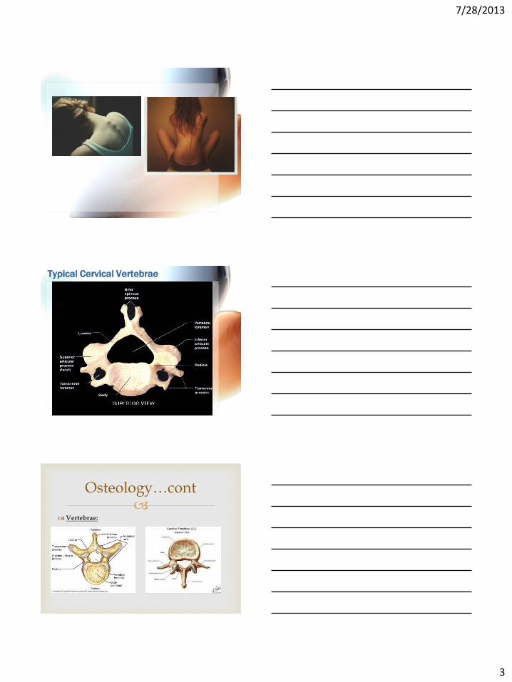
Intervertebral Disks:
23 disks located between vertebrae, starting between C2 and C3
Function = absorb and transmit shock and maintain flexibility of the vertebral column
Disks make up approx 25% of the total length of the vertebral column
Osteology…cont
Lippert, p214-215
Intervertebral Disks…cont:
Annulus Fibrosus: outer portion of the disk consisting of several fibrocartilagenous rings that contain the nucleus pulposus
Nucleus Pulposus: pulpy, gelatinous substance with high water content in the center of the disk
Osteology…cont
Lippert, p215
Atlas: Caudal aspect

7/28/2013
5
Axis: 2nd Cervical Vertebra
Posterior Aspect
C7:
Osteology…cont
7/28/2013
6
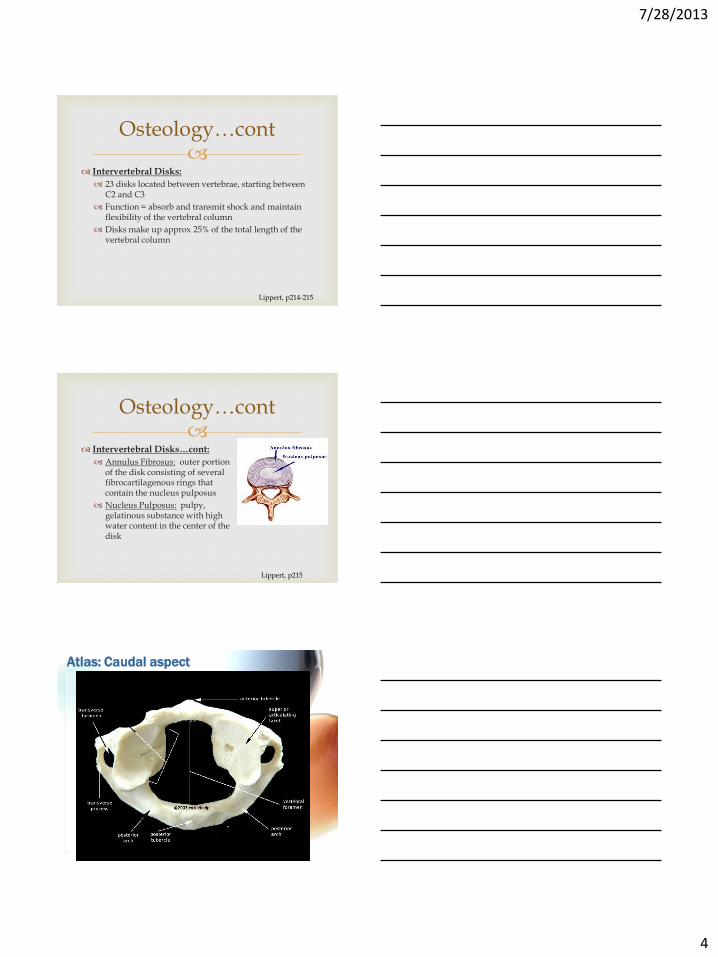
Examples of Vertebrae
What can you palpate?
What can you NOT palpate?
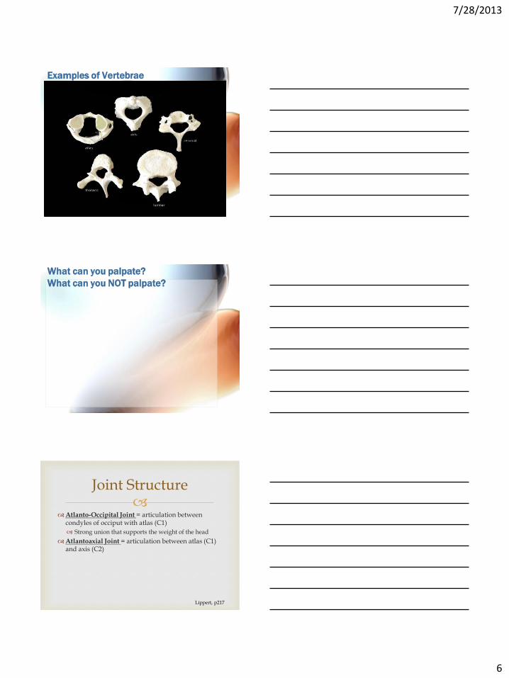
Atlanto-Occipital Joint = articulation between
condyles of occiput with atlas (C1)
Strong union that supports the weight of the head
Atlantoaxial Joint = articulation between atlas (C1) and axis (C2)
Joint Structure
Lippert, p217

7/28/2013
7
Articulations between C2 through S1 = all basically
the same
Strong, weight bearing articulations occur anteriorly between the vertebral bodies
Posteriorly, there are 2 articulations (one on each side) called facet joints (formed by the articular processes of adjacent vertebrae)
Each facet joint = synovial joint with synovial membrane and capsular ligament
The direction the facets face largely determine the type and amount of motion possible at that part of the vertebral column
Joint Structure…cont
Lippert, p217
Atlanto-Occipital Joint Flexion and extension, no rotation
Atlantoaxial Joint Rotation and some lateral flexion (aka sidebending)
Cervical Spine Flexion, extension, rotation, sidebending Retraction = combined head flexion on C1 and C2-C7 extension Protraction = combined head extension C1 and C2-C7 flexion
Thoracic Spine Facets in frontal plane Mostly rotation and lateral flexion Attachment of ribs contributes to lack of flexion and extension
Lumbar Spine Facets in sagittal plane Most flexion and extension of vertebral column occurs in lumbar spine
Joint Movement
Lippert, p219
Supporting Structures
Anterior longitudinal
ligament
Attaches the bodies of the
vertebrae on the anterior
surface
Prevents excessive hyper
extension
Thin superiorly and thick
inferiorly to fuse the
sacrum
Found in the thoracic and
lumbar regions deep to the
aorta
Lippert, 218

7/28/2013
8
Supporting Structures…cont
Posterior longitudinal
ligament
Attaches to the bodies of the
vertebrae on the posterior
surfaces inside the vertebral
foramen
Prevents excessive flexion
Thick superiorly to help
support the skull and thin
inferiorly
Contributes to instability and
increased disk injury in the
lumbar region.
Lippert, 218
Supporting Structures…cont
Supraspinal ligament Extends from the 7th cervical
vertebra distally to the sacrum
posteriorly along the tips of the
spinous processes
Interspinous ligament Attaches successive spinous
processes
Nuchal ligament Interspinous ligament in the
cervical spine
Lippert, 218
Supporting Structures…cont
Ligamentum Flavum
Connects adjacent laminae on the anterior surface
Lippert, 218
7/28/2013
9
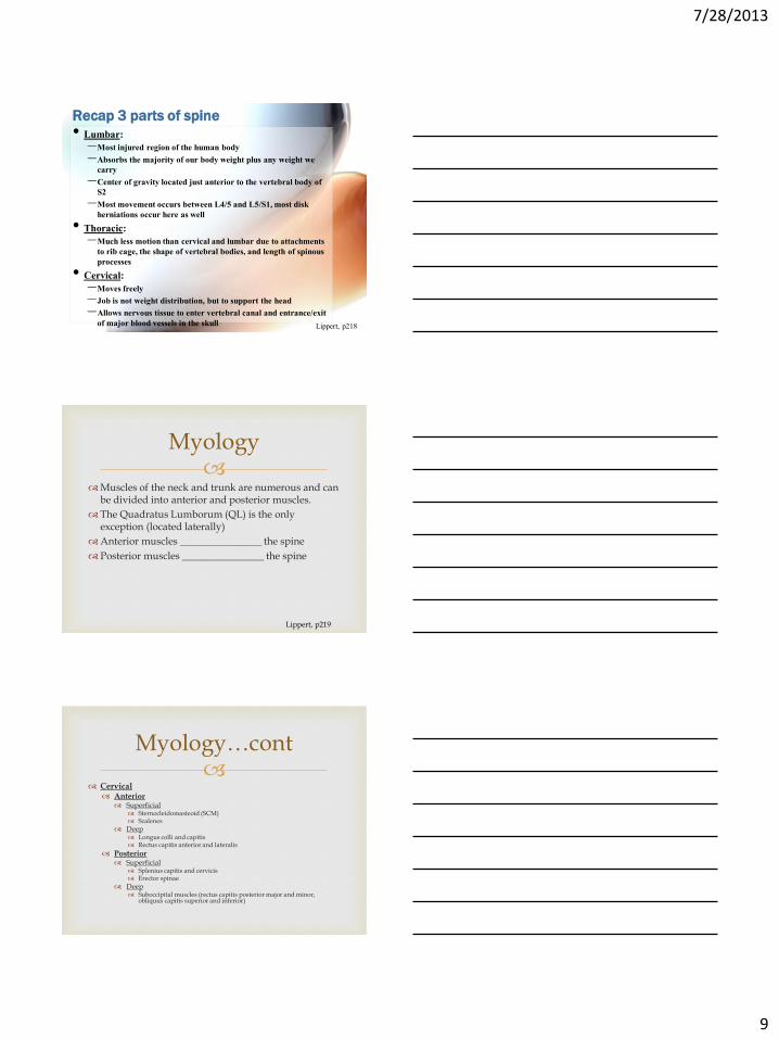
Recap 3 parts of spine
• Lumbar:
–Most injured region of the human body
–Absorbs the majority of our body weight plus any weight we
carry
–Center of gravity located just anterior to the vertebral body of
S2
–Most movement occurs between L4/5 and L5/S1, most disk
herniations occur here as well
• Thoracic:
–Much less motion than cervical and lumbar due to attachments
to rib cage, the shape of vertebral bodies, and length of spinous
processes
• Cervical:
–Moves freely
–Job is not weight distribution, but to support the head
–Allows nervous tissue to enter vertebral canal and entrance/exit
of major blood vessels in the skull Lippert, p218
Muscles of the neck and trunk are numerous and can
be divided into anterior and posterior muscles.
The Quadratus Lumborum (QL) is the only exception (located laterally)
Anterior muscles ________________ the spine
Posterior muscles ________________ the spine
Myology
Lippert, p219
Cervical Anterior Superficial
Sternocleidomasteoid (SCM) Scalenes
Deep Longus colli and capitis Rectus capitis anterior and lateralis
Posterior Superficial
Splenius capitis and cervicis Erector spinae
Deep Suboccipital muscles (rectus capitis posterior major and minor,
obliquus capitis superior and inferior)
Myology…cont

7/28/2013
10
Cervical
Anterior
Superficial
Sternocleidomasteoid (SCM)
Scalenes
Myology…cont
Sternocleidomastoid
Origin Sternal head: superior aspect
of the manubrium of the
sternum
Clavicular head: medial 1/3
of the clavicle
Insertion Mastoid process of the
temporal bone
Innervation Spinal accessory n. (cranial
n. XI)
Action Bilateral: Flexion of the
head & neck
Unilateral: Contralateral
rotation of the head and
neck
Lippert, p219-220
Scalenes
Origin Ant. Scalene: transverse processes
of C3-C7
Middle Scalene: transverse
processes of C2-C7
Posterior Scalene: transverse
processes of C5-C7
Insertion Ant. Scalene: 1st rib
Middle Scalene: 1st rib
Posterior Scalene: external surface
of the 2nd rib
Innervation Ventral rami (C3-C7)
Action Bilateral: flexion of the neck, assist
with inspiration by elevating ribs
1&2
Unilateral: lateral flexion
Lippert, p220
7/28/2013
11
Myology…cont
Cervical
Anterior
Deep
Longus colli = flexes neck
Longus capitis = flexes head
Rectus capitis anterior = flexes head
Rectus capitis lateralis = laterally bends head
Lippert, p221
Posterior Neck
Superficial (Erector Spinae)
Splenius capitis & cervicis= bilaterally = extends head and neck, unilaterally = rotates and laterally bends the face to same side
Erector spinae = extensors which bring the head back from a flexed position
Deep
Suboccipital muscles (rectus capitis posterior major* and minor, obliquus capitis superior and inferior *) = extend the head and * also lateral bend and rotate to the same side
Myology…cont
Lippert, p221-222
Neck:
Posterior:
Superficial: Splenius capitis and cervicis
Myology…cont

7/28/2013
12
Neck:
Posterior:
Deep:
Suboccipital Muscles
Myology…cont
Trunk Anterior Superficial to Deep:
Rectus Abdominis External Oblique Internal Oblique Transverse Abdominis
Posterior Superficial
Erector spinae
Deep Transverse spinal muscles
Lateral Deep
Quadratus Lumborum
Myology…cont
Trunk
Anterior
Superficial to Deep:
Rectus Abdominis
External Oblique
Internal Oblique
Transverse Abdominis
Myology…cont

7/28/2013
13
Rectus Abdominis
Origin Crest of the pubis
Insertion Xiphoid process and cartilages
of ribs 5-7
Innervation Intercostal n. (T7-T12)
Action Flexion of the trunk, posterior
pelvic tilt, increases intra-
abdominal and intrathoracic
pressure
“tidbits” pregnancy?
tendinous inscriptions?
Lippert, p223
Strengthening Rectus Abdominis
• Crunch: to hold the ankles/feet down or not?
Lippert, p222-223
External Oblique
Origin Lateral side of ribs 4-12
Insertion Iliac crest and linea alba
Innervation Intercostal nerves (T8-
T12)
Action Bilateral: Flexion of
the trunk, posterior
pelvic tilt, increased
intra-abdominal and
intra-thoracic pressure
Unilateral: Rotation of
the trunk to the
contralateral side,
ipsilateral lateral
flexion of the trunk
Lippert, p223

7/28/2013
14
Strengthening External Oblique
•When you do a crunch on a diagonal and bring
your right shoulder toward your left knee,
which external oblique is responsible for this
motion? The right or left?
Internal Oblique
Origin Iliac crest, inguinal
ligament &
thoracolumbar fascia
Insertion Ribs 9-12, linea alba
Innervation Intercostal n. (T8-T12)
Action Bilateral: flexion of
the trunk, posterior
pelvic tilt, increases
intra-abdominal and
intra-thoracic pressure
Unilateral: ipsilateral
lateral flexion of the
trunk, rotation of the
trunk to the ipsilateral
side Lippert, p223
Strengthening Internal Oblique
•When you do a crunch on a diagonal and bring
your right shoulder toward your left knee,
which internal oblique is responsible for this
motion? The right or left?

7/28/2013
15
Lippert, p224
Strengthening Transverse Abdominis
Myology…cont
Trunk: Posterior: Superficial: Erector Spinae
3 muscles make up the erector spinae
Medially = spinalis
Attaches spinous process to spinous process
Action = extension
Intermediate = longissimus
Attaches transverse process to transverse process
Action = extension and lateral bending
Laterally = iliocostalis
Attaches transverse process to rib or rib to rib
Action = extension and lateral bending
Lippert, p224

7/28/2013
16
Trunk: Posterior: Deep: Transverse Spinal Muscle
Group
3 muscles make up the Transverse Spinal Group
They attach from transverse to spinous process
Action = extension and rotation to the opposite side
Most superficial = semispinalis (tend to span ≥ 5 vertabrae)
Intermediate = multifidus (tend to span 2-4 vertebrae)
Deepest = rotatores (span 1 vertebrae)
(All run the entire vertebral column in layers)
Myology…cont
Lippert, p225-226
Trunk: Posterior: Deep: Transverse Spinal Muscle
Group
Myology…cont
Trunk:
Lateral:
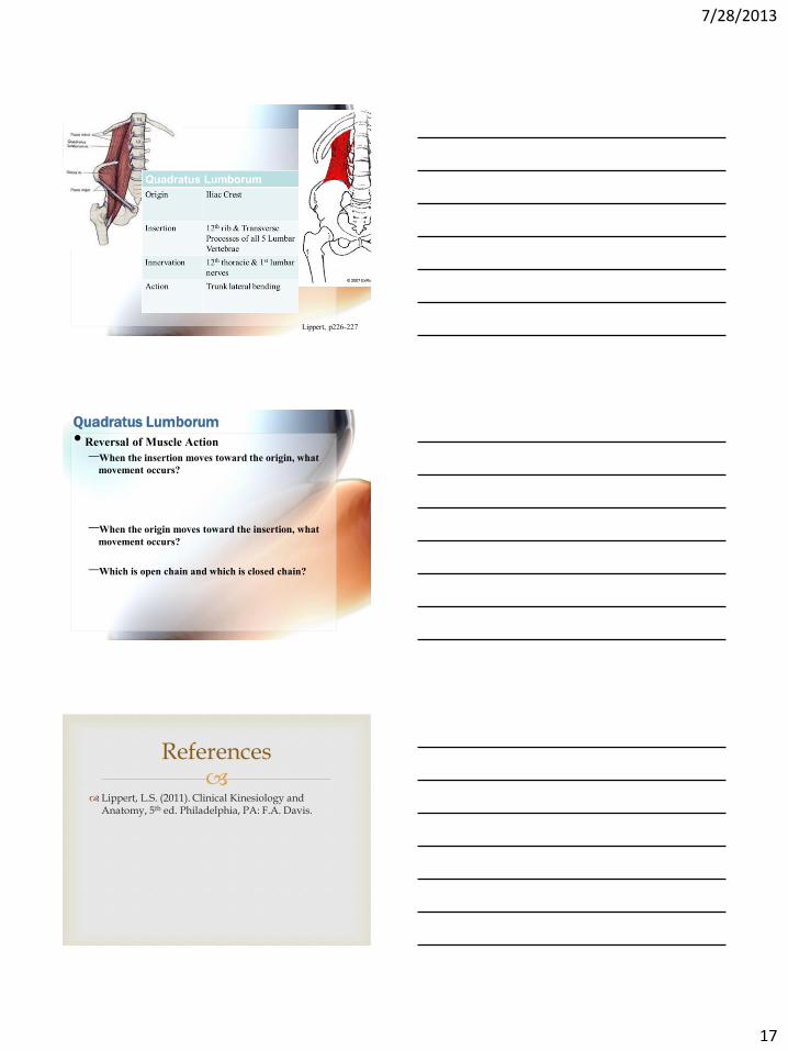
Deep: Quadratus Lumborum
Myology…cont
7/28/2013
17
Lippert, p226-227
Quadratus Lumborum
• Reversal of Muscle Action
–When the insertion moves toward the origin, what
movement occurs?
–When the origin moves toward the insertion, what
movement occurs?
–Which is open chain and which is closed chain?
Lippert, L.S. (2011). Clinical Kinesiology and
Anatomy, 5th ed. Philadelphia, PA: F.A. Davis.
References