The Vertebral Column - JU...
52
The Vertebral Column Dr.Ahmed Salman Assistant Prof. of Anatomy. The University Of Jordan The University Of Jordan Faculty Of Medicine
Transcript of The Vertebral Column - JU...
The Vertebral Column
Dr.Ahmed SalmanAssistant Prof. of Anatomy. The University Of Jordan
The University Of Jordan Faculty Of Medicine
It is composed of 33 vertebrae;
7 cervical,
12 thoracic,
5 lumbar,
5 sacral,
4 coccygeal separated from each other
by intervertebral discs.
Vertebral Column
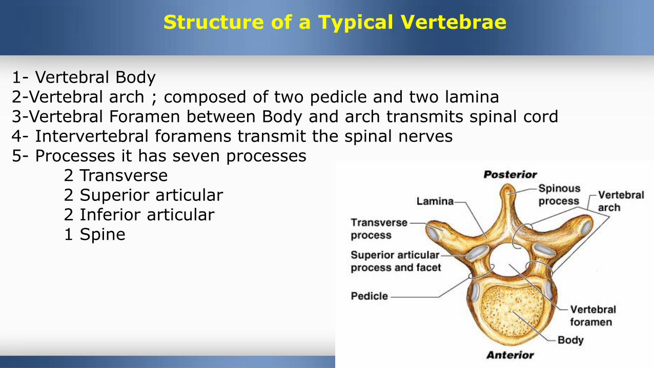
Structure of a Typical Vertebrae
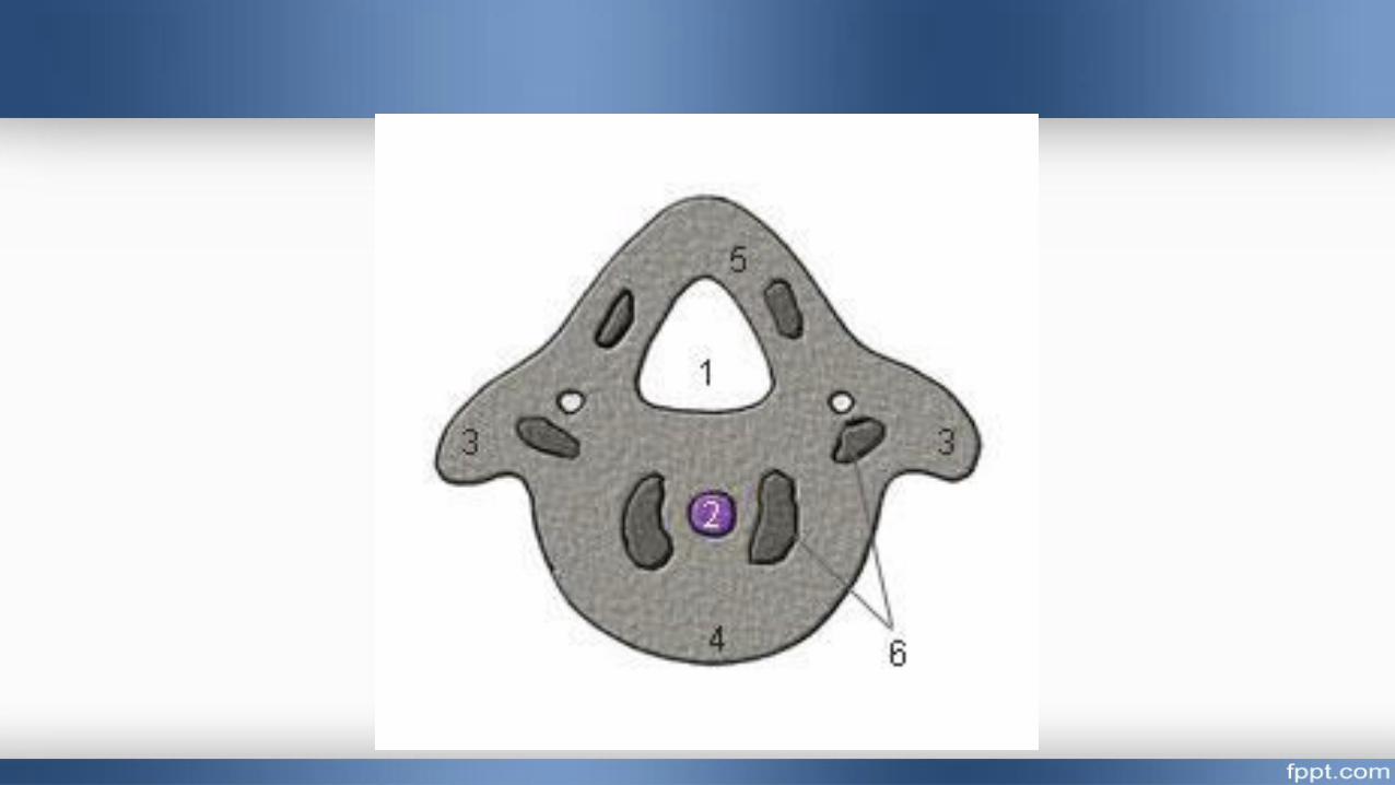
1- Vertebral Body2-Vertebral arch ; composed of two pedicle and two lamina3-Vertebral Foramen between Body and arch transmits spinal cord4- Intervertebral foramens transmit the spinal nerves 5- Processes it has seven processes
2 Transverse2 Superior articular2 Inferior articular1 Spine
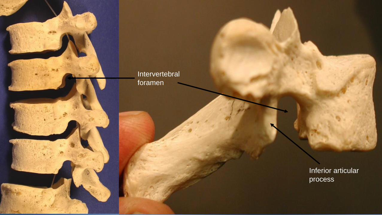
Intervertebral
foramen
Inferior articular
process
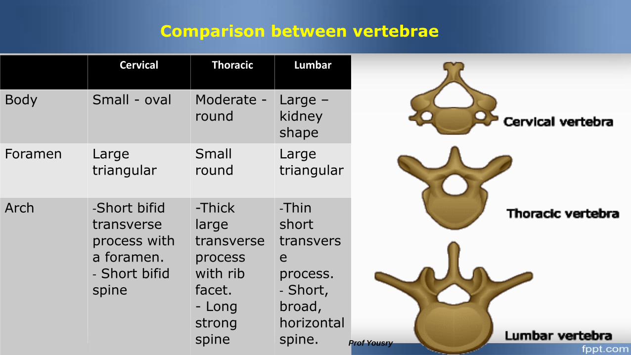
Cervical Thoracic Lumbar
Body Small - oval Moderate -
round
Large –
kidney
shape
Foramen Large
triangular
Small
round
Large
triangular
Arch -Short bifid
transverse
process with
a foramen.
- Short bifid
spine
-Thick
large
transverse
process
with rib
facet.
- Long
strong
spine
-Thin
short
transvers
e
process.
- Short,
broad,
horizontal
spine. Prof Yousry
Comparison between vertebrae
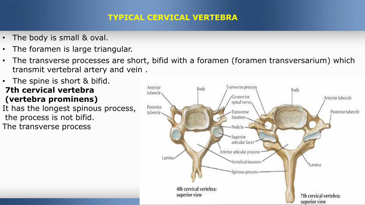
• The body is small & oval.
• The foramen is large triangular.
• The transverse processes are short, bifid with a foramen (foramen transversarium) which transmit vertebral artery and vein .
• The spine is short & bifid.7th cervical vertebra(vertebra prominens)It has the longest spinous process,the process is not bifid. The transverse process
Prof Yousry
TYPICAL CERVICAL VERTEBRA
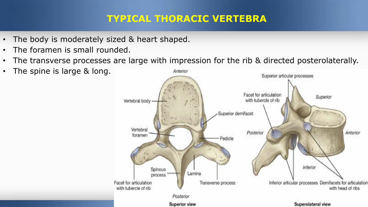
• The body is moderately sized & heart shaped.
• The foramen is small rounded.
• The transverse processes are large with impression for the rib & directed posterolaterally.
• The spine is large & long.
TYPICAL THORACIC VERTEBRA
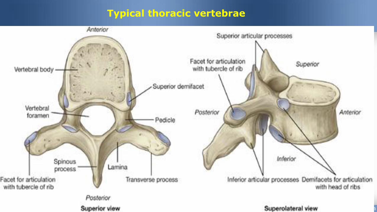
Typical thoracic vertebrae
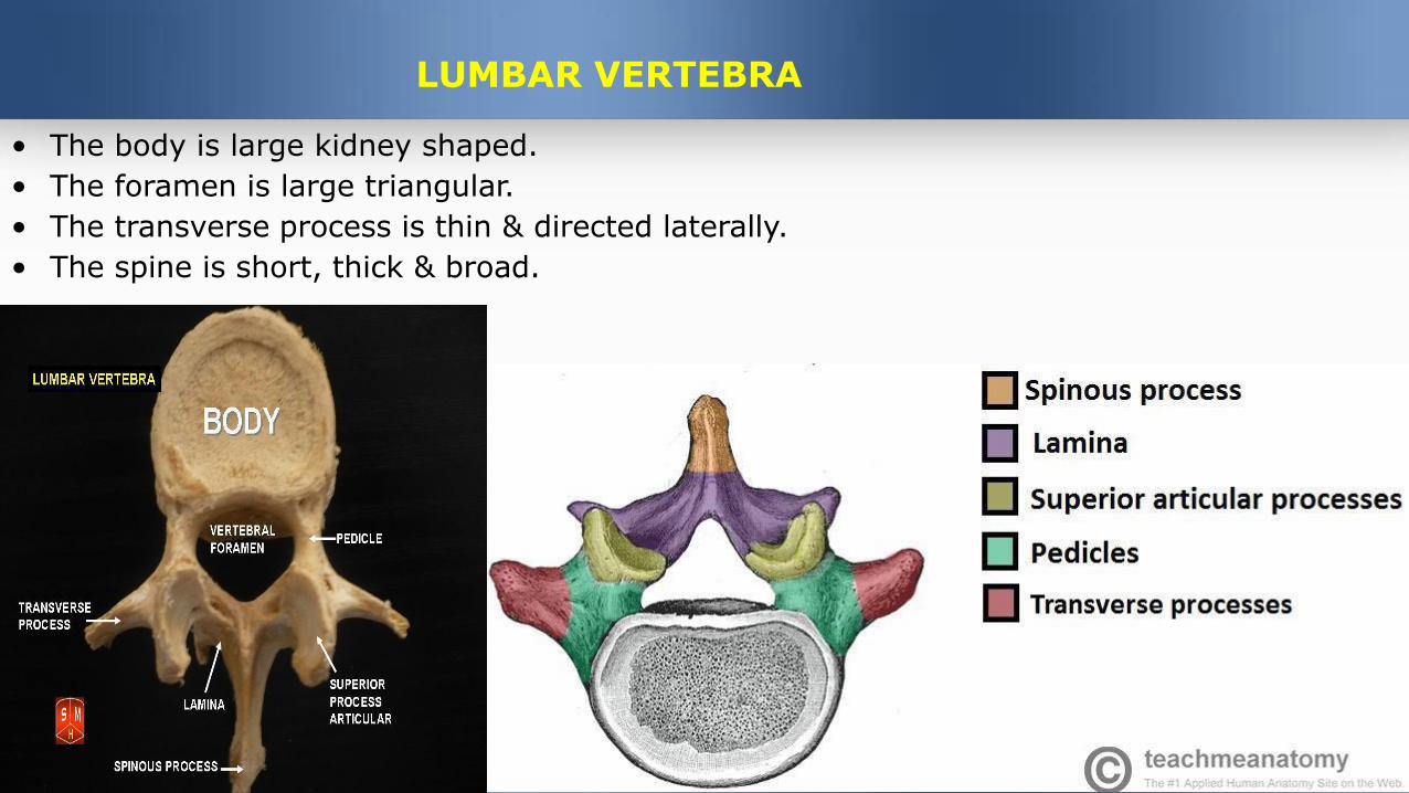
• The body is large kidney shaped.
• The foramen is large triangular.
• The transverse process is thin & directed laterally.
• The spine is short, thick & broad.
LUMBAR VERTEBRA
THE SACRUM & COCCYX
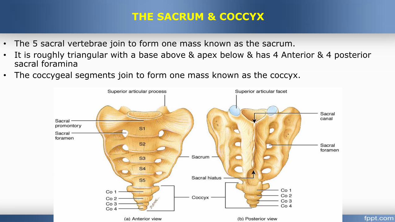
• The 5 sacral vertebrae join to form one mass known as the sacrum.
• It is roughly triangular with a base above & apex below & has 4 Anterior & 4 posterior sacral foramina
• The coccygeal segments join to form one mass known as the coccyx.
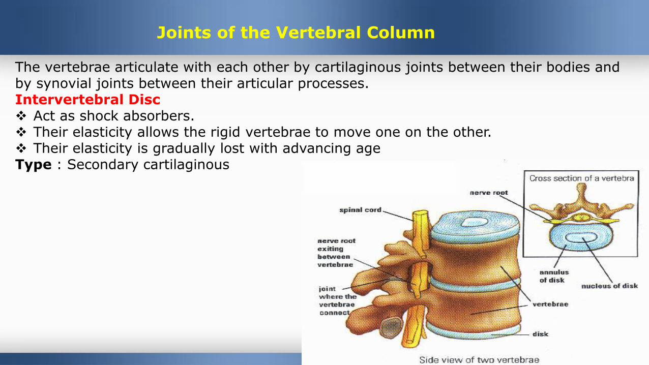
Joints of the Vertebral Column
The vertebrae articulate with each other by cartilaginous joints between their bodies and by synovial joints between their articular processes.Intervertebral Disc Act as shock absorbers. Their elasticity allows the rigid vertebrae to move one on the other. Their elasticity is gradually lost with advancing ageType : Secondary cartilaginous
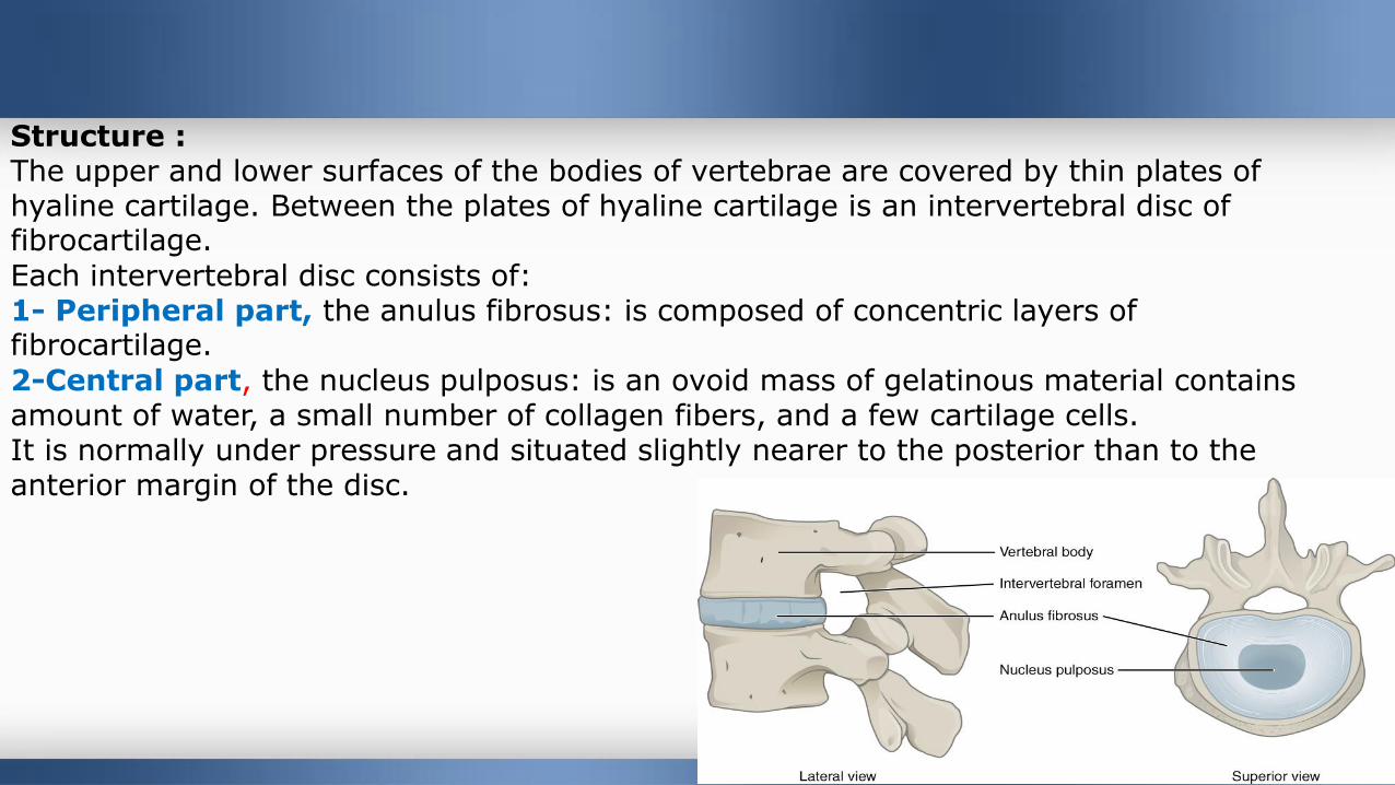
Structure :The upper and lower surfaces of the bodies of vertebrae are covered by thin plates of hyaline cartilage. Between the plates of hyaline cartilage is an intervertebral disc of fibrocartilage.
Each intervertebral disc consists of:1- Peripheral part, the anulus fibrosus: is composed of concentric layers of fibrocartilage.
2-Central part, the nucleus pulposus: is an ovoid mass of gelatinous material contains amount of water, a small number of collagen fibers, and a few cartilage cells. It is normally under pressure and situated slightly nearer to the posterior than to the anterior margin of the disc.
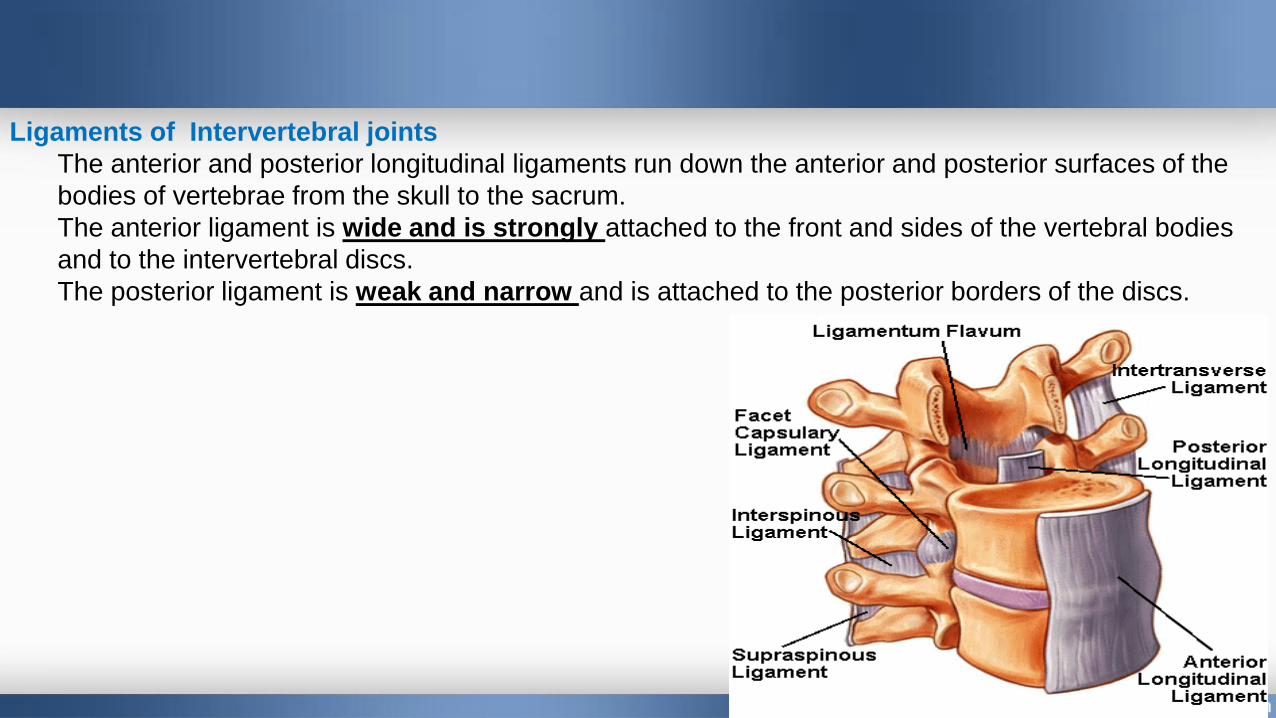
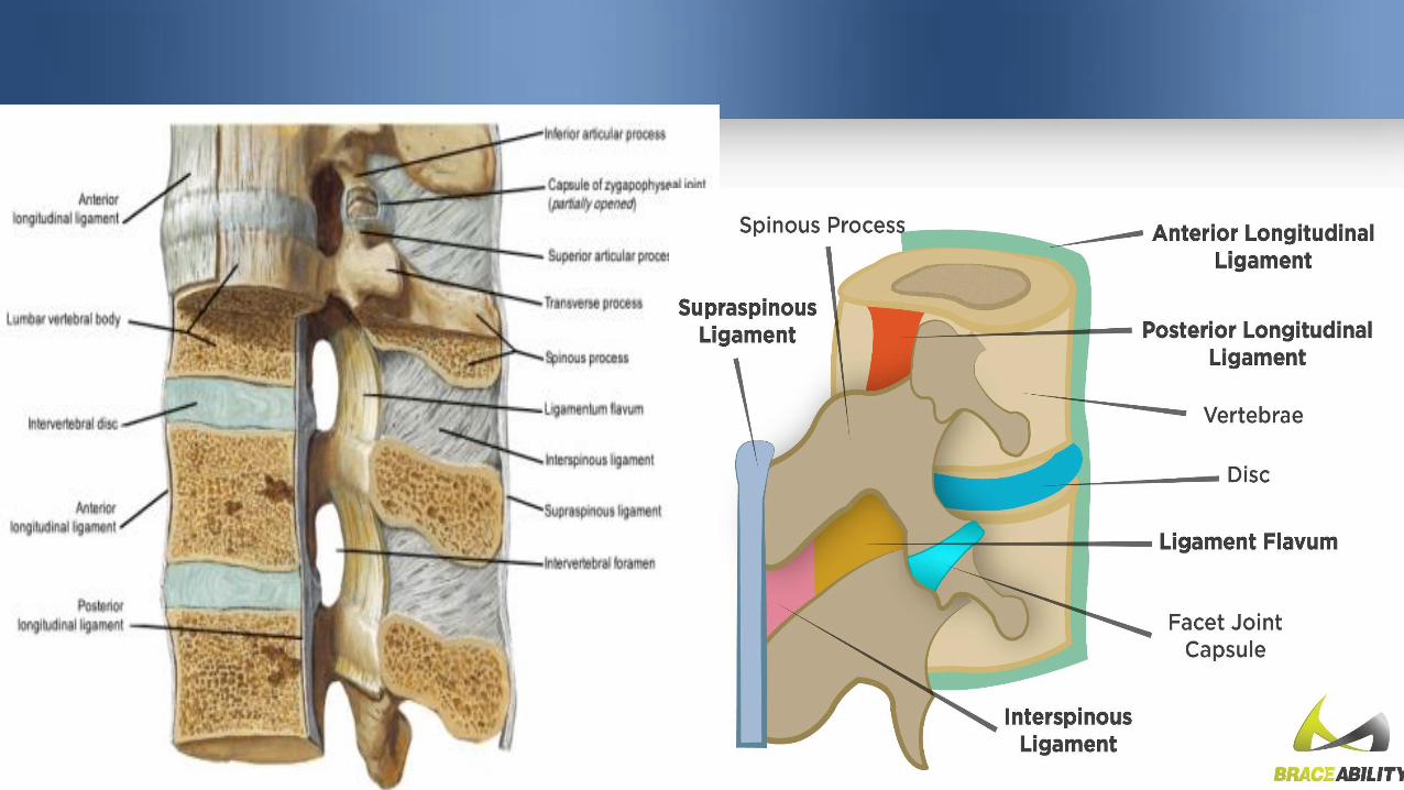
Ligaments of Intervertebral joints
The anterior and posterior longitudinal ligaments run down the anterior and posterior surfaces of the
bodies of vertebrae from the skull to the sacrum.
The anterior ligament is wide and is strongly attached to the front and sides of the vertebral bodies
and to the intervertebral discs.
The posterior ligament is weak and narrow and is attached to the posterior borders of the discs.
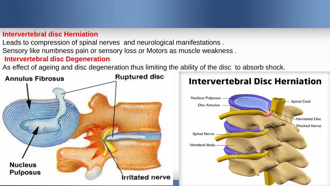
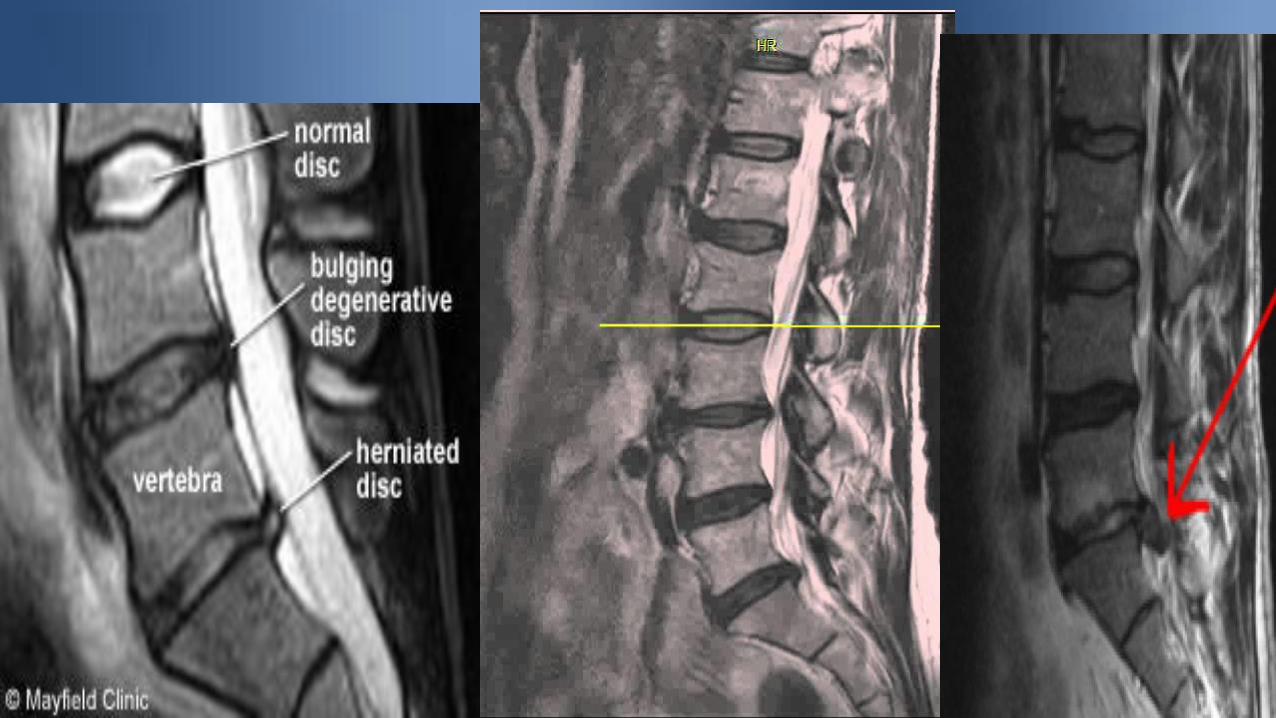
Intervertebral disc Herniation
Leads to compression of spinal nerves and neurological manifestations .
Sensory like numbness pain or sensory loss or Motors as muscle weakness .
Intervertebral disc Degeneration
As effect of ageing and disc degeneration thus limiting the ability of the disc to absorb shock.
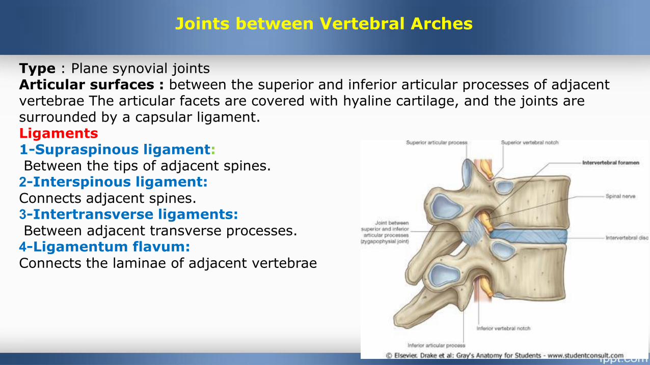
Joints between Vertebral Arches
Type : Plane synovial joints Articular surfaces : between the superior and inferior articular processes of adjacent vertebrae The articular facets are covered with hyaline cartilage, and the joints are surrounded by a capsular ligament.Ligaments1-Supraspinous ligament:Between the tips of adjacent spines.2-Interspinous ligament: Connects adjacent spines.3-Intertransverse ligaments:Between adjacent transverse processes.4-Ligamentum flavum: Connects the laminae of adjacent vertebrae
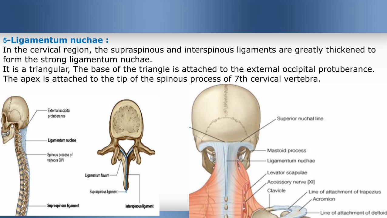
5-Ligamentum nuchae :In the cervical region, the supraspinous and interspinous ligaments are greatly thickened to form the strong ligamentum nuchae.It is a triangular, The base of the triangle is attached to the external occipital protuberance. The apex is attached to the tip of the spinous process of 7th cervical vertebra.
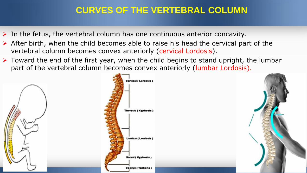
CURVES OF THE VERTEBRAL COLUMN
In the fetus, the vertebral column has one continuous anterior concavity.
After birth, when the child becomes able to raise his head the cervical part of the vertebral column becomes convex anteriorly (cervical Lordosis).
Toward the end of the first year, when the child begins to stand upright, the lumbar part of the vertebral column becomes convex anteriorly (lumbar Lordosis).
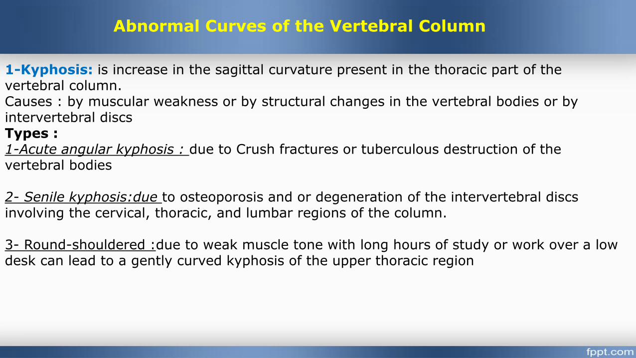
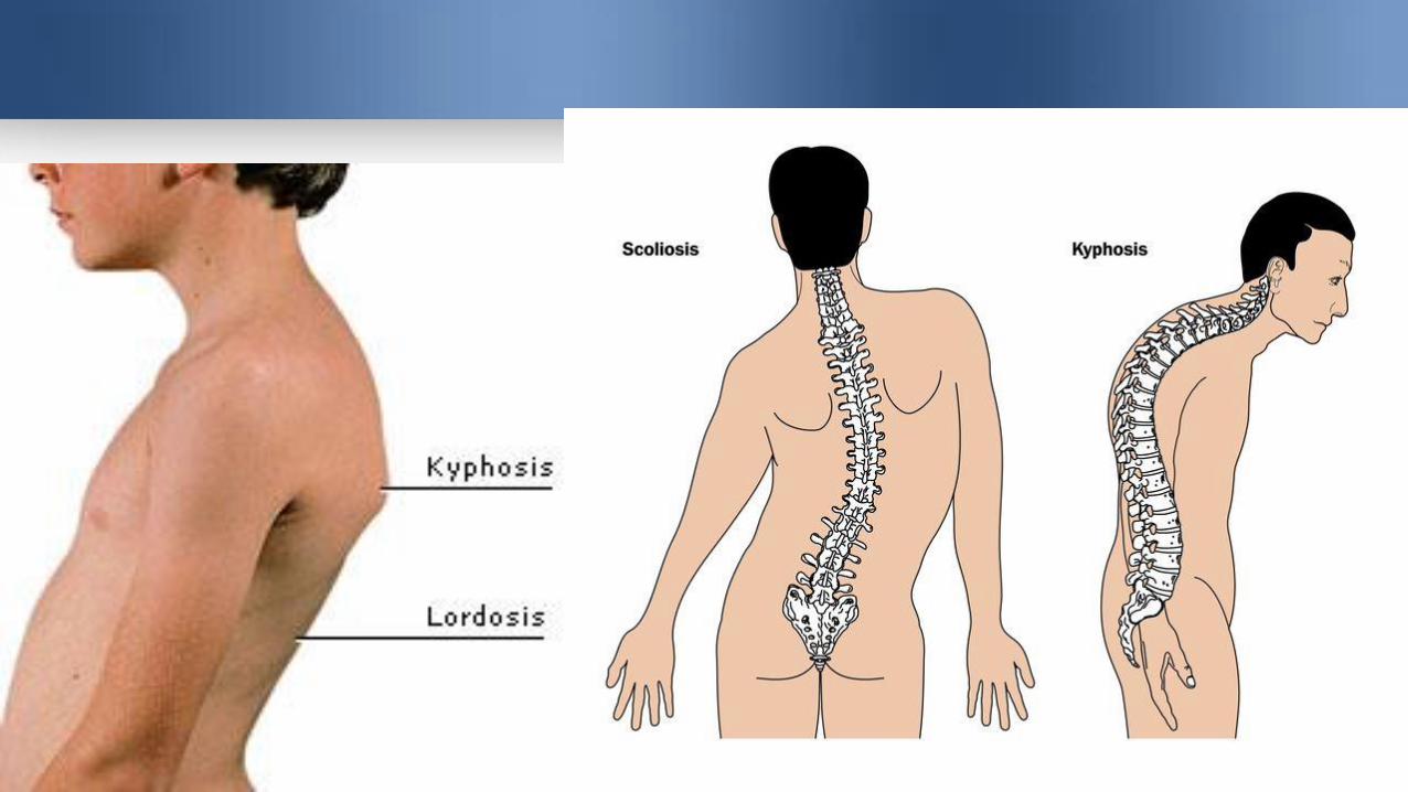
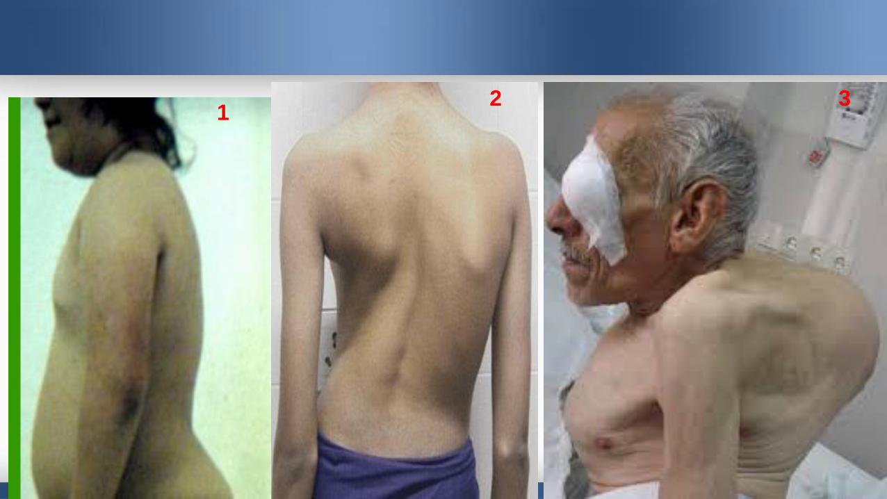
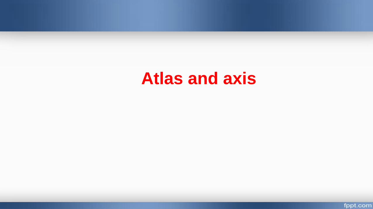
1-Kyphosis: is increase in the sagittal curvature present in the thoracic part of the vertebral column. Causes : by muscular weakness or by structural changes in the vertebral bodies or by intervertebral discsTypes :1-Acute angular kyphosis : due to Crush fractures or tuberculous destruction of the vertebral bodies
2- Senile kyphosis:due to osteoporosis and or degeneration of the intervertebral discs involving the cervical, thoracic, and lumbar regions of the column.
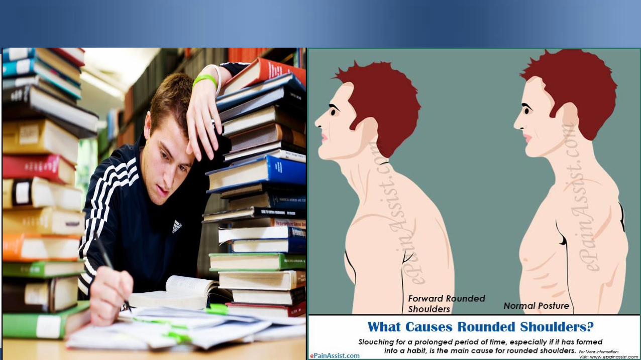
3- Round-shouldered :due to weak muscle tone with long hours of study or work over a low desk can lead to a gently curved kyphosis of the upper thoracic region
Abnormal Curves of the Vertebral Column
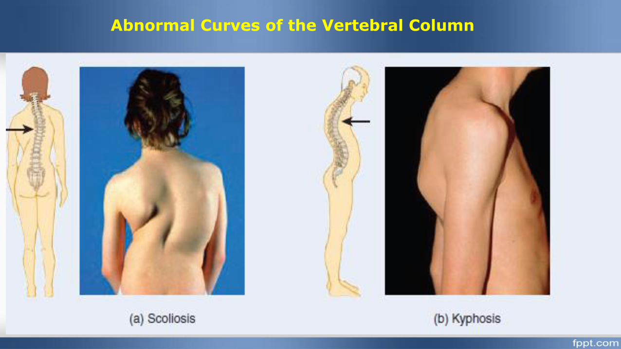
Abnormal Curves of the Vertebral Column
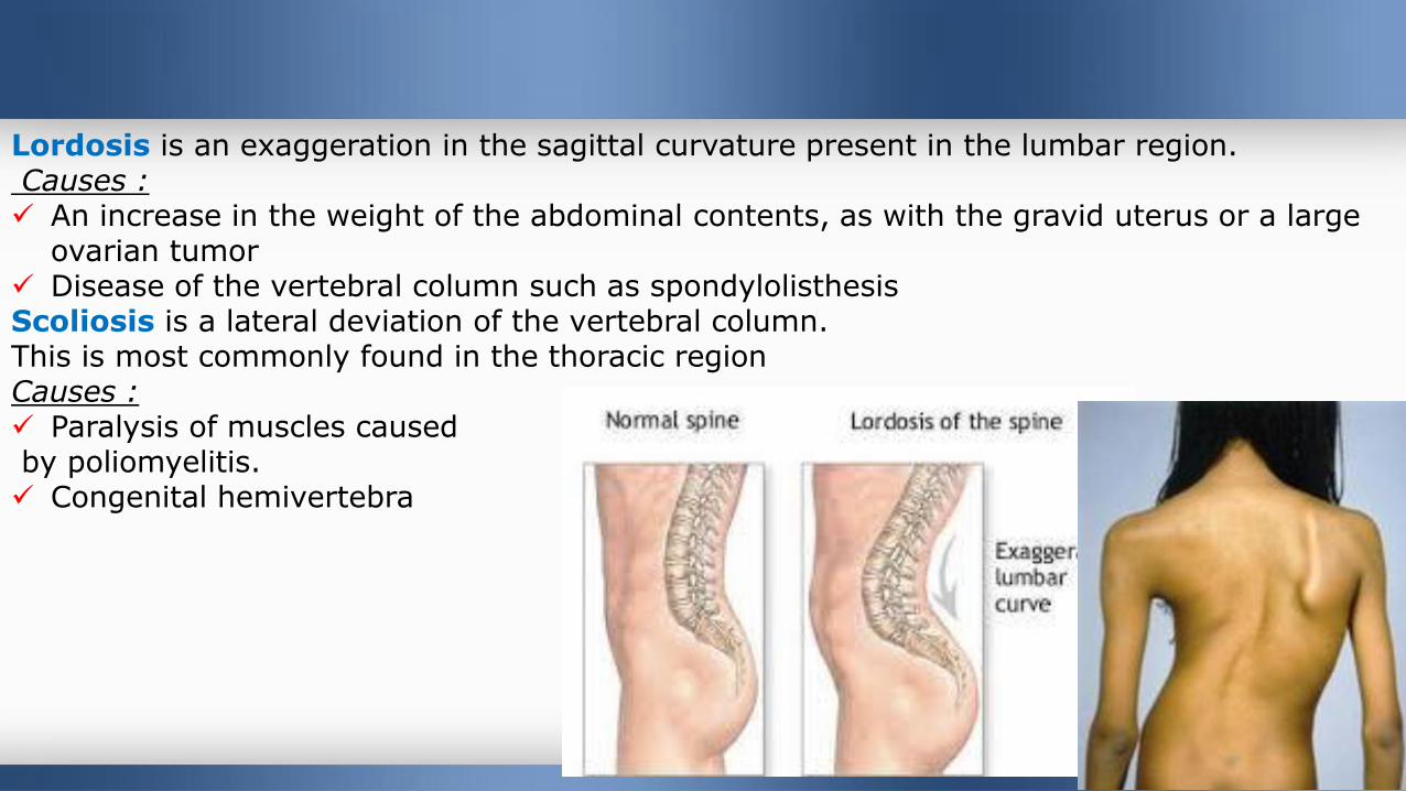
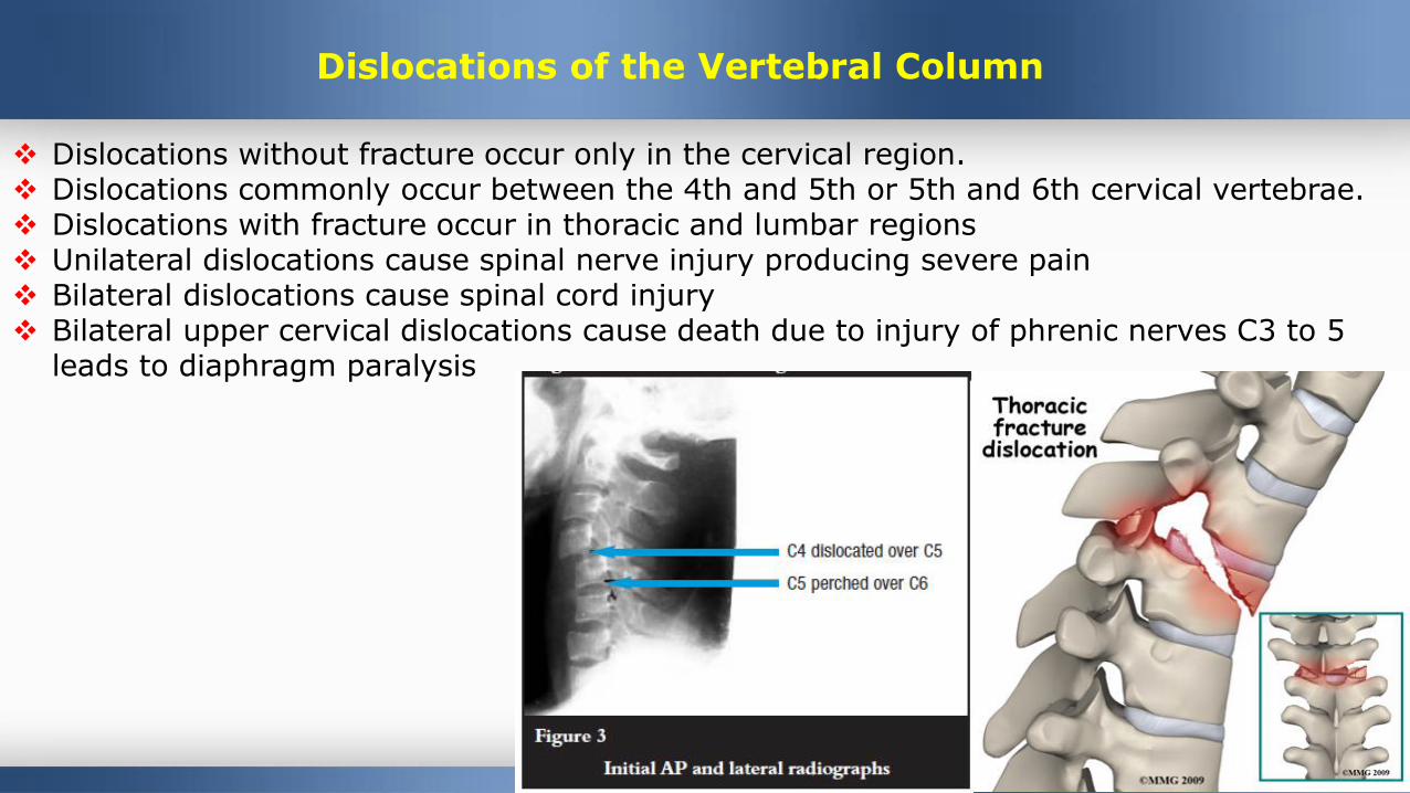
Lordosis is an exaggeration in the sagittal curvature present in the lumbar region.Causes : An increase in the weight of the abdominal contents, as with the gravid uterus or a large
ovarian tumor Disease of the vertebral column such as spondylolisthesisScoliosis is a lateral deviation of the vertebral column.This is most commonly found in the thoracic region Causes : Paralysis of muscles causedby poliomyelitis. Congenital hemivertebra
12 3
Dislocations of the Vertebral Column
Dislocations without fracture occur only in the cervical region. Dislocations commonly occur between the 4th and 5th or 5th and 6th cervical vertebrae. Dislocations with fracture occur in thoracic and lumbar regions Unilateral dislocations cause spinal nerve injury producing severe pain Bilateral dislocations cause spinal cord injury Bilateral upper cervical dislocations cause death due to injury of phrenic nerves C3 to 5
leads to diaphragm paralysis
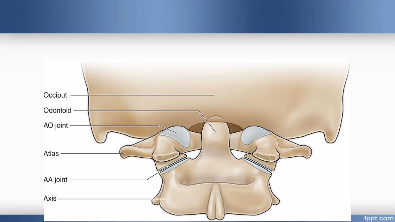
Atlas and axis
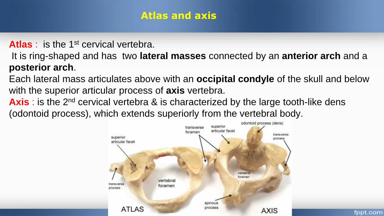
Atlas : is the 1st cervical vertebra.
It is ring-shaped and has two lateral masses connected by an anterior arch and a
posterior arch.
Each lateral mass articulates above with an occipital condyle of the skull and below
with the superior articular process of axis vertebra.
Axis : is the 2nd cervical vertebra & is characterized by the large tooth-like dens
(odontoid process), which extends superiorly from the vertebral body.
Atlas and axis
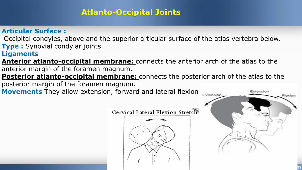
Articular Surface :Occipital condyles, above and the superior articular surface of the atlas vertebra below. Type : Synovial condylar jointsLigamentsAnterior atlanto-occipital membrane: connects the anterior arch of the atlas to the anterior margin of the foramen magnum.Posterior atlanto-occipital membrane: connects the posterior arch of the atlas to the posterior margin of the foramen magnum.Movements They allow extension, forward and lateral flexion.
Atlanto-Occipital Joints
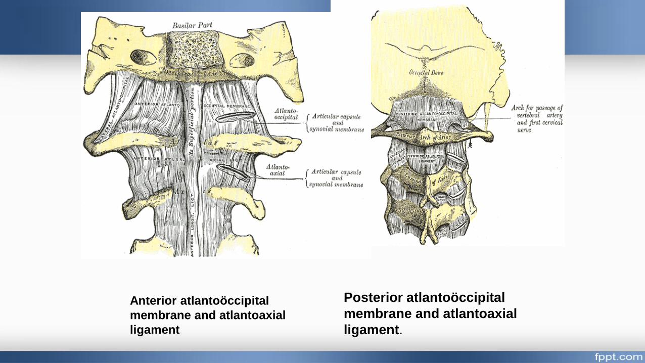
Anterior atlantoöccipital
membrane and atlantoaxial
ligament
Posterior atlantoöccipital
membrane and atlantoaxial
ligament.
Atlantoaxial Joints
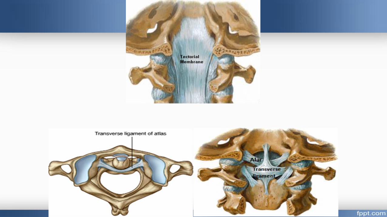
A. The Median atlanto-axial Joint : Articular Surface : between the odontoid process and the anterior arch of the
atlasType: Pivot synovial jointLigaments1-Apical ligament: connects the apex of the odontoid process to the anterior margin of foramen magnum.2-Alar ligaments: connect the odontoid process to the occipital condyles.
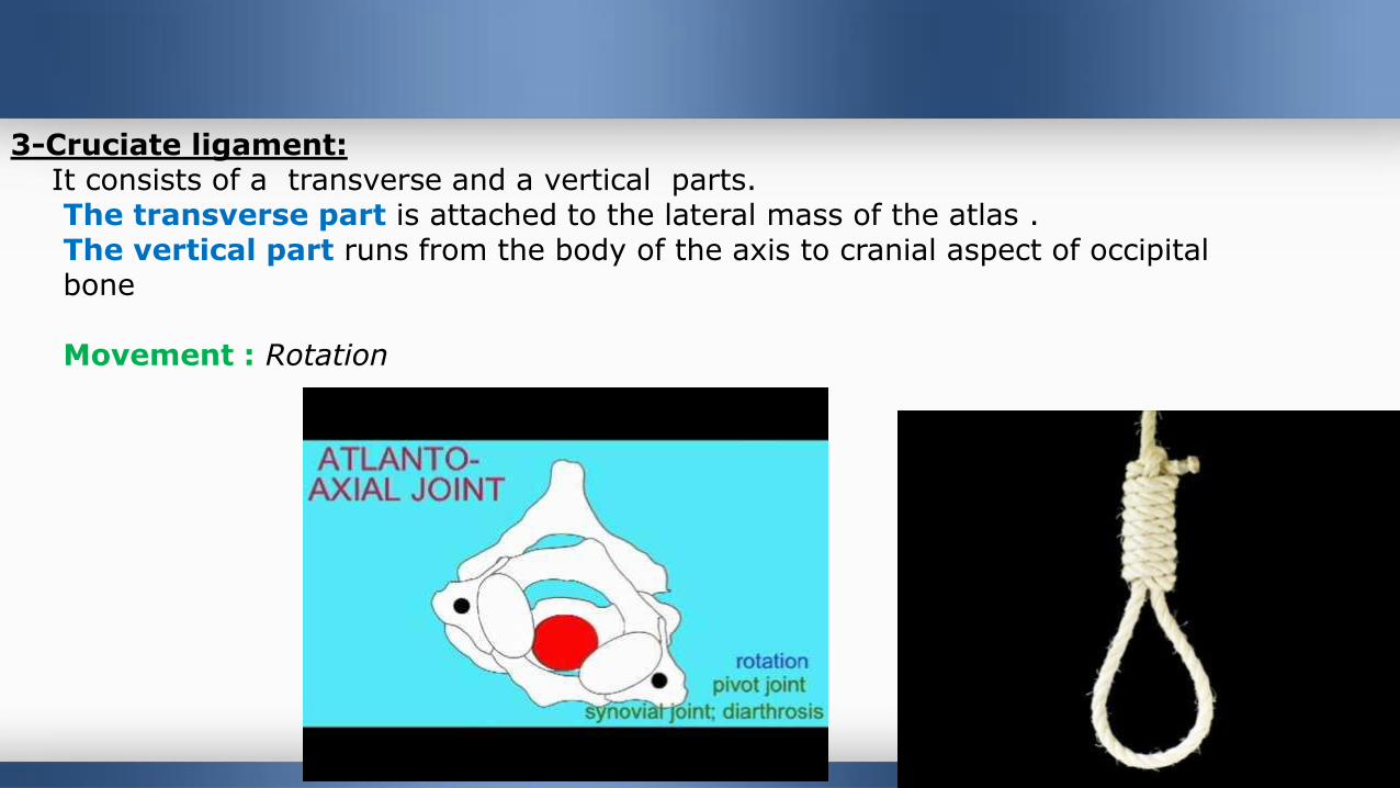
3-Cruciate ligament: It consists of a transverse and a vertical parts. The transverse part is attached to the lateral mass of the atlas . The vertical part runs from the body of the axis to cranial aspect of occipital bone
Movement : Rotation
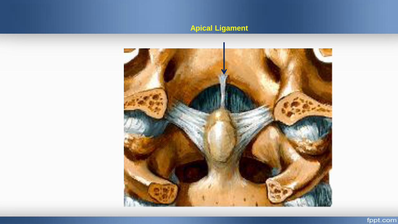
Apical Ligament
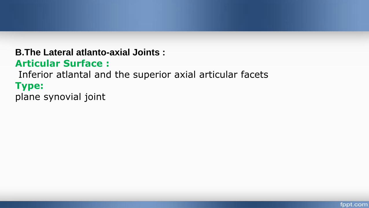
B.The Lateral atlanto-axial Joints :
Articular Surface :Inferior atlantal and the superior axial articular facets
Type: plane synovial joint
Sacroiliac Joint
Type: plane synovial joint
Articular surfaces : between sacral and iliac auricular surfaces.
Ligaments:
o The ventral sacroiliac ligament
o The interosseous sacroiliac ligaments .
o The dorsal sacroiliac ligament.
Functions:
o It transmits the body weight from lumbar spine to the hip bones.

Development of Vertebral column
Stage 1
Stage of formation of
mesenchymal vertebral column
Time of development: 4th week
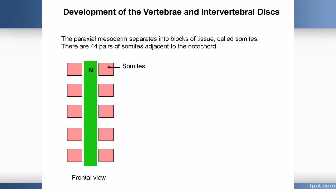
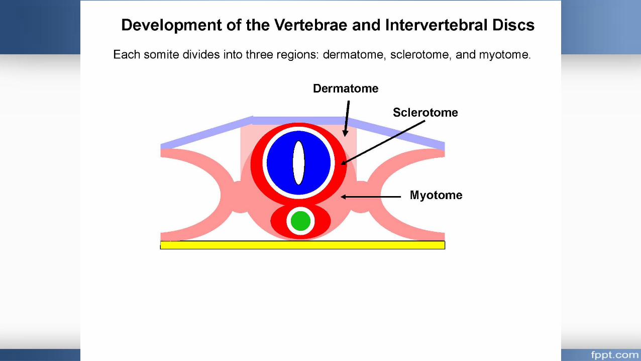
Differentiation of sclerotomic segments
Each segments differentiated into
Cephalic part (less condensed or light)
Caudal part (more condensed or dark)
The caudal condensed part of each sceleretome fuses with
the cranial light part of the sceleretome below it forming a
mesenchymal centrum (future vertebral body).
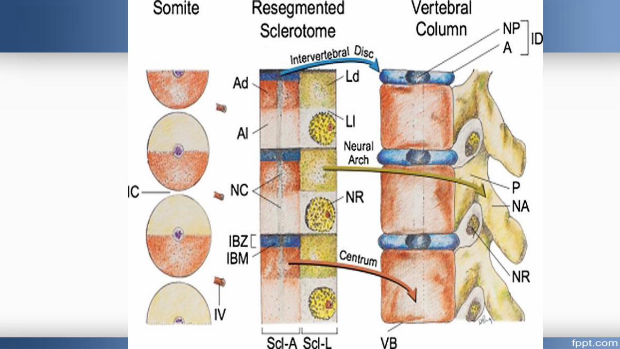

- Cells from that centrum migrate dorsally to surround and fuse behind the
neural tube, forming the vertebral or neural arch (future pedicles, laminae
and spinous process).
- At the junction of the vertebral body and vertebral arch, the following
processes project laterally.
• Two (right and left) costal processes.
• Two (right and left) primitive transverse processes lie dorsal to the costal
processes.
II. Cartilaginous stage:
In the 6th week of development, the mesenchymal vertebrae are chondrofied, so that a mesenchmyal vertebral column is transformed into a cartilaginous one.
III. Bony stage
By the 8th week of development, three primary centers appear whichtransform each vertebra into a bony structure.
Fate of the notochord :
The notochord has the following fate:
• In the region of the vertebral bodies, it degenerates completely.
• In the intervertebral regions, it forms the nucleus pulposus of each
intervertebral disc while the annulus fibrosus of each disc is derived from
the scleretomic mesenchyme,

Congenital anomalies
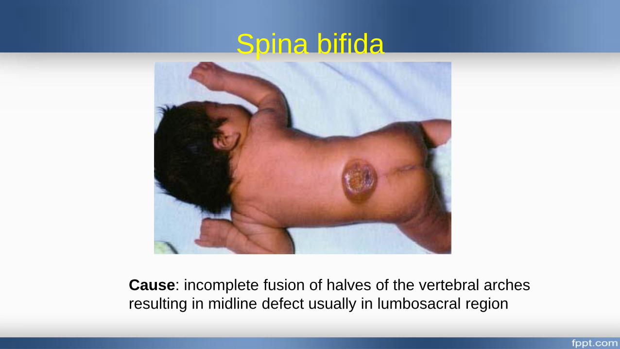
Spina bifida
Cause: incomplete fusion of halves of the vertebral arches
resulting in midline defect usually in lumbosacral region
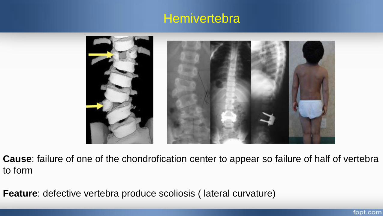
Hemivertebra
Cause: failure of one of the chondrofication center to appear so failure of half of vertebra
to form
Feature: defective vertebra produce scoliosis ( lateral curvature)
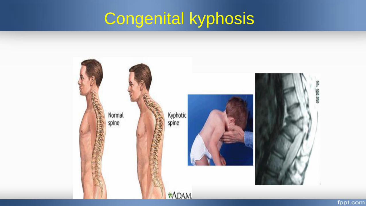
Congenital kyphosis