The use of transmission electron microscopy in the study ... · The use of transmission electron...
9
The use of transmission electron microscopy in the study of gametogenesis in Gnathostomulida A. Falleni and C. Ghezzani Department of Clinical and Experimental Medicine, Unit of Experimental Biology and Genetics, University of Pisa, Via A. Volta 4, 56126 Pisa, Italy The chapter reports a study on the oogenesis, spermatogenesis and spermiogenesis of the gnathostomulid bursovaginoid Austrognatia sp. by means of transmission electron microscopy and cytochemical techniques. In particular, in this review, we describe how the use of transmission electron microscope and cytochemical methods can help the researcher to understand issues and to solve problems related to germ cell differentiation. The aim of the investigation was to obtain information on the male and female gonad and functional significance of some oocyte and sperm structures to compare with those from other lower metazoans. Keywords: gametes; ultrastructure; cytochemistry; Gnathostomulida 1. Introduction Gnathostomulids are minute (0,3-3 mm in length), free-living, acoelomate, unsegmented marine worms which occupy the interstices of sand grains, often of detritus-rich sediments with low oxygen concentrations. The group is cosmopolitan and currently comprises about 100 species [1, 2]. Gnathostomulida has long been considered an enigmatic taxon. They were first described as an order of the “turbellarian” Platyhelminthes by Ax [3], then elevated to the class of Platyhelminthes [4, 5] and finally to the rank of phylum [6, 7]. Gnathostomulida are now considered to be related to Syndermata (Rotifera + Acanthocephala) [8, 9] and are included together with Micrognathozoa in the taxon Gnathifera within Protostomia-Spiralia [10, 11]. The peculiar characteristics of the group are an entirely monociliated epidermis and a bilaterally symmetric pharynx containing complex cuticular mouth parts [12, 13, 14, 15]. All gnathostomulids are hermaphrodites with simple, unpaired, mid-dorsally located ovary and paired or unpaired testes in the posterior body region. Sterrer [7, 16] on the basis of the organization of the reproductive system and the high diversity in sperm morphology, distinguished two orders within the group: the Filospermoidea, with a filiform, monoflagellate sperm and the Bursovaginoidea consisting of the suborders Scleroperalia and Conophoralia, with aberrant, aflagellate sperm (dwarf type and conulus type respectively). Moreover, the Bursovaginoidea possess a peculiar female reproductive system also having a vagina, and a bursa for sperm storage. Fertilization seems to occur by ipodermal injection of sperm (in the Filospermoidea) or after copulation. In the latter case transfer of sperm may be assisted by accessory copulatory structures (a female bursa and a male penis stylet). Ovoposition occurs by rupture of the dorsal epidermis at least for the Scleroperalia. Some years ago we investigated the gametogenesis of some gnathostomulids [17, 18, 19] in order to obtain ultrastructural data to compare with other lower metazoans, in particular platyhelminths; in this review we report findings on Austrognathia sp. (Bursovaginoidea, Conophoralia), with the aim of showing how transmission electron microscopy can be a useful tool of investigation for the study of male and female gametes in this group of animals, in particular for highlighting features that are not resolvable with light microscopy. 2. Ovary and female germ cell differentiation The ovary of Austrognathia sp. is unpaired and located dorsally in the mid body region between the epidermis and the gut. It consists of 3-6 developing germ cells whose sizes progressively increase towards the caudal end (Figs 1, 2). Usually one mature oocyte occupies the entire body breadth. This feature is similar to that observed with light microscopy in other species of gnathostomulids [16] even though the number of developing oocytes seems to vary according to the size of the organisms. Ultrastructural investigations evidenced that each differentiating oocyte is surrounded by several elongated accessory cells (Figs 3, 4), a feature never observed with light microscopy. A tunica consisting of a basement membrane internally reinforced by thin muscle cells surrounds the accessory cells (Fig. 3). Accessory cells completely envelope the oocytes throughout oogenesis with their long cytoplasmic processes. The nucleus shows clumps of condensed chromatin adjacent to the nuclear envelope and the cytoplasm contains free ribosomes, mitochondria and cisternae of rough endoplasmic reticulum which become abundant in the early phase of vitellogenesis (Fig. 4). The oogenesis of Austrognathia can be subdivided into previtellogenic and vitellogenic phases as is the case in other lower metazoans with entolecithal eggs [20, 21, 22, 23]. Microscopy: advances in scientific research and education (A. Méndez-Vilas, Ed.) © FORMATEX 2014 __________________________________________________________________ 103
Transcript of The use of transmission electron microscopy in the study ... · The use of transmission electron...

The use of transmission electron microscopy in the study of gametogenesis in Gnathostomulida
A. Falleni and C. Ghezzani Department of Clinical and Experimental Medicine, Unit of Experimental Biology and Genetics, University of Pisa, Via
A. Volta 4, 56126 Pisa, Italy
The chapter reports a study on the oogenesis, spermatogenesis and spermiogenesis of the gnathostomulid bursovaginoid Austrognatia sp. by means of transmission electron microscopy and cytochemical techniques. In particular, in this review, we describe how the use of transmission electron microscope and cytochemical methods can help the researcher to understand issues and to solve problems related to germ cell differentiation. The aim of the investigation was to obtain information on the male and female gonad and functional significance of some oocyte and sperm structures to compare with those from other lower metazoans.
Keywords: gametes; ultrastructure; cytochemistry; Gnathostomulida
1. Introduction
Gnathostomulids are minute (0,3-3 mm in length), free-living, acoelomate, unsegmented marine worms which occupy the interstices of sand grains, often of detritus-rich sediments with low oxygen concentrations. The group is cosmopolitan and currently comprises about 100 species [1, 2]. Gnathostomulida has long been considered an enigmatic taxon. They were first described as an order of the “turbellarian” Platyhelminthes by Ax [3], then elevated to the class of Platyhelminthes [4, 5] and finally to the rank of phylum [6, 7]. Gnathostomulida are now considered to be related to Syndermata (Rotifera + Acanthocephala) [8, 9] and are included together with Micrognathozoa in the taxon Gnathifera within Protostomia-Spiralia [10, 11]. The peculiar characteristics of the group are an entirely monociliated epidermis and a bilaterally symmetric pharynx containing complex cuticular mouth parts [12, 13, 14, 15]. All gnathostomulids are hermaphrodites with simple, unpaired, mid-dorsally located ovary and paired or unpaired testes in the posterior body region. Sterrer [7, 16] on the basis of the organization of the reproductive system and the high diversity in sperm morphology, distinguished two orders within the group: the Filospermoidea, with a filiform, monoflagellate sperm and the Bursovaginoidea consisting of the suborders Scleroperalia and Conophoralia, with aberrant, aflagellate sperm (dwarf type and conulus type respectively). Moreover, the Bursovaginoidea possess a peculiar female reproductive system also having a vagina, and a bursa for sperm storage. Fertilization seems to occur by ipodermal injection of sperm (in the Filospermoidea) or after copulation. In the latter case transfer of sperm may be assisted by accessory copulatory structures (a female bursa and a male penis stylet). Ovoposition occurs by rupture of the dorsal epidermis at least for the Scleroperalia. Some years ago we investigated the gametogenesis of some gnathostomulids [17, 18, 19] in order to obtain ultrastructural data to compare with other lower metazoans, in particular platyhelminths; in this review we report findings on Austrognathia sp. (Bursovaginoidea, Conophoralia), with the aim of showing how transmission electron microscopy can be a useful tool of investigation for the study of male and female gametes in this group of animals, in particular for highlighting features that are not resolvable with light microscopy.
2. Ovary and female germ cell differentiation
The ovary of Austrognathia sp. is unpaired and located dorsally in the mid body region between the epidermis and the gut. It consists of 3-6 developing germ cells whose sizes progressively increase towards the caudal end (Figs 1, 2). Usually one mature oocyte occupies the entire body breadth. This feature is similar to that observed with light microscopy in other species of gnathostomulids [16] even though the number of developing oocytes seems to vary according to the size of the organisms. Ultrastructural investigations evidenced that each differentiating oocyte is surrounded by several elongated accessory cells (Figs 3, 4), a feature never observed with light microscopy. A tunica consisting of a basement membrane internally reinforced by thin muscle cells surrounds the accessory cells (Fig. 3). Accessory cells completely envelope the oocytes throughout oogenesis with their long cytoplasmic processes. The nucleus shows clumps of condensed chromatin adjacent to the nuclear envelope and the cytoplasm contains free ribosomes, mitochondria and cisternae of rough endoplasmic reticulum which become abundant in the early phase of vitellogenesis (Fig. 4). The oogenesis of Austrognathia can be subdivided into previtellogenic and vitellogenic phases as is the case in other lower metazoans with entolecithal eggs [20, 21, 22, 23].
Microscopy: advances in scientific research and education (A. Méndez-Vilas, Ed.)
© FORMATEX 2014
__________________________________________________________________
103

Early pre-vitellogenic oocytes are oblong cells measuring about 15 µm in maximum diameter. They show a large nucleus with a nuclear envelope very rich in pores, containing mainly diffuse chromatin and a well-developed nucleolus (Fig. 5). The cytoplasm contains abundant free-ribosomes, some mitochondria and numerous membrane-unbound
Fig. 1 Schematic drawing of Austrognathia. b, bursa; o, ovary; ph, pharynx; t, testis; v, vagina. Fig. 2 Portion of the ovary of Austrognathia sp. located under the epidermis. ec, epithelial cell; po, previtellogenic oocyte; vo, early vitellogenic oocyte. Fig. 3 Portion of a late vitellogenic oocyte (vo) surrounded by an accessory cell (ac). Arrows point to the tunica internally reinforced by muscle cells (mc). n, accessory cell nucleus. Fig. 4 Cytoplasmic projections of accessory cells (ac) showing elongated cisternae of rough endoplasmic reticulum (arrowheads) are visible between two early vitellogenic oocytes (o). Fig. 5 A previtellogenic oocyte with chromatoid bodies (cb) in the perinuclear cytoplasm. m, mitochondria; n, nucleus; nu, nucleolus. Fig. 6 A chromatoid body surrounded by mitochondria from a previtellogenic oocyte. Fig. 7 Stacks of annulate lamellae from a previtellogenic oocyte. aggregates of finely granular or fibrillar electron-dense material, the so-called chromatoid bodies (Fig. 5), often surrounded by mitochondria (Fig. 6). Stacks of annulate lamellae, sheets of endoplasmic reticulum-like membranes interrupted by pore complexes, are also visible in proximity of the nuclear region or in the deep cytoplasm of late previtellogenic oocytes (Fig. 7). The presence of chromatoid bodies is a common feature of the male and female germ line cells of many invertebrates and vertebrates [24, 25] but their components and functions have remained obscure for decades and only recently been revealed. These subcellular structures are now viewed as a storage/processing site of
Microscopy: advances in scientific research and education (A. Méndez-Vilas, Ed.)
© FORMATEX 2014
__________________________________________________________________
104

mRNA [26, 27] but also as a degradation site involving the ubiquitin-proteasome system and the lysosome system [28, 29]. Vitellogenic oocytes are oblong cells measuring about 45-50 µm in maximum diameter. The nucleus exhibits completely diffuse chromatin and a prominent nucleolus (Fig. 8). The cytoplasm is characterized by the appearance and
Fig. 8 Early vitellogenic oocyte showing elongated cisternae of rough endoplasmic reticulum (arrows) and some electron-dense yolk globules (arrowheads). ec, epithelial cell; n, nucleus. Fig. 9 Golgi complexes (gc) and nascent yolk vacuoles (arrows) from a vitellogenic oocyte. y, yolk globule. Fig. 10 A Golgi complex (gc) and forming yolk globules (arrows). y, mature yolk globule. Fig. 11 Coated pits (arrows) along the oolemma. Fig. 12 Endocytotic vesicles without coating in the process of fusing with a nascent yolk globule (arrows). Fig. 13 Early and late vitellogenic oocytes (vo). The electron-dense yolk globules are scattered in the ooplasm. Fig. 14 Enzymatic protein extraction (pronase E, 6h incubation). Late vitellogenic oocyte (vo). The content of yolk globules is digested by protease. Fig. 15 Thiéry test, unstained section. A fine silver precipitate indicating the presence of glycoproteins is visible on the peripheral component of the yolk globule. In the cytoplasm a number of glycogen rosettes are visible.
Microscopy: advances in scientific research and education (A. Méndez-Vilas, Ed.)
© FORMATEX 2014
__________________________________________________________________
105
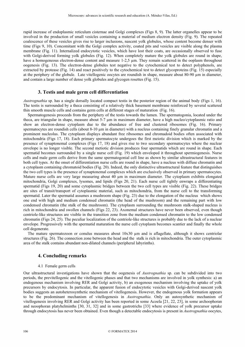
rapid increase of endoplasmic reticulum cisternae and Golgi complexes (Figs 8, 9). The latter organelles appear to be involved in the production of small vesicles containing a material of medium electron density (Fig. 9). The repeated coalescence of these vesicles gives rise to larger inclusions, nascent yolk globules, whose content become denser with time (Figs 9, 10). Concomitant with the Golgi complex activity, coated pits and vesicles are visible along the plasma membrane (Fig. 11). Internalized endocytotic vesicles, which have lost their coats, are occasionally observed to fuse with Golgi-derived forming yolk globules (Fig. 12). When completely mature the yolk globules are round in shape, have a homogeneous electron-dense content and measure 1-2,5 µm. They remain scattered in the ooplasm throughout oogenesis (Fig. 13). The electron-dense globules test negative to the cytochemical test to detect polyphenols, are extracted by protease (Fig. 14) and react positively to the cytochemical test to detect glycoproteins (Fig. 15) especially at the periphery of the globule. Late vitellogenic oocytes are roundish in shape, measure about 80-90 µm in diameter, and contain a large number of dense yolk globules and glycogen rosettes (Fig. 15).
3. Testis and male germ cell differentiation
Austrognathia sp. has a single dorsally located compact testis in the posterior region of the animal body (Figs 1, 16). The testis is surrounded by a theca consisting of a relatively thick basement membrane reinforced by several scattered thin smooth muscle cells and contains germ cells at different stages of maturation (Fig. 16). Spermatogenesis proceeds from the periphery of the testis towards the lumen. The spermatogonia, located under the theca, are triangular in shape, measure about 5-7 µm in maximum diameter, have a high nucleo/cytoplasmic ratio and show an electron-dense cytoplasm due to the abundance of free and clustered ribosomes (Fig. 16). Primary spermatocytes are roundish cells (about 8-10 µm in diameter) with a nucleus containing finely granular chromatin and a prominent nucleolus. The cytoplasm displays abundant free ribosomes and chromadoid bodies often associated with mitochondria (Figs 17, 18). Each primary spermatocyte undergoes the first meiotic division which is marked by the presence of synaptonemal complexes (Figs 17, 18) and gives rise to two secondary spermatocytes where the nuclear envelope is no longer visible. The second metiotic division produces four spermatids which are round in shape. Each spermatid becomes surrounded by a single nurse cell (Fig. 19) which enveloped it throughout spermiogenesis. Nurse cells and male germ cells derive from the same spermatogonial cell line as shown by similar ultrastructural features in both cell types. At the onset of differentiation nurse cells are round in shape, have a nucleus with diffuse chromatin and a cytoplasm containing chromatoid bodies (Fig. 20). Indeed, the only distinctive ultrastructural feature that distinguishes the two cell types is the presence of synaptonemal complexes which are exclusively observed in primary spermatocytes. Mature nurse cells are very large measuring about 40 µm in maximum diameter. The cytoplasm exhibits elongated mitochondria, Golgi complexes, lysomes, and lipids (Figs 20, 21). Each nurse cell tightly enwraps a differentiating spermatid (Figs 19, 20) and some cytoplasmic bridges between the two cell types are visible (Fig. 22). These bridges are sites of transit/transport of cytoplasmic material, such as mitochondria, from the nurse cell to the transforming spermatid. Later the spermatid assumes a mushroom shape (Fig. 23) due to the elongation of the nucleus which shows one end with high and medium condensed chromatin (the head of the mushroom) and the remaining part with low condensed chromatin (the stalk of the mushroom). The cytoplasm surrounding the mushroom stalk-shaped nucleus is rich in mitochondria and swollen channels (Figs 22, 23). Axonemal structures have never been observed, even though centriole-like structures are visible in the transition zone from the medium condensed chromatin to the low condensed chromatin (Figs 24, 25). The peculiar localization of the centriole-like structures is probably due to the lack of a nuclear envelope. Progressively with the spermatid maturation the nurse cell cytoplasm becomes scantier and finally the whole cell degenerate. The mature spermatozoon or conulus measures about 10x30 µm and is aflagellate, although it shows centriolar structures (Fig. 26). The connection zone between the head and the stalk is rich in mitochondria. The outer cytoplasmic area of the stalk contains abundant non-dilated channels (peripheral labyrinths).
4. Concluding remarks
4.1 Female germ cells
Our ultrastructural investigations have shown that the oogenesis of Austrognathia sp. can be subdivided into two periods, the previtellogenic and the vitellogenic phases and that two mechanisms are involved in yolk synthesis: a) an endogenous mechanism involving RER and Golgi activity, b) an exogenous mechanism involving the uptake of yolk precursors by endocytosis. In particular, the apparent fusion of endocytotic vesicles with Golgi-derived nascent yolk bodies suggests an autoheterosynthetic mechanism of vitellogenesis. However, the endogenous yolk formation appears to be the predominant mechanism of vitellogenesis in Austrognathia. Only an autosynthetic mechanism of vitellogenesis involving RER and Golgi activity has been reported in some Acoela [21, 22, 23], in some archoophoran and neoophoran platyhelminths [30, 31, 32] and in some gastrotrichs [33] where evidence of yolk precursor uptake through endocytosis has never been obtained. Even though a detectable endocytosis is present in Austrognathia oocytes,
Microscopy: advances in scientific research and education (A. Méndez-Vilas, Ed.)
© FORMATEX 2014
__________________________________________________________________
106

it occurs at a very low level as is the case in some jellyfish [34]. The autosynthetic mechanism of vitellogenesis is widespread in the animal kingdom [35] and is generally considered the original primitive mechanism of yolk formation, while heterosynthesis is considered a derived mode of vitellogenesis [36, 37, 38]. Protein yolk is not the only reserve material for embryonic development in Austrognathia because late vitellogenic oocytes contain large amounts of glycogen. Large amounts of glycogen have been also observed in the oocytes of some nematodes [39].
Fig. 16 Portion of the testis of Austrognathia sp. showing male germ cells at different stages of maturation. Arrows point to the theca delimiting the testis. nc, nurse cell; sc, primary spermatocytes; sp, spermatogonium; spe, spermatid. Figs 17, 18 Primary spermatocytes (sc) showing a nucleus with synaptonemal complexes (arrows) and a cytoplasm with numerous chromatoid bodies (cb). Fig. 19 Developing spermatid (spe) surrounded by a nurse cell (nc). gc, Golgi complex; n, spermatid nucleus. Fig. 20 Portion of the testis showing primary spermatocytes (sc) and nurse cells (nc). spe, developing spermatid; t, theca; ec, epithelial cell. Fig. 21 A differentiating nurse cell with a nucleus (n) containing a nucleolus (nu) and a cytoplasm showing mitochondria (m), free ribosomes, lysosomes (ly) and a cluster of lipids (l).
Microscopy: advances in scientific research and education (A. Méndez-Vilas, Ed.)
© FORMATEX 2014
__________________________________________________________________
107

No eggshell granules have been found in the mature oocytes of Austrognathia. In particular, neither eggshell granules containing polyphenols, the precursors of the sclerotized eggshell of most platyhelminths [30, 40], nor exocytosis of protein granule content to form a soft, non-sclerotized eggshell as is typical of some Acoela [21, 22, 23] have been observed in Austrognathia. However, a thin, translucent egg capsule was observed by Sterrer [16] in the gnathostomulid Haplognathia with light microscopy. It could be hypothesized that some epithelial or gut gland cells provide oocytes with a soft covering during deposition which occurs through rupture of the body wall.
Fig. 22 A nurse cell (nc) enveloping a transforming spermatid (spe). Note the large cytoplasmic bridges (arrows) for the passage of nutrients from the nurse cell to the transforming spermatid and the dilated channels in the spermatid cytoplasm (arrowheads). n, spermatid nucleus. Fig. 23 Longitudinal section of a late mushroom-shaped spermatid surrounded by a nurse cell (nc). The spermatid nucleus shows three degrees of chromatin condensation: high (*) medium (**) and low (***). The cytoplasm surrounding the nucleus is rich in mitochondria. ac, apoptotic cell; gc, Golgi complexes; n, nurce cell nucleus. Figs 24, 25 Arrows point to centriole-like structures located in the transition zone, from the head to the stalk, of the mushroom-shaped nucleus of the spermatid. Fig. 26 Mature spermatozoon. The enveloping nurse cell is no longer visible. The cytoplasm shows a tubular peripheral labyrinth. m, mitochondrium.
Microscopy: advances in scientific research and education (A. Méndez-Vilas, Ed.)
© FORMATEX 2014
__________________________________________________________________
108

Our ultrastructural investigations have shown that flattened accessory cells surround the oocytes throughout oogenesis. These cells display ultrastructural similarities to those described in some acoels [21, 22, 23] and rhabditophoran platyhelminths [31, 32, 41, 42, 43]. The abundance of RER cisternae in their cytoplasm at the onset of vitellogenesis could suggest a proteosynthetic activity of these cells. Yolk precursors could be released into the narrow, periocytic space and then absorbed by the oocyte through endocytosis. In addition, these accessory cells may have a supporting function for the ovary.
4.2 Male germ cells
In Gnathostomulida sperm structure differs so widely between orders and suborders that sperm phylogeny remains uncertain [44, 45]. Three types of sperm have been observed in the group. The order Filospermoidea shows filiform sperm, up to 100 µm in length, with a spiral nucleus topped by an acrosome, a middle piece with mitochondria and a flagellum with a 9+2 axoneme [8, 46]. The Bursovaginoidea-Scleroperalia have “dwarf” type sperm usually measuring about 2-5 µm up to the “megasperm” of Gnathostomaria lutheri about 18 µm in length. The dwarf type sperms are similar being spherical or spindle-shaped, aflagellate, and provided with micropodia (microvilli-like membrane protrusions) [45, 47]. The Bursovaginoidea-Conophoralia have “conulus” type sperms measuring up to 70 µm in length, aflagellate and cone-shaped [7, 8, 46 ]. According to the observations made by Sterrer [7, 46] under the light microscope, the sperm of Austrognathia riedli consisted of four different parts: a hat, a cingulum, a body and a matrix. In our study on Austrognathia sp. the use of transmission electron microscope allowed us to clarify that the matrix corresponds to the nurse cell, the sperm body to the mushroom stalk consisting of the low density nuclear region + the surrounding cytoplasm, the cingulum to the stalk connection and the hat to the pyknotic electron-dense chromatin of the mushroom head. In Austrognathia sp. no type of compartmentalization defining an acrosomal vesicle has been observed. However, a regular (paracrystalline) arrangement of macromolecules closely opposed to the anterior end of the nucleus suggestive of an acrosomal function has been reported by Lanfranchi and Falleni [18]. In addition, the transmission electron microscope allowed us to show that the nurse cells (trophocytes) provide the developing spermatids with cytoplasmic materials and organelles such as ribosomes, mitochondria through cytoplasmic bridges. No nurse cells have been observed around male gametes in Platyhelminthes. Among the Nematoda the trichuroids show non-germinal sustentacular cells which extend cytoplasmic projections around developing male germ cells and site of exchange have been hypothesized [48, 49]. The nurse cell-spermatid interaction in Austrognathia recalls the nurse cell-oocyte interaction in some invertebrates such as insects (e.g., Drosophila) where nurse cells pump large amounts of ribosomes and macromolecules into the oocytes through cytoplasmic bridges [50, 51] or the situation in some annelid Hirudinea where mitochondria, ER, and vast amount of ribosomes are transferred from the trophocytes via the cytophores to the oocyte [52]. Contrary to the rule, the male gamete of Austrognathia sp. is a large sperm since the giant mushroom-shaped spermatid (about 30-40 µm in length) does not produce residual bodies and does not decrease in size during its differentiation into mature sperm as is the rule in the animal kingdom since a nurse cell encompasses it and elaborates nutrients to supply during spermiogenesis. The existence of this type of nurse cell-spermatid interaction in Austrognathia leads us to hypothesize that in a certain period of biological evolution male and female gametes could have been similar and both provided with nurse cells. Then the male gametes progressively differentiated eliminating most of cytoplasmic organelles in order to become small, compact, tapered cells suitable for realizing a fast and efficient fertilization. Thus the giant mushroom sperm of the gnathostomulid Conophoralia may represent an ancestral model of a male gamete which has remained practically unchanged. In conclusion we believe that ultrastructural microscopy will continue to provide important information on subcellular structures and cell mechanisms underlying gametogenesis in lower invertebrates. In addition, some ultrastructural features of germ cells can provide an excellent framework for assessing and consolidating molecular data in relation to phylogenetic issues.
References [1] Sterrer W, Sørensen MV. Chirognathia dracula gen. et spec. nov. (Gnathostomulida) from the west coast of north America.
Mar. Biol. Res. 2006; 2:296-302. [2] Sterrer W. Two species (one new) of Gnathostomulida (Bursovaginoidea: Conophoralia) from Barbados. P. Biol. Soc. Wash.
2011; 124:141-146. [3] Ax P. Die Gnathostomulida, eine rätselhafte wurmgruppe aus dem meeressand. Akad. Wiss. Lit. Mainz. Abh. Math. Nat. Kl.
1956; 8:1-3. [4] Ax P. Die Entdeckung neuer Organisationstypen im Tierreich. A. Ziemsen Verlag, Wittenberg, Germany; 1960. [5] Ax P. Eine neue Tierklasse aus dem Litoral des Meeres-Gnathostomulida. Umschau Wissenschaft Technik. 1966; 66:17-23. [6] Riedl R.J. Gnathostomulida from America. Science 1969; 163:445-452. [7] Sterrer W. Systematics and evolution within the Gnathostomulida. Syst. Zool. 1972; 21:151-173.
Microscopy: advances in scientific research and education (A. Méndez-Vilas, Ed.)
© FORMATEX 2014
__________________________________________________________________
109

[8] Sterrer W, Mainitz M, Rieger RM. Gnathostomulida: Enigmatic as ever. In: S. Conway-Morris, J.D. George, R. Gibson and H.M. Platt, editors. The origins and relationship of lower invertebrates. Oxford, Systematics Association, Clarendon Press; 1985. p. 181-199.
[9] Rieger RM, Tyler S. Sister-group relationship of Gnathostomulida and Rotifera-Acanthocephala. Invertebr. Biol. 1995; 114:186-188.
[10] Giribet G. Current advances in the phylogenetic reconstruction of metazoan evolution. A new paradigm for the Cambrian explosion? Mol. Phyl. Evol. 2002; 24:409-47.
[11] Witek A, Herlyn H, Ebersberger I, Mark Welch DB, Hankeln T. Support for the monophyletic origin of Gnathifera from phylogenomics. Mol. Phyl. Evol. 2009; 53:1037-1041.
[12] Sterrer W. Gnathostomulida. In: Westheide W, Rieger R, editors. Spezielle Zoologie. Einzeller und Wirbellose Tiere. Fischer, Stuttgart; 1996. p. 259-264.
[13] Sterrer W. Gnathostomulida from the (sub)tropical northwestern Atlantic. Studies on the Natural History of the Caribbean Region. 1998; 74:1-178.
[14] Sterrer W. Gnathostomulida from Australia and Papua New Guinea. Cash. Biol. Mar. 2001; 42:363-395. [15] Sørensen MV, Sterrer W. New characters in the gnathostomulid mouth parts revealed by scanning electron microscopy. J.
Morphol. 2002; 253:310-334. [16] Sterrer W. Gnathostomulida. In: Giese AC, Pierce JS, editors. Reproduction of Marine Invertebrates, Vol 1, Acoelomate and
Pseudocoelomate Metazoa. New York: Academic Press; 1974. p. 345-357. [17] Falleni A. Ultrastructure of growing oocytes and accessory cells in Austrognathia (Gnathostomulida, Bursovaginoidea). Tissue
Cell. 1993; 25:777-790. [18] Lanfranchi A, Falleni A. Ultrastructural observations on the male gametes in Austrognathia sp. (Gnathostomulida,
Bursovaginoidea). J. Morphol. 1998; 237:165-176. [19] Lanfranchi A, Falleni A. Spermatids, like oocytes, can have nurse cells. Proceedings of the 15th International Congress on
Electron Microscopy. 2002; 2:445-446. [20] Adiyodi KG, Adiyodi RG. Reproductive Biology of Invertebrates. In: J. Wiley and Sons, editors. Oogenesis, Ovoposition and
Oosorption, Vol 1. Chichester; 1983. p. 1-770. [21] Falleni A, Gremigni V. Ultrastructural study of oogenesis in the acoel turbellarian Convoluta. Tissue Cell. 1990; 22:301-310. [22] Falleni A, Raikova O, Gremigni V. Ultrastructural and cytochemical features of the ovary in Paratomella rubra
(Platyhelminthes, Acoela). J Submicr. Cytol. Pathol. 1995; 27:511-523. [23] Raikova O, Falleni A, Gremigni V. Oogenesis in Actinoposthia beklemishevi (Platyhelminthes, Acoela): an ultrastructural and
cytochemical study. Tissue Cell 1995; 27:621-633. [24] Eddy EM. Germ plasm and the differentiation of the germ line. Int. Rev. Cytol.1975; 43:229-280. [25] Saffman EE, Lasko P. Germline development in vertebrates and invertebrates. Cell Mol. Life Sci. 1999; 65:1141-1163. [26] Parvinen M. The chromatoid body in spermatogenesis. Int. J. Androl. 2005; 28:189-201. [27] Kotaja N, Sassone-Corsi P. The chromatoid body: a germcell-specific RNA-processing centre. Nat. Rev. Mol. Cell. Biol. 2007;
8:85-90. [28] Haraguchi CM, Mabuchi T, Hirata S, Shoda T, Hoshi K, Akasaki K, Yokota S. Chromatoid bodies: Aggresome-like
characteristics and degradation sites for organelles of spermiogenic cells. J. Histochem. Cytochem. 2005; 54:455-465. [29] Yokota S. Historical survey on chromatoid body research. Acta Histochem. Cytochem. 2008; 41:65-82. [30] Gremigni V. A comparative ultrastructural study of homocelluler and heterocellular female gonads in fre-living
Platyhelminthes-Turbellaria. Fortsch. Zool. 1988; 36:245-261. [31] Falleni A, Lucchesi P, Ghezzani C, Silveira M, Gremigni V. Ultrastructural and Cytochemical Aspects of the Female Gonad of
Geoplana burmeisteri (Platyhelminthes, Tricladida, Terricola). J. Morphol. 2006; 267:318-332. [32] Falleni A, Lucchesi P, Ghezzani C, McDonald JC, Jones HD. The female gonad in two species of Microplana (Platyhelminthes,
Tricladida, Rhynchodemidae): ultrastructural and cytochemical investigations. J. Morphol. 2009; 270:1042-1054. [33] Hummon WD, Hummon MR. Gastrotrichia. In: KG Adiyodi, RG Adiyodi, editors. Reproductive Biology of Invertebrates.
Oogenesis, ovoposition, and Oosorption, Vol.1. Chichester: J. Wiley and Sons; 1983. p. 211-221. [34] Eckelbarger KJ, Larson R. Ultrastructure of the ovary and oogenesis in the jellyfish Linuche unguicolata and Stomolopus
meleagris, with a review of ovarian structure in Scyphozoa. Mar. Biol. 1992; 114:633-643. [35] Huebner E, Anderson E. 1976. Comparative spiralian oogenesis-structural aspects: an overview. Am. Zool. 16:315-348. [36] Boyer BC. Ultrastructural studies of differentiation in the oocyte of the polyclad turbellarian, Prosteceraeus floridanus. J.
Morphol. 1972; 136: 273-296. [37] Eckelbarger KJ. Evolutionary radiation in polychaete ovaries and vitellogenic mechanisms and their role in life history patters.
Can. J. Zool. 1983; 61:487-504. [38] Porchet M, Baert J-L, Dhainaut A. Evolution of the concepts of vitellogenesis in Polychaete Anellids. Invert. Repr. Dev. 1989;
16:53-61. [39] Foor WG. Nematoda. In: KG Adiyodi, RG Adiyodi, editors. Reproductive Biology of Invertebrates. Oogenesis, Ovoposition,
and Oosorption, vol. 1. Chichester: J. Wiley and Sons; 1983. p. 223-256. [40] Falleni A, Lucchesi P, Ghezzani C. Comparative analysis of female gonad characters in neoophorans Platyhelminthes: an
ultrastructural and cytochemical overview. In: A. Méndez-Vilas, editor. Current microscopy contribution to advances in science and technology. Badajoz, Spain: Formatex Research Center. 2012. p. 711-722.
[41] Falleni A, Gremigni V. Ultrastructural study of growing oocytes in Nematoplana riegeri (Platyhelminthes). J. Submicr. Cytol. Phatol. 1992; 24:51-52.
[42] Falleni A, Lucchesi P. Ultrastructural and cytochemical aspects of oogenesis in Castrada viridis (Platyhelminthes, Rhabdocoela) J. Morphol. 1992; 213:241-250.
[43] Falleni A, Lucchesi P, Ghezzani C, Brogger MI. Ultrastructural ad Cytochemical Aspects of the Germarium and the Vitellarium in Syndesmis patagonica (Platyhelminthes, Rhabdocoela, Umagillidae). J. Morphol. 2014; 275:703-719.
Microscopy: advances in scientific research and education (A. Méndez-Vilas, Ed.)
© FORMATEX 2014
__________________________________________________________________
110

[44] Sterrer W. Gnathostomulida. In: Knobil E, Neill JD, editors. Encyclopedia of Reproduction, Vol. 2. San Diego, CA: Academic Press; 1999. p. 461-463.
[45] Sterrer W, Salvenmoser W, Rieger RM. Ultrastructure of Gnathostomaria lutheri Ax (Gnathostomulida: Scleroperalia). I. An Hypothesis about the origin of Micropodia in Scleroperalian sperm. Marine Ecology. 2001; 22:3-11.
[46] Lammert V. Gnathostomulida. In: Harrison FW, Ruppert EE, editors. Microscopic Anatomy of Invertebrates, Vol.4: Aschelminthes. New York: Wiley-Liss; 1991. p.19-39.
[47] Alvestad-Graebner I, Adam H. Gnathostomulida. In: Adiyodi KG, Adiyodi RG editors. Reproductive Biology of Invertebrates. Spermatogenesis and sperm function, Vol 3. Chichester: J. Wiley and Sons; 1983. p. 171-180.
[48] Neil BW, Wright KA. Spermatogenesis in the hologonic testis of the trichuroid nematode, Capillaria hepatica (Bancroft, 1893). J. Ultrastruct. Res. 1973; 44:210-234.
[49] Jenkins T, Larman A. Funnel M. Spermatogenesis in a trichuroid nematode, Trichurus muris – I. Fine structure of spermatogonia. Int. J. Invert. Repr.1979; 1:371-385.
[50] Spradling A. Development genetics of oogenesis. In: M. Bate and A. Martinez-Arias, editors. Drosophila Development. Cold Spring harbor, NY: Cold Spring Harbor laboratory Press; 1993. p. 1-70.
[51] Kim C. Vesicular tubular-shaped particles transported from nurse cells to oocytes in Drosophila egg chambers. Mol. Cell. 2003; 17:140-143.
[52] Spalek-Wolczynka A, Klag J, Bielecki A, Swiatek P. Oogenesis in Four Species of Piscicola (Hirudinea, Rhynchobdellida) J. Morphol. 2008; 269:18-28.
Microscopy: advances in scientific research and education (A. Méndez-Vilas, Ed.)
© FORMATEX 2014
__________________________________________________________________
111