The use of kisspeptin as a novel physiological trigger for ...
131
1 The use of kisspeptin as a novel physiological trigger for oocyte maturation in IVF treatment MD(Res) Thesis Department of Investigative Medicine Imperial College London Rumana Islam
Transcript of The use of kisspeptin as a novel physiological trigger for ...

1
The use of kisspeptin as a novel physiological trigger for oocyte maturation in IVF
treatment
MD(Res) Thesis Department of Investigative Medicine
Imperial College London
Rumana Islam
2
Table of Contents
Abstract ......................................................................................................................... 5
Background: ............................................................................................................... 5
Methods: .................................................................................................................... 5
Results ........................................................................................................................ 6
Conclusion ................................................................................................................. 7
Declaration of Contributors ........................................................................................ 8
List of abbreviations .................................................................................................. 11
List of Figures: ........................................................................................................... 14
List of Tables .............................................................................................................. 16
Chapter 1 .......................................................................................................................... 17
Introduction ...................................................................................................................... 17
Infertility ..................................................................................................................... 18
In vitro fertilisation .................................................................................................... 19
Current IVF protocols ............................................................................................... 19
1. FSH stimulation for folliculogenesis ................................................................. 22
2. Prevention of premature LH surge ................................................................... 22
(i) GnRHa long protocol .......................................................................................... 23
(ii) GnRH antagonist short protocol ........................................................................ 24
3. Triggering of oocyte maturation........................................................................ 26
(i) hCG trigger.......................................................................................................... 26
(ii) GnRH agonist trigger ......................................................................................... 28
4. Transvaginal oocyte retrieval ............................................................................ 29
5. Embryogenesis..................................................................................................... 29
6. Embryo development .......................................................................................... 32
7. Luteal phase support. ......................................................................................... 35
Ovarian Hyperstimulation Syndrome (OHSS) ....................................................... 39
Risk Factors ............................................................................................................. 39
Clinical features ....................................................................................................... 39

3
Management of OHSS ............................................................................................. 42
Prevention of OHSS ................................................................................................. 42
Kisspeptin ................................................................................................................... 43
Kisspeptin and its receptor ....................................................................................... 44
The neuroanatomical distribution and expression of KISS-1/Kiss-1 ....................... 44
Kisspeptin stimulates GnRH secretion .................................................................... 45
Kisspeptin neurons display sexual dimorphism ....................................................... 46
Kisspeptin and metabolism ...................................................................................... 47
Effects of kisspeptin administration to humans ....................................................... 49
(ii) Effect of Kisspeptin application in patients with subfertility. ........................... 55
(iii) Patients undergoing IVF treatment ................................................................... 58
CHAPTER 2 ................................................................................................................... 64
Kisspeptin as a novel trigger of oocyte maturation during IVF treatment ...................... 64
Introduction: .............................................................................................................. 65
Hypothesis: ................................................................................................................. 65
Methods: ..................................................................................................................... 66
Regulatory approval: ................................................................................................ 66
Peptide synthesis ...................................................................................................... 66
Sample Size and Power:........................................................................................... 66
Study participants: ................................................................................................... 67
Randomisation and masking: ................................................................................... 68
Study outcomes: ....................................................................................................... 70
Embryo Grading: ..................................................................................................... 73
Assessment of Ovarian Hyperstimulation Syndrome (OHSS): ............................... 73
Hormonal assay methodology: ................................................................................ 74
Statistical analysis: ................................................................................................... 75
Results: ........................................................................................................................ 77
Baseline characteristics: ........................................................................................... 77
Primary outcome ...................................................................................................... 77
Secondary outcomes: ............................................................................................... 82
Adverse events ............................................................................................................ 88
Discussion.................................................................................................................... 89
Conclusion and future work: .................................................................................... 94

4
Chapter 3 ........................................................................................................................ 95
Assessment of OHSS following ovarian stimulation with different triggers of oocyte maturation: kisspeptin, GnRH agonist and hCG.............................................................. 95
Introduction ................................................................................................................ 96
Risk factors .............................................................................................................. 98
Pathophysiology ....................................................................................................... 98
Triggering of oocyte maturation .............................................................................. 99
Methods ..................................................................................................................... 102
Statistical analysis .................................................................................................. 102
Results ....................................................................................................................... 104
Discussion.................................................................................................................. 110
Limitations: .............................................................................................................. 112
Conclusion and future work: .................................................................................. 113
References ................................................................................................................. 114
Appendices………………………………………………………………………….131
Copyright request and permission……………………………………………….131

5
Abstract
Background:
Infertility affects one in six couples (1) and hence is an important health issue,
which can have significant medical, psychological and financial implications for
couples. In vitro fertilization (IVF) is an effective treatment for infertility. However,
one of the major complications that can arise from IVF treatment is ovarian
hyperstimulation syndrome (OHSS). OHSS is predominantly related to the mode of
triggering oocyte maturation during IVF treatment. It has been previously reported
that a single dose of kisspeptin results in an LH-surge of ~12-14hrs duration,
which safely triggers oocyte maturation in women at high risk of OHSS, but has yet
to be directly compared with other triggers. I hypothesised that increasing the
duration of LH-exposure by administering a second dose of kisspeptin could
further optimise oocyte maturation without increasing the incidence of OHSS.
Ovarian volume and ascitic fluid are commonly used to categorise the severity of
OHSS in diagnostic guidelines. I also aimed to address if ovarian volume at early
OHSS screening (day 2-10 following oocyte retrieval) altered when using different
triggers of oocyte maturation (hCG, GnRH agonist or kisspeptin).
Methods:
This was a phase-2 single-blinded randomised placebo-controlled trial of 62
women at high risk of OHSS undergoing IVF treatment at Hammersmith Hospital,
6
London, UK. Following a recombinant FSH/GnRH-antagonist superovulation
protocol, all patients (n=62) received a subcutaneous injection of kisspeptin-54
(9.6nmol/kg) 36hrs prior to oocyte retrieval. Patients were then randomised 1:1 to
receive either a second dose of kisspeptin (D; Double, n=31), or saline (S; Single,
n=31) 10hrs thereafter. Oocytes were retrieved transvaginally 36h after kisspeptin
injection, assessed for maturation (primary outcome), and fertilised by
intracytoplasmic sperm injection with subsequent transfer of one or two embryos.
A second retrospective study was conducted assessing all women at high risk of
OHSS (antral follicle count ≥23), aged <35yrs, BMI <30 kg/m2 with both ovaries
intact, for sonographic signs and for OHSS symptoms during early OHSS screening
(2-10 days following oocyte retrieval) at Hammersmith Hospital, London, UK
(2013-2016).
Ovarian volume on ultrasound, ascitic volume and OHSS symptoms were
determined when patients were triggered with (hCG) (n=44), GnRH agonist
(GnRHa) (n=94) or kisspeptin (n=115) at time of early OHSS screening.
Results
A second injection of kisspeptin significantly induced further LH-secretion at 4hrs
and 10hrs after injection compared to saline (P<0.0001). A higher proportion of
patients achieved satisfactory oocyte yield following a second dose of kisspeptin
(S:45.2%, D:71.0%; absolute difference +25.8%, CI 2.1-49.5%, P=0.042). There was
no difference in the frequency of OHSS between the two groups. The mean

7
implantation rates following a double dose were higher (37.1±48.2%) when
compared to a single dose (23.3±43%) of kisspeptin.
Average mean ovarian volume (MOV) following hCG trigger was 172mls ±89mls,
88 ±60mls after GnRHa trigger and 53±37mls following kisspeptin trigger
(p<0.0001). The mean change in ovarian volume from baseline to OHSS screening
for hCG was 166±87mls (25-fold), 75±56mls (10-fold) with GnRHa and 41±35mls
(6-fold) following kisspeptin (p<0.0001). OHSS symptoms were reported most
frequently following hCG and least with kisspeptin.
Conclusion
Administering a second dose of kisspeptin safely improves oocyte yield in women
undergoing IVF treatment. Kisspeptin may thus present a safer alternative than
GnRHa or hCG triggering in patient at high risk of OHSS.
Trial registration: ClinicalTrials.gov NCT01667406
Funding: Medical Research Council, Wellcome Trust & National Institute of Health Research.

8
Declaration of Contributors
The author of this thesis performed the screening and recruitment of the patients
included in the study. The author supervised the ovarian stimulation phase,
transvaginal oocyte retrieval and embryo transfer of the included patients.
The collected oocytes were assessed for maturity and underwent intracytoplasmic
sperm injection at the Embryology Laboratory of the IVF Unit at Hammersmith
Hospital.
The author also performed further data collection and analysis of ovarian
hyperstimulation screening for matched patients who had a GnRH agonist or hCG
as oocyte maturation trigger.
Dr Ali Abbara and Dr Sophie Clarke had a pivotal role in administering the
kisspeptin as well as collection and analysis of data.
Professor Waljit Dhillo supervised the progress of the study throughout via regular,
structured meetings. Mr Geoffrey Trew provided senior clinical input regarding the
individual management of patients during the ovarian stimulation phase of the
study.
Declaration of originality
The work presented in this thesis is my own and where another’s work is included,
it is appropriately referenced

9
Declaration of copyright
The copyright of this thesis rests with the author and is made available under a
Creative Commons Attribution Non-Commercial No Derivatives licence.
Researchers are free to copy, distribute or transmit the thesis on the condition that
they attribute it, that they do not use it for commercial purposes and that they do
not alter, transform or build upon it. For any reuse or redistribution, researchers
must make clear to others the licence terms of this work.

10
Acknowledgements
I would like to extend my sincere gratitude to my supervisor, Professor Waljit
Dhillo. His invaluable guidance and support throughout this time has enabled me to
complete this research study and, for the future, has helped to equip me with the
skills needed and develop my curiosity to pursue further research projects.
I would also like to thank Dr Ali Abbara for his continuous support, patience and
generosity with his time throughout this study. Both Professor Dhillo and Dr
Abbara’s input was essential to completing this MD. I am also grateful to Dr Channa
Jayasena for his advice and guidance.
Mr Geoffrey Trew’s contribution to my professional career and development
cannot be overestimated. I am so deeply grateful to him for believing in my ability
to complete this research and always encouraging my endeavors.
Finally, I would like to thank my family for their unwavering confidence in me and
their support.

11
List of abbreviations
2PN Two pronuclei stage
AMH Anti-mullerian Hormone
ART Assisted Reproduction Techniques
ARDS Adult Respiratory Distress Syndrome
ASRM American Society of Reproductive Medicine
AVPV Anteroventral Periventricular nucleus
BMI Body Mass Index
EGF Epidermal Growth Factor
FSH Follicle Stimulating Hormone
Gm Geometric mean
GnRH Gonadotrophin Releasing Hormone
GnRHa GnRH agonist
hCG Human Chorionic Gonadotrophin
HFEA Human Fertilisation and Embryology Authority
hMG Human Menopausal Gonadotrophin
ICM Inner Cell Mass
ICSI Intracytoplasmic Sperm Injection
IGF-1 Insulin-like Growth Factor 1
IVF In Vitro Fertilisation
IL-1b Interleukin 1b
IL-6 Interleukin 6
IR Immunoreactivity
Kg Kilogram

12
Kg/m2 Kilogram per square metre
KP Kisspeptin
L Litre
LH Luteinising Hormone
LPS Luteal Phase Support
Lq Lower quartile
Mm Millimetre
M2 Metaphase 2
mIU milli- International units
ml millilitre
NICE National Institute for Health and Clinical Excellence
nmol nano mole
ng nano gram
OHSS Ovarian Hyperstimulation Syndrome
PDGF Platelet Derived Growth Factor
Pg Pico gram
Pmol Pico mole
POPV Preoptic Periventricular Nucleus
rFSH Recombinant FSH
SEM Standard Error of the Mean
SD Standard Deviation
TGF Transforming Growth Factor
Uq Upper quartile
VEGF Vascular Endothelial Growth Factor

13
VEGFR-2 Vascular Endothelial Growth Factor Receptor-2

14
List of Figures:
Figure 1: Phases of IVF treatment
Figure 2: Intracytoplasmic sperm injection (ICSI).
Figure 3: Embryo development day 1-5
Figure 4: Mean plasma kisspeptin-IR, LH, FSH, inhibin B and testosterone after
kisspeptin-54 and control saline infusions.
Figure 5: Mean ± SEM increase in plasma LH after saline or kisspeptin injection in
the various phases of the female menstrual cycle
Figure 6: Mean ± SEM increase in plasma FSH after saline or kisspeptin injection in
the various phases of the female menstrual cycle
Figure 7: Effects of kisspeptin-54 and saline injection on the first day (A,B,C) and
the 14th day (D,E,F) to plasma levels of LH, FSH and serum oestradiol
Figure 8: A subgroup of women receiving the two doses kisspeptin-54 (6.4 or 12.8
nmol/kg; n = 10 per group) at t = 0 minutes. Overnight measurements of
circulating kisspeptin-54, LH, FSH, oestradiol, and progesterone at t = –30, –15, 0,
30, 60, 90, 120, 150, 180, 240, 360, 480, 600, and 705 minutes.
Figure 9: Enrolment of patients into the study who were administered one or two
doses of kisspeptin-54 as their oocyte maturation trigger in their ICSI cycle.
Figure 10: Superovulation protocol using either one or two doses of subcutaneous
kisspeptin-54 as a trigger to induce oocyte maturation

15
Figure 11: Histogram showing the number of oocytes retrieved by kisspeptin dose
group.
Figure 12: Levels of serum FSH, LH, oestradiol and progesterone (Mean ± SD) after
administration of single and double dose kisspeptin at 9.6 nmol/kg at 4, 10, 14 and
20 hours after 1st kisspeptin dose
Figure 13: Change in serum LH at 4hrs following the second dose of kisspeptin by
categories of serum LH just prior to administration of the second dose of kisspeptin
Figure 14: Histograms demonstrating the percentage of patients reporting OHSS
symptoms by trigger: namely abdominal pain and bloating, nausea.

16
List of Tables
Table 1: Embryo grading system for embryos on day 2, 3 and day 5/6.
Table 2: Royal College of Obstetricians and Gynaecologists classification of severity
of OHSS
Table 3: Summary of total number of metaphase 2 oocytes retrieved following
trigger with kisspeptin-54; percentage of oocytes that were mature; oocyte yield
(%) (the proportion of eggs recovered from the number of follicles > 14 mm
diameter) per dose (1.6, 3.2, 6.4, or 12.8 nmol/kg)
Table 4: Baseline characteristics of patients who received kisspeptin-54 trigger
Table 5: Primary and secondary outcomes with clinical outcomes for IVF following
kisspeptin-54 trigger. Primary Outcome: proportion of patients achieving
satisfactory oocyte maturation; defined as the proportion of patients achieving an
oocyte yield ≥ 60%. Secondary Outcomes: Implantation rate, occurrence of OHSS,
and reproductive hormone levels
Table 6: Early OHSS screening (3-5days following oocyte retrieval) and late OHSS
screening (11days following embryo transfer).
Table 7: Classification of OHSS including symptoms, ultrasound and
haematological markers
Table 8: Baseline characteristocs of patients screened for OHSS having hcg, GnRH
and kisspeptin as triggers
Table 9: Average mean ovarian volume and mean ascitic volume ± standard
deviation following oocyte maturation triggers hCG, GnRHa and kisspeptin.

17
Table 9: Percentage of patients reporting symptoms of OHSS following oocyte
maturation triggers hCG, GnRHa and kisspeptin
Chapter 1
Introduction

18
Infertility
Infertility is defined by the National Institute for Health and Clinical Excellence
(NICE) as the inability of a couple to conceive after two years of regular
unprotected intercourse. They also estimate that infertility affects one in seven
couples in the UK making it an important health issue that can have significant
implications medically, psychologically and financially on those finding it difficult
to conceive. The Human Fertilisation and Embryology Authority (HFEA) has
reported that after pregnancy, infertility is the most common reason for women
aged 20–45 to seek medical advice in primary care.

19
In vitro fertilisation
The birth of Louise Brown in 1978, the first baby born via in vitro fertilisation
(IVF), revolutionised assisted conception and IVF has become the mainstay of
fertility treatment worldwide. In the early stages of IVF, single oocytes were
collected from natural cycles. However, the development of ovarian stimulation
regimens promoting multifollicular growth, timed oocyte maturation, luteal phase
support and the improvement of embryo culture conditions have had a radically
positive effect on the success rates of treatment. IVF is now used routinely to treat
infertility associated with a variety of conditions such as anovulation, tubal
blockage, male infertility, endometriosis and also where no cause is found.
Current IVF protocols
A cycle of IVF aims to mimic a normal menstrual cycle with the exception of
inducing multifollicular development. There are several distinct steps by how this
is achieved (Figure1):-
1. FSH stimulation
Exogenous FSH is administered to the patients to stimulate the ovaries in order that multiple follicles containing oocytes develop simultaneously.
2. Prevention of premature ovulation
A premature surge of luteinising hormone (LH) could lead to premature ovulation and to a decrease in the number of oocytes retrieved. The use of

20
GnRH analogues and antagonists during the ovarian stimulation phase can prevent the LH surge through reversible blockade of the pituitary GnRH receptors.
3. Triggering of oocyte maturation
In order for oocytes to become mature and gain the competence for fertilisation by sperm, LH exposure is required. This can be achieved through the bolus administration of pharmacological agents such as hCG, which simulates the effects of the natural mid-cycle LH surge.
4. Oocyte retrieval The oocytes are usually retrieved transvaginally. The surgeon passes a needle through the vaginal wall under ultrasound guidance and aspirates the follicular fluid to retrieve the egg.
5. Embryogenesis
The collected oocytes are then placed adjacent to a high concentration of sperm in laboratory petri dish for in vitro fertilisation (IVF). Alternatively the embryologist may perform a procedure called intracytoplasmic sperm injection (ICSI), during which a single spermatozoon is injected directly into the oocyte for fertilisation.
6. Luteal phase support
After the embryos have been replaced in the uterus, the patients are given exogenous progestogens to enhance the luteal phase and promote decidualisation of the endometrium and implantation of an embryo.

21
Metaphase 2 (M2)
LH LH
Mature
3. LH Exposure to Mature Eggs
Diploid
Metaphase 1 (M1)
Maturation
Immature
1. FSH stimulation (superovulation)
2. Prevent Premature ovulation
4. Embryogenesis
Haploid
Figure 1:Phases of IVF treatment: 1) FSH is administered to the patients to stimulate simultaneous development of multiple follicles in the ovaries 2) Prevention of premature ovulation is achieved via administration of GnRH analogues and antagonists during the ovarian stimulation phase through reversible blockade of the pituitary GnRH receptors 3) Follicles undergo maturation via administration of triggers such as hCG, which simulates the effects of the natural mid-cycle LH surge 4) The collected oocytes are placed in a high concentration of sperm for in vitro fertilisation (IVF) or fertilised by injection of a single spermatozoon is directly into the oocyte during a process known as intracytoplasmic sperm injection (ICSI).

22
1. FSH stimulation for folliculogenesis
This initial phase of treatment is aimed at promoting multifollicular growth within
the ovaries. This is achieved by administration of exogenous follicle-stimulating
hormone (FSH) which can be either in the recombinant form (rFSH) or human
menopausal gonadotrophins (hMG) which are manufactured from the urine of
menopausal women. The main distinction between the two preparations is that
hMG has both FSH and luteinising hormone (LH) activity whereas rFSH exerts FSH
activity alone (2-4)
2. Prevention of premature LH surge
Ovarian stimulation protocols were developed in order to induce the growth of
multiple follicles during the initial phase of IVF treatment. With higher numbers of
eggs being collected and subsequently more embryos being created, the success
rate of treatment has dramatically increased (5-8). During the spontaneous
menstrual cycle, the mean follicular diameter reaches an average of 20 mm (range
18-26 mm) at the beginning of the LH surge ((9-11). However, during controlled
ovarian stimulation (COH) for IVF the occurrence of an endogenous LH surge has
been observed in approximately 13% of cycles despite the largest follicle having a
mean diameter of less than 16 mm (12). Premature luteinisation, referring to a rise
in serum progesterone prior administration of the oocyte maturation trigger, can
be associated with adverse effects on the mean number of retrieved oocytes, the
number of recovered mature oocytes and embryo quality when compared to a non-
prematurely luteinized patient cohort (13, 14). The clinical pregnancy rate has also
shown to be higher in non-prematurely luteinized patients (15, 16).

23
(i) GnRHa long protocol
The use of gonadotrophin releasing hormone agonists (GnRHa) has allowed for
prevention of a premature LH surge via reversibly blocking pituitary
gonadotrophin secretion. GnRHa bind to their pituitary receptors and lead to an
initial stimulatory release of FSH and LH, also termed the ‘flare’ effect. After more
prolonged administration, the GnRHa/receptor complex is internalised into the
gonadotroph cell. This desensitisation of the pituitary gland usually occurs after at
least 14 days of GnRHa administration. Functional recovery time of the GnRHa
receptor after cessation of GnRHa administration is variable, which has
implications for the luteal phase hormonal profile (17, 18).
Two distinct GnRHa protocols exist: one where the GnRHa agonist is commenced in
the mid-luteal phase (day 21 of a 28 day menstrual cycle), and the other where it is
commenced in the follicular phase on day 2. The GnRHa agonist is administered for
two weeks whereupon pituitary suppression is confirmed via surrogate pelvic
ultrasound markers. Once this is confirmed, concurrent administration of GnRHa
and gonadotrophins are continued until at least 3 follicles reach a mean diameter of
18mm. It is at this stage that a bolus injection of human chorionic gonadotrophin
(hCG) is administered to induce oocyte maturation prior to ooctye recovery. The
clinical pregnancy rate is similar between the two protocols (19). However, one
study reported a higher incidence of functional ovarian cysts in the follicular phase
long protocol (20) which has the potential of disrupting the planning and progress
of an IVF cycle.
24
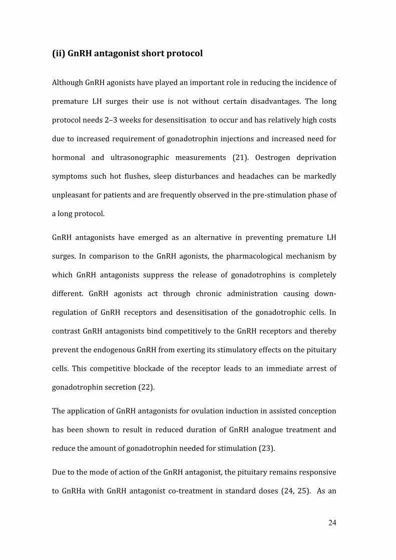
(ii) GnRH antagonist short protocol
Although GnRH agonists have played an important role in reducing the incidence of
premature LH surges their use is not without certain disadvantages. The long
protocol needs 2–3 weeks for desensitisation to occur and has relatively high costs
due to increased requirement of gonadotrophin injections and increased need for
hormonal and ultrasonographic measurements (21). Oestrogen deprivation
symptoms such hot flushes, sleep disturbances and headaches can be markedly
unpleasant for patients and are frequently observed in the pre-stimulation phase of
a long protocol.
GnRH antagonists have emerged as an alternative in preventing premature LH
surges. In comparison to the GnRH agonists, the pharmacological mechanism by
which GnRH antagonists suppress the release of gonadotrophins is completely
different. GnRH agonists act through chronic administration causing down-
regulation of GnRH receptors and desensitisation of the gonadotrophic cells. In
contrast GnRH antagonists bind competitively to the GnRH receptors and thereby
prevent the endogenous GnRH from exerting its stimulatory effects on the pituitary
cells. This competitive blockade of the receptor leads to an immediate arrest of
gonadotrophin secretion (22).
The application of GnRH antagonists for ovulation induction in assisted conception
has been shown to result in reduced duration of GnRH analogue treatment and
reduce the amount of gonadotrophin needed for stimulation (23).
Due to the mode of action of the GnRH antagonist, the pituitary remains responsive
to GnRHa with GnRH antagonist co-treatment in standard doses (24, 25). As an

25
alternative to hCG, GnRHa have been used to trigger the endogenous release of LH
and FSH resembling the physiological mid-cycle surge of gonadotrophins (26).
GnRHa have also been shown to be as efficacious as hCG for induction of ovulation
(27-29).
hCG and LH share the same alpha subunit and 81% of the amino acid residues of
their beta subunit; hence they bind to the same receptor (30). The half-life of hCG,
however, is 24 hours (31) whereas the half-life of LH is 60 minutes (32). A GnRH
agonist-induced LH surge has been shown to effectively stimulate ovulation and
final oocyte maturation (27, 28).
There are two variations of the GnRH antagonist protocol: namely the fixed and
flexible. During the fixed protocol the GnRH antagonist (Cetrotide or Ganirelix) is
administered from day six of controlled ovarian stimulation onwards (33). In the
flexible protocol administration of the GnRH antagonist is withheld until the lead
follicle reaches a mean diameter of at least 14mm.
A meta-analysis comparing the fixed and flexible GnRH antagonist protocols
demonstrated no statistically significant difference in pregnancy rates, although
there appeared to be a trend towards higher pregnancy rates when the fixed
protocol was used (34). However, the flexible protocol was associated with a
decrease in the number of doses of GnRH antagonist required and the total amount
of gonadotrophins used.

26
FSH is traditionally commenced on day 2 of the menstrual cycle during a GnRH
antagonist protocol and therefore the onset of menses was normally a prerequisite.
However more recently, with the development of emergency fertility preservation
ovarian stimulation protocols prior to chemotherapy for cancer, the antagonist
protocol can also be started in the luteal phase which is termed a ‘random start’.
Studies have shown that both late follicular phase and luteal phase start antagonist
ovarian stimulation protocols were as effective as the conventional early follicular
phase start. The number of total and mature oocytes retrieved, oocyte maturity
rate, mature oocyte yield, and fertilisation rates were similar across protocols (35).
Early meta-analyses comparing the use of GnRHa ovulation induction with the
antagonist protocol demonstrated a 5% reduction in clinical pregnancy rates (36).
These findings were also corroborated by Griesenger et al (2005) with the analysis
of a large German national IVF database and, as a consequence, the antagonist
protocol became the second-line choice for a significant numbers of clinicians (37).
However, the most recent meta-analysis has failed to identify any significant
difference in live birth rates between the two protocols (38).
3. Triggering of oocyte maturation
(i) hCG trigger
Ovulation in a natural cycle is initiated and perpetuated by a mid-cycle surge of LH
released by the pituitary gland. The LH surge acts to induce final oocyte maturation

27
whereby the first meiotic division is completed with extrusion of the polar body
and it is then arrested in prophase of the second meiotic division until fertilisation.
hCG shares the same alpha subunit and 81% of the amino acids of the beta subunit
of LH and hence binds to the same receptor (30). Initially high doses of urinary hCG
(5000-10,000iu) were administered to women to successfully mimic the
physiological LH surge and induce oocyte maturation. More recently the use of
recombinant hCG has become widespread following on from the randomised
controlled trial (RCT) conducted by Papanikolaou et al (2010) comparing 250 μg
recombinant hCG versus 10,000iu urinary hCG. This study concluded the
pregnancy rate was significantly higher using recombinant hCG (40).
hCG also has a stimulating action on the development and function of the corpus
luteum; namely a luteotrophic effect, that is more potent owing to its longer half-
life compared to the endogenous LH surge and as such hCG can still be detected in
the serum 10 days after the preovulatory bolus injection (27, 31, 41). In contrast
the physiological LH surge in a normal menstrual cycle lasts 48 hours.
Consequently, continued support of the corpus luteum by hCG elicits
supraphysiological luteal phase steroid concentrations. In the context of an
exaggerated ovarian response secondary to stimulation (i.e. a large number late
follicular phase follicles and very high oestradiol levels), both recombinant and
urinary hCG have clearly been associated with an increased risk of OHSS (42-
44).The induction and stimulation of multiple corpora lutea may represent a key
phenomenon in this regard. Consequently, the investigation and development of
alternative triggering agents continues.

28
(ii) GnRH agonist trigger
As previously outlined, GnRHa initially induce a surge in gonadotrophin production
termed the ‘flare’ effect. During the long agonist protocol, it is its continued
administration that leads to downregulation of its receptor and desensitisation of
the pituitary. With the advent of the GnRH antagonist protocol, where the GnRH
receptor is not unresponsive rather competitively inhibited, GnRHa can be used as
an alternative to hCG to induce oocyte maturation. The GnRHa displaces the GnRH
antagonist from the pituitary GnRH receptor thereby initiating an endogenous
surge of gonadotrophins, namely LH and FSH.
The gonadotrophin surge generated by administration of GnRHa differs from a
physiogical LH surge that occurs in a normal menstrual cycle. The physiological LH
surge has three distinct phases: the ascending, the plateau and the descending.
Each of these phase durations are 14, 14 and 20 hours respectively (45). In
comparison, the GnRHa-induced LH surge has two phases: the rapid ascending
phase lasting approximately 4 hours and the longer descending phase of 20 hours
(28).
The shortened duration of the LH surge with a GnRHa trigger significantly reduces
the total amount of LH released when compared to a normal menstrual cycle (27,
28). As a result, the GnRHa trigger is associated with a deficient luteal phase steroid
profile and subsequently lower pregnancy rates (46, 47). However, despite this, the
GnRHa trigger has a much better safety profile with regards to OHSS when
compared to hCG (48, 49). Another observed benefit is the retrieval of a larger

29
proportion of metaphase II (MII) oocytes when compared to hCG (49). This can be
in part attributed to the concomitant FSH surge that occurs with the LH surge when
GnRHa are administered. Theoretically the concurrent surge in FSH can promote
oocyte nuclear maturation, through resumption of the second meiotic division and
potentiate the formation of LH receptors in the luteinised granulosa cells, thus
supporting the function of multiple corpora lutea (50-53).
4. Transvaginal oocyte retrieval
Transvaginal ultrasonography-guided oocyte retrieval is performed approximately
36 hours following the ovulation induction trigger injection. The surgeon inserts a
single or double lumen needle through the lateral fornix of the vagina into the
peritoneal cavity and then into the ovary. Care is taken to avoid injuring adjacent
structures. The proximal end of the needle is attached to suction apparatus and
after each ovarian follicle is penetrated the follicular fluid is aspirated under
suction ranging from 100-150mmHg for single and double lumen respectively. The
follicular fluid is then transferred to an embryologist in the adjacent laboratory
who then examines the aspirate to identify and quantify the number of oocytes
collected. Once all of the accessible follicles are aspirated, the needle is withdrawn
from the lateral fornix and the procedure is repeated in the contralateral ovary.
5. Embryogenesis
Fertilisation of oocytes by sperm can be achieved in the laboratory via two
methods: IVF or IVF with intracytoplasmic sperm injection (ICSI). Conventional IVF
involves placement of an oocyte in culture medium containing motile sperm at a

30
concentration of 50–100,000 sperm per millilitre (ml). Fertilisation occurs when a
sperm binds to the surface of the extracellular zona pellucida that surrounds the
egg. Following zona penetration and fusion with the oocyte plasma membrane,
peripherally located cortical granules within the egg exocytose their contents,
which modifies the zona matrix such that sperm no longer bind. These events are
orchestrated to ensure that a single sperm fertilises a single egg.
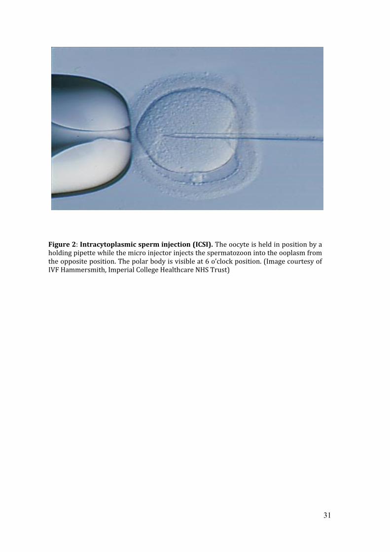
ICSI fertilisation involves oocytes being denuded of cumulus cells using
hyaluronidase and subsequently being assessed for maturity. Mature oocytes are
identified if extrusion of the first polar body (metaphase II) has occurred. The
oocyte is immobilised using a holding pipette; an injection pipette is used to
aspirate a single spermatozoon. Immobilisation of the spermatozoon can be
achieved by crushing the tail with the injection pipette and aspirating it into the
injection needle. The spermatozoon is then passed through the zona pellucida and
directly injected into the ooplasm (Figure 2). ICSI is recommended for male factor
infertility where there is a reduced sperm count, poor motility or a low number of
morphologically normal sperm (<4%). ICSI can also be used in cases where there
was low proportion of fertilisation or complete failure of fertilisation with previous
IVF.
31
Figure 2: Intracytoplasmic sperm injection (ICSI). The oocyte is held in position by a holding pipette while the micro injector injects the spermatozoon into the ooplasm from the opposite position. The polar body is visible at 6 o’clock position. (Image courtesy of IVF Hammersmith, Imperial College Healthcare NHS Trust)

32
6. Embryo development
Oocytes are assessed for fertilisation 16-18 hours after the ICSI procedure. The
oocytes are considered fertilised when two pronuclei are identifiable in the
ooplasm (2PN stage). The female and male pronuclei contain haploid copies of
their respective chromosomes. The 2-PN zygotes then enter the cleavage stage. The
resultant embryos enter the four-cell stage on day 2 and the 6-8-cell stage on day 3,
and following full compaction of the embryonic cells on day 4 (morula stage) a
blastocoele cavity forms on day 5 (Figure 3). The blastocoele separates the
embryonic components to the inner cell mass and the trophoectoderm which will
subsequently form the foetus and placenta respectively. Given that the embryonic
genome becomes fully activated at the 8-cell stage, preference is given to using
embryos that have undergone this transition from the maternal to the embryonic
genome (Braude et al, 1988). Embryo selection following IVF for return to the
uterus for implantation depends on a grading system, which is based on
morphological criteria (Table 1).

33
Figure 3: Timeline of the fertilised 2PN zygote entering the cleavage stage containing 4 blastomeres on day 2, 6-8 blastomeres on day 3, blastomeres compacting into a morula on day 4, and reaching blastocyst at day 5. On day 6 then embryo hatches out to the zona pellucida. Images taken from: http://yaolab.stanford.edu/images/Slide7_ Retrieved on 30/07/12)

34
Table 1: Grading system for day 2/3 embryos (blastomere) and day 5/6 embryos (blastocyst) (54).

35
A significant increase in biochemical pregnancy (confirmed by the presence of a
positive pregnancy test) (OR 1.45, 95% CI 1.03–2.04) and clinical pregnancy rates
(defined by the presence of an active foetal heartbeat at the 6th gestational week
ultrasound) (OR 1.63, 95% CI 1.12–2.35) was observed after single blastocyst
transfer compared with transfer of a single cleavage stage (day 3) embryo in
patients aged less than 36 years old (55, 56). Similar pregnancy outcomes have
been reported following the transfer of a single versus double blastocyst transfer
(57, 58). However, twin pregnancies have been reported to be significantly higher
following double embryo transfer (3.5% in single embryo transfer versus 37.5%
with double P < 0.05) (58).
7. Luteal phase support.
The luteal phase is defined as the period between ovulation and either the
establishment of a pregnancy or the onset of menses two weeks later (59). During
normo-ovulatory cycles, the corpus luteum remains dependent on support from
the pituitary gonadotrophins throughout the luteal phase. The spontaneous LH
surge results in luteinisation of theca and granulosa cells, a switch from the
predominant production of oestrogen to progesterone, the induction of the final
stages of oocyte maturation. The luteinised granulosa cells represent the most
important progesterone-producing cells of the corpus luteum. LH is the principal
trophic hormone for the corpus luteum (60, 61) and decreasing LH concentration
induces the structural and functional degradation of the corpus luteum termed
luteolysis . (62). Luteolysis can also be induced by the luteal phase administration
of a GnRH agonist (62, 63) or an antagonist (64). Continued support of the corpus

36
luteum by LH is critical and luteal regression becomes non- reversible when
endogenous LH is lacking beyond 72 hours (65, 66). The pulsatile release of LH is
closely controlled by oestradiol and progesterone feedback loops (67). Adequate
corpus luteum function is necessary for conception to occur. Luteal regression
during the normal menstrual cycle is caused by reduced responsiveness of the
aging corpus luteum to LH (68), which can be overcome by administering
increasing doses of LH (69) or hCG (either administered exogenously, or as occurs
during pregnancy).
The luteal phase of stimulated IVF cycles is abnormal (70, 71) and is likely to be
related to the supraphysiological levels of steroids secreted by a high number of
corpora lutea during the early luteal phase. This directly inhibits the release of LH
via negative feedback actions at the hypothalamic-pituitary level (72).
The profound suppression of pituitary gonadotrophin release after cessation of
GnRHa is well established (73, 74). Earlier cessation of GnRHa administration
during follicular phase stimulation aimed at enabling recovery from pituitary
desensitisation was not shown to be successful (41, 75). Due the known rapid
recovery of pituitary gonadotrophin release following cessation of GnRH
antagonists (76), researchers speculated that GnRH antagonist co-treatment could
be applied to IVF without disrupting the luteal phase steroid profile. Although
initial studies were inconclusive (77, 78), a recent study focusing on the non-
supplemented luteal phase endocrinology following GnRH antagonist co-treatment
clearly demonstrated abnormal steroid profiles, along with extremely low LH
concentrations, profoundly reduced luteal phase lengths and significantly
compromised chances of pregnancy (79).

37
Progesterone induces secretory transformation of the endometrium in the luteal
phase (80). By inducing this change following oestrogen priming in the follicular
phase, progesterone improves endometrial receptivity (81). During this period of
receptivity, the endometrial epithelium develops a functional and transient ovarian
steroid-dependent status that facilitates embryo attachment (82). Decreased
endometrial receptivity is considered largely responsible for the low implantation
rates in IVF (83).
Csapo et al. (1972, 1973) highlighted the importance of progesterone during the
first weeks of a pregnancy. The removal of the corpus luteum prior to 7 weeks of
gestation led to miscarriage in their initial study (84). However, they found that the
pregnancy could be maintained even after removal of the corpus luteum if
exogenous progesterone was administered (85). Therefore, luteal phase
supplementation with hCG or progesterone following IVF has become ubiquitous
as it results in an increased pregnancy rate (PR) (93). The use of progesterone in
those with a history of recurrent miscarriage following spontaneous pregnancy
remains contentious. A Cochrane review demonstrated a significant improvement
in miscarriage rates in this subgroup of women (205) whereas a recent multicentre
RCT did not (86).
In 1985, Leeton et al. first demonstrated the extension of the luteal phase of
stimulated IVF cycles by administering 50 mg intramuscular (IM) progesterone
(87). Subsequent trials have yielded conflicting results. In an open label study
evaluating 1184 women receiving either vaginal and IM progesterone for luteal
phase support (LPS), the clinical and ongoing PRwere comparable (35.1 versus

38
35.2%, respectively) and (30.2 and 33.6%, respectively)(88). However, meta-
analysis of 5 studies comparing IM administration of progesterone with vaginal
application concluded that clinical PR and delivery rate were significantly higher
when IM progesterone was used (89).
However, this route of administration is often associated with a number of side
effects, including painful injections and rash (90) causing difficulties with patient
tolerance and compliance (91). In addition to this, injections of progesterone in oil
can also cause inflammatory reactions and abscesses (92). Vaginal administration
of progesterone is a viable alternative to the IM injections of progesterone if the
side effects of IM injections are proving too severe.

39
Ovarian Hyperstimulation Syndrome (OHSS)
One of the major complications of IVF treatment is a condition known as OHSS. It is
a systemic disease that can affect the cardiovascular, hepatorenal and central
nervous systems leading to significant morbidity and even mortality. The severity
of OHSS can vary with mild forms accounting for up to 33% of all IVF cycles. Severe
OHSS is reported to occur between 3-8% of IVF cycles (194)
Risk Factors
Certain patient and cycle characteristics increase the risk of OHSS; women with a
previous history of OHSS, polycystic ovary syndrome (94) increased antral follicle
count (AFC) or high levels of anti-Mu llerian hormone (AMH) (95, 96), elevated
serum oestradiol during ovarian stimulation (98) and high doses of exogenous FSH
during ovarian stimulation (99). Some studies have also noted that mean body
mass index (BMI) (97) and age (94, 97) were lower in women that developed OHSS
compared to those that did not.
The risk of OHSS is also related to the number of developing follicles during the
follicular phase of ovarian stimulation (42) and the number of retrieved oocytes at
the end of the cycle (100). The risk also appears to increase when hCG is
administered to induce oocyte maturation. OHSS manifestation becomes less likely
when progesterone-based luteal phase support is used instead of hCG (101).
Clinical features
Cardinal symptoms of OHSS are abdominal bloating, pain and nausea or vomiting
related to ascites and large ovarian volumes. As the disease progresses more

40
serious sequelae can manifest as outlined below. The Royal College of Obstetricians
and Gynaecologists (RCOG) has employed the following classification for OHSS
(Table 2).
OHSS can also be classified as early or late onset. The cause of early onset OHSS is
typically due to the use of hCG to induce oocyte maturation prior to retrieval.
Although hCG and LH share the same alpha subunit, the circulating half-life of hCG
is much longer than that of LH; circulating for up to 7 days post administration (31)
compared to the physiological LH surge lasting approximately 48 hours (102). As a
consequence hCG can excessively stimulate the ovary, driving the pathophysiology
of this condition. The hallmark of OHSS is an increase in capillary permeability
resulting in a fluid shift from the intravascular space to third space compartments
(103, 104). Vascular endothelial growth factor (VEGF), has emerged as one of the
factors most likely involved in the pathophysiology of OHSS (105). VEGF is an
angiogenic cytokine that is a potent stimulator of the vascular endothelium and
appears to play an integral role in follicular growth, corpus luteum function, and
ovarian angiogenesis. Recent studies also indicate that hCG increases VEGF
expression in human granulosa cells and raises serum VEGF concentrations (106,
107). VEGF has been implicated as it known to increase vascular permeability to
high molecular weight proteins promoting capillary leakage (108).

41
Table 2: RCOG classification of severity of OHSS

42
Management of OHSS
The management of patients with mild or moderate OHSS can be on an outpatient
basis. These patients mainly require supportive therapy, in the form of analgesia
and antiemetics, and monitoring for progression of disease severity. Clinical review
of symptoms and ultrasound examination to measure ovarian size and to
determine the presence ascites is useful in assessing the severity of OHSS.
Recommended laboratory investigations include haemoglobin, haematocrit, liver
function tests, serum creatinine, urea and electrolytes.
Women are encouraged to drink to thirst to reduce the risk of haemoconcentration
and physical activity should be maintained as reduced mobility may increase the
risk of thrombosis. Women should continue luteal supoort with progesterone and
not hCG. Patients are advised to avoid sexual intercourse and strenuous exercise as
it may worsen pain and may increase the risk of ovarian torsion. Hospitalisation is
indicated in cases of severe or critical OHSS (Table 2).
Prevention of OHSS Various strategies have been adopted in order to reduce the risk of the
development of OHSS. Administering the lowest effective dose of FSH in women
with polycystic ovarian syndrome (PCOS) has been shown to reduce the incidence
(109). Pre-treatment with metformin prior to commencing IVF also has a
protective effect in the same patient cohort (110). Similarly, dopamine agonists
have been shown to reduce the incidence of OHSS by binding to VEGFR-2 receptor,
reducing its activity and decreasing vascular permeability (111).
The most effective development in combatting the incidence of OHSS, especially in

43
the high-risk population, has been the introduction of the GnRH antagonist
protocol. This protocol allows the use of a GnRHa instead of hCG to induce oocyte
maturation. Administration of a single dose of GnRH agonist, after co-treatment
with GnRH antagonist, induces an endogenous rise of both FSH and LH which
results in the induction of the final stages of oocyte maturation (112). The shorter
half-life of the endogenous LH surge and the subsequent pituitary suppression due
to GnRH receptor desensitisation, leads to a withdrawal of LH support for the
corpora lutea that may lead to early luteolysis and possible abolition of the
secretion of the vasoactive peptides responsible for causing OHSS (113). However,
the abnormal luteal phase steroid profile after GnRHa trigger (79) has been shown
to be associated with impaired implantation and lower on-going pregnancy rates
compared to hCG (46, 114).
As a result, there remains a need to identify novel oocyte maturation triggers that
have an effective safety profile in terms of reducing the risk of OHSS but that
maintain ongoing pregnancy success rates. Kisspeptin is a neuropeptide that has
such potential.
Kisspeptin
The KISS1 gene, encoding for the neuropeptides termed kisspeptins, was first
identified in 1996 as a tumour suppressor in malignant melanoma (115) The gene
was named after the famous chocolate ‘kisses’ produced in the town of its
discovery, Hershey (PA, USA). The KISS1 gene is located on chromosome 1q32
consisting of 4 exons where the first two are not translated (116).

44
Kisspeptin and its receptor
G protein-coupled receptors (GPR) are the largest category of cell membrane
receptors in humans (117). The GPR54 receptor was first described in the rat
model (118) and subsequently the human brain (119). The GPR54 gene is located
at chromosome 19p13.3 consisting of five exons encoding a protein of 398 amino
acids with seven hydrophobic, trans-membrane domains (119). Upon kisspeptin
binding to it, the GPR54 receptor activates phospholipase C and subsequently
recruits intracellular messengers, such as inositol triphosphate and diacylglycerol.
These messengers mediate a biphasic rise of intracellular calcium (120) and
protein kinase C activation which execute kisspeptin’s function (119, 121).
The KISS-1 gene initially encodes for a precursor 145- amino acid peptide, which is
subsequently cleaved into a 54-amino acid product called kisspeptin-54 (116). This
can be cleaved further to 14,13 and 10-amino acid peptides all sharing a common
C-terminal that is responsible for the high affinity binding and activation of the
GPR54 receptor (122).
The neuroanatomical distribution and expression of KISS-1/Kiss-1
Kiss-1 mRNA is predominantly expressed in the hypothalamic anteroventral
periventricular (AVPV) nucleus and the preoptic periventricular (POPV) nucleus in
rodents (123, 124). This differs from humans who have a more scattered
distribution of in the preoptic region (125, 126). Furthermore, kisspeptin and
GnRH neuronal networks lie in close proximity in the hypothalamus as
demonstrated in animal models (127-129) and also in humans (130) suggesting

45
that kisspeptin has direct involvement in GnRH neurosecretion. The physiological
role of kisspeptin-regulated GnRH secretion is demonstrated via abolition of
kisspeptin-induced GnRH neuron depolarisation by a kisspeptin antagonist (131-
133). Kisspeptin acts upstream of GnRH as demonstrated by the prevention of
kisspeptin-induced LH release by pretreatment with a GnRH antagonist (123, 134).
Kisspeptin stimulates GnRH secretion
Exogenous administration of kisspeptin was first shown to potently stimulate the
hypothalamic-pituitary-gonadal axis in the rat model (135). Intra-
cerebroventricular and intravenous administration of kisspeptin-10 to adult male
rats, results in a dose-dependent increase in plasma LH, FSH and testosterone. This
was also demonstrated in sheep where central administration of kisspeptin
intracerebroventricularly produced a dramatic release of GnRH into the
cerebrospinal fluid, with a parallel rise in serum LH (127).
In addition, intravenous infusion of kisspeptin in oophorectomised sheep produced
an increase of serum FSH and LH release (136). In the primate, repetitive
kisspeptin administration to agonadal prepubertal males, primed with GnRH to
enhance pituitary responsiveness, elicited GnRH-dependent LH secretion similar to
that of adults (134, 137). Conversely, Clarkson et al (2008) were able to
demonstrate the absence of the LH surge in GPR54 knockout mice (138).
GPR54 is predominantly expressed in the central nervous system (spinal cord,
caudate nucleus, substantia nigra, hippocampus, amygdala and thalamus),
endocrine organs (pituitary gland and pancreas) and the placenta. The highest
expression of the GPR54 receptor is in the pituitary gland and the placenta (122).

46
There is significant overlap between kisspeptin, neurokinin B and dynorphin
neurons in the human hypothalamus (130, 140). These neurons expressing all
three neuropeptides are termed KNDy neurons (141). KNDy neurons make direct
contact with GnRH cell bodies in humans (142). In the murine model dynorphin
and NKB act autosynaptically on kisspeptin neurons coordinate the pulsatile
release of GnRH into the portal circulation (143). In this study, neurokinin B was
found to have a stimulatory effect and dynorphin an inhibitory one on kisspeptin
neurons. The pulsatile release of GnRH leads to the secretion of LH and FSH from
the gonadotroph cells in the anterior pituitary gland (124, 131). This observation
was consistent with those seen in primates showing the coinciding pulsatile release
of kisspeptin and GnRH secretion, as measured in the median eminence of the
monkey (144). Furthermore, kisspeptin’s pivotal role in controlling reproductive
function is highlighted by the fact that mutations of the GPR54 gene are the cause
of hypogonadotrophic hypogonadism and delayed puberty in humans (145, 146);
whereas activating mutation of GPR54 results in central precocious puberty (147).
Kisspeptin-mediated GnRH release is sex steroid dependent. In humans, the
infundibulum relays oestrogen and progesterone signalling whereas in the rodent,
the AVPV relays positive feedback and the arcuate nucleus negative (148).
Kisspeptin neurons display sexual dimorphism
Interestingly, in rodents, the kisspeptin neurons in the hypothalamus appear to
display sexual dimorphism (124). This is particularly true in the AVPV, where there
are approximately 25 times more kisspeptin cell bodies in adult females compared

47
with males (149). Furthermore, kisspeptin neurons located in the AVPV of the rat
express estrogen receptor α (206, 207), which indicates that these kisspeptin
neurons are equipped with the appropriate gonadal steroid hormone receptors
required for positive feedback to generate the LH surge. This is important when
considering that pre-ovulatory positive steroid feedback is exclusively female. The
arcuate nucleus, which controls negative feedback in rodents, does not display this
kind of dimorphism (124, 149). More recently, there is also evidence to support the
possibility of sexually dimorphic kisspeptin neuron populations in humans. The
female hypothalamus has significantly more kisspeptin neurons in the
infundibulum and the rostral periventricular area of the third ventricle (RP3V)
(130). Humans also display this dimorphism in their response to kisspeptin. While
kisspeptin potently stimulates LH release in men (150) the effect of kisspeptin is
far more variable in women depending on which phase of the menstrual cycle
kisspeptin is administered (151). The sexual variation in the neuroanatomical
distribution of the kisspeptin pathway, and the differing responses to kisspeptin
administration, may be a reflection of kisspeptin’s distinct or differing role between
the sexes; notably the generation of the preovulatory LH surge which is unique to
females.
Kisspeptin and metabolism
It is well established that reproductive function in humans can be greatly
influenced by nutritional status; in that either extreme (being underweight or
obese) can have a detrimental effect. Although GnRH neurons do not express the
leptin receptor (Ob-Rb), a significant proportion of kisspeptin neurons in the
arcuate nucleus of mice do (152). In this way, kisspeptin may provide the link

48
between nutritional status and reproduction. While the role of leptin in the control
of KiSS-1 expression is being investigated, the effects of other metabolic regulators
on the KiSS-1 system have remained relatively unexplored so far. In principle,
considering the proven reproductive roles of a number of neuropeptides and
hormones that are primarily involved in the control of food intake and energy
homeostasis (208, 209, 210) a hypothesis that some of these regulators may have
also have an impact on KiSS-1 expression or function, in addition to leptin, is a
feasible one.
Kiss1 mRNA is significantly reduced in obese mice (153) compared with wild-type
controls (152). In rats with streptozotocin-induced diabetes, hypothalamic levels of
Kiss1 mRNA are decreased, which is associated with subsequently reduced levels of
circulating gonadotrophins. However, this hypogonadotropic state can be reversed
by kisspeptin administration, implying that diminished kisspeptin signaling may
explain the reproductive dysfunction that can accompany diabetes (154, 155).
Decreased testosterone levels have been observed in obese men with type 2
diabetes with reduced GnRH secretion thought to be the root cause (156). Possible
mechanisms for this down-regulation of kisspeptin and subsequent GnRH
signalling include increased oestrogen negative feedback in the obese (157) leptin
resistance (158) hyperglycaemia and insulin resistance (155, 159). George et al
(2013) demonstrated that administration of kisspeptin-10 increased the frequency
of LH pulsatility in hypogonadal, type 2 diabetic men (160).
In the catabolic state, gonadotrophin secretion and expression of Kiss1 mRNA is
reduced in rodents and monkeys (161, 162). However, exogenous administration
of kisspeptin can restore reproductive function (163).

49
Humans with leptin or leptin receptor mutations demonstrate biochemical
hypogonadism (164) and the majority do not have normal pubertal development
(165). Leptin deficient mice demonstrate a decrease in the expression of Kiss1
mRNA, which can be partially restored by leptin (152). However, mice with a
selective deletion of the leptin receptor from kisspeptin neurons exhibit normal
pubertal development and fertility (166) leading to speculation that leptin’s action
on kisspeptin neurons is not obligatory for reproductive function. Furthermore, in
human studies where leptin mutations were present, a small number of females
underwent delayed but spontaneous menses (167, 168) suggesting that even in the
absence of leptin activity, some activation of the hypothalamic–pituitary–gonadal
axis is possible. Collectively, these findings point to a potentially important role of
kisspeptin neurons in regulation of the reproductive axis via metabolic signaling.
Effects of kisspeptin administration to humans (i) Healthy volunteers Kisspeptin is a potent stimulator of the reproductive axis in both animal models
and in humans. As stated previously, kisspeptin exerts its effects directly on GnRH
neurons via its receptor initiating the release of GnRH into the portal circulation.
This in turn leads to the secretion of FSH and LH from the anterior pituitary.
Although kisspeptin stimulates the release of gonadotrophins (FSH and LH), its
effect on LH secretion is more marked (169, 170). Kisspeptin-54 was first
administered into 6 healthy male volunteers in 2005. They received a 90-minute
intravenous infusion of kisspeptin at a dose of 4pmol/kg/min for the first 30
minutes and half that dose (2pmol/kg/min) for the remaining 60 minutes. This was
50
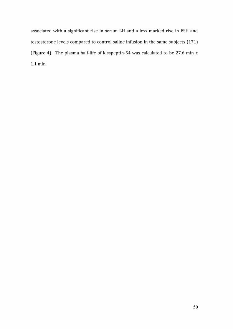
associated with a significant rise in serum LH and a less marked rise in FSH and
testosterone levels compared to control saline infusion in the same subjects (171)
(Figure 4). The plasma half-life of kisspeptin-54 was calculated to be 27.6 min ±
1.1 min.
51
Figure 4: Mean plasma kisspeptin-IR, LH, FSH, inhibin B and testosterone after kisspeptin-54 and control saline infusions. Mean 90-min LH: kisspeptin, 10.8 ± 1.5 vs. saline, 4.2 ± 0.5 U/L; mean 90-min FSH: kisspeptin, 3.9 ± 0.7 vs. saline, 3.2 ± 0.6 U/L mean 180-min testosterone: kisspeptin, 24.9 ± 1.7 vs. saline, 21.7 ± 2.2 nmol/liter (Dhillo et al., 2005)

52
Further studies confirmed the findings of Dhillo et al (2005) by infusing
(continually and in bolus-form) kisspeptin-10 into the a cohort of healthy male
volunteers (169). Interestingly, despite continuous infusion kisspeptin-10 for 22.5
hours, George et al (2011) demonstrated no tachyphylaxis of the GnRH/LH
pathway; indeed LH secretion tended to progressively increase. This may have
been due to lower doses of kisspeptin-10 being used and over a shorter duration
compared to other studies where tachyphylaxis was observed (172, 173)(Seminara
et al., 2006, Jayasena et al., 2009).
The investigation of the effects of kisspeptin-54 on LH secretion in healthy female
volunteers was undertaken by Dhillo et al., 2007. Every participant was
administered subcutaneous boluses of kisspeptin (0.4nmol/kg) and saline in
different menstrual cycles. This set of injections was performed once during the
follicular, once in the pre-ovulatory and again in the luteal phase of the menstrual
cycle. Kisspeptin-54 increased plasma LH compared with saline injection in all
phases of the cycle. A varied gonadotrophin secretory response was observed
depending on which phase of the menstrual cycle kisspeptin was administered. In
particular, the effect of kisspeptin-54 on LH and FSH in the pre-ovulatory phase
was 5-fold greater than in the follicular and luteal phases (151) (Figures 5 and 6).
These findings were supported by a further study where ten women in the early
follicular phase, three women in the pre-ovulatory phase, and 14 women in the
mid-luteal phase received an intravenous bolus of kisspeptin-10 at a dose of 0.24
nmol/kg. LH pulses were observed immediately after kisspeptin administration in
all luteal and pre-ovulatory women. However, only half the women in the early
follicular phase demonstrated a significant response to kisspeptin (170).

53
Figure 5: Mean ± SEM increase in plasma LH after saline or kisspeptin injection in the various phases of the female menstrual cycle. (A:follicular, B:pre-ovulatory, C:luteal, D: all three phases). ***, P<0.001. Mean increase in LH over baseline (IU/liter) ± sem for follicular phase was 0.12 ± 0.17; preovulatory phase, 20.64 ± 2.91 (P < 0.001 vs. follicular phase); luteal phase, 2.17 ± 0.79 (P < 0.01 vs. follicular phase (Dhillo et al, 2007)

54
Figure 6: Mean ± SEM increase in plasma FSH after saline or kisspeptin injection in the various phases of the female menstrual cycle (A:follicular, B:pre-ovulatory, C:luteal, D: all three phases). ***, P<0.001. (Dhillo et al, 2007)

55
Of note, kisspeptin-54 also stimulated progesterone release in the luteal phase of
the participants. This may be mediated via the secretion of GnRH and subsequently
LH (64, 174). However, a more recent study has shown that in rats there is strong
intensity of kisspeptin and GPR54 immunoreactivity in the corpus luteum,
particularly in the theca and granulosa cells (154). It is therefore plausible that the
increase in progesterone after peripheral kisspeptin-54 injection in healthy women
represents a direct effect of kisspeptin on the corpus luteum. Recently, kisspeptin
expression has been shown to be increased in endometrial stromal cells through
decidualisation (175) suggesting a role for kisspeptin in preparing the
endometrium for implantation.
(ii) Effect of kisspeptin application in patients with subfertility.
(a) Women with hypothalamic amenorrhoea
Women with hypothalamic amenorrhoea were the first patients in which the
therapeutic potential of kisspeptin was explored. This condition is associated with
slow GnRH pulsatility and subsequent decline in FSH and LH secretion. Decreased
levels of gonadotrophins lead to low ovarian follicular activity. Ten patients with
hypothalamic amenorrhoea were administered subcutaneous injections of
kisspeptin-54 at a dose of 6.4nmol/kg twice a day for a period of two weeks. Serum
gonadotrophin and oestradiol levels were measured on the day 1 and 14. The
results indicated an initial potent rise in serum LH and FSH on the first day of
injections (10-fold increase in LH and 2.5 fold in FSH) (173). This response,
however, was not sustained and appeared to be significantly reduced by day 14,

56
suggesting that chronic administration of kisspeptin can result in tachyphylaxis
(173) (Figure 7). However, the same researchers demonstrated that tachyphylaxis
could be avoided by intermittent twice-weekly injections of 6.4 nmol/kg of
kisspeptin-54 over an 8 week period (176). Even though an increase in LH and FSH
levels into the normal physiological range was observed in both studies, this did
not translate into increased ovarian follicular activity (persistent ovarian
quiescence on scan and no significant rise in serum oestradiol levels) (173, 176).

57
Figure 7: Effects of kisspeptin-54 and saline injection on the first day (A,B,C) and the
14th day (D,E,F) to plasma levels of LH, FSH and serum oestradiol. Data are shown at mean ± SEM. *,P<0.05. ***, P<0.001 (Jayasena et al., 2009)
58
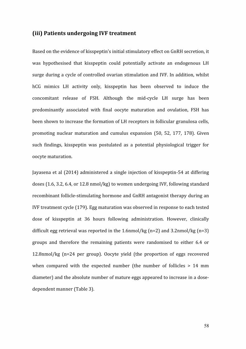
(iii) Patients undergoing IVF treatment
Based on the evidence of kisspeptin’s initial stimulatory effect on GnRH secretion, it
was hypothesised that kisspeptin could potentially activate an endogenous LH
surge during a cycle of controlled ovarian stimulation and IVF. In addition, whilst
hCG mimics LH activity only, kisspeptin has been observed to induce the
concomitant release of FSH. Although the mid-cycle LH surge has been
predominantly associated with final oocyte maturation and ovulation, FSH has
been shown to increase the formation of LH receptors in follicular granulosa cells,
promoting nuclear maturation and cumulus expansion (50, 52, 177, 178). Given
such findings, kisspeptin was postulated as a potential physiological trigger for
oocyte maturation.
Jayasena et al (2014) administered a single injection of kisspeptin-54 at differing
doses (1.6, 3.2, 6.4, or 12.8 nmol/kg) to women undergoing IVF, following standard
recombinant follicle-stimulating hormone and GnRH antagonist therapy during an
IVF treatment cycle (179). Egg maturation was observed in response to each tested
dose of kisspeptin at 36 hours following administration. However, clinically
difficult egg retrieval was reported in the 1.6nmol/kg (n=2) and 3.2nmol/kg (n=3)
groups and therefore the remaining patients were randomised to either 6.4 or
12.8nmol/kg (n=24 per group). Oocyte yield (the proportion of eggs recovered
when compared with the expected number (the number of follicles > 14 mm
diameter) and the absolute number of mature eggs appeared to increase in a dose-
dependent manner (Table 3).

59
Overnight blood sampling was performed to determine the time profile of hormone
release in 20 of the subjects treated with the two highest doses of kisspeptin-54
(6.4 or 12.8 nmol/kg; n = 10/dose). Peak levels of plasma kisspeptin were
observed approximately one hour after injection and then fell to preinjection levels
by 12 hours following injection (Figure 8). Serum LH levels peaked four to six
hours following kisspeptin-54 injection and decreased thereafter. The total length
of the LH surge was 12 hours following kisspeptin injection. Similar patterns were
observed in FSH and oestradiol secretion but to a lesser extent following
kisspeptin-54 injection. In contrast, progesterone levels rose continually during the
12 hours following kisspeptin-54 injection.
Fertilisation occurred in 92% of patients (defined as at least one fertilised egg) and
the rate of embryo transfer was 92%. In addition, 36 of 49 patients who had an
embryo transfer did so at the blastocyst stage (day 5 after egg collection).
Furthermore, the blastocyst formation rate (defined as the total number of
blastocysts divided by the total number of 2PN zygotes formed) in these patients
was 49.4% (129 blastocysts from 261 2PN zygotes). The biochemical pregnancy
rate was 40%, and the clinical pregnancy rate was 23% (12 of all 53 treated
patients). Pregnancy outcomes were as follows: ten women each gave birth to
healthy babies (eight women had singleton pregnancies and two women had twin
pregnancies); the remaining two women suffered miscarriages (one at nine weeks
and the other at 12 weeks gestation).

60
1.6(n=2) 3.2(n=3) 6.4(n=24) 12.8(n=24)
M2 4.5(3.5) 4.3(1.5) 7.5 (3.8) 8.8 (4)
%M2 75 (35) 79 (36) 79 (22) 85 (16)
Oocyte yield 49 (29) 36 (18) 76 (49) 103 (53)
Table 3: Summary of total number of metaphase 2 (M2) oocytes retrieved; percentage of oocytes that were mature; oocyte yield (%) (the proportion of eggs recovered from the number of follicles > 14 mm diameter) per dose (1.6, 3.2, 6.4, or 12.8 nmol/kg) (Jayasena et al., 2014)
Kisspeptin-54 dose (nmol/kg): mean(SD)

61
Figure 8: A subgroup of women receiving the two highest doses of kisspeptin-54 (6.4 or 12.8 nmol/kg; n = 10 per group) at t = 0 minutes underwent overnight measurements of circulating kisspeptin-54, LH, FSH, oestradiol, and progesterone at t = –30, –15, 0, 30, 60, 90, 120, 150, 180, 240, 360, 480, 600, and 705 minutes. Blue circles show median for 6.4 nmol/kg kisspeptin-54; red triangles show median for 12.8 nmol/kg kisspeptin-54. Vertical lines indicate the interquartile ranges. Conversion factor for serum oestradiol: 1 pmol/l = 0.27 pg/ml; conversion factor for serum progesterone: 1 nmol/l = 0.31 ng/ml (Jayasena et al., 2014)

62
In current practice hCG is the most commonly used hormone to trigger oocyte
maturation. hCG acts directly on ovarian LH receptors to stimulate egg maturation.
The use of hCG confers an increased risk of OHSS due to prolonged follicular
stimulation compared with the endogenous LH surge, and a lack of negative
feedback control. In contrast kisspeptin stimulates endogenous GnRH and
gonadotrophin release and hence was hypothesized to reduced risk of OHSS.
In order to test this hypothesis, Abbara et al. (2015) recruited sixty women
requiring IVF treatment at high risk of developing OHSS. Previous work has shown
that women with either a serum anti-Müllerian hormone (AMH) greater than 40
pmol/L, or total ovarian antral follicle count (AFC) greater than 23 on ultrasound
have a high risk of developing OHSS during IVF treatment (96, 180). Following a
standard recombinant FSH/GnRH antagonist protocol, patients were randomly
assigned to receive a single injection of kisspeptin-54 to trigger oocyte maturation
(3.2 nmol/kg, n = 5; 6.4 nmol/kg, n = 20; 9.6 nmol/kg, n = 15; 12.8 nmol/kg, n =
20).
Oocyte maturation (≥ 1 mature oocyte) was observed in 95% (57/60) of women
who received kisspeptin-54. The highest oocyte yield (121%) was observed
following 12.8nmol/kg kisspeptin-54. Oocyte maturation did not occur in three
women and another three women suffered failed fertilisation of oocytes following
ICSI. A clinical decision to freeze all embryos was made in three women who were
deemed at high risk of developing OHSS should they have an embryo transfer.
Therefore, embryo formation occurred in 90% (54/60) of women who received
kisspeptin-54 and 51 of 60 women had a fresh embryo transfer.

63
At all doses of kisspeptin-54 tested, biochemical pregnancy, clinical pregnancy, and
live birth rates per transfer (n = 51) were 63, 53, and 45%, respectively. The
highest pregnancy rates were observed following 9.6 nmol/kg kisspeptin-54. Out of
60 participants none developed moderate, severe, or critical OHSS requiring
medical intervention confirming kisspeptin’s efficacy as a safe trigger of oocyte
maturation in those women at high risk of OHSS . The expected rate of moderate or
severe OHSS would normally be 10-20% in a high risk population (96). However,
only three women (5%) in this study were diagnosed with mild early OHSS and one
(2%) with mild late OHSS (181). Together this work suggests that kisspeptin is an
effective trigger of oocyte maturation and may be a safer trigger than the current
‘gold’ standard. The work in this thesis builds on these findings and aims to
optimise the protocol for kisspeptin administration to further improve outcomes
during IVF treatment and compare the rates of OHSS which occur in women
undergoing IVF treatment with different triggers for oocyte maturation (hCG,
GnRH, kisspeptin).

64
CHAPTER 2
Kisspeptin as a novel trigger of oocyte
maturation during IVF treatment

65
Introduction:
The available data, summarised in chapter one, demonstrates that kisspeptin can
induce oocyte maturation, embryogenesis and produce live births. It is also
associated with low rates of OHSS, even in a high risk population. However, the
optimal kisspeptin dosing regime remains unknown. In both IVF studies the oocyte
yield, defined as the percentage of mature oocyte collected out of all follicles ≥
14mm, was lower than may have been expected in a significant number of cases. A
possible explanation for this is that a single injection of kisspeptin results in an LH
surge which lasts for 12 hours (179). In contrast, during a normal menstrual cycle
the physiological LH surge lasts a total of 48 hours (102). Given this observation,
the question arises as to whether administration of a second dose of kisspeptin
would lengthen the duration of the LH surge compared to a single injection of
kisspeptin resulting in improved oocyte yield for a higher proportion of patients.
The work in this thesis attempts to address this important question.
Hypothesis:
Using two doses of kisspeptin to trigger oocyte maturation in IVF treatment will
improve oocyte yield compared to administration of a single injection of kisspeptin.
Aim:
To determine if two doses of kisspeptin improve the oocyte yield compared to a
single injection in women undergoing IVF treatment.

66
Methods:
Regulatory approval:
The Hammersmith and Queen Charlotte’s Research Ethics Committee, London, UK,
approved this study (reference: 10/H0707/2). The study was carried out in the IVF
Unit at Hammersmith Hospital under a license from the UK Human Fertilisation
and Embryology Authority. Written informed consent was obtained from all
subjects prior to inclusion in the study. Approval of this study as a Clinical Trial of
an Investigative Medicinal Product was granted by the Medicines and Healthcare
Products Regulatory Agency, UK. The study was registered on the National
Institutes of Health Clinical Trials database (NCT01667406). The study was
performed in accordance with the Declaration of Helsinki.
Peptide synthesis
Kisspeptin-54 was synthesised and purified by Bachem (Bachem Holding AG,
Bubendorf. Switzerland). Sterile vials of Kisspeptin-54 were produced by the
Clinalfa brand of Bachem (Bachem Distribution Services GmbH, Weil am Rhein,
Germany). Both productions were prepared according to Good Manufacturing
Practice (GMP). Vials of freeze-dried kisspeptin-54 were stored at -20OC and
reconstituted in 0.5ml of 0·9% saline as previously described (179).
Sample Size and Power:
The sample size was calculated based on oocyte yield data from previous studies
using kisspeptin to trigger oocyte maturation (179, 181). Optimal oocyte yield was
achieved in 43% of patients using a single kisspeptin injection to trigger oocyte

67
maturation. The oocyte yield was expected to rise to 80% using two kisspeptin
injections (based on published data using triggers for oocyte maturation which
result in a 24h rise in LH (182). Using Fisher's exact conditional test for two
proportions, a sample size of 31 patients per group (±5% drop-out rate) was
determined to provide an 80% power using a two-sided α of 0.05 to detect an
expected increase in the proportion of patients achieving satisfactory oocyte yield
of rising from 43% to 80%.
Study participants:
Seventy-six women requiring IVF treatment for infertility at Hammersmith
Hospital, London, UK, were screened for participation between October 2014 and
August 2015. Eligible patients received one IVF treatment cycle: all treatment costs
for this IVF cycle were funded by study participation. The first two inclusion
criteria aimed to select women at high risk of OHSS: serum anti-Müllerian hormone
(AMH) ≥40pmol/L (≥5·6ng/mL) or total antral follicle count (AFC) ≥23; age 18-
34yrs; early follicular phase serum follicle stimulating hormone (FSH) ≤12iU/L;
both ovaries intact; body mass index 18-29kg/m2. Exclusion criteria were
moderate/severe endometriosis and poor response to ≥2 previous cycles of IVF
treatment.
Ten patients were not eligible for inclusion, two patients withdrew consent prior to
commencing the study protocol and a further two patients developed conditions
requiring additional management during the stimulation phase of treatment
beyond the realms of the study protocol (e.g. hydrosalpinx) and were therefore
excluded prior to randomisation (Figure 9). In total sixty-two eligible patients

68
underwent a single IVF treatment cycle and were randomised to receive either one
(n=31) or two doses (n=31) of kisspeptin to trigger oocyte maturation.
Randomisation and masking:
Randomisation was performed using printed lists that were prepared by the trial
statistician (L.H.) using SAS 9.3. Sixty-two patients were randomised 1:1 to receive
either one or two doses of kisspeptin. All randomised patients were included in the
analysis of primary and secondary outcomes. A saline-placebo injection was
administered, 10 hours after the first, to participants randomised to a single
kisspeptin injection such that participants, embryologists, and IVF clinicians
remained blinded to the single or double dose allocation.

69
Figure 9: Recruitment of patients. Inclusion criteria were: serum anti-Müllerian hormone (AMH) ≥40pmol/L or total antral follicle count (AFC) ≥23(15,16); age 18-34yrs; early follicular phase serum follicle stimulating hormone (FSH) ≤12iU/L; both ovaries intact; body mass index 18-29kg/m2. Exclusion criteria were moderate/severe endometriosis and poor response to ≥2 previous cycles of IVF treatment. Patients were then randomised to receive a single or double dose of kisspeptin at 9.6nmol/kg.
Single Dose of Kisspeptin 54
(n=31)
Double Dose of Kisspeptin 54
(n=31)
10 Not eligible 2 Withdrew
2 Excluded prior to randomisation
Randomised (n=62)
Assessed for Eligibility
(n=76)
70
Study outcomes:
The primary outcome was the proportion of patients achieving optimal oocyte
maturation; this was defined as the proportion of patients achieving an oocyte
yield (percentage of mature oocytes collected from the number of follicles ≥14mm
in diameter on ultrasound scan performed on the morning of first kisspeptin
trigger administration) of at least 60%.
Secondary outcomes were the incidence of OHSS; reproductive hormone profiles
following kisspeptin administration; and implantation rate (proportion of embryos
transferred which implanted at 6weeks gestation. Biochemical pregnancy was
defined as a serum hCG level >10mIU/mL 11 days following the embryo transfer
procedure and clinical pregnancy was defined as the presence of an intrauterine
gestational sac with an active fetal heart pulsation at 6 weeks’ gestation).
The study protocol is summarised in Figure 10. Participants received ovarian
stimulation via daily subcutaneous injections of rFSH (Gonal F 112.5 iU, Merck
Serono, Geneva, Switzerland, administered at midday) commencing on day 2 or 3
of their menstrual cycle. Pelvic ultrasound scans were performed five days after
commencing rFSH to determine ovarian follicular development and appropriately
titrate the dose of rFSH.

71
Figure 10: Study superovulation protocol using either one or two doses of subcutaneous kisspeptin-54 as a trigger to
induce oocyte maturation. On day 2 of the menstrual cycle, daily subcutaneous recombinant FSH (Gonal F IU) was commenced. After 5 days of recombinant FSH injections, daily GnRH antagonist injections (Cetrotide 0.25mg) were started. Patients were randomised to receive either one, or two injections of kisspeptin-54 10hours apart. Transvaginal ultrasound-directed egg retrieval was carried out 36h following kisspeptin-54 injection. Oocyte yield was calculated. Intracytoplasmic sperm injection was then performed using sperm from the male partner. Intramuscular progesterone and oral oetradiol LPS were given from the day of egg collection to 12 weeks gestation. One or two embryos were be transferred to the uterine cavity 3-5 days following egg collection. Biochemical pregnancy (βHCG >10iU/L) was assessed 11 days following embryo transfer and clinical pregnancy assessed at 6 weeks gestation.
72
Daily subcutaneous GnRH antagonist injections (Cetrotide 0·25mg, Merck Serono,
injected at 9pm) were commenced from day 5 of rFSH injections until 24 hours
prior to kisspeptin trigger injection. If serum LH was undetectable (<0·5 iU/L) on
Day 7 of rFSH injections, then the dose of cetrotide was halved to 0·125mg. When
at least three ovarian follicles reached a mean diameter of ≥18mm on ultrasound
scan, all patients received a subcutaneous bolus injection of kisspeptin
administered by a study investigator to trigger oocyte maturation at 36hrs prior to
oocyte retrieval. The dose of kisspeptin was weight-adjusted (9.6nmol/kg), and
injection volumes ranged from a minimum of 300µl to 580µl. Patients were then
randomised to receive either a second dose of kisspeptin (9.6nmol/kg), or saline
placebo injection at 10 hours following the first kisspeptin trigger injection.
Patients, IVF physicians and embryologists were blinded to the randomisation.
Serum reproductive hormones (LH, FSH, oestradiol and progesterone) were
measured immediately prior to, as well as at 4, 10,14 and 20 hours following the
first kisspeptin injection.
Transvaginal ultrasound-guided oocyte retrieval (TVOR) was carried out 36 hours
following kisspeptin administration under intravenous propofol sedation. ICSI was
performed in all study cycles using sperm from the male partner to allow for
stripping of the cumulus oophorus and assessment of oocyte maturation. All
oocytes were fertilised using ICSI as couples with male factor infertility were
included in the study.
Definitions of clinical outcomes reported include fertilisation rate (percentage of
mature oocytes which fertilised to form two pronuclear (2PN) zygotes following

73
ICSI, embryo formation and quality, biochemical pregnancy rate (serum βhCG
>10iU/L 11days after embryo transfer) and clinical pregnancy rate (intrauterine
gestational sac with fetal heart pulsation on ultrasound at 6 weeks gestation).
Embryo Grading:
All embryos were assessed on day 3 post TVOR by an embryologist, using the
British Fertility Society (BFS) and Association of Clinical Embryologist (ACE)
embryo grading scheme for cleavage stage embryos. They describe embryos based
on cell number, blastomere size and fragmentation (Table 1). If at day 3 at least 2
embryos had 6 or more cells, <20% difference in blastomere diameter and <20%
fragmentation, they were cultured to day 5 post oocyte retrieval in order that the
strongest embryos could be identified for transfer. On Day 5, embryos were graded
for blastocyst expansion (1 to 6), inner cell mass (A to E) and trophectoderm (A to
C). At the blastocyst stage, embryos which scored at least 3 for blastocyst
expansion and A or B for inner cell mass and trophectoderm were classified as
‘high quality embryos’.
Assessment of Ovarian Hyperstimulation Syndrome (OHSS):
Participants were screened for OHSS by symptoms (abdominal pain and bloating,
nausea, vomiting, diarrhoea and subjective reduction in urine output), blood
analysis (haemoglobin, haematocrit, white cell count, liver function, renal function,
coagulation profile) and ultrasound parameters (ovarian size, presence of ascites in
the Pouch of Douglas, adnexae, abdomen or pleural cavity). Women who did not
have fresh embryo transfer due to lack of oocyte maturation or failed fertilisation
were screened by symptoms alone due to the subsequent low risk of OHSS. For all

74
patients with ≥18 follicles ≥11mm in mean diameter, or serum oestradiol level
18,000pmol/L (ie high risk of OHSS), the clinical decision with regards to safety
as to whether to proceed with fresh embryo transfer, or to cryopreserve all
embryos and conduct embryo transfer in a subsequent cycle (segmentation), was
requested from the senior IVF study clinician (G.T.) prior to kisspeptin trigger
administration. Women who had segmentation were screened for early OHSS at 5
days following oocyte retrieval, but were only screened for late OHSS by symptoms
alone unless the results of early OHSS screening were abnormal.
OHSS was graded according to RCOG classification (183, 184). The severity of OHSS
was independently graded by two experienced IVF clinicians external to the study
team (S.L. and R.S.), who were provided with blinded data concerning patient
symptoms, blood analysis and ultrasound parameters. In the event of any
discrepancy in the categorisation of OHSS, the more severe classification was
reported.
Hormonal assay methodology:
Serum LH, FSH, oestradiol, and progesterone were measured using automated
chemiluminescent immunoassays (Abbott Diagnostics, Maidenhead, UK).
Interassay coefficients of variation were as follows: LH, 3.4%; FSH, 3.5%;
oestradiol, 3.4%; progesterone, 1.8%. Limits of detectability for each assay were as
follows: LH 0.07mIU/ml; FSH 0.05mIU/ml; oestradiol 70pmol/l (19pg/mL);
progesterone 0.3nmol/L (0.1ng/mL).
AMH was measured using an electrochemiluminescence immunoassay (Roche
Cobas e411 assay, Switzerland). The analytical range of detection was 0.01–23

75
ng/ml (0.071-164.2 pmol/L). The lower limit of detection is 0.01 ng/ml (0.071
pmol/L) and the limit of quantitation = 0.030 ng/mL. The interassay coefficient of
variance is between 2.9% and 4.4%: 4.4% at 0.232ng/ml (1.66 pmol/L) and 3.8%
at the higher end of 18.8ng/ml (134.23 pmol/L). The intraassay coefficient of
variance is between 1.1% and 1.8%: 1.8% at 0.232ng/ml (1.66 pmol/L) and 1.5%
at the higher end of 18.8ng/ml (134.23 pmol/L).
Statistical analysis:
The study data were summarised using standard descriptive methods with mean
(SD) presented for parametric variables and median (interquartile range) for non-
parametric variables. Data analysis was performed by L.H., the study statistician,
who had no involvement in patient management or data collection. The primary
outcome (proportion of patients achieving an oocyte yield 60%) was compared
between treatment groups using a logistic regression model. From the model, an
estimate of the difference in success rates and an estimate of the odds ratio for
success were derived, together with a p-value and 95% confidence intervals,
comparing the two treatment groups. Similar models and analyses were performed
for the other binary outcomes: clinical pregnancy rate and implantation rate.
Relevant baseline co-variables were tested in all of these models, but none were
statistically significant, and so were excluded from these models for derivation of
the final results.
Linear logistic regression models with a gamma error distribution were used to
analyse the continuous secondary outcomes (LH, FSH, oestradiol, progesterone
levels; mean volume and maximum diameter of ovaries), since exploratory
76
analyses suggested that the data was not normally distributed. These linear models
are exactly analogous to conventional analysis of variance/covariance models, but
do not assume normal distributions for the outcomes. Estimates of the difference in
response between treatment groups, together with 95% confidence intervals and
p-values, were derived from each of these models. Relevant baseline co-variables
were again tested in all these models. The only co-variables that were statistically
significant were the pre-trigger values for the LH, FSH, oestradiol and progesterone
fold-change analyses. The results from these models are therefore adjusted for the
relevant pre-trigger values. For the analysis of mean volume and maximum
diameter of ovaries, no co-variables were statistically significant in exploratory
analyses and so were excluded from these models for derivation of the final results.

77
Results:
Baseline characteristics:
No statistically significant differences in baseline characteristics were identified
between the dose groups (Table 4).
Primary outcome
Oocyte maturation
The study was designed to assess if oocyte maturation can be improved in women
having IVF treatment following 2 doses of kisspeptin-54 administration. The
proportion of patients achieving an optimal oocyte yield (≥60%) was improved
from 45.2% in the single kisspeptin group to 71.0% in the double kisspeptin group
(P<0.042). The absolute risk difference in the proportion of patients achieving a
satisfactory oocyte yield between the single and double groups was 25.8% (CI
2.1%-49.5%). The odds of achieving an optimal oocyte yield in the double group
was increased 2.97-fold (CI 1.04- 8.48).
Oocyte maturation was observed in the majority of patients following kisspeptin
trigger (61/62; 98.4%): (30/31; 96.8%) in the single group and all patients in the
double group (31/31; 100%) had at least one mature oocyte retrieved and
fertilised to form an embryo. In the single group 3 out of 31 (9.7%) patients had
less than 4 oocytes collected. However, no patients in the double dose group had
less than 4 oocytes retrieved (see Table 5 for details of oocyte maturation by
kisspeptin dosing group and Figure 11 for histogram of number of oocytes
retrieved by kisspeptin dosing group).

78
Table 4: Baseline characteristics of patients who received kisspeptin-54 trigger. Contains medians (lower quartile, upper quartile) for continuous variables and numbers of patients (percentages) for categorical variables.
* AMH = serum anti-Müllerian hormone in pmol/L
Single Double Both
N 31 31 62
Age 31.2 ((27.9,33.4) 30.9(29.1,32.6) 31.1(29,32.7)
Weight(kg) 61.6(58.4,68.2) 68.1(57.4,76.1) 64.5(58.4,73)
Body mass index (kg/m2)
23.9(21.9,27.2) 26.6(22.8,28.9) 25.5(22.7,28)
Antral Follicle Count 39(30,52) 33(29,40 36(29,48)
AMH* (pmol/L) 52.4(36,81.4) 44(34.6,56.4) 45.7(36,63.5)
Cause of infertility
PCOS 17 (54.8%) 18 (58.1%) 35 (56.5%)
Tubal 1 (3.2%) 0 (0%) 1 (1.6%)
Male 4 (13%) 2 (7%) 6 (9.7%)
Mixed 1 (3.2%) 1 (3.2%) 2 (3.2%)
Unexplained 8 (26%) 10 (32.3%) 18 (29%)

79
Single (n=31)
(CI)
Double (n=31)
(CI)
AD (CI, P value)
Both (n=62)
(CI)
Primary outcome: No. of patients
achieving ≥60% oocyte yield
(%)
14 (45.2%) 22 (71.0%) 25.8% (2.1%, 49.5%, P=0.042)
36 (58.1%)
Number of Oocytes 12 (7, 18) 13 (10, 17) 12 (8, 17)
Oocyte Maturation Rate (%)
83.0% (67.0%, 92.0%)
80.0% (67.0%, 92.0%)
82.0% (67.0%, 92.0%)
Oocyte Yield* (%) 52.9% (25.0%, 75.0%)
63.6% (40.0%, 75.0%)
58.1% (37.5%, 75.0%)
Number of patients with
≥4 eggs collected (%)
28 (90.3%)
31 (100%)
59 (95.2%)
Fertilisation rate (%)
78.9% (55.6%, 83.3%)
78.6% (66.7%, 92.9%)
78.6% (64.2%, 88.9%)
No. of patients with at least one 2PN)
zygote
30 (96.8%) 31 (100%) 61 (98.4%)
No. of cleaved embryos
at day 3 post ICSI
6 (3, 9) 8 (5, 10) 7 (4,10)
Number of embryos at day 3 graded as ≥623 (high grade)
6 (3, 8) 6 (3, 9) 6 (3,9)
Number of patients with embryo
transfer
29 (93.5%) 31 (100%) 60 (96.8%)
Number of Embryos at day 5
6 (3, 9) 6 (4, 9) 6 (3, 9)
Number of High quality
blastocysts (≥3A/B) at day 5
1 (0, 2) 1 (0, 3) 1 (0, 3)
Number of patients with day 5 embryo
transfer
26 (83.9%) 29 (93.5%) 55 (88.7%)
Number of patients with transfer of
high quality embryo at day 5
17 (54.8%) 17 (54.8%) 34 (54.8%)
Number of patients with segmentation
1 (3.2%) 0 (0.0%) 1 (1.6%)
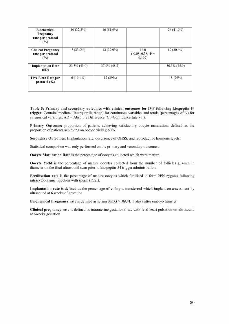
80
Biochemical Pregnancy
rate per protocol (%)
10 (32.3%) 16 (51.6%) 26 (41.9%)
Clinical Pregnancy rate per protocol
(%)
7 (23.0%) 12 (39.0%) 16.0 (-0.08, 0.38, P =
0.199)
19 (30.6%)
Implantation Rate (SD)
23.3% (43.0) 37.0% (48.2) 30.3% (45.9)
Live Birth Rate per protocol (%)
6 (19·4%)
12 (39%)
18 (29%)
Table 5: Primary and secondary outcomes with clinical outcomes for IVF following kisspeptin-54 trigger. Contains medians (interquartile range) for continuous variables and totals (percentages of N) for categorical variables, AD = Absolute Difference (CI=Confidence Interval).
Primary Outcome: proportion of patients achieving satisfactory oocyte maturation; defined as the proportion of patients achieving an oocyte yield ≥ 60%.
Secondary Outcomes: Implantation rate, occurrence of OHSS, and reproductive hormone levels.
Statistical comparison was only performed on the primary and secondary outcomes.
Oocyte Maturation Rate is the percentage of oocytes collected which were mature.
Oocyte Yield is the percentage of mature oocytes collected from the number of follicles ≥14mm in diameter on the final ultrasound scan prior to kisspeptin-54 trigger administration.
Fertilisation rate is the percentage of mature oocytes which fertilised to form 2PN zygotes following intracytoplasmic injection with sperm (ICSI).
Implantation rate is defined as the percentage of embryos transferred which implant on assessment by ultrasound at 6 weeks of gestation.
Biochemical Pregnancy rate is defined as serum βhCG >10iU/L 11days after embryo transfer
Clinical pregnancy rate is defined as intrauterine gestational sac with fetal heart pulsation on ultrasound at 6weeks gestation
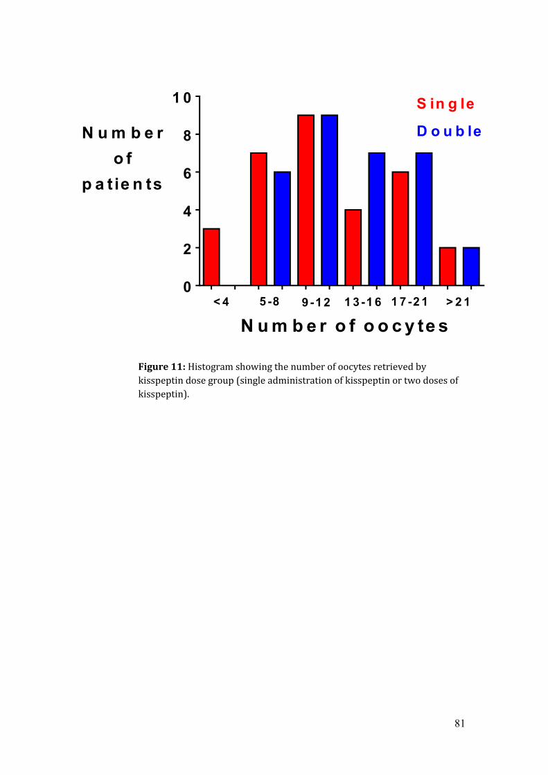
81
Figure 11: Histogram showing the number of oocytes retrieved by kisspeptin dose group (single administration of kisspeptin or two doses of kisspeptin).
< 4 S< 4 D
4 -8 S
4 -8 D
9 -12 S
9 -12 D
1 3 -15 S
1 3 -15 D
1 6 -21 S
1 6 -21 D
> 2 1 S
> 2 1 D0
2
4
6
8
1 0
N u m b e ro f
p a t ie n ts
< 4 5 -8 9 -1 2 1 3 -1 6 1 7 -2 1
S in g le
D o u b le
> 2 1
N u m b e r o f o o c y te s

82
Secondary outcomes:
(i) Reproductive Hormone Levels:
Injection with kisspeptin led to a similar rise in LH and FSH between the single and
double dose groups. Patients receiving kisspeptin as their second injection (double
group) had significantly higher serum LH and FSH levels at 4 hours and 10 hours
after the second injection, when compared to those receiving saline as their second
injection (single group). The mean change in serum LH was increased 3.3-fold in
single group and 14.7-fold in double group at 4hrs following the second injection
when compared with pre-trigger levels;( P<0.0001; see Figure 12 for serum
hormonal profiles by kisspeptin dosing group).
For those patients randomised to receive a double dose of kisspeptin, those with a
lower serum LH-rise following the first dose of kisspeptin had a more pronounced
LH-response following the second dose of kisspeptin (see Figure 13 for bar-chart of
change in serum LH at 4hrs following the second dose of kisspeptin by levels of
serum LH just prior to administration of the second dose of kisspeptin). However,
this did not reach statistical significance.

83
Figure 12: Levels of serum FSH,LH, oestradiol and progesterone (Mean ± SD) after administration of single and double dose kisspeptin at 9.6 nmol/kg at 4, 10, 14 and 20 hours after 1st kisspeptin dose; n=31 per dose
0 2 00
1 0
2 0
3 0
4 0
5 0
6 0
T im e (h o u rs )
S e r u mL H
(iU /L )
S in g le
D o u b le
4 1 0 1 4
s e c o n dK P d o s e
* * *
* * *
0 2 00
2
4
6
8
1 0
1 2
T im e (h o u rs )
S e r u mF S H
(iU /L )S in g le
D o u b le
4 1 0 1 4
s e c o n dK P d o s e
* * ** * *
0 2 00
5 0 0 0
1 0 0 0 0
1 5 0 0 0
2 0 0 0 0
T im e (h o u r s )
S e r u mE s tra d io l(p m o l/L )
S in g le
D o u b le
4 1 0 1 4
s e c o n dK P d o s e
0 2 00
5
1 0
1 5
2 0
2 5
T im e (h o u r s )
S e r u m P ro g e s te ro n e
(n m o l/L )S in g leD o u b le
4 1 0 1 4
s e c o n dK P d o s e
A B
C D
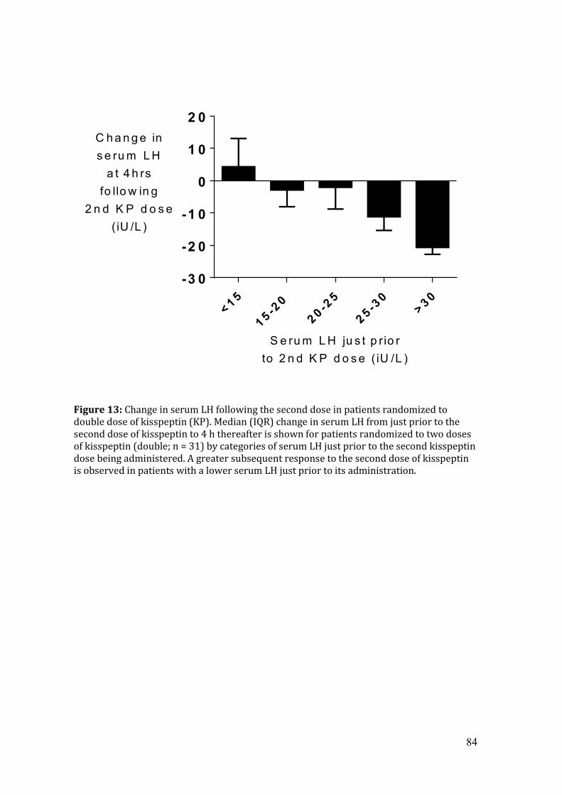
84
Figure 13: Change in serum LH following the second dose in patients randomized to double dose of kisspeptin (KP). Median (IQR) change in serum LH from just prior to the second dose of kisspeptin to 4 h thereafter is shown for patients randomized to two doses of kisspeptin (double; n = 31) by categories of serum LH just prior to the second kisspeptin dose being administered. A greater subsequent response to the second dose of kisspeptin is observed in patients with a lower serum LH just prior to its administration.
< 1 5
1 5 -20
2 0 -25
2 5 -30
> 3 0-3 0
-2 0
-1 0
0
1 0
2 0
S e ru m L H ju s t p r io rto 2 n d K P d o s e ( iU /L )
C h a n g e ins e ru m L H
a t 4 h rsfo llo w in g
2 n d K P d o s e(iU /L )

85
(ii) Rates of OHSS:
The incidence of OHSS did not vary between those who received a single or double
dose of kisspeptin (Table 6). Only one woman from sixty-two at high risk of OHSS
(1.6%) was diagnosed with moderate early OHSS. She was admitted for analgesia
for less than 24hrs on the day following oocyte retrieval and although no medical
intervention was required, she was treated with segmentation to safeguard her
well-being. No patients were diagnosed with mild early OHSS but one patient in the
double group was diagnosed with mild late OHSS (1.6%).

86
OHSS Screening Early Late
Single Double Both AD (CI)
P value Single Double Both
AD (CI)
P value OHSS Symptoms
Number of patients screened by OHSS symptoms (%)
31 (100%)
31 (100%)
62 (100%) 31
(100%) 31
(100%) 62
(100%)
Number of patients with 1 symptom potentially
consistent with OHSS (%)
9 (29.0%)
7 (22.6%)
16 (25.8%) 9
(29.0%) 9
(29.0%) 18
(29.0%)
Number of patients requiring medical intervention for
OHSS (%)
0 (0.0%)
0 (0.0%)
0 0.0%) 0
(0.0%) 0
(0.0%) 0
(0.0%)
Number of patients requiring hospital admission for OHSS
(%)
1 (3.2%)
0 (0.0%)
1 (1.6%) 0
(0.0%) 0
(0.0%) 0
(0.0%)
Sonographic Screening
Number of patients screened with pelvic ultrasound (%)
31 (100%)
30 (96.8%)
61 (98.4%) 30
(96.8%) 31
(100%) 61
(98.4%)
Mean ovarian volume in mls (SD) 68.6 (52.1) 48.2
(28.9) 58.6
(43.2)
-20.4 (-39.3, -1.5) P = 0.034
28.9 (40.9) 16.1 (15.9) 22.4 (31.3)
-12.8 (-22.5, -3.3) P = 0.006
Mean ovarian diameter in mm (SD) 41.5 (10.1) 38.7
(7.9) 40.2 (9.1) 29.6
(9.9) 25.9 (6.0)
27.8 (8.3)
Maximal ovarian diameter in mm (SD) 64.2 (15.1) 58.1 (11.0) 61.3 (13.6)
-6.1 (-12.9, 0.60)
P = 0.079 44.7 (14.0) 42.4 (9.5) 43.5
(11.9)
-2.3 (-7.7, 3.1) P = 0.410
Number of patients with maximum ovarian diameter
5cm (%)
24 (77.4%)
22 (73.3%)
46 (75.4%) 7
(23.3%) 7
(22.6%) 14
(23.0%)
Number of patients with maximum ovarian diameter
8cm (%)
5 (16.1%)
1 (3.3%)
6 (9.8%) 1
(3.3%) 0
(0.0%) 1
(1.6%)
Number of patients with maximum ovarian diameter
12cm (%)
0 (0.0%)
0 (0.0%)
0 (0.0%) 0
(0.0%) 0
(0.0%) 0
(0.0%)
Total free fluid in abdomen, POD / Adnexa in mls (SD)
5.8 (8.1)
3.4 (5.8)
4.6 (7.1) 3.0
(11.8) 0.8
(2.3) 1.9
(8.4)
Number of patients with grade 2 ascites (%)
0 (0.0%)
0 (0.0%)
0 (0.0%) 0
(0.0%) 0
(0.0%) 0
(0.0%)
Number of patients with pleural effusion (%)
0 (0.0%)
0 (0.0%)
0 (0.0%) 0
(0.0%) 0
(0.0%) 0
(0.0%)
Blood parameters
Number of patients screened with Blood analysis
31 (100%)
29 (93.5%)
60 (96.8%) 30
(96.8%) 31
(100%) 61
(98.4%)
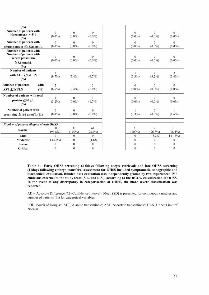
87
Table 6: Early OHSS screening (3-5days following oocyte retrieval) and late OHSS screening (11days following embryo transfer). Assessment for OHSS included symptomatic, sonographic and biochemical evaluation. Blinded data evaluation was independently graded by two experienced IVF clinicians external to the study team (S.L. and R.S.), according to the RCOG classification of OHSS. In the event of any discrepancy in categorisation of OHSS, the more severe classification was reported.
AD = Absolute Difference (CI=Confidence Interval). Mean (SD) is presented for continuous variables and number of patients (%) for categorical variables.
POD, Pouch of Douglas; ALT, Alanine transaminase; AST, Aspartate transaminase; ULN, Upper Limit of Normal.
(%) Number of patients with
Haematocrit >45% (%)
0 (0.0%)
0 (0.0%)
0 (0.0%) 0
(0.0%) 0
(0.0%) 0
(0.0%)
Number of patients with serum sodium 132mmol/L
0 (0.0%)
0 (0.0%)
0 (0.0%) 0
(0.0%) 0
(0.0%) 0
(0.0%)
Number of patients with Number of patients with
serum potassium 5.0mmol/L
(%)
0 (0.0%)
0 (0.0%)
0 (0.0%) 0
(0.0%) 0
(0.0%) 0
(0.0%)
Number of patients with ALT 2xULN
(%)
3 (9.7%)
1 (3.4%)
4 (6.7%) 1
(3.3%) 1
(3.2%) 2
(3.4%)
Number of patients with AST 2xULN (%)
2 (6.5%)
1 (3.4%)
3 (5.0%) 0
(0.0%) 0
(0.0%) 0
(0.0%)
Number of patients with total protein 80 g/L
(%)
1 (3.2%)
0 (0.0%)
1 (1.7%) 0
(0.0%) 0
(0.0%) 0
(0.0%)
Number of patient with creatinine 110 µmol/L (%)
0 (0.0%)
0 (0.0%)
0 (0.0%) 1
(3.3%) 0
(0.0%) 1
(1.6%)
Number of patients diagnosed with OHSS
Normal 30 (96.8%)
31 (100%)
61 (98.4%) 31
(100%) 30
(96.8%) 61
(98.4%)
Mild 0 0 0 0 1 (3.2%) 1 (1.6%) Moderate 1 (3.2%) 0 1 (1.6%) 0 0 0
Severe 0 0 0 0 0 0 Critical 0 0 0 0 0 0

88
(iii) Implantation rate:
A higher implantation rate was observed following a double dose of kisspeptin
(37.1%) when compared to a single dose (23.3%) (OR 2.1; CI 0.68-6.3), but this
was not statistical significant. The live birth rate was 19·4% in the single group and
39% in the double group
Adverse events
The administration of kisspeptin-54 was tolerated well in all patients included in
this study. This finding is in keeping with the safety profile reported in previous
human studies (151, 170, 171, 173, 185). One woman in the single dose group had
an unexplained second trimester miscarriage at 19 weeks of gestation.

89
Discussion
Kisspeptin signaling is essential for mammalian fertility (145, 186) and exogenous
administration of kisspeptin can potently stimulate the release of endogenous
reproductive hormones in the mammalian species studied (see chapter 1). A single
injection of kisspeptin has been shown to result in an LH surge of ~12-14hrs
duration that was sufficient to safely trigger oocyte maturation in women at high
risk of OHSS and subsequently result in fertilisation, implantation of the embryo
and, most importantly, result in live births for infertile women undergoing IVF
treatment (181).
The primary objective of this study was to assess if oocyte maturation can be
improved in women having IVF treatment following 2 doses of kisspeptin-54
administration without increasing the incidence of OHSS. In order to investigate
the safety and efficacy of kisspeptin in women at high risk of developing OHSS, the
inclusion criteria for this trial were a high serum AMH ≥40pmol/L or AFC ≥23. The
women in this study had a mean AFC of 40 and mean serum AMH of 52pmol/L
confirming successful recruitment of women at high risk of developing OHSS
during IVF treatment (96, 180).
It is reasonable to assume that prolonging the duration of the LH-surge with a
second dose of kisspeptin could potentially increase the risk of developing OHSS in
the high-risk patient. Such reports have been described in the literature following
GnRH-a triggering (187-189).
This study shows that patients who received two doses of kisspeptin to trigger
oocyte maturation had an increased duration of LH and FSH exposure compared to

90
patients receiving a single dose, leading to an increased proportion of patients
achieving an optimal oocyte yield. Oocyte maturation was assessed in two ways:
the absolute number of mature oocytes; and the proportion of collected oocytes
that were mature (metaphase II).
The hormonal profiles of patients receiving a second dose of kisspeptin were
variable with regards to their LH-response following the second dose of kisspeptin.
The response appeared to be dependent on the LH-rise following the first injection
of kisspeptin. Patients with a more robust LH-rise following the first dose of
kisspeptin had a lower secondary rise following the further dose of kisspeptin.
Conversely, those patients with a lower initial LH-response following the first dose
of kisspeptin responded with a more potent LH-rise following the second dose of
kisspeptin. Therefore, the second ‘rescue’ dose of kisspeptin was able to eliminate
the retrieval of fewer than 4 oocytes from 9.7% in the single group to 0% in the
double group.
There are a number of plausible hypotheses as to the variable response that was
observed; kisspeptin pharmacodynamics may be explained by its unique
mechanism of action. Kisspeptin acts at the level of the hypothalamus to cause the
release of a pre-formed endogenous pool of GnRH (190). Patients with a greater
LH-rise following the first dose of kisspeptin may have depleted their hypothalamic
GnRH reserves to the extent that a second injection of kisspeptin only resulted in a
modest further LH-response of what remained. Whereas patients with a lower rise
in LH following the first dose of kisspeptin may have a greater residual
hypothalamic GnRH store, allowing a second dose of kisspeptin to induce a larger
LH-response.
91
Another possibility is that the variability in LH-response is a result of tachyphylaxis
at the kisspeptin receptor. This hypothesis, however, seems less likely as
tachyphylaxis is more usually associated with high-dose and frequent
administrations of kisspeptin rather than two intermediate doses administered 10
hours apart, as in this study (191).
Kisspeptin’s ability to stimulate gonadotrophin secretion based on patient’s
individually determined endogenous GnRH reserve, without causing excessive
response, is of key significance in women undergoing IVF treatment at high risk of
developing OHSS. Despite the study population recruited being at high risk of
developing OHSS, a second dose of kisspeptin did not increase the proportion of
patients developing OHSS.
There is data to suggest that kisspeptin may reduce the incidence of OHSS via
alternative factors other than its shorter duration of action. Firstly, kisspeptin has
been shown to inhibit the production of VEGF, one of the main angiogenic
cytokines implicated in the pathophysiology of OHSS, in studies conducted looking
at tumour angiogenesis (192). Moreover, kisspeptin acts via the release of a
preformed endogenous pool of GnRH from the hypothalamus rather than a
pharmacological dose of GnRH (171, 190). Therefore, as the quantity of
endogenous GnRH is individually determined and tailored to each patient,
kisspeptin is less likely to produce an excessive ovarian response (181).
Although 48% (30/62) of patients in the trial had more than 25 follicles ≥11mm
mean diameter on the day of trigger, the majority of patients (98.4%; 61/62) were
still able to safely proceed with a fresh embryo transfer following kisspeptin
triggering. Only one participant did not have a fresh embryo transfer: she had an

92
AFC of 84 and a serum AMH of 165pmol/L. This patient developed 73 follicles
≥11mm on the morning of trigger and her serum oestradiol was 38,264pmol/L. At
collection 46 oocytes were retrieved, of which 41 were mature. The day following
oocyte retrieval, she developed abdominal pain and was admitted for analgesia for
a period of less than 24hrs before her symptom resolved spontaneously. At
screening for early OHSS, her maximal ovarian diameter was 10cm, she had no
biochemical abnormalities suggestive of OHSS and the total amount of ascites was
50mls. She was graded as moderate OHSS by one reviewer and as mild OHSS by the
other.
Of important note, prospective clinical trials in which routine screening for OHSS is
conducted an increased frequency of OHSS is often reported when compared with
retrospective trials in which symptom-prompted, rather than universal, screening
of OHSS is conducted (193, 194). It is therefore reassuring still that despite routine
screening of women at high risk of OHSS, two doses of kisspeptin can be safely
administered without causing an increased risk of ovarian hyper-response and
OHSS.
Some researchers have proposed that all women with 25 or more follicles ≥11mm,
or those considered high risk of OHSS should freeze all their embryos and segment
their cycle in order to avoid late-onset OHSS (195, 196). However, a universal
segmentation policy may result in increased cost of treatment and increased time
to pregnancy when compared to fresh embryo transfer, which is not often a
favoured option by patients.
The clinical pregnancy rate in this trial was comparable to that observed following
hCG-triggering in clinical trials (194). Thus, using kisspeptin to trigger oocyte

93
maturation could provide a useful clinical option to all women, especially those at
high risk of OHSS, to enable fresh embryo transfer without compromising patient
safety or pregnancy rates.

94
Conclusion and future work:
A second dose of kisspeptin administered 10 hours following the first can improve
the proportion of patients achieving optimal oocyte maturation, but without
increasing the risk of OHSS. Therefore, a second dose of kisspeptin is a safe option
in patients undergoing IVF treatment to further optimise oocyte yield, even those
at high risk of OHSS.
The detailed analysis of the FSH, LH, oestradiol and progesterone levels during
ovarian stimulation and the luteal phase will allow us to gain further insight into
the reproductive hormonal milieu prior to and after the administration of
kisspeptin. Future prospective studies are needed to directly compare kisspeptin
and other triggers of oocyte maturation currently used in clinical practice: namely
hCG and GnRH agonist, to adequately assess the clinical and live birth rates in
particular as well as the incidence and severity of OHSS.

95
Chapter 3
Assessment of OHSS following ovarian stimulation with
different triggers of oocyte maturation:
kisspeptin, GnRH agonist and hCG

96
Introduction
OHSS is the main complication of IVF treatment. Several classifications have been
proposed based on symptoms, ultrasound findings and laboratory parameters
(Table 7). Historically, OHSS has been graded as mild, moderate, severe or critical
(183, 184). Further objective assessments including ovarian volume were used in a
recent RCT to compare the risk of severe OHSS between GnRH agonist versus
antagonist in 1,050 IVF cycles (194).
However, there has been no clear consensus between investigators regarding a
standardised classification for OHSS. In addition, there is a distinct lack of
normative data for ovarian volume following ovarian stimulation (this criteria is
present in the RCOG Greentop Guideline for the management of OHSS and in the
Golan classification). Some degree of ovarian hyperstimulation is expected with the
use of follicular stimulants, but the point at which symptoms transition from being
anticipated effects to those indicative of pathology remain unclear. There has,
however, been a recent review published to grade OHSS in the clinical trial setting
(199). With regards to laboratory parameters, again, situation appropriate,
standardised, normal ranges for women undergoing ovarian stimulation are
lacking. Indeed, elevated liver enzymes in a woman with moderate OHSS have been
reported (200). In addition the clinical significance of some of these parameters,
such elevated liver enzymes, have not been established.

97
Table 7: Classification of OHSS including symptoms, ultrasound and haematological markers (Golan et al., 1989, Navot et al., 1992)
Mild OHSS
Grade 1: Abdominal distension and discomfort, nausea, vomiting and enlarged ovaries- defined
as >80mm
Moderate OHSS
Mild OHSS with ultrasonic evidence of pelvic or abdominal ascites
Severe OHSS
Moderate OHSS with clinical evidence of ascites and/or hydrothorax
Critical OHSS
Severe OHSS with oliguria and renal failure, thromboembolism or adult respiratory distress
syndrome and abnormal laboratory parameters: haematocrit >45%; (ii) creatinine > 90 µmol/l;
(iii) alanine aminotransferase (ALT) >45 U/l.
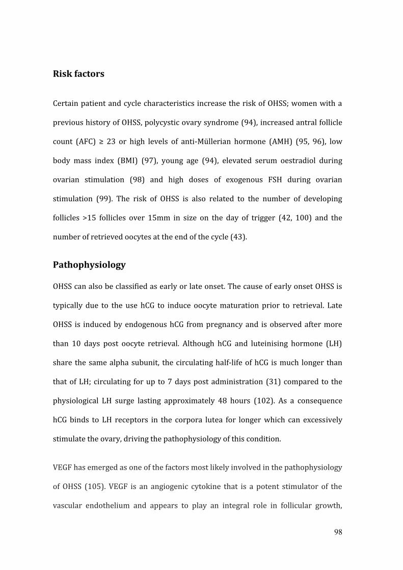
98
Risk factors
Certain patient and cycle characteristics increase the risk of OHSS; women with a
previous history of OHSS, polycystic ovary syndrome (94), increased antral follicle
count (AFC) ≥ 23 or high levels of anti-Mu llerian hormone (AMH) (95, 96), low
body mass index (BMI) (97), young age (94), elevated serum oestradiol during
ovarian stimulation (98) and high doses of exogenous FSH during ovarian
stimulation (99). The risk of OHSS is also related to the number of developing
follicles >15 follicles over 15mm in size on the day of trigger (42, 100) and the
number of retrieved oocytes at the end of the cycle (43).
Pathophysiology
OHSS can also be classified as early or late onset. The cause of early onset OHSS is
typically due to the use hCG to induce oocyte maturation prior to retrieval. Late
OHSS is induced by endogenous hCG from pregnancy and is observed after more
than 10 days post oocyte retrieval. Although hCG and luteinising hormone (LH)
share the same alpha subunit, the circulating half-life of hCG is much longer than
that of LH; circulating for up to 7 days post administration (31) compared to the
physiological LH surge lasting approximately 48 hours (102). As a consequence
hCG binds to LH receptors in the corpora lutea for longer which can excessively
stimulate the ovary, driving the pathophysiology of this condition.
VEGF has emerged as one of the factors most likely involved in the pathophysiology
of OHSS (105). VEGF is an angiogenic cytokine that is a potent stimulator of the
vascular endothelium and appears to play an integral role in follicular growth,
99
corpus luteum function, and ovarian angiogenesis. Recent studies also indicate that
hCG increases VEGF expression in human granulosa cells and raises serum VEGF
concentrations (106, 107). VEGF has been implicated as it known to increase
vascular permeability to high molecular weight proteins promoting capillary
leakage (108). As such there is a clinical need to improve IVF protocols to eliminate
the occurrence of OHSS. Since triggering oocyte maturation with hCG is a major
cause of OHSS, newer triggers such as GnRHa and kisspeptin have been
investigated as potentially safer triggers.
Triggering of oocyte maturation
hCG : Ovulation in a natural cycle is initiated and perpetuated by a mid-cycle surge of LH
released by the pituitary gland. The LH surge acts to induce final oocyte maturation
whereby the first meiotic division is completed with extrusion of the polar body
and it is then arrested in prophase of the second meiotic division until fertilisation.
hCG shares the same alpha subunit and 81% of the amino acids of the beta subunit
of LH and hence binds to the same receptor (30) hCG also has a stimulating action
on the development and function of the corpus luteum; namely a luteotrophic
effect, that is more potent owing to its longer half-life compared to the endogenous
LH surge and as such hCG can still be detected in the serum 10 days after the
preovulatory bolus injection (31). Consequently, continued support of the corpus
luteum by hCG elicits supraphysiological luteal phase steroid concentrations. In
combination with an exaggerated ovarian reponse secondary to stimulation (i.e. a
large number late follicular phase follicles and very high oestradiol levels), hCG has
clearly been associated with an increased risk of OHSS (42-44).
100
GnRH agonist:
GnRHa admimistration initially induces a surge in gonadotrophin production
termed the ‘flare’ effect. With the advent of the GnRH antagonist protocol, where
the GnRH receptor competitively inhibited rather than desensitised, a GnRHa can
be used as an alternative to hCG to induce oocyte maturation. The GnRHa displaces
the GnRH antagonist from the pituitary GnRH receptor, and activates it, thereby
initiating an endogenous surge of gonadotrophins, namely LH and FSH.
The gonadotrophin surge generated by administration of GnRHa is shorter than
the physiogical LH surge that occurs in a normal menstrual cycle: 24 hours
compared to 48 hours respectively (28).
The shortened duration of the LH surge with a GnRHa trigger significantly reduces
the total amount of LH released when compared to a normal menstrual cycle (27,
28). In rodents GnRHa administration is associated with structural luteolysis of the
corpora lutea via matrix metalloproteinase activity (201). As a result, the GnRHa
trigger has a much better safety profile with regards to OHSS when compared to
hCG (48, 49, 202).
Kisspeptin:
Kisspeptin is a hypothalamic peptide, that is encoded by the KiSS1 gene, which is a
neuromodulator acting upstream of GnRH. Kisspeptin stimulates GnRH neurons
directly through its action on the kisspeptin receptor to release GnRH into the
hypothalamo-pituitary portal circulation, which in turn stimulates the secretion of
LH and FSH from the gonadotrophs of the anterior pituitary (131, 203).

101
Exogenous administration of kisspeptin has been shown to be able to effectively
induce a potent LH surge and subsequent oocyte maturation in women undergoing
IVF treatment (179) and also in those at high risk of OHSS (181). In these two
studies a single injection of kisspeptin resulted in an LH surge which lasted for 12
hours compared to the 24 hour LH surge with GnRHa as mentioned previously.
Peak levels of hCG occur at 24 hours post administration (182) whereas with
GnRHa (204) and kisspeptin (179) this occurs at 4 hours. Therefore, the total
amount of gonadotrophin released during the duration of the surge is significantly
reduced when GnRHa is used and even less so with kisspeptin owing to the shorter
duration of the LH surge (12 hours). The resulting lower circulating LH
concentrations will promote earlier luteolysis compared to hCG.
In another recent study directly comparing equimolar administration of
kisspeptin-10 and 54 with GnRH and on gonadotrophin secretion, GnRH was found
to be more potent than kisspeptin’s indirect hypothalamic stimulation. Therefore,
theoretically kisspeptin may have a therapeutic advantage over GnRH in mitigating
adverse clinical consequences secondary to excessive gonadotrophin stimulation;
namely OHSS (190).
To determine conclusively the efficacy and safety of hCG vs GnRHa vs kisspeptin in
IVF treatment, prospective studies are needed. However, in the absence of these
studies, I compared the incidence of OHSS symptoms and ultrasound markers in
our IVF unit who had hCG, GnRHa or kisspeptin as their trigger for oocyte
maturation in their IVF cycle.

102
Methods
The inclusion criteria selected women at high risk of OHSS: serum anti-Müllerian
hormone (AMH) ≥40pmol/L (≥5·6ng/mL) or total antral follicle count (AFC) ≥23;
age 18-34yrs; early follicular phase serum follicle stimulating hormone (FSH)
≤12iU/L; both ovaries intact; body mass index 18-29kg/m2. Women in the
kisspeptin group were universally screened sonographically and for symptoms of
OHSS 2-5 days following oocyte retrieval) at Hammersmith Hospital, London, UK
(2013-2016). Patients in the hCG and GnRHa groups were screened if clinicians
deemed them at high risk of developing OHSS or they developed symptoms of OHSS
post oocyte retrieval.
Ovarian volume on ultrasound, ascitic volume and OHSS symptoms were
determined when patients were triggered with (hCG) (n=44), GnRH agonist
(GnRHa) (n=94) or kisspeptin (n=115) at time of early OHSS screening (2-10 days
following oocyte retrieval). In the kisspeptin group data was collected as part of a
prospective clinical trial. In the hCG and GnRHa agonist groups, data was collected
from matched patients retrospectively that met the inclusion criteria.
Statistical analysis
The distribution of continuous variables was compared to the normal distribution
using the Kolmogorov-Smirnov test. Non-parametrically distributed variables were
log transformed to maximise power. The study data were summarised using
standard descriptive methods with mean (SD) presented for parametric variables
and median (interquartile range) for non-parametric variables. Non-
parametrically distributed, continuous variables (mean ovarian volume, change in
ovarian volume and mean ascitic volume) were log-transformed prior to analysis

103
to maximise power. Analysis of continuous variables was performed using One-
way ANOVA with post-hoc Tukey’s test to examine pairwise differences between
the three groups. Analysis of dichotomous variables (presence of individual OHSS
symptoms) was performed using the Chi-squared test, with examination of
standardised residuals to determine the significance of pairwise differences
between the three groups.

104
Results
Baseline characteristics
Two hundred and sixty-one women at high risk of OHSS received either hCG
(n=40), GnRHa (n=99), or kisspeptin (n=122) to trigger oocyte maturation. The
cohort studied was at high risk of OHSS with a median of 38 antral follicles, 24
follicles ≥11mm on the day of trigger and 19 oocytes retrieved. There were
significant imbalances between the treatment groups as clinicians are more likely
to recommend GnRHa trigger rather than hCG trigger for patients deemed at higher
risk of OHSS (see Table 8).
Average mean ovarian volume (MOV) following hCG trigger was 172mls ±89mls,
88 ±60mls after GnRHa trigger and 53±37mls following kisspeptin trigger
(p<0.0001) (see Table 8). The mean change in ovarian volume from baseline to
OHSS screening for hCG was 166±87mls (25-fold), 75±56mls (10-fold) with GnRHa
and 41±35mls (6-fold) following kisspeptin (p<0.0001) Mean total ascitic volume
following hCG, GnRHa and kisspeptin trigger was 92± 131mls, 5.7 ± 21.5mls and
4.8± 7.8mls respectively. These volumes were statistically significant when
comparing hCG with kisspeptin and GnRHa (p<0.0001). However these values did
not reach statistical significance between kisspeptin and GnRHa (see Figure 13).
Abdominal pain and bloating were reported in 73% and 84% respectively of
patients triggered with hCG, 20% and 28% with GnRHa trigger and 13% and 12%
following kisspeptin trigger. The incidence of bloating was statistically significant
between all three groups (p<0.0001). However, abdominal pain was only

105
statistically significant when comparing hCG with kisspeptin or GnRHa (p<0.0001).
Nausea and vomiting occurred in 43% and 9% respectively of patients triggered
with hCG, 12% and 4% following GnRHa trigger and 3% and <1% of patients
triggered with kisspeptin (p<0.0001). The incidence of nausea was statistically
significant between all three groups (p<0.0001). However, vomiting was only
statistically significant when comparing hCG with kisspeptin or GnRHa (p<0.05)
(see Figure 14). The percentage of patients where >10mls of ascitic fluid was
detected upon ultrasound during early OHSS screening following administration of
hCG, GnRHa and kisspeptin was 80%, 18% and 16% respectively (p<0.0001)(see
Table 10).

106
hCG GnRHa Kisspeptin All P value N 40 99 122 261
Age (y) 32·4 (30·0-35·3)
30·0 (28·0-33·0)
31·0 (29·0-33·0)
31 (29-33) 0·013
Body mass index (kg/m2) 24·6 (21·0-27·0)
24·7 (21·0-27·7)
24·5 (21·2-27·3)
24·6 (21·0-27·3) 0·97
Antral Follicle Count (AFC) 36 (20-40)
42 (33-55)
36 (29-49)
38 (29-50)
<0·0001
Total recombinant FSH dose (IU)
1781 (1325-2325)
1500 (1113-2025)
1238 (1050-1575)
1350 (1125-1950) 0·0002
Mean daily rec FSH dose (IU) 150 (113-225)
122 (111-150)
113 (112-131)
119 (113-150)
<0·0001
Long Protocol (%) 55% 0% 0% 8% <0·0001
Number of follicles on day of trigger
36 (32-41)
40 (35-48)
33 (26-41)
37 (29-34)
<0·0001
Number of follicles ≥11mm on day of trigger
23 (20-31)
27 (23-34)
22 (15-27)
24 (19-30)
<0·0001
Number of follicles ≥14mm on day of trigger
16 (10-20)
15 (13-21)
13 (9-17)
14 (10-19) 0·001
Number of oocytes retrieved 21 (17-24)
27 (20-34)
12 (8-15)
19 (12-27)
<0·0001
Mean day of OHSS assessment (% between days 3-5 post oocyte retrieval)
4·1 (74%)
4·1 (85%)
4·7 (99%)
4·4 (90%)
<0·0001
% treated with segmentation 51% 13% 8% 17% <0·0001
Table 8: Baseline characteristics. Median (IQR) is presented for continuous variables and were compared by Kruskal Wallis test. Proportion is presented for binary variables and compared by Chi squared test. Long protocol denotes the use of a GnRH agonist to prevent premature ovulation during controlled ovarian stimulation prior to trigger.
107
Trigger hCG GnRHa Kisspeptin P value
Mean ovarian volume (mls) (±SD)
172±89 88±60 53±37 <0.0001
Mean ascitic volume (mls) (±SD)
92± 131 5.7 ± 21.5 4.8± 7.8 <0.0001
Table 9: Mean ovarian volume and mean ascitic volume ± standard deviation following oocyte maturation triggers hCG, GnRHa and kisspeptin. . Analysis of continuous variables was performed using One-way ANOVA with post-hoc Tukey’s test to examine pairwise differences between the three groups
Trigger hCG (%) GnRHa (%) Kisspeptin (%) P value
Nausea 43 12 3 <0.0001
Vomiting 9 4 <1 <0.05
Abdominal pain
72 20 13 <0.0001
Abdominal bloating
84 28 12 <0.0001
Total ascites >10mls
80 18 16 <0.0001
Table 10: Percentage of patients reporting symptoms of OHSS: nausea, vomiting, abdominal pain and bloating, and the percentage of patients where >10mls of ascitic fluid was detected on ultrasound during early OHSS screening. Analysis of of individual OHSS symptom was performed using the Chi-squared test, with Bonferroni correction to determine the significance of pairwise differences between the three groups

108
Figure 13: Column graphs demonstrating average mean ovarian volume and mean ascitic volume ± standard deviation (mls) following oocyte maturation triggers hCG, GnRHa and kisspeptin **** = p<0.0001
0
50
100
150
200
250
300
hcg GnRHa Kisspeptin
Mean ovarian volume
hcg
GnRHa
Kisspeptin
ml
**** ****
****
0
50
100
150
200
hcg GnRHa Kisspeptin
Mean ascitic volume
hcg
GnRHa
Kisspeptin
**** **** ml

109
Figure 14: Column graphs demonstrating the percentage of patients reporting OHSS symptoms by trigger: namely abdominal pain and bloating, nausea and vomiting.
**** = p<0.0001 * = p<0.05

110
Discussion
It is well established that after administration of hCG, there is an increased risk of
developing OHSS compared to when a GnRHa agonist is used for triggering final
oocyte maturation. This is largely owing to the long half-life of hCG and its
subsequent potent luteotrophic effect. The excessive stimulation of multiple
corpora lutea following oocyte retrieval leads to multiple vasoactive active
substances being released by the ovary, altering capillary permeability, leading to
the large fluid shifts from the intravascular to the extravascular space which are
pathognomonic of OHSS.
An attractive approach to avoiding OHSS is to substitute hCG. GnRHa have been
successfully used in this manner and have led the way thus far in reducing the
incidence of OHSS. However, owing to the short half-life of the endogenous LH peak
with GnRHa triggering, most of the studies performed in patients showed reduced
implantation and pregnancy rates. This results in a need to identify novel oocyte
maturation triggers that have an effective safety profile in terms of reducing the
risk of OHSS, compared to what is currently available, but that also maintain on-
going pregnancy success rates.
Kisspeptin has been shown to effectively trigger oocyte maturation and phase 2
studies have identified the optimal dosing regimes for improving the oocyte yield
(see previous chapter). The rate of OHSS reported in these studies was low and
ranged from 1.6-5% (181). However, this study is the first direct comparison
between kisspeptin and the other triggers used for final oocyte maturation with
regards to assessing symptoms and ultrasonographic markers of OHSS.

111
Mean ovarian volume (MOV) was significantly lower following GnRHa when
compared to hCG and was significantly lower still when comparing kisspeptin to
GnRHa (p<0.0001). Compared to baseline ovarian volumes prior to commencing
ovarian stimulation, ovarian volumes at early OHSS screening remained enlarged
24-fold following hCG, 10-fold following GnRHa and 6-fold following kisspeptin
triggering. This observation could be explained by the reduced total length of the
LH surge (12-14 hrs single group; 20 hours in double group) following kisspeptin
injection. The reduced kisspeptin-induced LH surge duration and amplitude of the
LH peak when compared to GnRHa and hCG results in significantly reduced total
quantity of LH produced. As the corpora lutea are dependent on pituitary LH
support, this is likely to be associated with increased luteolysis, structural and
functional regression of the corpora lutea and subsequently quicker reduction in
ovarian volume following stimulation. Again, the same trend was observed with
mean total ascitic volumes, with the lowest volumes being observed in those
triggered with kisspeptin followed by GnRHa and the most being reported with
hCG.
Patients reported symptoms of OHSS most frequently following hCG trigger,
followed by GnRHa and least of all with kisspeptin across parameters. Abdominal
pain was reported in 72% following hCG, 20% following GnRHa and 13% following
kisspeptin. Abdominal bloating was reported in 84% following hCG, 28% following
GnRHa and 12% following kisspeptin. Nausea was reported in 43% following hCG,
12% following GnRHa and 3% following kisspeptin. Vomiting was reported in 9%
following hCG, 4% following GnRHa and <1% following kisspeptin.

112
This data demonstrates a positive correlation between the mean ovarian volume,
the ascitic volume and presence of OHSS symptoms. An explanation for this could
be that the more active the corpora lutea are, due to more prolonged LH receptor
stimulation either via hCG or GnRHa-induced LH, the larger they remain and the
more vasoactive substances are released from the ovary. This alters capillary
permeability to a greater extent, causing larger fluid shifts and accumulation of
fluid in the third space compartment. This, in addition to the mechanical pain or
bloating associated with enlarged ovaries, can lead to initiation or exacerbation of
OHSS symptoms. The shorter duration of the LH surge observed following
kisspeptin is likely to be associated with quicker luteal regression and ovarian
recovery back to their physiological state.
Limitations:
As in any observational analysis there may be unmeasured confounding, and here
particularly a risk of confounding by indication. This study was non-randomised
and therefore managing clinicians may have been less likely to assign patients
thought to be at high risk of OHSS to an hCG trigger. There is also risk of differential
loss to follow up. Patients in the kisspeptin trials were uniformly asked to attend
for OHSS screening between days 2-5 following oocyte retrieval, and indeed more
than 97.5% had both sonographic and symptom screening. Women assigned to
GnRHa or hCG groups were screened between day 2-10 post oocyte retrieval
whenever they were considered at substantial risk of OHSS on the day of trigger or
if they presented with symptoms.

113
Conclusion and future work:
This study has shown for the first time, that kisspeptin is associated with a reduced
incidence of OHSS symptoms and a quantitative reduction in the ultrasound
parameters used to grade the severity of OHSS: namely ovarian size and the ascitic
volume. This pilot data lays the foundation for a similar comparison where
kisspeptin will be compared to GnRHa and hCG in a prospective, randomised trial
as well as analysis of the luteal phase hormonal profile following each trigger.
Based on the results of this study, kisspeptin has the potential to be the safest
trigger for oocyte maturation during IVF treatment.

114
References
1. Fertility problems: assessment and treatmentNICE guidelines [CG156] [Available from: nice.org.uk. 2. van Wely M, Westergaard LG, Bossuyt PM, van der Veen F. Effectiveness of human menopausal h versus recombinant follicle-stimulating hormone for controlled ovarian hyperstimulation in assisted reproductive cycles: a meta-analysis. Fertil Steril. 2003;80(5):1086-93. 3. Coomarasamy A, Afnan M, Cheema D, van der Veen F, Bossuyt PM, van Wely M. Urinary hMG versus recombinant FSH for controlled ovarian hyperstimulation following an agonist long down-regulation protocol in IVF or ICSI treatment: a systematic review and meta-analysis. Hum Reprod. 2008;23(2):310-5. 4. Ng EH LE, Yeung WS, Ho PC. HMG is as good as recombinant human FSH in terms of oocyte and embryo quality: a prospective randomized trial. . Human Reproduction. 2001;16(2):319-25. 5. Neveu S, Hedon B, Bringer J, Chinchole JM, Arnal F, Humeau C, et al. Ovarian stimulation by a combination of a h-releasing hormone agonist and gonadotropins for in vitro fertilization. Fertil Steril. 1987;47(4):639-43. 6. Chetkowski RJ, Kruse LR, Nass TE. Improved pregnancy outcome with the addition of leuprolide acetate to gonadotropins for in vitro fertilization. Fertil Steril. 1989;52(2):250-5. 7. Antoine JM S-BJ, Alvarez S, Cornet D, Tibi C, Mandelbaum J, Plachot M. Ovarian stimulation using human menopausal gonadotrophins with or without LHRH analogues in a long protocol for in-vitro fertilization: a prospective randomized comparison. Human Reproduction. 1990;1(5):565-9. 8. Tummon IS, Daniel SA, Kaplan BR, Nisker JA, Yuzpe AA. Randomized, prospective comparison of luteal leuprolide acetate and gonadotropins versus clomiphene citrate and gonadotropins in 408 first cycles of in vitro fertilization. Fertil Steril. 1992;58(3):563-8. 9. Hackeloer BJ, Fleming R, Robinson HP, Adam AH, Coutts JR. Correlation of ultrasonic and endocrinologic assessment of human follicular development. Am J Obstet Gynecol. 1979;135(1):122-8. 10. Funduk-Kurjak B, Kurjak A. Ultrasound monitoring of follicular maturation and ovulation in normal menstrual cycle and in ovulation induction. Acta Obstet Gynecol Scand. 1982;61(4):329-35. 11. Ritchie WG. Ultrasound in the evaluation of normal and induced ovulation. Fertil Steril. 1985;43(2):167-81. 12. Hamori M, Stuckensen JA, Rumpf D, Kniewald T, Kniewald A, Kurz CS. Premature luteinization of follicles during ovarian stimulation for in-vitro fertilization. Hum Reprod. 1987;2(8):639-43. 13. Silverberg KM, Burns WN, Olive DL, Riehl RM, Schenken RS. Serum progesterone levels predict success of in vitro fertilization/embryo transfer in patients stimulated with leuprolide acetate and human menopausal gonadotropins. J Clin Endocrinol Metab. 1991;73(4):797-803. 14. Bosch E, Labarta E, Crespo J, Simon C, Remohi J, Jenkins J, et al. Circulating progesterone levels and ongoing pregnancy rates in controlled ovarian stimulation cycles for in vitro fertilization: analysis of over 4000 cycles. Hum Reprod. 2010;25(8):2092-100.

115
15. Schoolcraft W, Sinton E, Schlenker T, Huynh D, Hamilton F, Meldrum DR. Lower pregnancy rate with premature luteinization during pituitary suppression with leuprolide acetate. Fertil Steril. 1991;55(3):563-6. 16. Ou YC, Lan KC, Chang SY, Kung FT, Huang FJ. Increased progesterone/estradiol ratio on the day of HCG administration adversely affects success of in vitro fertilization-embryo transfer in patients stimulated with gonadotropin-releasing hormone agonist and recombinant follicle-stimulating hormone. Taiwan J Obstet Gynecol. 2008;47(2):168-74. 17. Smitz J EP, Camus M, Devroey P, Tournaye H, Wisanto A, Van Steirteghem AC. Pituitary gonadotrophin secretory capacity during the luteal phase in superovulation using GnRH-agonists and HMG in a desensitization or flare-up protocol. Human Reproduction. 1992;7(9):1225-9. 18. Donderwinkel PF SD, Pache TD, De Jong FH, Hop WC, Fauser BC. . Luteal function following ovulation induction in polycystic ovary syndrome patients using exogenous gonadotrophins in combination with a gonadotrophin-releasing hormone agonist. Human Reproduction. 1993;1(8):2027-32. 19. Kondaveeti-Gordon U, Harrison RF, Barry-Kinsella C, Gordon AC, Drudy L, Cottell E. A randomized prospective study of early follicular or midluteal initiation of long protocol gonadotropin-releasing hormone in an in vitro fertilization program. Fertil Steril. 1996;66(4):582-6. 20. Ferraretti AP, Magli C, Feliciani E, Montanaro N, Gianaroli L. Relationship of timing of agonist administration in the cycle phase to the ovarian response to gonadotropins in the long down-regulation protocols for assisted reproductive technologies. Fertil Steril. 1996;65(1):114-21. 21. Olivennes F, Fanchin R, Bouchard P, de Ziegler D, Taieb J, Selva J, et al. The single or dual administration of the gonadotropin-releasing hormone antagonist Cetrorelix in an in vitro fertilization-embryo transfer program. Fertil Steril. 1994;62(3):468-76. 22. Diedrich K, Diedrich C, Santos E, Zoll C, al-Hasani S, Reissmann T, et al. Suppression of the endogenous luteinizing hormone surge by the gonadotrophin-releasing hormone antagonist Cetrorelix during ovarian stimulation. Hum Reprod. 1994;9(5):788-91. 23. Tarlatzis BC, Fauser BC, Kolibianakis EM, Diedrich K, Rombauts L, Devroey P. GnRH antagonists in ovarian stimulation for IVF. Hum Reprod Update. 2006;12(4):333-40. 24. Felberbaum RE, Reissmann T, Kupker W, Bauer O, al Hasani S, Diedrich C, et al. Preserved pituitary response under ovarian stimulation with HMG and GnRH antagonists (Cetrorelix) in women with tubal infertility. Eur J Obstet Gynecol Reprod Biol. 1995;61(2):151-5. 25. Reissmann T, Felberbaum R, Diedrich K, Engel J, Comaru-Schally AM, Schally AV. Development and applications of luteinizing hormone-releasing hormone antagonists in the treatment of infertility: an overview. Hum Reprod. 1995;10(8):1974-81. 26. Nakano R, Mizuno T, Kotsuji F, Katayama K, Wshio M, Tojo S. "Triggering" of ovulation after infusion of synthetic luteinizing hormone releasing factor (LRF). Acta Obstet Gynecol Scand. 1973;52(3):269-72. 27. GONEN Y BH, POWELL W, CASPER RF. Use of gonadotropin-releasing hormone agonist to trigger follicular maturation for in vitro fertilization. The Journal of Clinical Endocrinology & Metabolism. 1990;71(4):918-22.

116
28. Itskovitz J, Boldes R, Levron J, Erlik Y, Kahana L, Brandes JM. Induction of preovulatory luteinizing hormone surge and prevention of ovarian hyperstimulation syndrome by gonadotropin-releasing hormone agonist. Fertil Steril. 1991;56(2):213-20. 29. Imoedemhe DA, Sigue AB, Pacpaco EL, Olazo AB. Stimulation of endogenous surge of luteinizing hormone with gonadotropin-releasing hormone analog after ovarian stimulation for in vitro fertilization. Fertil Steril. 1991;55(2):328-32. 30. Parsons TF PJ. Biologically active covalently cross-linked glycoprotein hormones and the effects of modification of the COOH-terminal region of their alpha subunits. Journal of Biological Chemistry. 1979;254(13):6010-15. 31. Damewood MD, Shen W, Zacur HA, Schlaff WD, Rock JA, Wallach EE. Disappearance of exogenously administered human chorionic gonadotropin. Fertil Steril. 1989;52(3):398-400. 32. Yen SS, Llerena O, Little B, Pearson OH. Disappearance rates of endogenous luteinizing hormone and chorionic gonadotropin in man. J Clin Endocrinol Metab. 1968;28(12):1763-7. 33. Huirne JA HR, Lambalk CB. Are GnRH antagonists comparable to agonists for use in IVF. Human Reproduction. 2007;22(11):2805-13. 34. Al-Inany H AM, Mansour RT, Serour GI. Optimizing GnRH antagonist administration: meta-analysis of fixed versus flexible protocol. . Reproductive biomedicine online. 2005;10(5):567-70. 35. Cakmak H, Katz A, Cedars MI, Rosen MP. Effective method for emergency fertility preservation: random-start controlled ovarian stimulation. Fertil Steril. 2013;100(6):1673-80. 36. Al-Inany H, Aboulghar M. GnRH antagonist in assisted reproduction: a Cochrane review. Hum Reprod. 2002;17(4):874-85. 37. Griesinger G, Felberbaum R, Diedrich K. GnRH antagonists in ovarian stimulation: a treatment regimen of clinicians' second choice? Data from the German national IVF registry. Hum Reprod. 2005;20(9):2373-5. 38. Al-Inany HG, Youssef MA, Aboulghar M, Broekmans F, Sterrenburg M, Smit J, et al. GnRH antagonists are safer than agonists: an update of a Cochrane review. Hum Reprod Update. 2011;17(4):435. 39. Al-Inany HG, Abou-Setta AM, Aboulghar M. Gonadotrophin-releasing hormone antagonists for assisted conception. Cochrane Database Syst Rev. 2006(3):CD001750. 40. Papanikolaou EG, Bourgain C, Fatemi H, Verpoest W, Polyzos NP, De Brabanter A, et al. Endometrial advancement after triggering with recombinant or urinary HCG: a randomized controlled pilot study. Reprod Biomed Online. 2010;21(1):50-5. 41. Beckers NG, Laven JS, Eijkemans MJ, Fauser BC. Follicular and luteal phase characteristics following early cessation of gonadotrophin-releasing hormone agonist during ovarian stimulation for in-vitro fertilization. Hum Reprod. 2000;15(1):43-9. 42. Enskog A, Henriksson M, Unander M, Nilsson L, Brannstrom M. Prospective study of the clinical and laboratory parameters of patients in whom ovarian hyperstimulation syndrome developed during controlled ovarian hyperstimulation for in vitro fertilization. Fertil Steril. 1999;71(5):808-14. 43. Mathur RS, Akande AV, Keay SD, Hunt LP, Jenkins JM. Distinction between early and late ovarian hyperstimulation syndrome. Fertil Steril. 2000;73(5):901-7.

117
44. Soliman S DM, Collins J, Hughes EG. The role of luteal phase support in infertility treatment: a meta-analysis of randomized trials. Fertility and sterility. 1994;61(6):1068-76. 45. Hoff JD QM, Yen SS. Hormonal dynamics at midcycle: a reevaluation. The Journal of Clinical Endocrinology & Metabolism. 1983;57(4):792-6. 46. Kolibianakis EM S-MA, Schroer A, Van Steirteghem A, Devroey P, Diedrich K, Griesinger G. A lower ongoing pregnancy rate can be expected when GnRH agonist is used for triggering final oocyte maturation instead of HCG in patients undergoing IVF with GnRH antagonists. Human Reproduction. 2005;20(10):2887-92. 47. Humaidan P, Bredkjaer HE, Bungum L, Bungum M, Grondahl ML, Westergaard L, et al. GnRH agonist (buserelin) or hCG for ovulation induction in GnRH antagonist IVF/ICSI cycles: a prospective randomized study. Hum Reprod. 2005;20(5):1213-20. 48. Itskovitz-Eldor J KS, Mannaerts B. Use of a single bolus of GnRH agonist triptorelin to trigger ovulation after GnRH antagonist ganirelix treatment in women undergoing ovarian stimulation for assisted reproduction, with special reference to the prevention of ovarian hyperstimulation syndrome: preliminary report: short communication. Human Reproduction. 2000;15(9):1965-8. 49. Humaidan P, Kol S, Papanikolaou EG, Copenhagen Gn RHATWG. GnRH agonist for triggering of final oocyte maturation: time for a change of practice? Hum Reprod Update. 2011;17(4):510-24. 50. Eppig JJ. FSH stimulates hyaluronic acid synthesis by oocyte-cumulus cell complexes from mouse preovulatory follicles. Nature. 1979;281(5731):483-4. 51. Yding Andersen C. Effect of FSH and its different isoforms on maturation of oocytes from pre-ovulatory follicles. Reprod Biomed Online. 2002;5(3):232-9. 52. Yding Andersen C, Leonardsen L, Ulloa-Aguirre A, Barrios-De-Tomasi J, Moore L, Byskov AG. FSH-induced resumption of meiosis in mouse oocytes: effect of different isoforms. Mol Hum Reprod. 1999;5(8):726-31. 53. Zelinski-Wooten MB, Hutchison JS, Hess DL, Wolf DP, Stouffer RL. Follicle stimulating hormone alone supports follicle growth and oocyte development in gonadotrophin-releasing hormone antagonist-treated monkeys. Hum Reprod. 1995;10(7):1658-66. 54. Cutting R, Morroll D, Roberts SA, Pickering S, Rutherford A, Bfs, et al. Elective single embryo transfer: guidelines for practice British Fertility Society and Association of Clinical Embryologists. Hum Fertil (Camb). 2008;11(3):131-46. 55. Papanikolaou EG, Camus M, Kolibianakis EM, Van Landuyt L, Van Steirteghem A, Devroey P. In vitro fertilization with single blastocyst-stage versus single cleavage-stage embryos. N Engl J Med. 2006;354(11):1139-46. 56. Zech NH, Lejeune B, Puissant F, Vanderzwalmen S, Zech H, Vanderzwalmen P. Prospective evaluation of the optimal time for selecting a single embryo for transfer: day 3 versus day 5. Fertil Steril. 2007;88(1):244-6. 57. Criniti A, Thyer A, Chow G, Lin P, Klein N, Soules M. Elective single blastocyst transfer reduces twin rates without compromising pregnancy rates. Fertil Steril. 2005;84(6):1613-9. 58. Fauque P, Jouannet P, Davy C, Guibert J, Viallon V, Epelboin S, et al. Cumulative results including obstetrical and neonatal outcome of fresh and frozen-thawed cycles in elective single versus double fresh embryo transfers. Fertil Steril. 2010;94(3):927-35.

118
59. Fatemi HM, Kolibianakis EM, Camus M, Tournaye H, Donoso P, Papanikolaou E, et al. Addition of estradiol to progesterone for luteal supplementation in patients stimulated with GnRH antagonist/rFSH for IVF: a randomized controlled trial. Hum Reprod. 2006;21(10):2628-32. 60. Filicori M, Butler JP, Crowley WF, Jr. Neuroendocrine regulation of the corpus luteum in the human. Evidence for pulsatile progesterone secretion. J Clin Invest. 1984;73(6):1638-47. 61. HUTCHISON JS ZA. The rhesus monkey corpus luteum is dependent on pituitary gonadotropin secretion throughout the luteal phase of the menstrual cycle. Endocrinology. 1984;115(5):1780-6. 62. Casper RF, Yen SS. Induction of luteolysis in the human with a long-acting analog of luteinizing hormone-releasing factor. Science. 1979;205(4404):408-10. 63. Lemay A, Labrie F, Ferland L, Raynaud JP. Possible luteolytic effects of luteinizing hormone-releasing hormone in normal women. Fertil Steril. 1979;31(1):29-34. 64. McLachlan RI, Cohen NL, Vale WW, Rivier JE, Burger HG, Bremner WJ, et al. The importance of luteinizing hormone in the control of inhibin and progesterone secretion by the human corpus luteum. J Clin Endocrinol Metab. 1989;68(6):1078-85. 65. Polson DW, Sagle M, Mason HD, Kiddy D, Franks S. Recovery of luteal function after interruption of gonadotrophin secretion in the mid-luteal phase of the menstrual cycle. Clin Endocrinol (Oxf). 1987;26(5):597-600. 66. Dubourdieu S, Charbonnel B, Massai MR, Marraoui J, Spitz I, Bouchard P. Suppression of corpus luteum function by the gonadotropin-releasing hormone antagonist Nal-Glu: effect of the dose and timing of human chorionic gonadotropin administration. Fertil Steril. 1991;56(3):440-5. 67. Nippoldt TB, Reame NE, Kelch RP, Marshall JC. The roles of estradiol and progesterone in decreasing luteinizing hormone pulse frequency in the luteal phase of the menstrual cycle. J Clin Endocrinol Metab. 1989;69(1):67-76. 68. Zeleznik AJ. In vivo responses of the primate corpus luteum to luteinizing hormone and chorionic gonadotropin. Proc Natl Acad Sci U S A. 1998;95(18):11002-7. 69. Duffy DM SD, Stouffer RL. Titrating Luteinizing Hormone Replacement to Sustain the Structure and Function of the Corpus Luteum after Gonadotropin-Releasing Hormone Antagonist Treatment in Rhesus Monkeys. The Journal of Clinical Endocrinology & Metabolism. 1999;84(1):342-9. 70. Messinis IE, Templeton AA. Disparate effects of endogenous and exogenous oestradiol on luteal phase function in women. J Reprod Fertil. 1987;79(2):549-54. 71. Vandevoort CA, Baughman WL, Stouffer RL. Comparison of different regimens of human gonadotropins for superovulation of rhesus monkeys: ovulatory response and subsequent luteal function. J In Vitro Fert Embryo Transf. 1989;6(2):85-91. 72. Fauser BC, Devroey P. Reproductive biology and IVF: ovarian stimulation and luteal phase consequences. Trends Endocrinol Metab. 2003;14(5):236-42. 73. Smitz J, Devroey P, Camus M, Deschacht J, Khan I, Staessen C, et al. The luteal phase and early pregnancy after combined GnRH-agonist/HMG treatment for superovulation in IVF or GIFT. Hum Reprod. 1988;3(5):585-90.

119
74. Sungurtekin U, Jansen RP. Profound luteinizing hormone suppression after stopping the gonadotropin-releasing hormone-agonist leuprolide acetate. Fertil Steril. 1995;63(3):663-5. 75. Pantos K, Meimeth-Damianaki T, Vaxevanoglou T, Kapetanakis E. Prospective study of a modified gonadotropin-releasing hormone agonist long protocol in an in vitro fertilization program. Fertil Steril. 1994;61(4):709-13. 76. Dal Prato L BA. Use of antagonists in ovarian stimulation protocols. . Reproductive biomedicine online. 2005;10(3):330-8. 77. Albano C, Smitz J, Tournaye H, Riethmuller-Winzen H, Van Steirteghem A, Devroey P. Luteal phase and clinical outcome after human menopausal gonadotrophin/gonadotrophin releasing hormone antagonist treatment for ovarian stimulation in in-vitro fertilization/intracytoplasmic sperm injection cycles. Hum Reprod. 1999;14(6):1426-30. 78. de Jong D MN, Fauser BC. A pilot study involving minimal ovarian stimulation for in vitro fertilization: extending the “follicle-stimulating hormone window” combined with the gonadotropin-releasing hormone antagonist cetrorelix. Fertility and sterility. 2000;73(5):1051-4. 79. Beckers NG, Macklon NS, Eijkemans MJ, Ludwig M, Felberbaum RE, Diedrich K, et al. Nonsupplemented luteal phase characteristics after the administration of recombinant human chorionic gonadotropin, recombinant luteinizing hormone, or gonadotropin-releasing hormone (GnRH) agonist to induce final oocyte maturation in in vitro fertilization patients after ovarian stimulation with recombinant follicle-stimulating hormone and GnRH antagonist cotreatment. J Clin Endocrinol Metab. 2003;88(9):4186-92. 80. Bourgain C, Devroey P, Van Waesberghe L, Smitz J, Van Steirteghem AC. Effects of natural progesterone on the morphology of the endometrium in patients with primary ovarian failure. Hum Reprod. 1990;5(5):537-43. 81. Kolibianakis EM DP. The luteal phase after ovarian stimulation. . Reproductive biomedicine online. 2002;5:26-35. 82. Mart n J DnF, A vila S, Castrillo JL, Remoh J, ellicer A, Simo n C. Human endometrial receptivity: gene regulation. Journal of reproductive immunology. Journal of reproductive immunology. 2002;55(1):131-9. 83. Paulson RJ, Sauer MV, Lobo RA. Embryo implantation after human in vitro fertilization: importance of endometrial receptivity. Fertil Steril. 1990;53(5):870-4. 84. Csapo AI, Pulkkinen MO, Ruttner B, Sauvage JP, Wiest WG. The significance of the human corpus luteum in pregnancy maintenance. I. Preliminary studies. Am J Obstet Gynecol. 1972;112(8):1061-7. 85. Csapo AI, Pulkkinen MO, Wiest WG. Effects of luteectomy and progesterone replacement therapy in early pregnant patients. Am J Obstet Gynecol. 1973;115(6):759-65. 86. Coomarasamy A, Williams H, Truchanowicz E, Seed PT, Small R, Quenby S, et al. A Randomized Trial of Progesterone in Women with Recurrent Miscarriages. N Engl J Med. 2015;373(22):2141-8. 87. Leeton J, Trounson A, Jessup D. Support of the luteal phase in in vitro fertilization programs: results of a controlled trial with intramuscular Proluton. J In Vitro Fert Embryo Transf. 1985;2(3):166-9. 88. Levine H. Luteal support in IVF using the novel vaginal progesterone gel Crinone 8%: results of an open-label trial in 1184 women from 16 US centers. Fertil Steril. 2000;74(4):836-7.

120
89. Pritts EA, Atwood AK. Luteal phase support in infertility treatment: a meta-analysis of the randomized trials. Hum Reprod. 2002;17(9):2287-99. 90. Lightman A, Kol S, Itskovitz-Eldor J. A prospective randomized study comparing intramuscular with intravaginal natural progesterone in programmed thaw cycles. Hum Reprod. 1999;14(10):2596-9. 91. Costabile L, Gerli S, Manna C, Rossetti D, Di Renzo GC, Unfer V. A prospective randomized study comparing intramuscular progesterone and 17alpha-hydroxyprogesterone caproate in patients undergoing in vitro fertilization-embryo transfer cycles. Fertil Steril. 2001;76(2):394-6. 92. Propst AM, Hill JA, Ginsburg ES, Hurwitz S, Politch J, Yanushpolsky EH. A randomized study comparing Crinone 8% and intramuscular progesterone supplementation in in vitro fertilization-embryo transfer cycles. Fertil Steril. 2001;76(6):1144-9. 93. Daya S, Gunby J. Luteal phase support in assisted reproduction cycles. Cochrane Database Syst Rev. 2004(3):CD004830. 94. Navot D, Relou A, Birkenfeld A, Rabinowitz R, Brzezinski A, Margalioth EJ. Risk factors and prognostic variables in the ovarian hyperstimulation syndrome. Am J Obstet Gynecol. 1988;159(1):210-5. 95. Broer SL, Dolleman M, Opmeer BC, Fauser BC, Mol BW, Broekmans FJ. AMH and AFC as predictors of excessive response in controlled ovarian hyperstimulation: a meta-analysis. Hum Reprod Update. 2011;17(1):46-54. 96. Jayaprakasan K CY, Islam R, Haoula Z, Hopkisson J, Coomarasamy A, Raine-Fenning N. Prediction of in vitro fertilization outcome at different antral follicle count thresholds in a prospective cohort of 1,012 women. Fertility and sterility. 2012;98(3):657-63. 97. Delvigne A, Demoulin A, Smitz J, Donnez J, Koninckx P, Dhont M, et al. The ovarian hyperstimulation syndrome in in-vitro fertilization: a Belgian multicentric study. I. Clinical and biological features. Hum Reprod. 1993;8(9):1353-60. 98. Haning RV AC, Carlson IH, Kuzma DL, Shapiro SS, Zweibel WJ. Plasma estradiol is superior to ultrasound and urinary estriol glucuronide as a predictor of ovarian hyperstimulation during induction of ovulation with menotropins. Fertility and sterility. 1983;40(1):31-6. 99. Mizunuma H, Takagi, T., Yamada, K., Andoh, K., Ibuki, Y. and Igarashi, M. Ovulation induction by step‐down administration of purified urinary follicle‐stimulation hormone in patients with polycystic ovarian syndrome. Fertility and sterility. 1991;55(6):1195-6. 100. Blankstein J, Shalev J, Saadon T, Kukia EE, Rabinovici J, Pariente C, et al. Ovarian hyperstimulation syndrome: prediction by number and size of preovulatory ovarian follicles. Fertil Steril. 1987;47(4):597-602. 101. Herman A, Ron-El R, Golan A, Raziel A, Soffer Y, Caspi E. Pregnancy rate and ovarian hyperstimulation after luteal human chorionic gonadotropin in in vitro fertilization stimulated with gonadotropin-releasing hormone analog and menotropins. Fertil Steril. 1990;53(1):92-6. 102. Hoff JD, Quigley ME, Yen SS. Hormonal dynamics at midcycle: a reevaluation. J Clin Endocrinol Metab. 1983;57(4):792-6. 103. Tollan A, Holst N, Forsdahl F, Fadnes HO, Oian P, Maltau JM. Transcapillary fluid dynamics during ovarian stimulation for in vitro fertilization. Am J Obstet Gynecol. 1990;162(2):554-8.

121
104. Goldsman MP, Pedram A, Dominguez CE, Ciuffardi I, Levin E, Asch RH. Increased capillary permeability induced by human follicular fluid: a hypothesis for an ovarian origin of the hyperstimulation syndrome. Fertil Steril. 1995;63(2):268-72. 105. Geva E, Jaffe RB. Role of vascular endothelial growth factor in ovarian physiology and pathology. Fertil Steril. 2000;74(3):429-38. 106. Neulen J, Yan Z, Raczek S, Weindel K, Keck C, Weich HA, et al. Human chorionic gonadotropin-dependent expression of vascular endothelial growth factor/vascular permeability factor in human granulosa cells: importance in ovarian hyperstimulation syndrome. J Clin Endocrinol Metab. 1995;80(6):1967-71. 107. Pellicer A, Albert C, Mercader A, Bonilla-Musoles F, Remohi J, Simon C. The pathogenesis of ovarian hyperstimulation syndrome: in vivo studies investigating the role of interleukin-1beta, interleukin-6, and vascular endothelial growth factor. Fertil Steril. 1999;71(3):482-9. 108. Lee A, Christenson LK, Stouffer RL, Burry KA, Patton PE. Vascular endothelial growth factor levels in serum and follicular fluid of patients undergoing in vitro fertilization. Fertil Steril. 1997;68(2):305-11. 109. Marci R, Senn A, Dessole S, Chanson A, Loumaye E, De Grandi P, et al. A low-dose stimulation protocol using highly purified follicle-stimulating hormone can lead to high pregnancy rates in in vitro fertilization patients with polycystic ovaries who are at risk of a high ovarian response to gonadotropins. Fertil Steril. 2001;75(6):1131-5. 110. Tso LO, Costello MF, Albuquerque LE, Andriolo RB, Marjoribanks J, Macedo CR. Metformin treatment before and during in vitro fertilization or intracytoplasmic sperm injection in women with polycystic ovary syndrome: summary of a Cochrane review. Fertil Steril. 2015;104(3):542-4. 111. Soares SR. Etiology of OHSS and use of dopamine agonists. Fertil Steril. 2012;97(3):517-22. 112. Humaidan P, Bredkjaer HE, Bungum L, Bungum M, Grøndahl ML, Westergaard L, et al. GnRH agonist (buserelin) or hCG for ovulation induction in GnRH antagonist IVF/ICSI cycles: a prospective randomized study. Hum Reprod. 2005;20(5):1213-20. 113. Engmann L, DiLuigi A, Schmidt D, Nulsen J, Maier D, Benadiva C. The use of gonadotropin-releasing hormone (GnRH) agonist to induce oocyte maturation after cotreatment with GnRH antagonist in high-risk patients undergoing in vitro fertilization prevents the risk of ovarian hyperstimulation syndrome: a prospective randomized controlled study. Fertil Steril. 2008;89(1):84-91. 114. Griesinger G BH, Schultz L, Depenbusch M, Schultze-Mosgau A. Cumulative live birth rates after GnRH-agonist triggering of final oocyte maturation in patients at risk of OHSS: a prospective, clinical cohort study. European Journal of Obstetrics & Gynecology and Reproductive Biology. 2010;149(2):190-4. 115. Lee JH, Miele ME, Hicks DJ, Phillips KK, Trent JM, Weissman BE, et al. KiSS-1, a novel human malignant melanoma metastasis-suppressor gene. J Natl Cancer Inst. 1996;88(23):1731-7. 116. West A, Vojta PJ, Welch DR, Weissman BE. Chromosome localization and genomic structure of the KiSS-1 metastasis suppressor gene (KISS1). Genomics. 1998;54(1):145-8. 117. Bockaert J, Pin JP. Molecular tinkering of G protein-coupled receptors: an evolutionary success. EMBO J. 1999;18(7):1723-9.

122
118. Lee DK, Nguyen T, O'Neill GP, Cheng R, Liu Y, Howard AD, et al. Discovery of a receptor related to the galanin receptors. FEBS Lett. 1999;446(1):103-7. 119. Muir AI, Chamberlain L, Elshourbagy NA, Michalovich D, Moore DJ, Calamari A, et al. AXOR12, a novel human G protein-coupled receptor, activated by the peptide KiSS-1. J Biol Chem. 2001;276(31):28969-75. 120. Min L, Soltis K, Reis AC, Xu S, Kuohung W, Jain M, et al. Dynamic kisspeptin receptor trafficking modulates kisspeptin-mediated calcium signaling. Mol Endocrinol. 2014;28(1):16-27. 121. Constantin S, Caligioni CS, Stojilkovic S, Wray S. Kisspeptin-10 facilitates a plasma membrane-driven calcium oscillator in gonadotropin-releasing hormone-1 neurons. Endocrinology. 2009;150(3):1400-12. 122. Kotani M, Detheux M, Vandenbogaerde A, Communi D, Vanderwinden JM, Le Poul E, et al. The metastasis suppressor gene KiSS-1 encodes kisspeptins, the natural ligands of the orphan G protein-coupled receptor GPR54. J Biol Chem. 2001;276(37):34631-6. 123. Gottsch ML, Cunningham MJ, Smith JT, Popa SM, Acohido BV, Crowley WF, et al. A role for kisspeptins in the regulation of gonadotropin secretion in the mouse. Endocrinology. 2004;145(9):4073-7. 124. Clarkson J, Herbison AE. Postnatal development of kisspeptin neurons in mouse hypothalamus; sexual dimorphism and projections to gonadotropin-releasing hormone neurons. Endocrinology. 2006;147(12):5817-25. 125. Pompolo S, Pereira A, Estrada KM, Clarke IJ. Colocalization of kisspeptin and gonadotropin-releasing hormone in the ovine brain. Endocrinology. 2006;147(2):804-10. 126. Oakley AE, Clifton DK, Steiner RA. Kisspeptin signaling in the brain. Endocr Rev. 2009;30(6):713-43. 127. Messager S, Chatzidaki EE, Ma D, Hendrick AG, Zahn D, Dixon J, et al. Kisspeptin directly stimulates gonadotropin-releasing hormone release via G protein-coupled receptor 54. Proc Natl Acad Sci U S A. 2005;102(5):1761-6. 128. Ramaswamy S GK, Gibbs RB, Plant TM. Structural interactions between kisspeptin and GnRH neurons in the mediobasal hypothalamus of the male rhesus monkey (Macaca mulatta) as revealed by double immunofluorescence and confocal Endocrinology. 2008;9:4387-95. 129. Uenoyama Y, Inoue N, Pheng V, Homma T, Takase K, Yamada S, et al. Ultrastructural evidence of kisspeptin-gonadotrophin-releasing hormone (GnRH) interaction in the median eminence of female rats: implication of axo-axonal regulation of GnRH release. J Neuroendocrinol. 2011;23(10):863-70. 130. Hrabovszky E, Ciofi P, Vida B, Horvath MC, Keller E, Caraty A, et al. The kisspeptin system of the human hypothalamus: sexual dimorphism and relationship with gonadotropin-releasing hormone and neurokinin B neurons. Eur J Neurosci. 2010;31(11):1984-98. 131. Irwig MS, Fraley GS, Smith JT, Acohido BV, Popa SM, Cunningham MJ, et al. Kisspeptin activation of gonadotropin releasing hormone neurons and regulation of KiSS-1 mRNA in the male rat. Neuroendocrinology. 2004;80(4):264-72. 132. Roseweir AK, Kauffman AS, Smith JT, Guerriero KA, Morgan K, Pielecka-Fortuna J, et al. Discovery of potent kisspeptin antagonists delineate physiological mechanisms of gonadotropin regulation. J Neurosci. 2009;29(12):3920-9.

123
133. Liu X, Lee K, Herbison AE. Kisspeptin excites gonadotropin-releasing hormone neurons through a phospholipase C/calcium-dependent pathway regulating multiple ion channels. Endocrinology. 2008;149(9):4605-14. 134. Shahab M, Mastronardi C, Seminara SB, Crowley WF, Ojeda SR, Plant TM. Increased hypothalamic GPR54 signaling: a potential mechanism for initiation of puberty in primates. Proc Natl Acad Sci U S A. 2005;102(6):2129-34. 135. Thompson EL, Patterson M, Murphy KG, Smith KL, Dhillo WS, Todd JF, et al. Central and peripheral administration of kisspeptin-10 stimulates the hypothalamic-pituitary-gonadal axis. J Neuroendocrinol. 2004;16(10):850-8. 136. Caraty A, Smith JT, Lomet D, Ben Said S, Morrissey A, Cognie J, et al. Kisspeptin synchronizes preovulatory surges in cyclical ewes and causes ovulation in seasonally acyclic ewes. Endocrinology. 2007;148(11):5258-67. 137. Plant TM, Ramaswamy S, Dipietro MJ. Repetitive activation of hypothalamic G protein-coupled receptor 54 with intravenous pulses of kisspeptin in the juvenile monkey (Macaca mulatta) elicits a sustained train of gonadotropin-releasing hormone discharges. Endocrinology. 2006;147(2):1007-13. 138. Clarkson J dAdTX, Moreno AS, Colledge WH, Herbison AE. Kisspeptin-GPR54 signaling is essential for preovulatory gonadotropin-releasing hormone neuron activation and the luteinizing hormone surge. Journal of Neuroscience. 2008;28(35):8691-7. 139. Horikoshi Y MH, Takatsu Y, Ohtaki T, Kitada C, Usuki S, Fujino M. Dramatic elevation of plasma metastin concentrations in human pregnancy: metastin as a novel placenta-derived hormone in humans. The Journal of Clinical Endocrinology & Metabolism. 2003;88(2):914-9. 140. Rometo AM, Krajewski SJ, Voytko ML, Rance NE. Hypertrophy and increased kisspeptin gene expression in the hypothalamic infundibular nucleus of postmenopausal women and ovariectomized monkeys. J Clin Endocrinol Metab. 2007;92(7):2744-50. 141. Cheng G, Coolen LM, Padmanabhan V, Goodman RL, Lehman MN. The kisspeptin/neurokinin B/dynorphin (KNDy) cell population of the arcuate nucleus: sex differences and effects of prenatal testosterone in sheep. Endocrinology. 2010;151(1):301-11. 142. Dahl SK, Amstalden M, Coolen L, Fitzgerald M, Lehman M. Dynorphin immunoreactive fibers contact GnRH neurons in the human hypothalamus. Reprod Sci. 2009;16(8):781-7. 143. Navarro VM, Gottsch ML, Chavkin C, Okamura H, Clifton DK, Steiner RA. Regulation of gonadotropin-releasing hormone secretion by kisspeptin/dynorphin/neurokinin B neurons in the arcuate nucleus of the mouse. J Neurosci. 2009;29(38):11859-66. 144. Keen KL, Wegner FH, Bloom SR, Ghatei MA, Terasawa E. An increase in kisspeptin-54 release occurs with the pubertal increase in luteinizing hormone-releasing hormone-1 release in the stalk-median eminence of female rhesus monkeys in vivo. Endocrinology. 2008;149(8):4151-7. 145. Seminara SB, Messager S, Chatzidaki EE, Thresher RR, Acierno JS, Jr., Shagoury JK, et al. The GPR54 gene as a regulator of puberty. N Engl J Med. 2003;349(17):1614-27. 146. de Roux N, Genin E, Carel JC, Matsuda F, Chaussain JL, Milgrom E. Hypogonadotropic hypogonadism due to loss of function of the KiSS1-derived peptide receptor GPR54. Proc Natl Acad Sci U S A. 2003;100(19):10972-6.

124
147. Teles MG, Bianco SD, Brito VN, Trarbach EB, Kuohung W, Xu S, et al. A GPR54-activating mutation in a patient with central precocious puberty. N Engl J Med. 2008;358(7):709-15. 148. Smith JT, Cunningham MJ, Rissman EF, Clifton DK, Steiner RA. Regulation of Kiss1 gene expression in the brain of the female mouse. Endocrinology. 2005;146(9):3686-92. 149. Kauffman AS, Gottsch ML, Roa J, Byquist AC, Crown A, Clifton DK, et al. Sexual differentiation of Kiss1 gene expression in the brain of the rat. Endocrinology. 2007;148(4):1774-83. 150. Jayasena CN, Nijher GM, Comninos AN, Abbara A, Januszewki A, Vaal ML, et al. The effects of kisspeptin-10 on reproductive hormone release show sexual dimorphism in humans. J Clin Endocrinol Metab. 2011;96(12):E1963-72. 151. Dhillo WS, Chaudhri OB, Thompson EL, Murphy KG, Patterson M, Ramachandran R, et al. Kisspeptin-54 stimulates gonadotropin release most potently during the preovulatory phase of the menstrual cycle in women. J Clin Endocrinol Metab. 2007;92(10):3958-66. 152. Smith JT, Acohido BV, Clifton DK, Steiner RA. KiSS-1 neurones are direct targets for leptin in the ob/ob mouse. J Neuroendocrinol. 2006;18(4):298-303. 153. Quennell JH, Howell CS, Roa J, Augustine RA, Grattan DR, Anderson GM. Leptin deficiency and diet-induced obesity reduce hypothalamic kisspeptin expression in mice. Endocrinology. 2011;152(4):1541-50. 154. Castellano JM, Gaytan M, Roa J, Vigo E, Navarro VM, Bellido C, et al. Expression of KiSS-1 in rat ovary: putative local regulator of ovulation? Endocrinology. 2006;147(10):4852-62. 155. Castellano JM, Navarro VM, Roa J, Pineda R, Sanchez-Garrido MA, Garcia-Galiano D, et al. Alterations in hypothalamic KiSS-1 system in experimental diabetes: early changes and functional consequences. Endocrinology. 2009;150(2):784-94. 156. Dandona P, Dhindsa S, Chaudhuri A, Bhatia V, Topiwala S, Mohanty P. Hypogonadotrophic hypogonadism in type 2 diabetes, obesity and the metabolic syndrome. Curr Mol Med. 2008;8(8):816-28. 157. Schneider G, Kirschner MA, Berkowitz R, Ertel NH. Increased estrogen production in obese men. J Clin Endocrinol Metab. 1979;48(4):633-8. 158. Finn PD, Cunningham MJ, Pau KF, Spies HG, Clifton DK, Steiner RA. The Stimulatory Effect of Leptin on the Neuroendocrine Reproductive Axis of the Monkey* *This work was supported by USPHS (NIH) Grants P50-HD-12629, T32-HD-07453, RR-00166, HD-18185, RR-00166, HD-16631, and HD-08610; the Andrew W. Mellon Foundation; and ZymoGenetics, Inc. Endocrinology. 1998;139(11):4652-62. 159. Castellano JM, Navarro VM, Fernandez-Fernandez R, Roa J, Vigo E, Pineda R, et al. Expression of hypothalamic KiSS-1 system and rescue of defective gonadotropic responses by kisspeptin in streptozotocin-induced diabetic male rats. Diabetes. 2006;55(9):2602-10. 160. George JT, Veldhuis JD, Tena-Sempere M, Millar RP, Anderson RA. Exploring the pathophysiology of hypogonadism in men with type 2 diabetes: kisspeptin-10 stimulates serum testosterone and LH secretion in men with type 2 diabetes and mild biochemical hypogonadism. Clin Endocrinol (Oxf). 2013;79(1):100-4.

125
161. Cota D, Proulx K, Smith KA, Kozma SC, Thomas G, Woods SC, et al. Hypothalamic mTOR signaling regulates food intake. Science. 2006;312(5775):927-30. 162. Wahab F, Ullah F, Chan YM, Seminara SB, Shahab M. Decrease in hypothalamic Kiss1 and Kiss1r expression: a potential mechanism for fasting-induced suppression of the HPG axis in the adult male rhesus monkey (Macaca mulatta). Horm Metab Res. 2011;43(2):81-5. 163. Navarro VM, Fernandez-Fernandez R, Castellano JM, Roa J, Mayen A, Barreiro ML, et al. Advanced vaginal opening and precocious activation of the reproductive axis by KiSS-1 peptide, the endogenous ligand of GPR54. J Physiol. 2004;561(Pt 2):379-86. 164. von Schnurbein J, Moss A, Nagel SA, Muehleder H, Debatin KM, Farooqi IS, et al. Leptin substitution results in the induction of menstrual cycles in an adolescent with leptin deficiency and hypogonadotropic hypogonadism. Horm Res Paediatr. 2012;77(2):127-33. 165. Strobel A, Issad T, Camoin L, Ozata M, Strosberg AD. A leptin missense mutation associated with hypogonadism and morbid obesity. Nat Genet. 1998;18(3):213-5. 166. Donato J, Jr., Cravo RM, Frazao R, Gautron L, Scott MM, Lachey J, et al. Leptin's effect on puberty in mice is relayed by the ventral premammillary nucleus and does not require signaling in Kiss1 neurons. J Clin Invest. 2011;121(1):355-68. 167. Farooqi IS, O'Rahilly S. Leptin: a pivotal regulator of human energy homeostasis. Am J Clin Nutr. 2009;89(3):980S-4S. 168. Ozata M, Ozdemir IC, Licinio J. Human leptin deficiency caused by a missense mutation: multiple endocrine defects, decreased sympathetic tone, and immune system dysfunction indicate new targets for leptin action, greater central than peripheral resistance to the effects of leptin, and spontaneous correction of leptin-mediated defects. J Clin Endocrinol Metab. 1999;84(10):3686-95. 169. George JT VJ, Roseweir AK, Newton CL, Faccenda E, Millar RP, Anderson RA. Kisspeptin-10 is a potent stimulator of LH and increases pulse frequency in men,. The Journal of Clinical Endocrinology & Metabolism. 2011;96(8):1228-36. 170. Chan YM, Butler JP, Sidhoum VF, Pinnell NE, Seminara SB. Kisspeptin administration to women: a window into endogenous kisspeptin secretion and GnRH responsiveness across the menstrual cycle. J Clin Endocrinol Metab. 2012;97(8):E1458-67. 171. Dhillo WS, Chaudhri OB, Patterson M, Thompson EL, Murphy KG, Badman MK, et al. Kisspeptin-54 stimulates the hypothalamic-pituitary gonadal axis in human males. J Clin Endocrinol Metab. 2005;90(12):6609-15. 172. Seminara SB, Dipietro MJ, Ramaswamy S, Crowley WF, Jr., Plant TM. Continuous human metastin 45-54 infusion desensitizes G protein-coupled receptor 54-induced gonadotropin-releasing hormone release monitored indirectly in the juvenile male Rhesus monkey (Macaca mulatta): a finding with therapeutic implications. Endocrinology. 2006;147(5):2122-6. 173. Jayasena CN, Nijher GM, Chaudhri OB, Murphy KG, Ranger A, Lim A, et al. Subcutaneous injection of kisspeptin-54 acutely stimulates gonadotropin secretion in women with hypothalamic amenorrhea, but chronic administration causes tachyphylaxis. J Clin Endocrinol Metab. 2009;94(11):4315-23. 174. Loucopoulos A, Ferin M. The treatment of luteal phase defects with pulsatile infusion of gonadotropin-releasing hormone. Fertil Steril. 1987;48(6):933-6.

126
175. Baba T, Kang HS, Hosoe Y, Kharma B, Abiko K, Matsumura N, et al. Menstrual cyclic change of metastin/GPR54 in endometrium. Med Mol Morphol. 2015;48(2):76-84. 176. Jayasena CN, Nijher GM, Abbara A, Murphy KG, Lim A, Patel D, et al. Twice-weekly administration of kisspeptin-54 for 8 weeks stimulates release of reproductive hormones in women with hypothalamic amenorrhea. Clin Pharmacol Ther. 2010;88(6):840-7. 177. Byskov AG, Yding Andersen C, Hossaini A, Guoliang X. Cumulus cells of oocyte-cumulus complexes secrete a meiosis-activating substance when stimulated with FSH. Mol Reprod Dev. 1997;46(3):296-305. 178. Downs SM, Daniel SA, Eppig JJ. Induction of maturation in cumulus cell-enclosed mouse oocytes by follicle-stimulating hormone and epidermal growth factor: evidence for a positive stimulus of somatic cell origin. J Exp Zool. 1988;245(1):86-96. 179. Jayasena CN, Abbara A, Comninos AN, Nijher GM, Christopoulos G, Narayanaswamy S, et al. Kisspeptin-54 triggers egg maturation in women undergoing in vitro fertilization. J Clin Invest. 2014;124(8):3667-77. 180. Lee TH, Liu CH, Huang CC, Wu YL, Shih YT, Ho HN, et al. Serum anti-Mullerian hormone and estradiol levels as predictors of ovarian hyperstimulation syndrome in assisted reproduction technology cycles. Hum Reprod. 2008;23(1):160-7. 181. Abbara A, Jayasena CN, Christopoulos G, Narayanaswamy S, Izzi-Engbeaya C, Nijher GM, et al. Efficacy of Kisspeptin-54 to Trigger Oocyte Maturation in Women at High Risk of Ovarian Hyperstimulation Syndrome (OHSS) During In Vitro Fertilization (IVF) Therapy. J Clin Endocrinol Metab. 2015;100(9):3322-31. 182. Fauser BC, de Jong D, Olivennes F, Wramsby H, Tay C, Itskovitz-Eldor J, et al. Endocrine profiles after triggering of final oocyte maturation with GnRH agonist after cotreatment with the GnRH antagonist ganirelix during ovarian hyperstimulation for in vitro fertilization. J Clin Endocrinol Metab. 2002;87(2):709-15. 183. Golan A, Ron-el R, Herman A, Soffer Y, Weinraub Z, Caspi E. Ovarian hyperstimulation syndrome: an update review. Obstet Gynecol Surv. 1989;44(6):430-40. 184. Navot D, Bergh PA, Laufer N. Ovarian hyperstimulation syndrome in novel reproductive technologies: prevention and treatment. Fertil Steril. 1992;58(2):249-61. 185. Young J, George JT, Tello JA, Francou B, Bouligand J, Guiochon-Mantel A, et al. Kisspeptin restores pulsatile LH secretion in patients with neurokinin B signaling deficiencies: physiological, pathophysiological and therapeutic implications. Neuroendocrinology. 2013;97(2):193-202. 186. de Roux N GE, Carel JC, Matsuda F, Chaussain JL, Milgrom E. Hypogonadotropic hypogonadism due to loss of function of the KiSS1-derived peptide receptor GPR54. Journal of Clinical Endocrinology & Metabolism. 2003;19:10972-6. 187. Fatemi HM, Popovic-Todorovic B, Humaidan P, Kol S, Banker M, Devroey P, et al. Severe ovarian hyperstimulation syndrome after gonadotropin-releasing hormone (GnRH) agonist trigger and "freeze-all" approach in GnRH antagonist protocol. Fertil Steril. 2014;101(4):1008-11.

127
188. Gurbuz AS GF, Ozcimen N, Isik AZ. Gonadotrophin-releasing hormone agonist trigger and freeze-all strategy does not prevent severe ovarian hyperstimulation syndrome: a report of three cases. Reproductive biomedicine online. 2014;29(5):541-4. 189. Ling LP, Phoon JW, Lau MS, Chan JK, Viardot-Foucault V, Tan TY, et al. GnRH agonist trigger and ovarian hyperstimulation syndrome: relook at 'freeze-all strategy'. Reprod Biomed Online. 2014;29(3):392-4. 190. Jayasena CN, Abbara A, Narayanaswamy S, Comninos AN, Ratnasabapathy R, Bassett P, et al. Direct comparison of the effects of intravenous kisspeptin-10, kisspeptin-54 and GnRH on gonadotrophin secretion in healthy men. Hum Reprod. 2015;30(8):1934-41. 191. Abbara A, Ratnasabapathy R, Jayasena CN, Dhillo WS. The effects of kisspeptin on gonadotropin release in non-human mammals. Adv Exp Med Biol. 2013;784:63-87. 192. Cho SG, Yi Z, Pang X, Yi T, Wang Y, Luo J, et al. Kisspeptin-10, a KISS1-derived decapeptide, inhibits tumor angiogenesis by suppressing Sp1-mediated VEGF expression and FAK/Rho GTPase activation. Cancer Res. 2009;69(17):7062-70. 193. Serour GI, Aboulghar M, Mansour R, Sattar MA, Amin Y, Aboulghar H. Complications of medically assisted conception in 3,500 cycles. Fertil Steril. 1998;70(4):638-42. 194. Toftager M, Bogstad J, Bryndorf T, Lossl K, Roskaer J, Holland T, et al. Risk of severe ovarian hyperstimulation syndrome in GnRH antagonist versus GnRH agonist protocol: RCT including 1050 first IVF/ICSI cycles. Hum Reprod. 2016;31(6):1253-64. 195. Humaidan P, Thomsen LH, Alsbjerg B. GnRHa trigger and modified luteal support with one bolus of hCG should be used with caution in extreme responder patients. Hum Reprod. 2013;28(9):2593-4. 196. Atkinson P, Koch J, Ledger WL. GnRH agonist trigger and a freeze-all strategy to prevent ovarian hyperstimulation syndrome: a retrospective study of OHSS risk and pregnancy rates. Aust N Z J Obstet Gynaecol. 2014;54(6):581-5. 197. Delvigne A, Rozenberg S. Epidemiology and prevention of ovarian hyperstimulation syndrome (OHSS): a review. Hum Reprod Update. 2002;8(6):559-77. 198. Papanikolaou EG, Pozzobon C, Kolibianakis EM, Camus M, Tournaye H, Fatemi HM, et al. Incidence and prediction of ovarian hyperstimulation syndrome in women undergoing gonadotropin-releasing hormone antagonist in vitro fertilization cycles. Fertil Steril. 2006;85(1):112-20. 199. Humaidan P, Nelson SM, Devroey P, Coddington CC, Schwartz LB, Gordon K, et al. Ovarian hyperstimulation syndrome: review and new classification criteria for reporting in clinical trials. Hum Reprod. 2016;31(9):1997-2004. 200. Wakim AN, Fox SD. Elevated liver function tests in a case of moderate ovarian hyperstimulation syndrome. Hum Reprod. 1996;11(3):588-9. 201. Goto T, Endo T, Henmi H, Kitajima Y, Kiya T, Nishikawa A, et al. Gonadotropin-releasing hormone agonist has the ability to induce increased matrix metalloproteinase (MMP)-2 and membrane type 1-MMP expression in corpora lutea, and structural luteolysis in rats. J Endocrinol. 1999;161(3):393-402. 202. Griesinger G, Kolibianakis EM, Papanikolaou EG, Diedrich K, Van Steirteghem A, Devroey P, et al. Triggering of final oocyte maturation with gonadotropin-releasing hormone agonist or human chorionic gonadotropin. Live
128
birth after frozen-thawed embryo replacement cycles. Fertil Steril. 2007;88(3):616-21. 203. Castellano JM, Navarro VM, Fernandez-Fernandez R, Nogueiras R, Tovar S, Roa J, et al. Changes in hypothalamic KiSS-1 system and restoration of pubertal activation of the reproductive axis by kisspeptin in undernutrition. Endocrinology. 2005;146(9):3917-25. 204. Vuong TN, Ho MT, Ha TD, Phung HT, Huynh GB, Humaidan P. Gonadotropin-releasing hormone agonist trigger in oocyte donors co-treated with a gonadotropin-releasing hormone antagonist: a dose-finding study. Fertil Steril. 2016;105(2):356-63. 205. Oates-Whitehead RM, Haas DM, Carrier JA. Progestogen for preventing miscarriage. Cochrane Database Syst Rev. 2003 Jan 1;4.
206. Smith JT, Popa SM, Clifton DK, Hoffman GE, Steiner RA. Kiss1 neurons in the forebrain as central processors for generating the preovulatory luteinizing hormone surge. Journal of Neuroscience. 2006 Jun 21;26(25):6687-94.
207. Adachi S, Yamada S, Takatsu Y, Matsui H, Kinoshita M, Takase K, Sugiura H, Ohtaki T, Matsumoto H, Uenoyama Y, Tsukamura H. Involvement of anteroventral periventricular metastin/kisspeptin neurons in estrogen positive feedback action on luteinizing hormone release in female rats. Journal of Reproduction and Development. 2007;53(2):367-78.
208. Castellano JM, Navarro VM, Fernandez-Fernandez R, Roa J, Vigo E, Pineda R, Steiner RA, Aguilar E, Pinilla L, Tena-Sempere M. Effects of galanin-like peptide on luteinizing hormone secretion in the rat: sexually dimorphic responses and enhanced sensitivity at male puberty. American Journal of Physiology-Endocrinology and Metabolism. 2006 Dec 1;291(6):E1281-9.
209. Fernandez-Fernandez R, Aguilar E, Tena-Sempere M, Pinilla L. Effects of polypeptide YY3–36 upon luteinizing hormone-releasing hormone and gonadotropin secretion in prepubertal rats: in vivo and in vitro studies. Endocrinology. 2005 Mar 1;146(3):1403-10.
210. Fernandez-Fernandez R, Aguilar E, Tena-Sempere M, Pinilla L. Effects of polypeptide YY3–36 upon luteinizing hormone-releasing hormone and gonadotropin secretion in prepubertal rats: in vivo and in vitro studies. Endocrinology. 2005 Mar 1;146(3):1403-10.

129
Appendices Appendix A
Copyright permission from Journal of Clinical Investigation
130
Appendix B
Copyright permission request form submitted to Oxford University Press on 22/11/17
E-theses: request to reproduce an extract from a third party's published work
[22/11/2017]
Dear Oxford University Press,
I have completed my MD(Res) thesis at Imperial College London entitled ‘The use of kisspeptin as a novel physiological trigger for oocyte maturation in IVF treatment’.
I seek your permission to reprint, in my thesis an extract from:-
x Dhillo WS, Chaudhri OB, Thompson EL, Murphy KG, Patterson M, Ramachandran R, et al. Kisspeptin-54 stimulates gonadotropin release most potently during the preovulatory phase of the menstrual cycle in women. J Clin Endocrinol Metab. 2007;92(10):3958-66.The extracts to be reproduced are Figures 3 and 4.
x Dhillo WS, Chaudhri OB, Patterson M, Thompson EL, Murphy KG, Badman MK, et al. Kisspeptin-54 stimulates the hypothalamic-pituitary gonadal axis in human males. J Clin Endocrinol Metab. 2005;90(12):6609-15. The extract to be reproduced is Figure 5
x Jayasena CN, Nijher GM, Chaudhri OB, Murphy KG, Ranger A, Lim A, et al. Subcutaneous injection of kisspeptin-54 acutely stimulates gonadotropin secretion in women with hypothalamic amenorrhea, but chronic administration causes tachyphylaxis. J Clin Endocrinol Metab. 2009;94(11):4315-23. The extract to be reproduced is Figure 2
I would like to include the extracts in my thesis which will be added to Spiral, Imperial's institutional repository http://spiral.imperial.ac.uk/ and made available to the public under a Creative Commons Attribution-NonCommercial-NoDerivs licence.
If you are happy to grant me all the permissions requested, please return a signed copy of this letter. If you wish to grant only some of the permissions requested, please list these and then sign.
Yours sincerely,

131
Dr Rumana Islam
Permission granted for the use requested above:
I confirm that I am the copyright holder of the extract above and hereby give permission to include it in your thesis which will be made available, via the internet, for non-commercial purposes under the terms of the user licence.
[please edit the text above if you wish to grant more specific permission]
Signed:
Name:
Organisation:
Job title: