THE UNIVERSITY OF TEXAS SOUTHWESTERN MEDICAL … Notebook-Lectures-Labs 1.pdf · THE UNIVERSITY OF...
111
I THE UNIVERSITY OF TEXAS SOUTHWESTERN MEDICAL CENTER AT DALLAS SOUTHWESTERN SCHOOL OF HEALTH PROFESSION ‐ DEPARTMENT OF PHYSICAL THERAPY ORTHOPEDIC PHYSICAL THERAPY RESIDENCY EDUCATION CURRICULUM COURSES Course Name: Advanced Physical Therapy Practice: Clinical Orthopedic Residency Education Series Weekend 2: Physical Therapy Management of the Cervicothoracic Region and TMJ Course Description: This is the first in a series of five courses designed to provide comprehensive, advanced training in manual therapy and orthopedic rehabilitation. This program will provide content specific to the Cervicothoracic region and TMJ using the framework of differential diagnosis, clinical reasoning, and treatment planning as introduced in Weekend 1. Program participants will receive hands‐on lab instruction in systematic examination testing essential for making pathology and impairment‐based diagnoses in the Cervicothoracic region and TMJ. They will also receive extensive hands‐on training in exercise and manual therapy techniques, including spinal thrust manipulation, for treatment of the Cervicothoracic region and TMJ. Instructional Level: Intermediate/Advanced Target Audience: Licensed Physical Therapists Behavioral/Learning Objectives: Behavioral and Instructional Objectives: At the completion of this course, the student will be able to: 1. Discuss key considerations regarding the anatomy and kinematics of the Cervicothoracic region and TMJ as they contribute to pathology, movement analysis, and treatment planning 2. Gather all relevant information from the subjective and objective examination for making a sound impairment and pathology‐based diagnosis in the Cervicothoracic region and TMJ 3. Perform detailed movement analysis of the Cervicothoracic region and TMJ for the purpose of pathology and impairment‐based diagnosis 4. Perform special testing for the Cervicothoracic region and TMJ for the purpose of pathology‐based diagnosis 5. Perform impairment‐based strength and flexibility testing of the Cervicothoracic region and TMJ 6. Perform manual techniques, including spinal thrust manipulation, to the Cervicothoracic region and TMJ for the purpose of relieving pain and increasing mobility 7. Perform exercise techniques to the Cervicothoracic region and TMJ for the purpose of relieving pain, increasing mobility, and increasing strength Directed Independent Learning Contact Hours: 5 On‐Site Contact Hours: 16 Total Contact Hours: 21 Course Coordinator: Jason Zafereo, PT, OCS, FAAOMPT Additional Lecturers: Julie DeVahl, PT, MS, OCS Lab Instructors: Jason Zafereo, Julie DeVahl, PT, MS, Ed Mulligan, PT, DPT, OCS, SCS, ATC, Emily Middleton, PT, DPT, CSCS
-
Upload
nguyendung -
Category
Documents
-
view
212 -
download
0
Transcript of THE UNIVERSITY OF TEXAS SOUTHWESTERN MEDICAL … Notebook-Lectures-Labs 1.pdf · THE UNIVERSITY OF...

I
THE UNIVERSITY OF TEXAS SOUTHWESTERN MEDICAL CENTER AT DALLAS SOUTHWESTERN SCHOOL OF HEALTH PROFESSION ‐ DEPARTMENT OF PHYSICAL THERAPY
ORTHOPEDIC PHYSICAL THERAPY RESIDENCY EDUCATION CURRICULUM COURSES Course Name: Advanced Physical Therapy Practice: Clinical Orthopedic Residency Education Series
Weekend 2: Physical Therapy Management of the Cervicothoracic Region and TMJ Course Description: This is the first in a series of five courses designed to provide comprehensive, advanced training in manual therapy and orthopedic rehabilitation. This program will provide content specific to the Cervicothoracic region and TMJ using the framework of differential diagnosis, clinical reasoning, and treatment planning as introduced in Weekend 1. Program participants will receive hands‐on lab instruction in systematic examination testing essential for making pathology and impairment‐based diagnoses in the Cervicothoracic region and TMJ. They will also receive extensive hands‐on training in exercise and manual therapy techniques, including spinal thrust manipulation, for treatment of the Cervicothoracic region and TMJ. Instructional Level: Intermediate/Advanced Target Audience: Licensed Physical Therapists Behavioral/Learning Objectives: Behavioral and Instructional Objectives: At the completion of this course, the student will be able to: 1. Discuss key considerations regarding the anatomy and kinematics of the Cervicothoracic region
and TMJ as they contribute to pathology, movement analysis, and treatment planning 2. Gather all relevant information from the subjective and objective examination for making a sound
impairment and pathology‐based diagnosis in the Cervicothoracic region and TMJ 3. Perform detailed movement analysis of the Cervicothoracic region and TMJ for the purpose of
pathology and impairment‐based diagnosis 4. Perform special testing for the Cervicothoracic region and TMJ for the purpose of pathology‐based
diagnosis 5. Perform impairment‐based strength and flexibility testing of the Cervicothoracic region and TMJ 6. Perform manual techniques, including spinal thrust manipulation, to the Cervicothoracic region
and TMJ for the purpose of relieving pain and increasing mobility 7. Perform exercise techniques to the Cervicothoracic region and TMJ for the purpose of relieving
pain, increasing mobility, and increasing strength Directed Independent Learning Contact Hours: 5 On‐Site Contact Hours: 16 Total Contact Hours: 21 Course Coordinator: Jason Zafereo, PT, OCS, FAAOMPT Additional Lecturers: Julie DeVahl, PT, MS, OCS Lab Instructors: Jason Zafereo, Julie DeVahl, PT, MS, Ed Mulligan, PT, DPT, OCS, SCS, ATC, Emily Middleton, PT, DPT, CSCS

I
Instructor Biographies:
Jason Zafereo, MPT, OCS, FAAOMPT (Lead Instructor) Mr. Zafereo is an Assistant Professor and Clinical Director of the faculty practice at UT Southwestern. Mr. Zafereo received his Bachelor of Arts in Biology from Baylor University and his Master of Physical Therapy from the University of Texas Southwestern Medical Center at Dallas. He received his fellowship training in orthopedic manual physical therapy from The Manual Therapy Institute. A board certified specialist in orthopedics, Mr. Zafereo’s clinical interests include orthopedic manual therapy with an emphasis in treatment of the neck and low back.
Julie DeVahl, PT, MS,OCS (Assistant Instructor and Independent Study Contributor) Ms. DeVahl is an Assistant Professor and the Director of Clinical Education at UT Southwestern. She received her B.S. in Physical Therapy from the University of North Dakota in 1979 and her Master of Science degree from the University of Minnesota in 1984. She joined the faculty of UT Southwestern department of physical therapy as the Director of Clinical Education in 2002. She has been teaching continuing education courses on electrotherapy topics throughout her career.
Beth Deschenes, PT, MS, OCS (Independent Study Contributor) Ms. Deschenes is a Clinical Assistant Professor at UT Southwestern and she received her Master of Science degree in Physical Therapy from the University of Kansas Medical Center in 1996. Prior to becoming a physical therapist, she worked in healthcare marketing and health/fitness management. Ms. Deschenes also holds a Master of Science degree in Health/Fitness Management from The American University, which she received in 1984. She was board certified in orthopedics since 2003.
Ed Mulligan, PT, DPT, OCS, SCS, ATC (Lab Assistant) Dr. Mulligan is an Assistant Professor in the Physical Therapy Department at UT Southwestern. His undergraduate degree is from the University of Nebraska and he received his physical therapy training at UTMB – Galveston. He completed the post‐professional master’s degree program at Texas Woman’s University‐Dallas in 1995 and his DPT at Regis University in 2008. He was recognized as a clinical specialist in sports physical therapy by the APTA in 1988 and orthopedic physical therapy in 2009.
Emily Middleton, PT, DPT, CSCS (Lab Assistant) Dr. Middleton is a Faculty Clinical Associate at UT Southwestern. She is the lead therapist at the department’s satellite sports medicine clinic on the UTD campus in Richardson, TX. Emily is a graduate of the UT Southwestern Orthopedic Physical Therapy Residency Program. She served as a lab assistant for a musculoskeletal spine curriculum and has been a speaker at the past two TPTA annual conferences.
Course Requirements:
1. 100% attendance is mandatory. 2. Students must be prepared and dressed appropriately for lab. Students MUST be able to expose
appropriate body parts for joint examination and intervention.
Required Reading/Pre‐Course Assignments:
On‐line Audiovisual Presentations: Cervical Anatomy/Kinematics/Pathoanatomy; TMJ Anatomy/Kinematics/Pathoanatomy; Thoracic Anatomy/Kinematics/Pathoanatomy Recommended References:
1. Magee, D. Orthopedic Physical Assessment. Elsevier, 2007, ISBN‐10: 0721605710 2. Flynn, T. User’s Guide to the Musculoskeletal Examination: Fundamentals for the Evidence‐Based Clinician.
Evidence in Motion, 2008, ISBN‐13: 9780971479234 3. Cleland, J. Orthopedic Clinical Examination: An Evidence‐Based Approach for Physical Therapists. Icon,
2005, ISBN‐10: 1929007876

I
4. Cook C. Orthopedic Manual Therapy: An Evidence‐Based Approach. Prentice Hall, 2007, ISBN‐10: 0131717669
5. Butler D, Jones MA. Mobilisation of the Nervous System, Churchill Livingstone; 1991. ISBN‐10: 0443044007 6. Olson KA. Manual Physical Therapy of the Spine, Saunders; 2009. ISBN‐13: 9781416047490 7. Sahrmann, S. Diagnosis and Treatment of Movement Impairment Syndromes. Mosby, 2002, ISBN‐10:
0801672058 8. Dutton, M. Orthopaedic Examination, Evaluation, and Intervention. 2nd Ed. The McGraw Hill Companies,
Inc., 2008. ISBN‐13: 978‐0‐07‐147401‐6
Resources:
1. Description of Advanced Orthopedic Clinical Practice 2. AAOMPT/APTA Manual Therapy Guidelines 3. Bibliography and References Provided in Course Notebook
Means of Participant Course Evaluation:
1. Pre/Post Course self‐assessment of skill and knowledge 2. Course Evaluation and Feedback
Description of Teaching Methods and Learning Experiences:
1. Pre‐course on‐line audiovisual presentations 2. Lecture, Laboratory Demonstration, Practice, and Critique 3. Case Study Reviews 4. Question/Answer, Discussion, and Role Playing Opportunities 5. Direct observation and critique of skill during laboratory activities 6. Written and Live‐Patient Examinations (Residents only)
Course Agenda and Pre‐Course Audiovisual Presentations:
Content Instructors
Pre‐Course Cervical Anatomy/Kinematics/Pathoanatomy Deschenes
Pre‐Course TMJ Anatomy/Kinematics/Pathoanatomy DeVahl
Pre‐Course Thoracic Anatomy/Kinematics/Pathoanatomy Deschenes
Saturday Cervical Pre‐Course Review Discussion Zafereo
Cervical Examination Lecture Zafereo
Cervical Examination Lab Zafereo
Cervical Treatment Lecture Zafereo
Lunch
Cervical Treatment Lab Zafereo
TMJ Pre‐Course Review Discussion DeVahl
TMJ Examination/Treatment Lecture DeVahl
TMJ Examination/Treatment Lab DeVahl
Sunday TMJ Examination/Treatment Lab DeVahl
Thoracic Pre‐Course Review Discussion Zafereo
Thoracic Examination/Treatment Lecture Zafereo
Lunch
Thoracic Examination Lab Zafereo
Thoracic Treatment Lab Zafereo
Clinical Reasoning Practice Lab Zafereo

1
Cervical Spine Applied Anatomy
Jason Zafereo, PT, OCS, FAAOMPT
Clinical Orthopedic Rehabilitation Education
Objectives
Discuss concepts relevant to pathophysiology and differential diagnosis for headache
Discuss concepts relevant to pathophysiology and differential diagnosis for cervical radiculopathy
Objectives
Discuss concepts relevant to pathophysiology and differential diagnosis for cervical disc and joint disorders
Discuss concepts relevant to pathophysiology and differential diagnosis for cervical instability
HEADACHE
Pathophysiology of Headache
Pain referred to TCN from structures innervated by the C1-3 spinal nerves
– Upper cervical synovial joints
– Upper cervical muscles
– C2-3 disc
– Dura mater of upper SC and posterior cranial fossa
Pain perceived based on higher center activity– Cortex
– Brainstem
Bogduk N, Curr Pain Headache Rep, 2001
Pathophysiology of Headache
Boyling et al., Grieve's Modern Manual Therapy: The Vertebral Column, 2005

2
Differential Diagnosis of Headache (IHS)
Primary Headaches– Tension-type– Migraine– Cluster– Exertional
Other Headaches– Neuralgias– Central Facial Pain
Secondary Headaches
– Trauma
– Vascular
– Intracranial
– Substance/Withdrawal
– Infection
– Homeostasis
– Cervical/Cranial
– Psychiatric
Migraine Headache (IHS)
Headache attacks lasting 4-72 hours (untreated or unsuccessfully treated)
Headache has at least two of the following characteristics:
– unilateral location
– pulsating quality
– moderate or severe pain intensity
– aggravation by or causing avoidance of routine physical activity
During headache at least one of the following:
– nausea and/or vomiting
– photophobia and phonophobia
Aura consisting of at least one of the following, but no motor weakness:
– fully reversible visual symptoms including positive features (eg, flickering lights, spots or lines) and/or negative features (ie, loss of vision)
– fully reversible sensory symptoms including positive features (ie, pins and needles) and/or negative features (ie, numbness)
– fully reversible dysphasic speech disturbance
Cluster Headache (IHS)
Severe or very severe unilateral orbital, supraorbital and/or temporal pain lasting 15-180 minutes if untreated
Headache is accompanied by at least one of the following:
– ipsilateral conjunctival injection and/or lacrimation
– ipsilateral nasal congestion and/or rhinorrhea
– ipsilateral eyelid edema
– ipsilateral forehead and facial sweating
– ipsilateral miosis and/or ptosis
– a sense of restlessness or agitation
Attacks have a frequency from one every other day to 8 per day
Occipital Neuralgia (IHS)
Paroxysmal stabbing pain, with or without persistent aching between paroxysms, in the distribution(s) of the greater, lesser and/or third occipital nerves
Tenderness over the affected nerve
Pain is eased temporarily by local anesthetic block of the nerve
Dx Secondary Headaches
Pre-test likelihood (27%) pts presenting to ER
Presence of comorbidity*
Patient’s age > 50*
Existence of trigger factor*
Age > 60 with absence of pain in other body parts (neck/back) and diffuse headache of > 24 h duration
* 9.3 fold increased risk of secondary HA– Mert et al, J Headache Pain 2008
RADICULOPATHY

3
Pathophysiology of Radiculopathy
Tension event associated with herniated intervertebral disc
Compression event associated with degenerative disc changes
– Zygapophyseal joint– Uncovertebral joint
Sizer et al, Pain Practice, 2001
Soft Herniation (C5/6 - C7/T1)
Degeneration occurs from the inside to outside (similar to lumbar discs)
Treatment focused on axial decompression
Irritated posterior longitudinal ligament leads to neck andarm pain
Pain with sagittal plane movements
Hard Herniation (C2/3 – C4/5)
Degeneration occurs from the outside to inside
Smallest A/P diameter and highest uncinate processes C4-6 (Ebraheim et al, Clin Orthop RelRes, 1997)
Treatment focused on A/P decompression
IVF stenosis creates isolated arm pain
Pain with foraminal closing
LOCAL CERVICAL PAIN
Pathophysiology of Local Cervical Pain
Disc disorders– Soft disc herniation
C5/6 and C6/7– Degenerative disc
disease Joint disorders
– Zygapophyseal joint– Uncovertebral joint
Differential Diagnosis of Disc Disorders
Soft disc herniation C5/6 and C6/7– Acute torticollis positional fault– Pain with sagittal plane motions primary – Pain with ipsilateral sidebending/rotation
secondary– Change with Repeated movements– Positive Dural tension testing
Degenerative disc disease– Diagnosis of exclusion– Reduced cervical lordosis– Pain with 3-D motion testing uncoupled

4
Repeated Movements
McKenzie theory (Stevens and McKenzie 1988)
– Alteration of gelatinous nucleus position through loading of IVD
– Requires intact annulus
Alternate mechanism for effectiveness in cervical spine, possibly neurophysiological (Mercer and Jull 1996)
Dural Testing
Anchoring of C5-7 roots to sulcus of transverse processes decreases effectiveness of neural testing
Alternate mechanism for dural testing (Sizer et al 2001)
– Neck flexion with scapular retraction
– Tension on T1 root level
Differential Diagnosis of Joint Disorders
Zygapophyseal joint– Pain with 3-D motion
testing coupled– Primary restriction is into
rotation
Uncovertebral joint– Pain with 3-D motion
testing coupled– Primary restriction is into
sidebending
INSTABILITY
Pathophysiology of Instability
Degeneration and mechanical injury causes (Panjabi, J Spinal Disord, 1992)
– Poor posture
– Repetitive occupational trauma
– Acute trauma
– Weakness of cervical musculature
Increase in neutral zone of a spinal segment
Pathophysiology of Instability
Healthy versus microtrauma versus macrotrauma (Jull et al 2004)
– Excessive SCM activation in trauma groups during Craniocervical flexion
Chronic neck pain (Falla 2004)
– Decreased deep neck flexor activation with SCM overactivation

5
Cervicothoracic Musculature
Global muscles– Upper trapezius/Levator– Splenius capitis/cervicis– Semispinalis capitis– SCM– Scalenes
Local muscles– Semispinalis cervicis– Multifidus– Longus colli/capitis (deep
neck flexors)
Differential Diagnosis Instability
Directional Susceptibility to Movement (DSM)
– Uni-planar motion Extension
Flexion
Rotation
– Combined motion Extension-Rotation
– Most common syndrome (Sahrmann 2011)
Flexion-Rotation
Differential Diagnosis Instability
DSM into extension– History of whiplash
– Older patient
– Forward head/Increased thoracic kyphosis
– Pain/Hinge point with cervical extension
– Weak DNF/Thoracic extensors
– Stiffness thoracic extension, SCM, scalene
Differential Diagnosis Instability
DSM into flexion– Exaggerated “correct” posture
– Younger patient
– Flat thoracic spine
– Pain with cervical flexion
– Weak intrinsic neck extensors
– Stiffness DNF and thoracic flexion
Differential Diagnosis Instability
Scapula is the key for determining asymmetrical rotation forces on neck
Patients with rotation syndromes have pain/clicking during rotation/sidebending
Dominance of scapular elevators create global muscle overuse into the neck, which leads to inhibition of local muscles
Most common scapular impairment (Sahrmann 2002)– Scapular downward rotation
– Scapular depression
Scapular Downward Rotation/Depression Syndrome
Compensatory cervical extension with movements of upper extremity
– Levator scapula creates ipsilateral cervical rotation
– Upper trapezius creates contralateral cervical rotation
TOS
Shoulder impingement

6
Impairments
Tight– Levator scapula* and
Rhomboid
– Pec minor
– Latissimus major and dorsi
Weak– Serratus anterior
– Lower and Upper* trapezius
Case Review
Jason Zafereo, PT, OCS, FAAOMPT
Clinical Orthopedic Rehabilitation Education
Objectives
Review concepts for history-taking, examination, and treatment planning in the context of a hypothesis-testing framework
Apply clinical reasoning process to orthopedic patient cases
Patient Cases
Data collection and hypothesis formation
Subjective exam– History of present illness
Onset, Location, Nature, Aggravating/easing, Intensity, Associated symptoms, Timing
– Functional status
– Medical History Co-morbidities, radiology, prior
treatment, patient goal(s)
Patient Cases
Hypothesis testing during objective exam and treatment
Objective exam– Impairment: ROM, Palpation for
position, Flexibility, MMT
– Pathology: ROM, Palpation for condition, Neurological exam, Special testing, Resisted testing
Treatment– Pain, Stiffness, Weakness
Patient Cases
Hypothesis categories– Pathology
Contractile/non-contractile
– Contributing factors Environmental, Behavioral, Emotional, Physical,
Biomechanical
– Contraindications/precautions
– Prognosis Co-morbidities, Flags, Healing phase, Exam findings
– Management Yellow flags, Pain, Stiffness, Weakness, Education

7
Assignment
Pick a partner
Pick case 1-2 or 3-4
Assign roles of patient/therapist
Therapist: interview, pre-exam pathology hypothesis, verbal exam, post-exam hypotheses (including treatment)
Switch roles/cases

1
Cervical Spine Differential Diagnosis
Jason Zafereo, PT, OCS, FAAOMPT
Clinical Orthopedic Rehabilitation Education
Objectives
Describe the relevant findings from the history and examination consistent with a contractile tissue source of symptoms
Describe the relevant findings from the history and examination consistent with a non-contractile tissue source of symptoms
Objectives
Describe the relevant findings from the history and examination consistent with stiffness as a primary impairment to movement
Describe the relevant findings from the history and examination consistent with instability/weakness as a primary impairment to movement
CONTRACTILE TISSUE PATHOLOGY
Myofascial Pain Syndrome
55% of head and neck pain cases (Fricton et al 1985)
95% of chronic cases 95% of chronic cases referred to pain management (Gerwin 1995)
41% of new patients (over a 5-month period) referred to otolaryngologist practice (Teachey 2004)
Myofascial Pain Syndrome
Elevation of contractile substancessubstances
– Acetylcholine
– Calcium
Hypoxia and low pH
Contraction knots– Contracted
sarcomeres
Sensitization Travell and Simons 1999

2
Trigger Points (TrPs)
Active– Spontaneous pain
at rest
Latent– No
spontaneous
– Pain on contraction or stretching of muscle involved
– Mirror image motor unit activation in 61.5% patients with chronic neck pain (Audette et al 2004)
ppain at rest
– May have pain on contraction or stretching
– No mirror image activation
Subjective Exam Findings
Nature– Aching, cramping, difficult to localize and
referred to deep somatic tissues
Associated symptoms– Affective-emotional pain component w/
heightened attention to painful stimuliMyofascial connection to anterior cingulate
cortex/ periaquaductal gray (PAG)
– Headache
Subjective Exam Findings
TrPs present in 93.9% of migraineurs, 29% of asymptomatic controls (Calandre et al 2004)
Migraine location consistent with site of TrP referral from temples, suboccipitals (Giamberardino et al 2007)
Location of TrPs in TTHA– UT 75%
– Temporalis 74%
– SCM 60%
Subjective Exam Findings
Tension type headache has at least two of the following characteristics (IHS):
– bilateral locationbilateral location
– pressing/tightening (non-pulsating) quality
– mild or moderate intensity
– not aggravated by routine physical activity such as walking or climbing stairs
Plus, both of the following: – no nausea or vomiting (anorexia may occur)
– no more than one of photophobia or phonophobia
Subjective Exam Findings--Location
Travell and Simons 1999
Subjective Exam Findings--Location
Travell and Simons 1999

3
Subjective Exam Findings--Location
B involvement in fibromyalgia/ sensitizationsensitization
Multiple active TrP sites in UTs (7.4/13), particularly mid-belly of muscle, compared to 0/13 active in normal controls– Ge et al 2009
Objective Exam Findings
Test Response
ROM/Flexibility Restricted flexibility of involved muscle; Active and Passive ROM painful in opposite directions; CROM t i ifi tl li it d t ithCROM not significantly limited except with Levator and Splenius Cervicis TrPs
Muscle Provocation Testing Painful, possibly weak (no atrophy)
Palpation 1) Focal tenderness with concordant sign reproduction (about 3kg of pressure)
2) Twitch response3) Taut band4) Often referred pain (non dermatomal) on
continued (~5sec) pressure
Objective Exam Findings
Palpation reliability– Inter-rater reliability upper torso, k=0.74 (Gerwin et al
1997)1997)
– Intra-rater reliability upper trap, ICC=.61-.82 (Barbero et al 2012)
– Inter-rater reliability upper trap Experienced, k=.63
Inexperienced, k=.22– (Myburgh et al 2011)
NON-CONTRACTILE TISSUE PATHOLOGY
Nerve
Interface sites– Disc
(protrusion/prolapse)(protrusion/prolapse)
– IVF (reduced AP diameter)
Subjective Exam Findings
Demographics– <45 years (disc)
– >45 years (IVF)
Nature– Sharp, shooting, linear,
catching45 years (IVF)
Aggravating– Nerve tension positions of
neck or UE
– Coughing/sneezing/straining (disc)
– Closing positions neck (IVF)
Intensity– High severity and irritability
g
Easing (meds)– Less responsive to
NSAIDs, more to anti-epileptic (Neurontin-Lyrica) or anti-depression (Amitriptyline) meds
Associated neuro sx

4
Subjective Exam Findings--Location Objective Exam Findings
Test Response
ROM UE: Active and Passive ROM equal and painful in same direction; Cervical spine painful in same direction; Cervical spine rotation <60deg (IVF); Changes with repeated movements (disc)
Special Testing Positive Cervical distraction; positive Spurling’s; Positive ULTT
Neurological exam Sensation, strength, and reflex may be altered at key sensory/motor points
Palpation Tenderness over nerve trunks and involved segment
CPR for Cervical Radiculopathy
Key Tests (Wainner et al 2003)– ULTT
(K=.76, LR+=3.5, LR-=.58)(K .76, LR 3.5, LR .58)
– Spurling’s test
(K=.60, LR+=3.50, LR-=.58)
– Distraction test
(K=.88, LR+=4.40, LR-=.62)
– Cervical rotation <60deg
Cluster– 3/4 positive, +LR = 6.1 (65%)– 4/4 positive, +LR = 30.3 (90%)
Joint/Disc
Pain of joint/disc origin can be hard to distinguish
– Pathology specific examPathology specific exam usually not helpful
– ROM exam serves as primary means of identification
Diagnosis of exclusion after ruling out nerve/muscle with pathology-specific exam
Subjective Exam Findings--Disc
Age– 30-45 years old
Onset– Chronic, history of acute torticollis
Nature– Aching
Associated symptoms– May report pain with swallowing
Subjective Exam Findings—Disc Location
C3/4– Mastoid, temple, TMJ, Parietal cranium
C3/4 to C5/6 C3/4 to C5/6– Occipital cranium, OA, Neck, Throat
C3/4 to C6/7– Upper back, trapezius, Superior shoulder, UE
C4/5 to C6/7– Anterior chest
C6/7– Scapula

5
Subjective Exam Findings-Cervicogenic Headache (IHS***)
Dull, not throbbing or lancinating***
Unilateral, in ram’s horn distribution (may project to forehead orbits) ***forehead, orbits)
HA affected by cervical ROM/posture ***
Migraine meds not helpful
Largely female, mean age at onset 33-43
History of trauma
No significant nausea, phot/phon-ophobia, vertigo
Subjective Exam Findings- Joint Location
Dwyer et al 1990; Fukui et al 1996
Objective Exam Findings
Test Response
ROM Active and Passive ROM painful in same pdirection***; Sagittal plane: Disc; Frontal plane:UVJ; Transverse plane: Joint/Disc;Repeated movements: Disc
Special Testing (Disc) Cervical flexion with scapular retraction
Special Testing (HA) Limited Cervical Flexion Rotation Test;Decreased performance on Craniocervical Flexion Test
Palpation Tenderness over involved joints***
Diagnostic Accuracy of Special Testing
FRT– Positive: ROM ≤32deg is
significantg
– 91% sens, LR-=.09; 90% spec, LR+=9.32 (Ogince et al 2007)
– 63% C1/2 involvement in CGH (Hall et al 2010)
CCFT (Jull et al 2007)– Limited performance 26-
30mmHg
– 100% sens, 94% spec when combined with ROM, palpation findings
PRIMARY STIFFNESS IMPAIRMENT
Subjective Exam Findings

6
Objective Exam Findings
Objective Exam Variable Response
ROM Limited ROM
Passive physiological movement Capsular pattern; characteristic motion loss with firm end feel
Passive accessory movement R1 occurs before P1
Palpation/Observation Tenderness, tightness, and presence of positional fault (TP/facet rotation/scapula)
Flexibility Limited in muscles prone to hypertonicity
Cervical Alignment Testing StabilityData from Normal 20-year Olds
Forward HP (ICC = 0.82 - 0.91; SEM = 1.42° - 1.70°) and side flexion (ICC = 0.63 - 0.85; SEM = 0 83° 1 27°) t bl ithi0.83° -1.27°) were stable within a session, within a day, and over a 7-day period, regardless of time of day for testing
Head extension was found to be less stable (ICC = 0.71 - 0.83; SEM = 2.69° - 3.72°)
Average position = -0.15° (side flexion) and 50° (forward HP)
Abnormals vary by 2.2° - 6.7° for Forward HP
Silva et al, Physiotherapy Theory and Practice, 2011)
Cervical ROM Diagram
RL
RL
Cervical Cardinal Plane Patterns
Upper Cervical Cardinal Plane Testing
OA flexion/extension C1/2 rotation
Reliability of Motion Testing
Physiological– Mobility K = .78 to 1.0
for C0 C3for C0-C3 Jull et al, Aust J
Physiother, 1997
– Mobility K =.03-.63 for C2-T2 (PA or 1-D tests)
– Pain ICC =.22-.80 for C2-T2 (PA or 1-D tests) Pool et al, J Manip
Physiol Ther, 2004

7
Reliability of Motion Testing
Seated cervical sidebend test (with 3-D coupled motion) C2-3 tocoupled motion) C2 3 to C6-7
Assess pain provocation, hypomobility, end feel
K = Fair to moderate most painful side
K = Fair to substantial least painful side
– (Manning et al 2012)
Common Motor Patterns
Ventral hyperactive musculature
– Pec minorPec minor
– Scalenes
– SCM
– Biceps
Dorsal hyperactive musculature
– Middle and upper trapezius
– Levator scapulae
Flexibility Testing
Traditional, passive length assessment
versus
Active dominance assessment– Upper trap dominance creates
ipsilateral C2 SP movement with UE elevation
– Levator dominance creates contralateral C2 SP movement with UE elevation
PRIMARY WEAKNESS IMPAIRMENT
Subjective Exam Findings
Subjective Exam Variable Response
Mechanism Remote history of trauma; frequentMechanism Remote history of trauma; frequent episodes of acute attacks
Aggravating factors Sustained weight-bearing posture; sharp pain with sudden movements
Easing factors Manipulation; Non-weight bearing; external support (hands and collar)
Associated factors Popping, clicking, lockingFatigue and inability to hold head up
Objective Exam Findings
Objective Exam Variable Response
Active movements Full general mobility with painful arc; aberrant motion; hinging, pivoting, abe a t ot o ; g g, p ot g,fulcruming. Greater ROM in supine than in sitting/standing
Passive physiological movement Full with decreased resistance to end range
Passive accessory movement Increased neutral zone
Strength testing Weakness/poor coordination longuscolli/capitus
Palpation Atrophy of multifidus segmentally

8
Common Motor Patterns
Dorsal hypoactive musculature
– Lower trapezius
– Serratus
– Supra- and infraspinatus
– Deltoid
– Triceps
Ventral hypotonic musculature
– Deep neck flexors
Strength Testing
Trapezius– Lower: Association
between low trap pweakness and side of neck pain (Petersen and Wyatt, 2011)
– Upper: Standing shrug with UEs overhead (Sahrmann 2002)
Strength Testing
Serratus Anterior– Position: Seated, shoulder
120-130deg. Resistance gat upper arm downward and backward
– Normal: Holds scapular abduction/upward rotation
– Considerations: Pec minor and levator substitution
Details on Craniocervical Flexion Test (Jull et al 2004)
Procedure– Stabilizer to 20mmHg
Chi d i h– Chin nod without superficial activity
– 10sec x 10
Test results– Normal = 26 mmHg
– Ideal = 26-28 mmHg
Details on Neck Flexor Endurance Test
Procedure– Max chin retraction
– Lift head 1in above plinthLift head 1in above plinth
Test results (Harris et al 2005)
– Mean without neck pain = 38.95s
– Mean with neck pain = 24.1s
Significant effect of gender (not age/activity) in normals (Domenech et al 2011)
– Men = 38.9s
– Women = 29.4s
Deep Neck Flexor Assessment
CSA of DNF group (McGaugh and Ellison, 2011)
– Moderate-good ICC intrasession reliability
M d ICC i li bili– Moderate ICC interrater reliability

9
Deep Posterior Neck Muscle Assessment
Semispinalis cervicis, multifidus, and rotatores imaged togetherimaged together
CSA=2.6 (f) - 3.15 (m) cm2
Age, gender (normalized to BMI) not associated with muscle size differences
Asymmetry >8% could be indicative of pathology, best evidence in chronic WAD
– Rankin et al, 2005

Cervical Examination Lab
I. Palpation/observation for position
a. Head: Chin in line with sternum; Ear over shoulder
b. Neck: Torticollis, Lordosis, Hinge points
c. Scapula: 3 inches from thorax, medial border parallel to spine, T2‐7, 30deg anterior tilt
d. Shoulder: Slightly below horizontal axis of T1; central acromium at midaxillary line
II. ROM testing a. Cardinal plane movements (AROM) with Overpressure (PROM)
i. Isolated upper cervical movements 1. OA flexion/extension in maximum end range rotation 2. C1/2 rotation in maximum end range flexion
ii. Whole cervical movements (25, 50, 75% limited)
b. Quadrant tests (3‐D movement primarily affects posterior spinal elements) i. Combined flexion with contralateral sidebend/rotation ii. Combined extension with ipsilateral sidebend/rotation
c. Repeated movements i. Retraction with posterior glide/Protraction with anterior glide ii. Foraminal opening with posterior glide
d. Shoulder screen i. ROM and affect on concordant sign ii. Bakody’s sign: reduced C4‐6 radiculopathy pain with hand on head
OA ROM Flexion with overpressure Sidebend with overpressure
C1/2 ROM Extension with overpressure Rotation with overpressure
Closing quadrant Re/Pro‐traction Foraminal opening Bakody’s sign

III. Flexibility testing
a. Upper trapezius
i. Maximally sidebend neck with contralateral scapula in elevation. Depress
contralateral scapula. Cervical sidebending should remain constant.
ii. C2 SP moves toward arm (UT tight) with UE elevation
b. Levator scapula
i. Maximally flex and contralaterally sidebend rotate neck. Maximally abduct arm
on side of testing. Arm should touch the head without losing any cervical
sidebending.
ii. C2 SP moves away from arm (levator tight) with UE elevation
IV. Strength testing
a. DNF
i. Craniocervical flexion test
1. Stabilizer to 20mmHg
2. Target 22‐30mmHg
3. Criteria for test termination: inability to hold 10secs and repeat 10x at a
level
4. Goal ≥ 26mmHg
ii. Neck flexor endurance test
1. Cranium in max retraction, 1inch off table
2. Criteria for test termination: Loss of chin retraction
3. Goal ≥ 30secs
V. Palpation for condition
a. Myofascial pain syndrome
Upper trap flex. Levator flex. C2 stability
CCFT Neck flexor endurance

i. Assess tissue sliding using flat fingertip palpation
ii. Assess tissue splay using pincer palpation
iii. Assess taut bands/trigger points using deep pressure fingertip palpation
VI.
VII. Joint mobility testing
a. OA
i. Flexion 10deg to Extension 30deg
1. Move through sagittal plane on axis through ears
ii. Sidebending 5‐15deg
1. Move through frontal plane on axis through nose
iii. Sidegliding
1. Flex head up to lock out lower cervical spine. Glide side to side.
2. Expect limitations opposite OA sidebend limitations
b. C1/2
i. Maximum cervical flexion with bilateral rotation ≥ 32deg (AKA Flexion‐Rotation
Test)
ii. Maximum cervical sidebending with contralateral rotation
1. Important to maintain head in line with neck while rotating (ex: apply
right sideglide at C2 while testing right rotation)
iii. Unilateral PA articular pillar of C2 with head turned 30‐40deg ipsilateral rotation
OA flexion/extension OA sidebending OA sidegliding
Flexion‐Rotation Test C1/2 phys. Rotation C1/2 PA
Tissue sliding Tissue splay Taut bands

c. C2‐7
i. Sideglide
1. Position hands in cradle around the neck. Contact articular pillars with
palmar MCPs. Apply pressure through MCPs. Glide side to side
2. Closing restriction: Loss of contralateral sideglide, more limited in
extension
3. Opening restriction: Loss of ipsilateral sideglide, more limited in flexion
ii. PA
1. Contact SP or Facet with B thumbs or “dummy thumb”
2. Glide P to A in plane of facet
3. Assess size of neutral zone and resistance at end range
iii. 3‐D PPIVM
1. Contact articular pillar with radial DIP
2. Assess folding (collapse) of segmental level with closing quadrant
3. Compare level to level, assessing for pain and loss of movement
VIII. Upper Quadrant Neurological Testing 1. DTRs
a. Testing sites i. C5‐biceps ii. C6‐brachioradialis iii. C7‐triceps
b. Outcomes i. Increased‐Segmental facilitation or central compression ii. Decreased‐Root compression
2. Sensation (pin prick) a. Testing sites
i. C4‐shawl area ii. C5‐lateral deltoid/lateral elbow iii. C6‐posterior thumb iv. C7‐posterior distal middle finger
Sideglide hand position Sideglide application Central PA 3‐D PPIVM

v. C8‐lateral border of little finger vi. T1‐medial forearm vii. T2‐axilla
b. Outcomes i. Increased‐Segmental facilitation ii. Decreased‐Central or root compression
3. Strength a. Testing sites
i. C4‐scapular elevation (trapezius) ii. C5‐deltoid iii. C6‐biceps/wrist extension iv. C7‐triceps/wrist flexion v. C8‐thumb extension vi. T1‐finger abduction/adduction
b. Outcomes i. Decreased without fatigability‐ Segmental facilitation ii. Decreased with fatigability‐Central or root compression
4. Peripheral nerve tests a. Radial nerve
i. Resisted wrist extension b. Median nerve
i. Resisted DIP flexion of index finger (OK sign) c. Ulnar nerve Resisted abduction of index finger (lateral key pinch)
IX. Special testing
a. Spurling’s
i. Ipsilateral sidebending with axial
compression (A)
ii. Ipsilateral closing quadrant with axial
compression (B)
b. Distraction
i. Seated or Supine, ensure patient relaxation
c. Dural tension test
i. Neck flexion with scapular retraction
ii. Positive: Concordant sign reproduction more pronounced with retraction
Spurling’s A Spurling’s B
Distraction supine Distraction seated Dural tension test

d. ULTT
i. ULTT 1 (median nerve)
1. Scapular depression
2. GHJ ABD to 110deg
3. Forearm supination
4. Wrist and finger extension with radial
deviation
5. GHJ ER
6. Elbow extension
7. Cervical sidebending
ii. Normal findings (ULTT 1)
1. Deep Stretch or ache in cubital fossa extending down the anterior and
radial aspects of the forearm and in the radial hand
2. Definite tingling sensation in the thumb and first three fingers
3. Stretch in anterior shoulder area
4. Increased symptoms with contralateral cervical sidebending
5. Decreased symptoms with ipsilateral cervical sidebending
6. Range of elbow extension deficit 16.5‐53.2deg
iii. ULTT 2a (median nerve)
1. Scapular depression
2. GHJ ABD to 10deg
3. Elbow extension
4. GHJ ER
5. Wrist and finger extension with radial
deviation
6. Increase GHJ ABD 40‐50deg or Cervical
sidebending
iv. ULTT 2b (radial nerve)
1. Scapular depression
2. GHJ ABD to 10deg
3. Elbow extension
4. GHJ IR
5. Wrist and finger flexion with ulnar deviation
6. Increase GHJ ABD 40‐50deg or Cervical
sidebending
v. ULTT 3 (ulnar nerve)
1. Scapular depression
2. Forearm supination

3. Wrist and finger extension with radial deviation
4. Elbow flexion
5. GHJ ABD to the point of placing patient’s hand over ear
6. Cervical sidebending
X. Craniovertebral Scan
1. Cervical Myelopathy
a. Pathological reflexes i. Babinski
1. Pointed object run along lateral to medial sole of foot 2. Positive: Extension of big toe and splaying of other toes
ii. Hoffman’s Test (K=.76, Cook et al, JOSPT, 2009) 1. Flick patient’s middle finger 2. Positive: Flexion pattern of index finger and thumb
iii. Inverted supinator sign 1. absence of contraction of the brachioradialis muscle when the styloid
process of the radius is tapped 2. hyperactive response of the finger flexor muscles
iv. Pathological reflexes, gait deviation, and age >45 years (3/5 positive: +LR=30.9; 1/5 positive: ‐LR=.18) Cook et al, JMMT, 2010
b. Brisk DTRs‐UE/LE (K=.68) c. Clonus of wrist or ankle
i. Positive: Greater than 3 beats or sustained activity d. Lhermitte’s sign
i. Long sitting, passive flex patient’s head and one hip ii. Positive: Sharp, electric shock pain in spine or extremities
e. Romberg’s Test i. Patient standing with eyes closed for 20‐30secs ii. Positive: Excessive sway or loss of balance
2. Cervical Spine Fracture/Instability a. Canadian C‐spine Rule (Spec 42.5%, Sens 100%), Stiell et al, JAMA, October 2001
i. Imaging required 1. High risk: Age>65 or dangerous mechanism or paresthesias in
extremities ii. No imaging required
Babinski Hoffman’s Inverted Supinator Lhermitte’s

1. Low risk: Simple rear‐end collision or able to tolerate sitting position in ER or ambulatory at any time after onset or delayed onset of neck pain or absence of mid line cervical spine tenderness
and 2. ROM: At least 45deg rotation each direction
b. Jefferson Fracture Test i. Medial pressure on TP of C1 while stabilizing opposite TP ii. Positive: Movement or crepitus or cardinal symptoms
c. Alar Ligament Test i. Patient seated, palpate C2 while sidebending patient’s head ii. Normal: Immediate movement of C2 opposite of sidebending
d. Transverse Ligament Test i. Patient supine, palpate C2. Move occiput and C1 anteriorly on C2. Hold 20sec ii. Normal: C2 should follow, no cardinal symptoms
e. Sharp‐Purser Test (Spec 96%, LR+=17.25, Sens 69%, LR‐=.32), Uitvlugt et al, Arthritis &
Rheumatism, July 1988
i. Patient seated, hand on forehead, finger pressing in on C2 SP. Passively flex head while pressing occiput posteriorly
ii. Positive: Reduction of atlas on axis
3. Vertebral artery Insufficiency
a. Vertebral artery Test (Sens 9.3%,Spec 97.8%, LR+=4.2) Sakaguchi et
al, Neurology, Sept 2003
i. Head and neck in closing quadrant, hold up to 30secs ii. Positive: 5 Ds or Nystagmus
b. Hautant’s Test i. Patient seated with arms flexed to 90deg, eyes closed.
Hold up to 30secs. c. Results: Arms move with head in neutral, nonvascular (articular);
arms move with head in closing quadrant, vascularCranial nerve testing
i. CN 2‐optic‐visual acuity ii. CN 3, 6‐occulomotor, abducens‐look up and out iii. CN 4‐trochlear‐look down and in iv. CN 5‐trigeminal‐facial sensation v. CN 7‐facial‐smile/frown
Alar Ligament Transverse Ligament Sharp‐Purser
Hautant’s

vi. CN 8‐vestibulocochlear‐hearing/body tilt vii. CN 9‐glossopharyngeal‐ gag reflex viii. CN 10‐vagus‐elevation of soft palate with “ah” ix. CN 11‐spinal accessory‐upper trapezius strength x. CN 12‐hypoglossal‐tongue movements

1
Cervical Spine Management
Jason Zafereo, PT, OCS, FAAOMPT
Clinical Orthopedic Rehabilitation Education
Objectives
Discuss the components of a classification scheme as proposed by Childs et al.
Describe the treatment interventions used for the management of pain from contractile and non-contractile tissue sources
Objectives
Describe the treatment interventions used for the management of stiffness from contractile and non contractile sourcesand non-contractile sources
Describe the treatment interventions used for the management of instability of non-contractile sources
Describe treatment considerations for post-op ACDF
TREATMENT-BASED CLASSIFICATION
“Old” Classification Categories
Treatment-based system (Childs et al 2004)– Pain-dominant treatment
Acute pain/WAD Acute pain/WAD Headache Radiculopathy
– Impairment-dominant treatment Mobilization Exercise and conditioning
“New” Classification Categories
ICF-based system (Childs et al, JOSPT 2008)– Pain-dominant treatment
Neck pain with headaches Neck pain with headaches Neck pain with radiating pain
– Impairment-dominant treatment Neck pain with mobility deficits Neck pain with movement coordination impairments

2
Category Characteristics
Acute Pain/WAD – Onset by MVA
D ti f <30
Re-distributed among– Neck pain with mobility
deficits– Duration of sx <30 days
– Initial pain rating >7 or NDI>52
– ~6% of cervical population Fritz et al 2007
deficits
– Neck pain with movement coordination impairments Childs et al 2008
Category Characteristics
Headache– CC HA with neck pain– HA affected by
movement
Neck pain with headaches– Unilateral HA associated with
neck/suboccipital area symptoms movement
– No dx of migraines– No WAD– No radiating sx below
elbow– ~9% of cervical
population Fritz et al 2007
that are aggravated by neck movements or positions
– Headache produced or aggravated with provocation of ipsilateral posterior cervical myofascia and joints
– Restricted cervical ROM/segmental mobility
– Substandard performance on cranial cervical flexion test Childs et al 2008
Category Characteristics
Radiculopathy– Signs of nerve
root i
Neck pain with radiating pain– Upper extremity symptoms, usually
radicular or referred, pain, that are compression
– Sx distal to the elbow
– No WAD– ~35% of
cervical population Fritz et al
2007
produced or aggravated with Spurling’s maneuver and upper limb tension tests, and reduced with the neck distraction test
– CROM rotation <60° ipsilateral
– Signs of nerve root compression
– Success with reducing UE symptoms with initial examination and intervention procedures Childs et al 2008
Category Characteristics
Mobilization– Duration of sx <30days– Age <60
Neck pain with mobility deficits– Duration <12 weeksg
– No WAD– No radiating sx below
elbow– ~ 18% of cervical
population Fritz et al 2007
– Age <50– Symptoms isolated to
the neck– Restricted cervical
ROM Childs et al 2008
Category Characteristics
Exercise and conditioning
– Duration of sx >30days
Neck pain with movement coordination impairments
– Duration of sx >12 weeks– Substandard performance on cranialy
– Age >60– No WAD– No radiating
sx below elbow
– ~33% of cervical population Fritz et al
2007
Substandard performance on cranial cervical flexion test and deep neck flexor endurance test
– Coordination, strength and endurance deficits of neck and and upper quarter muscles (longus colli, middle trapezius, lower trapezius, serratus anterior)
– Flexibility deficits of upper quarter muscles (anterior/middle/posterior scalenes, upper trapezius, levator scapulae, pec minor, pec major)
– Ergonomic inefficiencies with repetitive activities Childs et al 2008
PAIN-DOMINANT TREATMENT

3
Pain-Dominant Treatment
Mixed tissue-type– Acute/Chronic WAD
– Neck pain with headachesNeck pain with headaches
– Torticollis
Contractile– Myofascial pain syndrome
Non-contractile– Neck pain with radiating pain
Acute WAD Management
General guidelines– Relative rest
Intermittent cervical collar– Intermittent cervical collar – Physical modalities
Tissue specific guidelines– Graded AROM (with directional pref) – Graded mobilization (with directional pref)– Submaximal isometrics– Soft tissue massage
# Active TrPs related to loss of CROM and pain intensity (Fernandez-Perez et al. 2012)
Acute WAD Management
Contributing Impairments– Emotional/Behavioral/Physical (Poor Px variables)
Hi h l l f i /NDI ( 5 5/10 14 5/50) HA t High levels of pain/NDI (>5.5/10, >14.5/50), HA at inception, < postsecondary education, female, and WAD grade 2 or 3 (Walton et al 2013)
Cold hyperalgia (Goldsmith 2012)
– Mechanical TSM (Fernandez de las Penas et al 2004)
– 40% VAS reduction and increased rotation by 20° (4th visit)
DNF strengthening to address underlying instability (often Sahrmann extension syndrome)
Scapular stabilization/passive support
Chronic WAD Characteristics
Central sensitization– Signs and symptoms
Wid d t i l i di t ib ti Widespread, non-anatomical pain distribution
Hypersensitivity to pressure pain threshold, touch, vibration, temperature, mental load
Chronicity, failed conservative treatment
Unresponsive to NSAIDs
Disproportionate/inconsistent response to movement
More constant/intense/irritable pain, including night pain
Strong association with maladaptive psychosocial factors
Nijs et al 2010
Chronic WAD Management
Multidisciplinary pain program
– Pain versus time-Pain versus timecontingent treatment
– Specific stabilization exercise*
– Low velocity mobilization*
– Ergonomic education*
– Psychosocial intervention program
*Jull et al 2007; Nijs et al 2009
Neck pain with HeadacheManagement
Contractile (TTHA)– Soft tissue mobilization/massage
– Relaxation (Breathing, Direct/Indirect muscleRelaxation (Breathing, Direct/Indirect muscle energy techniques)
– Needling (Karakurum et al 2001)
– Conventional TENS (Ahmed et al, Headache 2000)
Non-contractile (Cervicogenic)– Graded mobilization (C0-C3)
– Graded ROM Cervical spine directional pref or restricted ROM
Neural gliding PRN

4
Neck pain with HeadacheManagement
Contributing Impairments– Environmental/Emotional
Sitti t di l i k Sitting, standing, sleeping, work ergonomics
Stress management (TTHA)
– Mechanical Underlying joint dysfunction
(Cervicogenic)
Facilitated segment (TTHA)
DNF strengthening (Cervicogenic)
Scapular stabilization/TSM
Support for Headache Management
Contractile (Hammill et al 1996)– 2 visits for ergonomics and HEP instruction for cervical
stabilizer strengtheningg g
– 4 visits for massage and stretching
– Significant improvement compared to control
Non-contractile (Jull et al 2002)– Comparison of mobilization, neck and scapular stabilization,
combined treatment, and control
– Significant improvement in individual and combined groups; greater proportion of good to excellent results in combined group
Myofascial Pain Syndrome
General guidelines– High freq/intensity TENS (Graff-Radford et al 1989)
– Iontophoresis with lidocaine (Kaya et al 2009)Iontophoresis with lidocaine (Kaya et al 2009)
– Laser (Kannan 2012)
– Thermal ultrasound (Srbely and Dickey 2007)
Tissue specific guidelines– Soft tissue mobilization/massage (Sefton et al 2011)
Improved CROM/decreased EMG activity compared to light touch or control
– Relaxation (Breathing, Direct/Indirect METs)
– Needling local or remote (Tsai et al 2010) Improved CROM and UT pain with needling at ECRL
Myofascial Pain Syndrome
Contributing Impairments– Environmental/Emotional/Physical
Sitting standing sleeping work ergonomics Sitting, standing, sleeping, work ergonomics
Stress, increased sympathetic output, fatigue
Hormone (estrogen/thyroid) deficiencies
Vitamin and mineral deficiencies
– Mechanical Treatment of underlying facilitated segment
– C3-4 dysfunction associated with TrPs in UT, SCM, LS (Fernandez de las Penas 2009)
Synergist/agonist strengthening
Thoracic Spine Manipulation
Torticollis Management
Noncontractile Disc– Graded Axial distraction
– Graded ROM (into
Noncontractile Joint– Graded mobilization
Direct isometric Graded ROM (into directional preference)
Contractile SCM– STM and MET
mobilization
Indirect manipulation
Webb et al 2011
Torticollis Management
Contributing Impairments– Environmental
E i t i d d d Ergonomics on sustained and end range postures
– Mechanical Treatment of underlying instability
Mobilization of hypomobile segments above/below level

5
Neck pain with Radiating PainManagement
General guidelines– TENS
M h i l T i– Mechanical Traction
Tissue specific guidelines– Graded AROM
Neural gliding
Directional preference– Retraction/Protraction
– Flexion with contralateral rotation
Neck pain with Radiating PainManagement
Contributing Impairments– Environmental
E i t i / h i l Ergonomics on nerve tension/mechanical interface aggravating/easing positions
– Mechanical Thoracic spine manipulation
Mobilization of hypomobile segments above/below level (UCS/CTJ)
Strengthening of hypotonic cervical/scapular muscles in the region to address underlying instability
Support for Traction
CPR for traction in patients with neck pain
– Peripheralization with C4-7 mobility testingmobility testing
– Positive shoulder abduction test– Age ≥ 55– Positive ULTT A– Positive neck distraction test
4/5 criteria (LR+=23.1), Posttest probability 44% to 94.8% (Raney et al 2009)
CPR not validated!
Lack of Support for Traction
2008 Cochrane Review– No evidence that clearly
supports or refutes the use of
Systematic review: 10 trials
Population: Cervicobrachial syndromepp
either continuous or intermittent traction for individuals with chronic neck disorders (Graham et al 2008)
RTC comparing multimodal treatment for cervical radiculopathy with and without mechanical traction
– No difference between groups Young et al 2009
syndrome
Intervention: ICT or CCT compared to placebo
– No significant difference pain, disability short/long term (A)
– Potential change using sustained traction Salt et al 2011
Support for Directional Pref. ROM
Intervention: General exercise versus directional pref. ROM (McKenzie) versus control (low(McKenzie) versus control (low intensity US and education) x 8 weeks (Kjellman et al 2002)
– Up to 3w: McKenzie group for pain and disability
– 1 year: All groups for pain and disability
– Similar recurrence rates at 1 year
– C level evidence
Support for Thoracic Manipulation in Radiculopathy/Myelopathy
Traction, TSM, Exercise (ROM/DNF PRE)
B d t l JOSPT 2004– Browder et al, JOSPT 2004
– Cleland et al, JOSPT 2005
– Murphy et al, J Manipulative Physiol Ther 2006
– Waldrop, MA, JOSPT 2006
Significant reductions in pain and disability short term
Mostly B/C level evidence

6
Support for Cervical Manipulation in Degenerative Radiculopathy
Multimodal program consistingconsisting of STM, CSM, TSM, ROM, Traction, and DNF training
Forbush et al 2011
STIFFNESS-DOMINANT TREATMENT
Treatment Considerations
Primary treatment for patients in neck pain with mobility deficits category
Cl ifi ti f ti t ith diff ti t d i– Classification for patients with undifferentiated pain
– Treatment primarily focused on non-contractiletissue mobilization/stretching at the site of pain
May include later-stage treatment for tissues transitioning from pain-dominant state– Myofascial pain syndromes (MPS)/TTHA
– Neck pain with radiating pain
Progression of MPS/TTHA
Initiate higher intensity manual and self stretching once irritability hasonce irritability has subsided
Ischemic pressure– Sustained holds 30-60sec x 4
Travell and Simons 1999
End-range stretching– Sustained holds 30-60sec x 4
Support for Ischemic Pressure
Cervicocogenic Headache and Dizziness Management
5x30sec ischemic 5x30sec ischemic compression to posterior nuchal muscle
Significant, immediate improvement in CROM, isometric strength, and sensory organization test scores (improved ankle strategy in conditions 4/5)
– Lin et al 2012
Progression of Neck pain with Radiating Pain
Disc– PA mobilization
Nerve Nerve– AP mobilization to increase IVF
diameter (Sizer et al 2001)
– Lateral glide with neural glide Significant improvement in pain
compared to ultrasound or articular mobilization techniques (Salt et al 2011)
Significant benefit and no increased risk of adverse events compared to controls (Nee et al 2012)

7
Primary Treatment for Joint Stiffness
Contributing Impairments– Physical
Spondylosis
Adhesion formation from healing
– MechanicalShort and strong musculature (Janda)
Underlying joint instability (lock)
Examples of Therapist-Administered Treatment
OA distraction
C1/2 rotation
Subaxial sideglides
APs or PAs
Subaxial Closing/opening mobilization
Soft tissue glides/ischemic pressure
Examples of Patient-Administered Treatment
Tennis/golf balls, Pivot therapy, Theracane
– Ischemic pressure andIschemic pressure and PA mobilization
Towel/hand-assisted mobilization
– Mobilization with movement
– Isolated ROM training
Support for Manual Therapy
Cochrane Review (33 trials)
Population: Mechanical neck pain with/without HA, acute to chronic
Intervention: 1-11 weeks– No significant short term effects (B)
Manipulation/mobilization alone
Manipulation/mobilization with physical agents
– Maintained long term effect (A) Manipulation/mobilization with exercise
– Gross et al, Spine, 2004
Manipulation versus Mobilization?
Cochrane Review (27 trials)
Population: Neck pain with/without HA/radic acutewith/without HA/radic, acute to chronic
Intervention– Manipulation and mobilization
equivocal at intermediate followup(B)
– Manipulation superior to control for pain at short term (C)
Gross et al 2010
The Risks of Cervical Spine Manipulation
Incidence of adverse events (AEs) reported between 1/50,000 to
Contraindications– Acute - fracture/dislocation/soft
tissue injury/myelopathybetween 1/50,000 to 1/5.85 million
44.8% of AEs potentially preventable by r/o contraindications
10.4% of AEs unpreventable
j y y p y
– Osteoporosis/recent surgery
– Ligamentous rupture/instability
– RA/AS/Connective tissue disease
– Vascular disease
– Tumor/Infection
– Anticoagulant therapy
– VA insufficiencies (5Ds)
– Nausea/Tinnitus/facial paresthesia
– No change or worse with manipsPuentedura et al 2012

8
CPRs for Mid-Cervical Manipulation
Tseng et al 2006– NDI < 11.5/50
– Bilateral involvement
Puentedura et al 2012– Symptoms < 38 days
– Positive expectation thatBilateral involvement
– No sedentary work >5 h/d
– Relief with neck motion
– No worsening with neck motion
– Spondylosis without radiculopathy
4/6 criteria (LR+=5.33), Posttest probability 60% to 89%
Positive expectation that manipulation will help
– Asymmetrical CROM rotation ≥ 10°
– Pain with PAs mid C-spine
¾ criteria (LR+=13.5), Posttest probability 39% to 90%
CPRs Not Validated!
CPR for Thoracic Spine Manipulation in Neck Pain
CPR for TSM in patients with neck pain (Cleland et al, PT, 2007)
– Symptom duration less than 30 daysNo symptoms distal to the shoulder– No symptoms distal to the shoulder
– No worsening with looking up– FABQ (PA) <12– Decreased T3-5 kyphosis– Cervical extension <30°
3/6 criteria (LR+=5.5), Posttest probability 54% to 86%; 4/6 (LR+=12), 93% probability
CPR for TSM Validation Study
140 patients with mechanical neck pain randomized to exercise (5v) or TSM (2v) plus exercise (3v)
– TSM CPR not validatedTSM CPR not validated
– Manipulation plus exercise group exhibited significantly greater benefits in disability at short and long term (1-4 wk, 6 mos) followups, pain in short term (1 wk) regardless of fitting CPR Cleland et al, PT 2010
TSM should be considered in all patients with mechanical neck pain (systematic review)
– Increased ROM, decreased pain, improved function Cross et al, JOSPT 2011
CPR for TSM Revisited
Patients fitting 4/6 TSM CPR
Received either TSM or Received either TSM or CSM
CSM group demonstrated more favorable response and fewer transient side effects
– Puentedura et al, JOSPT 2011
Superior Benefits of Cervical Spine Manipulation
Comparison of thrust and non-thrust upper cervical and upper
jospt perspectives for patients
Neck Paincervical and upper thoracic manipulation
– Significant improvements in ROM, pain, disability, and DNF function in thrustgroup Dunning et al, JOSPT
2012
Neck PainManipulation of Your Neck and Upper Back Leads to
Quicker Recovery
Equivocal Benefits of Cervical Spine Manipulation
1 week Kinesiotape v. 1 session CSM
– Similar reductions in
2x/w x 3 weeks, subacute-chronic, Nonthrust v. CSMSimilar reductions in
pain/disability and increased CROM Saavedra-Hernandez et
al 2012
1 time TSM v. CSM– Equivocal improvement in
CROM, pain pressure threshold, pain intensity Martinez-Segura 2012
Nonthrust v. CSM– Equivocal improvement
short and long term pain/disability Boyles et al 2010
2x/w x 2 weeks, acute, Nonthrust v. CSM
– No difference in number of days to achieve recovery Leaver et al 2010

9
The Bottom Line
No clear “best” approach
Apply clinical reasoning
Expert review of 736 typical patient cases– After consideration of patient
history, symptoms, radiology, and response to past treatment, CSM deemed appropriate in only 11.1% of cases
PRIMARY INSTABILITY IMPAIRMENT
Treatment Considerations
Primary treatment for patients in neck pain with movement coordination impairments
Cl ifi ti f ti t ith diff ti t d i– Classification for patients with undifferentiated pain
– Treatment primarily focused on contractile tissue neuromuscular re-education/strengthening at the site of pain
Should be terminal classification for all tissues transitioning from pain-dominant state
Primary Treatment for Joint Instability
Contributing Impairments– Environmental/Physical
Poor posture, repetitive occupational trauma, acute trauma (Panjabi 1992)
Bifocal use
– Mechanical Treatment of flexion, extension, rotation
syndromes
Examples of Ergonomic Emphasis
Standing bra/purse adjustments
Sitting workstation positionFoot support– Foot support
– Seat support
– Lumbar support
– Arm support Passive correction of scapular
downward rotation improves pain, CROM, proprioception (Ha et al 2011)
– Neutral neck for sustained phone use
Sleeping pillow support– Neck support versus head support
Support for Pillow Selection
Comparison of polyester, foam, feather, and rubber pillows to a patient’s usualpillows to a patient s usual pillow in side-sleepers
Outcome measures were waking/temporal pain, sleep quality, pillow comfort
– Rubber pillow superior
– Foam/polyester equivocal
– Foam contour/regular equivocal
– Feather pillows inferiorGordon et al 2009

10
Steps to Exercise Application
1. Manual Therapy– Cervical PAs increase
deep neck flexor
2. Neuro re-ed– Begin with muscles
that have oppositedeep neck flexor activation and decrease superficial muscle activity Jesus-Moraleida et al
2011
– Thoracic manipulation reduced lower trap inhibition Cleland et al 2004
that have oppositefunction of syndrome Avoids aggravation
associated with loading into DSM
– Progress to working muscles actions intosyndrome Ensure activation of
weakest synergist
Initial Therapeutic Exercise Emphasis
Independent activation and tonic hold
DNF i t i ( t d )– DNF isometrics (ext. syndrome) Emphasis on good standing
posture with neck lengthening improves CCFT performance (Beer et al 2012)
– Cervical multifidus isometrics (flexion syndrome)
– Lower scapular isometrics (rotation syndrome)
Progression of Therapeutic Exercise Emphasis
Integrated tonic hold– Trunk flexion with head
in neutral (flexionin neutral (flexion syndrome)
– Head neutral with trunk lift (extension syndrome)
– Combined scapula and GHJ loading (rotation syndrome)
PRE/Function
Support for Primary Treatment
Deep Neck Flexor Strengthening– Local to global (Falla 2004)– Global versus local (O’Leary 2007)( y )
In chronic patients, each effective at increasing strength
Multifidus Strengthening– Significant changes in thickness up to 50% of maximum with
isometric head extension (Lee et al 2009)
Support for Primary Treatment
Neck flexors and Scapula
Global strength– Global strength (1x15) versus endurance (3x20) Strength group:
Increased ROM/strength
Both showed reduced pain and disability at 1 and 3 years
– Ylinen et al 2003
Support for Primary Treatment
Cochrane review (31 trials)
Population: Mechanical neck pain p pwith/without HA, acute to chronic
Intervention: 2-52 weeks– Stretching, strengthening, eye fixation
exercises supported (C)
– Combined manual therapy and exercise (A)– Kay et al, Cochrane Database Syst Rev, 2005

11
ACDF POST-OP MANAGEMENT(MAXEY AND MAGNUSSON 2013)
Specific Tissue Considerations
Contractile tissue incision– SCM, platysma, Anterior
scalene, Middle scalene, Longus colli
Non-contractile tissue resection
– ALL, PLL, Joint capsule, Synovium
Bone graft from iliac crest often used in disc space
Phase I-II Management (0-21 days)
Hospitalization 1-2 days
ContraindicationsNo CROM
Independent HEP– Lower quarter
stabilization– No CROM testing/intervention
– No UE MMT
Patient education– No lifting > ½ gallon of milk
– UE ROM ≤ 90degrees
– Wear cervical collar as directed
– Dysphagia in 28-51% of patients
stabilization
– UE ROM ≤ 90degrees
– Pec stretching (corner)
– Ergonomics
– Walking program
Phase IIIa Management (4-8 weeks post-op)
First appearance in outpatient PT (6w)
Treatment– CROM
U Contraindications– PRE above 90deg
elevation
– Mobilization to CT spine before radiographic evidence of callus formation
– Upper quarter stabilization Scapula
DNF
– UE PRE<90deg
– Neural gliding (not tensioning)
– Proprioception training
Phase IIIb-c Management (9-12+ weeks post-op)
No contraindications remaining
Treatment focus– Progression of PRE
– Restoration of full CROM and TROM C5-6 fusion will demonstrate 65-70deg max cervical
rotation post-op
– Incorporation of previously restricted functional overhead movements

Cervical Treatment Lab
I. Pain‐dominance treatment
a. Centralization (See patient handouts)
i. Retraction/Protraction
ii. Foraminal opening
iii. Traction
b. MET for meniscoid entrapment
i. Passive positioning to the barrier using
turban grip, with little finger hooked
around involved segment
ii. Opposite hand around inferior segment,
providing counter‐rotation
iii. Direct MET applied through turban grip arm
II. Stiffness‐dominance treatment
a. Post op considerations: Interventions to address ROM first allowed at 8 weeks post
ACDF, but may be delayed based on evidence of radiographic healing (Maxey and
Magnusson 2013)
b. Manual mobilization
i. OA distraction
1. Stand on side to be mobilized
2. Cradle head in contralateral rotation
3. Mobilizing hand on occiput, contact with palmar
MCP
4. Distract superior, in direction of forearm
5. Add contralateral sideglide to further achieve
end range
6. Apply HVLA thrust to comfort
ii. OA sideglide, C1/2 PA, C1/2 physiological rotation
1. Same as exam
2. Apply HVLA thrust to C1/2 physiological rotation
to comfort
iii. C2‐7 closing (downglide)
1. Contact involved articular pillar with 1st radial
MCP
2. Ipsilateral sidebend/contralateral rotation down
to the level
3. Sideglide contralateral at the level
4. Slight distraction with opposite hand
5. Mobilize into sidebend and sideglide, in the
direction of opposite axilla
6. HVLA thrust to comfort
MET

iv. C2‐7 opening (upglide)
1. Contact involved articular pillar with palmar
MCPs of index fingers (wrap the level)
2. Extend and sidebend (ipsilateral) down to the
level
3. Use component technique to take up slack
by squeezing down on the level with MCPs
4. Perform contralateral sideglide to take up
additional slack
5. Perform Contralateral rotation at the level, in the direction of the
opposite eye, to end range
6. HVLA thrust to comfort
v. C2‐7 AP mobilization
1. Slide pads of fingers one inch anterior/medial
from TP
2. Apply A to P graded mobilization
3. Simultaneously flex the neck to comfort
4. Avoid carotid artery with this technique
vi. C2‐7 sideglide with neural glide
1. Hand position for sideglide same as exam
2. Sideglide segment away from symptomatic
UE
3. Sustain sideglide as patient performs neural
glide on/off distally
vii. Subaxial PAs
1. Same as exam, central or unilateral
viii. Soft tissue mobilization
1. Suboccipital release
a. Achieve B MCP flexion and IP extension
b. Position digits 2‐5 at base of occiput
c. Allow for relaxation; supplement distraction
2. SCM, Scalene, Levator, Upper trap
a. Longitudinal or perpendicular tissue glides
b. Work from slack to stretch position
3. TrP Pressure Release
a. Increase pressure on TrP until barrier engaged
b. Maintain until release, then take up slack
Suboccipital release Scalene mobilization

c. Self mobilization (See patient handouts)
i. UCS Stretching
1. Towel for segmental mobilization
a. C1/2 rotation
b. OA flexion with C1 anterior glide
2. OA sidebending with hand block
ii. LCS Stretching
1. Hand collar with cervical closing
2. Upper trapezius, scalene, and levator stretching
iii. Neural gliding (shared exercises with upper thoracic dysfunction)
1. Put primary interface site on slack initially
2. Glide distally, progressing more proximally to spine as able
III. Weakness‐dominance treatment (See patient handouts)
a. Post op considerations: Multi‐plane cervical isometrics and UE PRE above 90degrees
allowed at weeks 9‐12 post ACDF (Maxey and Magnusson 2013)
b. Cervical extension (cervical flexion syndrome)
i. Tonic hold: Isometric cervical retraction
ii. Integrated tonic hold: Cervical retraction/rotation.
c. Thoracic flexion (cervical flexion syndrome)
i. Tonic hold: Isometric thoracic flexion
ii. Integrated tonic hold: trunk curl with thoracic flexion emphasis with/without
towel
d. Cervical flexion (cervical extension syndrome)
i. Tonic hold: Craniocervical flexion with stabilizer (not on patient handout, same
as exam)
ii. Tonic hold: Isometric craniocervical flexion
iii. Integrated tonic hold: Chin tuck with head lift with/without arm raises
e. Thoracic extension (cervical extension syndrome)
i. Tonic hold: Isometric thoracic extension
ii. Integrated tonic hold: scapular retraction, chest lift
f. Cervical rotation syndrome (see scapular exercises in thoracic lab)

Clinical Reasoning Lab Case 1 SUBJECTIVE Age: 63 Sex: Female Marital status: Married Occupation: Retired Recreational activities: Tennis, traveling, computer classes Chief Complaint: Low back pain and B LE pain History of Present Illness: 1. Onset: Initial 22 years ago with lifting. At the time of her original injury, she was diagnosed with a HNP by her orthopedic surgeon. Self-managed well with intermittent LBP until recent episode, beginning 4 months ago. No apparent trauma or predisposing factors. 2. Location: LBP, and new onset of B LE pain, which was not present before 4 months ago. Pain in LEs extended down into buttocks and posterior legs as far as calves and heels, but not into feet, seemingly following L5 or S1 dermatome. 3. Nature: Severe, sharp, shooting in LEs 4. Aggravating: Playing tennis, slow walking, prolonged standing greater than 1 hour, bending slightly forward to do dishes or vacuum, lifting heavy loads. Easing: sitting, lying down 5. Intensity: 0/10 best, 4/10 currently, 5/10 at worst 6. Associated symptoms: Patient denied numbness or tingling, bowel/bladder problems, or pain with cough/sneeze Functional limitations: Tennis limited to 15-20 minutes before significant onset of pain. Medical History: Unremarkable: denied weight loss/gain, history of trauma, HTN, TB, anemia, CA, heart problems, depression, thyroid problems, emphysema, hepatitis, asthma, kidney disease, or diabetes. One epileptic seizure many years ago and hospitalization for facial injuries after MVA at age 17, no residual impairments from either. Radiology: CT scan revealed central spinal canal stenosis with multiple level lumbar degenerative disc disease and a grade 1 spondylolisthesis at L5/S1. Prior treatment: ESI 4 months ago, resulting in 40-50% pain relief, but no change in location of symptoms. Anti-inflammatory med (nabumetone) decreased pain by another 20%. No previous PT. Physician said she might be a surgical candidate if PT not helpful.

Patient goal: Return to playing tennis and avoid lumbar surgery. OBJECTIVE Alignment: hyperlordosis with palpable step at L4/5, flattening of LS junction. Leg length symmetrical in prone. ROM: lumbar flexion full (fingertips to toes) and without pain provocation. Lumbar sidebending WNL, symmetrical, and painless, with normal pelvic coupling. Lumbar extension not pain provocative but significantly restricted at LS junction, with most extension occurring in upper lumbar spine. Active trunk rotation in sitting symmetrical and no pain provocation. Hip flexion, adduction, IR restricted R compared to L, with pinching in R anterior hip and groin. Active hip extension in prone limited by over 50%, less than 10deg in range B, with decreased tone noted in gluteus maximus, especially on the R. Flexibility: Tight hip flexors R compared to L, especially iliopsoas. Passive SLR 80deg B. Neurological: No sensory deficits and no motor weakness in LEs. Reflexes not tested. Palpation: ILA position with patient fully flexed in sitting revealed L posterior/inferior ILA. Lumbar TPs with patient fully flexed in sitting revealed no asymmetry L2-L5, R rotation of L1. Pubic symphysis inferior pube on R, with tenderness of R inguinal ligament. Marked increase in tone of R lower abdominal quadrant for iliacus and psoas. Significant tightness and tenderness noted on palpation of R long dorsal SI ligament. Palpation in prone on elbows revealed R TP of L5 posterior, L TP of L4 posterior. Accessory mobility testing: AP translation of innominates revealed restriction on R compared to L. AP glide of R hip limited compared to L. Loss of anterior nutation at R sacral base. Unilateral Pas on R TPs of L1-S1 produced significant local pain at L4 and S1. Hypomobility L T11-12 for closing. Special Testing: positive standing/seated flexion test for SIJ R. Positive one legged stork test R. Negative SLR B for symptom provocation. Active heel slide revealed significant imbalance in muscle control on R versus L.

Clinical Reasoning Lab Case 2 SUBJECTIVE Age: 14 Sex: Female Marital status: Single Occupation: Student Recreational activities: Cheerleading/tumbling Chief Complaint: Lack of improvement from sprained ankle History of Present Illness: 1. Onset: Initial 3-4 weeks ago while participating in cheerleading practice. MOI was inversion sprain from landing on another team member’s foot during a lift maneuver. She rested for a few moments, and although the ankle was tender, she was able to continue the work-out session. Three days later she returned to the gym for a follow-up work out and reinjured the same ankle following a jumping maneuver. She landed on the outside aspect of the foot and described hearing a pop, immediately experienced severe pain and was unable to place any weight on the right foot. She was referred to an athletic trainer, who advised she apply ice, elevation, and a compression wrap, and exercise the foot as much as possible. He encouraged her to walk with axillary crutches WBAT. After 2 days of no improvement, she saw an orthopedic surgeon. He ordered her to use the crutches NWB and perform ankle DF/PF to pain tolerance for 2 weeks. After 2 weeks of no improvement, he ordered PT. 2. Location: inferior to lateral malleolus, anterolateral aspect of ankle. 3. Nature: throbbing (lateral malleolus), tingling over anterolateral aspect of ankle 4. Aggravating: any weight bearing or pressure. Throbbing and tingling increased with dependent position for 1-2mins. 5. Intensity: 0/10 rest, very intense with any weight bearing or pressure 6. Associated symptoms: discoloration to dark purple and spotted when in gravity dependent position for even a few seconds. Toes and forefoot would begin to shake sometimes, lasting only a few seconds, occurring when she tried to move or position her toes or ankle. R toes and ankle cooler than L. Patient denies numbness. Limitations: NWB R LE, reluctant to place foot on floor. Unable to wear shoe because of slight ankle and foot swelling, and because of increased sensitivity, primarily over all her toes and the anterolateral aspect of the foot and ankle. Only able to tolerate elastic bandage and an oversized fleece stocking for ankle support and protection. . Medical History:

Unremarkable for any prior history of ankle injuries or any history of spinal complaints. No symptoms/problems elsewhere and her general health was unremarkable. Radiology: Posteroanterior, lateral, and ankle mortise views were negative for fracture at first orthopedic visit, and 2 weeks later. Prior treatment: trainer and orthopedic surgery recommendations as described. Anti-inflammatory med (naproxen 375mg, 2x/d) taken sporadically. Unsure whether it had any benefit. Extra moisturizing lotion applied to forefoot daily to overcome dryness. Patient goal: Return to cheerleading squad ASAP and participate in cheerleading competition in 2 weeks. OBJECTIVE Observation: mottled foot in gravity-dependent, blanched when elevated for 20-30secs. ROM: All active movement of toes, subtalar and ankle joints guarded and incomplete as a result of pain. Slight quivering of the toes noted during movement. Active assisted DF L ankle 8deg, 2deg R. PF limited R to 25deg, compared to 50deg L. Active and passive PF with subtalar supination produced discomfort in the lateral ankle region. Other active motions not quantified. End feel of passive calcaneal inversion soft and painful before tension perceived. End feel of calcaneal eversion normal. Normal end feel and no pain with midtarsal oblique and longitudinal axes motion. Hip and knee motion unremarkable. Thoracic rotation limited to R and slightly uncomfortable at end range locally compared to L. Strength: MMT deferred because of discomfort. Neurological: decreased sensation to touch in superficial peroneal nerve distribution. Palpation: cooler R foot than L, with calf measuring 2deg F cooler than L. Plantar and dorsal aspects of R foot dryer than L. Capillary filling time of distal right great toe prolonged compared with L. Posterior tibial and dorsalis pedis pulses present and equal B. Accessory mobility testing: Not tested Special Testing: R SLR without ankle/foot positioning produced discomfort and tingling in R lateral ankle at 50deg. Peroneal nerve sensitization with SLR extended discomfort and tingling into lateral forefoot and toes. No increase in tingling when tibial or sural nerves sensitized. SLR L 95-100deg with stretching in thigh only.

Clinical Reasoning Lab Case 3 SUBJECTIVE Age: 14 Sex: Male Marital status: Single Occupation: Student Recreational activities: Competitive Karate, former champion Chief Complaint: Severe B groin pain History of Present Illness: 1. Onset: 18month history of severe B groin pain. Occasional pain noted in R groin before severe onset, but pain was not present during activity and did not interfere with activities such as walking, running, or sports. Patient had tragedy involving another sibling about 20months ago, at which time he had to halt participation in all sports for about a month. As patient resumed participation in karate, previous pain returned, more intense, and began on L. 2. Location: R>L groin 3. Nature: intense ache 4. Aggravating: pain with walking for 15mins, requiring him to walk slowly. Pain with attempting to lift L or R thigh (knee to chest) greater than 100deg. Pain with squatting. Pain after 20mins of sustained standing. Occasional pain with rolling or standing from sitting. Easing: sitting or when in the recumbent position. 5. Intensity: 2/10 at best, 8-9/10 at worst. 30-40mins to subside once severe pain hits. 6. Associated symptoms: None reported Medical History: Unremarkable. Thorough workup for systemic, visceral, genitourinary disease explaining current complaints was negative. Radiology: Negative for hip or pelvic lesions. Prior treatment: “Take it easy, avoid karate” recommended by pediatric orthopedic surgeon, adhered to for over a year, condition not improving. Patient goal: Return to karate OBJECTIVE Alignment: thoracic and lumbar spinal curves normal. Iliac crest level without pelvic rotation or tilt. Patient stood in B hip abduction and slight ER.

ROM: Lumbar flexion primarily in lumbar spine with hip flexion limited to 60deg, painfree. Sidebending and rotation with pelvis stabilized were painfree and symmetrical. Lumbar extension not tested. Passive hip flexion limited B to 90deg by pain in the groin. Passive ER and posterior femur loading increased range by 10deg before pain. Active hip flexion limited by groin pain at 80deg. Passive hip ER 75deg B. Passive hip IR B 10deg. Active hip extension full range, hamstring dominant, anteromedial femoral glide. Seated knee extension accompanied by femoral IR was -30deg B, -35deg when hip IR prevented. Flexibility: Thomas test: R hip 25deg from full extension, painless. Extension full when hip allowed to abduct. L hip 20deg from full extension, painless, in neutral, full extension in abducted position. SLR limited to 50deg B with anteromedial femoral motion. FABER WNL B with groin pain at end range. Ober 10deg from neutral abd B. Strength: IO and rectus abdominus 4/5. EO and rectus abdominus 4/5. Posterior gluteus medius 3/5 R, 3+ L. Hip in TFL test position, 5/5 B. Gluteus maximus B 4-/5. Iliopsoas 3+/5 B. Hip ER 4-/5 B. Accessory mobility testing: Not tested Special Testing: SLS resulted in R>L hip medial rotation. Iliopsoas isometric testing painfree, difficult to maintain.

Clinical Reasoning Lab Case 4 SUBJECTIVE Age: 48 Sex: Male Marital status: Unknown Occupation: Self-employed graphic designer Recreational activities: None reported Chief Complaint: intermittent L sided mid to low lumbar deep aching, when severe would spread across to R side History of Present Illness: 1. Onset: 10 weeks ago, when patient tripped (unsure why, thinks he stumbled on a paving stone) in the street and fell forward with hands in pockets, more towards the R. He was unable to break his fall, but he did not lose consciousness. At the time patient felt unharmed, but symptoms began over next 3 days. 2. Location: L sided mid to low lumbar deep aching, when severe would spread across to R side. Pain would radiate B towards outer hips, over the greater trochanter. Pain also radiated posteriorly and down through the L buttock, posterior thigh and calf, but not into the foot. No pain noted in R LE. Band of L thoracic pain intermittently, from lower L scapula to L thoracolumbar level. Intermittent ache over dorsum of B hands. Pain in superior B scapulae. Patient denied neck or head pain, throat, chest, or abdominal symptoms. 3. Nature: intermittent ache in lumbar spine. Intermittent deep ache also in L LE. Posterolateral L calf was extra sensitive. L thoracic pain described as “electric sensitivity. Intermittent ache over B hands and superior scapulae. 4. Timing: Symptoms initially the same one week after fall, now improved slightly. 5. Aggravating: Standing and walking, especially noted with distance greater than 100m. Working seated on the edge of his stool also increased back and leg pain. Easing: Lying supine 6. Associated symptoms: slight numbness in 3rd and 4th toes of L foot. Weakness in R LE causing him to drag the leg as he walked, with his foot dropping towards the end of the day. Increasing weakness R hand, making it difficult to hold a pencil. Intermittent swelling over dorsum of L hand. No pain with cough/sneeze. Micturition normal though slow.

Functional limitations: difficulty walking, carried a stick when his back was bad. Medical History: HTN controlled with medication. Gout controlled with allopurinol. No diabetes, weight steady, overweight but not obese. Radiology: No radiographs taken. Prior treatment: Occasional ibuprofen, little effect. Osteopathic manipulation to neck and shoulders—eased shoulder pains, but not lower quarter symptoms. Patient goal: Decrease pain. OBJECTIVE ROM: Lumbar flexion fingers to tibial tuberosities, limited by increased LBP. Passive neck flexion at end range lumbar flexion increased L buttocks pain. Trunk extension limited by stiffness at 20deg with central LBP. Sidebending L restricted but painless; sidebending R restricted and painful R low back. Gait: Wide BOS, unsteady without stick, decreased R knee flexion during swing with limb circumduction. Decreased R foot DF during lift off and swing phases. Gait suggestive of extensor spasm. Balance L LE steady, wobbly R LE. Neurological: B calf muscle weakness in standing. R ankle DF and evertors considerably weak. Decreased sensation to light touch and pinprick throughout L LE. DTRs hyper-reflexic B; ankle clonus present R. Positive Babinski B. Special Testing: Passive neck flexion in supine to 70deg symptom free. SLR 80deg L painfree without sensitizer, reproduction of familiar pain with DF. R SLR limited to 60deg with hard resistance.

Cervicothoracic Case History: 45 year-old female. Accountant. No regular exercise. Very sedentary. Mildly overweight. Chief complaint is pain. Pain location: Right cervical spine (from about C4) that runs down the right lateral shawl region. Pain onset: Insidious over the past 4 weeks. Pain nature: Ache, worsening over the past 4 weeks Aggravating: Extended computer use or driving, looking up, turning right, right S/L at night. Easing factors: Warm shower on neck and looking downward Timing: Pain is worse in the afternoon at work after sustained postures for computer use; best in the morning after her shower. Intensity: At best the pain is 3/10, at worst 8/10. Currently a 5/10 Associated symptoms: No numbness/tingling, weakness, dizziness, or headache. PMH: Significant for Osteopenia. R shoulder impingement diagnosis 1 year ago. R shoulder pain resolved with steroid injection; no PT intervention. No previous complaints of neck pain. Imaging: Xrays have been taken and are remarkable for degenerative changes at C4/5. Prior treatment: No medical care, besides visit to PCP, for this episode Medications: No meds. Extra information that may help describe the situation: Pt is an accountant. As tax season is upon us, she is spending long hours at work on her computer. She relays her posture to you as one in which she must look up slightly to see her computer screen. Her chair does not have armrests and her keyboard is sitting atop her desk. She sometimes spends four hours at the computer without getting up. She sleeps on one pillow and prefers her right side, which is painful at present. She is a single mother of two living in a single story home. She does report stress at home and work, and feels that pain is worse when stress is highest.
STOP & Plan for Exam List your pathology hypotheses and procedures for testing (Contractile, non-contractile): List your contributing factor hypotheses and procedures for testing where appropriate (Physical, Behavioral, Emotional, Environmental, Biomechanical): List your precautions/contraindications:

Exam: Alignment: R scapula adducted and downwardly rotated compared to left. Right shoulder depressed when viewed anteriorly. Moderate forward head and anteriorly tilted scapulae bilateral. Skin crease at C4/5. R rib elevated. Increased CT kyphosis. Decreased mid thoracic kyphosis. Thoracic TPs deep on the R from T3-5. ROM: Cervical spine: Flex: Full ROM with pulling at CT junction and upper thoracic spine at end ROM Ext: ROM limited to 50% with pain at right mid cervical spine and shawl. Crease deepens at C4/5 with movement. Sidebending: Left SB pulls right lateral cervical muscles with ROM limited 25%. Right SB
increased pain right cervical spine to shawl, full ROM. Rotation: ROM limited by 25% to left with end ROM pulling R shawl; ROM limited by 50% to right with pain limitation right cervical to shawl. CT differentiation test resulted in decreased neck and shawl pain when thoracic R rotation applied. Pain eliminated when C spine returned to neutral and R thoracic rotation maintained. Passive overpressure to neck ROM revealed firm end feel and pain with significant motion restriction into R closing quadrant, primarily at C2/3. Repeated movements were not tested. Shoulder screen revealed symmetrical B shoulder elevation limited 15deg without pain. Patient reported pulling into R cervical spine with elevation of R shoulder. C2 SP moved ipsilateral with R shoulder elevation. Flexibility testing: Pec minor 2” R, 1.5” L. Levator length limited R compared to L. Strength testing: Neck flexor endurance test 12secs. Serratus 3+ R, 4 L. Low traps 3+ B. Neurological testing: Not performed Palpation: concordant sign tenderness to palpation and trigger point at R levator. Concordant sign tenderness with slight referral to R shawl at R C4/5 articular pillar Segmental mobility testing: C1/2 rotation symmetrical to 40deg. PA to C4 has empty end feel. PA to C2/3 and C7/T1 stiff, especially on R. PA to T3-5 very stiff centrally. First rib depression R is stiff compared to L. Sidegliding revealed decreased left sideglide, more limited in extension, at C2/3. Special testing: Positive CRLF test when head rotated L and flexed to R. Negative dural tension test cervical. Negative traction testing. Cervical pain with positioning for Spurling’s B, but no increased pain with compression.
ASSESS & TREAT List your pathology hypothesis (Contractile, non-contractile): List your biomechanical and other contributing factor hypotheses: State your prognosis: State your management plan within the context of pain-dominant versus impairment-dominant treatment and the directional susceptibility to movement for the cervical spine:

1
Thoracic Spine Applied Anatomy
Jason Zafereo, PT, OCS, FAAOMPT
Clinical Orthopedic Rehabilitation Education
Objectives
Discuss concepts relevant to thoracic pain of red flag origin
Discuss concepts relevant to pathophysiology Discuss concepts relevant to pathophysiology and differential diagnosis for neuropathic pain originating from the thorax
Discuss concepts relevant to pathophysiology and differential diagnosis for disc and joint disorders of the thorax
Discuss concepts relevant to pathophysiology and differential diagnosis for mobility of the thorax
RED FLAGS
Vertebral Compression Fracture
Female Age > 70 Significant trauma
Female
Age > 52
No leg pain Significant trauma Prolonged corticosteroid
use ¾ = + LR of 218
– Henschke et al, Arthritis Rheum 2009
No leg pain
BMI ≤ 22
No regular exercise
4/5 = + LR of 9.6
2/5= - LR of 0.16– Roman et al, JMMT 2010
Spinal Cancer
Age > 50
Previous hx cancer
Failure to improve in 1 mo of therapy
Unexplained weight loss– Sens = 1.0; Spec =
.60– Deyo and Jarvk, Ann Intern
Med 2002
NERVE

2
Pathophysiology of Neural Structures
Radiculopathy rare due to high exit point of roots in IVF
Myelopathy possible due to small cord and canal T4-T9
Tension on sensory branches and sympathetic ganglion, most commonly T2 to T4
– Sizer et al, Pain Practice, 2001
Pathophysiology of T4 Syndrome
Intercostobrachial nerves contribute to
– Posterior brachial cutaneous branch of radial nerve
– Medial cutaneous nerve of the arm and forearm
T2 sympathetic ganglion (Fraser, J Orthop Med, 1993)
– Extremity swelling– Weakness of grip– Difficulty breathing
Pathophysiology of Sympathetic/Somatic Pain
Extensive branching between systems
Close proximity to heads of ribs Close proximity to heads of ribs, ventral aspect of vertebral column (Nathan, Spine, 1987)
Sympathetic ganglion– Head and neck C8-T5
– UE T2-T10
– LE T10-L2
Evidence of T10 Syndrome?
52-year old female with low back and B LE symptoms
Unremarkable exam except– T10-12 stiffness
– Positive sympathetic slump Longsitting slump with
contralateral thoracic sidebend/rotation and Grade 4 AP pressure on ribs corresponding to sympathetic ganglia referral distribution
– Cleland and McRae 2002
Geerse 2012
Differential Diagnosis of Neural Structures
Radiculopathy– Spinal levels in relation to
Sensory/motor levels 2 segments above for T1-6 3 segments above for T7-
10
– Abdominal weakness T6-12
Myelopathy– Mild paraparesis of LEs– Positive Babinski– Wide-based gait– Occasional sensory
disturbances
LOCAL THORACIC PAIN

3
Pathophysiology of Local Thoracic Pain
Disc disorders– Disc Protrusion
Disc Prolapse– Disc Prolapse Joint disorders
– Zygapophyseal joint– Ribs
Costotransverse joint Costovertebral joint Costochondral joint
Pathophysiology of Disc Disorders
Protrusion– More common than once
believed– Commonly seen in mid-
thoracic segments– 11% of documented
lesions on MRI were symptomatic on discography (Errico et al 1997)
Pathophysiology of Disc Disorders
Prolapse– Tend to occur in mid
to lower thoracicto lower thoracic More flexion/extension
available Increased load bearing
(Edmondston and Singer, Manual Therapy, 1997)
– 33% of body weight at T8
– 47% of body weight at T12
Pathophysiology of Joint Disorders
Zygapophyseal joint– T1-4
Morphologically similar to i l icervical spine
Moves with cervical motion
– T5-8 UE elevation creates
movement down to T5/6
– T9-12 Morphologically similar to
lumbar spine LE motion creates
movement up to T8
Pathophysiology of Joint Disorders
Zygapophyseal arthropathy often associated with secondary disc disorder
Most common sites– C7-T1– T3-5– T11-L1– Sizer et al, Pain Practice,
2001– Shore, British J Surg, 1985
Pathophysiology of Joint Disorders
Ribs– 1-4
Often seen in MVA/whiplashp
– 2-3 Costochondritis Women>men, Left>right
– 3-5 Disc narrowing leads to CV joint
arthrosis
– 8-10 Slipped rib tip Hyperkyphotic posture or direct
trauma

4
Differential Diagnosis of Disc Disorders
Disc (Sizer et al 2001)– Uniplanar rotation or
flexion provocativeflexion provocative– Breathing provocative– Dural irritation
PLL/posterior annulus attached to dural sac
Production of positive dural signs
– Neck flexion at end range thoracic rotation
– Thoracic slump
Differential Diagnosis of Joint Disorders
Zygapophyseal Joint– 3-D motion testing
most provocative, butmost provocative, but coupling is variable
Rib Joints– Uniplanar
sidebending most provocative
– Breathing provocative
MOBILITY
Pathophysiology of Mobility
Posture and ROM– Non-contractile
determinants Vertebral body/disc
shape
Tension in associated ligaments
– Contractile determinants Deep one joint muscles
and thoracic long extensors
Differential Diagnosis Mobility
Age predictive of impairment– Old likely related to anatomical changes
Postural correction not likely except through cervical and lumbar spine
– Young likely related to postural muscle weakness/stiffnessPoor correlation between thoracic kyphosis angle
and thoracic extension ROM (Edmondston et al 2011)
Thoracic postural correction possible Flexion-rotation syndrome most common
– Sahrmann 2011
Differential Diagnosis Mobility
Rotation syndrome– Pain/asymmetry with
rotation/sidebendrotation/sidebend– Pain with unilateral
shoulder flexion– SB/rotation postural
deviation around area of pain (possibly scoliosis)
– Weak intrinsic thoracic paraspinals, lower scapula, and obliques
– Stiff ribs, lats

5
Differential Diagnosis Mobility
Flexion syndrome– Young: worse pain with
prolonged flexed sitting– Older: Pain with reversal of
flexion/walking/standing– Pain/Increased thoracic flexion– Limited thoracic extension– Thoracic kyphosis, swayback– Weak intrinsic thoracic
paraspinals, scapula retractors, TRA, and external obliques
– Stiff lumbar paraspinals, rectus abdominus, internal obliques
Differential Diagnosis Mobility
Extension syndrome– Pain with extension or return
from flexionfrom flexion– Limited thoracic flexion,
tilted/winged scapulae– Military posture– Weak abdominals (rectus
and IO) and serratus– Stiff thoracic extensors and
scapular adductors

1
Thoracic Spine Differential Diagnosis
Jason Zafereo, PT, OCS, FAAOMPT
Clinical Orthopedic Rehabilitation Education
Objectives
Describe the relevant findings from the history and examination consistent with a contractile tissue source of symptoms
Describe the relevant findings from the history and examination consistent with a non-contractile tissue source of symptoms
Objectives
Describe the relevant findings from the history and examination consistent with stiffness as a primary impairment to movement
Describe the relevant findings from the history and examination consistent with instability/weakness as a primary impairment to movement
CONTRACTILE TISSUE PATHOLOGY
Subjective Exam Findings
Demographics– Age 6 to 40, Females (3:1)
Nature– Deep aching, occasionally lancinating
Aggravating– Cold– Psychological stressors– Anxiety– Sustained postures
Associated symptoms– Paresthesias (nondermatomal)
Subjective Exam Findings--Location
Travell and Simons 1999

2
Subjective Exam Findings--Location
Travell and Simons 1999
Subjective Exam Findings--Location
Travell and Simons 1999
Objective Exam Findings
Test Response
Alignment P. min: Ant tilted scapula; Lat and P. maj: IRGHJ; Serr post: Leg length discrepancyGHJ; Serr post: Leg length discrepancy
ROM/Flexibility Restrictions noted primarily in GHJ and STflexibility. Patient may demonstrate decreased rib expansion with Serr ant TrP
Muscle Provocation Testing Painful, possibly weak (no atrophy)
Palpation 1) Focal tenderness with concordant sign reproduction (about 3kg of pressure)
2) Twitch response3) Taut band4) Often referred pain (non dermatomal) on
continued (~5sec) pressure
NON-CONTRACTILE TISSUE PATHOLOGY
Nerve
T4 Syndrome
Thoracic Outlet Syndrome– Scalenes
– First rib/ Costoclavicular space
– Pec minor
T4 Subjective Exam Findings
Demographics– Females 4:1, Age 30-50
Location
Timing– Night or early morning
pain/paresthesia Location– Upper A/P thoracic spine
– Unilateral to bilateral UEs
– Craniofacial
p p
Associated Symptoms– Glove distribution of
paresthesia into hands
– Swelling of extremity
– Weakness of grip
– Difficulty breathing

3
TOS Subjective Exam Findings
Demographics (neurogenic TOS)– Females 3-4:1
Onset Onset– History of neck trauma or work repetitive stress
Aggravating Factors– Arms in elevated position**
– Sustained postures
Timing– Night or early morning pain/paresthesia (release phenomenon)
Associated Symptoms– Paresthesias C8/T1**
Objective Exam Findings
Test Response
ROM UE: Active and Passive ROM equal and painful in same direction; TOS: Cervical spine motion non-same direction; TOS: Cervical spine motion nonprovocative except when scalene on stretch
Accessory mobility T4: Concordant sign with T4 PA
Special Testing TOS: Positive ROOS**; T4/TOS: Positive ULTT; TOS: Positive Adson’s
Neurological exam TOS: Sensation, strength may be altered at C8/T1T4: Sensation/Pain in non-dermatomal pattern
Palpation TOS: Supraclavicular tenderness over brachial plexus**
Diagnostic Accuracy of Special Testing
TOS provocation testing– ROOS, Supraclavicular
pressure Costoclavicularpressure, Costoclavicular maneuver, Adson’s, Wright’s, Cyriax release, ULTT
– ROOS Most specific TOS test
LR+=1.2-5.2
– Adson’s Most sensitive TOS test
LR-=.28 Hooper et al 2010
Joint/Disc
Thoracic Spine– Difficult differential diagnosis
– Rotation testing is keyRotation testing is key
– Combined or coupled motions: Joint
– Uniplanar motions: Disc
Thoracic Ribs– Sidebending testing is key
– Uniplanar motions: CV, CC, or CT joints
Subjective Exam Findings--Disc
Location– Midline mid thoracic
– Paravertebral T9 or lower
Aggravating– Prolonged flexion/stooping
– Coughing, sneezing, deepParavertebral T9 or lower
– Band like lower chest wall pain
Onset (49%)– Result of axial trauma
– Lifting with twisting Linscott and Heyborne
2007
Nature– Aching
Coughing, sneezing, deep breathing
– Lifting
Easing– Recumbency
Associated symptoms – Paresthesias, weakness
Subjective Exam Findings--Joint
Location– Typically unilateral, not midline
O i l di i i h ll (CV/CT)– Occasional radiation to anterior chest wall (CV/CT)
– 2nd/3rd ribs anterior (CC)
Aggravating factors (Ribs)– Deep breathing
– Coughing/sneezing

4
Subjective Exam Findings- Costotransverse Joint Location
Young et al, BMC Musc Disorders, 2008
Subjective Exam Findings- Zygapophyseal Joint Location
Dreyfuss et al 1994; Fukui et al 1997
Objective Exam Findings
Test Response
ROM Active and Passive ROM painful in same direction; CV/CT: Ipsilateral sidebendingdirection; CV/CT: Ipsilateral sidebending, Flexion; 1st rib: Cervical rotation ipsilateral, SB contralateral; Disc: Flexion, Rotation; ZJ: Flexion, Rotation, Sidebending
Special Testing Disc: Positive dural tension thoracic slump; Rib: Pain with thorax compression
Palpation CV/CT: Tender CT joint and rib angle
PRIMARY STIFFNESS IMPAIRMENT
Objective Exam Findings
Same underlying ROM characteristics as cervical hypomobility
Other unique impairments Other unique impairments– Rib positional faults
– Positive CRLF (Lindgren) test for first rib stiffness
Interrater reliability K = 1
Correlation with radiologic findings K = .84
– (Lindgren et al 1989)
Thoracic ROM Diagram

5
Patterns of Motion Loss
Z Joints– T1-4 follows
opening/closing patterns p g g pof Cervical spine
– Mid thoracic patterns inconsistent due to coupling characteristics
Ribs (CV)– Inhalation restriction
– Exhalation restriction
Reliability of Motion Testing
Physiological– Mobility K = .27-.65 for T5-7
– Brismee et al, J Manip PT, , p ,2006
Accessory– Mobility K = .2-.4 for Thoracic
– Haas et al, Chiropr Tech, 1995
– Pain K = .12 for T7-10– Horneij et al, J Rehabil
Med, 2002
Reliability of Palpation Testing
Palpation for position– SPs primarily affected
by pull of largeby pull of large, powerful trunk muscles
– Thoracic spine common site for normal variations in skeletal anatomy
Rib Mobility Assessment
Ankylosing Spondylitis– Chest expansion
Circumferential measurement at axilla, 4th
intercostal space, nipple line, 10th rib taken at maximal exhalation and inhalation
Less than 2.5cm difference is 94% specific for AS (Rigby and Wood 1993)
Common Motor Patterns
Ventral hyperactive musculature
– Pec minorPec minor
– Biceps
Dorsal hyperactive musculature
– Middle and upper trapezius
– Levator scapulae
Flexibility Testing
Pec minor– Position: Supine
hooklying, low back flat; y gArms at side, elbows flexed, hands on abdomen
– Normal: Posterior acromium ≤ one inch from table
Short head of biceps– Increased stiffness with
elbow extended

6
Flexibility Testing
Latissimus dorsi– Position: Subject
raises arm in flexionraises arm in flexion overhead
– Normal: 180deg of flexion with back flat and arms close to head
– Considerations: Kyphosis, tight pec minor
PRIMARY WEAKNESS IMPAIRMENT
Subjective Exam Findings
Same underlying characteristics as cervical instability
Other factors to consider– Systemic hypermobility
– Post MVA
– Post thoracotomy or laminectomy
Objective Exam Findings
Same underlying ROM characteristics as cervical instability
Other possible impairments– Excessive/Reduced kyphosis
– Rotational positional fault/scoliosis
– Weakness of longissimus (erector spinae)
– Atrophy of multifidus
– Weakness of abdominals
Strength Testing
Multifidus active with contralateral rotation
Longissimus active with Longissimus active with ipsilateral rotation
Both active with ipsilateral sidebending/extension
– Lee et al, J Electromyography and Kinesiology, 2009
Strength Testing
Thoracic extensors
Grading– 5 = hands behind
head, clears ribs
– 4 = hands at side, clears ribs
– 3 = hands at side, clears sternum
– 2 = hands at side, clears head

7
Strength Testing
Rectus and obliques
Grading– 5 = hands behind head,
clears scapulae
– 4 = hands across chest, clears scapulae
– 3 = hands at side, clears scapulae
– 2 = hands at side, partial lift-off

Thoracic Examination Lab
I. Palpation/observation for position
a. Spine: Scoliosis, Rib hump; Kyphosis
b. First Rib: Symmetry of position
c. 2‐10 Ribs (Flex and rotate trunk away to expose ribs)
i. Stuck in inspiration
1. Palpate increased spacing between ribs (suspect superior rib stuck)
2. Confirmation: space remains open when patient exhales
ii. Stuck in expiration
1. Palpate decreased spacing between ribs (suspect superior rib stuck)
2. Confirmation: space remains closed when patient inhales
II. ROM testing
a. Cardinal plane movements (AROM) with Overpressure (PROM) i. CT testing
1. Cervical rotation to pain 2. T1‐4 ipsilateral manual rotation 3. Increased ROM or decreased pain confirms CT junction stiffness
ii. Thoracic testing (25, 50, 75% limited)
First rib symmetry Rib 2‐10 symmetry
CT junction testing Upper thoracic SB with overpress. Lower Thor. SB with overpress.
Flexion with overpress. Extension with overpress. Rotation with overpress.

b. Quadrant tests (3‐D movement) i. Same combinations as cervical spine
c. Repeated movements i. Press up with emphasis on thoracic extension ROM
and anterior spinal translation ii. Quadruped rocking back with emphasis on thoracic
flexion ROM and posterior spinal translation
d. Shoulder screen i. ROM and effect on concordant sign
e. Chest expansion (assess difference between max inhalation/exhalation at 4 points. Normal difference is 1‐3in)
i. Axilla ii. Fourth intercostal space iii. Nipple line iv. 10th rib
III. Flexibility testing
a. Pectoralis minor
i. Posterior acromium ≤ one inch from table
b. Latissimus
i. 180deg of flexion with back flat and arms close to head
IV. Strength testing
a. Back extensors
i. 5 = hands behind head, clears ribs
ii. 4 = hands at side, clears ribs
iii. 3 = hands at side, clears sternum
iv. 2 = hands at side, clears head
Thoracic Closing Quadrant
Pec minor flex. Latissimus flex.
Prone press up Quadruped flexion Chest expansion test

b. Rectus and obliques
i. 5 = hands behind head, clears scapulae
ii. 4 = hands across chest, clears scapulae
iii. 3 = hands at side, clears scapulae
iv. 2 = hands at side, partial lift‐off
c. Serratus
i. Test of scapular upward rotation hold capacity at 120deg elevation. Avoid
compensatory anterior scapular tilt or elevation.
d. Trapezius
i. Lower: resisted scap adduction/depression with UE at 120 (avoid compensatory
scapular elevation)
ii. Middle: resisted scap adduction with UE at 90 (avoid compensatory scapular
elevation/depression)
iii. Upper: resisted shrug with UEs overhead (test at full scapular elevation)
V. Palpation for condition
a. Myofascial pain syndrome
i. Same considerations as cervical spine
ii. Bordering the scapula
1. Assess ease of lifting medial scapula
border away from trunk
2. Assess ease of sliding under lateral
scapular border
b. Muscle atrophy Multifidus
i. Assess for asymmetry in muscle bulk, just adjacent
to spinous process
VI. Joint mobility testing
a. T1‐12 PA
i. Contact SP or TP with hypothenar eminence using “dummy hand” technique
ii. Glide P to A in plane of facet
iii. Assess size of neutral zone and resistance at end range
Serratus MMT Low trap MMT Mid trap MMT Upper trap MMT

b. 1‐2 Rib depression
i. 1st: Sidebend head ipsilateral to slacken scalenes. Contact first rib with radial
first MCP. Keep palm of mobilizing hand facing anterior. Glide medial/inferior.
ii. 2nd: Located 1 thumbs‐width down 45deg from first rib. Mobilize inferior/lateral
using hypothenar emimence and “dummy hand”
c. 3‐10 Ribs PA spring assessment CT or CC
i. Stand opposite side to spring
ii. Stabilize same side TP of rib level
iii. Contact rib angle with lateral border of hand, running hand in direction of rib.
Keep hand flat (diffuse contact).
iv. Take up soft tissue slack (screw mechanism)
v. Spring anterior/lateral
d. 3‐10 Ribs Superior/Inferior assessment CV
i. Patient sidelying with test side up
ii. Using two hand grip, grasp rib to be tested between
thumb and forefinger
iii. Push up and over to assess s superior/outflare glide
iv. Push down and in to assess inferior/inflare glide
VII. Special testing
a. CRLF (Test of 1st rib elevation)
i. Rotate head contralateral to rib being tested and
maximally side‐flex neck
ii. Look for asymmetry of flexion between sides
b. ROOS
i. Arms in 90/90 position, rapidly open and close hands
ii. Positive: Arms drop, ischemic pain, weakness, N/T of
hand before 3mins
c. Adson’s
i. Rotate head ipsilateral, abduct, extend, and ER shoulder, hold breath
ii. Positive: Pulse obliteration and concordant symptoms within 10secs
d. Thoracic dural tension
i. Longsitting with one leg flexed
Thoracic PA 1st rib glide 2nd rib glide Rib spring 3‐10

ii. Flex neck, slump, and DF ankle. Look for concordant sign
iii. Positive: maintain slump and symptoms change with alteration of neck flexion
or DF
e. Thorax compression
i. Concordant sign reproduction with rib compression—specific for rib dysf.
f. Sympathetic Slump
i. Longsitting with legs extended
ii. Flex thoracic spine maximally and neck extended
iii. Contralaterally rotate and sidebend thoracic spine (away from pain)
iv. Apply Grade 3‐4 P to A force to ribs of suspected level of involvement
v. Positive: concordant sign reproduction local or remote, change with neck flexion
ROOS Adson’s Thoracic dural tension Rib compression
Sympathetic Slump

1
Thoracic Spine Management
Jason Zafereo, PT, OCS, FAAOMPT
Clinical Orthopedic Rehabilitation Education
Objectives
Describe the treatment interventions used for the management of pain from contractile and non-contractile tissue sourcescontractile tissue sources
Describe the treatment interventions used for the management of stiffness from contractile and non-contractile sources
Describe the treatment interventions used for the
management of instability of non-contractile sources
PAIN-DOMINANT TREATMENT
Pain-Dominant Treatment
Contractile– Myofascial pain syndrome
– Treatment prescribed asTreatment prescribed as for cervical spine, including progression
Non-contractile– T4 syndrome
– TOS
– Disc
T4 Management
General guidelines– TENS
Tissue specific Tissue specific guidelines– Graded mobilization
T3-5 – Graded ROM
Neural gliding Cervicothoracic spine,
opposite spinal curve
T4 Management
Contributing Impairments– Environmental
E i t i d d d Ergonomics on sustained and end range slumped postures
– Mechanical Underlying rib dysfunction (1-3 ribs)
into exhalation
Mobilization of hypomobile spinal segments above/below level
Stretching of hypertonic muscles (Upper trap/levator/scalenes)
Spinal/Scapular stabilization

2
Support for T4 Management
Case study (28 year old female, 2 months onset B UE, neck, head and upper back pain)
6 i it 3 k– 6 visits over 3 weeks
– Heavy emphasis postural education
– T4 Graded PAs in flexion (position of provocation)
– Referral to Pilates class (did not attend)
Outcome: Elimination of B UE, neck, and head pain by 4th visit, lost to long-term followup
– Conroy and Schneiders, Manual Therapy, 2005
TOS Management
General guidelines– TENS
Sinkus and Stragier 1994g
Tissue specific guidelines
– Graded AROM Neural gliding
– Crosby and Wehbe, Hand Clinics, 2004
TOS Management Contributing Impairments
Environmental– Ergonomics on correction of
depressed shouldersdepressed shoulders Cyriax release maneuver
before bed, up to 30mins
Gradual increase in sx before decrease
– Education on sleeping position Avoid overhead
Physical– Education on diaphragmatic
breathingHooper et al 2010
TOS Management Contributing Impairments
Mechanical– Mobilization of hypomobile
first rib, ACJ, SCJ**
– Stretching of hypertonic scalene/pec minor
– Mobilization/stabilization of facilitated cervical segment (C3/4) accounting for scalene hypertonicity
– Scapular stabilization*
– Taping into upward* rotation
*Watson et al 2010**Hooper et al 2010
Support for TOS Management
Interventions– ROM: Shoulder rolls and seated upper cervical flexion
– Stretching: Levator, UT, SCM, scalenes, pec minorStretching: Levator, UT, SCM, scalenes, pec minor
– Strengthening: Serratus anterior
Outcomes– Satisfied with outcome 88.1%
– CRLF test negative and a normal range of cervical spine motion 81.5%
– Grip strength normal if reduced at admission 64.9%
– Tinel’s sign normal if positive at admission 58.5% Lindgren, Arch Phys Med Rehabil, 1997
Disc Management
Tissue specific guidelines– Graded Axial distraction
Sizer et al 2001
– Graded ROM (direc. pref.)
Contributing impairments– Environmental
Ergonomics on sustained and end range postures
– Mechanical Mobilization of hypomobile
segments above/below level
Treatment of underlying instability spinal/scapular

3
Thoracic Traction Considerations
T1-3 use cervical setup at 30deg
T4-9 use cervical setup T4-9 use cervical setup with bolster under thoracic spine
T9-12 use lumbar setup with higher pelvic belt placement
– Grieve, Common Vertebral Joint Problems, 1988
STIFFNESS-DOMINANT TREATMENT
Classification Categories
Treatment-based system (Olson 2009)– Impairment-dominant treatment
Thoracic hypomobility Thoracic hypomobility Thoracic hypomobility with UE referred pain (T4 syndrome) Thoracic instability
Thoracic hypomobility with neck pain Thoracic hypomobility with shoulder impairments Thoracic hypomobility with low back pain
Treatment Considerations
Immediate focus for hypomobile structures in and around the area of undifferentiated pain
Gradual inclusion for tissues transitioning from pain-dominant state– Contractile myofascial pain syndrome
Ischemic pressure and sustained end range stretching
– Non-contractile nerve and disc
Progression of Nerve Tissue
TOS/T4– Progress neural
glides to end rangeglides to end range positions
– Simultaneous mobilization of mechanical interface sites First rib
T4 in sympathetic slump position
Manipulation: Grade V technique– According to (Shekelle 1994), best for:
Entrapped synovial folds or plica
Mobilization vs Manipulation
Entrapped synovial folds or plica
Hypertonic muscle
Articular adhesions
Segmental displacement
– Technique may be accurately localized or globally applied (short vs long lever)
– Requires prepositioning at end range
– Applied one time (repeat once PRN if no cavitation)

4
Examples of Therapist-Administered Treatment
PAs– SP, TP
– CT, CCCT, CC
Inhalation/Exhalation– CT, CV
Rotation– TPs
Distraction– ZJ
Examples of Patient-Administered Treatment
Theracane/Tennis balls – Soft tissue and joint mobilization
upper thoracic spine and ribspp p
Supine foam roller– Extension T3 and below (flexion
syndrome)
– Horizontal for targeted stretch, including ribs (rotation syndrome)
– Vertical for regional stretch
Sidelying foam roller– 3-10 rib mobilization
Support for Soft Tissue Treatment
Population: 40 subjects with active trigger points
Intervention: HEP consisting of theracane selfIntervention: HEP consisting of theracane self mobilization and stretching versus neck ROM, 2x/d x 5days
Outcomes: Significant decrease in pain (VAS) and pressure pain threshold in mobilization group– Hanten et al, PT, 2000
Support for TSM for Thoracic Pain
Population: 30 patients with mechanical thoracic spinal pain
Intervention: US placebo versus spinal manipulation
Outcomes: Short term improvements in pain reduction and lateral flexion ROM; no difference at one month– Schiller, J Manip Phys Ther, 2001
PRIMARY INSTABILITY IMPAIRMENT
Treatment Considerations
Immediate focus for joint instability with undifferentiated pain
Terminal classification for all tissues transitioning from pain-dominant state and hypomobility categories

5
Primary Treatment for Joint Instability
Contributing Impairments– Environmental
Ed ti li iti d Education on limiting end range positions, including rotation
– Mechanical Strengthening of Thoracic
spine away from syndrome
Stretching of CT and TL spine into syndrome
Motor control of Thoracic spine into syndrome
Examples of Primary Treatment for Instability
Independent activation/ Tonic hold
– Isometrics/isotonics ofIsometrics/isotonics of thoracic multifidus and scapular retractors (flexion syndrome)
– Isometrics/Isotonics of RA/IO and serratus (extension syndrome)
Examples of Primary Treatment for Instability
Integrated tonic hold– Supine lying trunk flexion
with cervical or lumbar emphasis (flexion syndrome)
– Prone lying trunk extension with cervical or lumbar emphasis (extension syndrome)
Examples of Primary Treatment for Instability
Integrated tonic hold (rotation syndrome)
P l i ith– Prone lying with scapular retraction/depression, neutral spine
– UE PRE Lee, Manual Therapy,
1996

Thoracic Treatment Lab
I. Pain‐dominance treatment
a. Centralization (See patient handouts)
i. Press ups
ii. Seated extension
iii. Quadruped flexion
iv. Prone on elbows flexion
II. Stiffness‐dominance treatment
a. Manual mobilization (time your mob/manip with patient’s exhalation)
i. Thoracic PAs
1. Same as exam, central or unilateral
2. Supine HVLA thrust
a. Patient reaches to opposite shoulders or
hands clasped behind head
b. Caudal hand in hook fist, eminences on one
row of TPs, fingertips on opposite row of TPs
c. Use cranial arm to flexes patient up to level
(flexion bias) or past the level (extension bias)
d. Lean onto patient’s trunk to take up slack
e. Manipulate with flexion or extension bias
i. Thrust in inferior/posterior direction
(flexion bias, good for flat mid thoracic)
ii. Thrust in superior/posterior direction
(extension bias, good for kyphosis and
required at CT junction)
ii. Thoracic physiological rotation
1. Prone HVLA thrust
a. Hypothenar eminences on adjacent TPs
with hands running across patient’s back
b. Use screw‐down mechanism to take up
tissue slack (hands should end up running up
patient’s back and arms should be crossed)
c. Lean onto patient’s trunk to take up additional
slack
d. PA thrust through both hands
2. CT junction rotation
a. Stand at patient’s head in power stance.
Patient should be resting on chin with nose
flattened
b. Hypothenar eminence on TP of segment
(arms should be crossed)

c. Take up soft tissue by pushing anterior/lateral through TP (in
the line of the levator muscle)
d. Rotate the patient’s head away until they are facing the side of
manipulation and you have created maximum locking out
e. Thrust anterior/lateral on TP
iii. Thoracic distraction HVLA thrust
1. Mid thoracic spine
a. Patient grasps their opposite shoulders
b. Therapist reaches to patient’s bottom arm
and cradles patient to achieve firm contact
against the levels to manipulate. Therapist
may stand off‐center as needed to achieve
firm contact.
c. Flex patient down and pull patient even
tighter into your chest
d. Rapid thrust through patient’s trunk moving posterior and
superior
2. CT junction distraction
a. Patient clasps hands behind head
b. Therapist reaches up under patient’s
armpits and squeezes their forearms
against the patient’s sides (therapist’s
hands can rest on patient’s forearms
or be free)
c. Therapist tightens forearm squeeze
and plants their chest into the levels to manipulate. Stance
may be off‐center to achieve best contact.
d. Keeping patient’s arms horizontally adducted, flex
patient down and scoop them into your chest to
achieve even tighter contact
e. Rapid thrust through patient’s trunk moving
posterior and superior
f. Alternate technique: Decrease emphasis on
forearm trunk squeeze and keeping patient’s arms
adducted. Instead, use patient’s arms as a lever
for taking up tissue slack.
iv. Rib PA
1. Prone

a. Same as test
2. Supine HVLA thrust
a. Stand on opposite
side of rib to be
manipulated
b. Caudal hand in gun
grip with tip of index
finger on SP, MCP on
rib head, and thenar
eminence on rib
angle
c. Cranial arm flexes patient up to level
d. Roll patient onto rib (past midline). Ensure maximum contact
of rib shared between MCP and thenar eminence.
e. Lean onto patient to take up slack. PA down with cranial arm
and trunk
v. Rib physiological inhalation/exhalation
1. Ribs 3‐10
a. Sidelying with treatment side up
b. Arm abducted until movement occurs
at site of dysfunction
c. Stuck in expiration: Mobilize the rib
below inferiorly. Take up tension with
increased GHJ ABD/inspiration
d. Stuck in inspiration: Mobilize the
dysfunctional rib inferiorly. Take up tension with increased GHJ
ABD/inhalation
2. First and Second rib exhalation mobilization
a. Same as exam
vi. First rib mobilization with neural gliding
1. Same rib mobilization as exam
2. Increase tension on system by sidebending
away and increasing UE ROM to the point of
symptom provocation
vii. Soft tissue mobilization
1. Medial scapular bordering

a. Put patient’s UE in IR reach position as needed
b. Maintain posterior scapular tilt and encourage upward rotation
c. Slide fingertips under scapula
d. Mobilize in direction of restriction
2. Pec minor
a. Sidelying with shoulder 90/90
b. Cranial hand mobilizes pec while
pulling superior scapula posteriorly
c. Caudal thumb encourages lateral
positioning of inferior angle
b. Self mobilization (See patient handouts)
i. CT spine
1. Tennis balls with cervical neutral, hands behind head
2. Unilateral tennis balls with rotation
3. Pec stretch
4. Tennis balls with CT extension
ii. Thoracic spine
1. Side stretch (rib superior glide)
2. Lat stretch
3. Foam roller for multisegment/soft tissue mobilization supine
4. Foam roller for multisegment/soft tissue mobilization sidelying
III. Weakness‐dominance treatment (See patient handouts)
a. Thoracic extension (thoracic flexion syndrome)
i. Tonic hold: Isometric thoracic extension
ii. Integrated tonic hold: Scapular retraction, Chest lift
b. Cervical‐lumbar flexion (thoracic flexion syndrome)
i. Tonic hold: Isometric craniocervical flexion
ii. Integrated tonic hold: Double leg lifts for lower abdominals
c. Thoracic flexion (thoracic extension syndrome)
i. Tonic hold: Isometric thoracic flexion
ii. Integrated tonic hold: Trunk curl with thoracic flexion emphasis
d. Cervical‐lumbar extension (thoracic extension syndrome)
i. Tonic hold: Isometric cervical retraction
ii. Integrated tonic hold: Trunk extension with trunk unsupported (lumbar focus)
e. Serratus
i. Tonic hold: Punches
ii. Integrated tonic hold: CKC push ups
iii. Integrated tonic hold or dynamic activation: Overhead press

f. Mid/Low trap
i. Tonic hold: Scapular retraction/depression with GHJ unsupported
ii. Integrated tonic hold: Scapular retraction/depression with GHJ flexion,
abduction
iii. Integrated tonic hold or dynamic activation: Snow angels
g. Upper trapezius
i. Tonic hold: Arms on wall, shrugs
ii. Integrated tonic hold: resisted shrugs with arms unsupported, overhead
iii. Integrated tonic hold or dynamic activation: High rows resisted

Julie DeVahl, MS, PT, OCSAssistant Professor
Department of Physical Therapy
Temporomandibular Disorders
Learning Objectives:
Conduct initial examination and classify patients with TMD.
Determine plan of care for patients with TMD.
Perform manual therapy for soft-tissue and joint restrictions.
Provide patient education for self-management.
Examination
History: – Chief complaint– Onset, mechanism of injury– Pain behavior patterns– Red flag screening– Medical interventions– Occupation– Activities of daily living– Recreational activities
Examination
Specific Questions may include:– Pain when talking, singing, yawning or chewing?– Clicking, popping or gravel noises in the jaw?– The feeling that your jaw catches or locks?– Problems opening or closing your mouth? – The habit of grinding or clenching your teeth?– A recent change in your bite? – Splint or night guard?– Dental history, including orthodontics?– Ear symptoms?
Examination
Specific Questions (cont’d)– Symptoms change (better or worse) with neck
movements?– Headaches? If yes, where do they start?– Neck, shoulder, or back pain?– Whiplash or recent injury to your head or neck?– Increased stress in your life?– A history of arthritis or other medical conditions?– Any prescription or over-the-counter medications,
herbs, or supplements?
Examination
Functional Questionnaires:– Jaw Functional Limitation Scale
Ohrbach et al, 2008
20 item version with good reliability and validity for assessing limitations in mastication, jaw mobility, verbal and emotional expression
8 item version for assessment of global functional limitation
– Temporomandibular Disorder Disability Index Streigerwald and Maher, 1997
No psychometrics available

Examination
What is the patient’s goal or expectation?
Checklist of Psychological and Behavioral Factors From McNeill 1990 in Dutton 2004
Inconsistent, inappropriate, or vague reports of pain
Over dramatization of symptoms
Symptoms that vary with life events
Significant pain of > 6 months’ duration
Repeated failures with conventional therapies
Inconsistent response to medications
History of other stress-related disorders
Major life events (e.g. new job, marriage, divorce, death)
Evidence of drug abuse
Clinically significant anxiety or depression
Evidence of secondary gain
Examination: Posture
Sitting and/or standing alignment– Occipital protuberance aligned with C7
– 2 finger-widths of space between base of occiput and C2
– McGregor’s plane is horizontal
– Forehead, lips and chin aligned (orthognathic) Retrognathic-posterior
Prognathic-anterior
Examination: Posture
Forward head posture is common– Adaptive: greater mandibular depression
– Maladaptive: functional malocclusion and spasm of the lateral pterygoid
Examination
Facial symmetry– Bottom 1/3 of face = top 1/3 of face
– Masseter hypertrophy or atrophy
Examination
Facial symmetry– Lat. eye-mouth = nose-chin
– Alignment of central incisors

Examination: AROM
Cervical – Examine c-spine gross and accessory
mobility prior to TMJ
– Can the patient keep the mouth closed during maximum flexion and extension? During cervical flexion: mandible moves up and forward
During cervical extension: mandible moves down and back
Examination
– Mandible AROM Depression-normal
opening3 Fingers
40 mm
Lateral deviation
(excursion)10 mm
Protrusion4-6 mm past upper incisors
(8-10 mm total)
Examination
Reliability (ICC)Standard Ruler Walker 2000
– Depression
Intra-rater reliability .94
Inter-rater reliability of .99
– Lateral deviation
Intra-rater reliability .75-.92
Inter-rater reliability of .94-.96
– Protrusion
Intra-rater reliability .89-93
Inter-rater reliability of .98
MDD =6 mmMagee 2008
Examination: AROM
– Mandible AROM1. Observe
2. Palpate
3. Measure
P
R L
O
Draw opening/closing pattern.Use “x” to mark joint soundsand “” to mark end range
Examination: AROM
Palpate condylar movement– Anterior to tragus
over condyle
– Posterior to tragus behind condyle
– 1# pressure used to palpate for tenderness
Examination: AROM
Auscultation– Click, pop, crepitus
– Document when it occurs
– Painful or not

Examination: PROM
Maximum Assisted Opening
PT uses thumb on maxillary incisors and index finger on mandibular incisors to assist with moderate pressure.
Examination: Strength
MMT– Grades 0-5/5
– Qualifiers:
Strong/pain free, Strong/painful, Weak/pain free, Weak/painful
Examination
Overpressure (end feel)– Normal: tissue stretch
– Abnormal Hard: osseous abnormalities
Springy: displacement of the disc
Capsular: adaptive shortening of the periarticular tissues
Examination: Accessory Motions
TMJ accessory motions– Distraction (inferior, caudal)
– Anterior glides
– Medial and lateral glides
Medial Lateral
Examination: Palpation
Muscle Palpation: – 2# pressure for extra-oral, 1# for intra-oral
– Mandible in rest position without teeth contact
– Press in multiple areas to locate tenderness
– Note:Hypertonus
Local tenderness
Referred pain
Examination: Palpation
Temporalis– Posterior
– Middle
– Anterior

Examination: Palpation
Temporalis tendon-intraoral along ramus of mandible– Use index finger pad
Examination: Palpation
Masseter– Origin
– Body
– Insertion
Examination: Palpation
Medial Pterygoid
Examination: Palpation
Lateral Pterygoid
Examination: Palpation
Accessory Muscles
SCM, Scalenes,Suboccipitals
Ant. Digastrics
Hyoid bone mobility(infrahyoids)
Examination: Special Tests
Jaw reflex: tap examiner’s thumb
Chvostek Test: tap parotid gland overlying masseter.– Positive test: facial
muscles twitch. Implicates involvement of CN VII.

Examination: Special Tests
Bite Test: place cotton roll or double tongue blades between molars and bite gradually; assess pain
– Ipsilateral pain-muscle/tendon irritation
– Contralateral pain-capsulitis/synovitis
– Test both sides to confirm
Diagnostic Value of Orthopedic Tests in TMD Lobbezoo-Scholte 1993
Tests Used1. Active movement (all directions)
2. Passive opening
3. Joint Play
4. Compression (manual)
5. Static pain test (MMT)
6. Palpation (muscles)
Diagnostic Value of Orthopedic Tests in TMD Lobbezoo-Scholte 1993
Distinguish Patient or Control– Passive opening (OR 20.6)
– Active movements and palpation (OR 35.6)
Distinguish Myogenous or Arthrogenous Patients– Active movements (OR 15.36)
Distinguish Internal Derangement or Osteoarthritis– Active movements (OR 80.0)
Distinguish ID w/ Reduction or w/o Reduction– Active movements (288.00)
Evaluation
PT Diagnosis– TMD Classification (Olson 2009)
Capsulitis/synovitisCapsular fibrosisMasticatory muscle disordersHypermobilityAnterior disc displacement with reductionAnterior disc displacement without reductionOsteoarthritis
Evaluation
Anterior disc displacement with reduction– Stage I:
Disc slightly anterior, little to no pain. Repetitive trauma begins to deform disc.
– Stage II: Reciprocal click early in opening and late in closing phase. Loss of integrity of ligamentous and intracapsular
structures, disc deformation and impingement May develop open lock
Evaluation
Anterior disc displacement without reduction– Stage III
Most painful stage
Reciprocal click occurs later in opening and earlier in closing
Closed lock-disc becomes lodged anteriorly (adhesions)
– Stage IV Clicking is rare, or single opening click
Chronic locking w/ soft-tissue remodeling
Ant. displaced disc common, but may be post.

Evaluation
Osteoarthritis
– Stage V Radiographic degenerative changes on condylar head
and articular eminences
Evidence of remodeling and osteophytes
Marked deformity and thickening of disc
Narrowed joint space
Research Diagnostic CriteriaDworkin and LeResche 1992
Axis I: Clinical TMD ConditionsGroup I:Muscle disorders
I.a. Myofascial Pain I.b. Myofascial Pain with Limited Opening (<40 mm)
Group II: Disc DisplacementsII.a. With reduction, normal openingII.b. Without reduction, limited openingII.c. Without reduction, normal opening
Group III: JointIII.a. Arthralgia-capsulitis/synovitisIII.b. Osteoarthritis-arthralgia and crepitisIII.c. Osteoarthrosis-absence of arthralgia
with bony changes
Interventions
Education– Rest Position of Jaw and Neck
Tongue tip on top (rugae)
Lips closed
Teeth parted
Erect posture
Diaphragmatic breathing
– Cervical Posture Sleep: supine preferred
Work/school/ADLs
Education
Soup
Smoothies
Milkshakes
Ice cream
Applesauce
Bananas
Gelatin
Eating Modifications– Soft foods include:
Scrambled eggs
Quiche
Baked fish
Yogurt
Tofu
Mashed potatoes
Pasta
Education
Eating Modifications– Don’t bite into foods such as:
Whole apples
Carrots or celery
Corn on the cob
Sandwiches with lettuce
Hamburgers
– Cut foods into bite-sized pieces
– Grind or finely chop meats or
other tough foods
Education
Eating Modifications– Avoid hard or chewy foods:
Nuts
Popcorn
Gum
Carmel
Gummy candies
Bread crusts or bagels
Ice

Education
Other Modifications– Support your jaw when yawning
“Tongue tip on top”
Manual support
– Avoid loud singing or yelling
– Avoid biting nails or pencils
For desk workers:– Headset
– Computer monitor at eye level
– Sit with good back support and don’t slouch
Interventions: Manual Therapy
Soft tissue techniques– Massage – longitudinal, cross friction or circular
SCM/Scalenes Submandibular Pterygoids/Masseter
Interventions: Manual Therapy
Myofascial Release – take up the slack and hold (1-2 min)
Occipital Parietal/Temporalis
Masseter-elevation Masseter-depression
Interventions: Manual Therapy
Myofascial Release
Ear Pull
Interventions: Manual Therapy
Joint mobilization– Distraction-for pain
control, general joint mobility or reduce condylar head if it is displaced
– Distraction and anterior translation-improve opening and protrusion or reduce an anteriorly displaced disc
Interventions: Manual Therapy
Joint mobilization– Lateral/Medial glides-prep
joint for ROM activities, break adhesions, stretch joint capsule or improve lateral deviation. Lateral-thumb on molars
(lingual side), fingers on mandible near front teeth
Medial-thumb near front teeth (lingual side), fingers on posterior mandible
Lateral
Medial

Interventions: Mobility Exercises
TMJ rotation and translation control– Restore proper tracking to the TMJ
– Decrease or eliminate clicking, popping or excessive movement
– Emphasize rotationPhase I: Active assisted-finger on chin and TMJ, tongue on top
Phase II: Active-fingers on TMJ, tongue on top
Phase III: Fingers in Phase I position, drop tongue at max opening
Phase IV: Fingers in Phase II position, drop tongue at max opening
Mobility: TMJ rotation and translation control “Turn the knob and open the door”
Phase I
Phase II
Phase III
Phase IV
Interventions: Mobility Exercises
Controlled ROM with Tongue Blade
– Requires visual cues with mirror
– Muscle re-ed and AROM post-op
– Initial training with tongue blade and progress to without Straight opening
Lateral deviation
Protrusion
Interventions: Mobility Exercises
PROM– Finger assisted method
Interventions: Mobility Exercises
PROM– Therabite
Atosmedical.com
Interventions: Stability Exercises
Phase I: one finger resistance in rest position– Lateral R and L, up, in, diagonal R and L

Interventions: Stability Exercises
Phase II: one finger resistance with opening one knuckle width
Phase III (opt): one finger resistance with opening two knuckles wide
Interventions: Stability Exercises
Rhythmic Stabilization: resist depression/ elevation in neutral – Progress to one
knuckle width
Rocabado 6x6 Program
6 exercises, 6 reps, 6x/day1. Tongue Clucks
Promotes correct rest position
2. Controlled TMJ Rotation on Opening
Promotes rotation and prevents excessive protrusion
3. Mandibular Rhythmic Stabilization
Promotes normal position of jaw with proper postural alignment
Rocabado 6x6 Program
4. Upper Cervical DistractionRelieve neurovascular compression by distracting occiput from atlas
5. Axial Extension of Cervical Spine
Normalize posture
6. Shoulder Girdle Retraction & Depression
Normalize posture
Impairments: Posture-related
Education
Soft-tissue mobilization
Posture exercises (Wright, et al 2000)
Myofacial classification (>6 mos)
3 visits
Signif. improvement in MMO, Sx severity, PPT, perceived improvement
Impairments: Cervical Muscle Endurance
Flexors (Armijo-Olivo, et al. 2010)
Extensors (Armijo-Olivo, et al. 2012)

Interventions: Modalities
Home: heat and/or cold, TENS Clinic
– US– E-stim: IFC, iontophoresis – Spray and Stretch– EMG biofeedback– Low Level Laser
Interventions: Modalities
TENS electrode placements
Interventions: Modalities Post-operative Considerations
Arthrocentesis
Arthroscopy
Arthroplasty with or without Autograft
Partial or Complete Joint Replacement
Occlusal Splints
Common intervention used by dentists
Goal- create single contact for all posterior teeth
Evidence does not support general use for nonacute TMD, bruxism and headaches

1
Lab Activities for TMD
I. Observe cervical posture and facial symmetry A. Side view: ortho-, retro-, prognathic? B. Frontal view:
1. Upper 1/3 vs lower 1/3 2. Nose-eye = nose-chin 3. Masseter bulk
C. Supine: cranial-caudal
II. Mandibular range of motion A. Observe pattern of movement (depression, elevation, protrusion, lateral
deviation) Midline? S or C curve? Deviation? B. Auscultation: silent, clicks, crepitus C. Measure active range of motion:
depression, lateral deviation R/L and protrusion-Therabite ruler
Depression= R lat dev= L lat dev= Protrusion=
D. Measure passive depression.
III.Palpation (self and partner) A. Lateral and posterior joint capsule
1. At rest for tenderness 2. During movement for motor control, joint noises
B. Extraoral trigger points: Masseter, Temporalis, Digastric,
C. Intraoral palpation:
Masseter Temporalis tendon, Lateral and Medial Pterygoid (ramus of condyle)

2
IV. Manual Muscle Test of mandibular musculature A. Depression B. Elevation C. Lateral deviation
V. Special Tests
Forced retrusion Bite test
VI.Mandibular accessory motion
Distraction Medial glide Lateral glide
Distraction and anterior translation
Mobilization is the same as accessory motion testing. Oscillations vs holds
VII. Soft-tissue Intervention Techniques A. Massage
Masseter Supra/Infrahyoids SCM
B. Myofascial release
Occipital Parietal (2 phases) Masseter (2 phases)
Ear Pull
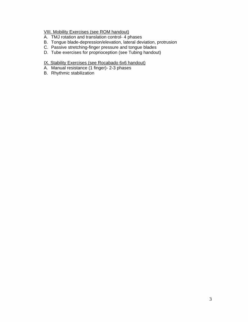
3
VIII. Mobility Exercises (see ROM handout) A. TMJ rotation and translation control- 4 phases B. Tongue blade-depression/elevation, lateral deviation, protrusion C. Passive stretching-finger pressure and tongue blades D. Tube exercises for proprioception (see Tubing handout)
IX. Stability Exercises (see Rocabado 6x6 handout) A. Manual resistance (1 finger)- 2-3 phases B. Rhythmic stabilization

4
TMD Cases and Problem Solving (adapted from Olson KA. Manual Physical Therapy of the Spine, 2009)
Case 1 History A 23 year old college student has tightness, discomfort and clicking in the right TMJ with intermittent occipital headaches. Pain is provoked with stressful situations and with chewing meat and crunchy foods. Test and Measures
Posture: moderate forward head posture with protracted scapulae AROM:
o Cervical (in standing): 85% in all planes of motion and pain free except for extension, which is 50% and provokes occipital area pain
o Thoracic: 75-85% in all planes of motion and pain free o Mandible: opening to 35 mm with mid range deviation to the right and
return to midline after opening joint sound (also noted at mid range closing. Lateral deviation right 10 mm, left 5 mm. Protrusion 5 mm with midrange click.
o Shoulder screen: full and pain free bilateral shoulder AROM Passive intervertebral motion (PIVM) testing: limited craniovertebral flexion, right
side bending, and left rotation; mid cervical spine PIVM testing reveals hypermobility; upper thoracic is slightly restricted at T1-T2 left and right rotation, and flexion.
Muscle length: mild tightness right levator scapula and minimally tight bilateral pectoralis major and minor
Strength: lower and middle trapezius are 4-/5; deep neck flexors are 3+/5 Neurological screen: negative Special tests:
o Bite test: painful right TMJ with biting on the left side o Retrusive overpressure: provokes pain on right TMJ
Palpation: tender and guarded right muscles of mastication with intraoral and extraoral palpation, tender at lateral pole right TMJ; tender at C2-C3 right facet joint
Evaluation
Diagnosis:
Impairments:
Prognosis:
Interventions: Goals:

5
Case 2 History A 50 year old construction worker has difficulty opening his mouth after trauma from being hit in the jaw during a bar fight 3 months prior to the initial evaluation. The patient has no history of TMJ sounds. Recent radiographic results were negative for signs of mandibular fracture. Tests and Measures
Posture: mild forward head posture with protracted scapulae AROM:
o Cervical in standing: 85% in all planes of motion and pain free o Thoracic: 75% upper thoracic rotation motion and pain free o Mandible: 20 mm opening with deviation to the right, lateral deviation right
7 mm, left 4 mm; protrusion 3 mm with deviation to the right; no joint sounds noted
o Shoulder screen: full ROM and normal strength Accessory motion testing TMJ: hypomobility with lateral and medial glide and
joint distraction right TMJ PIVM testing: slight hypomobility craniovertebral flexion and right side bending;
hypomobility T1-T2 left and right rotation Muslce length: no limitations noted Strength lower and middle trapezius are 4-/5; deep neck flexors are 3+/5 Neurologic screen: negative Special tests:
o Bite test: negative o Retrusive overpressure: negative
Palpation: tender and guarded right muscles of mastication intraoral and externally, and tender at right lateral mandibular condyle
Evaluation
Diagnosis:
Impairments:
Prognosis:
Interventions: Goals:

Jaw Functional Limitation Scale
For each of the items below, please indicate the level of limitation during the last month. If the activity has been completely avoided because it is too difficult, then circle ‘10'. If you avoid an activity for reasons other than pain or difficulty, then leave the item blank.
No limitation
Severe Limitation
1. Chew tough food 0 1 2 3 4 5 6 7 8 9 10
2. Chew hard bread 0 1 2 3 4 5 6 7 8 9 10
3. Chew chicken (for example, preparedin oven) 0 1 2 3 4 5 6 7 8 9 10
4. Chew crackers 0 1 2 3 4 5 6 7 8 9 10
5. Chew soft food (for example, macaroni,canned or soft fruits, cooked vegetables,fish)
0 1 2 3 4 5 6 7 8 9 10
6. Eat soft food requiring no chewing (forexample, mashed potatoes, applesauce, pudding, pureed food)
0 1 2 3 4 5 6 7 8 9 10
7. Open wide enough to bite from a wholeapple
0 1 2 3 4 5 6 7 8 9 10
8. Open wide enough to bite into asandwich
0 1 2 3 4 5 6 7 8 9 10
9. Open wide enough to talk 0 1 2 3 4 5 6 7 8 9 10
10. Open wide enough to drink from a cup 0 1 2 3 4 5 6 7 8 9 10
11. Swallow 0 1 2 3 4 5 6 7 8 9 10
12. Yawn 0 1 2 3 4 5 6 7 8 9 10
13. Talk 0 1 2 3 4 5 6 7 8 9 10
14. Sing 0 1 2 3 4 5 6 7 8 9 10
15. Putting on a happy face 0 1 2 3 4 5 6 7 8 9 10
16. Putting on an angry face 0 1 2 3 4 5 6 7 8 9 10
17. Frown 0 1 2 3 4 5 6 7 8 9 10
18. Kiss 0 1 2 3 4 5 6 7 8 9 10
19. Smile 0 1 2 3 4 5 6 7 8 9 10
20. Laugh 0 1 2 3 4 5 6 7 8 9 10
Ohrbach R, Larsson P, List T. The jaw functional limitation scale: development, reliability, and validity of 8-item and 20-item versions. J Orfac Pain. 2008;22:219-230.

Temporomandibular Disorders Classification
Adapted from Olson KA. Manual Physical Therapy of the Spine. 2009
Classification ROM Accessory Motion Pain Palpation Joint Sounds
Capsulitis/Synovitis Variable-often limited
by pain
Pain with testing Bite on opposite side
Retrusive overpressure
Accessory motion
testing
Tender over lateral
and posterior condyle
None
Capsular Fibrosis <25 mm
Deviates toward
involved side
Hypomobile Mild-none at rest
Pain at end range
Variable None
Masticatory Muscle AROM<PROM Normal Bite on same
Chewing, eating
Tender/spasm of
masseter, temporalis,
lateral pterygoid
None
Hypermobility >40 mm (subluxation)
>60 mm (dislocation)
Hypermobile Unually none Negative At end range
Anterior Disc Displacement
With Reduction
Normal opening with S
curve
Normal Variable Negative Reciprocal click
Anterior Disc Displacement
Without Reduction
<25 mm
Deviates toward
involved side
Hypomobile Generally pain at rest
and movement
Variable None
Osteoarthritis Limited AROM=PROM Hypomobile Variable Usually negative Crepitus

Temporomandibular Disorder Disability Index
Please check the statement that best pertains to you (not necessarily exactly) in each of the following categories.
Adapted from Streigerwald DP, Maher JH: The Streigerwald/Maher TMD disability questionnaire. Today Chiropract 23.86.91. 1997
1. Communication (talking) ___ I can talk as much as I want without pain, fatigue, or discomfort. ___ I can talk as much as I want, but it causes some pain, fatigue or discomfort. ___ I can’t talk as much as I want because of pain, fatigue or discomfort. ___ I can’t talk much at all because of pain, fatigue, or discomfort. ___ Pain prevents me from talking at all.
2. Normal living activities (brushing teeth/flossing) ___ I am able to care for my gums and teeth in a normal fashion without restriction and without pain, fatigue or discomfort. ___ I am able to care for all my teeth and gums, but I must be slow and careful, otherwise pain/discomfort or jaw tiredness results.
___ I do manage to care for my teeth and gums in a normal fashion, but it usually causes some pain/discomfort or jaw tiredness no matter how careful I am. ___ I am unable to properly clean all my teeth and gums because of restricted opening or pain. ___ I am unable to care for most of my teeth and gums because of restricted opening or pain.
3. Normal living activities (eating, chewing) ___ I can eat and chew as much of anything I want without pain/discomfort or jaw tiredness. ___ I can eat and chew most anything I want, but it sometimes causes pain/discomfort or jaw tiredness. ___ I can’t each much of anything I want because it often causes pain/discomfort or jaw tiredness or because of restricted opening. ___ I must eat only soft foods (consistency of scrambled eggs or less) because of pain/discomfort, jaw fatigue, or restricted opening. ___ I must stay on a liquid diet because of pain or restricted opening.
4. Social/recreational activities (singing, playing musical instruments, cheering, laughing, social activities, playing amateur sports/hobbies etc.)
___ I am enjoying a normal social life or recreational activities without restriction. ___ I participate in a normal social life or recreational activities, but pain/discomfort is increased. ___ The presence of pain or fear of likely aggravation only limits the more energetic components of my social life (sports, exercise, dancing, playing musical instruments singing). ___ I have restrictions socially as I can’t even sing, shout, cheer, play, or laugh expressively because of increased pain/discomfort. ___ I have practically no social life because of pain.
5. Nonspecialized jaw activities (yawning, mouth opening, and opening my mouth wide)
___ I can yawn in a normal fashion, painlessly. ___ I can yawn and open my mouth fully wide open, but sometimes there is discomfort. ___ I can yawn and open my mouth wide in a normal fashion, but it almost always causes discomfort. ___ Yawning and opening my mouth wide are somewhat restricted by pain. ___ I cannot yawn or open my mouth more than two finger widths (2.8 to 3.2 cm) or, if I can, it always causes
greater than moderate pain.
6. Sexual function (including kissing, hugging, and any and all sexual activities to which you are accustomed) ___ I am able to engage in all my customary sexual activities and expressions without limitation or causing headache, face, or jaw pain. ___ I am able to engage in all my customary sexual activities and expressions, but it sometimes causes some headache, face, or jaw pain or jaw fatigue. ___ I am able to engage in all my customary sexual activities and expressions, but it usually causes enough headache, face, or jaw pain to markedly interfere with my enjoyment, willingness, and satisfaction. ___ I must limit my customary sexual activities and expressions because of headache, face, or jaw pain, or limited mouth opening. ___ I abstain from almost all sexual activities and expression because of the head, face, or jaw pain it causes. 7. Sleep (restful, nocturnal sleep patterns) ___ I sleep well in a normal fashion without any pain medication, relaxants, or sleeping pills. ___ I sleep well with the use of pain pills, anti-inflammatory medication or medicinal sleeping aids. ___ I fail to realize 6 hours of restful sleep even with the use of pills. ___ I fail to realize 4 hours of restful sleep even with the use of pills ___ I fail to realize 2 hours of restful sleep even with the use of pills. 8. Effects of any form of treatment, including, but not limited to, medications, in-office therapy, treatments, oral orthotics (e.g., splints, mouthpieces), ice/heat, etc. ___ I do not need to use treatment of any type to control or tolerate headache, face or jaw pain and discomfort. ___ I can completely control my pain with some form of treatment. ___ I get partial, but significant, relief through some form of treatment. ___ I don’t get “a lot” of relief from any form of treatment. ___ There is no form of treatment that helps enough to make me want to continue. 9. Tinnitus, or ringing in the ear(s) ___ I do not experience ringing in my ear(s). ___ I experience ringing in my ear(s) somewhat, but it does not interfere with my sleep or my ability to perform my daily activities. ___ I experience ringing in my ear(s) and it interferes with my sleep or daily activities, but I can accomplish set goals and can get an acceptable amount of sleep. ___ I experience ringing in my ear(s), and it causes a marked impairment in the performance of my daily activities or results in an unacceptable loss of sleep. ___ I experience ringing in my ear(s) and it is incapacitating or forces me to use a masking device to get any sleep. 10. Dizziness (lightheadedness, spinning, or balance disturbance) ___ I do not experience dizziness. ___ I experience dizziness, but it does not interfere with my daily activities. ___ I experience dizziness that interferes somewhat with my daily activities, but I can accomplish my set goals. ___ I experience dizziness that causes a marked impairment in the performance of my daily activities. ___ I experience dizziness that is incapacitating.

Cervical and Thoracic Bibliography
1. Ahmed HE, White PF, Craig WF, Ghoname ES, Gajraj NM. Use of percutaneous electrical nerve stimulation (PENS) in the short-term management of headache. Headache 2000; 40(4): 311-315.
2. Audette JF, Wang F, Smith H. Bilateral activation of motor unit potentials with unilateral needle stimulation of active myofascial trigger points. Am J Phys Med Rehabil 2004; 83(5): 368-374.
3. Beer A, Treleaven J, Jull G. Can a functional postural exercise improve performance in the cranio-cervical flexion test? – A preliminary study. Manual Therapy Volume 17, Issue 3 , Pages 219-224, June 2012.
4. Bogdurk, N. Cervicogenic headache: anatomic basis and pathophysiologic mechanisms. Current Pain and Headache Reports 2001; 5(4): 382-386.
5. Boyles RE, Walker MJ, Young BA, Strunce JB, Wainner RS. The addition of cervical thrust manipulations to a manual physical therapy approach in patients treated for mechanical neck pain: A Secondary Analysis. Journal of Orthopaedic & Sports Physical Therapy 2010; 40(3): 133-140.
6. Boyling JD, Jull GA. 2005. Grieve's Modern Manual therapy: the vertebral column. Churchill Livingstone.
7. Brismee JM, Gipson D, Ivie D, Lopez A, Moore M, Matthijs O, Phelps V, Sawyer S, Sizer P. Interrater reliability of a passive physiological intervertebral motion test in the mid-thoracic spine. Journal of Manipulative and Physiological Therapeutics 2006; 29(5): 368-373.
8. Browder DA, Erhard RE, Piva SR. Intermittent cervical traction and thoracic manipulation for management of mild cervical compressive myelopathy attributed to cervical herniated disc: a case series. Journal of Orthopaedic and Sports Physical Therapy 2004; 34(11): 701-712.
9. Calandre EP, Hidalgo J, Garcia-Leiva JM, Rico-Villademoros F. Trigger point evaluation in migraine patients: an indication of peripheral sensitization linked to migraine predisposition? Eur J Neurology 2006; 13:244-249.
10. Childs JD, Fritz JM, Piva SR, Whitman JM. Proposal of a classification system for patients with neck pain. Journal of Orthopaedic and Sports Physical Therapy 2004; 34(11): 686-696.
11. Childs JD, Cleland JA, Elliott JM, Teyhen DS, Wainner RS, Whitman JM, Sopky BJ, Godges JJ, Flynn TW. Neck pain: Clinical practice guidelines linked to the International Classification of Functioning, Disability, and Health from the Orthopedic Section of the American Physical Therapy Association. J Orthop Sports Phys Ther. 2008 Sep;38(9):A1-A34.
12. Cleland J, McRae M. Complex regional pain syndrome I: Management through the use of vertebral and sympathetic trunk mobilization. J Manual Manipulative Ther 2002;10:188–199.
13. Cleland J, Selleck B, Stowell T, Browne L, Alberini S, St. Cyr H, Caron T. Short-Term Effects of Thoracic Manipulation on Lower Trapezius Muscle Strength. Journal of Manual & Manipulative Therapy, Volume 12, Number 2, 2004 , pp. 82-90(9).

14. Cleland JA, Flynn TW, Childs JD, Eberhart S. The audible pop from thoracic spine thrust manipulation and its relation to short-term outcomes in patients with neck pain. Journal of Manual and Manipulative Therapy 2007; 15(3): 143-154.
15. Cleland JA, Mintken PA, Carpernter K, Fritz JM, Glynn P, Whitman J, Childs JD. Examination of a clinical prediction rule to identify patients with neck pain likely to benefit from thoracic spine thrust manipulation and a general cervical range of motion exercise: Multi-center randomized clinical trial. Journal of the American Physical Therapy Association 2010; 90(9): 1239-1253.
16. Cleland JA, Whitman JM, Fritz JM, Palmer JA. Manual physical therapy, cervical traction, and strengthening exercises in patients with cervical radiculopathy: a case series. Journal of Orthopaedic and Sports Physical Therapy 2005; 35(12): 802-811.
17. Cleland, J. Orthopedic Clinical Examination: An Evidence-Based Approach for Physical Therapists. Icon (2005)
18. Conroy JL, Schneiders AG. The T4 syndrome. Manual Therapy 2005; 10(4): 292-296. 19. Cook, C. Orthopedic Manual Therapy: An Evidence Based Approach. Pearson Prentice
Hall (2007) 20. Crosby CA, Wehbe MA. Conservative treatment for thoracic outlet syndrome. Hand
Clinics 2004; 20(1): 43-49. 21. Cross KM, Kuenze C, Grindstaff TL, Hertel J. Thoracic spine thrust manipulation
improves pain, range of motion, and self-reported function in patients with mechanical neck pain: a systematic review. J Orthop Sports Phys Ther. 2011 Sep;41(9):633-42.
22. Domenech MA, Sizer PS, Dedrick GS, McGalliard MK, Brismee JM. The deep neck flexor endurance test: normative data scores in healthy adults. PM & R 2011; 3(2): 105-110.
23. Dreyfuss P, Tibiletti C, Dreyer SJ. Thoracic zygapophyseal joint pain patterns. A study in normal volunteers. Spine 1994; 19(7): 807-811.
24. Dunning JR, Cleland JA, Waldrop MA, Arnot CF, Young IA, Turner M, Sigurdsson G. Upper cervical and upper thoracic thrust manipulation versus nonthrust mobilization in patients with mechanical neck pain: a multicenter randomized clinical trial. J Orthop Sports Phys Ther. 2012 Jan;42(1):5-18.
25. Dutton, M. Orthopaedic Examination, E valuation & Intervention. McGraw-Hill (2004) 26. Dwyer A, April C, Bogduk N. Cervical zygapophyseal joint pain patterns. I: A study in
normal volunteers. Spine 1990; 15(6): 453-457. 27. Ebraheim NA, Lu J, Biyani A, Brown JA, Yeasting RA. Anatomic consideration for
uncovertebral involvement in cervical spondylosis. Clinical Orthopaedics and Related Research 1997; (334): 200-206.
28. Edmondston SJ, Singer KP. Thoracic spine: anatomical and biomechanical considerations for manual therapy. Manual Therapy 1997; 2(3): 132-143.
29. Edmondston SJ, Waller R, Vallin P, Holthe A, Noebauer A, King E. Thoracic spine extension mobility in young adults: influence of subject position and spinal curvature. J Orthop Sports Phys Ther. 2011 Apr;41(4):266-73.

30. Errico TJ, Stecker S, Kostuik JP. Thoracic pain syndromes. In: Frymoyer JW, ed. The Adult Spine: Principles and Practice. Philadelphia, Pa: Lippincott-Raven; 1997:1623–1637.
31. Falla D. Unravelling the complexity of muscle impairment in chronic neck pain. Manual Therapy 2004; 9(3): 125-133.
32. Fernandez-De-Las-Penas C, Fernandez-Carnero J, Palomeque del Cerro L, Miangolarra-Page JC. Manipulative treatment vs conventional physiotherapy treatment in whiplash injury a randomized controlled trial. Journal of Whiplash and Related Disorders 2004; 3(2): 73-90.
33. Fernandez-De-Las-Penas C. Interaction between trigger points and joint hypomobility: A clinical perspective. The Journal of Manual and Manipulative Therapy 2009; 17(2): 74-77.
34. Fernández-Pérez AM, Villaverde-Gutiérrez C, Mora-Sánchez A, Alonso-Blanco C, Sterling M, Fernández-de-Las-Peñas C. Muscle trigger points, pressure pain threshold, and cervical range of motion in patients with high level of disability related to acute whiplash injury. J Orthop Sports Phys Ther. 2012;42(7):634-41.
35. Flynn, T. User’s Guide to the Musculoskeletal Examination: Fundamentals for the Evidence-Based Clinician. Evidence in Motion (2008)
36. Forbush SW, Cox T, Wilson E. Treatment of patients with degenerative cervical radiculopathy using a multimodal conservative approach in a geriatric population: a case series. J Orthop Sports Phys Ther. 2011 Oct;41(10):723-33.
37. Fraser DM. T-3 Revisited. Journal of Orthopaedic Medicine 1993; 5:3-4. 38. Friction JR, Kroening R, Haley D, Siegert R. Myofascial pain syndrome of the head and
neck: a review of clinical characteristics of 164 patients. Oral Surgery, Oral Medicine, and Oral Pathology 1985; 60(6): 615-623.
39. Fritz JM, Brennan GP. Preliminary examination of a proposed treatment-based classification system for patients receiving physical therapy interventions for neck pain. Physical Therapy 2007; 87(5): 513-524.
40. Fukui S, Ohseto K, Shiotani M, Ohno K, Karasawa H, Maganuma Y, Yuda Y. Referred pain distribution of the cervical zygapophyseal joints and cervical dorsal rami. Pain 1996; 68(1): 79-83.
41. Fukui S, Ohseto K, Shiotani M. Patterns of pain induced by distending the thoracic zygapophyseal joints. Regional Anesthesia 1997; 22(4): 332-336.
42. Ge HY, Nie H, Madeleine P, Danneskiold-Samsøe B, Graven-Nielsen T, Arendt-Nielsen L. Contribution of the local and referred pain from active myofascial trigger points in fibromyalgia syndrome. Pain 2009;147(1-3):233-40.
43. Geerse WK. Bilateral leg symptoms--the T10 syndrome? Man Ther. 2012 Jun;17(3):251-4.
44. Gerwin RD. Neurobiology of the myofascial trigger point. Baillieres Clinical Rheumatology 1994; 8(4): 747-762.
45. Giamberardino MA, Tafuri E, Savini A, Fabrizio A, Affaitat G, Lerza R, Dilanni L, Lapenna D, Mezzetti A. Contribution of myofasical trigger points to migraine symptoms. J Pain 2007; 8(11): p. 869-78.
46. Goldsmith R, Wright C, Bell SF, Rushton A. Cold hyperalgesia as a prognostic factor in whiplash associated disorders: a systematic review. Man Ther. 2012 Oct;17(5):402-10.

47. Gordon SJ, Grimmer-Somers K, Trott P. Pillow use: The behaviour of cervical pain, sleep quality and pillow comfort in side sleepers. Manual Therapy 2009; 14(6): 671-678.
48. Graham N, Gross A, Goldsmith CH, Klaber Moffett J, Haines T, Burnie SJ, Peloso PMJ Mechanical traction for neck pain with or without radiculopathy. Cochrane Database of Systematic Reviews 2008, Issue 3.
49. Graff-Radford SB, Reeves JL, Baker Rl, Chiu D. Effects of transcutaneous electrical nerve stimulation on myofascial pain and trigger point sensitivity. Pain 1989; 37(1): 1-5.
50. Grieve GP. 1988 Common Vertebral Joint Problems. Elsevier Health Sciences. 51. Gross A, Miller J, D’Sylva J, Burnie SJ, Goldsmith CH, Graham N, Haines T, Bronfort
G, Hoving JL. Manipulation or mobilization for neck pain: A Cochrane Review. Manual Therapy 2010; 15(4): 315-333.
52. Gross AR, Hoving JL, Haines TA, Goldsmith CH, Kay T, Aker P, Bronfort G, Cervical Overview Group. A Cochrane review of manipulation and mobilization for mechanical neck disorders. Spine 2004; 29(14): 1541-1548.
53. Ha SM, Kwon OY, Yi CH, Jeon HS, Lee WH. Effects of passive correction of scapular position on pain, proprioception, and range of motion in neck-pain patients with bilateral scapular downward-rotation syndrome. Man Ther. 2011 Dec;16(6):585-9.
54. Haas M, Raphael R, Panzer D, Peterson D. Reliability of manual end-play palpation of the thoracic spine. Chiropractic Technique 1995; 7: 120-124.
55. Hall T, Briffa K, Hopper D, Robinson K. Reliability of manual examination and frequency of symptomatic cervical motion segment dysfunction in cervicogenic headache. Manual Therapy 2010; 15(6): 513-606.
56. Hammil JM, Cook TM, Rosecrance JC. Effectiveness of a physical therapy regimen in the treatment of tension-type headache. Headache 1996; 36(3): 149-153.
57. Hanten WP, Olson SL, Butts NL, Nowicki AL. Effectiveness of a home program of ischemic pressure followed by sustained stretch for treatment of myofascial trigger points. Journal of the American Physical Therapy Association 2000; 80(10): 997-1003.
58. Harris KD, Heer DM, Roy TC, Santos DM, Whitman JM, Wainner RS. Reliability of a measurement of neck flexor muscle endurance. Physical Therapy 2005; 85(12): 1349-1355.
59. Henschke N, Maher CG, Refshauge KM, Herbert RD, Cumming RG, Bleasel J, York J, Das A, McAuley JH. Prevalence of and screening for serious spinal pathology in patients presenting to primary care settings with acute low back pain. Arthritis and Rheumatism 2009; 60(10) 3072-3080.
60. Hooper TL, Denton J, McGalliard MK, Brismee JM, Sizer PS. Thoracic outlet syndrome: a controversial clinical condition. Part 1: anatomy, and clinical examination/diagnosis. Journal of Manual and Manipulative Therapy 2010; 18(2): 74-83.
61. Hooper TL, Denton J, McGalliard MK, Brismee JM, Sizer PS. Thoracic outlet syndrome: a controversial clinical condition. Part 2: non-surgical and surgical management. Journal of Manual and Manipulative Therapy 2010; 18(3): 132-138.
62. Horneij E, Hemborg B, Johnsson B, Ekdahl C. Clinical tests on impairment level related to low back pain: a study of test reliability. Journal of Rehabilitation and Medicine 2002; 34(4): 176-182.
63. Jarvik JG, Deyo RA. Diagnostic evaluation of low back pain with emphasis on imaging. Annals of Internal Medicine 2002; 137(7): 586-597.

64. Jesus-Moraleida FR, Ferreira PH, Pereira LS, Vasconcelos CM, Ferreira ML. Ultrasonographic analysis of the neck flexor muscles in patients with chronic neck pain and changes after cervical spine mobilization. J Manipulative Physiol Ther. 2011 Oct;34(8):514-24.
65. Jull G, Amiri M, Bullock-Saxton J, Darnell R, Lander C. Cervical musculoskeletal impairment in frequent intermittent headache. Part 1: Subjects with single headaches. Cephalalgia 2007; 27(7): 793-802.
66. Jull G, Kristjansson E, Dall’Alba P. Impairment in the cervical flexors: a comparison of whiplash and insidious onset neck pain patients. Manual Therapy 2004; 9(2): 89-94.
67. Jull G, Trott P, Potter H, Zito G, Niere K, Shirley D, Emberson J, Marschner I, Richardson C. A randomized controlled trial of exercise and manipulative therapy for cervicogenic headache. Spine 2002; 27(17): 1835-1843.
68. Jull G, Zito G, Trott P, Potter H, Shirlery D. Inter-examiner reliability to detect painful upper cervical joint dysnfunction. The Australian Journal of Physiotherapy 1997; 43(2): 125-129.
69. Kannan P. Management of myofascial pain of upper trapezius: a three group comparison study. Glob J Health Sci. 2012 Jul 15;4(5):46-52.
70. Karakurum B, Karaalin O, Coskun O, Dora B, Uçler S, Inan L. The 'dry-needle technique': intramuscular stimulation in tension-type headache. Cephalalgia. 2001 Oct;21(8):813-7.
71. Kay TM, Gross A, Goldsmith C, Santaquida PL, Hoving J, Bronfort G, Cervical Overview Group. Exercises for mechanical neck disorders. Cochrane Database of Systematic Reviews 2005 (3).
72. Kaya A, Kamanli A, Ardicoglu O, Ozgocmen S, Ozkurt-Zengin F, Bayik Y. Direct current therapy with/without lidocaine iontophoresis in myofascial pain syndrome. Bratisl Lek Listy. 2009;110(3):185-91.
73. Kjellman G, Oberg B. A randomized clinical trial comparing general exercise, McKenzie treatment and a control group in patients with neck pain. Journal of Rehabilitation Medicine 2002; 34(4): 183-190.
74. Leaver AM, Maher CG, Herbert RD, Latimer J, McAuley JH, Jull G, Refshauge KM. A randomized controlled trial comparing manipulation with mobilization for recent onset neck pain. Arch Phys Med Rehabil. 2010 Sep;91(9):1313-8.
75. Lee DG. Rotational instability of the mid-thoracic spine: assessment and management. Manual Therapy 1996; 1(5): 234-241.
76. Lee JP, Wang Cl, Shau YW, Wang SF. Measurement of cervical multifidus contraction pattern with ultrasound imaging. Journal of Electromyography and Kinesiology 2009; 19(3): 391-397.
77. Lee LJ, Coppieters MW, Hodges PW. Anticipatory postural adjustments to arm movement reveal complex control of paraspinal muscles in the thorax. Journal of Electromyography and Kinesiology 2009; 19(1): 46-54.
78. Lin YC, Lai CH, Chang WH, Tu LW, Lin JC, Chou SW. Immediate effects of ischemic compression on neck function in patients with cervicogenic cephalic syndrome. J Manipulative Physiol Ther. 2012 May;35(4):301-7.

79. Lindgren KA, Leino E, Mannienen H. Cineradiography of the hypomobile first rib. Archives of Physical and Medical Rehabilitation 1989; 70(5): 408-409.
80. Lindgren KA. Conservative treatment of thoracic outlet syndrome: a 2 year follow-up. Archives of Physical Medicine and Rehabilitation 1997; 78(4): 373-378.
81. Linscott MS, Heyborne R. Thoracic intervertebral disk herniation: A commonly missed diagnosis. The Journal of Emergency Medicine 2007; 32(3): 235-238.
82. Magee, D. Orthopedic Physical Assessment, 5th Ed. Saunders (2008) 83. Maitland, GD. Peripheral Manipulation. Butterworths (1977) 84. Manning DM, Dedrick GS, Sizer PS, Brismee JM. Reliability of a seated three-
dimensional passive intervertebral motion test for mobility, end-feel, and pain provocation in patients with cervicalgia. Journal of Manual and Manipulative Therapy 2012; 20(3): 135-141.
85. Martínez-Segura R, De-la-Llave-Rincón AI, Ortega-Santiago R, Cleland JA, Fernández-de-Las-Peñas C. Immediate changes in widespread pressure pain sensitivity, neck pain, and cervical range of motion after cervical or thoracic thrust manipulation in patients with bilateral chronic mechanical neck pain: a randomized clinical trial. J Orthop Sports Phys Ther. 2012 Sep;42(9):806-14.
86. Maxey L, Magnusson J. 2013. Rehabilitation for the postsurgical orthopedic patient. Northwestern University: Mosby Elsevier
87. McGaugh J, Ellison J. Intrasession and interrater reliability of rehabilitative ultrasound imaging measures of the deep neck flexors: A pilot study. Physiother Theory Pract. 2011; 27(8):572-7.
88. Mercer SR, Jull GA. Morphology of the cervical intervertebral disc: implications for McKenzie’s model of the disc derangement syndrome. Manual Therapy 1996; 1(2): 76-81.
89. Mert E, Ozge A, Tasdele B, Yilmaz A, Bilgin NG. What clues are available for differential diagnosis of headaches in emergency settings? The Journal of Headache and Pain 2008; 9: 89-97.
90. Murphy DR, Hurwitz EL, Gregory AA. Manipulation in the presence of cervical spinal cord compression: a case series. Journal of Manipulative and Physiological Therapeutics 2006; 29(3): 236-244.
91. Nathan H. Osteophytes of the spine compressing the sympathetic trunk and splanchinic nerves in the thorax. Spine 1987; 12(6): 527-532.
92. Nee RJ, Vicenzino B, Jull GA, Cleland JA, Coppieters MW. Neural tissue management provides immediate clinically relevant benefits without harmful effects for patients with nerve-related neck and arm pain: a randomised trial. J Physiother. 2012;58(1):23-31.
93. Nijs j, Van Houddenhove B, Oostendorp RAB. Recognition of central sensitization in patients with musculoskeletal pain: Application of pain neurophysiology in manual therapy practice. Manual Therapy 2010; 15(2): 135-141.
94. Nijs J, Van Oosterwijck J, De Hertogh W. Rehabilitation of chronic whiplash: treatment of cervical dysfunctions or chronic pain syndrome? Clinical Rheumatology 2009; 28(3): 243-251.
95. O’Leary S, Jull G, Kim M, Vicenzino B. Specificity in retraining craniocervical flexor muscle performance. Journal of Orthopaedic and Sports Physical Therapy 2007; 37(1): 3-9.

96. Ogince M, Hall T, Robinson K, Blackmore AM. The diagnostic validity of the cervical flexion-rotation test in C1/2-related to cervicogenic headache. Manual Therapy 2007; 12(3): 256-262.
97. Olson KA. 2009. Manual Physical Therapy of the Spine. St. Louis, Missouri: Elsevier Mosby.
98. Panjabi MM. The stabilizing system of the spine. Journal of Spinal Disorders 1992; 5(4): 383-396.
99. Panjabi MM. The stabilizing system of the spine. Part II. Neutral zone and instability hypothesis. Journal of Spinal Disorders 1992; 5(4): 390-396.
100. Petersen SM, Wyatt SH. Lower trapezius muscle strength in individuals with unilateral neck pain. JOSPT 2011; 41(4): 260-265
101. Pool JJ, Hoving JL, de Vet HC, van Mameren H, Bouter LM. The interexaminer reproducibility of physical examination of the cervical spine. The Journal of Manipulative Physiological Therapeutics 2004; 27(2): 84-90.
102. Puentedura EJ, Cleland JA, Landers MR, Mintken PE, Louw A, Fernández-de-Las-Peñas C. Development of a clinical prediction rule to identify patients with neck pain likely to benefit from thrust joint manipulation to the cervical spine. J Orthop Sports Phys Ther. 2012;42(7):577-92.
103. Puentedura EJ, March J, Anders J, Perez A, Landers MR, Wallmann HW, Cleland JA. Safety of cervical spine manipulation: are adverse events preventable and are manipulations being performed appropriately? A review of 134 case reports. Journal of Manual & Manipulative Therapy, Volume 20, Number 2, 2012 , pp. 66-74(9).
104. Puentedura EJ, Landers MR, Cleland JA, Mintken PE, Huijbregts P, Fernández-de-Las-Peñas C. Thoracic spine thrust manipulation versus cervical spine thrust manipulation in patients with acute neck pain: a randomized clinical trial. J Orthop Sports Phys Ther. 2011 Apr;41(4):208-20.
105. Raney NH, Petersen EJ, Smith TA, Cowan JE, Rendeiro DG, Deyle GD, Childs JS. Development of clinical prediction rule to identify patients with neck pain likely to benefit from cervical traction and exercise. European Spine Journal 2009; 18(3): 382-391.
106. Rankin G, Stokes M, Newham DJ. Size and shape of the posterior neck muscles measured by ultrasound imaging: normal values in males and females of different ages. Man Ther. 2005; 10(2):108-15.
107. Rigby AS, Wood PH. Observations on diagnostic criteria for ankylosing spondylitis. Clinical and Experimental Rheumatology 1993; 11(1): 5-12.
108. Roman M, Brown C, Richardson W, Isaacs R, Howes C, Cook C. The development of a clinical decision making algorithm for detection of osteoporotic vertebral compression fracture or wedge deformity. Journal of Manual and Manipulative Therapy 2010; 18(1): 44-49.

109. Saavedra-Hernández M, Castro-Sánchez AM, Arroyo-Morales M, Cleland JA, Lara-Palomo IC, Fernández-de-Las-Peñas C. Short-term effects of kinesio taping versus cervical thrust manipulation in patients with mechanical neck pain: a randomized clinical trial. J Orthop Sports Phys Ther. 2012 Aug;42(8):724-30.
110. Sahrmann S. 2011. Movement System Impairment Syndromes of the extremities, cervical and thoracic spines. St. Louis, Missouri: Elsevier Mosby.
111. Sahrmann SA. 2002. Diagnosis and Treatment of Movement Impairment Syndromes. St. Louis, Missouri: Elsevier Mosby.
112. Salt E, Wright C, Kelly S, Dean A. A systematic literature review on the effectiveness of non-invasive therapy for cervicobrachial pain. Manual Therapy 2011; 16(1):53-65.
113. Schiller L. Effectiveness of spinal manipulative therapy in the treatment of mechanical thoracic spine pain: a pilot randomized clinical trial. Journal of Manipulative and Physiological Therapeutics 2001; 24(6): 394-401.
114. Sefton JM, Yarar C, Carpenter DM, Berry JW. Physiological and clinical changes after therapeutic massage of the neck and shoulders. Man Ther. 2011 Oct;16(5):487-94.
115. Shekelle PG. Spine manipulation. Spine 1994; 19(7) 858-861. 116. Shore LR. On osteo-arthritis in the dorsal intervertebral joints. The British Journal
of Surgery 1985; 22: 883-839. 117. Silva, AG, Punt, TD, Johnson, MI. Variability of angular measurements of head
posture within a session, within a day, and over a 7-day period in healthy participants. Physiotherapy Theory and Practice 2011; 27(7):503–511.
118. Sizer PS, Phelps V, Azevedo E. Disc related and non-disc related disorders of the thoracic spine. Pain Practice 2001; 1(2): 136-149.
119. Sizer PS, Phelps V, Brismee JM. Differential diagnosis of local cervical syndrome versus cervical brachial syndrome. Pain Practice 2001; 1(1): 21-35.
120. Srbely JZ, Dickey JP. Randomized controlled study of the antinociceptive effect of ultrasound on trigger point sensitivity: novel application in myofascial therapy? Clinical Rehabilitation 2007; 21(5): 411-417.
121. Sterling M. Identifying those at risk of developing persisten pain following a motor vehicle collision. Journal Rheumatology 2006; 33(5): 838-839.
122. Stevens BJ, McKenzie RA. Mechanical Diagnosis and Self Treatment of the Cervical Spine 1988.
123. Teachey, WS. Otolaryngic myofascial pain syndromes. Current Pain and Headache Reports 2004; 8:457–462.
124. The Manual Therapy Institute Clinical Fellowship Program: Foundations, Joint Manipulations and Adverse Neural Tension, Medical Exercise Therapy, Evaluation and Treatment of the Thoracic Spine, Evaluation and Treatment of the Cervical Spine, Advanced Cervical Spine (2003-2005)
125. Travell JG, Simons DG. 1999. Myofascial Pain and Dysfunction: The Trigger Point Manual. Williams and Wilkins, Baltimore.
126. Tsai CT, Hsieh LF, Kuan TS, Kao MJ, Chou LW, Hong CZ. Remote effects of dry needling on the irritability of the myofascial trigger point in the upper trapezius muscle. Am J Phys Med Rehabil. 2010 Feb;89(2):133-40.

127. Tseng YL, Wang WT, Chen WY, Hou TJ, Chen TC, Lieu FK. Predictors for the immediate responders to cervical manipulation in patients with neck pain. Manual Therapy 2006; 11(4): 306-315.
128. Wainner RS, Fritz JM, Irrgang JJ, Boninger ML, Delitto A, Allison S. Reliability and diagnostic accuracy of the clinical examination and patient self-report measures for cervical radiculopathy. Spine 2003; 28(1): 53-62
129. Waldrop MA. Diagnosis and treatment of cervical radiculopathy using a clinical prediction rule and a multimodal intervention approach: a case series. Journal of Orthopaedic and Sports Physical Therapy 2006; 36(3): 152-159.
130. Walton DM, Macdermid JC, Giorgianni AA, Mascarenhas JC, West SC, Zammit CA. Risk Factors for Persistent Problems Following Acute Whiplash Injury: Update of a Systematic Review and Meta-analysis. J Orthop Sports Phys Ther. 2013;43(2):31-43.
131. Watson LA, Pizzari T, Balster S. Thoracic outlet syndrome Part 2: Conservative management or thoracic outlet. Manual Therapy 2010; 15(4): 305-314.
132. Webb AL, Collins P, Rassoulian H, Mitchell BS. Synovial folds- A pain in the neck? Manual Therapy 2011; 16(2): 118-124
133. Ylinen J, Takala EP, Nykanen M, Hakkinen A, Malkia E, Pohjolainen T, Karppi SL, Kautianinen H, Airaksinen O. Active neck muscle training in the treatment of chronic neck pain in women: a randomized controlled trial. The Journal of American Medical Association 2003; 289(19): 2509-2516.
134. Young BA, Gill HE, Wainner RS, Flynn TW. Thoracic costotransverse joint pain patterns: a study in normal volunteers.BMC Musculoskeletal Disorders 2008; 9: 140.
135. Young IA, Michener LA, Cleland JA, Aguilera AJ, Snyder AR. Manual therapy, exercise, and traction for patients with cervical radiculopathy: a randomized clinical trial. Phys Ther. 2009 Jul;89(7):632-42.
Reference List for TMD Armijo-Olivo S, Fuentes JP, da Costa BR, et al. Reduced endurance of the cervical flexor muscles in patients with concurrent temporomandibular disorders and neck disability. Manual Therapy. 2010;15:586-592. Armijo-Olivo S, Silvestre RA, Fuentes JP, et al. Patients with temporomandibular disorders have increased fatigability of the cervical extensor muscles. Clin J Pain. 2012;28(1):55-64. Bakke M. Mandibular function in patients with temporomandibular joint pain: a 3-year follow up. Oral Surg Oral Med Oral Pathol Oral Radiol Endod. 2008;106:227-234. Chortis AG, Chorti AG, Forrester G, et al. Therapeutic exercise in the management of anterior disc displacement of the temporomandibular joint. Physical Therapy Reviews. 2006;11:117-123. Craane B, Dijkstra PU, Stappaerts K, et al. Randomized controlled trial on physical therapy for TMJ closed lock. J Dental Res. 2012;91(4):364-369. Da Silva MA, Botelho AM, Turim CV, et al. Low level laser therapy as an adjunctive technique in the management of temporomandibular disorders. Cranio. 2012;30(4):264-271. Desmons S, Graux F, Atassi M, et al. The lateral pterygoid muscle, a heterogeneous unit implicated in temporomandibular disorder: a literature review. J Craniomand Pract. 2007;25:283-291. Dimitroulis G. Temporomandibular disorders: a clinical update, BMJ. 1998;317:190-194. Dutton M. Orthopedic Examination, Evaluation, and Intervention. 2nd Ed. New York, NY: McGraw-Hill Companies, Inc; 2008. Dworkin SF, LeResche L. Research diagnostic criteria for temporomandibular disorders: review, criteria, examinations and specifications, critique. J Craniomandib Pract. 1992;6:301-345. Flynn TW, Cleland JA, Whitman JM. Users’ Guide to the Musculoskeletal Examination. Buckner, KY: Evidence in Motion, 2008. Furto ES, Cleland JA, Whitman JM, et al. Manual physical therapy interventions and exercise for patients with temporomandibular disorders. J Craniomandib Pract. 2006;24:283-291. Higbie, EJ, Seidel-Cobb D, Taylor, LF, et al. Effect of head position on vertical mandibular opening. J Orthop Sports Phys Ther. 1999;29:127-130. Ismail F, Demling A, Hessling K, et al. Short-term efficacy of physical therapy compared to splint therapy in treatment of arthrogenous TMD. J Oral Rehabil. 2007;34:807-813. Jedel E, Carlsson J. Biofeedback, acupuncture and transcutaneous electric nerve stimulation in the management of temporomandibular disorders: a systematic review. Physical Therapy Reviews. 2003;8:217-223.
1

John MT, Dworkin SF, Mancl LA. Reliability of clinical temporomandibular disorder diagnoses. Pain. 2005;118:61-69. Kropmans TJB, Dijkstra PU, Stegenga B, et al. Review: Therapeutic outcome assessment in permanent temporomandibular joint disc displacement. J Oral Rehabilitation. 1999;26:357-363. Lobbezoo-Scholte AM, Steenks MH, Faber JAJ, et al. Diagnostic value of orthopedic tests in patients with temporomandibular disorders. J Dent Res. 1993;72:1443-1453. Magee JD. Orthopedic Physical Assessment. St Louis, MO: Saunders; 2008. Morrone L, Makofsky H. TMJ home exercise program. Clinical Management. 1991;11:20-28. McNeely ML, Olivo SA, Magee DJ. A systematic review of the effectiveness of physical therapy interventions for TMD. Phys Ther. 2006;86:710-725. Medlicott MS, Harris SR. A systematic review of the effectiveness of exercise, manual therapy, electrotherapy, relaxation training, and biofeedback in the management of temporomandibular disorder. Phys Ther. 2006;86:955–973. Melis M, Di Giosia M, Zawawi K. Low level laser therapy for the treatment of temporomandibular disorcers: a systematic review of the literature. Cranio. 2012;30(4):304-312. Monaco A, Sgolasta F, Ciarrocchi I, et al. Effects of transcutaneous electrical stimulation on electromyographic and kinesiographic activity of patients with temporomandibular disorders: a placebo-controlled study. Journal of electromyography and kinesiology. 2012;22:463-468. Munhoz WC, Marques AP. Body posture evaluations in subjects with internal temporomandibular joint derangement. J Craniomandib Pract. 2009;27:231-242. Nicolakis P, Erdogmus B, Kopf A, et al. Effectiveness of exercise therapy in patients with internal derangement of the temporomandibular joint. J Oral Rehabil. 2001;28:1158-1164. Olson KA. Manual Physical Therapy of the Spine. St Louis, MO: Saunders; 2009. Ohrbach R, Larsson P, List T. The jaw functional limitation scale: development, reliability, and validity of 8-item and 20-item versions. J Orofac Pain. 2008;22:219-230. Peterson C. The TMJ Healing Plan. Alameda, CA: Hunter House; 2010. Travell J: Temporomandibular joint pain referred muscles of the head and neck. J Prosthet Dent. 1960;10:745-763. Rocabado M. Arthrokinematics of the temporomandibular joing. In: Gelb H, ed. Clinical Management of Head, Neck and TMJ Pain and Dysfunction. Philadelphia, PA: WB Saunders; 1985.
Simons DG, Travell JG, Simons LS. Myofacial Pain and Dysfunction: The Trigger Point Manual, Volume 1. Upper Half of Body. Baltimore,MD: Williams & Wilkins; 1999.
Tengrungsun T, Mitriattanakul S, Buranaprasertsuk P, et al. Is low level laser effective for the treatment of orofacial pain?: A systematic review. Cranio. 2012;30(4):280-285.
2

Walker N, Bohannon RW, Cameron D. Discriminant validity of temporomandibular joint range of motion measurements obtained with a ruler. J Orthop Sports Phys Ther. 2000; 30:484-492. Wolford LM, Rodrigues DB, Limoeiro E. Orthognathic and TMJ Surgery: postsurgical patient management. J Oral Maxillofac Surg. 2011;69:2893-2903. Wright EF, Domenech MA, Fischer JR. Usefulness of posture training for patients with temporomandibular disorders. JADA. 2000;131:202-210. Yoshida H, Sakata T, Hayashi T, et al. Evaluation of mandibular condylar movement exercise for patients with internal derangement of the temporomandibular joint on initial presentation. Br J Oral Maxillofacial Surg. 2011;49:310-313.
3