The Tourniquet Manual Principles and Practicelibvolume5.xyz/industrialmanagementengineering/... ·...
115
The Tourniquet Manual: Principles and Practice Leslie Klenerman Springer
Transcript of The Tourniquet Manual Principles and Practicelibvolume5.xyz/industrialmanagementengineering/... ·...

The Tourniquet Manual:Principles and Practice
Leslie Klenerman
Springer
www.anaesthesia-database.blogspot.com

The Tourniquet Manual – Principles and Practice

SpringerLondonBerlinHeidelbergNew YorkHong KongMilanParisTokyo
1111234561178910111123111456789201111234567893011112345678940111211
Leslie Klenerman
The Tourniquet Manual – Principles and Practice

Leslie Klenerman, MBBCh, ChM, FRCSEd, FRCSEngEmeritus Professor of Orthopaedic and Accident Surgery, University of Liverpool, Liverpool, UK
British Library Cataloguing in Publication DataKlenerman, Leslie
The tourniquet manual : principles and practice1. TourniquetsI. Title617.9′178ISBN 1852337060
Library of Congress Cataloging-in-Publication DataKlenerman, Leslie.
The tourniquet manual : principles and practice/Leslie Klenerman.p. ; cm.
Includes bibliographical references and index.ISBN 1-85233-706-0 (alk. paper)1. Tourniquets–Handbooks, manuals, etc. I. Title.[DNLM: 1. Hemostatic Techniques. 2. Tourniquets. 3. Extremities–surgery. 4. Intraoperative Complications–prevention & control. 5. Orthopedic Procedures–methods. 6. Postoperative Complications–prevention & control. 7. Tourniquets–adverse effects.
WH 310 K644t 2003]RD73.T6K54 2003617′.9–dc21 2003045601
Apart from any fair dealing for the purposes of research or private study, or criticism or review,as permitted under the Copyright, Designs and Patents Act 1988, this publication may only bereproduced, stored or transmitted, in any form or by any means, with the prior permission inwriting of the publishers, or in the case of reprographic reproduction in accordance with the termsof licences issued by the Copyright Licensing Agency. Enquiries concerning reproduction outsidethose terms should be sent to the publishers.
ISBN 1-85233-706-0 Springer-Verlag London Berlin Heidelberga member of BertelsmannSpringer Science+Business Media GmbHhttp://www.springer.co.uk
© Springer-Verlag London Limited 2003
The use of registered names, trademarks, etc. in this publication does not imply, even in the absenceof a specific statement, that such names are exempt from the relevant laws and regulations andtherefore free for general use.
Product liability: The publisher can give no guarantee for information about drug dosage andapplication thereof contained in this book. In every individual case the respective user must checkits accuracy by consulting other pharmaceutical literature.
Typeset by Florence Production, Stoodleigh, Devon, EnglandPrinted in the United States of America28/3830-543210 Printed on acid-free paper SPIN 10896266
1111234561178910111123111456789201111234567893011112345678940111211

This book could not have been started without the generous sponsorship of theMedical Defence Union, the Medical Protection Society, the British Associationfor Surgery of the Knee, the British Orthopaedic Foot Surgery Society, and AneticAid, a manufacturer of tourniquets. I am very grateful to these bodies for theirhelp in making this book possible.
Thanks are also due to my wife Naomi and my son Paul for their constant help,criticism and encouragement; to Professor Malcolm Jackson of the Departmentof Medicine, University of Liverpool, for help with biochemistry; to DerekEastwood, John Kirkup and Durai Nayagam for their useful comments and correc-tions; to Alun Jones and Andrew Biggs in the Photographic Department at theRobert Jones and Agnes Hunt Hospital, Oswestry, for invaluable help with theillustrations; and to Stephen White for allowing me access to the theatre duringhis operation list.
Acknowledgements
v

Introduction . . . . . . . . . . . . . . . . . . . . . . . . . . . . . . . . . . . . . . . . . . . . . . . . . ix
1 Historical Background . . . . . . . . . . . . . . . . . . . . . . . . . . . . . . . . . . . . . . . 11.1 Screw Tourniquet . . . . . . . . . . . . . . . . . . . . . . . . . . . . . . . . . . . . . . . . . . 41.2 Listerian Methods . . . . . . . . . . . . . . . . . . . . . . . . . . . . . . . . . . . . . . . . . . . 61.3 Esmarch’s Bandage . . . . . . . . . . . . . . . . . . . . . . . . . . . . . . . . . . . . . . . . . 71.4 The Pneumatic Tourniquet . . . . . . . . . . . . . . . . . . . . . . . . . . . . . . . . . . . 10References . . . . . . . . . . . . . . . . . . . . . . . . . . . . . . . . . . . . . . . . . . . . . . . . . . . 11
2 Effect of a Tourniquet on the Limb and the Systemic Circulation . . . . . 132.1 Application of the Tourniquet . . . . . . . . . . . . . . . . . . . . . . . . . . . . . . . . . 152.2 Sites of Application . . . . . . . . . . . . . . . . . . . . . . . . . . . . . . . . . . . . . . . . . 182.3 Effect on Muscle . . . . . . . . . . . . . . . . . . . . . . . . . . . . . . . . . . . . . . . . . . . . 192.4 Compression of Nerves . . . . . . . . . . . . . . . . . . . . . . . . . . . . . . . . . . . . . . 272.5 Effects on the Skin . . . . . . . . . . . . . . . . . . . . . . . . . . . . . . . . . . . . . . . . . . 292.6 Systemic and Local Effects of the Application of a Tourniquet . . . . . . . 292.7 Haemodynamic Changes . . . . . . . . . . . . . . . . . . . . . . . . . . . . . . . . . . . . . 332.8 Limb Blood Flow in the Presence of a Tourniquet . . . . . . . . . . . . . . . . . 342.9 Hyperaemia and Swelling of a Limb After Release of a Tourniquet . . . . 352.10 Haematological Effects . . . . . . . . . . . . . . . . . . . . . . . . . . . . . . . . . . . . . . 352.11 Temperature Changes . . . . . . . . . . . . . . . . . . . . . . . . . . . . . . . . . . . . . . 352.12 Tourniquet Pain . . . . . . . . . . . . . . . . . . . . . . . . . . . . . . . . . . . . . . . . . . . 36Summary . . . . . . . . . . . . . . . . . . . . . . . . . . . . . . . . . . . . . . . . . . . . . . . . . . . . 36References . . . . . . . . . . . . . . . . . . . . . . . . . . . . . . . . . . . . . . . . . . . . . . . . . . . 36
3 Ischaemia–Reperfusion Syndrome . . . . . . . . . . . . . . . . . . . . . . . . . . . . . 393.1 Metabolic Changes . . . . . . . . . . . . . . . . . . . . . . . . . . . . . . . . . . . . . . . . . 413.2 Reperfusion . . . . . . . . . . . . . . . . . . . . . . . . . . . . . . . . . . . . . . . . . . . . . . . 413.3 Modifying Ischaemia–Reperfusion . . . . . . . . . . . . . . . . . . . . . . . . . . . . . . 44Summary . . . . . . . . . . . . . . . . . . . . . . . . . . . . . . . . . . . . . . . . . . . . . . . . . . . . 48References . . . . . . . . . . . . . . . . . . . . . . . . . . . . . . . . . . . . . . . . . . . . . . . . . . . 49
4 Exsanguination of the Limb . . . . . . . . . . . . . . . . . . . . . . . . . . . . . . . . . . 514.1 External Compression . . . . . . . . . . . . . . . . . . . . . . . . . . . . . . . . . . . . . . . 534.2 Sickle Cell Disease . . . . . . . . . . . . . . . . . . . . . . . . . . . . . . . . . . . . . . . . . . 58References . . . . . . . . . . . . . . . . . . . . . . . . . . . . . . . . . . . . . . . . . . . . . . . . . . . 58
5 Complications . . . . . . . . . . . . . . . . . . . . . . . . . . . . . . . . . . . . . . . . . . . . . 615.1 Damage to Nerves . . . . . . . . . . . . . . . . . . . . . . . . . . . . . . . . . . . . . . . . . . 635.2 Damage to Muscle . . . . . . . . . . . . . . . . . . . . . . . . . . . . . . . . . . . . . . . . . . 655.3 Vascular Complications . . . . . . . . . . . . . . . . . . . . . . . . . . . . . . . . . . . . . . 69
vii
Contents

5.4 Damage to Skin . . . . . . . . . . . . . . . . . . . . . . . . . . . . . . . . . . . . . . . . . . . . 725.5 Post-tourniquet Syndrome . . . . . . . . . . . . . . . . . . . . . . . . . . . . . . . . . . . . 725.6 Potential of Cross-infection During Peripheral Venous Access
by Contamination of Tourniquets . . . . . . . . . . . . . . . . . . . . . . . . . . . . . . 74References . . . . . . . . . . . . . . . . . . . . . . . . . . . . . . . . . . . . . . . . . . . . . . . . . . . 74
6 The Tourniquet Used for Anaesthesia . . . . . . . . . . . . . . . . . . . . . . . . . . 776.1 Intravenous Regional Anaesthesia . . . . . . . . . . . . . . . . . . . . . . . . . . . . . . 796.2 Digital Tourniquets . . . . . . . . . . . . . . . . . . . . . . . . . . . . . . . . . . . . . . . . . 826.3 Regional Sympathetic Blockade . . . . . . . . . . . . . . . . . . . . . . . . . . . . . . . . 84References . . . . . . . . . . . . . . . . . . . . . . . . . . . . . . . . . . . . . . . . . . . . . . . . . . . 86
7 Technology and Practice . . . . . . . . . . . . . . . . . . . . . . . . . . . . . . . . . . . . . 877.1 Design of the Tourniquet Cuff . . . . . . . . . . . . . . . . . . . . . . . . . . . . . . . . . 897.2 Hand-powered Tourniquets . . . . . . . . . . . . . . . . . . . . . . . . . . . . . . . . . . . 907.3 Automatic Tourniquets . . . . . . . . . . . . . . . . . . . . . . . . . . . . . . . . . . . . . . 907.4 Safety Aspects . . . . . . . . . . . . . . . . . . . . . . . . . . . . . . . . . . . . . . . . . . . . . 927.5 Practical Problems . . . . . . . . . . . . . . . . . . . . . . . . . . . . . . . . . . . . . . . . . . 947.6 Golden Rules for the Safe Use of Tourniquets . . . . . . . . . . . . . . . . . . . . 99References . . . . . . . . . . . . . . . . . . . . . . . . . . . . . . . . . . . . . . . . . . . . . . . . . . . 100
Index . . . . . . . . . . . . . . . . . . . . . . . . . . . . . . . . . . . . . . . . . . . . . . . . . . . . . 103
viii
Contents ➀➁➂➃➄➅➆
Why write a book on the tourniquet? The tourniquet is used routinely in oper-ating theatres throughout the world, but as far as I know there is no single bookthat surveys the considerable literature that has accumulated. If used sensibly,the tourniquet is a safe instrument. Most of the few complications seen with itsuse are preventable. However, when something untoward happens, the tourni-quet suddenly becomes an interesting subject, particularly if there is thelikelihood of medicolegal consequences. This book summarises the scientificbackground of the tourniquet and describes a safe physiological approach topreventing complications. Examples of medicolegal problems are included.
Considerable progress had been made since Lister first excised a tuberculouswrist joint in a bloodless field. Many researchers have studied the effects ofischaemia and pressure on nerves and muscles. Tourniquets have entered theage of computers and are now much more sophisticated. Despite this, there isstill much dogma surrounding the tourniquet in operating theatres and in textbooks. This book is aimed at orthopaedic surgeons, anaesthetists and oper-ating-theatre staff.
I hope that this short text will stimulate a more widespread interest in the tourni-quet and improve safe practice.
Leslie KlenermanJune 2003
ix
Introduction
Chapter 1Historical Background

This page intentionally left blank
THE EARLY DEVELOPMENT of the tourniquet is bound up with the operation of ampu-tation. It was only about 140 years ago that the tourniquet was first used in otheroperations on the limbs. The introduction of the bloodless field was a landmark inthe development of orthopaedic operative technique, and it is interesting to recallhow this came about.
There is evidence that limbs were amputated as far back as the Neolithic age.Hippocrates recommended cutting through the dead limb at a joint, “care beingtaken not to wound any living part”.1 Only since Roman times have variousconstricting devices been employed to help the control of haemorrhage duringamputation. Archigenes and Heliodorus, who practised in Rome in the early part ofthe second century AD, used narrow bands of cloth placed directly above and belowthe line of incision, each passed two or three times about the limb and tied in asingle knot. This mainly controlled the venous bleeding. Heliodorus then relied ontight bandaging of the stump.
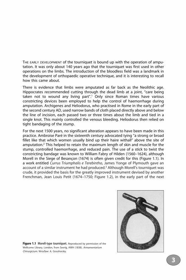
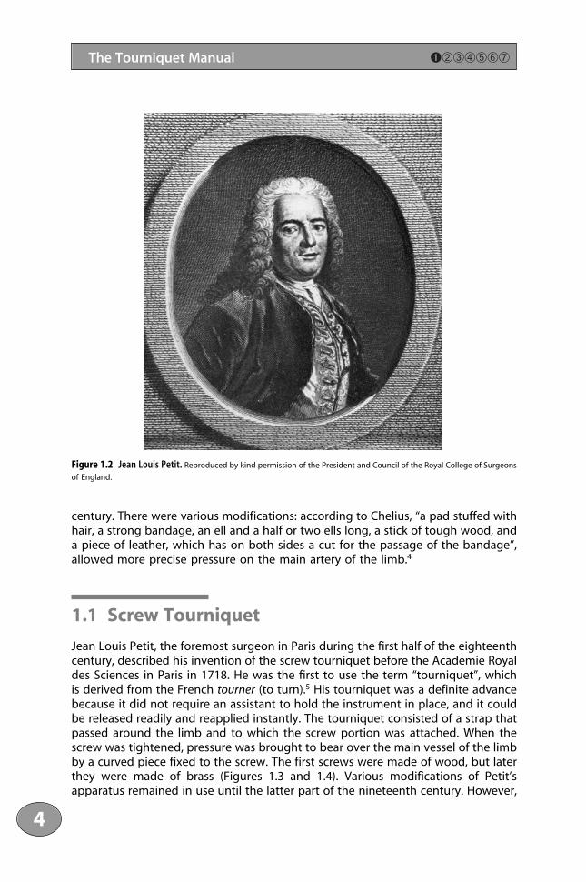
For the next 1500 years, no significant alteration appears to have been made in thispractice. Ambroise Paré in the sixteenth century advocated tying “a strong or broadfillet like that which women usually bind up their haire withall” above the site ofamputation.2 This helped to retain the maximum length of skin and muscle for thestump, controlled haemorrhage, and reduced pain. The use of a stick to twist theconstricting bandage was known to William Fabry of Hilden (1560–1624), althoughMorell in the Siege of Besançon (1674) is often given credit for this (Figure 1.1). Ina work entitled Currus Triumphalis e Terebintho, James Yonge of Plymouth gave anaccount of a similar instrument he had produced.3 Although Morell’s tourniquet wascrude, it provided the basis for the greatly improved instrument devised by anotherFrenchman, Jean Louis Petit (1674–1750; Figure 1.2), in the early part of the next
3
Figure 1.1 Morell-type tourniquet. Reproduced by permission of the
Wellcome Library, London, from Seerig, AWH (1838). Armamentarium
Chirurgicium. Wrocław: A. Gosohorsky.
century. There were various modifications: according to Chelius, “a pad stuffed withhair, a strong bandage, an ell and a half or two ells long, a stick of tough wood, anda piece of leather, which has on both sides a cut for the passage of the bandage”,allowed more precise pressure on the main artery of the limb.4
1.1 Screw Tourniquet
Jean Louis Petit, the foremost surgeon in Paris during the first half of the eighteenthcentury, described his invention of the screw tourniquet before the Academie Royaldes Sciences in Paris in 1718. He was the first to use the term “tourniquet”, whichis derived from the French tourner (to turn).5 His tourniquet was a definite advancebecause it did not require an assistant to hold the instrument in place, and it couldbe released readily and reapplied instantly. The tourniquet consisted of a strap thatpassed around the limb and to which the screw portion was attached. When thescrew was tightened, pressure was brought to bear over the main vessel of the limbby a curved piece fixed to the screw. The first screws were made of wood, but laterthey were made of brass (Figures 1.3 and 1.4). Various modifications of Petit’s apparatus remained in use until the latter part of the nineteenth century. However,
1111234561178910111123111456789201111234567893011112345678940111211
4
The Tourniquet Manual ➊➁➂➃➄➅➆
Figure 1.2 Jean Louis Petit. Reproduced by kind permission of the President and Council of the Royal College of Surgeons
of England.

5
➊➁➂➃➄➅➆ Historical Background
Figure 1.3 Petit’s tourniquet. Reproduced with
permission of the Wellcome Library, London, from
Savigny, JH (1798). A Collection of Engravings. The
Most Modern and Approved Instruments Used in the
Practice of Surgery. The Letter Press by T. Bensley.
Figure 1.4 Screw tourniquet in place. Reproduced by kind permission of the President and Council of the Royal
College of Surgeons of England from Sir Charles Bell (1821). Illustrations of the Great Operations of Surgery. London:
Longman, Hurst, Rees, Orme and Brown.

during the Crimean War, the British army reverted to using the simpler strap-and-buckle tourniquet.5
1.2 Listerian Methods
Joseph Lister (Figure 1.5), in the 1860s, was the first surgeon to use the bloodless fieldfor operations other than amputation, “long before the rest of the world had graspedthe idea of operating bloodlessly”.6 He described how his attention had first beendirected to this subject when trying to work out a satisfactory method for excision ofthe wrist joint in tuberculosis to save the hand from amputation and to overcome theprofuse bleeding associated with the procedure7:
And I found that when the hand was raised to the utmost degree and kept so for afew minutes and then while the elevated position was still maintained, a commontourniquet was applied to the arm being screwed up as rapidly as possible, so as to
1111234561178910111123111456789201111234567893011112345678940111211
6
The Tourniquet Manual ➊➁➂➃➄➅➆
Figure 1.5 Lord Lister

arrest all circulation in the limb and at the same time avoid venous turgescence, I hadpractically a bloodless field to operate on and thus gained the double advantage ofavoiding haemorrhage and inspecting precisely the part with which I was dealing.
Lister emphasised the importance of elevation of the limb before the tourniquetwas applied. He considered four minutes to be the best time to empty the bloodfrom the limb. There was thus drainage of all the venous blood and, in addition,arteriolar constriction. Lister gave experimental evidence to prove this point, basedon observations on his own hand and on the exposed metacarpal artery of a horse.7
1.3 Esmarch’s Bandage
Credit for the method of winding a strip of tensile material around the limb is usuallygiven to Johann T. Friederich August von Esmarch (1823–1908; Figure 1.6), Professorof Surgery at Kiel. Von Esmarch was not the first person to use such a device: hegave credit to Sartorius (in 1806), Brunninghausen (in 1818) and Sir Charles Bell (in1821) for having used methods of expressing venous blood from a limb in combi-nation with a tourniquet.8 Von Esmarch also acknowledged that Grandesso-Sylvestriin 1871 had used an elastic bandage to empty a limb of blood before amputation.The original Esmarch bandage was a rubber tube the thickness of a finger, woundtightly around the limb to serve as a tourniquet after the blood had been expressedfrom it by bandaging (Figure 1.7). The “Esmarch bandage” used today was actuallydesigned by von Langenbeck, based on equipment used by Esmarch; correctly, it istermed a “Langenbeck bandage”.9 Esmarch had been bandaging limbs firmly beforeamputation since 1855, in an effort to conserve blood because he had been disturbedat the amount of blood still present in an amputated limb after it had been severedfrom the patient. Subsequently, he adopted the technique for other operations onthe limb.
In The Surgeon’s Handbook on the Treatment of Wounded in War, Esmarch gives fulldetails of his technique10:
Operations on the extremities can be performed without loss of blood if they havepreviously been made bloodless in the following manner:
1 After the wounds or ulcers, which be present, have been well covered with somewaterproof material (varnished paper) the limb is firmly bandaged with an elas-tic roller from the tips of the fingers or toes upwards till it has reached beyondthe site of operation: by this means the blood is completely driven out of the vessels.
2 Where the bandage ends, an India rubber tube (elastic ligature) is wound withmoderately strong traction several times around the limb, so that no more bloodcan pass through the arteries. The ends of he tube are fastened together by aknot or a hook and chain.
3 The arteries can be compressed in most cases by an elastic bandage, firmlyapplied in many circular turns and at the end fastened with a safety pin (vanLangenbeck’s Schnurbinde).
7
➊➁➂➃➄➅➆ Historical Background

1111234561178910111123111456789201111234567893011112345678940111211
8
The Tourniquet Manual ➊➁➂➃➄➅➆
Figure 1.6 Friederich Augustvon Esmarch. Reproduced by kind
permission of the President and
Council of the Royal College of
Surgeons of England.
Figure 1.7 Application ofEsmarch’s bandage with a roller.

9
➊➁➂➃➄➅➆ Historical Background
Figure 1.8 (b) Application of thetourniquet. Reproduced by kind
permission of the President and Council
of the Royal College of Surgeons of
England.
Figure 1.8 Esmarch (von Langenbeck) bandagewith a rubber tourniquet. (a) Esmarch’s apparatusfor the bloodless operation.
4 When the elastic bandage is taken off . . . if the circulation has been effectively cutoff the limb exhibits a completely blanched appearance like that of a dead sub-ject, and any operation can be performed without loss of blood in dead subject.
Parts which contain unhealthy pus must not be firmly bandaged, for infecting mattermay thereby be driven upwards into the cellular tissue, and into the lymphatics. Insuch cases one must be satisfied with raising the limb on high for a few minutesbefore applying the bandage, so as to diminish the amount of blood in the vessels.
Instead of a chain and hook, a clasp can be used for fixing the ends or a ligatureemployed through the cleft of which the stretched ends can be easily passed [Figure 1.8].
When Esmarch published his method of bloodless operation, Lister changed fromusing a Petit-type tourniquet to using Esmarch’s rubber tourniquet, since the latterwas more trustworthy and more convenient. Throughout his practice, however, hecontinued to empty a limb of blood by simple elevation.5
1.4 The Pneumatic Tourniquet
Harvey Cushing (Figure 1.9) introduced the pneumatic tourniquet to limb surgeryin 1904.11 He abandoned the rubber tourniquet because it carried the danger ofnerve palsy: “out of a considerable number of pressure paralyses which have comeunder the writer’s observation during the past two years, eight of them have thusoriginated . . . the greater of these were of the brachial type”.11 In addition, therubber tourniquet was difficult to remove and reapply rapidly during operation. Theidea of an inflatable cuff originated from the use of the distensible armlet of therecently invented Riva-Rocci blood-pressure apparatus. As this armlet could beinflated only slowly, it allowed the limb to become engorged with blood beforefinally rendering it ischaemic; this made dissection difficult. Cushing then designed“a similar armlet, though broader, of less distensible rubber and of such quality thatit would stand boiling . . . and by connecting it with a bicycle pump of sufficientsize one or two quick strokes of the piston sufficed to fill it”.11 As a refinement, hesuggested inserting a manometer in the tube connecting the tourniquet pump anda tank of compressed air to maintain the required pressure. Cushing also used apneumatic tourniquet as a constricting band about the head to prevent loss of bloodwhile a skull flap was being raised. He later came to use a form of rubber ring inwhich a buckle was inserted so that a tube could be made into a ring of any sizeand could easily be removed at the end of the operation.
In his year abroad in 1900–1901, Cushing visited the Ospidale di S. Matteo clinic inPavia, Italy. There, he found a simple “home-made” adaptation of Riva-Rocci’s blood-pressure device, which was in daily routine use throughout the hospital. Cushingsketched this device and was given a model of the inflatable armlet, which he tookback to Baltimore. Cushing and George Washington Crile were the first to advocatemonitoring blood pressure during operations, and they introduced the first moni-toring device into the theatre.12
1111234561178910111123111456789201111234567893011112345678940111211
10
The Tourniquet Manual ➊➁➂➃➄➅➆

Nowadays, it is routine practice to always use pneumatic tourniquets to obtain abloodless field. These are found in increasing states of sophistication in all modernoperating theatres and will be described in detail later in this text. The effects ofthe tourniquet on the tissues of the limb have been studied both clinically andexperimentally in animals and form the basis of the following chapters.
References1 Adams, F (1849). The Genuine Works of Hippocrates. Baltimore: Williams & Wilkins, p. 259.2 Johnson, T (1649). The Workes of that Famous Chirurgion Ambrose Parey. London: Richard Cotes and Willi
Du-gard, p. 339.3 Yonge, J (1679). Currus Triumphalis e Terebintho. London: J. Martyn.4 Chelius, JM (1847). System of Surgery, Vol. 1. London: Henry Renshaw.5 Thompson, CJS (1942). The History and Evolution of Surgical Instruments. New York: Schuman’s, p. 85.6 Godlee, RJ (1924). Lord Lister, 3rd edn. Oxford: Clarendon Press, p. 632.7 Lister, J (1909). Collected Papers, Vol. 1. Oxford: Clarendon Press, p. 176.8 Von Esmarch, JFA (1873). Ueberkunstliche Blutleere bei Operationen. Sammlung Klinischer Vorträge in
Verbindung mit Deutschen Klinikern. Chirurgie 58(19): 373–384.9 Fletcher, IR, Healy, TEJ (1983). The arterial tourniquet. Annals of the Royal College of Surgeons 65: 410–417.
10 Von Esmarch, JFA (1878), transl. HH Clutton. The Surgeon’s Handbook on the Treatment of Wounded in War.London: Sampson Low, Marston, Searle & Rivington, p. 127.
11 Cushing, H (1904). Pneumatic tourniquets: with especial reference to their use in craniotomies. MedicalNews 84: 577–580.
12 Wangensteen OH, Wangensteen SD. The Rise of Surgery. Folkestone: William Dawson & Sons.
11
➊➁➂➃➄➅➆ Historical Background
Figure 1.9 Harvey Cushing. Reproduced with permission from Fulton, J (1946). Harvey Cushing. Oxford: London.

This page intentionally left blank

Chapter 2Effect of a Tourniquet on the Limb
and the Systemic Circulation

This page intentionally left blank

THE DEPRIVATION OF blood, although temporary, tests the reserves of a limb, and itis important to use the tourniquet with discretion so that no permanent damageoccurs. Essentially, a tourniquet should be applied only to limbs with normal bloodsupply. The effects of a tourniquet must be considered:
• on the tissues beneath the cuff, where there is both compression and ischaemia;
• distal to the cuff, where the effect is of ischaemia alone;
• on the systemic circulation.
2.1 Application of the Tourniquet
According to the American Heart Association, for accurate measurement of the bloodpressure in the arm, the inflatable bag surrounded by an unyielding covering calledthe cuff must be the correct width for the diameter of the patient’s arm.1 If it is toonarrow, the blood-pressure reading will be erroneously high; if it is too wide, thereading will be too low (Figure 2.1). The inflatable bag should be 20% wider thanthe diameter of the limb on which it is used. For an average adult, a bag of width12–14 cm has been found to be satisfactory. The inflatable bag should be longenough to go halfway around the limb if care is taken to put the bag over thecompressible artery. A bag of length 30 cm that nearly or completely encircles thelimb obviates the risk of misapplication.
15
Figure 2.1 Diagram to show the difference in the transmission of pressure from a narrow cuff and a wide cuffto limbs of varying thickness.
The cuff should be made of non-distensible material so that, as far as possible, aneven pressure is exerted throughout the cuff. Modern cuffs have fasteners that makeit unnecessary to wrap a long cuff around the limb. Blood pressure in the thigh ismeasured by an 18–20-cm bag and an appropriately larger cuff. Although there isno consensus about the exact cuff size for thighs of different diameters and shapes,it is important that the cuff is wider and longer than that for the arm, in order toallow for the greater girth.
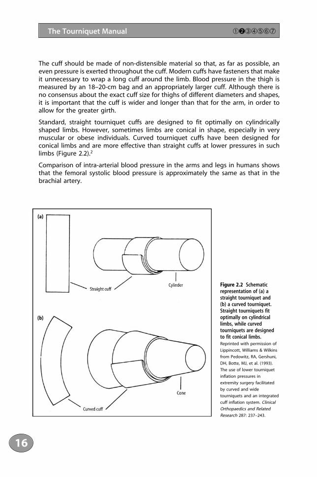
Standard, straight tourniquet cuffs are designed to fit optimally on cylindricallyshaped limbs. However, sometimes limbs are conical in shape, especially in verymuscular or obese individuals. Curved tourniquet cuffs have been designed forconical limbs and are more effective than straight cuffs at lower pressures in suchlimbs (Figure 2.2).2
Comparison of intra-arterial blood pressure in the arms and legs in humans showsthat the femoral systolic blood pressure is approximately the same as that in thebrachial artery.
1111234561178910111123111456789201111234567893011112345678940111211
16
The Tourniquet Manual ➀➋➂➃➄➅➆
Figure 2.2 Schematicrepresentation of (a) astraight tourniquet and (b) a curved tourniquet.Straight tourniquets fitoptimally on cylindricallimbs, while curvedtourniquets are designedto fit conical limbs.Reprinted with permission of
Lippincott, Williams & Wilkins
from Pedowitz, RA, Gershuni,
DH, Botte, MJ, et al. (1993).
The use of lower tourniquet
inflation pressures in
extremity surgery facilitated
by curved and wide
tourniquets and an integrated
cuff inflation system. Clinical
Orthopaedics and Related
Research 287: 237–243.

The principles for cuffs used as sphygmomanometers are also applicable to cuffsused as tourniquets, although allowances must be made for the situation in theoperating theatre. Fluctuations of blood pressure may occur due to operative trauma;for the upper limb, an additional pressure of 50–75 mm Hg above systolic pressure(limb occlusion pressure) should be sufficient to prevent bleeding at the operationsite. In the thigh, higher pressures are needed (Table 2.1): since the girth of the thighis large and there is a need to keep well clear of the field of operation, the cuffs arenarrower than those used to measure blood pressure. A simple rule of thumb in thethigh is to use double the systolic pressure in the arm.3 This does not apply to chil-dren, for whom lower pressures can be used.4
Measurements on five cadaveric lower limbs were made directly beneath an 8.5-cm Kidde tourniquet cuff to establish the relationship beneath the pressureexerted by the tourniquet and that transmitted to the underlying soft tissues. Thetissue pressure was consistently lower than the tourniquet pressure. The percentageof transmitted tourniquet pressure varied inversely with the circumference of thethigh.5 There is a tendency for the soft-tissue pressure beneath a tourniquet cuff todecrease with the depth of soft tissue. This is minimal but becomes more pronouncedas the circumference of the limb increases (Figure 2.3).
A tourniquet pressure of more than 300–350 mm Hg should rarely be required innormotensive individuals of normal habitus with compliant vessels. With the use ofwide cuffs, the limb circumference is a determining factor in the transmission ofpressure to the deep tissues. A cuff that is as wide as possible and is compatiblewith the surgical exposure should be used. Neimkin and Smith recommended usingtwo tourniquet cuffs inflated alternately at hourly intervals without a reperfusionperiod; this avoided prolonged compression under the cuff.6
In another trial, tourniquet cuffs with widths varying from 4.5 to 80 cm were appliedto the upper and lower extremities of 34 healthy, normotensive volunteers. Occlusionpressure was estimated by determining the level of cuff inflation at which the distalpulse became detectable by Doppler flow measurement. The occlusion pressure was
17
➀➋➂➃➄➅➆ Effect of the Tourniquet on the Limb
Table 2.1 Posterior tibial pressure measured using Doppler ultrasound and lower-calf tourniquet, and pressureof thigh tourniquet when Doppler signal has disappeared (occluding pressure). These measurements suggest thatthe commonly used pressure of 500 mm Hg (66.5 kPa) on the adult thigh is too high.Sex of patient Age (years) Pedal pressure (posterior Occluding pressure
tibial) (mm Hg)* in thigh (mm Hg)*
Right Left Right Left
Male 22 150 150 240 230Female 26 160 160 200 200Female 21 130 130 160 160Female 38 130 130 180 190Female 20 130 130 170 160Female 23 130 130 180 190Male 28 150 150 180 190Female 25 155 160 200 200Female 28 130 130 170 170
1 mm Hg ≈ 0.133 kPa.Reprinted with permission from Klenerman, L (1978). A modified tourniquet. Journal of the Royal Society of Medicine 71:
121–122.

inversely proportional to the ratio of tourniquet cuff width to limb circumference.It was in a subsystolic range at a ratio above 0.5. The manner in which arterial flowis impeded by a wide tourniquet inflated to subsystolic pressure is not known.Accumulation of frictional resistance along a segment of a blood vessel that ispartially collapsed under a low-pressure pneumatic tourniquet may completely eliminate flow without actual occlusion of the lumen region under the inflated cuff.7
Under the inflated cuff, there is a distribution of tissue from compressed to non-com-pressed zones, which includes mechanical deformation of all underlying tissuesincluding muscles and nerve. This deformation is greatest under the edges of the cuff.Thus, the pressure gradient that is greatest at the edge of the compressed segmentis the key factor in the occurrence of injuries to underlying tissues. Experimental stud-ies confirm that muscle and nerve exhibit the most severe injuries at the upper andlower edges of the tourniquet cuff.8 In experiments in which tourniquets were appliedto the thighs of rabbits, the proximal border of the tourniquet induced severe vas-cular damage contributing to a “no-reflow phenomenon” in the muscle.9
2.2 Sites of Application
It has generally been considered that it is safest to apply a tourniquet to the proxi-mal part of the limb, where the bulk of soft tissue provides the best protection for theunderlying nerves and vessels. In addition, it was thought that a tourniquet appliedto the forearm or calf might predispose to compartment syndrome. Recent reports
1111234561178910111123111456789201111234567893011112345678940111211
18
The Tourniquet Manual ➀➋➂➃➄➅➆
Figure 2.3 Mean soft-tissue pressure as a function of applied tourniquet pressure for thighs of different circum-ference. The vertical bars represent differences between the subcutaneous pressure and the pressure adjacent tothe bone. Values for a 46-cm thigh are not shown in order to increase the clarity of the graph and avoid over-lapping, but they were consistent with the pattern shown. Reproduced with permission from Shaw, JA, Murray, DG
(1982). The relationship between tourniquet pressure and underlying soft tissue pressure in the thigh. Journal of Bone and
Joint Surgery 64A: 1148–1151.

have shown that this is not correct. Yousif and colleagues concluded that patients tol-erate tourniquets on the upper arm and forearm equally well.10 Hutchinson andMcClintock found that in volunteers, tourniquets were tolerated for 44 minutes onthe forearm and for 31 minutes on the upper arm.11
In a randomised, prospective trial on the position of the tourniquet on either theupper arm or the forearm in patients with carpal-tunnel decompression, both groupsof patients tolerated the tourniquet equally well. However, the surgeons had somedifficulty with the tourniquet on the forearm, as the patient’s fingers may curl upand the tourniquet may be in the way when operating.12 The authors concludedthat there are very few indications for placing the tourniquet on the forearm in clin-ical practice.
A study of the use of a proximal calf tourniquet in 446 patients who had surgeryon the foot and ankle showed that there were no complications to nerves or vessels.13
The mean tourniquet time was 49.2 minutes for a single application and 131.1minutes if there were two periods of tourniquet ischaemia. A tourniquet applied tothe supramalleolar region of the leg has also been shown to be safe for surgery onthe foot. The pressure was 100–150 mm Hg above systolic pressure and did notexceed 325 mm Hg. There were no complications. An ankle tourniquet with aregional ankle block provides a reasonable alternative to the standard thigh tourni-quet for surgery of the foot.14 For forefoot surgery, it has been shown that patientswith an ankle tourniquet had significantly less pain during the operation thanpatients with a tourniquet on the calf, probably because of the smaller bulk of non-anaesthetised tissue.15 Calf tourniquets are thus suitable only for hindfoot surgery.
2.3 Effect on Muscle
With the tourniquet in place in both animal models and human studies, oxygentension and concentrations of creatine phosphate, glycogen and adenosine triphos-phate (ATP) in muscle cells decrease with time, whereas carbon dioxide tension andlactate concentration increase as anaerobic metabolism occurs. Intracellular pHremains constant for 15 minutes, followed by a linear decrease to 6.0 after four hoursof ischaemia. Intracellular creatine phosphate and ATP are depleted after two andthree hours of ischaemia, respectively.16
Following the release of a tourniquet after two to four hours of ischaemia, an increasein microvascular permeability in muscle and nerve (demonstrated by the extrava-sation of Evans blue dye in animal studies) occurs as a result of both directmicrovascular injury from compression and endothelial injury from superoxide radi-cals. Animal studies have also shown that after prolonged use of a tourniquet, thereis a marked decrease in the production of force in muscles beneath and distal tothe tourniquet (21–70% of control values).
Nevertheless, during tourniquet ischaemia, the muscle is at rest and the only expen-diture of energy is for basal metabolism. Thus, in spite of the circulatory arrest, thebiochemical changes are relatively slow.
19
➀➋➂➃➄➅➆ Effect of the Tourniquet on the Limb

2.3.1 Safe Period
The length of time that it is safe to leave a tourniquet in place on a healthy limbwithout causing irreversible damage to the skeletal muscle is of importance inorthopaedic practice. Present-day recommendations, based mainly on personalexperience, vary from one hour (the opinion of Bruner in 1951),17 to two hours(according to Boyes in 1964),18 with an upper limit of three hours (according toParkes in 1973).19 Compression and ischaemia are the factors most likely to contributeto the muscular damage. Changes in the muscle resulting from tourniquet-inducedischaemia have been studied from many aspects, including histological,20 histo-chemical,21 biochemical22 and ultrastructural.23–25 However, the effect of compressionon the muscle lying immediately under the tourniquet has received little attention.
2.3.2 Effects on the Ultrastructure of Muscle
Mammalian muscle is composed of three main types of fibre: fast-twitch white, fast-twitch red, and slow-twitch intermediate.25 The fast-twitch red and slow-twitchintermediate fibres rely primarily on oxidative metabolism and are more resistantto fatigue than the mainly glycolytic fast-twitch white fibres.
Using the soleus and the extensor digitorum longus, which between them containrepresentatives of all three types of fibre, the possibility of a differential response toischaemia was investigated in adult rhesus monkeys weighing 3.5–5 kg.26 Underanaesthesia, a Kidde tourniquet cuff of infant size was applied to the upper thigh ofthe right lower limb for periods lasting from one to five hours at a pressure of 300mm Hg. Immediately before the release of the tourniquet, samples from the soleusand the extensor digitorum longus were removed for biopsy and processed for elec-tron microscopy. Samples from the muscle lying under the tourniquet, the quadri-ceps, were taken after removal of the tourniquet.
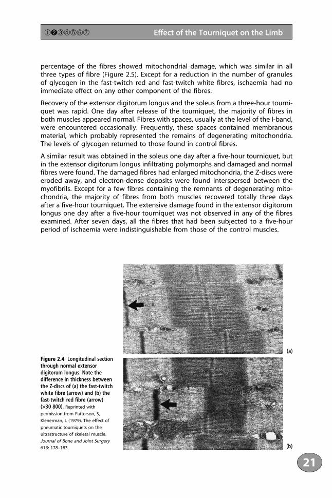
Recovery from three-hour and five-hour tourniquets was investigated. Samples fromthe quadriceps, extensor digitorum longus and soleus were taken one day, two orthree days, and seven days after release of the tourniquet. Only one sample wasremoved from any one particular muscle since, in trial experiments, repeatedsampling was found to cause marked damage to the fibres. Samples from the oppo-site limb were used as controls. The specimens were examined using electronmicroscopy (Figure 2.4).
2.3.3 Effects of Ischaemia on Muscles Distal to the Tourniquet
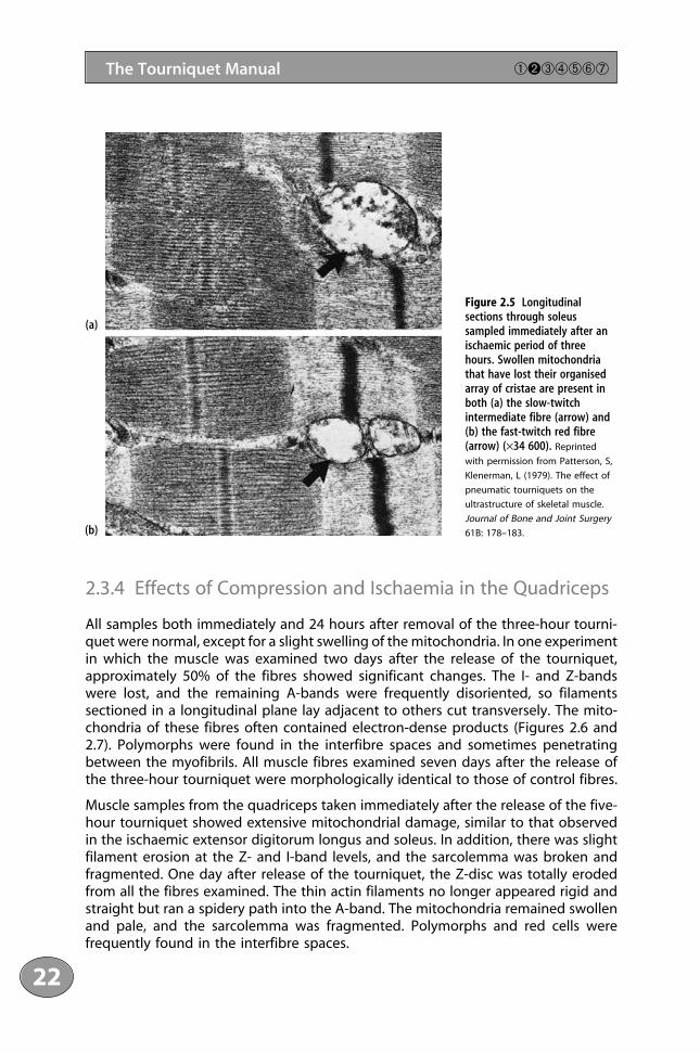
In the experiment described in the previous section, after one hour of ischaemiamarked changes in mitochondrial morphology were observed in the fibres of boththe extensor digitorum longus and the soleus. The fibres had become swollen andless electron-dense, and they had lost their organised network of cristae, althoughmany fibres displaying normal mitochondrial morphology were still present. As theperiod of ischaemia was increased progressively up to five hours, a greater
1111234561178910111123111456789201111234567893011112345678940111211
20
The Tourniquet Manual ➀➋➂➃➄➅➆
percentage of the fibres showed mitochondrial damage, which was similar in allthree types of fibre (Figure 2.5). Except for a reduction in the number of granulesof glycogen in the fast-twitch red and fast-twitch white fibres, ischaemia had noimmediate effect on any other component of the fibres.
Recovery of the extensor digitorum longus and the soleus from a three-hour tourni-quet was rapid. One day after release of the tourniquet, the majority of fibres inboth muscles appeared normal. Fibres with spaces, usually at the level of the I-band,were encountered occasionally. Frequently, these spaces contained membranousmaterial, which probably represented the remains of degenerating mitochondria.The levels of glycogen returned to those found in control fibres.
A similar result was obtained in the soleus one day after a five-hour tourniquet, butin the extensor digitorum longus infiltrating polymorphs and damaged and normalfibres were found. The damaged fibres had enlarged mitochondria, the Z-discs wereeroded away, and electron-dense deposits were found interspersed between themyofibrils. Except for a few fibres containing the remnants of degenerating mito-chondria, the majority of fibres from both muscles recovered totally three days after a five-hour tourniquet. The extensive damage found in the extensor digitorumlongus one day after a five-hour tourniquet was not observed in any of the fibresexamined. After seven days, all the fibres that had been subjected to a five-hourperiod of ischaemia were indistinguishable from those of the control muscles.
21
➀➋➂➃➄➅➆ Effect of the Tourniquet on the Limb
Figure 2.4 Longitudinal sectionthrough normal extensordigitorum longus. Note thedifference in thickness betweenthe Z-discs of (a) the fast-twitchwhite fibre (arrow) and (b) thefast-twitch red fibre (arrow)(×30 800). Reprinted with
permission from Patterson, S,
Klenerman, L (1979). The effect of
pneumatic tourniquets on the
ultrastructure of skeletal muscle.
Journal of Bone and Joint Surgery
61B: 178–183.
(a)
(b)
2.3.4 Effects of Compression and Ischaemia in the Quadriceps
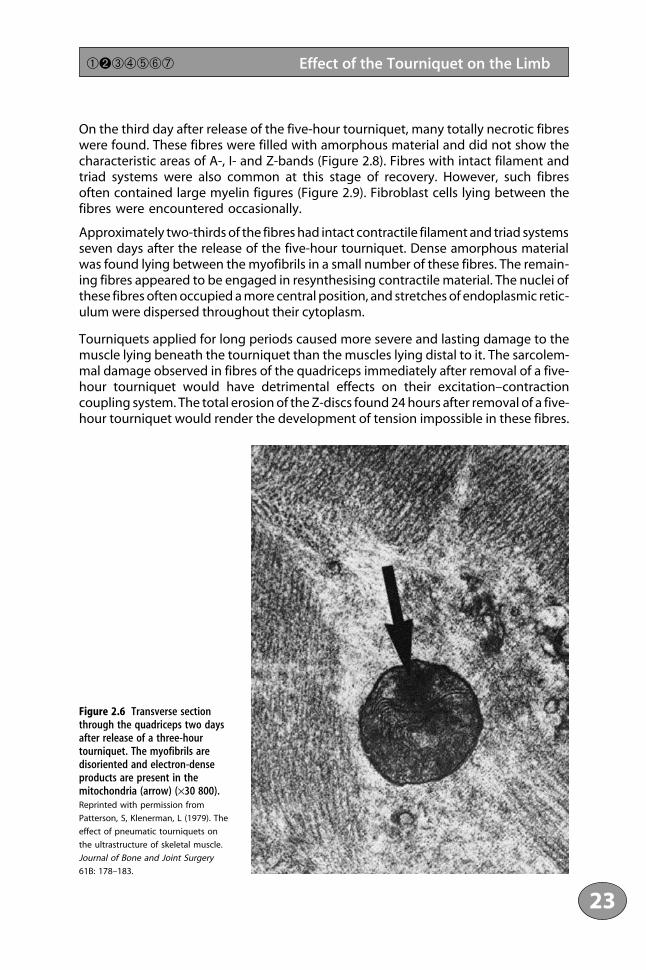
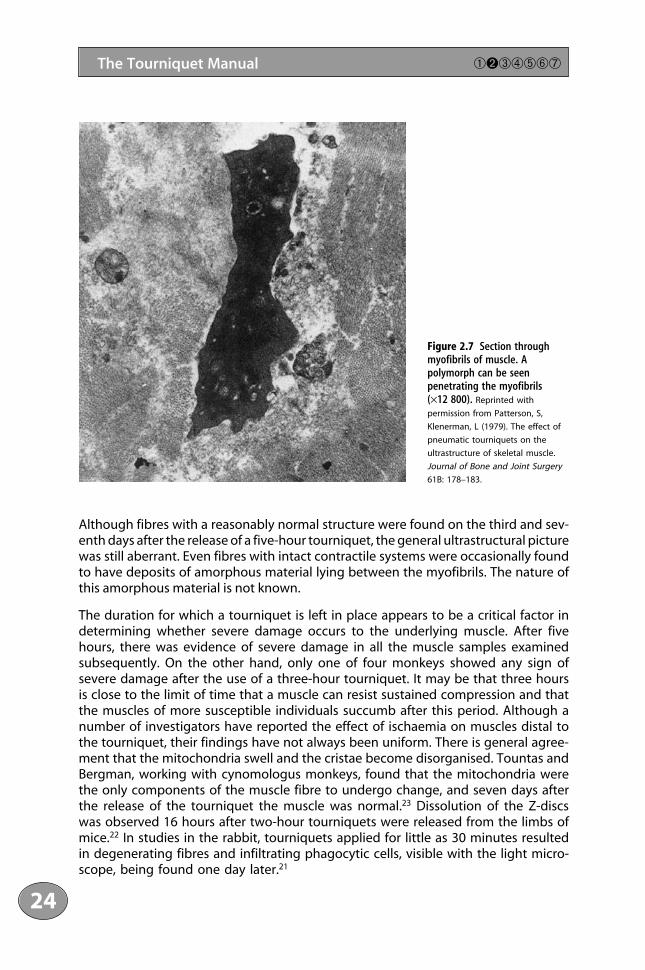
All samples both immediately and 24 hours after removal of the three-hour tourni-quet were normal, except for a slight swelling of the mitochondria. In one experimentin which the muscle was examined two days after the release of the tourniquet,approximately 50% of the fibres showed significant changes. The I- and Z-bandswere lost, and the remaining A-bands were frequently disoriented, so filamentssectioned in a longitudinal plane lay adjacent to others cut transversely. The mito-chondria of these fibres often contained electron-dense products (Figures 2.6 and2.7). Polymorphs were found in the interfibre spaces and sometimes penetratingbetween the myofibrils. All muscle fibres examined seven days after the release ofthe three-hour tourniquet were morphologically identical to those of control fibres.
Muscle samples from the quadriceps taken immediately after the release of the five-hour tourniquet showed extensive mitochondrial damage, similar to that observedin the ischaemic extensor digitorum longus and soleus. In addition, there was slightfilament erosion at the Z- and I-band levels, and the sarcolemma was broken andfragmented. One day after release of the tourniquet, the Z-disc was totally erodedfrom all the fibres examined. The thin actin filaments no longer appeared rigid andstraight but ran a spidery path into the A-band. The mitochondria remained swollenand pale, and the sarcolemma was fragmented. Polymorphs and red cells werefrequently found in the interfibre spaces.
1111234561178910111123111456789201111234567893011112345678940111211
22
The Tourniquet Manual ➀➋➂➃➄➅➆
Figure 2.5 Longitudinalsections through soleussampled immediately after anischaemic period of threehours. Swollen mitochondriathat have lost their organisedarray of cristae are present inboth (a) the slow-twitchintermediate fibre (arrow) and(b) the fast-twitch red fibre(arrow) (×34 600). Reprinted
with permission from Patterson, S,
Klenerman, L (1979). The effect of
pneumatic tourniquets on the
ultrastructure of skeletal muscle.
Journal of Bone and Joint Surgery
61B: 178–183.
(a)
(b)
On the third day after release of the five-hour tourniquet, many totally necrotic fibreswere found. These fibres were filled with amorphous material and did not show thecharacteristic areas of A-, I- and Z-bands (Figure 2.8). Fibres with intact filament andtriad systems were also common at this stage of recovery. However, such fibresoften contained large myelin figures (Figure 2.9). Fibroblast cells lying between thefibres were encountered occasionally.
Approximately two-thirds of the fibres had intact contractile filament and triad systemsseven days after the release of the five-hour tourniquet. Dense amorphous materialwas found lying between the myofibrils in a small number of these fibres. The remain-ing fibres appeared to be engaged in resynthesising contractile material. The nuclei ofthese fibres often occupied a more central position, and stretches of endoplasmic retic-ulum were dispersed throughout their cytoplasm.
Tourniquets applied for long periods caused more severe and lasting damage to themuscle lying beneath the tourniquet than the muscles lying distal to it. The sarcolem-mal damage observed in fibres of the quadriceps immediately after removal of a five-hour tourniquet would have detrimental effects on their excitation–contractioncoupling system. The total erosion of the Z-discs found 24 hours after removal of a five-hour tourniquet would render the development of tension impossible in these fibres.
23
➀➋➂➃➄➅➆ Effect of the Tourniquet on the Limb
Figure 2.6 Transverse sectionthrough the quadriceps two daysafter release of a three-hourtourniquet. The myofibrils aredisoriented and electron-denseproducts are present in the mitochondria (arrow) (×30 800).Reprinted with permission from
Patterson, S, Klenerman, L (1979). The
effect of pneumatic tourniquets on
the ultrastructure of skeletal muscle.
Journal of Bone and Joint Surgery
61B: 178–183.
Although fibres with a reasonably normal structure were found on the third and sev-enth days after the release of a five-hour tourniquet, the general ultrastructural picturewas still aberrant. Even fibres with intact contractile systems were occasionally foundto have deposits of amorphous material lying between the myofibrils. The nature ofthis amorphous material is not known.
The duration for which a tourniquet is left in place appears to be a critical factor indetermining whether severe damage occurs to the underlying muscle. After fivehours, there was evidence of severe damage in all the muscle samples examinedsubsequently. On the other hand, only one of four monkeys showed any sign ofsevere damage after the use of a three-hour tourniquet. It may be that three hoursis close to the limit of time that a muscle can resist sustained compression and thatthe muscles of more susceptible individuals succumb after this period. Although anumber of investigators have reported the effect of ischaemia on muscles distal tothe tourniquet, their findings have not always been uniform. There is general agree-ment that the mitochondria swell and the cristae become disorganised. Tountas andBergman, working with cynomologus monkeys, found that the mitochondria werethe only components of the muscle fibre to undergo change, and seven days afterthe release of the tourniquet the muscle was normal.23 Dissolution of the Z-discswas observed 16 hours after two-hour tourniquets were released from the limbs ofmice.22 In studies in the rabbit, tourniquets applied for little as 30 minutes resultedin degenerating fibres and infiltrating phagocytic cells, visible with the light micro-scope, being found one day later.21
1111234561178910111123111456789201111234567893011112345678940111211
24
The Tourniquet Manual ➀➋➂➃➄➅➆
Figure 2.7 Section throughmyofibrils of muscle. Apolymorph can be seenpenetrating the myofibrils (×12 800). Reprinted with
permission from Patterson, S,
Klenerman, L (1979). The effect of
pneumatic tourniquets on the
ultrastructure of skeletal muscle.
Journal of Bone and Joint Surgery
61B: 178–183.

25
➀➋➂➃➄➅➆ Effect of the Tourniquet on the Limb
Figure 2.8 Transverse section of the quadriceps three days after the release of a five-hour tourniquet. This fibrehas totally lost its characteristic areas of A-, I- and Z-bands (×30 800). Reprinted with permission from Patterson,
S, Klenerman, L (1979). The effect of pneumatic tourniquets on the ultrastructure of skeletal muscle. Journal of Bone and Joint
Surgery 61B: 178–183.
Figure 2.9 Longitudinal section through the quadriceps three days after the release of a five-hour tourniquet.The contractile filament system is intact but large myelin figures are present (arrows) (×9600). Reprinted with
permission from Patterson, S, Klenerman, L (1979). The effect of pneumatic tourniquets on the ultrastructure of skeletal muscle.
Journal of Bone and Joint Surgery 61B: 178–183.

In the experiments with rhesus monkeys, the mitochondria were the only compo-nents of the extensor digitorum longus and soleus to show any significant degreeof damage after a three-hour tourniquet. These organelles seem to be very sensi-tive to ischaemia, since changes were observed after only one hour. In studies onischaemic heart muscle, mitochondrial damage was observed after as little as 12minutes.27 The soleus and extensor digitorum longus of the rhesus monkeys showeda remarkable ability to regenerate their mitochondria. Mitochondrial structure wasnormal three days after a three-hour period of ischaemia and seven days after fivehours of ischaemia. It was interesting to find normal fibres interspersed with othersshowing mitochondrial damage. Moore and colleagues22 reported similar observa-tions in ischaemic muscle in mice.
Resistance or susceptibility of the mitochondria of muscle fibre to ischaemia doesnot appear to be related to the type of fibre, since similar changes were observedin all three types. The metabolism of fast-twitch red and slow-twitch intermediatefibres is primarily oxidative, while the fast-twitch white fibres are mainly glycolytic.Physiologically, this may be manifested in the former two types giving earlier fatiguetimes.
The more severe effects of ischaemia observed by some workers may be related tothe species of animal studied. In the rhesus monkey, a three-hour tourniquet didnot result in damage to the contractile filaments. However, in one monkey subjectedto a five-hour tourniquet, infiltrating cells and eroded Z-discs were found in theextensor digitorum longus one day after tourniquet release. This may indicate thatfive hours of ischaemia is close to the limit of time that the muscle can endureischaemia without becoming severely damaged.
Histologically, the contractile machinery of the muscle fibres of the rhesus monkeyappeared to be wholly unaffected by periods of ischaemia lasting up to three hours.However, from the functional aspect, this was not so. Tests for strength in cynomo-logus monkeys after three- or five-hour tourniquets using infant-size Kidde cuffswere carried out.28 Development of maximum isometric tension, contraction timesand half-relaxation times were measured in the muscles beneath and distal to thetourniquet. On release of the tourniquet, no consistent difference between controland experimental muscles was observed with respect to contraction and half-relax-ation times; however, there was a marked reduction in the development of isometrictension. On the sixth day after release of a five-hour tourniquet, isometric tensionwas reduced to 2–22% of the control value of the compressed muscle. Six days aftera three-hour tourniquet, isometric tension of the compressed muscle was 80% ofcontrol value; in the distal gastrocnemius–soleus complex, tension varied from 64%to control value. It is clear that the effect on muscle contraction after a three-hourperiod of ischaemia is not reversed immediately by restoration of the blood supply.It is surprising that the reduction in isometric tension was greater in the gastroc-nemius–soleus complex than in the compressed quadriceps muscle. A possibleexplanation for this is the morphology of the muscle. The fibres do not all run thewhole length of the quadriceps as the rectus femoris is bipennate. As the tourni-quet cuff was in the middle of the thigh, it is possible that this resulted in a mixedeffect on contractile power.
1111234561178910111123111456789201111234567893011112345678940111211
26
The Tourniquet Manual ➀➋➂➃➄➅➆

Similar experiments on New Zealand white rabbits showed that tourniquet com-pression for two hours resulted in markedly reduced force production beneath andalso distal to the tourniquet cuff. Two days after compression, maximal quadricepsforce production was reduced to 40% of control values with 125 mm Hg compres-sion and to 21% of control values after 350 mm Hg compression. The maximal forceproduction of the tibialis anterior declined to 70% and 24%, respectively.29 Althoughtissues distal to the tourniquet were usually accepted as affected by ischaemia alone,these workers considered that proximal compression of the nerve supply to the tibi-alis anterior beneath the tourniquet had interfered with nerve function.
In summary, the experiments on rhesus monkeys have shown more marked changesin the muscles beneath the cuff than those distal to the cuff. Three hours seems tobe close to the time limit for the safe application of a tourniquet because of thecombined effects of compression and ischaemia on the muscle beneath the cuff.
2.3.5 No-reflow Phenomenon
Attempts at reperfusion may not always be successful because of progressive micro-circulatory obstruction. This process, called the “no-reflow phenomenon”, is relatedto the length of the ischaemic interval. Muscles that have been ischaemic for oneto three hours are easily reperfused, but the no-reflow phenomenon occurs in40–50% of cases after five hours of ischaemia.30 The exact cause is not understoodfully. Microscopy has shown large numbers of red cells in the microcirculation.Cellular oedema may also lead to capillary plugging during reperfusion.31
2.3.6 Effect on the Function of the Underlying QuadricepsMuscle
It has been assumed that movement of the muscle is restricted by an inflated tourni-quet. An investigation was carried out in five healthy male volunteers using ultra-sound to measure the movement of the quadriceps muscle above and below thetourniquet, before and after inflation.32 A tourniquet of standard size was applied tothe thigh for five minutes. A bubble of air was injected into the muscle above thetourniquet and was the proximal point of reference. The musculotendinous junctionwas the distal point. The movement of the reference point was measured by ultra-sound before and after each inflation of the tourniquet. Each measurement wasrepeated with either the knee flexed and the hip extended, or the hip flexed and theknee extended. The ultrasound findings consistently showed no evidence of restric-tion of the quadriceps muscle by an inflated tourniquet. The authors recommend thatthe tourniquet be inflated in the most convenient position for the surgeon.
2.4 Compression of Nerves
According to Lundborg, inflation of a cuff to suprasystolic pressure around the arm results in a conduction block, rapidly reversible upon the release of the cuff.9
27
➀➋➂➃➄➅➆ Effect of the Tourniquet on the Limb

Pressure just sufficient to occlude the underlying blood vessels results in a block of nerve conduction in 15–45 minutes. At a cuff pressure of 150 mm Hg, sensoryloss and paralysis develop at the same rate as when a pressure of 300 mm Hg isused. This indicates that ischaemia rather than mechanical pressure is the under-lying cause of such conduction block, which is rapidly reversible and physiological.When the cuff is inflated to a higher pressure, there is a risk of mechanical damageto the nerve fibres, resulting in a longer-lasting conduction block – a local demy-elinating block, which has been called “tourniquet paralysis”.33, 34 The underlyingforce seems to be the pressure gradient within the nerve between its compressedand uncompressed portions, the displacements being away from the region of high pressure towards the uncompressed region beyond the edge of the cuff tourniquet.
The biological basis of localised conduction blocks induced by direct pressure hasbeen analysed extensively in a series of experimental studies.33–35 These experimentswere carried out on baboons, with a tourniquet cuff pressure of about 1000 mm Hgfor 90–180 minutes. When single teased fibres were examined within a few hours ordays, they showed a specific morphological phenomenon: under each border zoneof the compressed segment, the nodes of Ranvier had been displaced along eachfibre, so that the paranodal myelin was stretched on one side of the node and invagi-nated on the other. The whole picture is strongly reminiscent of an intussusception,as it occurs in the bowel. The underlying force seemed to be the pressure gradientwithin the nerve between its compressed and uncompressed portions. In each case,the displacement was away from the region of high pressure towards the uncom-pressed region beyond the edge of the cuff (Figure 2.10).
The result was localised degenerative changes of the damaged myelin (paranodaldemyelination). Only large myelinated fibres were affected. In these experiments, a cuff pressure of 1000 mm Hg maintained for one to three hours produced paral-ysis of distal muscles lasting for up to three months. There was a significantcorrelation between the duration of compression and the duration of the sub-sequent conduction block. The effects of the block correspond with the type of
1111234561178910111123111456789201111234567893011112345678940111211
28
The Tourniquet Manual ➀➋➂➃➄➅➆
Figure 2.10 Diagram to show the direction of displacement of nodes of Ranvier in relation to the cuff. Reprinted
with permission from Ochoa, J, Fowler, TJ, Gilliatt, RW (1972). Anatomical changes in peripheral nerves compressed by a pneu-
matic tourniquet. Journal of Anatomy 113: 433–455.

nerve injury classified by Seddon in 1943 as neurapraxia.36 Gilliatt in 1980 showedby direct recordings from the exposed nerve “a double conduction block” affectingthe large myelinated fibres as two separate regions of the nerve trunk correspondingin position to both edges of the cuff, while the intermediate region showed littleor no change in conduction.37
2.5 Effects on the Skin
On the whole, the skin is resilient and unaffected in the vast majority of cases oftourniquet use. Damage at the site of the tourniquet may be caused by pressurenecrosis or friction burns. Such burns are thought to be caused by spirit-based anti-septic solutions that seep beneath the tourniquet and are held against the skinunder pressure (see Chapter 5).38
Friction burns may result during operations on the thigh due to a fully inflatedtourniquet cuff slipping down and away from the plaster wool padding.39 An inves-tigation on the effects produced by commonly used antiseptic paints and a knownchemical irritant, anthralin, was carried out on the upper arms and forearms of volun-teers.40 Site-related variations in anthralin-induced inflammation were observed, butthere was no demonstrable effect of either pressure or ischaemia on the inflam-matory response. It was not possible to keep the tourniquets in place for longerthan half an hour because it would have been too painful for the volunteers totolerate the pain of ischaemia. It was concluded that burns under tourniquets arelikely to be idiosyncratic reactions, and their further investigation required detailedexamination of individuals affected by chemical burns.
2.6 Systemic and Local Effects of the Application of a Tourniquet
There have been few reports describing the systemic effects of reperfusing theischaemic limb.41, 42 Complete arrest of the circulation to the limb produces acidosisand changes in levels of potassium,43, 44 which in theory could result in effects onthe rhythm of the heart when the tourniquet is released. Although changes in theacid–base status of the blood leaving the limb have been described, the state ofthe blood reaching the heart after the release of a tourniquet has received littleattention.45 An animal and clinical study was undertaken to establish whether anybiochemical changes in the limb are reflected in the right atrium. In addition, thetime taken for the ischaemic limb to recover was investigated.46
2.6.1 Animal Experiments
An infant-size Kidde tourniquet cuff 5 cm wide was applied to the experimental limbof a rhesus monkey and inflated to a pressure of 300 mm Hg for a predetermined
29
➀➋➂➃➄➅➆ Effect of the Tourniquet on the Limb
time from one to five hours. At regular intervals during the period when the tourni-quet was in place, samples were taken over a period of one minute from the cannulain the right atrium to establish control values for acid–base status and potassiumlevels. After the release of the tourniquet, further samples were taken simultane-ously from both the internal jugular route and the femoral vein for periods as longas two hours.
Whenever possible, all samples were measured immediately for pCO2, pH, excess ofbase, and standard bicarbonate. If this was not possible, samples were stored in icefor no longer than 30 minutes.
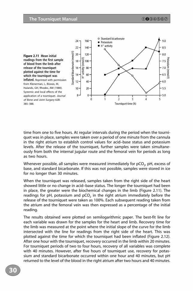
When the tourniquet was released, samples taken from the right side of the heartshowed little or no change in acid–base status. The longer the tourniquet had beenin place, the greater were the biochemical changes in the limb (Figure 2.11). Thereadings for pH, potassium and pCO2 in the right atrium immediately before therelease of the tourniquet were taken as 100%. Each subsequent reading taken fromthe atrium and the femoral vein was then expressed as a percentage of the initialreading.
The results obtained were plotted on semilogarithmic paper. The best-fit line foreach variable was drawn for the samples for the heart and limb. Recovery time forthe limb was measured at the point where the initial slope of the curve for the limbintersected with the line for readings from the right side of the heart. This wasplotted against the time for which the tourniquet had been inflated (Figure 2.12).After one hour with the tourniquet, recovery occurred in the limb within 20 minutes.For tourniquet periods of two to four hours, recovery of all variables was completewith 40 minutes. However, after five hours of tourniquet use, recovery for potas-sium and standard bicarbonate occurred within one hour and 40 minutes, but pHreturned to the level of the blood in the right atrium after two hours and 40 minutes.
1111234561178910111123111456789201111234567893011112345678940111211
30
The Tourniquet Manual ➀➋➂➃➄➅➆
Figure 2.11 Mean initialreadings from the first sampleof blood from the limb afterrelease of the tourniquetplotted against the time forwhich the tourniquet wasinflated. Reprinted with permission
from Klenerman, L, Biswas, M,
Hulands, GH, Rhodes, AM (1980).
Systemic and local effects of the
application of a tourniquet. Journal
of Bone and Joint Surgery 62B:
385–388.

2.6.2 Clinical Studies
Patients who were about to undergo total knee replacement or a high tibialosteotomy for rheumatoid arthritis or osteoarthritis were informed of the studies andconsented to participate. All patients received appropriate premedication ofpapaveretum and atropine. Anaesthesia was induced with thiopentone, an intra-venous injection of pancuronium was given, and intubation was carried out.Anaesthesia was maintained with nitrous oxide, oxygen and phenoperidine, andoccasionally halothane (less than 0.5%). Ventilation was adjusted for a standardpaCO2 of 5.4 kPa. A cannula was passed via the right internal jugular vein into theatrium and its position checked by looking for atrial oscillations; 5% dextrose solutionwas infused. An intravenous drip of Hartmann’s solution was set up in one forearm.The electrocardiogram was displayed continuously, and the temperature was moni-tored by a nasopharyngeal probe. An Esmarch bandage was used to exsanguinatethe site of operation, and a 10-cm Kidde tourniquet cuff was inflated to occlude thearterial flow at a pressure of twice the pre-induction systolic pressure. During theoperation, several samples were taken from the internal jugular cannula to establishbaseline values for blood analysis from the central venous pool. At the end of theoperation, pressure dressings were applied to the limb while the tourniquet was still inflated. Samples of blood were taken from the atrium via the internal jugular cannula and also from the femoral vein of the operated limb by direct needle stabjust before releasing the tourniquet. When the tourniquet was released, samples were taken simultaneously from the femoral needle and the internal jugular cannulafor a period of approximately 15 minutes and then intermittently from the jugularcannula for approximately two hours. These samples were analysed as describedabove.
There were nine patients (three men, six women), of average age 68 years (range51–80 years). The tourniquet was inflated for periods ranging from 70 to 186 minutes.
31
➀➋➂➃➄➅➆ Effect of the Tourniquet on the Limb
Figure 2.12 Estimated recoverytime for each variable in theblood supply in the limbsubjected to ischaemia inrelation to the time for whichthe tourniquet was used.Reprinted with permission from
Klenerman, L, Biswas, M, Hulands,
GH, Rhodes, AM (1980). Systemic
and local effects of the application
of a tourniquet. Journal of Bone
and Joint Surgery 62B: 385–388.

2.6.3 Results of Investigations
There were only minor fluctuations in the three variables – potassium, bicarbonateand pH – in the samples taken from the right atrium. These transiently reflected themarked changes that occurred in the blood from the limb. No cardiac dysrhythmiaswere detected on monitoring.
Neither the patients nor the experimental animals showed evidence of nerve palsies.
In a limb that has been rendered ischaemic, metabolites accumulate as a result ofhypoxia in the tissues. Theoretically, a rapid influx of some of these products, e.g.potassium, into the coronary circulation is likely to produce cardiac dysfunction. Inthese studies, although the potassium levels in the blood leaving the limb wereraised, at no time was a significant rise detected in the right atrium either in theanimals or in the patients. The most likely explanation for this is a dilutional effectdue to the larger volume of blood contained in the venous side of the circulation(50% of the circulating blood volume is accommodated on the venous side, butonly 15% is in the arterial system). Similarly, the fall in pH in the venous blood leavingthe acidotic limb was not reflected in the acid–base status of the blood samplesfrom the right atrium. Again, the effect of dilution is a factor here, but in additionthere is the efficient buffering capacity of the blood. A criticism of the samplingtechnique used could be based on the well-known streaming effect of blood fromthe venae cavae. This is well documented in relation to the measurement of venousoxygen in estimations of cardiac output. However, the authors were not aware ofwork showing that this effect was also applicable to other biochemical measure-ments. Although streaming within the atrium cannot be discounted, it is unlikely tobe an important factor as the results were consistent. These findings are essentiallyin agreement with those described in patients undergoing operations under tourni-quet with lumbar epidural anaesthesia.45
In the animal studies, it was found that the acid–base balance in the limb returnedto normal within 20 minutes of the release of a tourniquet that had been in placefor one hour, and within 40 minutes after four hours of ischaemia. The practice ofreleasing the tourniquet at two hours for a period of five to ten minutes to allow a“breathing period” therefore does not seem appropriate.
The investigations that have been described were undertaken in healthy animalsand fit patients who did not suffer from cardiovascular disease. When, as is notuncommon, the buffering capacity is reduced by anaemia, hypovolaemia, metabolicacidosis or pre-existing vascular disease, there is likely to be a reduction in the normal range of safety. In addition, under certain conditions a compromisedmyocardium may be sensitised to catecholamines by anaesthetic agents. In thesecircumstances, the period for which a tourniquet is used should be reduced to the minimum and full cardiovascular monitoring must be available. The changesnoted in the acid–base balance indicate that a period of three hours under a tourniquet is safe. This coincides with findings made in histological studies of theischaemic muscle.26
1111234561178910111123111456789201111234567893011112345678940111211
32
The Tourniquet Manual ➀➋➂➃➄➅➆

2.7 Haemodynamic Changes
The haemodynamic changes associated with the application and release of a tourni-quet are minimal in healthy adults, but they may not be tolerated by patients withpoor cardiac reserve. In a series of patients who were monitored for changes incentral venous pressure (CVP) and systolic blood pressure, it was found that themain rise in CVP with the application of bilateral tourniquets was 14.5 cm H2O.47 Thiswas maintained in 80% of patients until the tourniquets were released (Figure 2.13).It is likely that this was due to an increase of approximately 15% of circulating bloodvolume – about 700–800 ml of blood. In comparison, the CVP values when singletourniquets were applied showed that the circulation could deal with the smallerautotransfusion of blood more easily. The mean systolic pressure change was ±18.5mm Hg when the tourniquets were inflated. On deflation, the mean fall below theblood pressure at the start of surgery was 43.5 mm Hg. The initial rise in blood pres-sure either was sustained or fell gradually to the level before a tourniquet wasapplied, and then had a further dramatic fall within three minutes of release of thetourniquet. In a review of the records of 500 patients who had surgery under atourniquet, the frequency of intraoperative hypertension (defined as a 30% increasein either systolic or diastolic pressure compared with the first pressure recordingafter incision) was 11%. The probability of hypertension was increased if the patientwas elderly, had cardiac enlargement as shown by X-ray or electrocardiogram (ECG),or had nitrous oxide and narcotic anaesthesia. Pre-existing hypertension, increasedserum creatinine concentration, anaemia, or treatment with hypertensive drugs werenot associated strongly with intraoperative hypertension.48 Patients with headinjuries and multiple sites of trauma may have marked increases in intracranial pres-sure when lower limb tourniquets are released.49
Using transoesophageal echocardiography during 59 total knee replacements, it was found that showers of echogenic material traversed the right atrium, rightventricle and pulmonary artery after the tourniquets were deflated.50 This wasobserved in various degrees in all patients and lasted for 3–15 minutes. The meanpeak intensity occurred within 30 seconds (range 24–45 seconds) after the tourni-quet was released. Only three patients had evidence of clinical pulmonary embolism.These findings are similar to those described by Parmet and colleagues in a smallerseries of 29 patients.51 This group aspirated a 3 × 6-mm fresh thrombus from a centralcatheter in one patient. Another patient, who had a Greenfield filter in the inferiorvena cava to prevent emboli reaching the lungs from the legs following previousthromboembolism, showed very little echogenic material, indicating that the filter acted as an effective block. Inadequate exsanguination of the limb under-going surgery coupled with stasis and cooling may contribute to fresh thrombusformation. Nevertheless, these 29 patients had echogenic material with clinicallyadequate exsanguination. Bone cement activation of the coagulation cascade couldalso form fresh clot. It is likely that the pulmonary circulation is often exposed toembolic material during normal everyday life and that the lungs are able to clearsmall emboli.
33
➀➋➂➃➄➅➆ Effect of the Tourniquet on the Limb

2.8 Limb Blood Flow in the Presence of aTourniquet
The blood supply to the limbs of rhesus monkeys was studied with 50-� diametermicrospheres labelled with 51Cr and by the washout of 22Na injected into the tissues.One limb, upper or lower, was exsanguinated and the circulation was occluded witha pneumatic tourniquet. The opposite limb was used as a control. The blood to theoccluded limb was found to be less than 1% of the flow to the control limb. Thevenous return was less than 0.2% of that of the control limb. It was concluded that alimb with a tourniquet in place is virtually isolated from the circulation and theamount of blood reaching the tissues probably via the intramedullary circulation islikely to be of no significance to relieve the ischaemia.52 Added support for the isola-tion of the limb from normal blood flow is provided by the work of Santavirta andcolleagues, who studied tissue oxygen levels in rabbits.53 The tourniquet was in placefor 60, 80 or 120 minutes. The baseline PO2 in the tibialis anterior muscle was 22.6±0.6mm Hg. While the tourniquet was in place, the oxygen tension dropped to minimalvalues between 9.2±0.5 and 10.7±0.6 mm Hg in the three groups rendered ischaemicfor 60, 80 and 120 minutes, but the tissue microclimate never reached fully anoxicconditions. This minimal value was reached in 19–26 minutes and then remained con-stant during the remainder of the time that the tourniquet was in place, but it neverreached zero. The decline of PO2 and recovery after release of the tourniquet wasindependent of tourniquet time. Continuous oxygen during the experiment had noinfluence on the PO2.
1111234561178910111123111456789201111234567893011112345678940111211
34
The Tourniquet Manual ➀➋➂➃➄➅➆
Figure 2.13 Changes incentral venous pressure andblood pressure with atourniquet in place andafter release. Reproduced
with permission from Bradford,
EMW (1968). Haemodynamic
changes associated with the
application of lower limb
tourniquets. Anaesthesia 24:
190–197.

2.9 Hyperaemia and Swelling of a Limb AfterRelease of a Tourniquet
Using monkeys, a quantitative study was carried out to measure the effect of a tourniquet on the lower limb on peak flow, the amount of swelling, and the time for recovery. The disappearance of acute swelling is related to the period ofischaemia. As the duration of the tourniquet increased, no significant change in peak flow was demonstrated. The swelling that results from a tourniquet for one hour is overcome rapidly, but the effects are much more obvious for tourniquet times of two and three hours. When attempting to obtain haemostasis after releaseof a tourniquet, surgeons should remember that for a one-hour period of ischaemia,the hyperaemia falls to one-half in about five minutes, but that it takes 12 and 25 minutes, respectively, for this to take place after two and three hours of tourniquetuse.54 These times are of relevance to breathing periods. The onset of hyperaemia isrelated to the changes brought about by the effects of free oxygen radicals (seeChapter 3).
2.10 Haematological Effects
At the end of orthopaedic operations, there is a pronounced increase in fibrinolyticactivity in the blood from the systemic circulation, as well as from the operated limb,whereas there is only a small systemic increase after surgery on the leg without atourniquet. The vasa vasorum are probably the main source of plasminogen acti-vator in the vasculature and may be stimulated to respond maximally by completeischaemia; the increase in fibrinolytic activity does not appear to be related to theduration of the application of a tourniquet.55 However, there is no difference in theincidence of deep vein thrombosis in surgery on the lower limbs with and withouta tourniquet.56 The increase in fibrinolytic activity is short-lived; it is maximal at 15minutes and returns to preoperative levels within 30 minutes of the release of thetourniquet. It then falls below the preoperative levels, where it remains for at least48 hours. The tourniquet appears to alter the timing of a short period of increasedfibrinolytic activity without altering the overall pattern. It is unlikely that this wouldalter the incidence of deep vein thrombosis, but it may affect the degree of bleedingafter release of the tourniquet.57
2.11 Temperature Changes
An increased core body temperature occurs during the application of arterial tourni-quets, probably because of reduced metabolic heat transfer from the central to theperipheral compartments and from decreased heat loss from distal skin. When thetourniquet is released, there is a transient decrease in core temperature as a result ofredistribution of body heat from the return of hypothermic venous blood flow from
35
➀➋➂➃➄➅➆ Effect of the Tourniquet on the Limb
the tourniquet limb into the systemic circulation.58 A marked rise in temperature maycause the anaesthetist concern about the possibility of malignant hyperthermia. Anassociation between the use of tourniquets for limb surgery and a progressiveincrease in body temperature of greater than one degree with bilateral tourniquetshas been reported in children.59
With a tourniquet in place, the limb cools gradually; during the course of an oper-ation, the temperature may drop by 3–4 °C. Part of the cooling is counterbalancedby the effects of the lights and drapes in the operation theatre. There may be obviousdrying out of the issues exposed, which should always be kept moist with Hartman’ssolution or normal saline.
2.12 Tourniquet Pain
When a tourniquet is applied to the arms of volunteers, they experience a vague,dull pain in the limb, which is associated with an increase in blood pressure. Theaverage pain tolerance is 31 minutes, increasing to 45 minutes with sedation.Prolonged tourniquet inflation during general anaesthesia causes an increase inheart rate and blood pressure, which commonly leads the anaesthetist to increasethe depth of anaesthesia. A cutaneous neural mechanism is thought to be respon-sible for the tourniquet pain, and the rise in blood pressure follows a humoralresponse to the pain. Tourniquet pain and the associated hypertension can alsocomplicate spinal or epidural anaesthesia despite adequate sensory anaesthesia ofthe dermatome underlying the tourniquet.
Tourniquet pain is thought to be transmitted by unmyelinated, slow-conducting C-fibres, which are normally inhibited by fast pain impulses conducted by myelinatedA-delta-fibres. Mechanical compression causes loss of conduction due to ischaemia.Large A-delta nerve fibres are blocked, leaving C-fibres still functioning.16
Summary
The effect of a tourniquet on the tissues beneath and distal to it have been described.Nerves are vulnerable to high pressures, and muscle is vulnerable to prolongedischaemia. Based on a study of the ultrastructure of muscle and biochemical changesin the limb subjected to ischaemia in relation to their return to normal, three hoursis the upper limit of safety for a tourniquet to be kept in place.
References
1 American Heart Association (1967). Report of a subcommittee of the postgraduate education committee:recommendations for human blood pressure determination by sphygmomanometers. Circulation XXXVI;980–988.
1111234561178910111123111456789201111234567893011112345678940111211
36
The Tourniquet Manual ➀➋➂➃➄➅➆

2 Pedowitz, RA, Gershuni, DH, Botte, MJ, et al. (1993). The use of lower tourniquet inflation pressures inextremity surgery facilitated by curved and wide tourniquets and an integrated cuff inflation system.Clinical Orthopaedics and Related Research 287: 237–243.
3 Klenerman, L, Hulands, G (1979). Tourniquet pressures for the lower limb. Journal of Bone and Joint Surgery61B: 124.
4 Lieberman, JR, Staheli, LT, Dales, MC (1997). Tourniquet pressures on paediatric patients: a clinical study.Orthopaedics 20: 1143–1147.
5 Shaw, JA, Murray, DG (1982). The relationship between tourniquet pressure and underlying soft tissuepressure in the thigh. Journal of Bone and Joint Surgery 64A: 1148–1151.
6 Neimkin, RJ, Smith, RJ (1983). Double tourniquet with linked mercury manometers for hand surgery. Journalof Hand Surgery 8A: 938–941.
7 Graham, B, Breault, MJ, McEwen, JA, McGraw, RW (1993). Occlusion of arterial flow in the extremities atsubsystolic pressure through the use of wide cuffs. Clinical Orthopaedics and Related Research 286: 257–260.
8 Rydevik, BJ, Lundborg, G, Olmarker, K, Myers RR (2001). Biomechanics of peripheral nerves and spinalnerve roots. In Nordin, M, Frankel, VH, eds. Basic Biomechanics of the Musculoskeletal System, 3rd edn.Philadelphia: Lippincott, Williams & Wilkins.
9 Lundborg, G (1988). Nerve Injury and Repair. Edinburgh: Churchill Livingstone, p. 83.10 Yousif, NJ, Grunert, BK, Forte, RA, et al. (1993). A comparison of upper and forearm tourniquet tolerance.
Journal of Hand Surgery 18B: 639–641.11 Hutchinson, DT, McClinton, MA (1993). Upper extremity tourniquet tolerance. Journal of Hand Surgery 18A:
206–210.12 Odensson, A, Finsen, V (2002). The position of the tourniquet on the upper limb. Journal of Bone and Joint
Surgery 84B: 202–204.13 Michelson, JD, Perry, M (1996). Clinical safety and efficiency of calf tourniquets. Foot and Ankle International
17: 573–575.14 Lichtenfeld, NS (1992). The pneumatic tourniquet with ankle block anaesthesia for foot surgery. Foot and
Ankle International 13: 344–349.15 Finsen, V, Kasseth, A (1997). Tourniquets in forefoot surgery. Less pain when placed at the ankle. Journal
of Bone and Joint Surgery 79B: 99–101.16 Kam, PCA, Kanaugh, R, Yoong, FFY (2001). The arterial tourniquet: pathophysiological consequences and
anaesthetic implications. Anaesthesia 56: 534–545.17 Bruner, JW (1951). Safety factors in the use of the pneumatic tourniquet for haemostasis in surgery of the
hand. Journal of Bone and Joint Surgery 33A: 221–224.18 Boyes, JH (1964). Bunnell’s Surgery of the Hand. Philadelphia: J.B. Lippincott and Co., p. 133.19 Parkes, A (1973). Ischaemic effects of external and internal pressure of the upper limb. The Hand 5: 105–112.20 Harman, JW, Gwian, RP (1949). The recovery of skeletal muscle fibres from acute ischaemia by histologic
and chemical methods. American Journal of Pathology 24: 741–745.21 Dahlback, LO (1970). Effects of temporary tourniquet ischaemia on striated muscle fibres and motor end-
plates. Morphological and histological studies in the rabbit and electromyographical studies in man.Scandinavian Journal of Plastic and Reconstructive Surgery Suppl 7.
22 Moore, DH, Ruska, H, Copenhaver, WN (1956). Electromicroscopic and histochemical observations of muscledegeneration after tourniquet. Journal of Biophysical and Biochemical Cytology 2: 755–764.
23 Tountas, CP, Bergman, RA (1977). Tourniquet ischaemia: ultrastructural and histochemical observations ofischaemic human muscle and of monkey muscle and nerve. Journal of Hand Surgery 2: 31–37.
24 Strock, PE, Majino, G (1969). Microvascular changes in acutely ischaemic rat muscle. Surgery, Gynaecologyand Obstetrics 129: 1213–1224.
25 Barnard, RJ, Edgerton, VR, Furukaws, T, Peter, JB (1971). Histochemical, biochemical and contractile prop-erties of red, white and intermediate fibres. American Journal of Physiology 220: 410–414.
26 Patterson, S, Klenerman, L (1979). The effect of pneumatic tourniquets on the ultrastructure of skeletalmuscle. Journal of Bone and Joint Surgery 61B: 178–183.
27 McAlllister, LP, Munger, BL, Neel, JR (1977). Electron microscopic observations and acid phosphate activityin the ischaemic rat heart. Journal of Molecular and Cellular Cardiology 9: 353–364.
28 Patterson, S, Klenerman, L, Biswas, M, Rhodes, A (1981). The effect of pneumatic tourniquets on skeletalmuscle physiology. Acta Orthopaedica Scandinavica 52: 171–175.
29 Mohler, LR, Pedowitz, RA, Lopez, MA, Gershuni, DH (1999). Effects of tourniquet compression on neuro-muscular function. Clinical Orthopaedics and Related Research 359: 213–220.
30 Grace, PA (1994). Ischaemic–reperfusion injury. British Journal of Surgery 81: 637–647.
37
➀➋➂➃➄➅➆ Effect of the Tourniquet on the Limb

31 Leif, A (1973). Cell swelling a factor in ischaemic tissue injury. Circulation 8: 455–458.32 Herald, J, Cooper, L, Machart, J (2002). Tourniquet induced restriction of the quadriceps muscle mecha-
nism. Journal of Bone and Joint Surgery 84B: 856–857.33 Fowler, TJ, Danta, G, Gilliatt, RW (1972). Recovery of nerve conduction after pneumatic tourniquet, obser-
vations on the hindlimb of the baboon. Journal of Neurology, Neurosurgery and Psychiatry 35: 638–647.34 Ochoa, J, Fowler TJ, Gilliatt, RW (1972). Anatomical changes in peripheral nerves compressed by a pneu-
matic tourniquet. Journal of Anatomy 113: 433–455.35 Rudge, P, Ochoa, J, Gilliatt, RW (1974). Acute peripheral nerve compression in the baboon. Journal of
Neurological Science 23: 403–420.36 Seddon, H (1943). Three types of nerve injury. Brain 66: 237–288.37 Gilliatt, RW (1980). Acute compression block. In Sumner, AJ, ed. The Physiology of Peripheral Nerve Disease.
Philadelphia: W.B. Saunders, pp. 287–315.38 Dickinson, JC, Bailey, BN (1988). Chemical burns beneath tourniquets. British Medical Journal 297: 1513.39 Choudhary, S, Koshy, C, Ahmed, J, Evans, J (1998). Friction burns to thigh caused by tourniquet. British
Journal of Plastic Surgery 51: 142–143.40 Parslew, R, Braithwaite, J, Klenerman, L, Friedmann, P (1997). An investigation into the effect of ischaemia
and pressure on irritant inflammation. British Journal of Dermatology 136: 734–736.41 Dery, R, Pelletier, J, Jacques, A, et al. (1965). Metabolic changes induced in the limb during tourniquet
ischaemia. Canadian Anaesthetic Society Journal 12: 367–368.42 Solonen, KA, Takkanen, L, Narvenen, S, Gordin, R (1968). Metabolic changes in the upper limb during
tourniquet ischaemia. Acta Orthopaedica Scandinavica 39: 20–22.43 Stock, W, Bohn, HJ, Isselhard, W (1971). Metabolic changes in rat skeletal muscle after acute arterial occlu-
sion. Vascular Surgery 5: 249–255.44 Wilgis, EFS (1971). Observations on the effects of tourniquet ischaemia. Journal of Bone and Joint Surgery
53A: 1343–1346.45 Modig, J, Kolstad, K, Wigren, A (1978). Systemic reactions to tourniquet ischaemia. Acta Anaesthesiologica
Scandinavica 22: 609–614.46 Klenerman, L, Biswas, M, Hulands, GH, Rhodes, AM (1980). Systemic and local effects of the application of
a tourniquet. Journal of Bone and Joint Surgery 62B: 385–388.47 Bradford, EMW (1968). Haemodynamic changes associated with the application of lower limb tourniquets.
Anaesthesia 24: 190–197.48 Kaufman, RD, Walts, LF (1982). Tourniquet induced hypertension. British Journal of Anaesthesia 54: 333–336.49 Sparling, RJ, Murray, AW, Choksey, M (1993). Raised intracranial pressure associated with raised hyper-
tension after tourniquet removal. British Journal of Neurosurgery 7: 75–78.50 Berman, AT, Parmet, JR, Harding, SP, et al. (1998). Emboli observed with the use of transoesophageal
echocardiography immediately after tourniquet release during total knee arthroplasty with cement. Journalof Bone and Joint Surgery 89A: 389–396.
51 Parmet, JL, Berman, AT, Horrow, JC, et al. (1993). Thromboembolism coincident with tourniquet deflationduring total knee arthroplasty. Lancet 341: 1057–1058.
52 Klenerman, L, Crawley, J (1977). Limb blood flow in the presence of a tourniquet. Acta OrthopaedicaScandinavica 48: 291–295.
53 Santavirta, J, Hockerstedt, K, Niinikoski, J (1978). Effect of pneumatic tourniquet on muscle oxygen tension.Acta Orthopaedica Scandinavica 49: 415–419.
54 Klenerman, L, Crawley, J, Lowe, A (1982). Hyperaemia and swelling of a limb upon release of a tourniquet.Acta Orthopaedica Scandinavica 53: 209–213.
55 Klenerman, L, Mackie, I, Charabarti, R, et al. (1977). Changes in haemostatic system after application of atourniquet. Lancet 1: 970–972.
56 Angus, PD, Nakielny, R, Gordrum, DT (1983). The pneumatic tourniquet and deep venous thrombosis.Journal of Bone and Joint Surgery 65B: 336–339.
57 Price, AJ, Jones, NAG, Webb, PJ, et al. (1980). Do tourniquets prevent deep vein thrombosis? Journal ofBone and Joint Surgery 62B: 529.
58 Estebe, JP, Le Naoures, A, Malledant, Y, Ecoffey, C (1996). Use of the pneumatic tourniquet induces changesin central temperature. British Journal of Anaesthesia 77: 786–788.
59 Bloch, EC (1986). Hypothermia resulting from tourniquet application in children. Annals of the Royal Collegeof Surgeons of England 69: 193–194.
1111234561178910111123111456789201111234567893011112345678940111211
38
The Tourniquet Manual ➀➋➂➃➄➅➆

Chapter 3Ischaemia–Reperfusion Syndrome

This page intentionally left blank
BEFORE THE RECOGNITION of reperfusion injury, most studies attributed damage toischaemia alone and linked reperfusion to the beginning of repair processes andthe start of the return to normality. Knowledge of reperfusion injury has shifted theemphasis, perhaps too much, such that some authors have suggested that“ischaemic injury” is a misnomer and that all if not most damage occurs from reper-fusion.1 Probably the correct view is that cell damage following ischaemia is biphasic,with injury being initiated during ischaemia and exacerbated during reperfusion.2
Ischaemic injury has been characterised well: the cell is deprived of the energyneeded to maintain ionic gradients and homeostasis, and failure of enzyme systemssometimes leads to cell death. Reperfusion injury is mediated by the interaction offree radicals, endothelial factors and neutrophils. While several free-radical speciesare produced, the most reactive is the hydroxyl radical, which is capable of damagingproteins, DNA and lipids. Lipid peroxidation disrupts cell membranes, which arecomposed of polyunsaturated fatty acids and phospholipids. The endothelium is adynamic system that produces several agents that regulate the local environmentand may induce neutrophil chemotaxis, adherence and migration. Neutrophils playan important role in systemic injury and cause local tissue destruction by release ofproteins and free radicals. The tourniquet as used for orthopaedic surgery providesan excellent example of ischaemia and reperfusion.
3.1 Metabolic Changes
Oxygen as a basic fuel is crucial to all function. Aerobic metabolism replenishes thehigh-energy phosphate bonds required for cell function. Lack of oxygen results inanaerobic metabolism and an increased concentration of lactic acid. The depletionof cellular stores of energy, especially of adenosine 5′ triphosphate (ATP), results infailure of cellular homeostasis, characterised by loss of ion gradients across the cellmembranes.1 Plasma-membrane changes lead to a loss of sodium and calcium ionimbalance. Sodium ions move into the cell, drawing with them a volume of waterto maintain osmotic equilibrium with the surrounding interstitial space. Potassiumions escape from the cell into the interstitium. There is also calcium leakage intothe cell, which leads to mitochondrial membrane dysfunction.
3.2 Reperfusion
Restoration of the blood flow has two beneficial consequences for ischaemic tissue:the energy supply is restored and toxic metabolites are removed. The return of toxic
41

metabolites to the systemic circulation may have serious metabolic consequences,and reperfusion may also induce further local tissue injury. Free oxygen radicals havebeen identified as the cause of injury when reperfusion takes place (Figure 3.1).
3.2.1 Oxygen Free Radicals
A free radical is an unstable molecule containing one or more unpaired electrons.3
In chemical formulae, a superscript dot represents a free radical. Although normallyproduced in small quantities in a number of sites, including membrane-boundoxidases, phagocytic cells and the electron transport systems of mitochondria, freeradicals are rapidly scavenged by various antioxidants that are present locally. Freeradicals have extremely high reactivity and exist for a very short period. They arealso frequently involved in reactions that can be self-perpetuating, and they set thestage for the generation of multiple chain reactions, e.g. several oxygen free radi-cals may be produced by reduction or excitation of molecular oxygen. The unpaired
1111234561178910111123111456789201111234567893011112345678940111211
42
The Tourniquet Manual ➀➁➌➃➄➅➆
Figure 3.1 Generation offree radicals at reper-fusion. Reproduced with
permission of Elsevier
Science from Bulkley, GB
(1994). Reactive oxygen
metabolites and reperfusion
injury. Lancet 334: 934–936.
superoxide radical (·O2) is formed by the addition of one electron to a molecule ofoxygen. Superoxide can inactivate specific enzymes and is the precursor of hydrogenperoxide and the highly reactive hydroxyl radical. The superoxide dismutases (SODs)found in mitochondria and the cytoplasm convert superoxide to hydrogen peroxideand oxygen:
2 ·O2 + 2H+ → H2O2 + O2
The hydrogen peroxide generated is destroyed by catalase:
2 H2O2 → 2 H2O + O2
If it is not removed completely, the hydrogen peroxide may react with the super-oxide in the presence of iron to form the hydroxyl free radical:
Fe·O2 + H2O2 → OH· + O2
The hydroxyl radical is the most reactive of the free oxygen radicals in biologicalsystems and is probably responsible for most of the cellular damage that occursfrom free radicals.4 Hydrogen peroxide generated through the dismutation of super-oxide reacts with ferrous ions to produce a family of related free radicals. This secondreaction of hydrogen peroxide and ferrous ions is called the Haber–Weiss reactionand is noted for the generation of the highly cytotoxic hydroxyl (·OH) radical. Theenzyme xanthine oxidase is potentially a major source of free radicals in reperfusedischaemic tissue. Free radicals are capable of damaging all biomolecules and maytherefore lead to many of the features of reperfusion injury. Their inherent highreactivity means that they react close to the site of generation and hence may bedifficult to scavenge in the tissues.
3.2.2 Neutrophil Polymorphonuclear Leucocytes(Neutrophils)
Local and systemic damage is associated with neutrophil accumulation in themicrovasculature. Neutrophil–endothelial cell reactions are a prerequisite formicrovascular injury. Activated neutrophils adhere to and migrate across theendothelium and cause local destruction by releasing free radicals, proteolyticenzymes and peroxidases.
3.2.3 Defence Mechanisms
Several endogenous mechanisms exist to inhibit ischaemic reperfusion injury. Inaddition, some drugs have been found to be effective. Free-radical scavengersinteract with reactive oxygen species to render them harmless. Catalase is a naturally occurring metalloproteinase that catalyses the formation of water andoxygen from hydrogen peroxide and acts with superoxide dismutase in vivo.Mannitol has been used clinically for its hydroxyl-radical-scavenging effects for manyyears.5
43
➀➁➌➃➄➅➆ Ischaemia–Reperfusion Syndrome

3.2.4 Swelling After Ischaemia
Swelling is an invariable consequence of tourniquet use. It contributes to post-operative pain. The increase in vascular permeability is linked to the inflammatoryresponse started by free-radical action on endothelial cells. Water moves into the cell,accompanying the fluxes of sodium and calcium ions. There is an increased interstitialpressure within the rigid fascial compartments of the upper and lower limbs, whichcontributes to the collapse of the microvasculature and impairment of the blood sup-ply. In a study in which heparin was administered before application of the tourniquet,there was less oedema after the tourniquet was released, suggesting that someintravascular thrombosis is probably involved in the production of swelling.6
3.3 Modifying Ischaemia–Reperfusion3.3.1 Pharmacological Modification
Pharmacological modification of ischaemia-reperfusion injury has been directed atreducing the production and efforts of superoxide and secondary radicals at severallevels (Figure 3.2). Generation of superoxide has been modified using allopurinol, axanthine oxidase inhibitor,7 whereas secondary production of the more cytotoxichydroxyl radical is influenced by desferrioxamine, an iron chelator.8 The complexityof reperfusion injury and tissue differences in free-radical production accounts forthe lag in success in clinical compared with animal studies.
Pharmacological intervention to reduce post-ischaemic injury has been directed atother important mechanisms of damage. Intracellular accumulation of calcium, eitherfrom extracellular fluid or the sarcoplasmic reticulum, is implicated in this injury.Calcium-release modulators such as dantrolene have been shown to provide partialprotection against reperfusion injury.9 The effect of four hours of ischaemia followedby reperfusion for one hour was studied in anaesthetised rabbits. Muscles of the limbto which the tourniquets had been applied showed considerable ultrastructural dam-age, although the distribution of damage between muscles was not uniform (indescending order of damage: anterior tibial, soleus, quadriceps).
Damage to the muscle was associated with a significant increase in the concentra-tion of some indicators of free-radical-mediated processes (thiobarbituric acidreactive substances and diene conjugates), although others (glutathione and proteinsulphydryl groups) were unchanged. Reperfused muscles also showed considerableincrease in their calcium and sodium contents.
Treatment with dantrolene sodium (4 mg/h) throughout the periods of ischaemiaand reperfusion was found to preserve the ultrastructural appearance of the quadri-ceps, soleus and anterior tibial muscles. No effect of dantrolene sodium on indicatorsof free radical activity or muscle calcium contents was seen.
In a further investigation, the same group examined the potential protective effectof pretreatment with corticosteroids or antioxidants (ascorbic acid or allopurinol) in
1111234561178910111123111456789201111234567893011112345678940111211
44
The Tourniquet Manual ➀➁➌➃➄➅➆

45
➀➁➌➃➄➅➆ Ischaemia–Reperfusion Syndrome
Figure 3.2 Protective strategies in reducing ischaemia–reperfusion injury. Reproduced with permission from Kukreja,
RC, Hess, ML (1992). The oxygen free radical system: from equations through membrane–protein interactions to cardiovas-
cular injury and protection. Cardiovascular Research 26: 641–645.
rabbits with damage to skeletal muscle after reperfusion for one hour, followingfour hours of ischaemia with pneumatic tourniquets on a hind limb.10 There was aconsiderable amount of ultrastructural damage to the anterior tibial muscles accom-panied by a rise in circulating creatine kinase activity.
Pretreatment of animals with depot methylprednisolone by a single 8-mg intra-muscular injection led to preservation of the structure of tibialis anterior on bothlight and electron microscopy. High-dose, continuous intravenous infusion withascorbic acid (80 mg/h) throughout the period of ischaemia and reperfusion alsopreserved the structure of the muscle. Allopurinol in various doses had no effect.
These findings are fully compatible with a mechanism of ischaemia–reperfusion-induced injury, involving generation of oxygen radicals and neutrophil sequestrationand activation. The findings indicate that damage to human skeletal muscle caused by prolonged use of a tourniquet is likely to be reduced by simple pharmacologicalintervention.
3.3.2 Physical Modification
The practice of using breathing periods represents an attempt to reduce ischaemicinjury. This involves releasing the tourniquet after a set period of ischaemia to allowreperfusion, with the aim of returning tissue to its pre-ischaemic state, before subject-ing the limb to a further period of ischaemia. Several studies have defined the appro-priate breathing periods for the time ischaemia is required.
Newman, on the basis of studies in rats with nuclear magnetic resonance (NMR) spec-troscopy, suggested that the biochemical determinant of the speed of recovery afterthe release of the tourniquet was the level of ATP.10 Rapid recovery always occurredin the presence of ATP but not in its absence. He found that hourly ten-minutebreather periods prevented the depletion of ATP, and hence during three hours ofischaemia the metabolic demands for chemical energy were met. If the interval wasonly five minutes, this did not prevent ATP depletion and in addition to causing adeterioration of tissue pH did not shorten the recovery time. Pedowitz, using tech-netium uptake, found in a rabbit model that with a tourniquet time of four hours,skeletal muscle injury beneath the cuff was reduced significantly by hourly ten-minute reperfusion intervals.11 He noted that a ten-minute reperfusion period after atwo-hour tourniquet tended to exacerbate muscle injury. Reperfusion intervals couldprolong the duration of anaesthesia, increase blood loss, or produce haemorrhagicstaining and oedema.12 Nevertheless, Sapega and colleagues recommended on thebasis of studies on dogs that ischaemic injury to muscle can be minimised by limit-ing the initial period of tourniquet time to 1.5 hours.13 Release of the tourniquet forfive minutes permitted a further period of 1.5 hours. With knowledge of theischaemia–reperfusion syndrome, the use of breathing periods is not logical, as reper-fusion is now recognised as a major cause of damage to limbs after ischaemia. Furtherdamage by free-radical-mediated mechanisms is likely even after the biochemistry of the venous blood returns to normal equilibrium. Work in animals has suggested thatallowing reperfusion may actually increase the amount of damage to the ischaemiclimb in certain structures.14
1111234561178910111123111456789201111234567893011112345678940111211
46
The Tourniquet Manual ➀➁➌➃➄➅➆
3.3.3 Hypothermia
The beneficial effects of hypothermia on the survival of ischaemic tissue have beenshown in various studies.6 The underlying basis for protection is thought to be areduction in the rate of cellular metabolism and, as such, may influence bothischaemia and reperfusion mechanisms of injury. A successful, prospective, random-ised study was carried out on the hind limbs of pigs.15 Significant slowing of metab-olism was shown in hypothermic ischaemic limbs through measurement of high levels of muscle glycogen and phosphofructose. The difference was detectable several days after cooling and use of a tourniquet. In addition, the rate of return to normal levels of serum lactate, serum potassium and pH in hypothermic limbs suggested less tissue damage and a lower oxygen debt.
Nayagam carried out a randomised, prospective trial of the role of hypothermia ina series of 19 patients undergoing knee replacements, divided into cooled andcontrol groups.16 The effect of tourniquet ischaemia on muscle was considered sepa-rately from reperfusion injury. The effect of preoperative limb cooling was assessedusing clinical variables as well as biochemical assays of tissue samples. The RichardKing Limb Cooler (SI Industries, Croydon, UK) is simple and easy to apply and allowsprecise control of temperature. The target was 15 °C below skin temperature at thestart or the lowest cooling that was reached that could be tolerated. Muscle biop-sies from vastus medialis each amounting to 50–60 mg were taken at the start ofthe operation, just before release of the tourniquet and just before closure of theskin. Times were noted when the biopsies were taken, so the period of ischaemiaand reperfusion for each sample was known. All samples were wrapped in foil andstored immediately in liquid nitrogen. The assays showed highly significant rises inintramuscular sodium and calcium during the ischaemic period. In the control group,the influx of sodium was proportional to tourniquet time. Potassium and magne-sium levels remained unchanged during this period. On reperfusion, there was ahighly significant decrease in potassium in the control group. The cooled groupshowed a similar trend, but the values were not statistically significant. Sodium andcalcium levels did not change significantly in either group in this phase.
These results suggest that significant injury occurs during ischaemia, as shown bythe movement of calcium and sodium ions. The loss of ATP alters the function ofcation pumps within the plasma membrane. The influence of preoperative coolingis moderate; sodium influx, which was proportional to tourniquet time in the controlgroup, appears to lose the relationship on cooling. The effect of preoperative coolingon reperfusion is clear as there is a significant reduction in the loss of magnesiumand potassium. The manner by which this is brought about remains unclear, sincethe level of thiobarbituric acid reacting substances (TBARS) produced from lipidperoxidation of plasma membranes was the same in both groups. Preoperativecooling had no effect on TBARS. This may be due to the relatively small impact onmetabolism due to the 5°C difference in temperature on the cooled group, sincebreakdown of ATP to hypoxanthine may not have been reduced.
Patients who had preoperative cooling lost a significantly smaller volume of blood(P 0.05). This difference may have been a reflection of persistent vasoconstriction in
47
➀➁➌➃➄➅➆ Ischaemia–Reperfusion Syndrome

the cooled group after the release of the tourniquet. This was seen even 20 minutesafter release of the tourniquet. The difference in blood loss is approximately oneunit of blood and, at these volumes, may amount to a decision not to transfuse.
With regard to postoperative pain, there was a significant difference in visualanalogue scores measured eight and 24 hours postoperatively. A placebo effectcould not be excluded.
3.3.4 Ischaemic Preconditioning of Skeletal Muscle
Ischaemic preconditioning is a process by which exposure of a tissue to a shortperiod of (non-damaging) ischaemic stress leads to resistance to the deleteriouseffects of a subsequent prolonged ischaemic stress. It has been described extensivelyin the heart, but few studies have examined the possibility that it can occur inskeletal muscle. A rat model of unilateral limb ischaemia has been used to examinethis possibility. Exposure of the hind limb to a five-minute period of ischaemia anda five-minute period of reperfusion significantly protected the tibialis anterior muscleagainst the structural damage induced by a subsequent period of four hours of limb ischaemia and one hour of reperfusion.17, 18 This protection was evident onexamination of muscle by both light and electron microscopy. Longer or shortertimes of prior ischaemia had no effect. Prior exposure of the hind limb to five minutesof ischaemia and five minutes of reperfusion did not prevent the fall in ATP in the tibialis anterior muscle that occurred following a subsequent four-hour periodof ischaemia and one hour of reperfusion. Similarly, no effect of the programme ofpreconditioning on the elevated muscle myeloperoxidase (indicative of a raisedneutrophil content) or abnormal muscle cation contents was seen. Reperfusedischaemic muscle was found to have an increased content of heat shock protein(HSP) 72. The preconditioning protocol did not increase the content of this or other HSPs further, which indicates that it was not acting by increasing the protec-tion of these cytoprotective proteins. The protective effects of preconditioningappear to be reproduced by infusion of adenosine to animals immediately beforethe four-hour period of ischaemia. This indicates a potential mechanism by whichskeletal muscle may be preconditioned to maintain structural viability, as adenosineinhibits free-radical production from activated neutrophils via a receptor-mediatedmechanism.19
The use of breathing periods can now be abandoned and replaced by perioperativepharmacological protection, such as intravenous adenosine before release of thetourniquet, but proof of this is still required in clinical practice.
Summary
The role of free oxygen radicals has been described in relation to the ischaemia–reperfusion syndrome, which has given a new perspective on the changes after theapplication and release of a tourniquet. Protection by physical and pharmacologicalmeans is possible. Breathing periods are no longer applicable.
1111234561178910111123111456789201111234567893011112345678940111211
48
The Tourniquet Manual ➀➁➌➃➄➅➆

References1 McCord, JM (1985). Oxygen-derived free radicals in postischaemic tissue injury. New England Journal of
Medicine 312: 156–163.2 Grace, PA (1994). Ischaemia–reperfusion injury. British Journal of Surgery 81: 637–647.3 Dormandy, TL (1989). Free radical pathology and medicine. A review. Journal of the Royal College of
Physicians of London 23: 221–227.4 Haber, J, Weiss, J (1934). The catalytic decomposition of hydrogen peroxide by iron salts. Proceedings of
the Royal Society. Series A 147; 332–351.5 Magovem, GJ, Bolling, SF, Casale, AS, et al. (1984). The mechanism of mannitol in reducing ischaemic
injury: hyperosmolarity or hydroxyl scavenger. Circulation 10 (suppl 1): 191–195.6 Oredsson, S, Plate, G, Quarfordt, P (1991). Allopurinol – a free radical scavenger – reduces reperfusion
injury in skeletal muscle. European Journal of Vascular Surgery 5: 47–52.7 Ambrosio, G, Zweier, JL, Jacobus, WE, et al. (1987). Improvement of postischaemic myocardial function
and metabolism induced by administration of deferoxamine at the time of reflow: the role of iron in thepathogenesis of reperfusion injury. Circulation 76: 907–915.
8 Klenerman, L, Lowe, N, Miller, I, et al. (1995). Dantrolene sodium protects against experimental ischaemia.Acta Orthopaedica Scandinavica 66: 352–358.
9 Bushell, A, Klenerman, L, Davies, H, et al. (1996). Ischaemia–reperfusion-induced muscle damage. Protectiveeffect of corticosteroids and anti-oxidants in rabbits. Acta Orthopaedica Scandinavica 66: 393–398.
10 Newman, RJ (1984). Metabolic effects of tourniquets ischaemia studied by nuclear magnetic resonancespectroscopy. Journal of Bone and Joint Surgery 66B: 434–440.
11 Pedowitz, AR, Gershuni, DH, Friden, J, et al. (1992). Effects of reperfusion intervals on skeletal muscle injurybeneath and distal to a pneumatic tourniquet. Journal of Hand Surgery 17A: 245–255.
12 Concannon, MJ, Kester, CG, Welsh, CF, Puckett, CL (1992). Patterns of free radical production after tourni-quet ischaemia: implications for the hand surgeon. Plastic and Reconstructive Surgery 89: 846–851.
13 Sapega, AA, Heppenstall, RB, Chanc, B, et al. (1985). Optimising tourniquet application and release timesin extremity surgery. Journal of Bone and Joint Surgery 67A; 303–314.
14 Paletta, FX, Shehadi, SI, Mudd, JG, Cooper, T. Hypothermia and tourniquet ischaemia (1962). Plastic andReconstructive Surgery 29: 531–538.
15 Irving, G, Noakes, TD (1985). The protective role of local hypothermia in tourniquet induced ischaemia ofmuscle. Journal of Bone and Joint Surgery 67B: 297–301.
16 Nayagam, S (1995). Limb cooling in knee replacement surgery: modulating ischaemia–reperfusion injury.Master of Orthopaedic Surgery thesis. Liverpool: University of Liverpool.
17 Bushell, AJ, Klenerman, L, Taylor, S, et al. (2002). Ischaemic preconditioning of skeletal muscle. 1. Protectionagainst the structural changes induced by ischaemia/reperfusion injury. Journal of Bone and Joint Surgery84B: 1184–1187.
18 Bushell, A, Klenerman, L, Davies, H, et al (2002). Ischaemic preconditioning of skeletal muscle. 2.Investigation of the mechanisms involved. Journal of Bone and Joint Surgery 84B: 1189–1193.
19 Cronstein, BN, Rosenstein, ED, Kramer, SB, et al. (1985). Adenosine: a physiological modulator of super-oxide anion generation by human neutrophils. Journal of Immunology 135: 1366–1371.
49
➀➁➌➃➄➅➆ Ischaemia–Reperfusion Syndrome

This page intentionally left blank

Chapter 4Exsanguination of the Limb

This page intentionally left blank
BEFORE THE TOURNIQUET is inflated, it is essential to empty the limb of as much bloodas possible. This prevents unnecessary oozing and helps precise anatomical dissec-tion.
When Lister used a tourniquet in 1879 to help him radically excise tuberculous wristjoints, he was aware of the need for preliminary elevation of the limb.1 He was fasci-nated by the mechanism of blanching of the hand when it was elevated to 90degrees, and he was convinced that this was brought about by the nervous system.He described simple experiments in a volunteer, and he also experimented on ananaesthetised horse. In a lecture to the Harveian Society, Lister described how hisattention had been drawn to the problem about 15 years previously. He advocatedpreliminary elevation for a few minutes before the application of a tourniquet.
Distefano and colleagues, using impedance plethysmography, found that themaximal decrease in volume by elevation alone, with the limb at 45 degrees, occurredafter 15–20 seconds, with no noticeable change thereafter.2 While any changes inimpedance theoretically represent changes in intravascular and extravascularcompartments, in this study it would reflect only the former. Warren and colleagues,measuring changes of circumference of the limb with mercury in silastic straingauges, found that the optimal time for elevation was five minutes.3 For the maximaleffect, they suggested that the upper limb should be elevated at 90 degrees; forthe lower limb, they suggested 45 degrees of elevation, since further elevation waslikely to kink the femoral vein due to the flexion of the hip.
Using a gamma-camera technique and the injection of autologous 99m technetium-labelled erythrocytes, Blond and colleagues showed that there was little change inthe reduction of blood volume of the lower limb at 60 degrees with an increase ofthe duration of elevation.4, 5 The results after half a minute were 45%, one minute45%, two minutes 43%, four minutes 44%, six minutes 43%, and ten minutes 44%.This pattern was also seen in the upper limb.
4.1 External Compression
External compression in addition to elevation has been shown to improve the degreeof exsanguination However, it is contraindicated in patients who have a suspectedinfected or malignant lesion. Use of an Esmarch bandage or hand-over-hand manualexsanguination6 are more effective than elevation alone.
Use of an Esmarch bandage is time-consuming and can damage the skin over afracture or the atrophic skin of a patient with rheumatoid arthritis (Figure 4.1). It canalso detach pre-existing venous thromboses and produce pulmonary emboli.7–9
53

Furthermore, there is no effective control of the pressure with which an Esmarchbandage is applied. Undue stretching as each turn is applied results in increasedpressure. The average tension produced on routine application is 125 N; 175 N isnear the tensile limit of the bandage.10 Martin’s bandage made of cream-colouredlatex rubber is used in a similar manner.
Sterilisation of an Esmarch bandages requires care but is effective if the bandage isrolled loosely with a gauze bandage between the layers and placed in an autoclave.11
For knee surgery, the adequacy of exsanguination produced by an Esmarch bandagehas been compared with the effect of elevation for two minutes.12 A blinded,randomised, prospective trial was undertaken in 50 patients having total kneereplacement and 50 patients having arthroscopy. The mean blood loss during totalknee replacement was significantly greater in the group that was elevated. Thehaematocrit of samples of arthroscopy drainage was consistently less than 1%, irre-spective of the method of exsanguination. None of the operating surgeons reportedthat they considered that the surgery had been made more difficult by the use ofelevation alone. With elevation, the skin and superficial tissues are not cleared ofblood as effectively as with an Esmarch bandage. Minimal superficial bleeding didnot interfere with the surgical procedure at a deeper level. It was concluded thatconsidering the established risks of Esmarch bandages and the adequacy of the fieldprovided by elevation, the latter method was preferable. In contrast, Strover, in alarge personal series, did not use a tourniquet or mention exsanguination for eitherarthroscopies or total knee replacements.13
1111234561178910111123111456789201111234567893011112345678940111211
54
The Tourniquet Manual ➀➁➂➍➄➅➆
Figure 4.1 Application of an Esmarch bandage. By keeping close to the limb, one can avoid undue tension.

A technique described by Burchell and Stack14 was later modified in the form of theNorthwick Park Hospital Exsanguinator.15 Use of this apparatus did not require eleva-tion of the limb and thus could be used single-handedly. The apparatus consistedof a plastic cover applied to the limb distal to the tourniquet and inflated to systolicpressure for one minute for exsanguination, before the tourniquet was inflated. Thesplint was then deflated and removed.
In a similar manner, an arm splint used for the stabilisation of fractures for trans-port was inflated by compressed air to a pressure of 200 mm Hg. The tourniquetwas then inflated. The splint was deflated, unzipped and removed before intra-venous regional anaesthesia was given (see Chapter 6).16
The need for control of the pressure that is applied has led to the development ofappliances such as the Rhys-Davies Exsanguinator.17 This is an inflated elastic cylinderthat is rolled on to the limb (Figure 4.2). As the exsanguinator is applied, the pres-sure within the sleeve increases. With small limbs, this is not marked and the degreeof exsanguination is less. On a very large limb, the maximum pressure generated is
55
➀➁➂➍➄➅➆ Exsanguination of the Limb
Figure 4.2 Use of the Rhys-Davies Exsanguinator: (a) Preliminary grip.

1111234561178910111123111456789201111234567893011112345678940111211
56
The Tourniquet Manual ➀➁➂➍➄➅➆
Figure 4.2 (c) Completion.
Figure 4.2 (b) Starting on the limb.

only about 150 mm Hg, which is distributed uniformly over the whole exposedsurface of the limb. The exsanguinator does not produce localised ridges of highpressure or distort superficial tissues. Reinflation and direct measurement of theinflation pressure are done through a valve in the wall of the cylinder, and a sphyg-momanometer cuff is rolled up inside it. The exsanguinator requires regularmaintenance, and the manufacturer recommends that it be replaced annually.
External methods of exsanguination reduce limb volume by forcing blood from it.Using a water-displacement method, Silver and colleagues showed that a limb wouldswell immediately by approximately 10% of its original volume after release of apneumatic tourniquet (Figure 4.3).18 About half of the swelling is due to the returnof the exsanguinated blood to the limb. Further swelling is an effect of reactivehyperaemia, and additional swelling can occur following both haematoma forma-tion due to surgery and the accumulation of oedema from anoxia. Therefore, thepostoperative dressing must allow for this inevitable swelling (Figure 4.4). A plastershould never be completely circumferential; instead, it should be split in the midlineor applied only as a backslab, or there should be a well-padded dressing.
In summary, the combination of elevation and the use of a Rhys-DaviesExsanguinator is a safe and easy method for daily practice and can be used withoutfear of complications.
57
➀➁➂➍➄➅➆ Exsanguination of the Limb
Figure 4.3 To show changes involume occurring afterexsanguination. Reproduced with
permission of Lippincott, Williams &
Wilkins from Silver, R, de la Garza, J,
Koreska, J, Rang, M (1986). Limb
swelling after release of tourniquets.
Clinical Orthopaedics 206: 86–89.
Figure 4.4 Effect on the volumeof the limb, as seen in a rabbit’shind limb, following deflationafter application of a tourniquetfor three hours.

4.2 Sickle Cell Disease
The role of exsanguination and the use of tourniquets are controversial in patientswith sickle cell disease. Sickle cell disease is common in the West Indies and WestAfrica. The erythrocytes assume a crescent-like or sickle shape when deprived ofoxygen. Blood that remains in a limb distal to a tourniquet may sickle. Tourniquetsinduce the three most critical conditions known to produce sickling: circulatory stasis,acidosis and hypoxia. Homozygous patients with erythrocytes containing theabnormal haemoglobin S are at risk, but heterozygous patients who have the sicklecell trait but also normal haemoglobin A are not at risk. Sickle cell trait is the resultof inheritance of normal haemoglobin from one parent and haemoglobin S fromthe other. The small proportion of haemoglobin S is of less clinical significance,although it is not completely innocuous. The erythrocytes contain enough haemo-globin S to sickle in the laboratory preparation. Ludham and Jellis, working in Zambia,pointed out that a bloodless field may shorten the time taken for surgery, makesurgery safer, and minimise blood loss, especially if no blood is available for trans-fusion.19 Conversely, blood that remains in the limb may sickle, with potentiallyserious effects. With careful monitoring of systemic pO2 by pulse oximetry, Ludhamand Jellis have not seen any marked lowering after deflation of the tourniquet inpatients with sickle cell disease. The benefits afforded by the use of a tourniquetneed to be balanced against the dangers, and a decision should be made for eachindividual operation. Careful exsanguination is the key to safety. This approachaccords with the report of Stein and Urbaniak, who found in a series of 21 patientscarrying the sickle cell gene and who underwent 29 operations under tourniquetthat there was no statistically increased incidence of complications when comparedwith a control group of black patients without the sickle cell trait and who hadsimilar operations.20
References1 Godlee, RJ (1924). Lord Lister, 3rd edn. Oxford: Clarendon Press. p. 632.2 Distefano, V, Nixon, JE, Stone, RH (1974). Bioelectric impedance plethysmography as an investigative tool
in orthopaedic surgery – a comparative study of limb exsanguination techniques. Clinical Orthopaedics 99:203–206.
3 Warren, PJ, Hardman, PJ, Woolf, VJ (1992). Limb exsanguination. i. The arm: effects of angle of elevationand arterial compression. ii. The leg: effects of angle of elevation. Annals of the Royal College of Surgeonsof England 74: 320–322, 323–325.
4 Blond, L, Kirketorp-Moller, K, Sonne-Holm, S, Madsen, JL (2002). Exsanguination of lower limb in healthymale subjects. Acta Orthopaedica Scandinavica 73: 89–92.
5 Blond, L, Madsen, JL (2002). Exsanguination of the upper limb in healthy young volunteers. Journal of Boneand Joint Surgery 84B: 489–491.
6 Colville, J, Small, JO (1986). Exsanguination of the upper limb in hand surgery – comparison of fourmethods. The Hand 11B: 469–470.
7 Austin, M (1963). The Esmarch bandage and pulmonary embolism. Journal of Bone and Joint Surgery 45B:384–385.
8 Pollard, BJ, Lovelock, HA, Jones, RM (1983). Fatal pulmonary embolism secondary to limb exsanguination.Anaesthesiology 58: 373–374.
1111234561178910111123111456789201111234567893011112345678940111211
58
The Tourniquet Manual ➀➁➂➍➄➅➆

9 Hoffman, A, Wyatt, RWB (1985). Fatal pulmonary embolism following tourniquet inflation. Journal of Boneand Joint Surgery 67A: 633–634.
10 McClaren, AC, Rorabeck, CH (1985). The pressure distribution under tourniquets. Journal of Bone and JointSurgery 67A: 433–438.
11 O’Hara, JN, Coleman, M, Hutton, RM (1991). A simple and effective method of sterilizing Esmarch bandages.Journal of Arthroplasty 6: 95–96.
12 Marshall, PD, Patel, M, Fairclough, JA (1994). Should Esmarch bandages be used for exsanguination inknee arthroscopy and knee replacement surgery? A prospective trial of Esmarch exsanguination versussimple elevation. Journal of the Royal College of Surgeons of Edinburgh; 38: 189–190.
13 Strover, A (1996). Are tourniquets in total knee replacement and arthroscopy necessary? The Knee 3:115–119.
14 Burchell, G, Stack, G (1993). Exsanguination of the arm and hand. The Hand 5: 124–126.15 Klenerman, L (1978). A modified tourniquet: preliminary communication. Journal of the Royal Society of
Medicine 71: 121–122.16 Winnie, AP, Ramamurthy, S (1970). Pneumatic exsanguination for intravenous regional anaesthesia.
Anaesthesiology 33: 664–665.17 Rhys-Davies, NC, Stotter, AT (1985). The Rhys-Davies Exsanguinator. Annals of the Royal College of Surgeons
of England 67: 193–195.18 Silver, R, de la Garza, J, Koreska, J, Rang, M (1986). Limb swelling after release of tourniquets. Clinical
Orthopaedics 206: 86–89.19 Ludham, CA, Jellis, J (2002). Blood disorders and AIDS. In Benson, M, Fixsen, JA, MacNicol, M, Parch, K, eds.
Children’s Clinical Orthopaedics and Fractures, 2nd edn. London: Churchill Livingstone, p. 116.20 Stein, RE, Urbaniak, J (1980). Use of tourniquet during surgery in patients with sickle cell haemoglo-
binopathies. Clinical Orthopaedics 151: 231–233.
59
➀➁➂➍➄➅➆ Exsanguination of the Limb

This page intentionally left blank

Chapter 5Complications

This page intentionally left blank

A TOURNIQUET CAN damage any of the tissues of a limb. Complications arise because offailure to treat the limb physiologically and should be preventable. In addition, thereis an inevitable complication that always follows the use of a tourniquet to someextent: swelling. Swelling is related directly to the duration of ischaemia, and it mustbe anticipated. Release of the tourniquet and haemostasis before the wound is closedwill help to reduce swelling. There should be space available in well-padded postop-erative dressings, and the limb should be elevated for the first few hours after opera-tion. The effects of swelling must not be aggravated by constrictive dressings orplaster casts. Complete plasters must never be applied unless they are split immedi-ately. Backslabs are preferred, since these avoid the possibility of compartment syndrome due to external compression.
Complications often have medicolegal implications. The most common problems,in my experience, are nerve lesions, burns following spirit-based antiseptic solutionsseeping beneath the cuff, and failure to recognise peripheral vascular disease beforethe operation, which may lead to delayed wound healing or even amputation.
5.1 Damage to Nerves
Harvey Cushing introduced the use of a pneumatic tourniquet because of the prob-lems of nerve injuries produced by Esmarch bandages and solid rubber tourniquets.Speigel and Lewin1 claim that the first available reports of tourniquet paralysis arethose of Montes, recorded in a Mexican journal, and Putnam, recorded in a reportto the Boston Society for Medical Improvement.2 The largest series of similar casesis that collected by Eckhoff, who described 14 patients.3 Eckhoff stated: “no effortis too great in the prevention of this condition”. He hoped his article would stimu-late the routine use of a pneumatic tourniquet. In his series, most lesions recoveredwithin three months.
Middleton and Varian investigated the number of neurological complications afterthe use of a tourniquet by means of a questionnaire sent to 151 members of theAustralian Orthopaedic Association.4 The incidence of peripheral nerve lesions wasone in 5000 for the arm and one in 13 000 for the leg. The arm palsies fell into two main groups: the largest group involved median, ulnar and radial nerves belowthe tourniquet, while the slightly smaller group comprised isolated radial nervelesions. The lesions occurred with both Esmarch bandages and pneumatic cuffs. Allexcept one patient made a full recovery; the exception developed a complete radialnerve injury, which persisted. The approximate time for recovery was four to fivemonths, although some palsies were transient and others required up to 12 monthsto disappear.
63
In the lower limb, all the nerve lesions reported by Middleton and Varian wereproduced by Esmarch bandages. Rorabeck and Kennedy have reported five casesof sciatic nerve injury after pneumatic tourniquets.5 The pressure in all cases was500 mm Hg. A standard Kidde tourniquet was used. The tourniquet time varied from45 to 90 minutes. All patients had an obvious neurological defect immediately aftertheir operation. There was a complete absence of function in both the lateral andmedial popliteal divisions in one patient, and a partial foot drop was seen in fourpatients. Guanche reported a single case of posterior tibial nerve palsy followingthe use of a pneumatic tourniquet.6 This was in contrast to Rorabeck and Kennedy,who found the lateral popliteal to be affected most commonly. There was no clear-cut explanation for the nerve injuries, but it must not be forgotten that the pressurein the cuff can easily be raised by an assistant casually resting an elbow on it, orfrom the pressure effect of a small, firm sandbag when the thigh is positioned onit. In Guanche’s case, there was a large bruise on the posterior surface of the thighwhere the tourniquet had been applied.
The pathology of tourniquet paralysis was described in Chapter 2. It is a neurapraxia,a localised block with demyelination. Larger fibres are most susceptible to pres-sure. There is relative sparing of sensation compared with motor function. Small-diameter fibres are spared, which explains the preservation of pain and temperaturesensation and autonomic function. Because of the localised nature of the pathology,most lesions heal spontaneously in less than six months and permanent deficits are rare.7 The chance of complete recovery is excellent.1 Sensory defects are usuallyminor and tend to recover more rapidly than motor deficits.
The main cause of tourniquet paralysis is excessive pressure. This is avoided easilyif the apparatus has an accurate gauge. Faulty anaeroid gauges have been reportedfrequently.8 Nevertheless, it must not be forgotten that neural tissue may some-times be unusually susceptible to pressure.9 There may be neuropathy due torheumatoid arthritis, alcohol or diabetes. Wasting of muscles may reduce the pro-tection provided by muscles. Most lesions occur in the upper limb, where musclebulk is less. According to Saunders and colleagues, nerve injuries following the use of inflatable cuffs on the lower limb are more common than is generallythought.10 Postoperative weakness of the quadriceps may be due to pressure onthe femoral nerve and not simply disuse atrophy. Saunders and colleagues followed48 consecutive patients after arthrotomy with postoperative electromyography(EMG) of the quadriceps muscles. In cases where the duration of surgery exceededone hour, EMG changes were as high as 85%. Abnormal EMGs have also been noted in 72% of patients following menisectomy 10–45 days after operation.11
Perhaps the EMG changes and associated postoperative weakness have less sig-nificance in elderly or sedentary patients. The changes are much more importantfor active patients, especially athletes who need to become fully fit as rapidly aspossible.
The effect on the strength of the leg was studied recently in a prospective, double-blind, randomised trial of 48 patients who had an anterior cruciate ligamentreconstruction using an autologous graft from the patellar ligament.12 The patientswere randomised to having a tourniquet applied for the operation or to not havinga tourniquet applied. The preliminary measurements were made one week before
1111234561178910111123111456789201111234567893011112345678940111211
64
The Tourniquet Manual ➀➁➂➃➎➅➆

surgery. The average tourniquet time was 85±7 minutes (range 51–114) at 300 mmHg. Anterior cruciate ligament reconstruction resulted in a significant decrease inthigh and calf girth and dorsiflexion and plantar flexion strength measured isomet-rically three weeks after operation in both groups. The patients who had tourniquetsapplied had a greater decrease in thigh girth than the control group. The use of atourniquet had no effect on the strength of the quadriceps at six months, measuredisokinetically at 60 degrees per second, with the patient seated. It was concludedthat use of a tourniquet for less than 114 minutes had no effect on the strength ofthe lower limb after surgery.
The cases described so far have occurred after tourniquet times that vary from 28minutes to two hours and 45 minutes.13 The duration of ischaemia does not appearto be relevant to the occurrence of the nerve lesion, which is primarily the result ofcompression. Variations in time are related to differences in the magnitude of thedeforming force and the internal structure of the nerve at the site of compression.14
The radial nerve is the most vulnerable, followed by the median and ulnar nerves.The radial nerve is in the spiral groove adjacent to the humerus and therefore atrisk of compression. Disturbances of sweating and causalgia have been described,7
but this is rare.
A characteristic of the nerve lesion described in Chapter 2 is the relatively shortduration of compression required to produce it. Experimentally, Rudge andcolleagues produced a demyelinating block in the anterior tibial nerve of a baboonin one hour.15 They stressed the value of electrophysiological studies for followingprogress in cases of tourniquet palsy.
There should be no difficulty in the diagnosis of tourniquet palsy. Confusion mayoccur rarely when the operative field is near a main nerve trunk or when the lesionhas been undetected for a few days. As a rule, the sensory changes pass rapidly,whereas the motor symptoms last much longer. It is this dissociation between motorand sensory symptoms that helps to differentiate between a true division of one ormore nerves.3 In any event, the prognosis is good.
5.2 Damage to Muscle
5.2.1 Rhabdomyolysis
Sublethal damage to muscle is common and recovers rapidly (see Chapter 2).Rhabdomyolysis, the destruction of skeletal muscle, may occur occasionally. Highlevels of myoglobin result in acute renal failure. This situation is analogous to thecrush syndrome first described by Bywaters and Beall in 1941, which resulted fromfalling masonry in the Battle of Britain.16 It is rare following the use of a tourniquet,and only five cases have been recorded. The tourniquet time varies from 45 minutesin a case of burns17 to 4.5 hours.18
Many of the clinical features are non-specific, but pyrexia, pain and tenderness atthe site where the tourniquet cuff was applied, oedema, haemorrhagic discoloration,
65
➀➁➂➃➎➅➆ Complications

and oliguria are very suggestive of the diagnosis. It may follow severe compartmentsyndrome. Diagnosis is confirmed by a spot urine or serum myoglobin level.
Other features are cloudy urine, and significant elevation of serum creatinine phos-phokinase and lactate dehydrogenase. The serum creatine, urea, urate andphosphate and potassium levels are raised, whereas sodium and calcium concen-trates fall. Myoglobin is deposited in the distal convoluted tubule, ultimately causingocclusion. This may precipitate renal failure, but other factors such as anoxic renaltubules may be secondary. The treatment should be undertaken with a renal physi-cian. In severe cases, haemodialysis or peritoneal dialysis may be necessary.
Myoglobin is released after the routine use of a tourniquet. The concentration inthe plasma was assessed both before and up to 68 hours after release of the tourni-quet in 27 patients who had elective operations with no incisions into skeletal muscle.A control group underwent the same type of surgery but without a tourniquet.There was minimal elevation of myoglobin values after 65 and 90 minutes ofischaemia and a marked elevation after 150 minutes. Maximum values were reachedafter eight to ten hours, falling to preoperative values after 50–60 hours. In thisinvestigation, the disappearance of myoglobin took longer than after myocardialinfarction.19
The reported clinical cases have included a 73-year-old man with severe osteo-arthritis of the hips and knees.20 He developed symptoms after a total kneereplacement with a well-padded pneumatic tourniquet on the mid-thigh. His pastmedical history included angina, hypertension and cardiac failure. His serum crea-tine kinase reached 16 000 IU (normal = 250 IU). After initial treatment withintravenous fluids and then high doses of diuretics and renal-dose dopamine, thepatient’s renal function returned to normal.
5.2.2 Compartment Syndrome
Use of a pneumatic tourniquet experimentally in dogs has been effective inproducing a compartment syndrome in the fascial compartments distal to the site ofthe tourniquet application by creating post-ischaemic oedema of the muscles.Mubarak and Hargens found that tissue pressure rose above arterial diastolic pres-sure in 50% of hind limbs of dogs exposed to six hours of ischaemia after release ofthe tourniquet.21 Swelling of the muscle increased the tissue pressure. Capillary bloodflow was occluded by the high tissue pressure. These experimental conditions do notoccur in clinical practice, but compartment syndromes have been reported rarely,usually with prolonged tourniquet times.
Using baboons, Mars and Brock-Utne showed that release of a tourniquet after ithad been applied to the upper arm at a pressure of 100 mm Hg above systolic pres-sure for 90 minutes resulted in a transient increase in intracompartmental pressureof less than 30 minutes duration, in both bandaged and unbandaged limbs.22 Thiswas followed by a fall in intracompartmental pressure for up to three hours. Theauthors concluded that under normal circumstances, the release of a tourniquet and
1111234561178910111123111456789201111234567893011112345678940111211
66
The Tourniquet Manual ➀➁➂➃➎➅➆

the ensuing hyperaemia do not appear to put the limb at risk of developing compart-ment syndrome.
A case of transient compartment syndrome resulting from venous congestion has been described.23 During the procedure to internally fix a fractured proximal pha-lanx of the right index finger of an obese, hypertensive (blood pressure 160/90 mm Hg) patient, the procedure was complicated by venous bleeding. The pres-sure in the cuff fell to 80 mm Hg 30 minutes after the cuff was inflated. The cuff wasreinflated to 280 mm Hg, but the venous ooze persisted. The tourniquet was deflatedcompletely after 1.5 hours for 20 minutes. The air reservoir was changed and the cuffwas reinflated to 300 mm Hg. The procedure was completed after a total tourniquettime of two hours and 35 minutes. When the hand was dressed at the end of the oper-ation, the forearm flexor compartment was noted to be tense and rigid. The area ofarm beneath the tourniquet was ecchymotic, and both the arm and forearm distal tothe tourniquet were covered with petechiae. It was necessary to monitor the flexorcompartment pressure using the technique of Whitesides and colleagues 8 and 10cm distal to the antecubital fossa. Although these pressures were initially 50 and 55mg Hg, respectively, they then decreased gradually over the next four hours to 15mm Hg so that it was not necessary to decompress the forearm. The patient was dis-charged after 24 hours. After a week, the petechiae were still not resolved fully, butthe muscular compartments of the forearm remained soft and there were no clinicalsigns of ischaemic contracture.
A venous tourniquet that does not allow blood out of the limb but does not limit arterial inflow is a potentially dangerous situation and may, as seen above,result in compartment syndrome The author has seen a single case in a young, fit man with muscular thighs who, while having a patellar fracture repaired, devel-oped a gross compartment syndrome of the leg affecting all three compartmentsbecause of a venous tourniquet. After decompression, rhabdomyolysis followed;despite treatment in a renal unit, eventually the patient had to have an above-kneeamputation.
A compartment syndrome of the arm where the tourniquet was applied has beenreported.24 The patient, a 29-year-old woman, had an interfascicular dissection ofthe right median nerve in the distal forearm and palm for fibrofatty infiltration ofthe nerve, with the use of a dissecting microscope. The tourniquet was applied andreleased on four occasions during the course of the operation, which took 12.5hours. There were four breathing periods of 15 minutes, and the total tourniquettime was 655 minutes. The periods for which the tourniquet was inflated ranged inlength from 95 to 140 minutes. Twelve hours after the operation, the patient hadsevere pain and swelling of the arm. There was a tense circumferential swelling ofthe right arm from the elbow to the shoulder, with a few small blisters at the sitewhere the tourniquet had been applied. The forearm and hand were soft and notswollen. There was no pain on passive stretching of the digital flexors, but passiveextension of the elbow caused severe pain in the arm. The intracompartmental pres-sure in the arm measured by the needle and manometer method of Whiteside was70 mm Hg anteriorly and 50 mm Hg posteriorly. A fasciotomy was done through anincision over the medial intermuscular septum from the shoulder to the elbow. Thecompartment pressure dropped to 30 mm Hg in the anterior compartment of the
67
➀➁➂➃➎➅➆ Complications
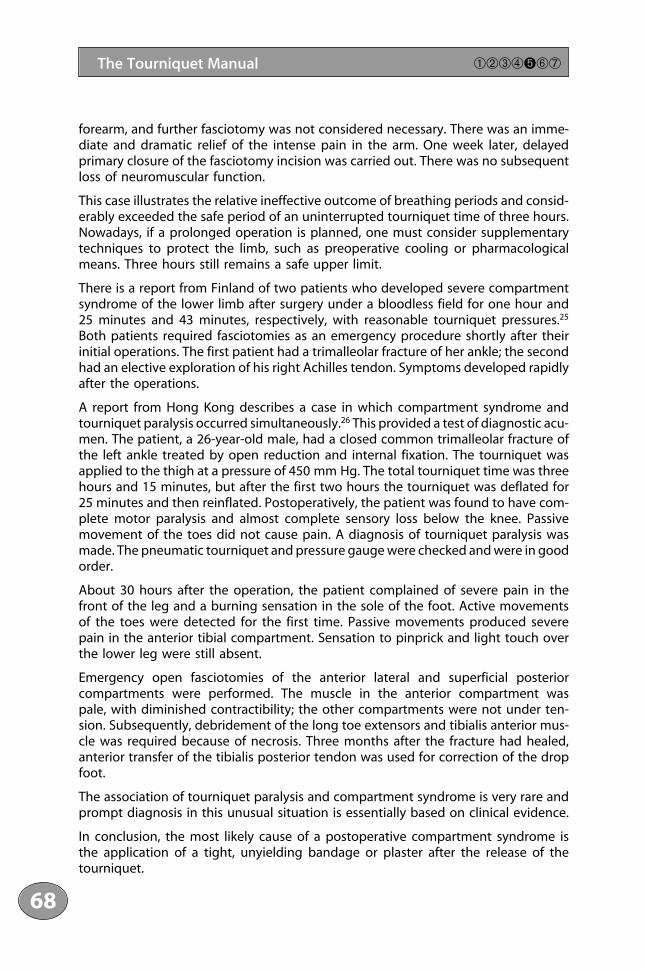
forearm, and further fasciotomy was not considered necessary. There was an imme-diate and dramatic relief of the intense pain in the arm. One week later, delayedprimary closure of the fasciotomy incision was carried out. There was no subsequentloss of neuromuscular function.
This case illustrates the relative ineffective outcome of breathing periods and consid-erably exceeded the safe period of an uninterrupted tourniquet time of three hours.Nowadays, if a prolonged operation is planned, one must consider supplementarytechniques to protect the limb, such as preoperative cooling or pharmacologicalmeans. Three hours still remains a safe upper limit.
There is a report from Finland of two patients who developed severe compartmentsyndrome of the lower limb after surgery under a bloodless field for one hour and25 minutes and 43 minutes, respectively, with reasonable tourniquet pressures.25
Both patients required fasciotomies as an emergency procedure shortly after theirinitial operations. The first patient had a trimalleolar fracture of her ankle; the secondhad an elective exploration of his right Achilles tendon. Symptoms developed rapidlyafter the operations.
A report from Hong Kong describes a case in which compartment syndrome andtourniquet paralysis occurred simultaneously.26 This provided a test of diagnostic acu-men. The patient, a 26-year-old male, had a closed common trimalleolar fracture ofthe left ankle treated by open reduction and internal fixation. The tourniquet wasapplied to the thigh at a pressure of 450 mm Hg. The total tourniquet time was threehours and 15 minutes, but after the first two hours the tourniquet was deflated for25 minutes and then reinflated. Postoperatively, the patient was found to have com-plete motor paralysis and almost complete sensory loss below the knee. Passivemovement of the toes did not cause pain. A diagnosis of tourniquet paralysis wasmade. The pneumatic tourniquet and pressure gauge were checked and were in goodorder.
About 30 hours after the operation, the patient complained of severe pain in thefront of the leg and a burning sensation in the sole of the foot. Active movementsof the toes were detected for the first time. Passive movements produced severepain in the anterior tibial compartment. Sensation to pinprick and light touch overthe lower leg were still absent.
Emergency open fasciotomies of the anterior lateral and superficial posterior compartments were performed. The muscle in the anterior compartment was pale, with diminished contractibility; the other compartments were not under ten-sion. Subsequently, debridement of the long toe extensors and tibialis anterior mus-cle was required because of necrosis. Three months after the fracture had healed,anterior transfer of the tibialis posterior tendon was used for correction of the dropfoot.
The association of tourniquet paralysis and compartment syndrome is very rare andprompt diagnosis in this unusual situation is essentially based on clinical evidence.
In conclusion, the most likely cause of a postoperative compartment syndrome isthe application of a tight, unyielding bandage or plaster after the release of thetourniquet.
1111234561178910111123111456789201111234567893011112345678940111211
68
The Tourniquet Manual ➀➁➂➃➎➅➆

5.3 Vascular Complications
A tourniquet should only be applied to a limb with a normal blood supply. Carefulpreoperative assessment of the circulation should be made of all patients havingoperations under tourniquet, especially on the lower limb. It is important to palpatethe pedal pulses, assess the capillary filling time, and note the state of the skin andnails. Brittle, dry nails, shining, scaly skin, and loss of hairs indicate poor circulatorynutrition. The presence of varicose veins is of importance in relation to postoperativedeep vein thrombosis and swelling. If pedal pulses are absent, then measurement ofthe ankle/brachial index (ABI) is essential. Using a Doppler probe, the blood pressureat the ankle is compared with the reading at the elbow; in normal circumstances, theratio should be 1. If there is any doubt about the circulation, the opinion of a vascu-lar surgeon is required. The application of a tourniquet to a limb with atheromatousvessels, commonly the superficial femoral artery, may result in poor wound healingand sepsis, ultimately requiring amputation (Figures 5.1 and 5.2).
A tourniquet should never be applied to a limb that has had an arterial prosthesisinserted .The implant is insufficiently elastic to dilate after release of the tourniquetand collateral circulation is likely to be defective.9
Fortunately, the incidence of arterial complications following total knee arthroplastyis lower than might be expected, particularly considering the proximity of the vesselsto the knee joint in both flexion and extension.27 Insall and Windsor state that the pre-operative absence of peripheral pulses has not been regarded as a contraindicationto surgery, provided that the capillary circulation is adequate.28 In their experience,seven cases of arterial compromise occurred in more than 5000 arthroplasties, threeresulting in amputation. Avoidance of a tourniquet may have prevented most, if notall, complications (see below). If a tourniquet is not used, bleeding can be profuse andtroublesome and may compromise fixation when cement is used. Insall and Windsorrecommend preoperative assessment by a vascular surgeon for those cases wherethere is doubt about the circulation. Acute vascular insufficiency following a totalknee replacement may be caused either by direct injury to a major vessel or by throm-bosis in an intact but diseased vascular system.
Rand, working at the Mayo Clinic, reported three cases of arterial injury associatedwith total knee arthroplasty in a series of 9022 patients during the period 1971–86.29
He suggested that pre-existing arterial disease and correction of extensive flexioncontractures appeared to be predisposing factors.
In a survey of the members of the British Association for Surgery of the Knee, therewere three cases due to direct injury. One resulted in a false aneurysm.30 There areseveral reports of similar occurrences. These three patients made a good recoveryafter corrective surgery. The outcome following thrombosis of an artery is different.Of the 11 patients in this survey, two died soon after surgery. Six required ampu-tation, one had persistent symptoms and died two years later, and only two werereported to have recovered after vascular surgery. McAuley and colleagues sug-gested that the problem could be a disruptive external force applied to chronicallydiseased vessels.31 Tethering of the proximal superficial femoral artery with stretching
69
➀➁➂➃➎➅➆ Complications
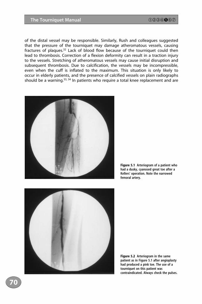
of the distal vessel may be responsible. Similarly, Rush and colleagues suggestedthat the pressure of the tourniquet may damage atheromatous vessels, causing fractures of plaques.32 Lack of blood flow because of the tourniquet could then lead to thrombosis. Correction of a flexion deformity can result in a traction injuryto the vessels. Stretching of atheromatous vessels may cause initial disruption andsubsequent thrombosis. Due to calcification, the vessels may be incompressible,even when the cuff is inflated to the maximum. This situation is only likely to occur in elderly patients, and the presence of calcified vessels on plain radiographsshould be a warning.33, 34 In patients who require a total knee replacement and are
1111234561178910111123111456789201111234567893011112345678940111211
70
The Tourniquet Manual ➀➁➂➃➎➅➆
Figure 5.1 Arteriogram of a patient whohad a dusky, cyanosed great toe after aKellers’ operation. Note the narrowedfemoral artery.
Figure 5.2 Arteriogram in the samepatient as in Figure 5.1 after angioplastyhad produced a pink toe. The use of atourniquet on this patient wascontraindicated. Always check the pulses.
not suitable for the use of a tourniquet, the operation can be done without a tourniquet.
Abdel-Salem and Eyres reported a controlled trial in a series of patients undergoingknee-replacement surgery.35 Eighty patients were allocated randomly to two groups:operation with or without a tourniquet. The patients were all operated on by thesame surgeon using the same prosthesis. There was no significant differencebetween the two groups in operating time or total blood loss. Postoperative painwas less in the patients in whom a tourniquet had not been used. They achievedstraight leg raising and knee flexion earlier and had fewer superficial wound infec-tions and deep vein thrombosis than the controls. Another prospective trial in 77patients found similar results, but there was no difference in the incidence of woundcomplications or deep vein thrombosis between the two groups of patients.36
A prospective study was undertaken on the blood flow of 44 patients who werehaving total knee replacements under tourniquet control and who had no evidenceof peripheral vascular disease.37 The ABI did not alter after operation, and there wereno changes in arterial waveforms. None of the limbs studied had an ABI below 0.87.Furthermore, none of the three limbs with ABI below 1 preoperatively showed anydeterioration after operation. Doppler velocity waveforms, which are considered amore sensitive index of stenosis, were unchanged .The authors considered that thepreoperative assessment should include duplex scanning as well as ABI measure-ments. Their final conclusion was that unless there is clinical evidence of peripheralvascular disease, then total knee replacement under a tourniquet is unlikely to causeischaemic complications.
Traumatic arteriovenous fistulae with a false aneurysm of the inferior medial genic-ular artery have been reported following arthroplasty of the knee. Both cases werediagnosed within the first two months following surgery.38 Both patients developedswellings in the regions of the incisions associated with a pulsatile swelling andaudible bruit. The diagnosis was confirmed by arteriography. Surgical excision withligation of the inferior medial genicular artery was effective in the relief of symp-toms. Strict haemostasis after release of the tourniquet at the end of the operationshould prevent this.
Giannestras and colleagues describe how an atheromatous plaque was displaced inthe left superficial femoral artery following use of a tourniquet on the thigh for abunion operation.39 The foot remained pale when the tourniquet was deflated. Theplaque was removed through an arteriotomy after an arteriogram had beenperformed. The tourniquet pressure used was 500 mg Hg – rather high and a possiblefactor – but there was no mention of the patient’s blood pressure. The patient’ssubsequent course was uncomplicated.
Vessels in the foot may also be damaged at operation but pass unnoticed at thetime. Webb Jones described three cases where aneurysms had developed after triplearthrodesis.40 Scott recorded the formation of an aneurysm of the peroneal arteryafter an operation on the ankle through a posterolateral approach.41
It is the surgeon’s duty to routinely inspect carefully the exposed digits after thetourniquet has been released in the operation theatre to ensure that there has been
71
➀➁➂➃➎➅➆ Complications

return of normal circulation. If there is any doubt about a digit, the bandages mayneed to be removed and reapplied. It is also important to continue to observe thecirculation for the first 12–24 hours.
5.4 Damage to Skin
Damage to the skin was mentioned in Chapter 2. The most common problem is burnsof the skin. Children appear to be particularly susceptible. Such burns have beenappreciated for many years: McElvenny drew attention to them in 1945.42 There are atleast six references to chemical burns under a tourniquet, and the complication can-not be regarded as inconsequentially rare. It is discussed specifically in Campbell’sOperative Orthopaedics.43 Burns occur when the padding under the tourniquetbecomes soaked by the antiseptic solution used to paint the skin. Aqueous solutionsare not recorded as causing burns, and proprietary antibacterial agents are notresponsible, except in specific allergic reactions, which are rare and result in skin irri-tation wherever applied. Alcohol-based solutions appear to be the most likely cause.The burns are due to prolonged contact of alcohol-based solutions since evaporationis prevented under the tourniquet (Figure 5.3).
These burns are easy to prevent using one of three methods. First, the skin prepa-ration can be applied well distally to the tourniquet in operations below the kneeor elbow. Second, the solutions should not be applied too liberally, as this promotesspillage and trickling towards the tourniquet. Finally, if the skin has to be preparedright up to the tourniquet, then it must be positively occluded from the preparedarea for operation by using a drape with a rubber membrane, as is used frequentlyin knee surgery, or a drape with an adhesive edge stuck to the skin; the latter is thesafest method.
A friction burn to the thigh has been described in a 48-year-old Caucasian man whounderwent a second-stage knee replacement following revision for an infected pros-thesis.44 After skin preparation with aqueous chlorhexidine, the tourniquet wassealed off from the operation site by an adhesive drape. At the end of the opera-tion, the tourniquet was found to have overrun the wool padding with almost halfof its width and was lying in direct skin contact. The whole complex had slippeddown the thigh by about 10 cm. The wool padding was not found to be soiled withblood or fluid. On the first postoperative day, the patient developed almost circum-ferential blisters on the thigh. It was presumed that the movement of fully inflatedtourniquet over the base skin due to slippage led to friction burns. It was thoughtthat the tourniquet had not been tied tightly enough before inflation.
5.5 Post-tourniquet Syndrome
This condition, according to Bruner, is the most common but least appreciatedmorbidity associated with the use of tourniquets.45 In my experience, however, it is
1111234561178910111123111456789201111234567893011112345678940111211
72
The Tourniquet Manual ➀➁➂➃➎➅➆

rare. The combined effect of muscle ischaemia, oedema, and microvascular con-gestion leads to a syndrome characterised by stiffness, pallor, weakness but notparalysis, and subjective numbness without anaesthesia. It probably represents theeffects of a situation in which the upper limits of tissue tolerance have been reached,and it does not occur with relatively short periods of ischaemia of less than twohours.
73
➀➁➂➃➎➅➆ Complications
Figure 5.3 Typical circumferential burn occurring under a tourniquet. This painful lesion is easily prevented.
5.6 Potential of Cross-infection DuringPeripheral Venous Access by Contamination of Tourniquets
Venepuncture for blood tests and intravenous cannulation are the most commoninvasive procedures carried out in hospitals. The usual method for providing venousstasis is the application of a reusable tourniquet. The use of such tourniquets inmany patients and many wards contravenes the basic principles of infection control.One study has revealed a substantial reservoir of potentially pathogenic bacteria onthese tourniquets, which can be transmitted from patient to patient on the handsof staff.46 Reusable tourniquets have been shown to be potential fomites. Since it is impossible to disinfect reusable tourniquets (they are not made of durable material), the use of disposable tourniquets is recommended.
References1 Speigel, IJ, Lewin, P (1943). Tourniquet paralysis. Analysis of three cases of surgically proved peripheral
nerve damage following use of a rubber tourniquet. Journal of the American Medical Association 129:432–435.
2 Putnam, JJ (1888). Peripheral paralysis following use of a rubber tourniquet. Boston Medical and SurgicalJournal 199: 1888.
3 Eckhoff, NI (1931). Tourniquet paralysis. Lancet 2: 343–345.4 Middleton, RWD, Varian, JP. Tourniquet paralysis (1974). Australia and New Zealand Journal of Surgery 44:
124–128.5 Rorabeck, CH, Kennedy, JC (1980). Tourniquet induced nerve ischaemia complicating knee ligament
surgery. American Journal of Sports Medicine 8: 98–102.6 Guanche, CJ (1995). Tourniquet-induced tibial nerve palsy complicating anterior cruciate reconstruction.
Journal of Arthroscopic and Related Surgery 11: 620–622.7 Bolton, CF, McFarlane, RM (1978). Human pneumatic tourniquet paralysis. Neurology 28: 787–793.8 Klenerman, L (1983). Tourniquet paralysis. Journal of Bone and Joint Surgery 65B: 374–375.9 Birch, R, Bonney, G, Wyn Parry, CB, eds (1998). Surgical Disorders of the Peripheral Nerves. London: Churchill
Livingston, pp. 301–302.10 Saunders, K, Louis, D, Weingarden, SI, Waylonis, GW (1979). Effect of tourniquet time on postoperative
quadriceps function. Clinical Orthopaedics and Related Research 143: 194–199.11 Weingarden, SI, Louise, DL, Waylonis, GW (1979). Electromyographic changes in postmeniscectomy
patients. Journal of the American Medical Association 241: 1248–1250.12 Nicholas, TJ, Tyler, TF, McHugh, MP, Gleim, GW (2001). Effect on leg strength of tourniquet use during
anterior cruciate reconstruction: a prospective randomised study. Journal of Arthroscopic and Related Surgery17: 603–607.
13 Aho, K, Saino, K, Kienta, M, Varpanen, E (1983). Pneumatic tourniquet paralysis. Journal of Bone and JointSurgery 65B: 441–443.
14 Sunderland, S (1968). Nerves and Nerve Injuries. Edinburgh: E & S Livingstone, p. 141.15 Rudge, P, Ochoa, J, Gilliatt, RW (1974). Acute peripheral nerve compression in the baboon. Journal of
Neurological Sciences 23: 403–420.16 Bywaters, EGL, Beall, D (1941). Crush injuries with impairment of renal function. British Medical Journal 1:
427–432.17 Pfeiffer, PM (1986). Acute rhabdomyolysis. Possible role of tourniquet ischaemia. Anaesthesia 41: 614–619.18 Williams, JE, Tucker, DB, Read, JM (1983). Rhabdomyolysis – myoglobin. Consequences of prolonged tourni-
quet. Journal of Foot Surgery 22: 52–56.
1111234561178910111123111456789201111234567893011112345678940111211
74
The Tourniquet Manual ➀➁➂➃➎➅➆
19 Jorgensen, HRI (1987). Myoglobin release after tourniquet ischaemia. Acta Orthopaedica Scandinavica 58:554–556.
20 Palmer, SH, Graham, G (1994). Tourniquet induced rhabdomyolysis after total knee replacement. Annalsof the Royal College of Surgeons of England 76: 416–417.
21 Mubarak, S, Hargens, A (1981). Compartment Syndrome and Volkmann’s Contracture. Philadelphia: W.B.Saunders, p. 50.
22 Mars, M, Brock-Utne, JG (1991). The effect of tourniquet release on intra-compartmental pressure in thebandaged and unbandaged limb. Journal of Hand Surgery 16B: 318–322.
23 O’Neil, D, Sheppard, JE (1989). Transient compartment syndrome of the forearm resulting from venouscongestion from a tourniquet. Journal of Hand Surgery 14A: 894–896.
24 Greene, TL, Dean, S (1983). Compartment syndrome of the arm – a complication of the pneumatic tourni-quet. Journal of Bone and Joint Surgery 65A: 270–273.
25 Hirvensalo, E, Tuomen, H, Lapensuo, M, Helio H (1992). Compartment syndrome of the lower limb causedby a tourniquet: a report of two cases. Journal of Orthopaedic Trauma 6: 469–472.
26 Luk, KD, Pun, WK (1987). Unrecognised compartment syndrome with tourniquet palsy. Journal of Bone andLimb Surgery 69B: 97–99.
27 Zaidi, SHA, Cobb, AG, Bentley, G (1995). Danger to popliteal artery in high tibial osteotomy. Journal ofBone and Joint Surgery 77B: 384–386.
28 Insall, JN, Windsor, RE, eds (1993). Surgery of the Knee, 2nd edn. New York: Churchill Livingstone, p. 896.29 Rand, JA (1987). Vascular complications of total knee arthroplasty. Journal of Arthroplasty 2: 89–93.30 Kumar, NS, Chapman, JA, Rawlins, I (1998). Vascular injuries in total knee arthroplasty. Journal of Arthroplasty
13: 211–216.31 McAuley, CE, Steed, DL, Webster, MW (1984). Arterial complications of knee replacement. Archives of Surgery
119: 960–962.32 Rush, J, Vidovich, H, Johnson, MA (1987). Arterial complications of knee replacement. Journal of Bone and
Joint Surgery 69B: 400–406.33 Klenerman, L, Lewis, JD (1976). Incompressible vessels. Lancet 1: 811–812.34 Jeyaseelan, S, Stevenson, TM, Pfitzner, J (1981). Tourniquet failure and arterial calcification. Anaesthesia
36: 48–50.35 Abdel-Salaam, A, Eyres, KA (1995). Effects of tourniquet during total knee arthroplasty. Journal of Bone and
Joint Surgery 77B: 250–253.36 Wakenkar, HM, Nicholl, JE, Koka, R, D’Arcy, J (1999). The tourniquet in knee arthroplasty. Journal of Bone
and Joint Surgery 81B: 30–33.37 Scriven, MW, Fligelstone, LJ, Oshodi, TO, et al. (1996). The influence of total knee arthroplasty on lower
limb blood flow. Journal of the Royal College of Surgeons of Edinburgh 41: 323–324.38 Dennis, DA, Newmann, RD, Toma, P, et al. (1987). Arteriovenous fistula with false aneurysm of the infe-
rior geniculate artery. Clinical Orthopaedics and Related Research 222: 255–259.39 Giannestras, NJ, Cranley, JJ, Lentz, M (1977). Occlusion of tibial artery after a foot operation under tourni-
quet. Journal of Bone and Joint Surgery 59A: 682–683.40 Webb Jones, A (1955). Aneurysm after foot stabilisation. Journal of Bone and Joint Surgery 37B: 440–442.41 Scott, JH (1955). Traumatic aneurysm of the peroneal artery. Journal of Bone and Joint Surgery 37B: 438–439.42 McElvenny, RT (1945). The tourniquet. Its clinical application. American Journal of Surgery LXIX: 94–10643 Crenshaw, A, ed. (1987). Cambell’s Operative Orthopaedics, Vol. 1, 7th edn. St Louis: CV Mosby, p. 111.44 Choudhary, S, Koshy, C, Ahmed, J, Evans, J (1998). Friction burns to the thigh caused by tourniquet. British
Journal of Plastic Surgery 51: 142–143.45 Bruner, JM (1951). Safety factors in the use of the pneumatic tourniquet in the hand. Journal of Bone and
Joint Surgery 33A: 221–224.46 Golder, M, Chan, CLH, O’Shea, S, et al. (2000). Potential risk of cross-infection during peripheral venous
access by contamination of tourniquets. Lancet 355: 44.
75
➀➁➂➃➎➅➆ Complications

This page intentionally left blank
Chapter 6The Tourniquet Used for Anaesthesia

This page intentionally left blank
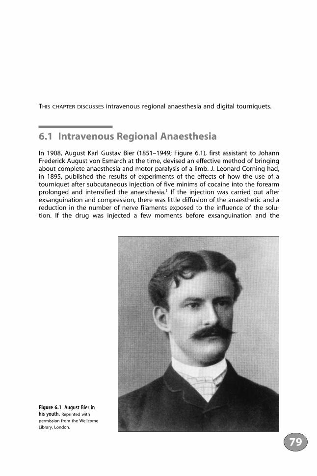
THIS CHAPTER DISCUSSES intravenous regional anaesthesia and digital tourniquets.
6.1 Intravenous Regional Anaesthesia
In 1908, August Karl Gustav Bier (1851–1949; Figure 6.1), first assistant to JohannFrederick August von Esmarch at the time, devised an effective method of bringingabout complete anaesthesia and motor paralysis of a limb. J. Leonard Corning had,in 1895, published the results of experiments of the effects of how the use of atourniquet after subcutaneous injection of five minims of cocaine into the forearmprolonged and intensified the anaesthesia.1 If the injection was carried out afterexsanguination and compression, there was little diffusion of the anaesthetic and areduction in the number of nerve filaments exposed to the influence of the solu-tion. If the drug was injected a few moments before exsanguination and the
79
Figure 6.1 August Bier in his youth. Reprinted with
permission from the Wellcome
Library, London.

application of the tourniquet, a sufficient saturation of the tissue was achieved toexpose a large number of the nerve filaments to the solution. Corning suggestedthat this technique was applicable to surgery of the extremities. Bier, a pioneer ofspinal anaesthesia, established this technique. It was cumbersome by modern stan-dards. He injected a solution of procaine into a subcutaneous vein on a section ofthe forearm that was visible between two constricting bands and that had beenrendered bloodless by an Esmarch bandage. A surgical cut-down was required tolocate the vein. The injected solution permeated through the exposed section ofthe limb very quickly and produced what Bier called “direct vein anaesthesia” in5–15 minutes. The anaesthesia lasted for as long as the upper constricting band wasleft in place. After its removal, sensation returned in a few minutes.
The development of techniques of regional anaesthesia followed that of drugs thatcould produce local anaesthesia. Gaedicke isolated cocaine in 1855. It was purifiedand named by Nieman in 1860 and first used as a local anaesthetic by Koller in 1864.Cocaine was used for infiltration anaesthesia and major nerve blocks, which werepopularised by both Halsted and Crile. The systemic toxicity of cocaine limited itsuse as a local anaesthetic. The practice of regional blocks was enhanced greatlywhen procaine was synthesised by Einhorn in 1904. Braun went on to establish theuse of procaine for regional anaesthesia.2
The technique did not become used widely, despite Bier’s reports of its use withEsmarch bandages in 134 patients, including ten amputations, 37 resections, 12sutures of bones, ten tendon transfers, two Dupuytren’s contractures, and sevencases of extirpation of varicose veins. Bier first conceived the possibility of themethod when he injected a solution of indigo carmine into the veins of an ampu-tated limb.3 He found that there was widespread diffusion of the dye. He consideredthat the anaesthesia was of two types: direct and indirect. Direct anaesthesia wasdue to infiltration of the fluid into the tissues and around the nerve filaments andoccurred rapidly. A little later, because of blocking of the larger nerve trunks passingthrough the exsanguinated region, further anaesthesia occurred distally (indirectanaesthesia) Today, the mechanism remains open to dispute and a combination oftheories may best explain the effects.
Despite Bier’s report of none of 134 patients showing any ill effects, his techniqueappears to have been forgotten until 1963, when it was reintroduced by Holmes.4
The technique was used on a limited scale but did not gain widespread popularity.There are a number of reasons for this5: the technique was cumbersome, requiringwrapping and unwrapping of Esmarch bandages in a precise manner; special equip-ment was required; and an operative procedure (cut-down) was used. It was Morrisonin 1931 who first suggested a venepuncture and made the method more practi-cable.6 Although Holmes used lignocaine, 0.5% prilocaine is now regarded as thedrug of choice.7 Only the 0.5% solution should be used, and the recommended doseis 3–4 mg/kg.
The method has now been refined. An intravenous cannula should be inserted intoeach hand. The limb should be exsanguinated before the tourniquet is inflated. Thiscan be done by elevation and careful application of an Esmarch bandage, takingcare not to dislodge the intravenous needle; alternatively, a pneumatic splint can
1111234561178910111123111456789201111234567893011112345678940111211
80
The Tourniquet Manual ➀➁➂➃➄➏➆
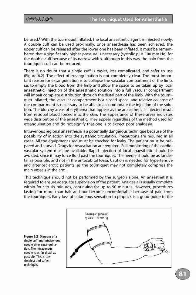
be used.8 With the tourniquet inflated, the local anaesthetic agent is injected slowly.A double cuff can be used proximally; once anaesthesia has been achieved, theupper cuff can be released after the lower one has been inflated. It must be remem-bered that a significantly higher pressure is necessary (systolic plus 100 mm Hg) forthe double cuff because of its narrow width, although in this way the pain from thetourniquet cuff can be reduced.
There is no doubt that a single cuff is easier, less complicated, and safer to use(Figure 6.2). The effect of exsanguination is not completely clear. The most impor-tant reason for exsanguination is to collapse the vascular compartment of the limb,i.e. to empty the blood from the limb and allow the space to be taken up by localanaesthetic. Injection of the anaesthetic solution into a full vascular compartmentwill impair complete distribution through the distal part of the limb. With the tourni-quet inflated, the vascular compartment is a closed space, and relative collapse ofthe compartment is necessary to be able to accommodate the injection of the solu-tion. The blotchy areas of erythema that appear as the anaesthetic is injected resultfrom residual blood forced into the skin. The appearance of these areas indicateswide distribution of the anaesthetic. They appear regardless of the method used forexsanguination and do not signify that one is to expect poor analgesia.
Intravenous regional anaesthesia is a potentially dangerous technique because of thepossibility of injection into the systemic circulation. Precautions are required in allcases. All the equipment used must be checked for leaks. The patient must be pre-pared and starved. Drugs for resuscitation are required. Full monitoring of the cardio-vascular system must be available. Rapid injection of local anaesthetic should beavoided, since it may force fluid past the tourniquet. The needle should be as far dis-tal as possible, and not in the antecubital fossa. Caution is needed for hypertensiveand arteriosclerotic patients, as the tourniquet may not completely compress themain vessels in the arm.
This technique should not be performed by the surgeon alone. An anaesthetist isrequired to ensure adequate supervision of the patient. Analgesia is usually completewithin four to six minutes, continuing for up to 90 minutes. However, procedureslasting for more than half an hour become uncomfortable because of pain from the tourniquet. Early loss of cutaneous sensation to pinprick is a good guide to the
81
➀➁➂➃➄➏➆ The Tourniquet Used for Anaesthesia
Figure 6.2 Diagram of asingle cuff and intravenousneedle after exsanguina-tion. The intravenousneedle is as far distal aspossible. This is thesimplest and safesttechnique.
effectiveness of the block. As muscle relaxation occurs, the limb should feel heavyto the patient.
This method is useful for distal fractures in the upper limb, ganglions and decom-pression of the median nerve. It is not used commonly for the lower limb, mainlybecause of the volume of local anaesthetic agent required.
After completion of the operation, the cuff should be deflated gradually in a step-wise manner to avoid a bolus injection of local anaesthetic. Sensation and motorpower return after several minutes. The minimum tourniquet time suggested is 20minutes to allow adequate diffusion into the ischaemic region.
The adverse effects of intravenous regional anaesthesia are due to accidental, suddendeflation of the cuff and deflation too soon after the injection of local anaesthetic.Sudden release results in a bolus of any unfixed local anaesthetic. Underinflation ofthe cuff will allow leakage to occur into the systemic circulation.
In 1982, in an editorial in the British Medical Journal, Margaret Heath noted that since1979 the Scientific and Technical Branch of the Department of Health and SocialSecurity in London had been informed of five deaths resulting directly from the useof intravenous regional analgesia.9 The drug used was bupivacaine. In three of themost recent cases, it was found that the equipment was in good order but the cuffhad been deflated when it should not have been.
The likelihood of direct intravenous injection during routine procedures has beenunderestimated. The effects of such a bolus are likely to be worse in frightenedpatients, as circulatory catecholamines will ensure that the maximum amount of thecardiac output reaches the brain and heart, which are the main target organs fortoxicity. The recognition and treatment of toxic reactions are crucial. There is alsothe problem of individual low thresholds. Major side effects of using prilocaine havenot been recorded in Britain. Prilocaine is the safest local anaesthetic for intravenouslocal anaesthesia. It is no longer available in North America in a form suitable forintravenous injection because of concern about methaemoglobinaemia, althoughthis does not occur in the dosages used; lidocaine is commonly used in North Americainstead.10
6.2 Digital Tourniquets
When performing a minor procedure on a single digit, finger or toe, it is convenientto apply a tourniquet at the base of the digit rather than use a proximal tourniquetfor the whole limb. A variety of techniques have been described. A common methodis to wrap a soft rubber catheter or Penrose drain around the base of the finger ortoe under tension and keep it in place with an artery forceps. Salem suggested amethod that is now used commonly11: he cut off the finger of a disposable glove,removed the tip, stretched and applied it to the digit for operation, and rolled itproximally to form a ring at the base of the digit. This provides a satisfactory blood-less field. The ring can readily be cut and removed when the operation has been
1111234561178910111123111456789201111234567893011112345678940111211
82
The Tourniquet Manual ➀➁➂➃➄➏➆

83
➀➁➂➃➄➏➆ The Tourniquet Used for Anaesthesia
Figure 6.3 Salem’s method of applying a digital tourniquet: (a) A finger from a rubber glove is rolled proximally.
Figure 6.3 (b) The tourniquet is grasped in an artery clip.

completed. A recent modification suggests that the tourniquet should be grippedby an artery clip, with the handle pointing proximally.12 The rubber ring is cut abovethe clip with scissors. This ensures that when the tourniquet is released, the dangerof the tourniquet remaining in place is avoided (Figure 6.3). A study of the pres-sures involved using a miniature pressure transducer and digital strain indicator hasshown that the Penrose drain generated highly variable pressure, often greater than500 mg Hg.13 In contrast, the rolled finger from a glove, in addition to producingexsanguination, uniformly generated pressures of less than 500 mg Hg, which isbelow the threshold required to produce nerve damage.
The main danger is that a digital tourniquet may be forgotten, covered with adressing, and left to cause gangrene in the anaesthetised finger or toe (Figure 6.4).A rare case of ischaemia of the distal half of a finger has been reported, which recov-ered with treatment by intravenous infusion of low-molecular-weight dextrose,intravenous dipyridamole, and alcohol. The finger was packed in ice and the armwas warmed. The report was from a unit using a tourniquet for at least 1000 oper-ations a year, and this was the first complication observed.14
6.3 Regional Sympathetic Blockade
For patients suffering complex regional pain syndrome (CRPS) type I (also knownas reflex sympathetic dystrophy), a technique similar to intravenous regional anaes-thesia has been used with a tourniquet in place.15 A solution of 20 mg (for the upper limb) or 30 mg (for the leg) of guanethidine in normal saline is injected. Thecuff is kept inflated for 15 minutes. This produces complete sympathetic blockade
1111234561178910111123111456789201111234567893011112345678940111211
84
The Tourniquet Manual ➀➁➂➃➄➏➆
Figure 6.3 (c) The rubber ring is cut above the clip. Reproduced with permission from Elsevier Science from Smith, IM,
Austin, OB, Knight, SL (2002). A simple and failsafe method for digital tourniquet. Journal of Hand Surgery 27B; 363–364.

for up to four days, reducing the pain in the affected limb and allowing active physiotherapy. Although this technique has been considered accepted practice formany years, recent work has shown that guanethidine offers no significant advan-tage in analgesia over a normal saline placebo block in the treatment of early CRPStype 1 of the hand after a fracture of the distal radius.16 Guanethidine does notimprove the outcome of this condition, and it may delay the resolution of vaso-motor instability when compared with placebo.
85
➀➁➂➃➄➏➆ The Tourniquet Used for Anaesthesia
Figure 6.4 Gangrenous thumb after a digital tourniquet had been left in place for too long. Reproduced with
permission from Elsevier Science from Smith, IM, Austin, OB, Knight, SL (2002). A simple and failsafe method for digital tourni-
quet. Journal of Hand Surgery 27B; 363–364.

References1 Corning, JL (1895). Anaesthetic effects of the hydrochlorate of cocaine when subcutaneously injected. New
York Medical Journal 42; 317–319.2 Hilgenhurst, G (1990). The Bier block after eighty years. Regional Anaesthesia 15: 2–5.3 Adams, RC (1944). Intravenous Anaesthesia. New York: Paul B. Hoeber, p. 111.4 Holmes, CM (1963). Intravenous regional anaesthesia. Lancet 1: 245–247.5 Colbern, EC (1970). The Bier block for intravenous regional anaesthesia. Technique and literature review.
Anaesthesia and Analgesia 49: 935–940.6 Morrison, JL (1931). Intravenous local anaesthesia. British Journal of Surgery 18: 641–647.7 Bader, AM, Concepion, M, Hurley, RJ, Arthur, GR (1998). Comparison of lidocaine and prilocaine for intra-
venous regional anaesthesia. Anesthesiology 69: 409–412.8 Winnie, AP, Ramamurthy, S ((1970.). Pneumatic exsanguination for intravenous regional anaesthesia.
Anesthesiology 33: 664–5.9 Heath, M (1982). Deaths after intravenous regional anaesthesia. British Medical Journal 285: 913–914.
10 Henderson, CL,Warriner, CB, McEwen, JA, Merrick, PM (1997). A North American survey of intravenousregional anaesthesia. Anaesthesia and Analgesia 85: 853–863.
11 Salem, MZA (1973). Simple finger tourniquet. British Medical Journal 2: 779.12 Smith, IM, Austin, OB, Knight, SL (2002). A simple and fail safe method for digital tourniquet. Journal of
Hand Surgery 27B: 363–364.13 Hixson, FP, Shafiroff, BB, Werner, FW, Palmer, AK (1986). Digital tourniquet a pressure study with relevance.
Journal of Hand Surgery 11A: 865–867.14 Dove, AF, Clifford, RP (1982). Ischaemia after use of a finger tourniquet. British Medical Journal 284:
1162–1163.15 Hannington-Kiff, JG (1974). Intravenous regional sympathetic block with guanethidine. Lancet i: 1019–1020.16 Livingstone, JA, Atkins, RM (2002). Intravenous regional blockade in the treatment of post-traumatic
complex regional pain syndrome type 1 (algodystrophy) of the hand. Journal of Bone and Joint Surgery84B: 380–386.
1111234561178910111123111456789201111234567893011112345678940111211
86
The Tourniquet Manual ➀➁➂➃➄➏➆

Chapter 7Technology and Practice

This page intentionally left blank

THE PNEUMATIC TOURNIQUET includes a pressure source, pressure gauge, regulator andinflatable cuff (Figure 7.1). There are two types of tourniquet: those inflated by directhand-pumping and those that are inflated “automatically” by the movement of aswitch that controls an air or gas supply.1 It is essential to ensure that the cuff isinflated rapidly to prevent venous congestion.
7.1 Design of the Tourniquet Cuff
Generally, the cuff is composed of one or two rectangular inflatable pneumatic bagsmade of rubber on plastic. These are enclosed in a cover of cloth or plastic, togetherwith a firm backing sheet known as a spine (Figure 7.2). The pneumatic bags areinflated from a hand pump or a regulated source of compressed air via connectingtubes and links. A strapping mechanism is provided to retain the tourniquet in placeon the arm or leg.
89
Figure 7.1 Components of an automatic tourniquet control unit. Reproduced with permission from Lippincott, Williams
& Wilkins from Hurst, LN, Weiglein, O, Brown, WE, Campbell, GJ (1981). The pneumatic tourniquet: a biomechanical and elec-
trophysiological study. Plastic and Reconstructive Surgery 648–652.
Figure 7.2 Diagram of a tourniquet cuff.

7.2 Hand-powered Tourniquets
A sphygmomanometer bulb or bicycle pump can be used for inflation. This type oftourniquet has the virtue of simplicity. It is not possible to preset a pressure. It isunlikely that accidental overinflation will occur since the tourniquet is inflated witha deliberate pumping action.
The pressure gauge is of an aneroid type (Figure 7.3). The gauge must be maintainedcarefully since it is vulnerable to knocks and is damaged readily. Displacement ofthe pin at the end of the range of pressure measurements has resulted in grossoverinflation of the cuff.2
7.3 Automatic Tourniquets
Development over the last 25 years has resulted in a variety of automatic systemsnow being available. Some use a self-contained gas source, such as a small cylinderof dichlorodifluoromethane or freon gas, to inflate the cuff. Others have an air reser-voir that may be charged from a compressed air line. The machines can sometimescompensate for small leakages of pressure.
The most common tourniquet systems now available include automatic safetydevices that operate from the main air supply or cylinders at a maximum pressureof 100 psi. A typical example is the Anetic Aid (Guiseley, Leeds, UK) APT Tourniquet.
1111234561178910111123111456789201111234567893011112345678940111211
90
The Tourniquet Manual ➀➁➂➃➄➅➐
Pressure to be measured
Flattened tube in250-degree arc
Figure 7.3 Diagram of aBourdon gauge commonly usedwith tourniquet equipment.Reprinted with permission from the
BMJ Publishing Group from
Hallett, J (1983). Inaccurate
tourniquet gauges. British Medical
Journal 286: 1267.
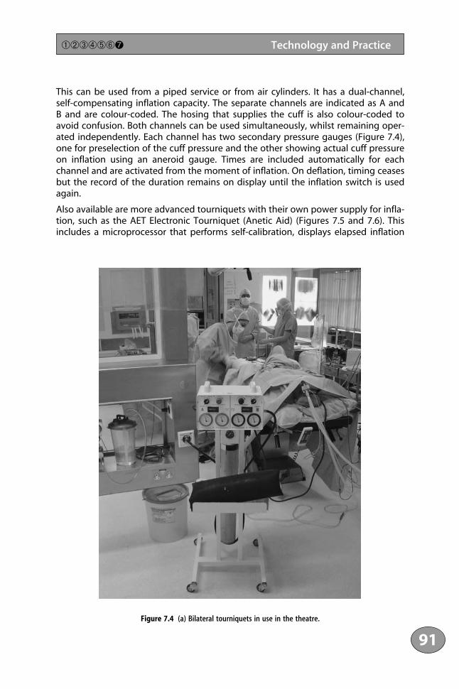
This can be used from a piped service or from air cylinders. It has a dual-channel,self-compensating inflation capacity. The separate channels are indicated as A andB and are colour-coded. The hosing that supplies the cuff is also colour-coded toavoid confusion. Both channels can be used simultaneously, whilst remaining oper-ated independently. Each channel has two secondary pressure gauges (Figure 7.4),one for preselection of the cuff pressure and the other showing actual cuff pressureon inflation using an aneroid gauge. Times are included automatically for eachchannel and are activated from the moment of inflation. On deflation, timing ceasesbut the record of the duration remains on display until the inflation switch is usedagain.
Also available are more advanced tourniquets with their own power supply for infla-tion, such as the AET Electronic Tourniquet (Anetic Aid) (Figures 7.5 and 7.6). Thisincludes a microprocessor that performs self-calibration, displays elapsed inflation
91
➀➁➂➃➄➅➐ Technology and Practice
Figure 7.4 (a) Bilateral tourniquets in use in the theatre.

time, and sounds an alarm half-hourly. It uses an internal electrical pump to compressthe ambient air, and pressure is shown on a microprocessor digital display. It canstore the details of up to 20 applications, which can be recalled if necessary.
7.4 Safety Aspects
The safety aspects of tourniquets are summarised in a paper in the AmericanOperating Room Nurses Journal.3 All parts of the tourniquet should be checked beforeuse to eliminate leaks from loose connectors, faulty tubing or the cuff bladder. Acommon source of leaks are worn O-rings, which help to seal the connection betweenthe tourniquet and the tubes for inflation. The tourniquet apparatus must be treatedwith respect, as unnecessary jarring or bumping will damage the gauge and resultin faulty readings. Routine and regular calibration of all tourniquets is mandatory.This can be done with a commercially available test gauge or a mercury column. Asimple precaution is readily available at all times4: before the use of any tourniquet,the pressure gauge is set at 100 mm Hg and connected to the mercury manometeron the anaesthetic machine. The pressure in the tourniquet line is then releasedand the pressure on the mercury manometer is read. The procedure is then repeated
1111234561178910111123111456789201111234567893011112345678940111211
92
The Tourniquet Manual ➀➁➂➃➄➅➐
Figure 7.4 (b) Tourniquet linked to compressed air, showing close-up of dials: one tourniquet is inflated and theother has been deflated.

with the pressure gauge set at 200 mm Hg. Inaccurate gauges should be returnedto the manufacturer. Inaccurate pressure gauges are one of the most common causesof pressure lesions in nerves (see Chapter 2).5 Once a gauge becomes inaccurate, itcannot be readjusted and it must be replaced The Bourdon gauge will suffer fromrough handling, and the flexibility of the metal coil will be altered with repeateduse.6
The aneroid gauge may not always be at fault. Durkin and Crabtree reported a casein which the indicator needle on the pressure gauge could travel around the dial asecond time, so that if the cuff was inflated quickly it might not be apparent thata reading of 250 mm Hg was actually representing a pressure of 1000 mg Hg.2 Thishazard could be avoided if all pressure gauges used for such purposes had a stopat the upper end of the scale. Cuff pressure within ±20 mm Hg of indicated dialpressure is acceptable.6 The complete system should be leak-proof, with no changein pressure with time, as demonstrated by the absence of variation of pressure at
93
➀➁➂➃➄➅➐ Technology and Practice
Figure 7.5 Two commonly used tourniquets: the left-hand tourniquet has a microprocessor, while the right-handone needs to be connected to piped air or cylinders. Reprinted courtesy of Anetic Aid, Guiseley, Leeds, UK.

400 mm Hg. Cuffs should exceed the circumference of the limb by 7–15 cm, shouldbe of a width appropriate to the size of the patient (see Chapter 2), and should bepositioned at the point of maximum circumference of the limb. As wide a cuff aspossible should be selected.
Tourniquet time, site of application, and pressure must be recorded at the start ofthe operation, including the identification number of the specific tourniquet appa-ratus that was used. Once applied, the cuff should not be rotated into a new position,as this may cause a shearing injury. As described previously (see Chapters 2 and 5),fluid from skin preparations should not be allowed to collect under the cuff.
Additional care must be taken in bilateral procedures in which tourniquets are usedon both limbs (see Chapter 2), since the risks of complications of the use of a tourni-quet may be increased in such cases. The tourniquets should not be deflatedsimultaneously, but should be deflated separately with an interval of a few minutesto allow circulatory adjustment to the autotransfusion that occurs.
7.5 Practical Problems
The use of a bloodless field is integral to limb surgery, but it may have become aritual. Stirling Bunnell stated7:
It is impossible to dissect a hand properly without the aid of ischaemia from a tourni-quet. It is also dangerous. Without a tourniquet the field is covered with blood, whichis opaque; therefore instead of progressing with the dissection the surgeon will be
1111234561178910111123111456789201111234567893011112345678940111211
94
The Tourniquet Manual ➀➁➂➃➄➅➐
Figure 7.6 The variety of cuffs available with Rhys-Davies Exsanguinators. Reprinted courtesy of Anetic Aid, Guiseley,
Leeds, UK.

fumbling, traumatising the tissues by sponging constantly, and the tissues will be crushed by too many haemostats. Could a jeweller repair a watch immersed in ink? Dissection of a limb in which blood is held back by a tourniquet makes itpossible to see every little nerve and vessel and to dissect with accuracy and minimaltrauma.
At the other extreme, Jahss, the editor of a three-volume treatise on the foot and ankle, stated8: “We never use a tourniquet in adults, thereby avoiding tissueanoxia.”
In contrast, Ward in 1976 reported a prospective, randomised trial in patients whorequired surgery for untreated Dupuytren’s disease in the hand.9 One group ofpatients were operated on using a tourniquet at 250 mm Hg after exsanguinationof the limb. The hands of the other patients were treated on an elevated hand tablewithout the use of a tourniquet. There were ten patients in each group, with an agerange of 42–81 years. All patients had normotensive general anaesthesia. The samesurgeon operated on all patients. It was found that when a tourniquet had beenused, the hands remained significantly increased in volume compared with thoseoperated upon without the use of a tourniquet. It was concluded that the use ofthe elevated hand table without a tourniquet placed no restrictions on the dura-tion of surgery and also reduced the subsequent oedema. This technique was feasiblebut, as far as I am aware, has not been adopted elsewhere. It does illustrate, however,that a tourniquet is not mandatory, even for the hand.
Tzarnas has reported that carpal tunnel decompression can be performed safelyunder local anaesthesia.10 The addition of a vasoconstrictor, adrenaline (epineph-rine), to the local anaesthetic will cause sufficient vasoconstriction to maintain a dryfield without the need for a tourniquet. A solution of 1% lidocaine and 1 : 100 000 adrenaline was injected into the line of the incision but not into thecarpal tunnel to avoid injury to the median nerve. Initially, this technique was usedfor patients with arteriovenous fistulae for haemodialysis; later, its use was extendedto other patients.
Similarly, there have been reports of randomised trials of tourniquet versus no tourni-quet in 80 patients undergoing total knee replacement. Abdel-Salem and Eyres founda reduced complication rate after knee arthroplasty carried out without a tourni-quet.11 There was a significant reduction in postoperative pain, and knee movementswere regained more rapidly. The authors did not have difficulty in creating a drybone surface for the insertion of methylmethacrylate cement. In the tourniquetgroup, four patients had superficial wound sepsis and one had an area of skinnecrosis that required grafting. Undertaking this operation without the use of atourniquet may be useful in some patients with calcified vessels (see Chapter 5).However, a similar prospective, randomised trial in 77 patients found there was nosignificant difference in the surgical time, postoperative pain, need for analgesia,volume collected in the drains, postoperative swelling, or incidences of either woundcomplications or deep vein thrombosis.12 The authors concluded that the use of atourniquet is safe and current practice can be continued.
95
➀➁➂➃➄➅➐ Technology and Practice

The use of a tourniquet for the internal fixation of fractures of the distal fibula has also been investigated.13 Forty patients formed part of each group in a prospective,randomised trial. The operation time when using a tourniquet was significantlyshorter (41±9 minutes) compared with not using a tourniquet (53±12 minutes).However, there were more complications when a tourniquet was used. Two patientsin the tourniquet group had deep vein thrombosis of the calf. In the tourniquet group,seven patients had wound infections, compared with four in the no-tourniquetgroup. Patients in whom a tourniquet had not been used returned to work on average one week earlier than those in whom a tourniquet had been applied. Theauthors concluded that it is not justified to use a tourniquet in the operative treat-ment of simple, isolated fibular fractures.
Similarly, a randomised trial on 60 closed fractures of the tibia treated by open reduc-tion and internal fixation with plates and screws was investigated.14 Half theoperations were performed with a thigh tourniquet and half were performedwithout. In the group in which tourniquets were used, there were six patients witherythema and induration of the wound; no such complications occurred in the no-tourniquet group. Despite negative cultures, superficial infection of the inflamedwounds was suspected. Surface swabs are known to be unreliable. It was suggestedthat the use of a tourniquet may predispose to infection, and their use was notrecommended for internal fixation of the tibia.
The use of a tourniquet has also been questioned in arthroscopy of the knee. Tworandomised trials have been reported, with opposite conclusions. In the first trial,109 patients were investigated.15 All patients had a tourniquet placed on the thighand were assigned to have it either inflated or not inflated. There were two compa-rable groups. There was no significant difference between the two groups withrespect to operative view, duration of operation, pain scores, analgesia requirements,or complications. The tourniquet required inflation during the operation in fourpatients who had been assigned to have it not inflated; in one patient, this madeno difference to the operative view. It was concluded that the findings indicatedthat arthroscopy may be performed adequately without a tourniquet, and theauthors recommended that the routine use of a tourniquet for this purpose bediscontinued.
In the second trial, the authors reached the conclusion that the use of a pneumatictourniquet at 300 mm Hg does not significantly affect the patient’s quality of life orclinical outcome.16 Visualisation was rated by surgeons to be three times better witha tourniquet than without, although operative time did not differ between thegroups.
Another randomised trial of tourniquet use, mentioned in Chapter 5, reported on48 patients undergoing anterior cruciate ligament reconstruction. This trial showedthat use of a tourniquet for less than 114 minutes had no affect on the strength ofthe lower extremity after surgery.17
The questions surrounding exsanguination and use of tourniquets are controversialissues for arthroscopic surgery. A bloodless field provides an excellent view inside the knee. Surgical manipulations can be done without clouding of the image and troublesome bleeding. Nevertheless, as discussed above, a tourniquet is not
1111234561178910111123111456789201111234567893011112345678940111211
96
The Tourniquet Manual ➀➁➂➃➄➅➐
necessary. A discriminatory approach is required, and this depends to a large extenton the surgeon’s preference. Not using a tourniquet may save time, produces no reac-tive hyperaemia, and carries less risk of postoperative bleeding. It may be necessaryto selectively coagulate bleeding sites. Minor bleeding from the skin incision for thearthroscopic portal is prevented by infiltration with local anaesthetic and adrenaline.
7.5.1 Use of Tourniquets in Cases of Trauma
The tourniquet plays a limited but important role in the management of open fractures and severe hand injuries.18 Proper use of a tourniquet in debridement ofopen fractures is essential. An open fracture is usually a high-energy injury; thedebridement, apart from cleansing, involves removing dead and devitalised tissuefrom living tissue. It is argued that intramedullary bleeding is not obscured by aninflated tourniquet, but this misses the point: what is of value is the vascularity of soft-tissue pedicles to butterfly fragments. Without a tourniquet, it is easy to identify suchpedicles when they are clearly non-viable and as such declare the attached butterflyas dead. In the presence of a tourniquet, one invariably declares such a butterfly frag-ment as viable purely on the strength of a pedicle being present, irrespective of anyblood flow within it. Perhaps it may be a comprise to suggest that a tourniquet maybe applied but not inflated unless unexpected bleeding is encountered, when it isnecessary to control bleeding to allow a clear view or to limit blood loss. The anoxiathat is produced by a tourniquet also interferes with the assessment of the viabilityof muscle and may add to a pre-existing ischaemic injury. Nevertheless, inflation forten minutes followed by release produces a capillary flush of the skin distal to thetourniquet, which is a helpful indicator of skin viability. This also allows the identifi-cation of neurovascular structures, particularly in the upper limb. A tourniquet is notrequired for intramedullary nailing, since it may potentiate thermal necrosis of bone.
It has also been shown that in multiply injured patients, the combination of reamedfemoral nailing with fracture fixation under tourniquet control increases pulmonarymorbidity.19 The data from a retrospective review show that multiply injured patientshave an increased length of dependence on a ventilator and a longer stay in theintensive care unit. It is suggested that tourniquets should not be used routinely inthis situation and, if necessary, total tourniquet time should be minimised.
Many surgeons routinely use pneumatic tourniquets in ankle fractures. A bloodlessfield facilitates anatomical reduction and may reduce the time for surgery. The useof a tourniquet in such cases may result in more postoperative pain and a higherincidence of wound infections. Using a slight Trendelenburg position of the opera-tion table will reduce bleeding and avoid the need for a tourniquet.
7.5.2 Drug Kinetics
A tourniquet can reduce the penetration of antibiotics into distal tissues, and the opti-mal time interval between administration of a prophylactic antibiotic and inflation of the tourniquet is important. Intravenous administration in the operating theatre
97
➀➁➂➃➄➅➐ Technology and Practice

ensures peak tissue levels at the time of surgery. Bannister and colleagues measuredantibiotic levels in bone and fat in patients having knee replacements to determinethe time that should elapse between the intravenous injection of the antibiotic andinflation of the tourniquet.20 The tissue levels increased progressively with time, andthere was wide variation in absorption rate between patients and between the twocephalosporins assessed. Five minutes is a safe time between systemic injection andinflation of the tourniquet. It was recommended that the antibiotic be given intra-venously at an early stage of induction of anaesthesia.
7.5.3 Application of the Cuff
When the cuff is applied to the limb, it is usual to cover the site with a layer ofplaster wool, applied carefully with no folds, i.e. wrinkle-free. An alternative has beendescribed, in which a piece of stockinet wrapping, three times the width of thetourniquet, is placed on the limb.21 The tourniquet is applied firmly and the fasten-ings are secured. The stockinet is then folded and a small cut is made to allowpassage of the air connection. Due to the elasticity of the stockinet, the tourniquetis held in place when inflated. There may be some difficulty in achieving a secureproximal position in obese patients. The patients who present the greatest difficultyare those who have copious subcutaneous, loose, flabby fat. Obese patientsfrequently require a relatively longer incision for adequate exposure at operation,so a more proximal position is important. The manoeuvre described by Krackow isachieved simply by having an assistant grasp the flesh just below the site wherethe tourniquet is to be applied and firmly pulling this loose tissue distally beforethe plaster wool is applied.22 Traction on the soft tissue is maintained while thetourniquet is applied and secured. When the flesh is released, the bulky tissuesupports the tourniquet and pushes it into a more proximal position. Excessive tight-ening of the tourniquet must be avoided, since it does not help to maintain theproximal position of the tourniquet but creates the effect of a venous tourniquet,which increases bleeding when the tourniquet is deflated at the end of an opera-tion. Using this technique, the tourniquet may be positioned securely 10–15 cmmore proximally on the fat thigh. A similar technique can be used on the upperlimb.
7.5.4 Release of the Tourniquet and Wound Closure
Opinion is divided as to the timing of removal of a tourniquet. Some surgeons main-tain the inflation of the tourniquet while closing the wound and applying a pressuredressing, releasing the tourniquet as the final step in the operation. Others release thetourniquet before wound closure to identify bleeding points and control haemor-rhage before closing the wound and applying a pressure dressing. The latter believethat their more elaborate approach minimises the risk of postoperative haematomaand the oedema. Himel and colleagues tested the effects of the two techniques in aseries of rabbits having standardised operations on both lower limbs.23 The animalswere injected with 99m technetium-labelled red cells and scanned to measure
1111234561178910111123111456789201111234567893011112345678940111211
98
The Tourniquet Manual ➀➁➂➃➄➅➐

haematoma formation. It was concluded that the release of a tourniquet after woundclosure was associated with greater haematoma formation. A similar conclusion wasreached by Mars in an experimental investigation in a primate model.24 The commonpractice of closing a wound and applying a compression bandage before deflation ofthe tourniquet was shown to raise intracompartmental pressure. When the tourniquetwas released, a further rise occurred. Postoperative intracompartmental pressureremained significantly higher during the first three hours of reperfusion in limbs ban-daged before release of the tourniquet, rather than in those in which the tourniquetwas released and haemostasis secured before the limb was bandaged.25
The effect of timing the release of the tourniquet on blood loss was investigated in81 patients undergoing knee replacements.26 The patients were allocated randomlyto one of two groups. In one group, the tourniquet was released for haemostasisbefore wound closure; in the other group, the tourniquet was not released until thewound was closed and a pressure dressing had been applied. No difference wasfound in total blood loss between the two groups. Nevertheless, with a large wound,early release of the tourniquet does give the opportunity to ensure that there is nomajor bleeding before the wound is sutured.
7.6 Golden Rules for the Safe Use of Tourniquets
To paraphrase Voltaire, “the price of safety is eternal vigilance”. Tourniquet time isprecious and must be used economically.
1 Only use a tourniquet on a limb with a normal, blood supply.
2 Always use a pneumatic tourniquet. Be sure that the gauge has been checkedand is correct.
3 Beware of thin, atrophied limbs. Apply protective padding without wrinkles.
4 Three hours is the final upper limit of safety, but try to keep well within thistime. The pressure applied to the limb is just as important as the duration ofischaemia: it should be related to the patient’s blood pressure, the lowest effec-tive pressure being used.
5 Do not exsanguinate the limb by external compression in the presence ofsuspected malignancy or infection.
6 Bilateral tourniquets can be a hazard in patients with poor left ventricular function.
7 There is no benefit from using breathing periods. Keep the tourniquet inflatedfor as long as required, but do not exceed three hours. Once the tourniquet hasbeen released, it should not be reinflated.
99
➀➁➂➃➄➅➐ Technology and Practice

8 Remember that limbs always swell after a tourniquet has been used. Reduceswelling to a minimum by releasing the tourniquet before suture of the woundwhere practicable. Never apply a complete plaster after the use of a tourniquet.
9 Release the tourniquet routinely before closing large wounds in order to securehaemostasis.
10 Check carefully that the circulation has returned to all digits after the plaster orbandage has been applied.
11 Seal off the tourniquet from the wound to prevent soaking of padding.
12 Always record the times of application and release of the tourniquet, the pres-sure, and the number of the tourniquet used.
References1 Department of Health and Social Security (1985). Evaluation of pneumatic tourniquet controllers. Health
Equipment Information Number 138. London: Department of Health and Social Security.2 Durkin, MAP, Crabtree, SD (1982). Hazard of pneumatic tourniquet application. Proceedings of the Royal
Society of Medicine 75: 658–660.3 Association of Operating Room Nurses (1990). Recommended practices. American Operating Room Nurses
Journal 52: 1041–1043.4 Prevoznik, SJ (1970). Injury from use of pneumatic tourniquet. Journal of Anaesthesiology 32: 177.5 Klenerman, L (1983). Tourniquet paralysis. Journal of Bone and Joint Surgery 65B: 374–375.6 Hallett, J (1983). Inaccurate tourniquet gauges. British Medical Journal 286: 1267.7 Boyes, JH, ed. (1964). Bunnell’s Surgery of the Hand, 4th edn. London: Pitman Medial, p. 132.8 Jahss, MH, ed. (1991). Disorders of the Foot and Ankle, 2nd edn. Philadelphia: W.B. Saunders Co.9 Ward, GM (1976). Oedema of the hand after fasciectomy with or without tourniquet. Hand 8: 179–185.
10 Tzarnas, CD (1993). Carpal tunnel release without a tourniquet. Journal of Hand Surgery 18A: 1041–1043.11 Abdel-Salam, A, Eyres, KS (1995). Effects of tourniquet during total knee arthroplasty. Journal of Bone and
Joint Surgery 77B: 250–253.12 Wakanker, HM, Nicholl, JE, Koka, R, D’Arcy, JC (1999). The tourniquet in knee arthroplasty. Journal of Bone
and Joint Surgery 81B: 30–33.13 Maffulli, N, Testa, V, Capesso, G (1993). Use of a tourniquet in the internal fixation of the distal part of the
fibula. Journal of Bone and Joint Surgery 75A: 700–703.14 Salam, AA, Eyres, KS, Cleary, J, El-Sayed, HH (1991). The use of a tourniquet when plating tibial fractures.
Journal of Bone and Joint Surgery 73B: 86–87.15 Johnson, DS, Stewart, H, Hirst, P, Harper, NJN (2000). Is tourniquet use necessary for arthroscopy? Journal
of Arthroscopic and Related Surgery 16: 648–651.16 Kirkley, A, Rampersaud, R, Griffin, S, et al. (2000). Tourniquet versus no tourniquet use in routine knee
arthroscopy: a prospective, double-blind, randomised clinical trial. Journal of Arthroscopic and RelatedSurgery 16: 121–126.
17 Nicholas, SJ, Tyler, TF, McHugh, MP, Gleim, GW (2001). Effects on leg strength of tourniquet use duringanterior cruciate ligament reconstruction. A prospective randomised study. Journal of Arthroscopic andRelated Surgery 17: 603–607.
18 Bucholz, RW, Heckman, J, eds. (2001). Rockwood and Green’s Fractures in Adults. Philadelphia: Lippincott,Williams & Wilkins, p. 294.
19 Pollak, AN, Battistella, F, Pelteg, J, et al. (1997). Reamed femoral nailing in patients with multiple injuries.Adverse affects of tourniquet use. Clinical Orthopaedics and Related Research 339: 41–46.
20 Bannister, GC, Auchencloss, JM, Johnson, DP, Newman, JH (1988). The timing of tourniquet application inrelation to prophylactic antibiotic administration. Journal of Bone and Joint Surgery 70B: 322–324.
1111234561178910111123111456789201111234567893011112345678940111211
100
The Tourniquet Manual ➀➁➂➃➄➅➐

21 Harland, P, Lovell, ME (1994). An alternative method of tourniquet padding. Annals of the Royal College ofSurgeons 76: 107.
22 Krackow, KA (1982). A manoeuvre for improved positioning of tourniquet in the obese patient. ClinicalOrthopaedics and Related Research 168: 80–82.
23 Himel, HN, Ahmed, M, Parmett, SR, et al. (1989). Effect of the timing of tourniquet release on postopera-tive haematoma formation: an experimental animal study. Plastic and Reconstructive Surgery 83: 692–697.
24 Mars, M (1994). The effect of postoperative bleeding on compartment pressure. Journal of Hand Surgery19B: 149–153.
25 Mars, M, Brock-Utne, JG (1991). The effect of tourniquet release on intra-compartmental pressure in thebandaged and unbandaged limb. Journal of Hand Surgery 16B, 318–322.
26 Widman, J, Isacson, J (1999). Surgical haemostasis after tourniquet release does not reduce blood loss inknee replacement. Acta Orthopaedica Scandinavica 70: 268–270.
101
➀➁➂➃➄➅➐ Technology and Practice

This page intentionally left blank

Abdel-Salem, AA 95ABI see ankle/brachial indexadenosine triphosphate (ATP), ischaemia-
reperfusion syndrome 46adrenaline 95AET Electronic tourniquet (Anetic Aid) 90–4Ahmed, M 98–9allopurinol, ischaemia-reperfusion syndrome 44–6American Operating Room Nurses Journal, safety
aspects 92amino acids, ischaemia-reperfusion syndrome 44–6anaesthesia 77–86—bupivacaine 82—cocaine 80—CRPS 84–5—digital tourniquets 82–5—guanethidine 84–5—intravenous regional 79–82—lignocaine 80—Penrose drain 82–3—prilocaine 80, 82—procaine 80—reflex sympathetic dystrophy 83–5—regional sympathetic blockade 83–5Anetic Aid APT tourniquet 90–4animal experiments, effects 29–31ankle/brachial index (ABI), vascular complications
69, 71antibiotics, drug kinetics 97–8antioxidants, ischaemia-reperfusion syndrome 44–6application—cuffs 98—sites 18–19, 29–32—tourniquet, effects 15–19arthroscopy, with/without tourniquets 96ascorbic acid, ischaemia-reperfusion syndrome 44–6ATP see adenosine triphosphateautomatic tourniquets 90–4
Bier, August 79–80bilateral tourniquets 33, 36, 90–2—safety aspects 99biochemical changes, muscle 19–27black patients, sickle cell disease 58blood flow, limb, effects 34blood pressure 15–18—haemodynamic changes 33–4blood supply, recovery time 32Bourdon gauge 90, 93
Bunnell, Stirling 94–5bupivacaine 82Burchell, G, exsanguination 55burns, skin 28–9, 72, 73
calf tourniquets 18–19Campbell’s Operative Orthopaedics 72carpal tunnel decompression 95catalase, ischaemia-reperfusion syndrome 43, 45central venous pressure (CVP) 33–4, 35clinical studies, effects 31cocaine 80compartment syndrome—muscles 18–19, 66–8—tourniquet paralysis 68complex regional pain syndrome (CRPS),
anaesthesia 84–5complications 61–75—cross-infection 74—muscle damage 65–8—nerve damage 63–5—post-tourniquet syndrome 72–3—skin damage 72—vascular 69–72compression—exsanguination 53–7—external 53–5—quadriceps 22–7contamination, tourniquets 74Corning, J. Leonard 79–80corticosteroids, ischaemia-reperfusion syndrome
44–6cross-infection 74CRPS see complex regional pain syndromecruciate ligament reconstruction 64–5cuffs—application 98—design 89—pressure 15–18—Rhys-Davies Exsanguinator 94—size 15–18—types 15–18Cushing, Harvey 10–11CVP see central venous pressure
dantrolene sodium, ischaemia-reperfusionsyndrome 44–6
de la Garza, J 57, 58
103
Index
defence mechanisms, ischaemia-reperfusionsyndrome 43
desferrioxamine, ischaemia-reperfusion syndrome44–6
digital tourniquets 82–5—Salem’s method 82–5Doppler flow measurement 17–18drug kinetics 97–8Dupuytren’s disease 80, 95duration see time, tourniquet
echocardiography, transoesophageal 33effects 15–38—animal experiments 29–30—application of tourniquet 15–19—blood flow, limb 34—clinical studies 31—haematological 35—haemodynamic 33–4—hyperaemia 35—limb 35—local 15–19, 29–32—muscle 19–27—nerves 27–9—pain 36—sites of application 18–19, 29–32—skin 29—swelling 35—systemic 29–32—temperature changes 35–6electromyography (EMG), tourniquet paralysis 64elevation, limb 53–7—knee surgery 54–7—with/without tourniquets 95embolism, pulmonary 53EMG see electromyographyEsmarch, Friederich August von 7–10Esmarch’s bandage 7–10—exsanguination 53–5—knee surgery 54–5exsanguination 51–9—external compression 53–5—postoperative dressing 55–7, 58—sickle cell disease 58extensor digitorum longus 21external compression, exsanguination 53–7Eyres, KS 95
fibula fracture fixation, with/without tourniquets 96
forearm tourniquets 18–19fractures, open 97free radicals, ischaemia-reperfusion syndrome
42–3
golden rules, safety aspects 99–100guanethidine, anaesthesia 84–5
Haber-Weiss/Fenton reaction, ischaemia-reperfusion syndrome 43
haematological effects 35haematoma formation 98–9haemodynamic effects 33–4hand-powered tourniquets 90hand surgery—trauma 97—with/without tourniquets 95Himel, HN 98–9historical background 3–11Holmes, CM 80hydroxyl radical, ischaemia-reperfusion syndrome
42–3hyperaemia, effects 35hypothermia, ischaemia-reperfusion syndrome 47–8
infection, cross-infection 74intravenous regional anaesthesia 79–82ischaemia—muscle 20–7—quadriceps 22–7ischaemia-reperfusion syndrome 39–49—defence mechanisms 43—hypothermia 47–8—metabolic changes 41—modifying 44–8—neutrophil polymorphonuclear leucocytes
(neutrophils) 43—oxygen free radicals 42–3—pharmacological modification 44–6—physical modification 46—preconditioning 45, 48—protective strategies 44–6—reperfusion 41–4—swelling 44
Jahss, MH 95
Keller’s operation 70knee surgery 69–72—Esmarch’s bandage 54–7—limb elevation 54–7—with/without tourniquets 95–7Koreska, J 57, 58Krackow, KA 98
lignocaine 80limb, effects 35
1111234561178910111123111456789201111234567893011112345678940111211
104
Index ➀➁➂➃➄➅➆

limb elevation 53–7—knee surgery 54–7—with/without tourniquets 95Lister, Joseph 5–6, 6–7, 53Listerian methods 5–6local effects 15–19, 29–32
mannitol, ischaemia-reperfusion syndrome 43Mars, M 98–9Martin’s bandage, exsanguination 54–7metabolic changes, ischaemia-reperfusion
syndrome 41methylprednisolone, ischaemia-reperfusion
syndrome 46mitochondria 21–6Morell-type tourniquet 3muscles—biochemical changes 19–21—compartment syndrome 18–19, 66–8—complications 65–8—damage 65–8—effects 19–27—extensor digitorum longus 21—ischaemia 20–7—quadriceps 22–7—rhabdomyolysis 65–6—ultrastructure 20
nerves—complications 63–5—compression 27–9—damage 63–5—effects 29—sciatic 64neutrophil polymorphonuclear leucocytes
(neutrophils), ischaemia-reperfusion syndrome 43no-reflow phenomenon 18, 27nodes of Ranvier 28Northwick Park Hospital Exsanguinator 55
open fractures 97oxygen free radicals, ischaemia-reperfusion
syndrome 42–3
pain, tourniquet 95—CRPS 84–5—effects 36—postoperative 97paralysis, tourniquet 28, 63–5, 68Parmett, SR 98–9Penrose drain, anaesthesia 82–3Petit, Jean Louis 3–4Petit’s tourniquet 4–6
pneumatic tourniquet 10–11polymorphs 22, 24positioning see sites of applicationpost-tourniquet syndrome 72–3postoperative dressing, exsanguination 55–7, 58practical problems, technology and practice 94–9practice see technology and practicepreconditioning, ischaemia-reperfusion syndrome
45, 48pressure—blood 15–18—cuffs 15–18—CVP 33–4, 35—measuring 17–18prilocaine 80, 82procaine 80pulmonary emboli 53
quadriceps—compression 22–7—EMG 64—ischaemia 22–7
Rand, JA 69Rang, M 57, 58recovery time, blood supply 32reflex sympathetic dystrophy, anaesthesia 83–5regional sympathetic blockade, anaesthesia 83–5release, tourniquet 98–100reperfusion 41–4reusable tourniquets, cross-infection 74rhabdomyolysis, muscles 65–6Rhys-Davies Exsanguinator 55–7—cuffs 94Riva-Rocci blood-pressure apparatus 10
safe period, tourniquet time 20, 99–100safety aspects, golden rules 99–100Salem’s method, digital tourniquets 82–5sciatic nerves 64screw tourniquet 4–6sickle cell disease, exsanguination 58Silver, R, exsanguination 55, 58sites of application, effects 18–19, 29–32skin—burns 28–9, 72, 73—complications 72—damage 72—effects 29Stack, G, exsanguination 55streaming of blood 32The Surgeon’s Handbook on the Treatment of
Wounded in War 7–10
105
➀➁➂➃➄➅➆ Index

surgery, with/without tourniquets 94–7swelling—effects 35—ischaemia-reperfusion syndrome 44—safety aspects 100systemic effects 29–32
TBARS see thiobarbituric acid reacting substances
technology and practice 87–101—Anetic Aid APT tourniquet 90–4—automatic tourniquets 90–4—bilateral tourniquets 33, 36, 90–2, 99—Bourdon gauge 90, 93—cuff design 89—drug kinetics 97–8—hand-powered tourniquets 90—practical problems 94–9—safety aspects 92–4
—trauma 97—with/without tourniquets 94–7temperature changes, effects 35–6thiobarbituric acid reacting substances (TBARS),
ischaemia-reperfusion syndrome 47time, tourniquet 18–19, 99–100—cruciate ligament reconstruction 64–5—muscle 19–27—removal of tourniquet 98–100—safe period 20, 99–100trauma 97Tzarnas, CD 95
vascular complications 69–72
Ward, GM 95water-displacement, exsanguination 55, 58wound closure 98–100
1111234561178910111123111456789201111234567893011112345678940111211
106
Index ➀➁➂➃➄➅➆