
The tonoplast proton-translocating ATPase of higher...
18
. - , I - ‘ I ! Biochimie, 68 (1986) 1263 - 1277 O Société de Chimie biologique/Elsevier, Paris 1263 The tonoplast proton- translocating ATPase of higher plants as a third class of proton-Jxmps + / I B. ,~ RI”, X. GIDROL2, H’. CHRESTIN’ and J. D’AUZAC3 P Unité de Recherches sur << les Mécanismes Biochimiques et Plzysiologiques de la Production Végé- tale P, ORSTOM, Institut Français de Recherche Scientifique pour le Développement en Coopération, 213, rue Lafayette, F-75480 Paris Cedex, 21.N.R.A., Centre de Recherches de Bordeaux, Station de Physiologie Végétale, B. P. 131, F-33140 * Pont-de-la-Maye, and 3Laboratoirede Physiologie VégétaleAppliquée, Universitédes Sciences et Techniques du Languedoc, place Eugène Bataillon, F-34060 Montpellier Cedex, France. (Received 31-7-1986,accepted after revision 3-10-1986) Summary - Taken together, all the data reported recently in the literature suggest that tonoplast ATPase belongs to a new class of proton pumps. To date, the most studied system is the proton-pumping ATPase from the tonoplast of Hevea latex. Its main characteristics are presented. It resembles the mitochondrial ATPase in its specificity, its substrate affinity, and its sensitivity to different inhibitors. However, for some aspects, it resembles the plasma mem- brane system in its response to other inhibitors tested (quercetin for example). It differs from both ATP- ases in its sensitivity to nitrate as well as by its molecular structure, i.e. a complex exhibiting a least 4 or 5 polypeptides. These results favor the existence of a third class of proton pumps, intermediate between the F,F,-class and the EIE,-class. tonoplast I lutoidal membrane I proton pump I subunits Résumé - L’ ATPase tonoplastique des plantes supérieures appartient à une troisième classe de pompe-à-proton. Le système le mieux décrit actuellement est l’A TPase tonoplastique du latex d’Hevea. Son étude ne se heurte pas aux difficultés rencontrées chez les autres végétaux: leur faible quantité, leur contamination par d’autres structures nzembranaires et une dénaturation non contrôlée à l’origine de nom- breux résultats difficiles à interpréter. Les propriétés de cette activité pompe-à-protonsont évoquées. Elle s’avère très proche de I ’actihtémito- chondriale en considérant une partie de ses propriét&s: son affinité pour le substrat, sa spécificité et sa sensibilité’à certains inhibiteurs caractéristiques des A TPases du type F,F, (trimethyltin, DCCD). De plus, elle est sensible aux protonophores. Mais, par d’autres aspects, elle se rapproche des ATPases du type EIE2. Dès lors, tout contribue àpenser que ce type d’ATPase appartiendrait à une troisième classe depompes- à-protons, intermédiaire entre les deux classes actuellement dkcrites. Les rares étudesfaites sur la structure moléculaire de cette activite“ solubilisée et purifiée le confirment. Par ailleurs, de par leurs propriétés com- parables à celles décrites pour I’ATPase tonoplastique, toutes les ATPases situées sur les membranes des compartiments endocellulaires appartiendraient à cette nouvelle classe de pompe-à-protons. tonoplaste I membrane lutoidique I pompe-à-protons I sous-unités This review represents part of the seminar given on this topic during the XIth Yamada Conference: Energy Transduction in ATPases (27-31 May 1985, Kobe, Japan). I Fonds Documentaire IRD 1
Transcript of The tonoplast proton-translocating ATPase of higher...

. - , I - ‘ I
!
Biochimie, 68 (1986) 1263 - 1277 O Société de Chimie biologique/Elsevier, Paris
1263
The tonoplast proton- translocating ATPase of higher plants as a third class of proton-Jxmps
+ /
I B. ,~ RI”, X. GIDROL2, H’. CHRESTIN’ and J. D’AUZAC3 P Unité de Recherches sur << les Mécanismes Biochimiques et Plzysiologiques de la Production Végé-
tale P, ORSTOM, Institut Français de Recherche Scientifique pour le Développement en Coopération, 213, rue Lafayette, F-75480 Paris Cedex, 21.N.R.A., Centre de Recherches de Bordeaux, Station de Physiologie Végétale, B. P. 131, F-33140 *
Pont-de-la-Maye, and 3Laboratoire de Physiologie Végétale Appliquée, Université des Sciences et Techniques du Languedoc, place Eugène Bataillon, F-34060 Montpellier Cedex, France. (Received 31-7-1986, accepted after revision 3-10-1986)
Summary - Taken together, all the data reported recently in the literature suggest that tonoplast ATPase belongs to a new class of proton pumps.
To date, the most studied system is the proton-pumping ATPase from the tonoplast of Hevea latex. Its main characteristics are presented. It resembles the mitochondrial ATPase in its specificity, its substrate affinity, and its sensitivity to different inhibitors. However, for some aspects, it resembles the plasma mem- brane system in its response to other inhibitors tested (quercetin for example). It differs from both ATP- ases in its sensitivity to nitrate as well as by its molecular structure, i.e. a complex exhibiting a least 4 or 5 polypeptides.
These results favor the existence of a third class of proton pumps, intermediate between the F,F,-class and the EIE,-class.
tonoplast I lutoidal membrane I proton pump I subunits
Résumé - L’ ATPase tonoplastique des plantes supérieures appartient à une troisième classe de pompe-à-proton. Le système le mieux décrit actuellement est l’A TPase tonoplastique du latex d’Hevea. Son étude ne se heurte pas aux difficultés rencontrées chez les autres végétaux: leur faible quantité, leur contamination par d’autres structures nzembranaires et une dénaturation non contrôlée à l’origine de nom- breux résultats difficiles à interpréter.
Les propriétés de cette activité pompe-à-proton sont évoquées. Elle s’avère très proche de I ’actihté mito- chondriale en considérant une partie de ses propriét&s: son affinité pour le substrat, sa spécificité et sa sensibilité’à certains inhibiteurs caractéristiques des A TPases du type F,F, (trimethyltin, DCCD). De plus, elle est sensible aux protonophores. Mais, par d’autres aspects, elle se rapproche des ATPases du type EIE2.
Dès lors, tout contribue àpenser que ce type d’ATPase appartiendrait à une troisième classe depompes- à-protons, intermédiaire entre les deux classes actuellement dkcrites. Les rares études faites sur la structure moléculaire de cette activite“ solubilisée et purifiée le confirment. Par ailleurs, de par leurs propriétés com- parables à celles décrites pour I’ATPase tonoplastique, toutes les ATPases situées sur les membranes des compartiments endocellulaires appartiendraient à cette nouvelle classe de pompe-à-protons.
tonoplaste I membrane lutoidique I pompe-à-protons I sous-unités
This review represents part of the seminar given on this topic during the XIth Yamada Conference: Energy Transduction in ATPases (27-31 May 1985, Kobe, Japan).
I Fonds Documentaire IRD 1

1264 B. Marin et al.
Table I. Some characteristic properties of the different ATPases from plant cells.
Introduction
The vacuoles of fungi and higher plants accumu- late, apparently irreversibly, a large variety of com- pounds involved in secondary metabolism. These molecules are synthesized in the cytoplasm, they cross the tonoplast often against a transmembrane concentration gradient in an energy-requiring reac- tion, and they are stored in vacuoles where they are not metabolized. This has been very well described in the latex of Hevea brusiliensis (see [l]). This il- lustrates a major type of regulation in plant cells, involving the compartmentation of solutes and its bioenergetic aspect.
Under the physiological conditions generally reported for the plant cell, the energy needed for solute transport across the tonoplast is provided by an Mg2+ -dependent ATPase which is located in this membrane and which translocates H+ elec- trogenically (see [ 11).
The primary active transport process across the plasmalemma and vacuolar membranes in plant tissues is considered to be the ATP-dependent elec- trogenic translocation of protons. Recently, two types of electrogenic H+-pumping ATPases in microsomal (non mitochondrial) membrane vesicles have been distinguished [2-61. The plasmalemma
H + -pumping ATPase is vanadate-sensitive, whereas the tonoplast H + -pumping ATPase is vanadate-insensitive (Table I). The tonoplast ATP- ase is also found to be anion-sensitive, i.e., stimulated by C1- and inhibited by NO,[1 , 7- 113.
By extension of the chemiosmotic hypothesis, the proton electrochemical potential difference generated by proton translocation is thought to provide the requisite motive force for the second- ary transport of a wide range of solutes. Such a hypothesis has been clearly verified for the com- partmentation of citrate in Hevea latex [12,13]. Consequently, the role of the tonoplast H t -translocating ATPase must be considered as fundamental under the physiological conditions observed and described in the literature (see [l]).
Recent data on the characterization of this ac- tivity and studies on its properties indicate that the tonoplast H+ -pumping ATPase could belong to the FoFl family of ATPases. Nevertheless, it could also be regarded as a species representative of a third type of ATP-driven pumps. This point of view has been discussed recently in light of the develop- ing knowledge concerning the evolution of ion pumps [14]. In this review, we will report all the data, published or not, which support such a hypothesis.
I)
Parameter Tonoplast ATPase Plasma membrane ATPase Mitochondrial ATPase ( E ~ E ~ Y P ~ ) (F,F,-tYPe)
K, (MgATP) 0.1-0.4 mM Substrate specificity
Anion sensitivity stimulated
Cation sensitivity
Inhibitors
ATP > GTP > PPi> NTP
C1- > Br- > I- > HCO? little or no effect
Optimum pH 6.5-8.5
vanadate - nitrate + DES + TMT + DCCD + oligomycin - azide - ouabain -
M, (kDa) 200 (or 400) Tentative identification
of subunits (if any) M, weight catalytic subunit 66 kDa DCCD-binding subunit 13 kDa
13, 23, 54, 66
0.3-0.4 mM ATP >>> NTP
little or no effect 6.5-7.0
stimulated K+ > Rb + > Cs + >Na+
- 100-110
none 100- 110 -
0.2-0.3 mM ATP >> NTP
stimulated HCO I; little or no effect
8.0-9.0
- + ? + + + + + 400 for F, part
14, 20, 31, 50, 55 50 10
$1
i

Intracellular p H in Dictyostelium 1261
shown to modulate stalk cell formation during developmental progression, decreases the in- tramitochondrial pH without altering cytosolic pH. The effect of weak acids on differentiation was perhaps linked to a quite specific perturbation of the transmitochondrial pH gradient and, in turn, of ion fluxes directly coupled to H+ gradients. Metabolically produced weak acids (or bases) might be the natural regulators of developmental path- ways in D. discoideum.
V
I
Acknowledgements
The authors would like to thank Professor P.V. Vignais for his generous support and helpful discussions. They are grateful to Mireille Bof for her skilled technical assistance and Jeannine Bournet for typing the manuscript. This work was funded in part by grants from the Centre National de la Recherche Scientifique and the Institut National pour la Santé et la Recherche Médicale.
References
1 Gillies R.J. & Deamer D.W. (1979) Curr. Top. Bioenerg. 9, 63-87
2 Boron W.F. (1980) in: Current Topics inMembrane and Transport (Bronner F. & Kleinzeller A., eds.), Vol. 13, Academic Press, New York, pp. 3-22
3 Roos A. & Boron W.F. (1981) Physiol. Rev. 61,
4 Nuccitelli R. & Heiple J.M. (1982) in: Intracellular pH: its Measurement, Regulation and Utilization in Cellular Functions (Nuccitelli R. & Deamer D.M., eds.), Alan Liss, New York, pp. 567-586
5 Aerts R.J., Durst0nA.J. &Moolenaar W.H. (1985) Cell 43, 653-657
6 Aerts R.J., Durst0nA.J. &Moolenaar W.H. (1986) FEBS Lett. 196, 167-170
q, 7 Sussman S. & Schindler J. (1978) Differentiation 10,
8 Gross J.D., Bradbury J., Kay R.R. & Peacey M. J. (1983) Nature 303, 244-245
9 Inouye K. (1985) J. Cell Sci. 76, 235-245 10 Jamieson G.A., Frazier W.A. & Schlesinger P.H.
(1984) J. Cell Biol. 99, 1883-1887 11 Moon R.B. & Richards J.H. (1973) J. Biol. Cheni.
12 Gadian D.G. & Radda G.K. (1981) Annu. Rev. Biochem. 50, 69-83
296-434
I 1-6
~ ’ 248, 7276-7278
13
14
15
16
17
18
Gillies R.J., Alger J.R., Den Hollander J.A. & Shulman R.G. (1982) in: Intracellular pH: its Measurement, Regulation and Utilization in Cellular Functions (Nuccitelli R. & Deamer D.W., eds.), Alan Liss, New York, pp. 79-104 Barany M. & Glonek T. (1982) Methods Enzyniol.
Bernard M., Canioni P. & Cozzone P.J. (1983) Biochimie 65, 449-470 Satre M. & Martin J.-B. (1985) Biochem. Biophys. Res. Commun. 132, 140-146 Jentoft J.E. & Town C.D. (1985) J. Cell Biol. 101,
Kay R.R., Gadian D.G. & Williams S.R. (1986) J.
85, 624-676
778-784
Cell Sci. 83. 163-179 19 Watts D.J. &Ashworth J.M. (1970) Biochein. J. 119,
20 Sussman M. (1966) in: Methods in Cell Physiology (Prescott D., ed.), Vol. 2, Academic Press, New York, pp. 387-410
21 Bass M.B. & Fromm H.J. (1985) Anal. Biocheni.
22 Martin J.B., Bligny R., Rebeille F., Douce R., Legay J.J., Mathieu Y. & Guern J. (1982) Plant Physiol. 70, 1156-1161
23 Gezelius K. (1974) Arch. Microbiol. 98, 311-329 24 Fosse1 E.T., Morgan H.E. & Ingwall J.S. (1980)
Proc. Natl. Acad. Sci. USA 77, 3654-3658 25 Hellstrand P. & Vogel H.J. (1985) Am. J. Physiol.
248 (Cell Physiol. 17), C320-C329 26 Pfeffer P.E., Tu S.I., Gerasimowicz W.V. &
Cavanaugh J.R. (1986) Plant Physiol. 80, 77-84 27 Ogawa S., Rottenberg H., Brown T.R., Shulman
R.B., Castillo C.L. & Glynn P. (1978) Proc. Natl. Acad. Sci. USA 75, 1796-1800
28 Iles R.A., Griffiths J.R., Stevens A.N., Gadian D.G. & Porteous R. (1980) Biochem. J. 192, 191-202
29 Srinivas U.K. & Katz E.R. (1980) FEMS Lett. 9, 53-55
30 Adler S., Shoubridge E. & Radda G.K. (1984) Am. J. Physiol. 247 (Cell Physiol. 16), C188-C196
31 Ratner D.I. (1986) Nature 321, 180-182 32 Fechheimer M., Denny C., Murphy R.F. & Taylor
D.L. (1986) Eur. J. Cell Biol. 40, 242-247 33 Stubbs M., Freeman D. & Ross B.D. (1984)
Biochem. J. 224, 241-246 34 Ryter A. &De Chastellier C. (1977) J. Cell Biol. 75,
35 De Chastellier C. & Ryter A. (1977) J. CellBiol. 75,
36 Martin F., Marcha1 J.P., Timinska A. & Canet D. (1985) New Phytol. 101, 275-290
37 Loomis W.F. (1975) in: Dictyosteliutn discoideum. A Developmental System, Academic Press, New York, pp. 214
171 - 174
145, 292-303
200-217
218-236

,
I
Tonoplast proton-translocating A TPase 1265
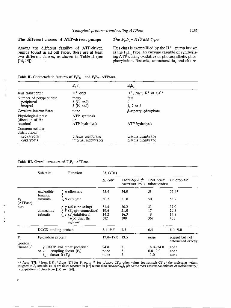
The different classes of ATP-driven pumps The F, FI - A TPase type
Among the different families of ATP-driven This class is exemplified by the H+ -pump known pumps found in all cell types, there are at least as the F,F, type, an enzyme capable of synthesiz- two different classes, as shown in Table II (see ing ATP during oxidative or photosynthetic phos- ~4,151). phorylation. Bacteria, mitochondria, and chloro-
Table II. Characteristic features of FIFO- and EIE2-ATPases.
I FOFI EIE2
1 Ions transported H+ only H+, Na+, K+ or Ca2+ Number of polypeptides : many few
peripheral 5 (E. coli) O integral 3 (E. coli) 1, 2 or 3
Covalent intermediates none P-aspartyl-phosphate Physiological poise ATP synthesis (direction of the or reaction) ATP hydrolysis ATP hydrolysis Common cellular distribution :
prokaryotes plasma membrane plasma membrane eukaryotes internal membranes plasma membrane
Table III. Overall structure of F,F,-ATPase.
Subunits Function ( k W
E. colia Thermophilicb Beef heartC Chloroplastd bacterium PS 3 mitochondria
a allosteric 55.4 54.0 53 55.4++
ß catalytic 50.2 51.0 50 53.9
I 6 (F,-aßy-connecting) 19.6 21.0 17 20.8
1
y (aß-connecting) 31.4 30.2 33 37.0
B (FI-inhibitory) 14.2 16.3 8 14.9 saccording the 382 380 367 401
I I
nucleotide binding
FI subunits (ATPase)
@3ß3Y6Ee
7 part connecting subunits
DCCD-binding protein 8.4-9.5 7.3 6.5 6.0-9.0
FO F,-binding protein 17.0-19.0 13.5 none present but not (proton channel)'
determined exactly OSCP and other proteins: 24.0 ? 18.0-24.0 none
coupling factor (F6) none ? 8.0-9.0 none Or I factor B (Fa none ? 13.0 none
a, from [17]; from [18]; from 1171 for F, part: * for tobacco CF,; other values for spinach CF,; e the molecular weight assigned to F, subunits (a-s) are those reported in [171 recent data consider a& y& as the most reasonable estimate of stoichiometry;
compilation of data from [19] and [20].
2

1266 B. Marin et al.
Plasts have very nearly the same molecular form of F,F,-ATPase. Recent information concerning their amino acid sequences is consistent with the en- zyme being derived from a common ancestral species [16]. Tables I, II and III summarize the structure and the chemistry of the different ATPases belonging to this type.
A characteristic feature of the F,F,-type ATP- ase is its extraordinary complexity. The enzyme of Escherichia coli plasma membrane, for exam- ple, consists of eight different proteins and approx- imately 18 peptide chains (Table III). The mito- chondrial enzyme is even more complex. In con- trast, the ATPase of animal (or plant) plasma mem- branes contains only two proteins (Tables I and 11).
The overall organization of this ATPase is as follows : the enzyme is built from two major func- tional units, distinguishable by their solubilities and their modes of binding to the membrane (Table III). The Fi part possesses all the catalytic
, sites. Usually, this part can be extracted from the ’ membrane by non-destructive treatments (dilution
in a solution of a chelating agent at low ionic strength, for example). All known examples of the F, part, described in the literature, comprise five different subunits, named a through e in order of decreasing size (Table III). The properties of each subunit are well established [19]. The isolation of
’ the F, and F, components of H + -ATPase from a thermophilic bacterium and E. coli has enabled us to determine their physicochemical and biological properties [18,19]. Five subunits are found in F,: a and ß are the nucleotide binding sites, 6 and E are the connecting bridge between F,
F, and link the aß complex to the SE complex. In addition, it is often suggested that a is an allosteric site, ß is a catalytic site, and $6 is an H + gate. The binding of nucleotides to a and ß changes the conformation of the latter. The protein com- ponents of F, of the ATPase are more integrated into the membrane than the F, part, and treatments with solvents or detergents (which destroy the membrane) are required to solubilize them. The complexity of F, polypeptides varies from one coupling membrane to another. It depends also upon the purity of the isolated H+-ATPase. Thus, the F, part of E. coli contains three polypeptides of approximate molecular weights of 24, 17 and 9.5 kDa [17]. Chloroplast F, also appears to contain only three or four polypep- tides, while mitochondrial F, contains two or three additional proteins [20]. In addition, the stoichiometry of F, polypeptides has not been established with certainty. Nevertheless, the smallest subunit which has the remarkable proper-
ty of being soluble in chloroform-methanol is pres- ent in 6-10 copies per F, [17]. All H+-ATPases of this FoFl type contain this hydrophobic subunit defined as the DCCD-binding protein. Thus, in E. coli, among the three subunits of F,, the DCCD- binding protein forms a highly hydrophobic part of the H f channel, and the F,-binding protein specifically binds F, 6 and e [19].
F,F,-activity can be reconstituted from a highly purified F! part and the crude F, part. F, is hydrophobic and embedded in biomembranes and thus is difficult to purify without a loss of activity. Fa's H + channel activity is measured after its in- corporation into liposomes. The flow of protons through F, could be coupled to ATP synthesis or hydrolysis by F,, under appropriate conditions [21]. When an artificial proton-motive force is provid- ed through imposed electrical and proton concen- tration gradients, the reconstituted vesicles catalyze ATP synthesis. The main function of this type of enzyme, the mitochondrial H+ -ATPase for ex- ample, is to utilize the transmembrane electrochem- ical gradient of protons for the synthesis of ATP.
F,F, is most often considered as an ATP- synthase, but it operates in either a synthetic or hydrolytic mode (Table II). In anaerobic growth of E. coli, for example, F,F, mediates ATP hydro- lysis and H + extrusion to maintain the proton- motive force used by secondary porters [15]. In- deed, this is normal for anaerobic bacteria. Similar- ly, F,F, (or a nearly related form, as suggested in Tables II and III) also functions as an ATP- synthase or hydrolase in eukaryotes, with its mode of operation depending upon location rather than circumstance. Thus, F,F, in mitochondria or chloroplasts is an ATP-synthase driven by the proton-motive force from respiratory or photoredox reactions. In the other eukaryotic F,F,-ATPases, its function is to hydrolyze ATP under the physiological conditions observed and described.
Many other biological membranes have F,F,-like ATPases, orientated to yield a reversed proton-motive force, acid and positive inside. Nevertheless, as suggested elsewhere [ 141 , assign- ment to the F,F, family requires an ATP- dependent H+ transport, sensitive to proton con- ductors but not affected by vanadate. A survey of the literature permits us to establish the list of candidates (Table IV). This table simply sug- gests that mechanistic homologies link these groups of H + pumps. We do not imply necessary struc- tural resemblances. In fact, the structure of such newly studied F,F,-like H + pumps is unclear and the published data on chromaffin granules, for
.

Tonoplast proton- translocating A TPase 1267
Table IV. Distribution of FIFO-ATPases.
Group Location References
Prokaryotes Eukaryotes
Eukaryotes fungi
plant cells .i
animal cells
all forms studied all mitochondria all chloroplasts other locations
vacuoles : N. crassa S. cerevisiae S. carlsbergensis
Avena sativa Beta vulgaris Glycine max Raphanus sativus Zea mays
Beta vulgaris Hevea brasiliensis Kalanchoe daìgreinontiana Tulipa sp.
Golgi vesicles lysosomes endoplasmic reticulum Golgi vesicles chromaffin granules synaptic vesicles synaptosomes mast cell granules pituitary storage granules (renal) exocytic vesicles platelet granules coated vesicles (endosomes) clathrin-coated vesicles
microsomes :
vacuoles :
[17, 22-24] [17, 251 [17, 26-29]
Table V. Distribution of EIE,-ATPases.
Location Ion substrate@) References
Prokaryotes ,Y
S. faecalis Na+ /(H+ ?) [911 S. faecalis Ca2+/? [921 S. faecalis K+/? 1931 E. coli K+/? [941 Acholeplasnia laidalwii (H+, Na+?)/? [951
1
Eukaryotes fungi
plant cells
animal cells
plasma membrane 1 H+/? [96-1031
plasma membrane H+ /(K ?) [104-1091
plasma membrane 3 Na+/2 K+ [ 110- 1 1 11 plasma membrane 1 Ca2+/2 H +
sarcoplasmic reticulum Ca2+ /? [1161 gastric mucosa 1 Hc / l K + [ 113- 1 151

1268 B. Marin et al.
0.2 0.1 -
example, indicate a very different subunit com- position [71,72].
!'\
L 0 . 3 : J II ' I
The E,E,-ATPase type
The second class of ATP-driven pumps has been collectively termed the ElE2-ATPase type accord- ing the terminology introduced recently [ 141. These ATP-driven pumps cycle between two stable states, with or without covalently bound phosphate (Table II). To classify an ATP-driven pump as EIE,-type, it must exhibit either of the two follow- ing criteria : ATP-driven transport sensitive to vanadate and/or identification of a phosphopro- tein. In Table V, the direction of ion transport is shown to be away/toward the ATP hydrolytic site. If known, stoichiometry is indicated. References cited are some recent examples that give an available entry into the literature concerning this class of ATP-driven pumps.
EIE2-type pumps show a broad ionic specificity (H+, Na+, K + , or Ca2+) accompanied by a remarkably simple structure. All known pumps of this class use a 100 kDa polypeptide that becomes phosphorylated at aspartate. The plasma mem- brane ATPase of Neurospora crassa and Beta vulgaris involves a covalent intermediate, the ß- aspartyl-phosphate [99, 106, 1071. Amino acids flanking this residue are nearly the same in ex- amples of very different origins [117]. The presence of phosphoprotein provides a useful operational
criterion for family membership, since its forma- tion confers sensitivity to the transition state in- hibitor, vanadate. As sequence data emerge, relatedness in the family may become even more apparent : J.E. Hesse et al. [94], for example, find a homology in the 100 kDa polypeptides of pro- karyote and eukaryote ElE2-ATPase.
Present knowledge about the structure of tonoplast ATPase
Little information exists about the polypeptide composition of tonoplast ATPase. This is due in part to the difficulty encountered in the solubiliza- tion and purification of the entire enzyme (see [ 1, 39, 52, 1181).
The tonoplast ATPase of Hevea latex has been solubilized with dichloromethane and purified 100-fold [52]. The resulting ATPase enzyme (bear- ing a hydrolytic enzyme) has a molecular weight of approximately 200 kDa (Fig. 1). Its probable assembly consists of five subunits with molecular weights of 66,23, 13 kDa and two 54 kDa subunits which normally remain together except for drastic SDS treatment. This composition indicates that this ATPase is probably not related to the plasma mem- brane system [97,98,101,104-1081. The subunit pattern is also not strictly comparable to those reported for the FI part of the FoFla-ATPase (see [18] and Tables I and III). In addition, the lack
i-

Tonoplast proton- translocating A TPase 1269
sugarcane tonoplast I
\ I / Hevea tonoplast
Fig. 2. Antibody diffusion test of Hevea tonoplast ATPase pro- teins with anti-F,. Solubilized proteins from tonoplast mem- branes were allowed to diffuse against antiserum to E. coli F,-ATPase. The reaction with bacterial F,-ATPase is also shown for control. These data clearly show that a polyclonal antibody against F,-ATPase of E. coli does not react with the vacuolar ATPase of Hevea latex (from [52]).
of cross-reaction between anti-F, and the ATPase does not suggest a close relationship with F,-type ATPases (Fig. 2).
Another tonoplast ATPase has been solubilized from vacuolar membranes of Saccharomyces cere- visiae with the zwitterionic detergent N-tetradecyl-N, N-dimethyl-3-ammonio-l-propane sulfonate and purified by glycerol density gradient centrifugation [33]. It was found to be composed of two major polypeptides a and b, respectively, of M, = 89 and 64 kDa. Such a subunit composition of the purified enzyme is entirely different from that of F,F,- ATPase and that of E,E2-ATPase. This enzyme was not inhibited by antiserum directed against mitochondrial F,-ATPase or mitochondrial F,-ATPase inhibitor protein. All these data clear- ly indicate that tonoplast ATPase is different from yeast plasma membrane H+ -ATPase and mitochondrial F, -ATPase.
A NO ;-sensitive ATPase activity, described as being of tonoplastic origin, has been solubilized from microsomal membranes of radish seedlings with octyl-P-D-glucoside [39]. The method led to an approximately 35-fold purification from the microsomes. The final preparation exhibited several bands. Eight components whose molecular masses were not indicated. It was strongly enriched in
t
J
an 86 kDa polypeptide. This author reported that if this large polypeptide belonged to the tonoplast ATPase, it would not agree at all with a structural similarity with the F,F,-ATPase.
Actually, on the basis of the recent data concern- ing the purified tonoplast ATPase from Hevea latex, a function has been assigned to each comp.0- nent. The 66 kDa subunit seems to bind nucleotides and SH reagents. Different studies with various ATP analogs indicate that this subunit may con- tain the catalytic binding site. Significant results have been obtained with NBD-C1 (7-chloro-4-
I ' I I I
o ' I I I I 1 I I O 2 4 6
inhibitor (pmol.mg protein -1)
Fig. 3. Effect of the different carbodiimides on Hevea tonoplast ATPase. Experiments were performed at 30°C with the follow- ing incubation medium : 50 mM Mes, 50 mM Hepes and 5 mM ,O-mercaptoethanol adjusted to pH 7.0 with Tris-base. The medium also contained 0.1 mM ammonium molybdate and 5 mM Mg*+ (as SO:- salt). Usually, the mixture was prein- cubated for 10 min before starting the reaction with the addi- tion of ATP (5 mM final concentration). Tonoplast membranes were assayed for ATPase activity in the presence of increasing amounts of inhibitor : 1 -cyclohexyl-3-(2-morpholinyl-4-ethyl)car- bodiimide metho-p-toluenesulfonate (CMCD) (-A-& ), N,N- dicyclohexylcarbodiimide (DCCD) ( -c+s- ), N,N-diisopropyl- carbodiimide (DIPCD) (-&A-), or l-ethyl-3-(3-dimethyl- aminopropy1)carbodiimide (EDAC) (-). Each assay con- tained 100 pg of protein/ml. Control activity was 0.21 (*mol/ min/mg of protein (from [47]).

1270 B. Marin et al.
Table VI. ATP-hydrolyzing activities present in plant homogenates.
Activity' Optimum pH Substrate specificity Inhibitors Requirements for divalent cations
Plasma membrane ATPase 6-7 Mitochondrial ATPase 8-9
Tonoplast ATPase 7-8 Pyrophosphatase 7.5-9
Acid phosphatase 5-6
Alkaline phosphatase 8-9
Apyrases (diphosphohydrolases) 6-7
absolute for ATP ATP, GTP, ITP, UTP
ATP, GTP, ITP, UTP pyrophosphate and nucleotide triphosphate many nucleotides and phosphoric esters many nucleotides and phosphoric esters
nucleotide di- and triphosphate
vanadate, DCCD, DES oligomycin, azide, DCCD DIDS, TMT, DCCD fluoride
vanadate
vanadate
unknown
Mg2+ (no Caz+) Mg2+ or Ca2+
Mg2+, Cazf? no effect
none
none
Mg2 +
and/or Ca2+
Abbreviations : DCCD : N,N-dicyclohexylcarbodiimide ; DES : diethylstilbestrol ; DIDS : 4,4'-diisothiocyano-2,2'-stilbene disulfonic acid ; TMT : trimethyltin.
nitrobenzo-2-2-oxa-l,3-diazole). The 53 kDa subunit can bind nucleotides with different affi- nities. Perhaps, it also participates in ATPase reac- tions. Such an interpretation must be confirmed. In addition, the 13 kDa subunit binds DCCD and TMT. Consequently it may be tempting to consider it as a part of the proton channel. Such results will reveal some components common to all ATPases capable of catalyzing a transmembrane flux of protons.
The usefulness of inhibitors in classifying the tonoplast ATPase
As described elsewhere [12,48,119], the lutoids of Hevea latex must be considered as a vacuolar com- partment. The lutoid preparations were uncon- taminated with membranes from other organelles of the plant cell (chloroplasts, mitochondria, etc.). Usually, plant homogenates contained a lot of activities capable of hydrolyzing ATP (Table VI). A problem of purification exists and many dif- ficulties are encountered when studying the tonoplast. Consequently, with Hevea latex, it has been possible to determine the precise sensitivity of the tonoplast ATPase to a great variety of agents, which selectively inhibit the different classes of H+-translocating ATPases (Table I and Figs. 3, 4, 5 and 6).
100
80
h - 2 $ 60 .I- S
s v
.I- .- > o a cl) > a
.= 40
.-
.I-
- 22 20
O
I I l l I I I
I I I I I 1
O 0.5 1 .o 1.5 inhibitor (pmol.mg protein-1)
Fig. 4. Effect of trimethyltin chloride on Hevea tonoplast AT- Pase. Reaction conditions are the same as those described for Fig. 3. Control activity was 0.24 pmol/min/mg of protein (from [471)*
b

Tonoplast proton - translocating A TPase 1271
Effect of DCCD, TMT, NBD-CI and ATP analogs on tonoplast A TPase. Relationship with F, F, -A TPases
DCCD has previously been described as an effec- tive inhibitor of Hevea tonoplast ATPase [43,44,47]. 50% inhibition was observed at 1.26 pmol of inhibitor/mg of protein with purified enzyme. DCCD is the most effective of the car- bodiimides tested [47] (Fig. 3).
Like DCCD, the organotins inhibit proton conductivity through the F, portion of the H+ - ATPase [120]. They act by binding to a proteo- lipid subunit, probably near but not identical with the site of DCCD binding. Among the different trialkyltin chlorides tested, trimethyltin and tributyltin were the most effective. Hevea tonoplast ATPase was inhibited 50% at 0.32pmol of in- hibitor/mg of protein and a nearly complete inhibi- tion was obtained at 1 pmol of inhibitor protein (Fig. 4). Such inhibition has also been reported for the vacuolar ATPase of fungi [3 1 , 331 and higher plants [l 1,39,53], The organotins inhibit the ATPases of mitochondria [121,122], chloroplasts [123] and chromaffin granules [77,79,80] in the same range of concentrations. These organotins also inhibit the plasma membrane ATPase but at significantly higher concentrations , as shown in N. crassa [3 11.
NBD-C1 constitutes a very powerful inhibitor of Hevea tonoplast ATPase. The half maximal effect was obtained at 1.4 pM and a value of 2.0 pM was calculated for Ki. Comparable values were obtain- ed for the vacuolar membrane of N. crassa [31]. NBD-C1 was also shown to inhibit F,Fl- ATPases from mitochondria [121,124,125] and chloroplasts [126,127]. It also inhibits the H+-ATPase of E. coli [128,129] and the ATPase from chromaffin granules [77-SO]. This molecule was effective on the soluble and membranous form of the F,F,-ATPase. It interacts specifically with the /3-subunit of the F,-ATPase [124,125,127,129]. The mitochondrial ATPase was inhibited 50% at 3.0 ,UM for N. crassa [31], whereas the plasma membrane ATPase of N. crassa showed a 5-10- fold lower affinity, with 50% inhibition being ob- tained at 14 pM [3 11.
Different ATP analogs have been described as effective inhibitors of F,F,-ATPase [126,130-1321. TNP-ATP and AMP-PNP were found to inhibit Hevea tonoplast ATPase with 50% inhibition at 10-15 pM and 3-5 pM, respectively, (Fig. 5). The Ki value calculated for the tonoplast enzyme was approximately 0.6-0.8 pM. The same results have been described for the mitochondrial and vacuolar
1
f
membrane ATPases of N. crassa with somewhat similar effectiveness (50% inhibition at 6 and 10 pM, respectively, and a Ki value of 0.6 pM for both enzymes) [3 11. The plasma membrane ATPase of N. crassa was found to be approximately 10-fold less sensitive (50% inhibition at 100 pM) to ATP analogs [3 11.
Effects of orthovanadate and quercetin on ton op last A TPase . R elat ions h ip w Ìt h E,E2-ATPases
Orthovanadate has been described as a specific in- hibitor of plasma membrane ATPases in animal and plant cells [101,133,134]. This molecule pro- duces no significant inhibition of Hevea tonoplast ATPase, even at a concentration as high as 1 mM (Fig. 6).
Hevea tonoplast ATPase was inhibited by quercetin with 50% inhibition at 20-25 pM (see [l , 118, 1351). This inhibition was competitive (Fig. 7). The Hevea enzyme was found to be near- ly as sensitive to this inhibitor as the tonoplast mem- brane ATPase of N. crassa (50% inhibition at 22 and 24 pM, respectively [31]). This molecule was 10-fold less effective against the plasma membrane ATPase of N. crassa (50% inhibition at 170 ,UM
Phenolic compounds, especially quercetin, have been reported to inhibit a great variety of mem- brane ATPases [101,127,128,136,137]. Quercetin is not a specific inhibitor of this class of ATPase because it can also inhibit the hydrolytic activity of the mitochondrial F,Fl enzyme, without inhibiting the ATP-synthase [121].
Such studies confirm the originality of the tonoplast ATPase. This ATPase activity is clearly distinguished from plasmalemma ATPase by its in- sensitivity to vanadate. It can also be casily distinguished from mitochondrial ATPase by its insensitivity to azide and oligomycin.
[311).
The sensitivity of tonoplast ATPase to anions
The effect of CI- and other anions on the tonoplast ATPase from Hevea latex has been in- vestigated [1,48,50].
C1- and other anions stimulated the ATPase ac- tivity of tightly-sealed vesicles prepared from Hevea tonoplast, with the following decreasing order of effectiveness : C1- > Br- >SO:- >NO;. NO; could be regarded as a potent inhibitor of this enzyme.

1272
100
ao n - 2 .I- S
60 s
> > o (d
a, > (d
v
+ .- 'G 40
.-
.I-
- 2 20
O
B. Marin et al.
, I l l I l
I l I l l I
O 5 i0 i 5 20 25 ATP analog (,UM)
Fig. 5. Effect of ATP analogs on tonoplast ATPase activity of Hevea latex. Reaction conditions are the same as those describ- ed for Fig. 3. 2;3'0-(2,4,6-trinitro-phenyl)-adenosine/-5'- triphosphate (TNP-ATP) (-o-+) ; 5'-adenylylimidodiphosphate (AMP-PNP) (-o-+). Control activity was 0.30 pmol/min/mg of protein (measured according to [47] and [52]).
'/" 2.5 1
io0
ao n - F 8 60 4- S
s A
>
(d
a, > (d
v
.I- .-
.- 40
.-
.I-
- 2 20
O
. . .
I l l l l I I
O 0.4 0.8 1.2 orthovanadate (mM)
Fig. 6. Effect of orthovanadate on Hevea tonoplast ATPase. Reaction conditions are the same as those described for Fig. 3. Control activity was 0.25 pmol/min/mg of protein (from [47]).
I , I I I I I , I I
O 0.1 0.2 0.3 quercetin (mM)
Fig. 7. Dixon plots for quercetin inhibition of Hevea tonoplast ATPase. The assay was started after 15 min of preincubation with either 2 mM ATP (-o+-), 4 mM ATP (-.t) or 6 mM ATP (U) (see [l] and [Ils]).
\
i
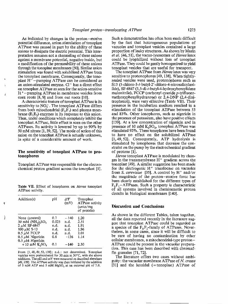
Tonoplast proton-translocating ATPase 1273
As indicated by changes in the proton-motive potential difference, anion stimulation of tonoplast ATPase was caused in part by the ability of these anions to dissipate the electric potential. This inter- pretation assumes not a channeling of these anions against a membrane potential, negative inside, but a modification of the permeability of these anions through the tonoplast membranes [50]. Similar anion stimulation was found with solubilized ATPase from the tonoplast membrane. Consequently, the tono- plast H+ -pumping ATPase can be considered as an anion-stimulated enzyme. C1- has a direct effect
H+-pumping ATPase in membrane vesicles from corn roots [8, 91 and from oat roots [lo].
A characteristic feature of tonoplast ATPase is its sensitivity to NO,. The tonoplast ATPase differs from both mitochondrial (FoFl) and plasma mem- brane (EIE,) enzymes in its response to this anion. Thus, under conditions which completely inhibit the tonoplast ATPase, little effect is seen on the other ATPases. Its activity is lowered by up to 90% by 50 mM nitrate [l, 39,521. The mode of action of this anion on the tonoplast ATPase is actually unknown, in spite of a considerable amount of work.
I
d
l on tonoplast ATPase as seen for the anion-sensitive
The sensitivity of tonoplast ATPase to pro- tonophores
Tonoplast ATPase was responsible for the electro- chemical proton gradient across the tonoplast [l],
Table VII. Effect of ionophores on Hevea tonoplast ATPase activity.
Addition(s) pH A Y Tonoplast
(unitdmg of protein)
4 (mV) ATPase activity
J None (control) 0.7 -140 1.20 50 mM (NH,),SO, 0.025 n.d. 2.51 2 pM SF-6847 n.d. n.d. 1.91 100 pM S-13 a d . n.d. 1.96 0.5 pM FCCP n.d. a d . 2.01 0.5 pM Nigericin 0.8 -136 1.14 0.5 pM Nigericin
+10mMK,SO4 0.1 -140 2.30
From [l , 48,49,52,139] ; n.d. : not determined. Tonoplast vesicles were preincubated for 30 min at 30°C, with the above additions. The pH and mV were measured as described elsewhere [49,139]. The ATPase activity was then initiated by the addition of 5 mM ATP and 5 mM MgSO, at an external pH of 7.0.
Such a demonstration has often been made difficult by the fact that heterogeneous populations of vacuoles and tonoplast vesicles contained a large proportion of leaky structures. As shown by Marin et al. [46,5 11 , the vacuo-lysosomes of Hevea latex could be lyophilized without loss of tonoplast ATPase. They could be gently homogenized to yield tonoplast vesicles that are useful for transport.
The tonoplast ATPase from Hevea latex was very sensitive to protonophores [49,138]. When tightly- sealed vesicles were used, protonophores such as S13 (5-chloro-3-t-butyl-2'-chloro-4-nitrosalicylani- lide), SF-6847 (3,5-di-t-butyl-4-hydroxybenzylidene malonitrile), FCCP (carbonyl cyanide p-trifluoro- methoxyphenylhydrazone) or 2,4-DNP (2,4-dini- trophenol), were very effective (Table VII). Their presence in the incubation medium resulted in a stimulation of the tonoplast ATPase between 53 and 83%. Other ionophores, such as nigericin in the presence of potassium, also have positive effects [139]. At a low concentration of nigericin and in presence of 10 mM K,SO,, tonoplast ATPase was stimulated 92%. These ionophores have been found to have no effect on the solubilized ATPase [l , 49,521. Consequently, ATP hydrolysis is stimulated by ionophores that decrease the con- straint on the pump by the electrochemical gradient of protons [l].
Hevea tonoplast ATPase is modulated by chan- ges in the transmembrane H+ gradient across the tonoplast [49]. A similar suggestion has been made for the electrogenic H+ translocase on vacuoles from S. cerevisiae [35]. A control by H+ and/or the magnitude of the proton-motive force has been clearly established for the different types of FoFl -ATPases. Such a property is characteristic of all systems involved in chemiosmotic proton circuits in biological membranes [ 1401.
Discussion and Conclusions
As shown in the different Tables, taken together, all the data reported recently in the literature sug- gest that tonoplast ATPase could be regarded as a species of the FoFl-family of ATPases. Never- theless, in some cases, since it will be difficult to be sure of having no contamination by other cellular membranes, a mitochondrial-type proton- ATPase could be present in the vacuolar prepara- tion. This case has been described with chromaf- fin granules [7 1 , 721.
The literature offers two cases without ambi- guity : the vacuolar membrane ATPase of N. crassa [31] and the lutoidal (=tonoplast) ATPase of

1274 B. Marin et al.
Hevea latex [1,43-52,118,119]. In the first case, a careful analysis of the different mutants of the mitochondrial ATPase allowed rigorous testing to determine whether the vacuolar mem- brane ATPase activity could be due to mitochon- drial contamination. In addition, it was possible to compare directly the vacuolar membrane ATPase with the mitochondrial and plasma membrane ATPases. This comparison is not possible with Hevea latex. Nevertheless, the principal advantage of this material is the ability to obtain a large frac- tion of uncontaminated vacuoles involving neither cell rupture treatments nor lytic attack by cell-wall- degrading enzymes. Consequently, the first evidence as to the nature of the tonoplast ATPase has come from the inhibitor studies on these two materials.
On thinking it over, it must be concluded that the tonoplast ATPase is different from the E,E,-ATPase type. It is insensitive to vanadate, even if it is sensitive to quercetin. The effect of vanadate on this ATPase class is to prevent the for- mation of a covalent phosphorylated intermediate (often a P-aspartyl-phosphate) during their reaction cycle [101,104-107,133,134]. At least with Hevea, such an intermediate has not been evidenced. Vanadate inhibits phosphorylation of the plasma membrane ATPase because this pentavalent ion competes with phosphate for binding and adopts a stable trigonal bipyramidal structure resembling the transition state of phosphate during the reac- tion. Consequently, the insensitivity of tonoplast ATPase to vanadate suggests that such a phos- phorylated intermediate is not involved in the mechanism of this enzyme. Certainly, quercetin is often regarded as a specific inhibitor of the E,E?-ATPase type, however, its effect is not con- clusive because it also inhibits the hydrolysis of ATP catalyzed by the mitochondrial ATPase [121,137].
The tonoplast ATPase also differs from the F,F, -ATPase type because it is insensitive to various chemicals such as azide and oligomycin. These molecules, described as typical inhibitors of the mitochondrial ATPase, have no effect on the tonoplast ATPase even at high concentra- tions. But, these differences are not sufficient to exclude the tonoplast ATPase from the F,F,- ATPase type. Its response to inhibitors which block proton translocation such as DCCD [138] and organotins [120,122,124,125] resembles the mitochondrial ATPase. In addition, the sensitivity of the tonoplast ATPase to inhibitors which pro- bably react at the active site of the enzyme (such as NBD-CI or the different ATP analogs) is very
similar to that of the mitochondrial ATPase. The substrate specificity of tonoplast ATPase is com- parable with that described for the mitochondrial enzyme. The K, values for MgATP are about 0.3 mM for both enzymes. In some cases, Ca2+ and Mgzt are similarly effective for their ATPase activities. Consequently, the tonoplast ATPase must be considered as a good candidate for the FqF,-ATPase class. It complies with all the criteria which define this class of ATP-driven ion pump (see Table II). Nevertheless, the structure is somewhat different, not as complex (at least 4 or 5 polypeptides for tonoplast ATPase instead of 20-24 subunits for the ATP-synthase). Such a molecular composition will be able to assume only the hydrolysis of ATP.
The different proton pumps located inside the eukaryotic cell could belong to the F,F, family. Table IV may be considered as speculative, link- ing all the ATPases by their mechanistic homologies. This does not necessarily imply struc- tural resemblances [14]. The different electropho- resis patterns of the purified tonoplast ATPase prepared from Hevea latex, Ruphunus seedlings and Saccharomyces confirm this point of view. The tonoplast ATPase is clearly distinguishable from the mitochondrial F,F,-ATPase and from the plasmalemma EIE,-ATPase. The same situation could be obtained with the ATPase of chromaffin granules : the purified enzyme consists of four ma- jor polypeptides with apparent molecular weights of about 125,80,40 and 20 kDa [72]. The ATPase of the cholinergic synaptic vesicle membrane cor- responds to a 250 kDa ATP-hydrolyzing molecule, composed of five polypeptides of 50 kDa each [141]. Any conclusions as to phylogenetic relation- ships have to wait for comparison of antibody specificities and amino acid sequences.
If the differences observed in the structure of these new F,F,-like ATPases justify the definition of another type, it could suggest the existence of a third class of ATP-driven pumps, intermediate between F,F,-ATPases and E,E,-ATPases. A characteristic feature of this class should be its sen- sitivity to anions : inhibition by NO, and stimula- tion by Cl-.
Acknowledgements
The authors are grateful to Dr. M. Czechowski for his help in preparing this manuscript and Mrs. M. Marin- Lanza for typing the manuscript and for drawing the dif- ferent figures.

Tonoplast proton-translocating A TPase 1275
References
3
4
5
6
7
8
9
10
11
12 13
14
15 16
17 18
19
v
I l
d
20
21
Y 22
23
b 24
25
26
27 28 29
30
1 Marin B. (1985) in: Biochenzistry and Function of Vacuolar Adenosinetriphosphatase in Fungi and Higher Plants, Springer-Verlag, Berlin, pp. 259
2 Bennett A.B., O’Neill S.D. & Spanswick R.M. (1984) Plant Physiol. 74, 538-544 Churchill K.A., Holaway B. & Sze H. (1983) Plant Physiol. 73, 921-928
I De Michelis M.I., Pugliarello M.C. & Rasi- Caldogno F. (1983) FEBS Lett. 62, 85-90 Lew R.R. & Spanswick R.M. (1984) Plant Sci. Lett. 36. 187-193 Poole R.J., Briskin D.P., Kratky Z. & Johnstone R.M. (1984) Plant Pliysiol. 74, 549-556 O’Neill S.D., Bennett A.B. & Spanswick R.M. (1983) Plant Physiol. 72, 837-846 Bennett A.B. & Spanswick R.M. (1983) J. Membr. Biol. 71, 95-107 Bennett A.B. & Spanswick R.M. (1983) J. Membr. Biol. 75, 21-31 Churchill K.A. & Sze H. (1983) Plant Physiol. 71,
Churchill K.A. & Sze H. (1984) Plant Physiol. 76,
Marin B. (1982) Trav. Doc. ORSTOM 144, 1-409 Marin B., Crétin H. & D’Auzac J. (1982) Physiol. Veg. 20, 333-346 Maloney P.C. &Wilson T.C. (1985) Bioscience 35, 43-48 Maloney P.C. (1982) J. Menzbr. Biol. 67, 1-12 Dayhoff M.O. &Schwartz R.M. (1981) Ann. N. Y. Acad. Sci. 361, 92-103 McCarty R.E. (1985) Bioscience 35, 27-30 KagawaY., SoneN., Hirta H. &Yoshida M. (1979) J. Bioenerg. Bionzeinbr. 11, 39-78 Kagawa Y. (1982) in: Membranes and Transport (Martonosi A.N., ed.), Vol. 1, Plenum Publishing Corp., New York, pp. 439-446 Houstek J., Kopecky J., Svoboda P. & Drahota Z. (1982) J. Bioenerg. Bionzenzbr. 14, 1-13 Knox B.E. &Tsong T.Y. (1984) J. Biol. Chem. 259,
Futai M. & Kanazawa H. (1980) Curr. Top. Bioenerg. 10, 181-215 Cross R.L. (1981) Annu. Rev. Biochem. 50,
Senior A.E. &Wise J.G. (1983) J. Membr. Biol. 73,
Senior A.E. (1973) Biochim. Biophys. Acta 301,
Nelson N. (1976) Biochinz. Biophys. Acta 456,
Nelson N. (1980) Ann. N. Y. Acad. Sci. 358,25-35 Nelson N. (1981) Curr. Top. Bioenerg. 11, 1-33 McCarty R.E. & Carmeli C. (1982) in: Photosyn- thesis (Govindjee R., ed.), Vol. 1, Academic Press, New York, pp. 647-690 Bowman E.J. &Bowman B.J. (1982) J. Bacteriol.
610-617
490-497
4757-4763
681 -714
105-124
249-277
314-338
151, 1326-1337
31 Bowman E.J. (1983) J. Biol. Chenz. 258,
32 Kakinuma Y., Ohsumi Y. & Anraku Y. (1981) J. Biol, Chem. 256, 10859-10863
33 Uchida E., Ohsumi Y. & Anraku Y. (1985) J. Biol. Chem. 260, 1090-1095
34 Okorokov L.A., Kulakovskaya T.V. & Kulaev IS. (1982) FEBS Lett. 145, 160-162
35 Okorokov L.A. & Lichko L.P. (1983) FEBS Lett.
36 Bennett A.B. & Spanswick R.M. (1984) Plant
37 Lew R.R. & Spanswick R.M. (1985) Plant Physiol.
38 De Michelis M.I., Rasi-Caldogno F, & Pugliarello
39 Tognoli L. (1985) Eur. J. Biochem. 146,
40 Hager A. & Helmle M. (1981) 2. Naturforsch. 36c,
41 Leigh R.A. & Walker R.R. (1980) Planta 150,
42 Walker R.R. & Leigh R.A. (1981) Planta 153,
43 D’Auzac J. (1975) Phytochemistry 14, 671-675 44 D’Auzac J. (1977) Phytochemistry 16, 1881-1885 45 Crétin H. (1982) J. Membr. Biol. 65, 174-184 46 Marin B. (1983) Planta 157, 324-330 47 Marin B. (1983) Plant Physiol. 73, 973-997 48 Marin B. (1984) in:Membrane Transport in Plants
(Cram W. J., Janacek K., Rybova R. & Sigler K., eds.), Academia Publishing House of C.S.S.R. Academy of Sciences, Prague, pp. 525-530
49 Marin B. (1985) Biochem. J. 229, 459-467 50 Marin B. & Gidrol X. (1985) Biochem. J. 226,
51 Marin B., Marin-Lanza M. & Komor E. (1981) Biochem. J. 198, 365-372
52 Marin B., Preusser J. & Komor E. (1985) Eur. J. Biochein. 131, 131-140
53 Smith J.A.C., Uribe E.G., Ball E., Heuer S. & Lut- tge U. (1984) Eur. J. Biochem. 141, 415-420
54 Jochem P., Rona J.P., Smith J.A.C. & Luttge U. (1984) Physiol. Plant. 62, 410-415
55 Aoki K. & Nishida K. (1984) Plant Physiol. 60,
56 Wagner G.J. & Mulready P. (1983) Biochinz.
57 Binari L.L.W. & Racusen R.H. (1983) Plant
58 Taiz L., Murray M. &Robinson D.G. (1983) Plan-
59 Chanson A., McNaughton E. & Taiz L. (1984) Plant
60 Njus D., Knoth J. & Zallakian M. (1981) Curr. Top.
61 Schneider D.L. (1981) J. Biol. Chem. 256,
62 Schneider D.L. (1983) J. Biol. Chem. 258,
63 Dell’Antone P. (1982) FEBS Lett. 146, 181-188
15238-15244
155, 102-106
Physiol. 74, 545-548
77, 352-357
M.C. (1984) Plant Sci. Lett. 36, 111-117
581-588
997-1008
222-229
140- 149
85-94
21-25
Biophys. Acta 728, 267-280
Physiol. 71, 594-597
ta 158, 534-539
Physiol. 76, 498-507
Bioenerg. 11, 107- 147
3858-3864
1833-1838

1276 B. Marin et al.
64 Moriyama Y., Takano T. & Ohkuma S. (1982) J.
65 Moriyama Y., Takano T. & Ohkuma S. (1984) J.
66 Moriyama Y., Takano T. & Ohkuma S. (1984) J.
67 Okhuma S., MoriyamaY. &Takano T. (1982) Proc.
68 Okhuma S., Moriyama Y. & Takano T. (1983) J.
69 Okhuma S. & Poole B. (1978) Proc. Natl. Acad. Sci.
70 Harikumar P. &Reeves J.P. (1983) J. Biol. Chem.
71 Cidon S. & Nelson N. (1982) J. Bioenerg.
72 Cidon S. & Nelson N. (1983) J. Biol. Chem. 258,
73 Cidon S., Ben-David H. &Nelson N. (1983) J. Biol.
74 Rees-Jones R. &Al Awqati Q. (1984) Biochemistry
75 Glickman J., Croen K., Kelly S. & AI Awqati Q. (1983) J. Cell Biol. 97, 1303-1308
76 Zhang R. & Schneider D.L. (1983) Biochem. Biophys. Res. Commun. 114, 620-625
77 Apps D.K. &Schatz G. (1979) Eur. J. Biochem. 100,
78 Apps D.K., Pryde J.G., Sutton R. & Phillips J.H.
79 Johnson R.G., Beers M.F. & Scarpa A. (1982) J.
80 Beers M.F., Carty S.E., Johnson R.G. & Scarpa A.
81 Anderson D.C., King S.C. &Parsons S.M. (1983)
82 Toll L. &Howard B.D. (1980) J. Biol. Chem. 255,
83 Johnson R.G., Carty S.E., Fingerhood B.J. &Scar- pa A. (1980) FEBS Lett. 120, 75-79
84 Carty S.E., Johnson R.G. & Scarpa A. (1982) J. Biol. Chem. 257, 7269-7273
85 Gluck S., Connan C. &Al Awqati Q. (1982)Proc. Natl. Acad. Sci. USA 79, 4327-4331
86 Fishkes H. & Rudnick G. (1982) J. Biol. Chem. 257,
87 Johnson R.G., Scarpa A. & Salganicoff L. (1978) J. Biol. Chem. 253, 7061-7068
88 Stone D.K., Xie X.S. & Racker E. (1984) J. Biol. Chem. 259, 2701-2703
89 Forgac M., Cantley L., Wiedenmann B., Altstill L. & Branton D. (1983) Proc. Natl. Acad. Sci. USA
90 Stone D.K., Xie X.S. & Racker E. (1983) J. Biol. Chem. 258, 4059-4062
91 Heefner D.L. & Harold F.M. (1982) Proc. Natl. Acad. Sci. USA 79, 2798-2802
92 Kobayashi H., Van Brunt J. &Harold F.M. (1978) J. Biol. Chem. 253, 2085-2092
93 Hugentobler G., Heid I. & Solioz M. (1983) J. Biol. Chem. 258, 7611-7617
Biochem. 92, 1333-1336
Biochem. 94, 995-1007
Biochem. 96, 927-930
Natl. Acad. Sci. USA 79, 2758-2762
Biochem. 94, 1935-1943
USA 75, 3327-3331
258, 10403-10410
Biomembr. 14, 499-512
2982-2988
Chem. 258, 11684-11688
23, 2236-2240
411-419
(1980) Biochem. J. 190, 273-282
Biol. Chem. 257, 10701-10707
(1983) Ann. N.Y. Acad. Sci. 402, 116-133
Biochemistry 21, 3037-3043
1787-1789
5671-5677
80, 1300-1303
94 Hesse J.E., Wieczorek L., Altendorf K., Reicin A.S., Dorus E. & Epstein W. (1984) Proc. Natl. Acad. Sci. USA 81, 4746-4750
95 Jinks D.C., Silvius J.R. & McElhaney R.N. (1978) J. Bacteriol. 136, 1027-1036
96 Scarborough G.A. (1976) Proc. Natl. Acad. Sci.
97 Malpartida F. & Serrano R. (1981) J. Biol. Chem. USA 73, 1485-1488
. .
256,- 4175-4177 98 Malpartida F. &Serrano R. (1981) FEBS Lett. 131,
351-354 99 Dame J.B. & Scarborough G.A. (1981) J. Biol.
Chem. 256, 10724-10730 100 Villalobo A., Boutry M. & Goffeau A. (1981) J.
Biol. Chem. 256, 12081-12087 101 Goffeau A. & Slayman C.W. (1981) Biochim.
Biophys. Acta 639, 197-223 102 Perlin D.S., Kasamo K., Brooker R.J. & Slayman
C.W. (1984) J. Biol. Chem. 259, 7889-7992 103 Slayman C.L. (1985) Bioscience 35, 34-37 104 Vara F. & Serrano R. (1982) J. Biol. Chem. 257,
105 Vara F. & Serrano R. (1983) J. Biol. Chem. 258,
106 Briskin D.P. &Poole R.J. (1983) Plant Physiol. 71,
107 Briskin D.P. &Poole R.J. (1983) Plant Physiol. 72,
12826-12830
5334-5336
507-512
1133-1 135 108 Serrano R. (1984) Cur. Top. Cell. Regul. 23,
87-126 109 O’Neill S.D. & Spanswick R.M. (1984) J. Membr.
Biol. 79, 231-243 110 Hoffman J.F. & Forbush B. III (1983) Curr. Top.
Membr. Transport, 19, 1-452 111 Craig W.S. & Kyte J. (1980) J. Biol. Chem. 255,
626216269 112 Carafoli E., Zurini M., NiggliV. &Krebs J. (1982)
Ann. N.Y. Acad. Sci. 402, 304-326 113 Sachs G., Chang H., Rabon E., Schackmann R.,
Lewin M. & Saccomani G. (1976) J. Biol. Chem.
114 Rabon E.C., McFall T.L. & Sachs G. (1982) J. Biol. Chem. 257, 6296-6299
115 Forte J.G. & Reenstra W.W. (1985) Bioscience 35,
116 Hasselbalch W. & Waas W. (1982) Ann. N. Y. Acad. Sci. 402, 459-469
117 Walderhaug M.O., Post R.L., Saccomani G., Leonard R.T. & Briskin D.P. (1985) J. Biol. Chem.
118 Marin B. (1987) in: Plant Vacuoles: Their Impor- tance in Plant Cell Compartmentation and Their Applications in Biotechnology, Plenum Publishing Corp., New York (in press)
119 D’Auzac J., Crétin H., Marin B. & Lioret C. (1982) Physiol. Veg. 20, 311-331
120 Dawson A.P., Farrow B.G. & Selwyn M.J. (1982) J. Biochem. 202, 163-169
121 Linnett P.E. & Beechey R.B. (1979) Methods En- zymol., 55, 472-518
122 Papa S., Guerrieri F., De Gomez-Puyou M.T., Bar-
251, 7690-7698
38-42
260, 3852-3859

Tonoplast proton - translocating A TPase 1277
ranco J. & Gomez-Puyou A. (1982) Eur. J. Biochem. 128, 1-7
123 Gould J.M. (1976) Eur. J . Biochein. 62,
124 Ferguson S.J., Lloyd W.J., Lyons M.H. & Radda G.K. (1975) Eur. J. Biochem. 54, 117-126
125 Ferguson S.J., Lloyd W.J. & Radda G.K. (1975) Eur. J. Biochem. 54, 127-133
126 Cantley L.C. Jr., & Hammes G.G. (1975) Biochemistry 14, 2968-2975
127 Deters D.W., Racker E., Nelson N. & Nelson H. Y (1975) J. Bio[. Chem. 250,, 1041-1047 I f 128 Futai M., Sternweis P.C. & Heppel L.A. i (1974) Proc. Natl. Acad. Sci. USA 71,
129 Nelson N., Kanner B.I. & Gutnick D.L. (1974) Proc. Natl. Acad. Sci. USA 71, 2720-2724
130 Penefsky H.S. (1979) Adv. Enzymol. Relat. Areas Mol. Biol. 49, 223-280
131 Grubmeyer C. & Penefsky H.S. (1981) J. Biol. Chern. 256, 3718-3727
567-575
, 2725-2729 I
132 Cross R.L. & Nalin C.M. (1982) J. Biol. Chem. 257,
133 Cantley L.C. Jr., Cantley L.G. & Josephson L. (1978) J. Biol. Chem. 253, 7361-7368
134 Cantley L.C. Jr., Gelles J. & Josephson L. (1978) Biochemistry 17, 418-425
135 Gidrol X. (1983) Thèse de Doctorat de Spécialité, Université d'Aix-Marseille II, pp. 138
136 Kuriki Y. & Racker E. (1976) Biochemistry 15,
137 Lang D.R. & Racker E. (1974) Biochim. Biophys. Acta 333, 180-186
138 Azzi A., CaseyR.P. &NaleczM.J. (1984) Biochinz. Biophys. Acta 768, 209-226
139 Marin B. (1986) Physiol. Plant. 66, 108-114 140 Skulachev V.P. & Hinckle P.C. (1981) in:
Cheniiosmotic Proton Circuits in Biological Mem- branes, Addison-Wesley Publishing Co. Inc., Reading, PA., p. 633
141 Harlos P., Lee D.A. & Stadler H. (1984) Eur. J. Biochem. 144, 441-446
2874-2881
4951-4956
Y i '
A
\
:' .


![V-ATPase · From Wiki: Vacuolar-type H+ -ATPase (V-ATPase) is a highly conserved evolutionarily ancient enzyme with remarkably diverse functions in eukaryotic organisms.[1] membranes](https://static.fdocuments.net/doc/165x107/5fa3fb056ad5ca477269e2ce/v-atpase-from-wiki-vacuolar-type-h-atpase-v-atpase-is-a-highly-conserved-evolutionarily.jpg)
