DISSERTATION Submitted to The Tamilnadu Dr. M.G.R. Medical ...
1
“CLINICAL STUDY ON TUBERCULOUS CERVICAL
LYMPHADENOPATHY”
Dissertation submitted
To
THE TAMILNADU DR. M.G.R.
MEDICAL UNIVERSITY, CHENNAI
With partial fulfilment of the regulations for the award of the degree of
M.S (General Surgery)
Branch-I
DEPARTMENT OF GENERAL SURGERY
Government Kilpauk Medical College
Chennai- 600010
April -2015
GOVT KILPAUK MEDICAL COLLEGE
CHENNAI

2
DECLARATION BY THE CANDIDATE
I hereby declare that this dissertation entitled “CLINICAL STUDY ON
TUBERCULOUS CLINICAL LYMPHADENOPATHY” is a bonafide and
genuine research work carried out by me under the guidance of
Prof. P. N. SHANMUGASUNDARAM M.S., Department of General Surgery,
Govt Kilpauk Medical College and Hospital, Chennai-10.
This dissertation is submitted to THE TAMILNADU DR. M.G.R.
MEDICAL UNIVERSITY CHENNAI in partial fulfilment of the degree of
M.S. General Surgery examination to be held in April 2015.
DATE:
PLACE: DR G. PRAMMARAJ @ SUBRAMANIAN
3
CERTIFICATE BY THE GUIDE
This is to certify that this dissertation is the bonafide work of
DR G. PRAMMARAJ @ SUBRAMANIAN
On
“CLINICAL STUDY ON TUBERCULOUS CERVICAL
LYMPHADENOPATHY”
During his course in M.S. General Surgery from August 2013 to August 2014 at
Government Kilpauk Medical College and Hospital, Chennai-10.
DATE:
PLACE: Prof. P. N. SHANMUGASUNDARAM M.S.
Professor and Head of the Department
Department of General Surgery
Govt Kilpauk medical college and hospital
Chennai - 600010
4
ENDORSEMENT BY THE HOD,
DEAN/ HEAD OF THE INSTITUTION
This is to certify that the dissertation entitled “CLINICAL STUDY ON
CERVICAL TUBERCULOUS LYMPHADENOPATHY’’ is a bonafide
research work done by Dr G. Prammaraj @ Subramanian, Post
Graduate in M.S. General Surgery, Kilpauk Medical College, Chennai-10 under
my direct guidance and supervision in my satisfaction, in partial fulfilment of
the requirements for the degree of M.S. General Surgery.
Prof P. N. SHANMUGASUNDARAM M.S. Prof. N. GUNASEKARAN M.D. D. T. C. D.
PROFESSOR & H. O.D DEAN/ HEAD OF THE INSTITUTION
DEPARTMENT OF GENERAL SURGERY GOVT KILPAUK MEDICAL COLLEGE,
GOVT KILPAUK MEDICAL COLLEGE, CHENNAI – 600010.
CHENNAI – 600010.
DATE: DATE:
PLACE: PLACE:
5
ACKNOWLEDGEMENT
I would like to begin by thanking the ALMIGHTY GOD for the things he
has bestowed upon me.
I would like to thank my parents for making me who I am today and for
their love, support and encouragement.
I take this opportunity to thank everyone who made this dissertation
possible.
I express my sincere thanks to Prof.N.GUNASEKARAN M.D, D.T.C.D
DEAN, Kilpauk Medical College and Hospital for giving me the opportunity to
conduct this study in the Department of General Surgery, Government Kilpauk
Medical College Hospital, Kilpauk Medical College, Chennai -10.
My deepest gratitude to my guide and mentor
Prof.P.N. SHANMUGASUNDARAM M.S., Department of General Surgery,
Kilpauk Medical College, for his invaluable guidance during my training as a
post graduate student and for guiding me with his professional expertise.
My special thanks to Dr. VIJAYLAKSHMI M.S,Dr. K.SRIDEVI M.S.
Dr. P. MATHUSOOTHANAN M.S. for their valuable suggestions and
guidance.

6
This study would have not been possible without the support of my
fellow post graduates and interns who had been a valuable support.
My sincere thanks to the laboratory technicians for their help during the
preparation of this dissertation. My sincere thanks to the Thoracic Medicine
Centre in KMCH and National Tuberculosis Research Institute, Chetpet,
Chennai.
The most important part of any medical research is patients. I owe great
deal of gratitude to each and every one of them for their willingness and
co – operation during my study.
DATE:
PLACE: Dr. G. PRAMMARAJ @ SUBRAMANIAN

7
LIST OF ABBREVIATIONS USED
AFB : Acid Fast Bacilli
ATT : Anti-tubercular treatment
BCG : Bacille Calmette-Guerin
CG/NCG : Caseating granuloma/ Non caseating granuloma
DC : Differential count
DOTS : Directly observed treatment short course
ESR : Erythrocyte Sedimentation Rate
FA-ABS :Fluoroscent treponemal antibody absorption
FIG : Figure
FNAC : Fine Needle Aspiration Cytology
GMS : Gomori methenamine silver
INH : Isoniazid
MDR-TB: Multi-drug resistant tuberculosis
MOTT : Mycobacteria other than tubercle
SCL : Supraclavicular
SM : Submandibular
LDC : Lower Deep Cervical
RNTCP : Revised National Tuberculosis Control Programme
SES : Socio-economic status
UDC : Upper deep cervical
WBC : White Blood Cell
Z : Pyrazinamide
ZN : Ziehl Neelson

8
ABSTRACT
BACKGROUND
Tuberculosis because of its increasing prevalence continues to be the
major burden in our country. Inspite of the advanced studies performed in the
field of medicine, it continues to be a major burden. Pulmonary tuberculosis is
most common. Among the extra-pulmonary infections, lymph node
involvement is the most common. It is the most common cause of
lymphadenopathy in the developing countries. Various modes of treatment are
available which includes radiation, chemotherapy and antibiotics.
AIMS AND OBJECTIVES
To study about the incidence of tuberculous cervical lymphadenopathy.
To study about the clinical presentations (signs, symptoms) of
tuberculous cervical lymphadenopathy.
To correlate clinical diagnosis with the histopathological findings of
tuberculous cervical lymphadenopathy and to interpret the results.
To study about the clinical work-up and the various management options,
their outcome and to follow up the clinical behaviour and improvement in
the course of tuberculous cervical lymphadenopathy for a period of not
more than 6 months.
9
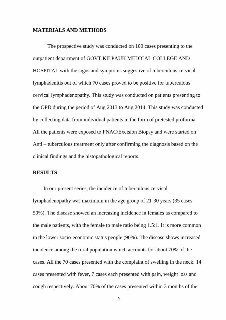
MATERIALS AND METHODS
The prospective study was conducted on 100 cases presenting to the
outpatient department of GOVT.KILPAUK MEDICAL COLLEGE AND
HOSPITAL with the signs and symptoms suggestive of tuberculous cervical
lymphadenitis out of which 70 cases proved to be positive for tuberculous
cervical lymphadenopathy. This study was conducted on patients presenting to
the OPD during the period of Aug 2013 to Aug 2014. This study was conducted
by collecting data from individual patients in the form of pretested proforma.
All the patients were exposed to FNAC/Excision Biopsy and were started on
Anti – tuberculous treatment only after confirming the diagnosis based on the
clinical findings and the histopathological reports.
RESULTS
In our present series, the incidence of tuberculous cervical
lymphadenopathy was maximum in the age group of 21-30 years (35 cases-
50%). The disease showed an increasing incidence in females as compared to
the male patients, with the female to male ratio being 1.5:1. It is more common
in the lower socio-economic status people (90%). The disease shows increased
incidence among the rural population which accounts for about 70% of the
cases. All the 70 cases presented with the complaint of swelling in the neck. 14
cases presented with fever, 7 cases each presented with pain, weight loss and
cough respectively. About 70% of the cases presented within 3 months of the
10
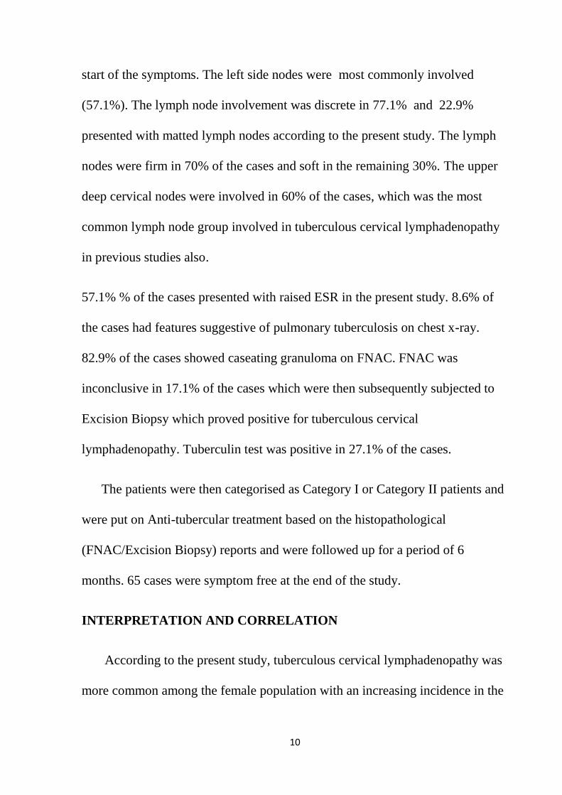
start of the symptoms. The left side nodes were most commonly involved
(57.1%). The lymph node involvement was discrete in 77.1% and 22.9%
presented with matted lymph nodes according to the present study. The lymph
nodes were firm in 70% of the cases and soft in the remaining 30%. The upper
deep cervical nodes were involved in 60% of the cases, which was the most
common lymph node group involved in tuberculous cervical lymphadenopathy
in previous studies also.
57.1% % of the cases presented with raised ESR in the present study. 8.6% of
the cases had features suggestive of pulmonary tuberculosis on chest x-ray.
82.9% of the cases showed caseating granuloma on FNAC. FNAC was
inconclusive in 17.1% of the cases which were then subsequently subjected to
Excision Biopsy which proved positive for tuberculous cervical
lymphadenopathy. Tuberculin test was positive in 27.1% of the cases.
The patients were then categorised as Category I or Category II patients and
were put on Anti-tubercular treatment based on the histopathological
(FNAC/Excision Biopsy) reports and were followed up for a period of 6
months. 65 cases were symptom free at the end of the study.
INTERPRETATION AND CORRELATION
According to the present study, tuberculous cervical lymphadenopathy was
more common among the female population with an increasing incidence in the

11
21-30 years of age. Swelling in the neck was the most common presenting
complaint followed by fever, pain, weight loss and cough. Upper deep cervical
group of nodes were the most commonly involved and most of the nodes were
discrete and firm in consistency. All the patients were subjected to
histopathological examination (FNAC/Excision Biopsy) and were started on
ATT based on the reports and as per the RNTCP guidelines. All the patients had
a 100% cure rate at the end of the study which included the follow up period of
6 months. Surgical treatments are only adjunctive to chemotherapy and not a
replacement.
12
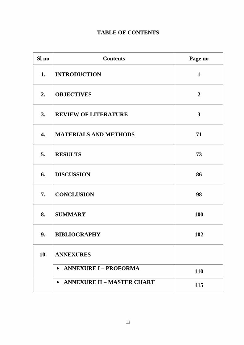
TABLE OF CONTENTS
Sl no Contents Page no
1.
INTRODUCTION
1
2.
OBJECTIVES
2
3.
REVIEW OF LITERATURE
3
4.
MATERIALS AND METHODS
71
5.
RESULTS
73
6.
DISCUSSION
86
7.
CONCLUSION
98
8.
SUMMARY
100
9.
BIBLIOGRAPHY
102
10.
ANNEXURES
ANNEXURE I – PROFORMA
110
ANNEXURE II – MASTER CHART
115

13
LIST OF FIGURES
Sl no Figure
Page no
1.
Embryology of the lymph node
6
2. Structure of the lymph node 8
3. Anatomy of the lymph nodes draining the head and neck
17
4. Anatomy of the lymph nodes of the head and neck
17
5. Mycobacterium tuberculosis colonies
21
6. Ziehl Neelson staining
28
7. Caseating granuloma
40
8. Stages of tuberculous cervical lymphadenitis
46
9. Gross picture of a tuberculous lymph node
47
10. Presentation of a tuberculous cervical lymph node
84
11. Cold aspirate
85
14
LIST OF TABLES
Sl no Table Page
no
1. Treatment categories in DOTS chemotherapy
67
2. Showing the incidence of tuberculous cervical
lymphadenopathy
73
3. Showing age incidence
74
4. Showing sex incidence
75
5. Showing socio – economic status
76
6. Showing geographical distribution
77
7. Showing clinical presentation
78
8. Showing duration of the disease
79
9. Showing group of lymph nodes involved
79
10. Showing side of lymph nodes involved
81
11. Showing the character of lymph nodes
81
12. Showing investigations
82
13. Showing results of FNAC and Excision Biopsy
84
14. Comparison of age incidence with various studies
86
15. Comparison of sex incidence with various studies
87
16. Comparison of socio – economic status in various studies
88
17. Comparison of clinical character in various studies
89
18. Comparison of lymph node character in various studies
91
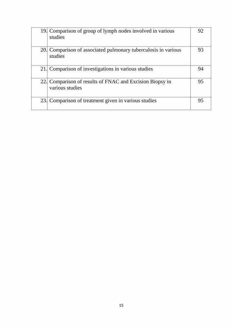
15
19. Comparison of group of lymph nodes involved in various
studies
92
20. Comparison of associated pulmonary tuberculosis in various
studies
93
21. Comparison of investigations in various studies
94
22. Comparison of results of FNAC and Excision Biopsy in
various studies
95
23. Comparison of treatment given in various studies
95
16
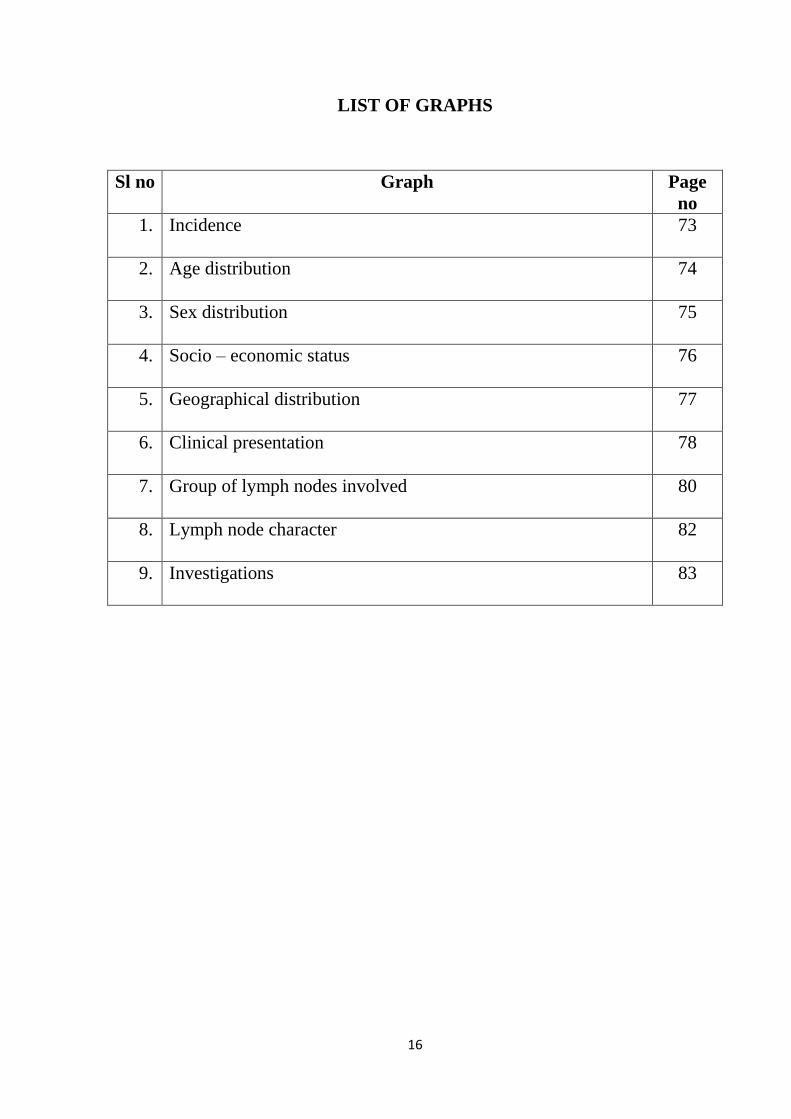
LIST OF GRAPHS
Sl no Graph Page
no
1. Incidence
73
2. Age distribution
74
3. Sex distribution
75
4. Socio – economic status
76
5. Geographical distribution
77
6. Clinical presentation
78
7. Group of lymph nodes involved
80
8. Lymph node character
82
9. Investigations
83
17
INTRODUCTION
Tuberculosis remains to be the major health burden primarily affecting the
lower socio-economic status people in our country.
In tuberculosis, the commonest type of infection is pulmonary tuberculosis.
In extra pulmonary tuberculosis, lymph node involvement is the most common
presentation. Cervical lymph nodes are affected frequently. Peripheral and
mediastinal nodes are seen in immunocompromised population like HIV. (1)
The general hospitals witness a staggering 2-3% of patients suffering from
glandular tuberculosis. It also leaves a permanent scar, often in parts of body
that are exposed ,affecting the general confidence of the patients. The treatment
modality for TB have changed from time to time , ranging from royal touch to
chemotherapy, antibiotics and surgery.
My study focussed on 70 patients with cervical lymph node during a period
of one year from August 2013 to August 2014.
18
AIMS AND OBJECTIVES
To study about the incidence of tuberculous cervical lymphadenopathy.
To study about the clinical presentations (signs, symptoms) of
tuberculous cervical lymphadenopathy.
To correlate clinical diagnosis with the histopathological findings of
tuberculous cervical lymphadenopathy and to interpret the results.
To study about the clinical work-up and the various management options,
their outcome and to follow up the clinical behaviour and improvement in
the course of tuberculous cervical lymphadenopathy for a period of not
more than 6 months.
19
REVIEW OF LITERATURE
HISTORY
There are evidences showing that ancient Indians like Manu and
Sushrutha have described tuberculous lymphadenopathy is a disease of the
unclean.
Hippocrates also described tuberculosis as „phthisis‟ meaning „I am
wasting‟. (2)
The old name for cervical tuberculous lymphadenitis is scrofula,
meaning „glandular swelling‟ in Latin. In French it is referred as „full necked
sow‟ because of its resemblance with pig‟s neck.
In early days it was thought that the king‟s touch can cure the disease
popularly known as „royal touch‟. There was also a saying dated back to 496
AD by a king „I touch thee, god heals thee‟. (3)
The royal touch was finally abandoned after being practised for a
period of 7 years by the English and 13 years by the French kings and queens.
The most popular theory put forward before 1886 was by Dole boe. He
proposed „THEORY OF OBSTRUCTION‟, which states that it is made up of
enlargement of numerous small glands made visible by obstruction. Large
caseating glands are produced by the fusion of multiple small glands.
20
Sylvius coined the term „Tubercle‟ following his serial observations in post
mortem examinations to the typical nodular lesions. Laennec proved that
tuberculosis can also occur in various parts of the body similarly by 2000 post
mortem examinations.
The infectious nature of tuberculosis was recognized in early days, only to
be proved experimentally by Villemin in 1865 by injecting the tubercle material
into a rabbit. (2)
Koch discovered the causative organism of tuberculosis as
„tubercle bacilli‟ in march 24 ,1882.
France became the first country to introduce the B.C.G. vaccination in 1921.
In developed countries, better economy, health education and awareness
reduced the incidence of tuberculosis.
The advent of chemotherapy revolutionised the treatment for
tuberculosis.(3)
The milestones in the development of chemotherapy (2)
are.,
1.1943- Streptomycin discovered by WALKSMAN.
2.1946-PAS discovered by LEHMANN.
3.1952-Isoniazid discovered by JACK BERNSTEIN ET AL.
4.1966- the discovery of Rifampicin.

21
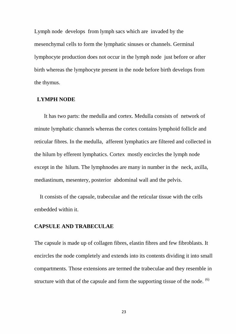
EMBRYOLOGY, ANATOMY AND PHYSIOLOGY OF LYMPH NODES
In embryo, at the end of 5th week the lymphatic system begins to develop. There
are two theories describing the development of the lymphatic system. First
theory was introduced by SABIN in 1902 which describes the lymphatic
System development as a diverticulae of the endothelium of vein. Another
Theory was introduced by HUNTINGTON & MCCLURE in 1909 regarding
the sprouting of endothelium in mesenchymal cleft which fuse and form the
lymphatic system.
In embryo, at the end of 6th week, local dilatations of the lymphatic channels
form 6 primary lymph sacs
1. Jugular lymph sacs are two in number which are present at the junction of
the subclavian vein with the anterior cardinal vein.
2. Iliac lymph sacs are two in number and present at the junction of the iliac
vein with the posterior cardinal vein.
3. Retroperitoneal lymph sac which is one in number present in the root of
the mesentery on the posterior abdominal wall.

22
4. At the level of the adrenal glands and posterior to the retroperitoneal
lymph sac (4)
is situated the cistern chyli.
Lymph vessels to the head, neck and upper limbs develop from the jugular
sacs
Lymph vessels to the lower limbs develop from the iliac sacs .
Lymph vessels to the gut develop from the retroperitoneal and cisternal sacs.
Right and left thoracic duct develop from the union of the cisterna chyli and the
two jugular sacs. These ducts join the venous system near the angle of
subclavian and internal jugular vein at base of the neck
Fig 1 – Embryology of the lymph node
23
Lymph node develops from lymph sacs which are invaded by the
mesenchymal cells to form the lymphatic sinuses or channels. Germinal
lymphocyte production does not occur in the lymph node just before or after
birth whereas the lymphocyte present in the node before birth develops from
the thymus.
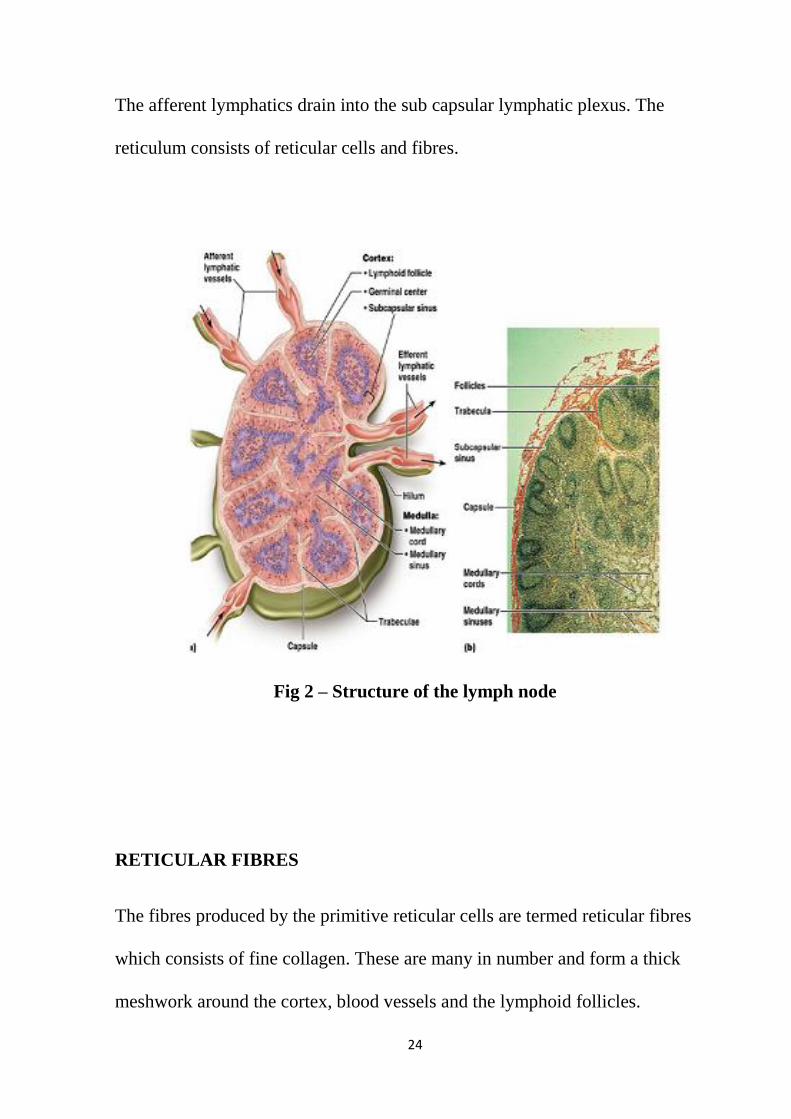
LYMPH NODE
It has two parts: the medulla and cortex. Medulla consists of network of
minute lymphatic channels whereas the cortex contains lymphoid follicle and
reticular fibres. In the medulla, afferent lymphatics are filtered and collected in
the hilum by efferent lymphatics. Cortex mostly encircles the lymph node
except in the hilum. The lymphnodes are many in number in the neck, axilla,
mediastinum, mesentery, posterior abdominal wall and the pelvis.
It consists of the capsule, trabeculae and the reticular tissue with the cells
embedded within it.
CAPSULE AND TRABECULAE
The capsule is made up of collagen fibres, elastin fibres and few fibroblasts. It
encircles the node completely and extends into its contents dividing it into small
compartments. Those extensions are termed the trabeculae and they resemble in
structure with that of the capsule and form the supporting tissue of the node. (6)
24
The afferent lymphatics drain into the sub capsular lymphatic plexus. The
reticulum consists of reticular cells and fibres.
Fig 2 – Structure of the lymph node
RETICULAR FIBRES
The fibres produced by the primitive reticular cells are termed reticular fibres
which consists of fine collagen. These are many in number and form a thick
meshwork around the cortex, blood vessels and the lymphoid follicles.
25
CELLS OF LYMPH NODES
The lymph nodule consists mainly of the B-lymphocytes. The lymphoblasts
occupy the paler germinal centre of the nodules which are stimulated by the
antigens. These divide more repeatedly giving rise to many B-lymphocytes and
they aggregate to form the dark rim around the germinal centre. They mature
into plasma cells which are seen mainly in the medullary cords which form the
antibodies which then ultimately reach the efferent vessel and the blood stream.
The medullary cords consist of the T-lymphocytes which form the intervening
tissue between the nodules.
The reticular fibres consist of the fibroblasts which were previously termed the
reticular cells. The lymphatic sinuses consist of numerous macrophages which
phagocytose the antigens and ultimately present to the lymphocytes. (7)
AFFERENT VESSELS
The vessels enter at different parts from the periphery which after dividing form
a dense plexus in the substance of the capsule and open into the sub capsular
lymphatic plexus which is in continuity with the lymphatic sinuses of the
cortex. The vessel while entering the node loses all its coverings except the
endothelial covering.
26
EFFERENT VESSELS
The vessels originate from the lymphatic sinuses of the medulla and terminate
at the hilum. The passage of the lymph though the sinus slows down gradually
which arrests the morphological elements carried in the lymph.
PHYSIOLOGY OF THE LYMPH NODE
Lymph node plays an important role in the defence mechanism of the body.
When there is an infection in the proximal part of the gland, the distal part gets
inflamed due to the localisation of the bacteria and their toxins. They play an
important role in the phagocytosis and destruction of the invading organisms.
No organism or foreign body can enter the blood stream without getting filtered
at the lymph node.
Bacteria enter the blood stream slowly through the lymphatics but the toxins
and the viruses of large molecular weight(>20,000 LMV) do not enter the blood
stream if the lymphatics are blocked. Immobilisation reduces the affected area
and aids in the resolution. It also plays an important role in the production of the
immunoglobulins. (8)
ANATOMY OF THE LYMPH NODES OF THE NECK
There are about 800 lymph nodes all over the body out of which around 300 lie
in the neck. Lymph nodes of the neck are particularly targeted by tuberculosis.
27
Nodes of the neck are involved in infections of oral cavity, nasal cavity, ear and
scalp. Neck nodes account for 90% of chronic lymphadenitis of the body. (9)
CLASSIFICATION OF LYMPH NODES OF NECK
Circular group of nodes
Vertical group of deep cervical nodes.
CIRCULAR CHAIN OF NODES:
It is composed of
A) OCCIPITAL NODES:
These are one or two nodes located between mastoid process and external
occipital protuberance. They are enlarged in infection of back of the scalp.
B) POST-AURICULAR NODES:
Present over the mastoid process behind the pinna. They are involved in
infections of temporal region of scalp, back of the pinna and external auditory
meatus.
C) PRE-AURICULAR NODES:
Located exactly in front of tragus, superficial to parotid fascia. Outer surface of
the pinna and side of the scalp drains into pre-auricular nodes.
28
D) PAROTID NODES:
They are located both in the substance of the parotid and deep to it., between it
and lateral wall of pharynx.
The deeper nodes drain
1.nasopharynx
2.back of the nose.
Superficial group drain
1.eye lids.
2.front of the scalp.
3.external auditory meatus.
4.tympanic cavity..
E) FACIAL NODES:
Composed of deep and superficial nodes.
Superficial group consists of
a) Infra-orbital :located just below orbit.
b) Buccinator: over the buccinator muscle , lateral to the angle of the
mouth.
29
c) Supra-mandibular: over the mandible, in front of the masseter
around the facial artery.
They drain the conjunctiva, eye lids, nose and cheek.
DEEP GROUP:
They are situated around the maxillary vessels and in correspondence to the
external pterygoid muscle. They are involved in infections of a) temporal fossa
b)infra-temporal fossa c) back of the nose d) pharynx.
F) SUB-MANDIBULAR NODES:
These are located in relation to the sub-mandibular salivary gland in the sub-
mandibular triangle. The lymph nodes are directly in contact with salivary gland
situated under the deep fascia.
They drain
a) Side of the nose.
b) Inner angle of eye.
c) The cheek.
d) Angle of mouth.
e) Upper lip
f) Outer aspect of lower lip.
g) Gums
h) Side of the tongue.
30
G) SUB-MENTAL NODES:
Situated in the sub-mental triangle. They are
involved in infections of central part of lower lip and floor of the mouth. They
also drain the apex of the tongue..
H) SUPERFICIAL CERVICAL NODES:
Situated on the outer border of sternomastoid around
the external jugular vein. They are involved in infections of the parotid gland
and lower ear.
I) ANTERIOR CERVICAL NODES:
Located in the midline of neck, in front of trachea and
larynx. Consists of deep and superficial group of nodes.
Superficial group: they are located around the anterior jugular vein and receive
lymph from skin of the neck. They are insignificant.
1) Deep group:
a) Infra-hyoid nodes: present over the thyrohyoid membrane
and receive lymph from front of the larynx
b) Pre-laryngeal nodes: located over the cricothyroid ligament.
31
They drain larynx. The afferent vessels pass through the
foramen in the middle of cricothyroid ligament. They also
assist in receiving lymph from the thyroid.
c) Pre-tracheal nodes: they are located in correspondence to the
inferior thyroid veins in front of the trachea and receive
lymph from the thyroid and trachea.
EFFERENT VESSELS OF CIRCULAR CHAIN:
The efferent vessels directly receive lymph from the nodes mentioned above to
the deep cervical nodes except from facial and submental nodes. The efferents
pass from facial and submental nodes pass to submandibular nodes first.
VERTICAL GROUP OF DEEP CERVICAL NODES:
These are situated in relationship to carotid sheath. A small group of nodes
are located behind the pharynx and they are called retropharyngeal nodes. They
receive lymph from pharynx, auditory tube and back of nose.
The vertical group of nodes lie along the pharynx, trachea and
oesophagus,from the base of the skull to the root of the neck. They are divided
conventionally into superior deep cervical and inferior deep cervical by
omohyoid. Some of the nodes project beyond the posterior margin of
32
sternomastoid into posterior triangle of neck. A small number of nodes are
situated in the groove between trachea and oesophagus along recurrent
laryngeal nerve. They are known as paratracheal nodes and they drain the
thyroid.
Two of the deep cervical nodes are specified:
1.Jugulo-digastric nodes: the node for tonsil.
2.Jugulo-omohyoid node: a node located on the common carotid above the point
just above where the anterior belly of omohyoid crosses the vessel. It plays a
vital role in the drainage of tongue.(10)
EFFERENTS OF VERTICAL CHAIN:
The lymph from the deep cervical trunk enters one trunk –jugular lymph trunk,
that leaves the inferior deep cervical nodes. On the right side it enters the
junction of subclavian vein and internal jugular vein. On the left side , it joins
the thoracic duct.
AFFERENTS OF THE VERTICAL CHAIN:
The deep cervical nodes drain the entire head and neck, directly or indirectly
from the circular chain. (10)
33
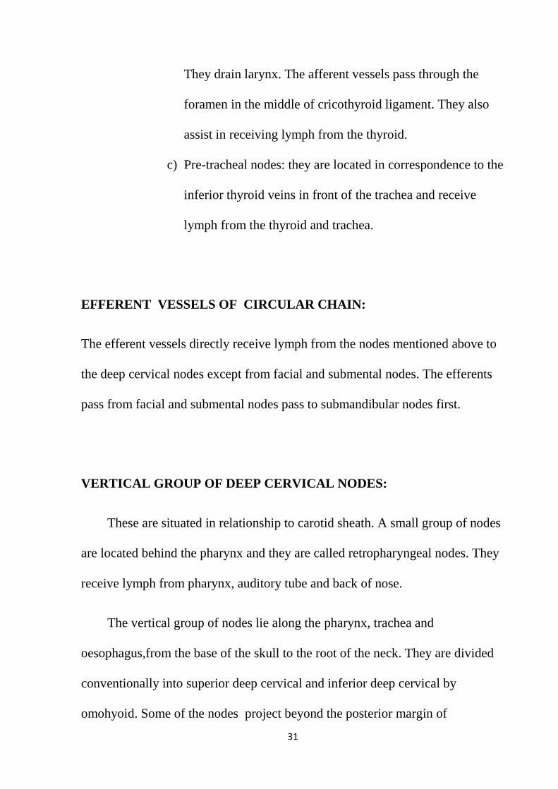
Fig 3 – Anatomy of the lymph nodes draining the head and neck
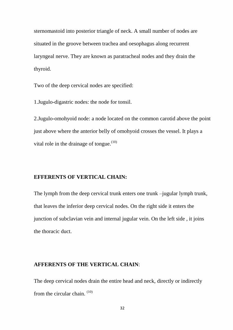
Fig 4 – Anatomy of the lymph nodes of the head and neck
34
BACTERIOLOGY
A) TUBURCLE BACILLI
1. Human mycobacterium tuberculosis
2. Bovine mycobacterium bovis
3. Murine mycobacterium microti
4. Avian –mycobacterium avium
5. Cold blooded – mycobacterium marinum
B) LEPRA BACILLI
1. Human mycobacterium leprae
2. Rat –mycobacterium lepral murium
C) MYCOBACTEERIA CAUSING SKIN ULCERS
1. Mycobacterium ulcerans
2. Mycobacterium balnei
D) ATYPICAL MYCOBACTERIA
1. Photo chromogens
2. Scoto chromogens
3. Non Photo chromogens
4. Rapid growers
E) JOHNE’S BACILLUS
1. Mycobacterium paratuberculosis

35
F) SAPROPHYTIC MYCOBACTERIA
1. Mycobacterium butyricum
2. Mycobacterium phlei
3. Mycobacterium stercoris
4. Mycobacterium smegmatis
5. Others
The term „mycobacteria‟ means fungus like bacteria which are nothing
but the slender rods showing branching filaments which resemble with that of
the fungus mycelium. Mycobacteria are aerobic, non motile, non capsulated,
non sporing. They are called the „Acid Fast Bacilli‟ because they do not stain
immediately but once stained, resist decolourisation with dilute mineral acids.
Growth is slow. The genus includes obligate parasites, opportunistic pathogens
and saprophytes.
Robert Koch announced his greatest discovery, the causative organism of
tuberculosis, to the Physiological society in Berlin on 24th March 1882.
Tuberculosis in human beings is caused by either the human type or the bovine
type of bacillus. Subsequently the other types were also described. The
generation time of the bacilli in vitro is 14-15 hours. Colonies are formed in
2weeks but can be delayed upto 6-8weeks. The growth is very slow and does
not occur below 25°c or above 40º. Optimum temperature is 37ºC. Optimum pH
is 6.4-7.0. Mycobacterium bovis is usually microaerophilic when it is primarily
isolated but becomes aerobic on subculture. Mycobacterium bovis grows
36
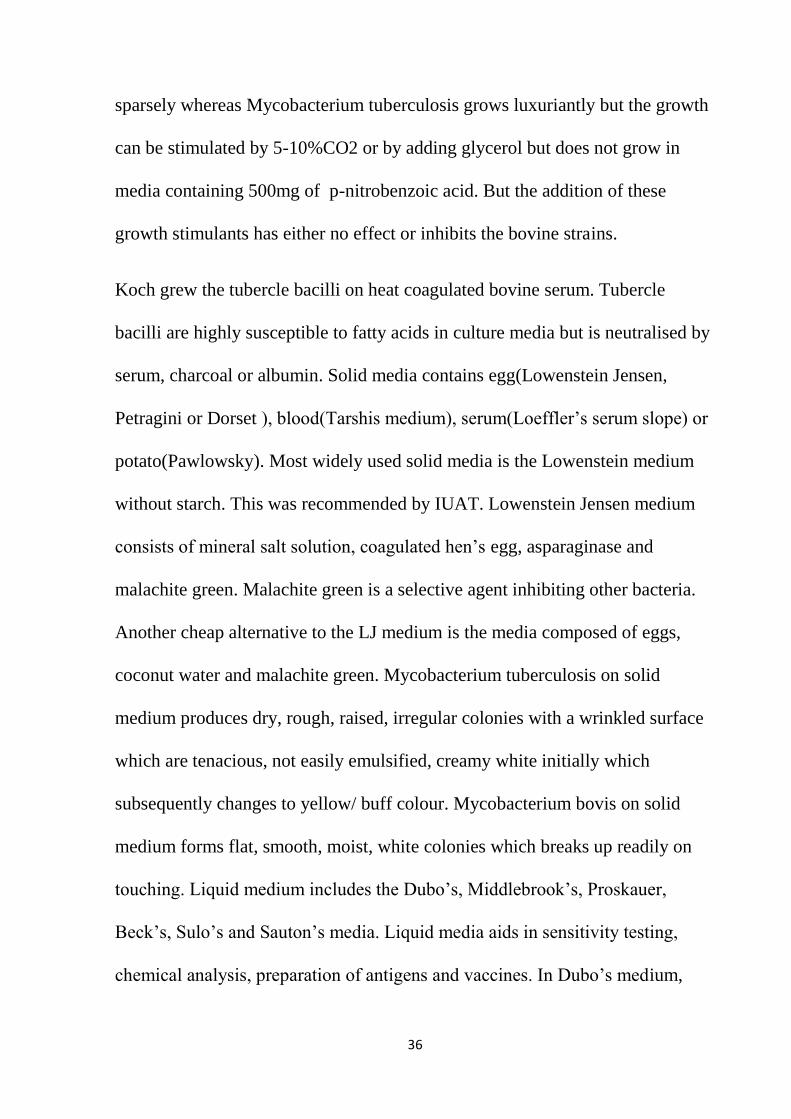
sparsely whereas Mycobacterium tuberculosis grows luxuriantly but the growth
can be stimulated by 5-10%CO2 or by adding glycerol but does not grow in
media containing 500mg of p-nitrobenzoic acid. But the addition of these
growth stimulants has either no effect or inhibits the bovine strains.
Koch grew the tubercle bacilli on heat coagulated bovine serum. Tubercle
bacilli are highly susceptible to fatty acids in culture media but is neutralised by
serum, charcoal or albumin. Solid media contains egg(Lowenstein Jensen,
Petragini or Dorset ), blood(Tarshis medium), serum(Loeffler‟s serum slope) or
potato(Pawlowsky). Most widely used solid media is the Lowenstein medium
without starch. This was recommended by IUAT. Lowenstein Jensen medium
consists of mineral salt solution, coagulated hen‟s egg, asparaginase and
malachite green. Malachite green is a selective agent inhibiting other bacteria.
Another cheap alternative to the LJ medium is the media composed of eggs,
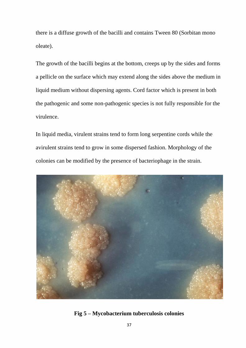
coconut water and malachite green. Mycobacterium tuberculosis on solid
medium produces dry, rough, raised, irregular colonies with a wrinkled surface
which are tenacious, not easily emulsified, creamy white initially which
subsequently changes to yellow/ buff colour. Mycobacterium bovis on solid
medium forms flat, smooth, moist, white colonies which breaks up readily on
touching. Liquid medium includes the Dubo‟s, Middlebrook‟s, Proskauer,
Beck‟s, Sulo‟s and Sauton‟s media. Liquid media aids in sensitivity testing,
chemical analysis, preparation of antigens and vaccines. In Dubo‟s medium,
37
there is a diffuse growth of the bacilli and contains Tween 80 (Sorbitan mono
oleate).
The growth of the bacilli begins at the bottom, creeps up by the sides and forms
a pellicle on the surface which may extend along the sides above the medium in
liquid medium without dispersing agents. Cord factor which is present in both
the pathogenic and some non-pathogenic species is not fully responsible for the
virulence.
In liquid media, virulent strains tend to form long serpentine cords while the
avirulent strains tend to grow in some dispersed fashion. Morphology of the
colonies can be modified by the presence of bacteriophage in the strain.
Fig 5 – Mycobacterium tuberculosis colonies
38
RESISTANCE:
Mycobacterial survival depends on the material in which the bacteria are
present. The bacilli are killed at 60°C in 15-20mins but the bacilli in the sputum
remains alive for 20-30hours and that present in the droplet remains viable for
8-10days. Cultures are killed by direct sunlight exposure for 2hours but remain
viable for 6-8months at room temperature and 2years in the deep freeze cabinet
at -20ºC.
Mycobacteria survives on exposure to 5%phenol, 15% sulphuric acid, 3% nitric
acid, 5% oxalic acid and 4% sodium hydroxide but destroyed by the contact of
the culture with tincture of iodine for 5minutes, and that of 80% ethanol in 2-
10minutes. Hence, 80% ethanol is used as a skin disinfectant and a disinfectant
for rubber gloves, clinical thermometer. Mycobacteria are sensitive to
formaldehyde and glutaraldehyde.
BIOCHEMICAL REACTIONS:
Mycobacterial species can be identified by biochemical tests which includes :
1. NIACIN TEST:
Addition of 10%cyanogen bromide and 4% aniline in 96% ethanol to a
suspension of culture containing human tubercle bacilli shows a canary
yellow colour (positive reaction) while the bovine type produces a
39
negative reaction. Tubercle bacilli produce niacin when grown on a
medium containing egg.
2. ARYL SULPHATASE TEST:
The atypical mycobacteria are grown in culture containing 0.001M tripotassium
phosphathalein disulfate and forms aryl sulphatase. If sodium hydroxide is
added to the medium drop by drop, a pink colour is produced which indicates a
positive reaction.
3. NEUTRAL RED TEST:
Virulent strain binds neutral red in alkaline buffer solution while the avirulent
strains do not.
4. CATALASE-PEROXIDASE TEST:
Aids in knowing the sensitivity of the strain to INH (Isotonic Acid
Hydrazine).Helps in distinguishing the tubercle bacilli which are weakly
catalase positive and peroxidise positive from atypical mycobacteria which are
strongly catalase positive. Tubercle bacilli resistant to INH lose their catalase
and peroxidise activity.
To 5ml of the test culture, equal volume of 30 vol of H2O2 and 0.2% catechol
in distilled water is added and allowed to settle for a few minutes. Catalase

40
production in indicated by effervescence and peroxidise activity by browning of
the colonies.
5. AMIDASE TEST:
5 amides namely the acetamide, benzamide, carbamide, nicosanamide and
pyracinamide are used which differentiates mycobacteria based on the ability to
split amides. The bacilli in the solution is incubated with 0.00165M solution of
the amide at 37°C and 0.1ml of MnSo4, 4H20, 1.0ml of phenol solution and
0.5ml of hypochlorite solution are added and placed in boiling water for
20minutes. A positive test is indicated by the presence of blue colour.
6. NITRATE REDUCTION TEST:
This is positive with Mycobacterium tuberculosis only. (11)
ANTIGENIC PROPERTIES:
Specificity of the group is due to polysaccharide and type specificity is due to
the protein antigens. Delayed hypersensitivity is developed to the protein of the
bacillus(tuberculin) following infection by the tubercle bacilli.
Tuberculin from Mycobacterium tuberculosis, Mycobacterium bovis and
Mycobacterium microti appears to be indistinguishable. There is some antigenic
relationship between the protein antigens of tubercle bacilli and atypical

41
mycobacteria shown by weak cross reactions in skin testing. A
ribonucleoprotein from Mycobacterium tuberculosis reacts with serum from
patients with lepromatous leprosy.
Mycobacterium tuberculosis has shown to be antigenically homogenous to
Mycobacterium bovis and Mycobacterium microti by agglutination, agglutinin
absorption, gel precipitation and passive hemagglutination.
BACTERIOPHAGE:
4 phage types of tubercle bacillus:
A-Commonest type worldwide
B-Occurs in Europe and North America
C-Seen rarely
D-Common in India and neighbouring countries. (11)
Phage 33D from a lysogenic environmental mycobacterium lyses all the types
of Mycobacterium tuberculosis but not BCG.
MOLECULAR TYPING:
Tubercle bacilli are genetically homogenous where certain strains contain a
chromosomal insertion sequence IS6110 which acts as a transposon. DNA
fingerprinting of these insertion sequences are employed for the diagnosis of the
infection, epidemiological studies for tracing the source of outbreaks.

42
HOST RANGE:
Natural infection in human beings, other primates, dogs and other animals who
have close contact with human beings is caused by Mycobacterium tuberculosis
which is highly infectious for guinea pigs and hamsters but non-pathogenic for
rabbits, cats, goats, bovines and fowls. Mice are moderately susceptible and
develop progressive infection following intra peritoneal, intravenous or
intracerebral inoculation.
Mycobacterium bovis produces tuberculosis in cattles, human beings, primate
carnivores which includes dogs, cats and badgers, swine and parrots. BCG, the
tuberculin vaccine is the attenuated strain of Mycobacterium bovis. It is highly
pathogenic for rabbits, guinea pigs and calves, moderately pathogenic for dogs,
cats, horses and rats and non-pathogenic for fowls.
Mammalian tubercle bacilli are considered as variants of single species of
Mycobacterium tuberculosis. Heterogenous group of tubercle bacilli isolated in
Africa which shows properties intermediate between the human and bovine
types are termed the African type. Human type of bacilli isolated in South India
which has low virulence for guinea pigs are termed the Asian type.
Mycobacterium avium causes tuberculosis in birds, „Yersin type ‟ tuberculosis
in rabbits and mice-proliferating without producing macroscopic tubercles. It is
non-pathogenic for guinea pigs and rats.

43
Mycobacteria microti causes focal lesions in human beings, guinea pigs, rabbits
and calves.(11)
It causes tuberculosis in voles.
MORPHOLOGY:
Tubercle bacilli vary in length from 0.8-5.5µ and width from 0.2-0.6µ.They
were examined under electron microscopy by Rosenblatt et al in 1942. Tubercle
bacilli are rod shaped with rounded/ slightly flattened ends. Other forms
includes curved, bean shaped and oval forms.
Human form is long, slender whereas the bovine form is short and stumpy.
Avian type is pleomorphic.
STAINING PROPERTY:
Tubercle bacilli are termed the acid fast bacilli due to the presence of lipoid
fraction containing free hydroxyl groups known as mycolic acid. These are
present in the capsule of the organisms (discovered by Anderson in 1938 ).
Ziehl Neelson developed a staining technique with 5% carbol fuchsin in
catechol for a few minutes at a higher temperature or for longer time at a lower
temperature following which they resist decolourisation with potent
hydrochloric acid. (12)

44
Fig 6 – Ziehl – Neelson staining
FLUOROSCEIN TECHNIQUE OF DETECTING BACILLI:
Tubercle bacilli absorb acromine fluorescent dye. When culture containing
tubercle bacilli is exposed to UV light , the bacilli is seen as brilliantly
illuminated structures against dark background even under the low power
microscope.
CHEMISTRY OF THE BACILLI: (12)
Bacilli contain the following constituents:
45
PROTEINS:
Upon injection, protein bound to a wax fraction induces tuberculin sensitivity.
Partially purified tuberculin contain a mixture of small proteins (10kDa).
CARBOHYDRATES:
Contains polysaccharide whose role in pathogenesis is uncertain.
LIPOID FRACTION:
Includes mycolipids (long chain fatty acids)(13)
, waxes and phospholipids.
Muramyl dipeptides with mycolic acids cause granuloma formation. Isolated by
Anderson in 1937.
Responsible for resistance to chemical
Adjuvant effect (waxD) enhancement of antigenic immunogenicity
Intracellular persistence in non-activated macrophages by means of
inhibition of the phagosome lysosome formation
Complement resistance
Virulence is due to the cord factor (trehalose 6,6-dimycolate) (14)
SOURCE OF INFECTION:
TONSILS:
In many cases, the tubercle bacilli enters through the tonsils of the
corresponding side so that the lesions are either minimal/ has healed which
46
makes its identification impossible. Upper deep cervical group of lymph nodes
are most commonly involved. The tuberculous process is usually restricted to
the clinically affected group of lymph nodes in more than 80% of the patients
but a primary focus in the lungs should always be suspected.(9)
On examination,
tonsils are not useful to aid in the presence or absence of infection due to
chronic tonsillitis. (15)
TOOTH:
This portal of entry should be suspected only when the submandibular or
submental group of lymph nodes are primarily involved. (16)
If the portal of
entry is the scalp, then the posterior triangle nodes are mainly involved which
receives the lymphatic vessels from the adenoid also.
PATH OF INFECTION:
Main source of infection are patients who are open case of tuberculosis or
carriers.
Contamination of the breast milk is extremely rare even when the mother is a
tuberculous patient. Contamination of the cow milk is possible but because of
the improvement in hygienic measures and the use of pasteurised milk,
contamination of the cow milk is almost eradicated.

47
PORTAL OF ENTRY: (2)
The bacilli are carried by air borne infections which gets filtered at the nostrils
and adenoids to produce lesions at the site of the draining lymph nodes.
DROPLET INFECTIONS:
TB carriers or patients possess thousands of bacilli in throat and nasal passages
which spread during the process of coughing/ sneezing. Bacilli enters the host
without losing much vitality even though some bacilli cling to the dust, clothes,
screens, utensils for several weeks and start losing their vitality.
DUST INFECTION:
TB bacilli in the sputum when spit on open streets gets dried and can remain
alive for several months which can enter the human body while inhaling but is
secondary to droplet infection.
INTRA-UTERINE INFECTIONS:
Previous theories showed that the infection is usually postnatal and placenta is
not a carrier of infection but recent theories have shown that the foetus can get
infected even upto the extent of septicaemia without positive tuberculin test.

48
RETROGRADE LYMPHATIC SPREAD:
Lymphatic spread is common in tuberculous adenitis. The involvement of the
lower deep cervical group of lymph nodes raises suspicion that it is involved
secondary to mediastinal group of nodes.
Hematogenous spread is a rare phenomenon. Hematogenous involvement
occurs in 50% instances of primary complex in younger age group and is
associated with multiple lymph node involvement before the tuberculin test
becomes positive. It is commonly associated with miliary tuberculosis or
tuberculous meningitis.
In progressive pulmonary tuberculosis, extra- pulmonary tuberculosis is
uncommon.
EPIDEMIOLOGY OF THE DISEASE:
TB, in the global prevalence, has been the most important of all the human
Infections, with massive morbidity and mortality. Evidence of spinal
tuberculosis has been identified in the Egyptian mummies. Prevalence increased
rapidly following urbanisation and overcrowding, but now the incidence has
come down with the improvement in standards of living. TB has been rightly
described as the “parameter of social welfare”.
About 20 million open cases of tuberculosis are reported at any point of time all
over the world, out of which 70-80% belong to the poorly developed nations.
49
About 4-5million new cases of open pulmonary tuberculosis arise each year out
of which 3million people die each year because of TB. (17)
TB is the highest burden in India where 1.8million persons develop TB out of
which 0.8million are smear positive. India accounts for 1/5th of the global
burden of TB. In India, 2 out of every 5 are infected with TB bacilli and 2
persons die of TB every 3minutes. 0.4million die of TB every year. (18)
In developing countries, prevalence of both infection and active disease is high
and mortality is increased in infants and children less than 5years of age. The
infective rate is high by the age of 20 with a rate of 10-15%. (19)
Predisposing factors which favours the development of tuberculosis includes
malnutrition, low socio-economic status, occupational exposure to silica,
medical and paramedical workers who are in direct contact with the patients and
infectious materials, population from the hilly regions who settle down in the
plains.
Infections like measles, typhoid, sore throat and acute respiratory tract ailments
may break down the host resistance and precipitate tuberculosis. Worldwide, the
most important risk factor for the development of tuberculosis is HIV induced
immunodeficiency. HIV and TB form a deadly combination. TB accounts for
25% of the deaths among people with HIV. (20)
In Western countries, because of the improvement in standards of living,
attention to diet, housing, sanitation, preventive interventions and specific anti-
tubercular drugs, the incidence of cervical lymphadenitis is so negligible which

50
is a successful attempt in eradicating tuberculosis. But in India, the problem still
remains unresolved due to poverty, uncooperative patients, infections and
irregular treatments.
ATYPICAL MYCOBACTERIA:
This group includes those bacilli other than the human or bovine tubercle bacilli
which can also cause the human disease clinically resembling that of
tuberculosis and has been termed the „atypical‟, „anonymous‟ or „unclassified‟
mycobacteria.(21)
„Environmental or opportunistic mycobacteria‟ is the best
suited term for these bacilli as their natural habitat is either soil or water
and cause opportunistic infection in human beings. Over recent years,
the term „non- tuberculous mycobacteria ‟ has gained worldwide
acceptance. They are also termed the „paratubercle‟, „tuberculoid‟ and
MOTT (mycobacteria other than tubercle ) bacilli. There is no proof of
evidence of direct human to human transmission.
Saprophytic mycobacteria such as M.smegamtis and M.phlei are not capable of
infecting human beings or animals.(21)
At times the human infection with them is
common while the disease is rare because, when injected into the guinea pigs,
they do not cause progressive disease.
In developed countries, where tuberculosis is almost eradicated or under
control, atypical mycobacteria is gaining importance. Some may produce
pulmonary disease which becomes almost indistinguishable from tuberculosis

51
while others may cause lymphadenitis. In Western countries, the spread of HIV
has increased the disease caused due to atypical mycobacteria. (22)
Atypical mycobacteria are almost resistant to the anti- tubercular drugs like
Streptomycin and Isoniazid and sensitive to Rifampicin. Usually a combination
of drugs is used but chemotherapy does not help in such cases and surgery
becomes mandatory.
CLASSIFICATION:
RUNYON’S CLASSIFICATION OF ATYPICAL MYCOBACTERIA
on the basis of pigment production and the growth rate).
Identification of the species is by other classifications.
Group I – Photo chromogens
Group II – Scoto chromogens
Group III – Non photo chromogens
Group IV – Rapid growers
GROUP I – PHOTO CHROMOGENS:
They are slow growing but the growth is faster than that of the tubercle bacilli.
They form colonies which produces no pigment in the dark but when exposed to
light for 1hour and after reincubation, a yellow orange pigment appears.
Mycobacterium kansasi causing pulmonary disease which resembles that of
tuberculosis.
Mycobacterium marinum causing swimming pool granuloma.
52
Mycobacterium simiae and Mycobacterium asiaticum causing pulmonary
disease.(23)
GROUP II SCOTOCHROMOGENS:
Do not cause human disease except for Mycobacterium scrofulaceum. Strains
produce yellow-orange-red pigmented colonies even in the dark.
M.scrofulaceum which causes cervical adenitis in children. (24)
M.gordonae which rarely produces human infection.
M.szulgai also belongs to this group.
GROUP III NON-PHOTO CHROMOGENS:
Colonies resembling that of tubercule bacilli do not form pigment even on
exposure to light. (25)
M.intracellulare was first discovered as a human pathogen at the Battey State
Hospital for TB in Georgia, USA. Hence, it is also known as the „Battey
Bacilli‟. This produces the pulmonary and renal infection and
lymphadenopathy.
M.avium rarely causes cervical adenitis in children and
lung disease in the elderly.
M.xenopii occasionally causes chronic lung disease in human beings.
There are many controversies about whether M.avium and M.intracellulare
should be considered as a variant of single species or are separate species and is
still a debate. (26)
As of now, they are considered in complex along with
M.scrofulaceum and the entire complex is called the M.avium-intracellulare-
scrofulaceum / MAIS complex.
53
GROUP IV RAPID GROWERS:
This group produces colonies within 7days of incubation at 37°C or 25°C.
The chromogenic rapid growers are saprophytes (e.g.: M.smegamtis, M.phlei).
M.fortuitum and M.chelonei which causes chronic abscess and do form any
pigment.
M.fortuitum produces pulmonary lesions which is indistinguishable
radiologically from typical tuberculosis.
SKIN PATHOGENS:
Skin lesions occur either as localised/ generalised in leprosy or tuberculosis.
Mycobacteria which causes skin pathogens are M.ulcerans and M.marinum.
They produce granulomatous lesion and chronic ulcers on the skin. (27)
These
mycobacteria multiply optimally at skin temperature and systemic invasion/
lymph node invasion does not occur.
PATHOGENESIS AND PATHOLOGY:
PATHOGENESIS:
Five factors aids in understanding the pathogenesis of tuberculosis which
includes:
Virulence of the mycobacteria which is not related to any endotoxin.
Relationship of the hypersensitivity to the immunity against the infection
Pathogenesis of caseation necrosis
This ability of Mycobacterium tuberculosis to induce the disease in
experimental animals is due to mycoside in the lipoid fraction of the bacterium.
54
Mycosides are covalently linked complex lipids and carbohydrates. Cord factor
is responsible for the serpentine, cord like growth of the Mycobacterium
tuberculosis in vitro and is toxic to mice. The cell wall of the bacilli contains
wax D (a glycolipid), and muramyl dipeptide and they are powerful adjuvants
and is important in the development of(28)
delayed hypersensitivity, skin
reactions and granulomatous inflammation at the sites of the infection.
In a previous unexposed immunocompetent individual, the pathogenesis of
tuberculosis is based on the development of the cell mediated immunity that
confers resistance to the organism and results in the development of tissue
hypersensitivity to the tuberculous antigens. Within 2-3weeks,a positive skin
reaction that is granulomatous occurs and the centre of the granulomas become
caseous forming typical soft tubercles.
Sequence of events from inhalation of the Mycobacteria to the development of
primary foci are:
Virulent strains enter the macrophage endosome and proliferate impairing the
phagolysosome formation. Antigens from the bacilli reach the draining lymph
nodes and is presented to the CD4+T cells which are sensitised and again
recirculate back to the site of infection where they release cytokines (IFN ɣ )
and
activate the macrophages forming granulomas which trap the residual micro-
organisms. These granulomas get caseated at the (29)
centre (soft tubercles), but
many a times there is no caseation (hard tubercles). The granulomas are
composed of epitheloid secretory cells and is responsible for necrosis.
Hypersensitivity is described by the caseating response to the tubercle bacillus,
the acquisition of the immunity and the resistance to the organism. The

55
sensitised host produces a defensive reaction and produces enhanced necrosis.
The protective or destructive response is determined based on the fact of
whether the primary focus of infection is localised/ generalised.
TISSUE REACTIONS IN TUBERCULOSIS:
Tissue response is based on the dose, virulence and viability of the bacilli and
whether the infection is primary or secondary (reinfection).
PRIMARY COMPLEX:
Bacilli usually gets lodged in the upper part of the lower lobe or the lower part
of the upper lobe. These undergo consolidation (Ghon‟s focus) and this
combined with the nodal involvement is termed the Ghon complex. In primary
infection, the lesion heals with resorption whereas in reinfection, the lesion is
progressive and reaction is accentuated due to the development of
hypersensitivity to the tubercle protein and localised in the area of lesion.
This lesion in glandular tuberculosis is due to primary infection in the lungs/
abdomen or the tonsils and the complementary lesion is in the lymph nodes.
TUBERCLE FORMATION:
In the initial 24 hours, leucocytes migrate to the area of infection to form a
centre of small abscess. The local reticulo endothelial cells / monocytes of the
blood produces mononuclear cells which migrate and covers the bacilli. In the
subsequent 15-20 days, nodules/tubercles are produced by groups of epitheloid
cells which produce the Langhan‟s giant cells encircled by the lymphocytes.
Tubercles consists of central area of necrosis and is surrounded by epitheloid
cells, giant cells and in the periphery by lymphocytes with surrounding
fibroblastic proliferation. (30)
56
If the resistance of the individual is good, infection may resolve without any
residual lesion. And the individual develops a positive reaction to tuberculin
in 4-6weeks following infection.
The susceptible individuals who are not on chemotherapy develop the following
changes:
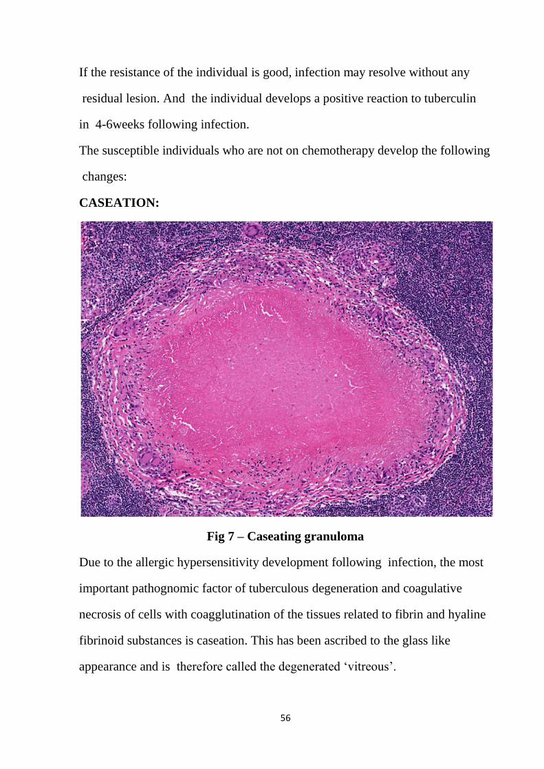
CASEATION:
Fig 7 – Caseating granuloma
Due to the allergic hypersensitivity development following infection, the most
important pathognomic factor of tuberculous degeneration and coagulative
necrosis of cells with coagglutination of the tissues related to fibrin and hyaline
fibrinoid substances is caseation. This has been ascribed to the glass like
appearance and is therefore called the degenerated „vitreous‟.
57
LIQUEFACTION:
Caseous focus which has arrested growth of the bacilli is less dangerous
compared to progressive lesion but once caseation occurs the healing is
disrupted. The proliferation of the bacilli, migration of the leucocytes and
monocytes cause the softening of caseous material forming a cold abscess
which may either get absorbed or burst forming a sinus. (31)
FIBROUS CALCIFICATION AND OSSIFICATION:
The epitheloid cells and fibroblasts are involved in the formation of precollagen
reticulin fibres and collagen and elastic fibres which aid in the conversion of
the caseous focus to collagen and subsequently into the hyaline scar.
Calcification occurs most commonly in the primary lesion. The combination
of Streptomycin, which hastens the fibroblastic activity and INH, which
enhances the revascularisation in caseous area thereby helping in the regression
of the swelling and healing of the fibrosis, should be used. Streptomycin alone
should not be used.
CLASSIFICATION OF THE TUBERCULOUS NODE BASED ON THE
PATHOLOGY:
1. PROLIFERATIVE:
Nodes are enlarged and hard. Capsule is intact.
No periadenitis and the lymph nodes are discrete.
2. FIBROUS:
Lymph nodes are enlarged, hard, mobile and shows calcification on x-ray.

58
3. CASEATION:
Caused by either blood or lymph borne infections. Nodes are matted. Cut
surface shows pale yellow cream caseating areas. The lymph nodes are usually
situated deep to the deep fascia of the neck and grows on to the skin forming a
track which ultimately leads to cold abscess. The healing of the sinus is
indicated with puckered scar. (31)
The healing of one sinus subsequently leads to
the formation of the other sinus such that multiple sinuses opens on the neck.
The scrofulodermic scar are prone for keloid formation.
CLINICAL FEATURES
Tuberculous lymphadenitis is more common in females compared to
males . Most commonly involves anterior triangle of neck .In the
anterior triangle , upper deep cervical group is most common . It
occurs in 2nd
decacade of life . In most of the patients onset is
insidious . It takes chronic course , it starts as a localized
infection . complication occurs when secondary infection takes
place . In 80% of the cases the disease is limited to the lymph
node group that is affected . In some cases there may be a
primary lung pathology and it has to be investigated .
Most common symptoms are evening rise of temperature , malaise
and appetite loss. Duration may vary from months to years.
59
On examination , the consistency of lymph node may be hard or
fluctuant .Inflammatory changes are minimal compared to other
bacterial lymphadenitis . The swelling is non tender and freely
mobile. skin changes are minimal .
The most common group affected is cervical(63%) , next group that is
involved is mediastinal(27%) and then axillary(8%). Mostly it is
unilateral and it involves deep cervical group of lymph nodes.
Multiple group of lymph nodes is involved if the origin is through
hematogeneus route. There may be splenomegaly .Toxic symptoms are
present
There are four clinical types:
ACUTE TYPE: common in infants and children , resembles acute
Lymphadenitis (32)
CASEATING TYPE : common in young adults ,neighbouring lymph nodes
are also involved . Typical matted nodes with caseation . cold abscess
and sinuses are common .
HYPERPLASTIC TYPE: lymphoid hyperplasia is predominant. Mostly
single group of lymph node is involved , usually it is firm and discrete
.
60
ATROPHIC TYPE: common in elderly where there is natural
involution. The nodes are usually small .
COURSE OF THE DISEASE:
There are 5 pathological stages (Jones and Campbell) . Patient may
present with any of these stages . (33)
STAGE 1 :
Lymph nodes are enlarged . They are firm , discrete and freely mobile.
There is solid inflammation and non specific hyperplasia.
STAGE 2:
Lymph nodes are rubbery and enlarged .They are matted together due to
periadenitis .
STAGE 3:
Lymph nodes break down and liquefy to form abscess due to caseation.
Pus collects beneath the fascia. There may be varying consistency . May
be fluctuant without inflammatory skin changes (cold abscess).
61
STAGE 4:
The deep cervical fascia may get eroded and the pus may get collected
beneath the superficial fascia which is laborious to form collar stud
abscess .
STAGE 5:
The superficial abscess enlarges and then eventually burst through
the skin when it gets ulcerated resulting in continuously discharging
sinus until the necrotic material beneath the deep cervical fascia
gets cleared . Healing may occur only when the conditions favour.

62
Fig 8 – Stages of tuberculous cervical lymphadenitis

63
Fig 9 – Gross picture of a tuberculous lymph node
OTHER PRESENTATIONS
In 10% individuals there may be long stem for the collar
stud abscesses. The feeding infected node may be located in
the other triangle of the neck with abscess presenting in
some other triangle .In such cases it is necessary to palpate
the full length of the stem.
In some other cases the abscess is situated over the affected
node and only small group of nodes are involved.
64
LABORATORY INVESTIGATIONS
RBC count
Hb% - may be microcytic hypochromic anemia
Differential count – lymphocytosis (34)
WBC count - not altered
ESR- raised . It is a prognostic factor
Mantoux test – positive test has no diagnostic value
Negative test excludes tuberculosis
PROCEDURE:
0.1 ml of PPD (purified protein derivative ) is injected in the volar
aspect of the forearm .(0.1ml =5Tu) After 48 to 72 hours look for
induration at the injected site. Induration is measured transversely
and the erythema is not taken into account. Positive test is
considered if the induration exceeds 10mm , Negative if it is < 5mm
and doubtful if it is 6-9mm.
POSITIVE TEST: suggestive of recent or past infection of the
tubercle bacilli or BCG vaccination. It is due to hypersensitivity to
65
tubercular protein. It becomes positive only after 4 to 6 weeks after
infection or vaccination.
NEGATIVE TEST: excludes tuberculous infection.
But it does not always rule out tuberculosis .
FALSE NEGATIVE: occurs in
immunosuppression
convalescence from viral infections
severe malnutrition
lymphoma , sarcoidosis
improper injection techniques
atypical Mycobacterial infection
Tubercular testing also used in the diagnosis of :
active infection in infants and young children
measuring TB prevalence in community
RADIOLOGICAL INVESTIGATIONS:
X - RAY NECK: look for calcification (34)
66
X – RAY CHEST : to rule out pulmonary tuberculosis
FNAC OF LYMPH NODE:
Smear is stained by Ziehl nelson technique, at least 10000 cells are
required for positive result .Histopathology shows : Tubercle (granuloma)
comprising area of caseation surrounded by epitheloid histiocytes ,giant
cell, lymphocytes and plasma cells.
BIOPSY OF THE LYMPH NODE:
It is a superior and reliable investigation for confirmation of diagnosis .
MACROSCOPIC FEATURES:
EARLY STAGE: Translucent and grey patches
ADVANCED STAGE: opaque and yellow due to caseation
MICROSCOPIC FEATURES:(35)
At early stage ,it shows epitheloid cells and giant cells with peripheral nuclei.
At the end of first week lymphocytes with dark nuclei and scanty cytoplasm .

67
at the end of third week caseation occurs at the centre with surrounding
epitheloid and giant cells , around which are lymphocytes and plasma
cells .
CULTURE:
Lowenstein Jensen media is used for culture . It takes 6 weeks for
positive culture. Serenity medium takes 5 days . Middle brook 7H12 and
7H13 (BACTEC 12B AND 13B) are most sensitive culture media .BACTEC
is an automated method which provides result in 4 days , radioactive C14
labelled broth culture is used. mycobacteria metabolises the culture to
liberate radioactive co2.
SEROLOGY:
Passive hemagglutination may be positive in advanced disease only and
negative in early stage , but no serological investigation is diagnostic of
pulmonary tuberculosis . (36)
68
SPUTUM FOR AFB :
Three samples are taken . First and third day sample are spot samples
.Second day sample is a early morning sample .Smear stained by Ziehl
nelson method . Atleast 2 smear positivity is required to declare positive
result. Smear is positive only when there is > 1-9 bacilli / 100 field.
MOLECULAR TESTING :
PCR testing is a very fast method . It is used to demonstrate
Mycobacterial DNA fragments .It is a very useful test in a suspected case
of Mycobacterial cervical lymphadenitis . 10 microorganisms are sufficient
for positive PCR . PCR testing can also be done on the FNAC or biopsy
smear and hence the need for open biopsy is reduced. Sensitivity of the
test is 43 to 84% . specificity is 75 to 100%. PCR is done in the smear and
culture negative cases.
69
IMMUNOPROPHYLAXIS
BACILLE CALMETTE GUERIN – ( BCG ) was identified by Calmette and
Guerin (1921). It is a live attenuated vaccine ,(37)
available in a freeze dried
form which is stable compared to liquid .It is given intradermally at birth
in infants . In developing countries ,it is given for children under 15 years.
It contains Mycobacterium bovis strain . It is attenuated by 239 serial
subculture in glycerine bile potato medium for a period of 13 years. It
induces delayed hypersensitivity reaction, hence shows positive tuberculin
reaction. It provides immunity for 10 to 15 years. Natural TB infection also
provides immunity for the same duration but there is progression of
disease when there is reactivation but this does not occur in immunization.
EFFICACY:
It varies between 80% to no immune response .It depends on various
factors .
prevalence and virulence of infection
type and potency of the vaccine used.

70
Age and nutritional status of the individual.
COMPLICATIONS:(38)
LOCAL: abscess , indolent ulcer , keloid , tuberculids – it is satellite nodule
developed adjacent to vaccinated site,lupus vulgaris ,lupoid reaction .
REGIONAL: axillary lymphadenitis or abscess of the lymph node.
SYSTEMIC: fever ,mediastinal lymphadenitis , erythema nodosum , otitis
media , fatal meningitis in rare cases.
Vaccination confers immunity to skeletal , meningeal and military
tuberculosis. It does not provide immunity for pulmonary TB especially in
infants and young children , but the disease takes milder course.
DIFFERENTIAL DIAGNOSIS:
Cervical lymphadenitis can be due to:
Viral
Bacterial- tubercular, atypical tuberculosis, syphilis, brucellosis, non-
Specific

71
Fungal-fungal granuloma
Organic-drug reaction, reactive follicular hyperplasia
Malignancy-which includes primary (Hodgkin‟s lymphoma,
lymphosarcoma) and secondary (with primaries in the neck, thorax and
abdomen)
Others
These can produce both acute or chronic lymphadenitis similar to
tuberculosis.
SARCOIDOSIS:
Granulomatous lesions are commonly seen in the lungs, lymph nodes, skin, eye,
liver, spleen, salivary glands, heart, skeleton and nervous system. The lymph
node involvement is restricted usually to the superficial group, about 2-3cm in
size, non-tender and no periadenitis. The lymph node involvement in
sarcoidosis is differentiated from tuberculous lymph node by the absence of
caseation. Mantoux test is negative and diagnosis is confirmed by biopsy and
Kveim test.
CAT- SCRATCH DISEASE:
Caused by B.henselae where initially there is enlargement of either the axillary
or the cervical lymph nodes. Primary skin lesion is indicated by a red papule
appearing between 7-12 days following contact. Diagnosis is confirmed by skin
72
testing and biopsy of lymph nodes which reveals lesion of histiocytic
proliferation and follicular hyperplasia.
INFECTIOUS MONONUCLEOSIS: (39)
Otherwise termed the GLANDULAR FEVER.
Rickettsia group of organisms causes this disease which is characterised by
fever, cervical lymph node enlargement (posterior triangle group of lymph
nodes are equally involved) which is painful and tender, sore throat,
splenomegaly, hepatomegaly and skin rashes. Diagnosis is confirmed by blood
investigations which include Paul-Bunnel test where there is an increase of
agglutinins for sheep RBCs during the acute phase of the disease and atypical
lymphocytosis. Epstein-Barr virus antibodies are detected by
immunofluorescence and ELISA.
SYPHILITIC LYMPHADENITIS:
PRIMARY STAGE:
Genital chancres and groin nodal enlargement.
SECONDARY STAGE:
Generalised involvement of the lymph nodes especially the epitrochlear and
occipital group of lymph nodes which are painless, discrete, firm and shotty.
TERTIARY STAGE:
Lymph nodes are rarely involved.
Presence of Treponema pallidum in dark ground illumination from the primary

73
lesion and positive W.R and Kahn tests confirms the diagnosis.
Specific tests includes FTA-ABS, MHA-TP. (40)
BRUCELLOSIS:
Otherwise termed the UNDULANT FEVER.
Brucella are small, gram negative, non-motile coccobacilli which presents with
fever, malaise, headache, myalgia and GI disturbances with enlargement and
tenderness of the spleen, liver and lymph nodes. Blood investigations which
shows hypochromic anaemia,leukopaenia, agglutination and culture of the
tissue confirms the diagnosis. Lymph node biopsy reveals features of either
tuberculosis or Hodgkin‟s disease. ELISA is used to detect either the IgG or the
IgM antibodies.
TULAREMIA:
It is highly infectious disease usually occurring in the farmers, hunters,
butchers, people who handle the contaminated skin or the internal organs of the
infected rabbits. It presents with symptoms like headache, feeling of cold
and pyrexial episode and resembles a plaque with ulcer at the site of infection,
enlargement of the regional lymph nodes where it becomes indistinguishable
from tuberculosis showing caseation, necrosis, epitheloid and giant cell
formation. The disease is caused by Pastuerella tularaemia. (41)
Diagnosis is
confirmed by the isolation of the organisms from the guinea pigs and rising
titre of the agglutinins against tularaemia in the serum.
TOXOPLASMOSIS:
It is caused by an intra cellular parasite Toxoplasma gondii. It exists in 4 forms
which includes cerebrospinal, lymphatic, exanthematous and the latent form out
74
of which lymphatic form is important. It presents with the enlargement of one
or more group of lymph nodes with fever of several weeks of duration and
marked constitutional symptoms. Diagnosis is confirmed by complement
fixation tests, neutralising antibody test and toxoplasmosis skin test.
FUNGAL:
A chronic granulomatous process with/ without caseation necrosis is noted.
Organisms like coccidiodomyces and blastomyces are identified with Pas
Grithley and Gomori methenamine silver.
HODGKIN’s DISEASE:
Lymph nodes are markedly enlarged with hard rubbery consistency where more
than one group is involved in different regions of the body with no features of
matting and periadenitis. Splenomegaly is present. Nodal enlargement in the
axilla and mediastinum will occur in the course of several weeks or months.
As in tuberculosis, non-specific inflammation or in secondary deposits, the
nodes do not break or suppurate but in this case the nodes break and suppurate.
Biopsy gives conclusive evidence.
Severe pain at the site of the disease after ingestion of alcohol, pyrexia,
leucocytosis, eosinophilia are the common features.
SECONDARY CARCINOMA:
Due to the lymphatic metastasis, there is enlargement of the cervical group of
lymph nodes in the elderly. The primary carcinoma is most often in the

75
mouth(tongue), lips, pharynx etc.,Malignant melanoma commonly leads to the
secondary involvement of the lymph nodes.
TREATMENT: (42)
CHEMOTHERAPY:
Treatment of tuberculous lymphadenitis includes either medical or surgery but
with the advancements of short course chemotherapy where the duration of
therapy has reduced from 18months to 6months and a cure rate of 95%, the role
of surgery is diminishing. Treatment is based on the WHO guidelines, where
the National Tuberculosis Programme worldwide follows DOTS approach
which provides intermittent chemotherapy for patients with tuberculous
lymphadenitis. Cat I treatment is for those who are smear positive tuberculous
lymphadenitis with pulmonary involvement or are severely ill.
Anti-tuberculous treatment was introduced in the early 1950s. The tuberculous
research centre in Chennai in 1990 reported results of a supervised short course
intermittent chemotherapy (6moths) consisting of streptomycin, rifampicin,
isoniazid, pyrazinamide 3times a week for 2 months followed by streptomycin
and isoniazid twice a week for 4 months on OP basis in children with
lymph node tuberculosis and a favourable clinical response was noted at the
end of the treatment. Hence in children, short course chemotherapy of 6 months
is effective in treating tuberculous lymphadenitis.
FACTORS WHICH MAKES TUBERCULOSIS DIFFICULT TO TREAT:
Caseation, an unique feature of tuberculosis, keeps the disease dormant
where the organisms multiply preventing the contact of the environment
of the body with the chemotherapeutic drugs.
76
Fibrosis reducing the blood supply.
The poor penetrating effect of the chemotherapeutic agents which allows
the tubercle bacilli to remain viable and multiply even after being
ingested by the macrophages.
Quick development of the resistance when administered improperly.
Prolonged duration of the treatment.
Expensive treatment.
ANTI-TUBERCULOUS DRUGS:
BACTERICIDAL DRUGS:
ISONIAZID (1952):
ISONICOTINIC ACID HYDRAZIDE
The drug acting on both the extra cellular and intra cellular bacilli kills the fast
multiplying organisms while the quiescent ones are only inhibited. It is
considered the most effective drug against Mycobacterium tuberculosis.
77
MECHANISM OF ACTION:
Exact mechanism not known but probably due to the inhibition of a unique fatty
acid component of a bacterial cell wall, MYCOLIC ACID. (43)
ABSORPTION AND RATE:
The drug penetrates through the caseous material with a peak plasma
concentration of 3-5mg/ml developing 1-2hours following an oral ingestion.
Being given orally/ parenterally, the drug is readily absorbed maintaining the
blood level and therapeutic concentration in the CSF. The drug is metabolised
by the liver by acetylation 75-95% of the dose is excreted in the urine in
24hours.
DOSAGE:
Daily dose of the drug is 5mg/kg with a maximum of 300mg and in children
<4years , dose is 10mg/kg/day.
Tablet: 50mg, 100mg, 300mg.
Syrup: 10mg/ml.
Injection: 10mg/ml.
Pyridoxine added in a dose of 10-50 mg/day to prevent peripheral neuritis
caused due to ISONIAZID.
Side effects includes hepatitis, rash, psychosis, acne and fever.
RIFAMPICIN:
It is a semi synthetic derivative of rifamycin-B.
This complex macrocyclic antibiotic is derived from Streptomyces
mediterranei.
Affects both the extra-cellular and intra-cellular bacilli and acts on
78
slowly/intermittently dividing organisms.
MECHANISM OF ACTION:
Inhibits DNA dependant RNA polymerase suppressing the initiation of chain
formation in RNA synthesis.
ABSORPTION AND FATE:
Penetrates all the body fluids, absorbed orally with a peak concentration in 2-
4hours and therapeutic levels in CSF with rapid elimination in bile by
deacetylation.
DOSAGE:
>50kg – 450mg/day (44)
Children – 10mg/kg/day.
CAPSULE:
150, 300, 450 and 600mg.
ADVERSE REACTIONS:
Flu like symptoms (fever, chills, headache and malaise), GI disturbances
(nausea, vomiting and abdominal cramps), flushing, pruritis, rash, hepatitis,
orange pink discolouration of body secretions.
STREPTOMYCIN:
Till 1952,it was the only effective drug available to treat tuberculosis. It crosses
the placenta and fetal serum levels are half of those in the maternal blood
reaching a peak concentration of 1hour when administered i.m.
It is excreted by glomerular filtration and hence the dosage should be titrated in
renal failure.
79
DOSAGE IN VARIOUS AGE GROUPS:
1gm - young individuals
0.75gm -40-60 years age group
0.5gm ->60years.
SIDE EFFECTS:
Ototoxicity (vestibular damage), anaphylaxis, renal tubular damage, blood
dyscrasias.
PYRAZINAMIDE:
It is the synthetic pyrazine analogue of nicotinamide. It is lethal to the intra
cellular bacilli exhibiting bactericidal activity only in acidic pH. Interacts with
the fatty acid synthase and inhibits the mycolic acid synthesis. It is well
absorbed from the GIT and widely distributed. Oral administration of 1gm
produces plasma concentration of 45mg/ml at 2hours. Drug is excreted by
glomerular filtration. The drug is hydrolysed and hydroxylated to 5-OH
pyrazinoic acid which is the major excretory product available as 500mg/
750mg tablets. Daily dose is 20-35mg/kg orally. Maximum dose is 3gm/day
regardless of the weight. Side effects includes hepatotoxicity, gout, arthralgias,
flushing, rashes and fever.
ETHAMBUTOL:
It is a bacteriostatic drug but in high concentrations it is bactericidal. The exact
mechanism is not known. It inhibits the arabinosyl transferases involved in
arabinogalactan synthesis and interferes with the mycolic acid incorporation
into the Mycobacterial cell wall. About 75-80% of the oral drug is absorbed
from the gastrointestinal tract and the peak plasma concentration is after

80
2-4hours.
The half life of the drug is 3-4hours. Two thirds is excreted in the urine and
15% is excreted in the form of the metabolites.
Tablet of 400mg and 800mg are available. Usual dose is 25mg/kg/day. Side
effects includes ocular toxicity, diminished visual activity, hyperuricaemia,
fever and rash.
DIFFERENT REGIMENS:
6 MONTH REGIME:
Initial phase of 2months – Ethambutol/Streptomycin + Isoniazid + Rifampicin
+ Pyrazinamide
Continuation phase of 4months – Isoniazid + Rifampicin
9 MONTH REGIME:
Initial phase of 2 months – Ethambutol/Streptomycin + Isoniazid + Rifampicin
Continuation phase of 7months – Isoniazid +Rifampicin
12 MONTH REGIME:
STM 1gm i.m for 3months with a total dose not exceeding 45gms.
It is given as follows:
Every day for the first 2weeks, every other day 1month, and every 3days for
1.5months.
RIF (600mg/day, p.o.) for 6months
INH (300mg/day p.o.) for 12 months or 18 months.
EMB (900mg/day p.o) for 12 months or 18 months.
PARA- AMINO SALICYLIC ACID:
This drug is rarely used with the advancement of short course chemotherapy. It
81
is not used because of the bulk and the unpleasant sensation.
THIOACETAZONE:
It is abandoned in developing countries but it is also used in some developing
countries because it is cheap. It is available as 150 mg tablet in combination
with Isoniazid. Usual dose is 150mg/day. Side effects are hypersensitivity
reactions as Steven Johnson syndrome which includes the symptoms such as
fever, rash, joint pain. This is a severe life threatening complication.
OTHERS:
Cycloserine, Kanamycin, Ethionamide, Capreomycin are the second line drugs.
NEWER ANTI-TUBERCULAR DRUGS:
The fluoroquinolone group of drugs are quite effective out of which ofloxacin is
most effective which showed a therapeutic effect on mice. Other drugs like
Sparfloxacin and Fleroxacin demonstrate anti tubercular activity. Rifabutin, a
drug belonging to rifamycetins is an effective drug. These drugs have been
effective in experimental animals and human cases but is not recommended for
general use.
Multi- drug resistant tuberculosis MDR-TB is defined as tuberculosis resistant
to at least Isoniazid and Rifampicin. When multi-drug resistance to the drugs
involves at least one drug in each group then it is termed as the extensive drug
resistant tuberculosis XDR-TB. MDR-TB can also involve the second line
drugs which includes the fluoroquinolones and the injectable drugs. Treatment
of MDR-TB must be done on the basis of sensitivity testing.

82
RISK FACTORS FOR MDR-TB INCLUDES:
Failed TB treatment
Failure to respond to TB treatment
Relapse following standard TB treatment
Previous incarceration
HIV infection
If the treatment is for a suspected case of MDR-TB, the patient is started
on SHREZ (Streptomycin+ Isonicotine hydrazine+ Rifampicin+ Ethambutol+
pyrazinamide) + MXF + Cycloserine until the report of the sensitivity testing is
received.
ASSESSMENT OF PROGRESS:
Improvement of ESR is of prognostic value.
Periodic check-up has to be done. If the patient shows no improvement when
under treatment then the doctor should find the cause and treat accordingly.
Before stopping the treatment, it is best to investigate twice since staring the
treatment all over again is difficult.

83
Treatment categories in DOTS chemotherapy in India:
Table 1 – Categories in DOTS chemotherapy
TREATMENT
CATEGORY
TYPE OF THE
PATIENT
TREATMENT
REGIMEN
CATEGORY I New sputum smear-
positive
2 (HRZE)3
4 (HR)3
Seriously ill sputum
smear-negative
Seriously ill extra-
pulmonary
New sputum smear-
negative, not seriously ill
New extra- pulmonary,
not seriously ill
CATEGORY II Sputum smear positive
Relapse, failure and
default
2 (HRZES)3
1 (HRZE)3
5 (HRE)3

84
The number before the letters in the treatment regimen indicates the number of
months of treatment. The number after the letters indicates the number of doses
per week.
H – Isoniazid (600mg)
R – Rifampicin (450mg)
Z – Pyrazinamide (1500mg)
E – Ethambutol (1200mg)
S – Streptomycin (750mg)
Patients more than 60kg receive an additional dose of rifampicin 150mg.
ROLE OF SURGERY
The role of surgery is decreasing with the advent of effective anti-tubercular
therapy.
Surgical procedures are ;
1. Biopsy.
2. Excision
3. Aspiration
85
4. Excision of sinus and scar.
5. Curettage.
6. Excision of cold abscess.
7. Tonsillectomy.
BIOPSY:
It is usually done for superficial mobile lymph node. Biopsy is for confirmation
of diagnosis before starting the short course chemotherapy.
EXCISION:
Excision of a mobile node with an intact capsule shortens the duration of
treatment, lessens the chance of recurrence and disfigurements and is
considered only when patient is not responding to chemotherapy. The lymph
node should be removed in toto.
Excision is considered in case of matted nodes, cold abscess, discharging sinus
and in patients who respond well to anti-tubercular drugs.
Contradiction to excision is the presence of secondary infection , multiple node
involvement, tender nodes and the presence of active lesion elsewhere in the
body.
Excision is not a contraindication in children but is better avoided in children.
ASPIRATION:
The presence of a big cold abscess, the chances of bursting open of the abscess,
the presence of the disease in the acute phase are indications of aspiration from
a non-dependent area with wide bore needle. Aspiration can also be repeated
which makes excision easy.
86
INCISION AND DRAINAGE:
It facilitates the spread of infection.
CURETTAGE:
Curettage is considered when the lesion is close proximity to the nerve or in the
presence of extensive skin necrosis. It has 70% cure rate.
OPERATION:
The presence of large, matted glands mandates removal but the removal is
dangerous if the node is attached to the wall of IJV. The nodes are removed
through a horizontal neck incision. If the bleeding of IJV is encountered, it is
stopped either by finger pressure or by dissection of the vein above and below
the matted glands, ligation and excision of the segment of the vein along with
the glands.
Functional neck dissection involves fat removal , associated lymph nodes in the
posterior and anterior triangles of the neck and is performed only when the
glands and vein is involved extensively. The sternocleidomastoid muscle,
accessory nerve and the IJV are to be preserved.
Removal of the gland should be followed with chemotherapy, otherwise sinuses
form with persistent drainage and scars. Mostly the infection is from the
ipsilateral tonsil , which mandates the removal of tonsil also.
87
MATERIALS AND METHODS.
The cases used for this study consists of elaborate study of 70 cervical
tubercular lymphadenitis patients, diagnosed and treated at Government
Kilpauk medical college , Chennai during the period August 2013 to August
2014. The patients were mainly from in and around Chetpet area.
The paper consists of seventy cases selected conveniently. For statistical
evaluation the data from my study is compared with that of data from literature.
METHODS:
The case details were recorded as depicted in the proforma. The data are
enclosed in the master chart for analysis.
INCLUSION CRITERIA:
1. Patients who were above 12 years of age and also confirmed to have
cervical tubercular lymphadenopathy both clinically and pathologically.
2. Patients who gave consent for investigations and willing to undergo
surgery if required.
EXCLUSION CRITERIA:
1. Patients less than 12 years of age.
2. Pregnant women with cervical lymphadenopathy.
3. Psychiatric patients.

88
4. Patients not willing to undergo investigations and surgery if required.
The patients are concluded to have cervical tubercular lymphadenopathy
based on history, clinical examination and confirmed by either FNAC or
histopathological examination.
Anti-tubercular chemotherapy was started only after confirmation of the
diagnosis.
Investigations such as Hb%, total count and differential count , ESR and
chest X-ray were done routinely. X-ray chest was done to rule out coexisting
pulmonary lesion.
Haematinics were given to anaemic patients to improve the general status.
For the patients who underwent surgery , the parts were prepared one night
before and kept on nil per oral for 12 hours . After getting the written
consent from patients, they were operated under general anaesthesia.
Antibiotics were given one hour before surgery prophylactically.
Follow of the patients was done from first post-operative day to the day of
discharge. Once the diagnosis was confirmed the patients were advised to
take short course anti-tubercular therapy for 6 months.

89
RESULTS
Table 2- Incidence of tuberculous cervical lymphadenitis
Total no of cases Total no of cases with
tuberculous cervical
lymphadenitis
Percentage
100 70 70
Graph 1 – Showing incidence of tuberculous cervical lymphadenopathy
The most dreadful disease in India is tuberculosis which is the most common
cause of cervical lymphadenopathy accounting for about 70 cases out of the 100
cases in the present study. Other causes of cervical lymphadenopathy were non-
specific lymphadenitis (28cases), lymphoma (2cases).
0
20
40
60
80
100
120
Total no of cases Total no of cases with tuberculous cervical
lymphadenitis
Percentage
Series1
90
Table 3 – Age Incidence
Age Group (years)
No of cases Percentage
11-20
7 10
21-30
35 50
31-40
14 20
41-50
7 10
51-60
7 10
61-70
- -
TOTAL
70 100
Graph 2 – Showing age incidence
The disease occurs mostly in the age group of 21-30 years. No cases were
0
20
40
60
80
100
120
11-20 21-30 31-40 41-50 51-60 61-70
Age Group (years)
No of cases
Percentage
91
reported in the age group of 61-70 years in the present study. The youngest in
the present series is 12 years old.
Table 4– Sex Incidence
Sex No of cases Percentage
Female
42 60
Male
28 40
Graph 3 – Showing sex incidence
There is a higher incidence in females. There are 28 male cases and 42 female
cases according to the present study with a female to male ratio of 1.5:1.
Female 4260%
Male 2840%
Sex Incidence
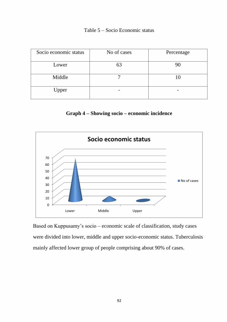
92
Table 5 – Socio Economic status
Socio economic status
No of cases Percentage
Lower
63 90
Middle
7 10
Upper
- -
Graph 4 – Showing socio – economic incidence
Based on Kuppusamy‟s socio – economic scale of classification, study cases
were divided into lower, middle and upper socio-economic status. Tuberculosis
mainly affected lower group of people comprising about 90% of cases.
0
10
20
30
40
50
60
70
Lower Middle Upper
Socio economic status
No of cases
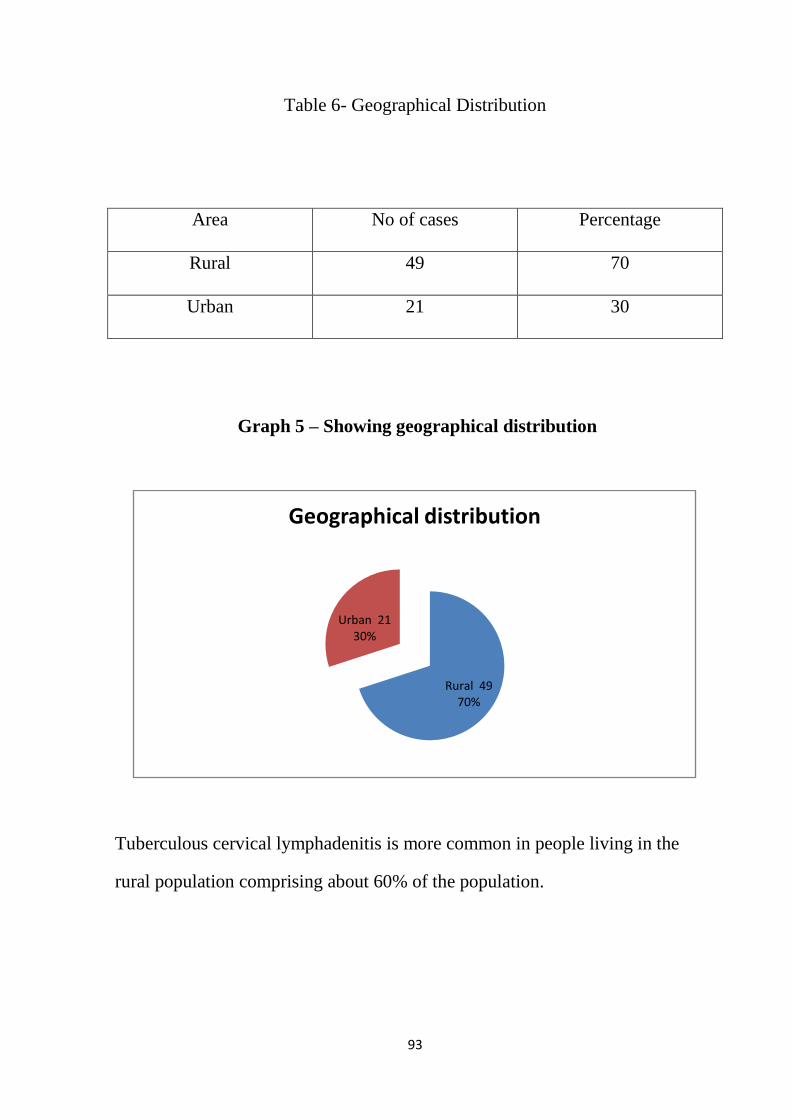
93
Table 6- Geographical Distribution
Area
No of cases Percentage
Rural
49 70
Urban
21 30
Graph 5 – Showing geographical distribution
Tuberculous cervical lymphadenitis is more common in people living in the
rural population comprising about 60% of the population.
Rural 4970%
Urban 2130%
Geographical distribution
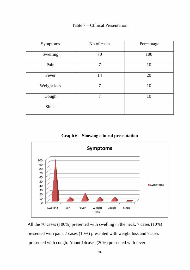
94
Table 7 – Clinical Presentation
Symptoms
No of cases Percentage
Swelling
70 100
Pain
7 10
Fever
14 20
Weight loss
7 10
Cough
7 10
Sinus
- -
Graph 6 – Showing clinical presentation
All the 70 cases (100%) presented with swelling in the neck. 7 cases (10%)
presented with pain, 7 cases (10%) presented with weight loss and 7cases
presented with cough. About 14cases (20%) presented with fever.
0
10
20
30
40
50
60
70
80
90
100
Swelling Pain Fever Weight loss
Cough Sinus
Symptoms
Symptoms

95
Table 8– Duration of the disease
Duration
No of cases Percentage
< 3months
49 70
>3 months
21 30
49 cases (70%) presented within the duration of 3 months.
Table 9– group of lymph nodes involved
Lymph node group
No of cases Percentage
UDC
42 60
LDC
8 11.4
SM
3 4.3
PT
14 20
MULTIPLE
3 4.3
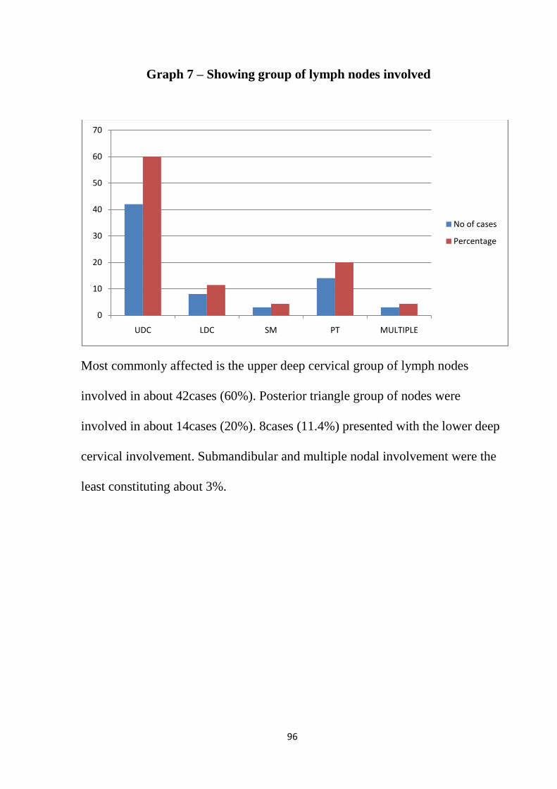
96
Graph 7 – Showing group of lymph nodes involved
Most commonly affected is the upper deep cervical group of lymph nodes
involved in about 42cases (60%). Posterior triangle group of nodes were
involved in about 14cases (20%). 8cases (11.4%) presented with the lower deep
cervical involvement. Submandibular and multiple nodal involvement were the
least constituting about 3%.
0
10
20
30
40
50
60
70
UDC LDC SM PT MULTIPLE
No of cases
Percentage
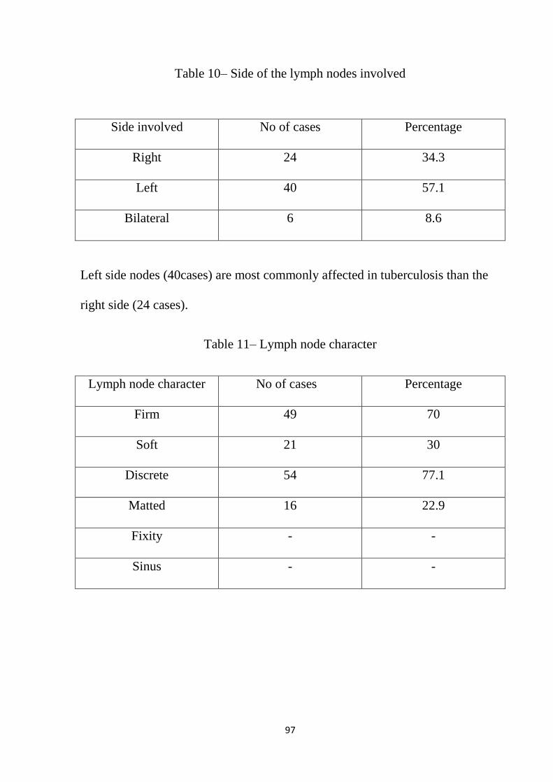
97
Table 10– Side of the lymph nodes involved
Side involved
No of cases Percentage
Right
24 34.3
Left
40 57.1
Bilateral
6 8.6
Left side nodes (40cases) are most commonly affected in tuberculosis than the
right side (24 cases).
Table 11– Lymph node character
Lymph node character
No of cases Percentage
Firm
49 70
Soft
21 30
Discrete
54 77.1
Matted
16 22.9
Fixity
- -
Sinus
- -

98
Graph 8 – Showing lymph node character
Firm lymph nodes were noted in about 49 cases (70%). 77.1% of the cases
presented with discrete nodes. Matted nodes were noted in 16 cases (22.9%).
None of them presented with fixed nodes or sinus according to the present
study.
Table 12 - Investigations
Investigations
No of cases Percentage
Raised ESR
40 57.1
Positive chest X-ray
6 8.6
Sputum for AFB positive
4 5.7
FNAC/Excision biopsy
70 100
Tuberculin test
19 27.1
0
10
20
30
40
50
60
70
80
Firm Soft Discrete Matted Fixity Sinus
Lymph node character
Lymph node character
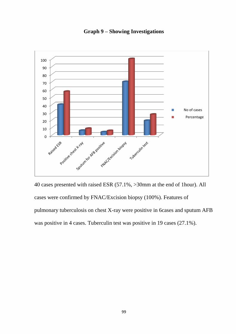
99
Graph 9 – Showing Investigations
40 cases presented with raised ESR (57.1%, >30mm at the end of 1hour). All
cases were confirmed by FNAC/Excision biopsy (100%). Features of
pulmonary tuberculosis on chest X-ray were positive in 6cases and sputum AFB
was positive in 4 cases. Tuberculin test was positive in 19 cases (27.1%).
0
10
20
30
40
50
60
70
80
90
100
No of cases
Percentage

100
Table 13– Results of FNAC and Excision Biopsy
Investigations Caseating granuloma
Inconclusive
No of
cases
Percentage No of
cases
Percentage
FNAC
58 82.9 12 17.1
Excision
Biopsy
12 100 - -
Fig 9 – Presentation of a tuberculous cervical lymph node

101
Fig 10 – Cold aspirate
All the 70 cases studied in the present series were put on short course
chemotherapy after thorough confirmation by FNAC or histopathological
examination of the excised lymph node. 58 cases were confirmed by FNAC
which revealed caseating granuloma. 12 cases (17.1%) were inconclusive and
these 12 cases were subjected to excision biopsy which showed caseating
granuloma. None of them presented with cold abscess.
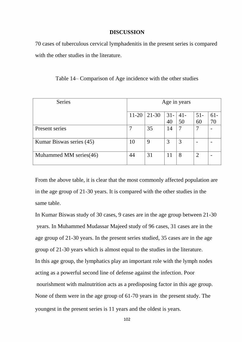
102
DISCUSSION
70 cases of tuberculous cervical lymphadenitis in the present series is compared
with the other studies in the literature.
Table 14– Comparison of Age incidence with the other studies
Series Age in years
11-20 21-30 31-
40
41-
50
51-
60
61-
70
Present series
7 35 14 7 7 -
Kumar Biswas series (45)
10 9 3 3 - -
Muhammed MM series(46)
44 31 11 8 2 -
From the above table, it is clear that the most commonly affected population are
in the age group of 21-30 years. It is compared with the other studies in the
same table.
In Kumar Biswas study of 30 cases, 9 cases are in the age group between 21-30
years. In Muhammed Mudassar Majeed study of 96 cases, 31 cases are in the
age group of 21-30 years. In the present series studied, 35 cases are in the age
group of 21-30 years which is almost equal to the studies in the literature.
In this age group, the lymphatics play an important role with the lymph nodes
acting as a powerful second line of defense against the infection. Poor
nourishment with malnutrition acts as a predisposing factor in this age group.
None of them were in the age group of 61-70 years in the present study. The
youngest in the present series is 11 years and the oldest is years.
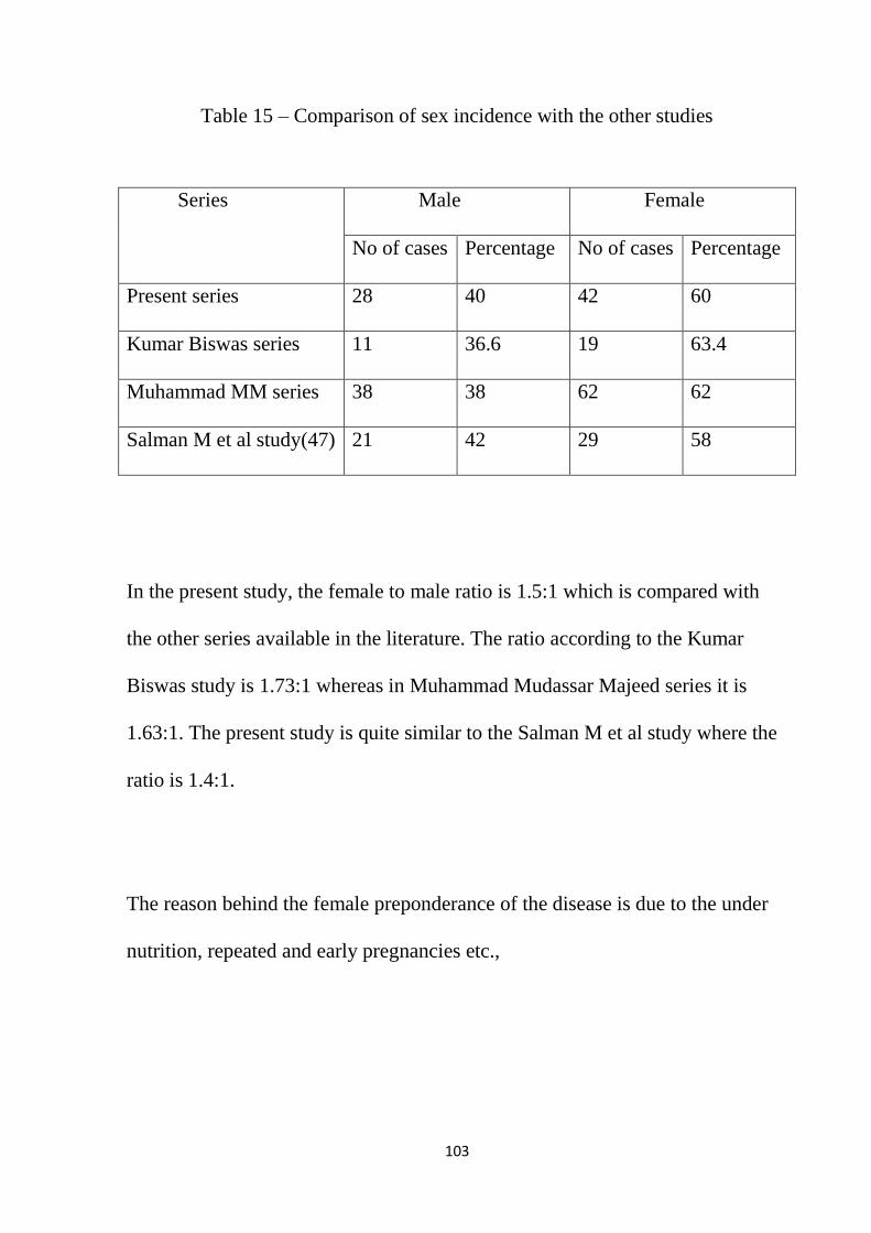
103
Table 15 – Comparison of sex incidence with the other studies
Series Male
Female
No of cases
Percentage No of cases Percentage
Present series
28 40 42 60
Kumar Biswas series
11 36.6 19 63.4
Muhammad MM series
38 38 62 62
Salman M et al study(47)
21 42 29 58
In the present study, the female to male ratio is 1.5:1 which is compared with
the other series available in the literature. The ratio according to the Kumar
Biswas study is 1.73:1 whereas in Muhammad Mudassar Majeed series it is
1.63:1. The present study is quite similar to the Salman M et al study where the
ratio is 1.4:1.
The reason behind the female preponderance of the disease is due to the under
nutrition, repeated and early pregnancies etc.,
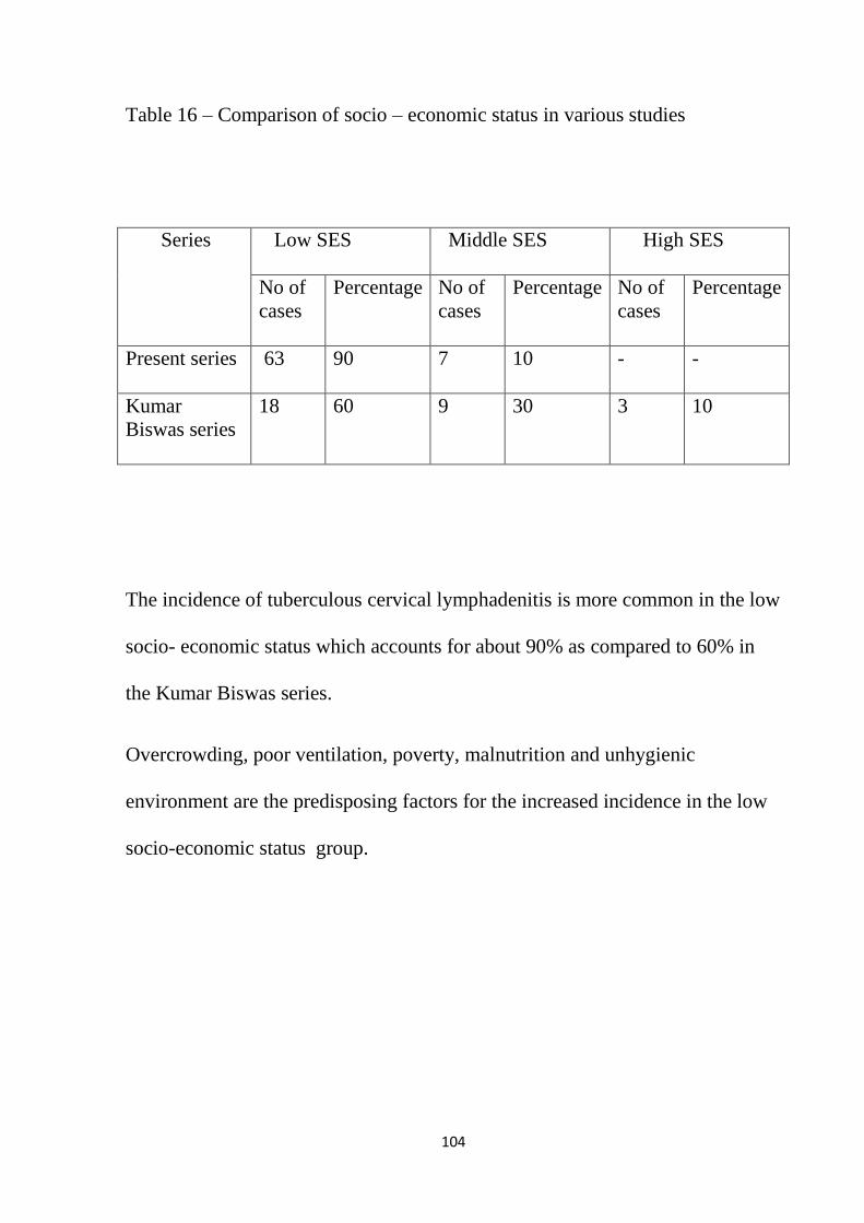
104
Table 16 – Comparison of socio – economic status in various studies
Series Low SES
Middle SES High SES
No of
cases
Percentage No of
cases
Percentage No of
cases
Percentage
Present series
63 90 7 10 - -
Kumar
Biswas series
18 60 9 30 3 10
The incidence of tuberculous cervical lymphadenitis is more common in the low
socio- economic status which accounts for about 90% as compared to 60% in
the Kumar Biswas series.
Overcrowding, poor ventilation, poverty, malnutrition and unhygienic
environment are the predisposing factors for the increased incidence in the low
socio-economic status group.
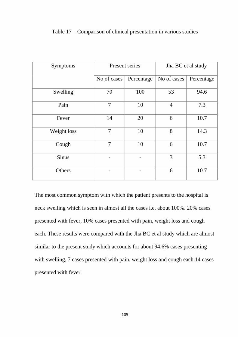
105
Table 17 – Comparison of clinical presentation in various studies
Symptoms
Present series
Jha BC et al study
No of cases
Percentage No of cases Percentage
Swelling
70 100 53 94.6
Pain
7 10 4 7.3
Fever
14 20 6 10.7
Weight loss
7 10 8 14.3
Cough
7 10 6 10.7
Sinus
- - 3 5.3
Others
- - 6 10.7
The most common symptom with which the patient presents to the hospital is
neck swelling which is seen in almost all the cases i.e. about 100%. 20% cases
presented with fever, 10% cases presented with pain, weight loss and cough
each. These results were compared with the Jha BC et al study which are almost
similar to the present study which accounts for about 94.6% cases presenting
with swelling, 7 cases presented with pain, weight loss and cough each.14 cases
presented with fever.

106
Cough is due to the upper respiratory tract involvement and is not a specific
feature of tuberculous adenitis.
Pain is due to the stretching of the capsule or the deep fascia of the neck by the
enlarging lymph nodes. Pain is more if superadded secondary infection is
present.
General symptoms like fever, loss of weight etc., are not specific for tubercular
adenitis.
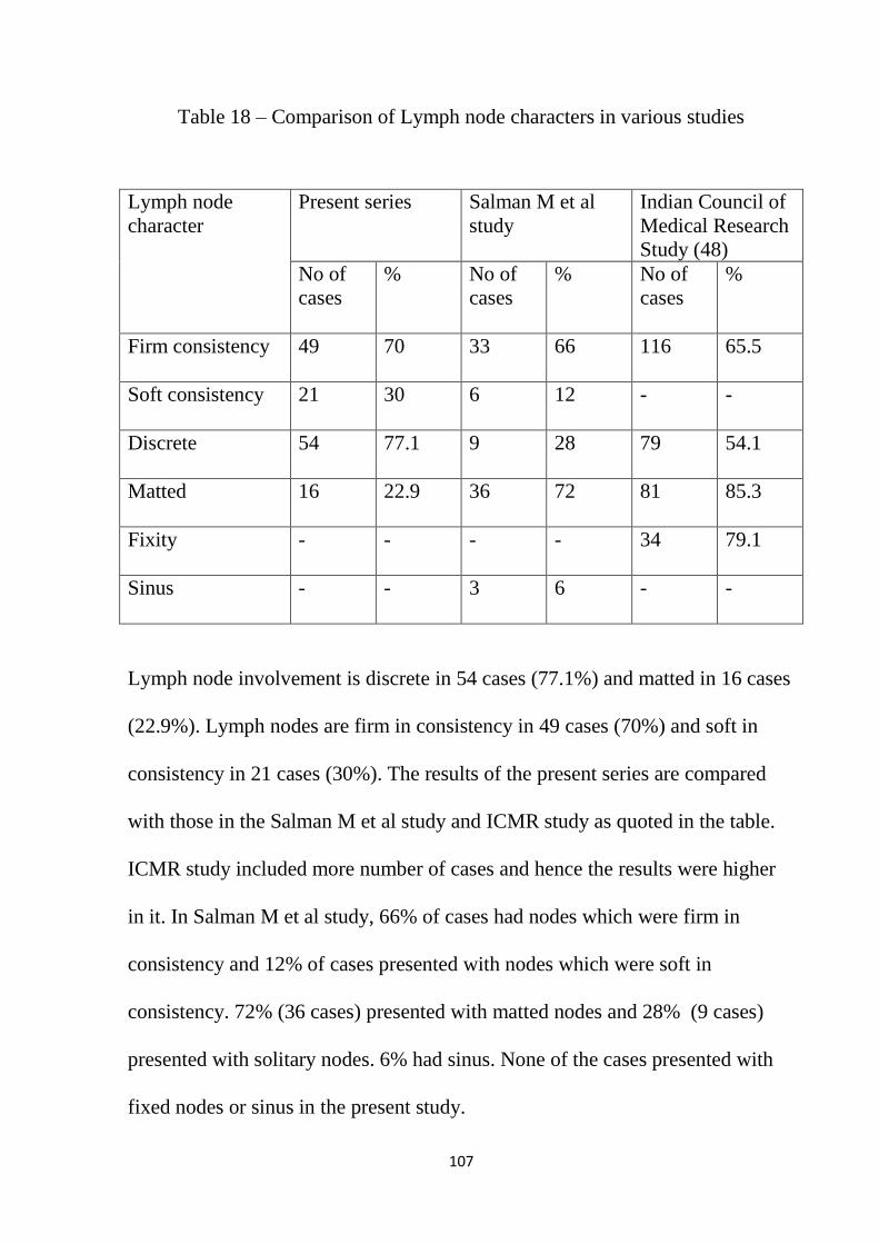
107
Table 18 – Comparison of Lymph node characters in various studies
Lymph node
character
Present series Salman M et al
study
Indian Council of
Medical Research
Study (48)
No of
cases
% No of
cases
% No of
cases
%
Firm consistency
49 70 33 66 116 65.5
Soft consistency
21 30 6 12 - -
Discrete
54 77.1 9 28 79 54.1
Matted
16 22.9 36 72 81 85.3
Fixity
- - - - 34 79.1
Sinus
- - 3 6 - -
Lymph node involvement is discrete in 54 cases (77.1%) and matted in 16 cases
(22.9%). Lymph nodes are firm in consistency in 49 cases (70%) and soft in
consistency in 21 cases (30%). The results of the present series are compared
with those in the Salman M et al study and ICMR study as quoted in the table.
ICMR study included more number of cases and hence the results were higher
in it. In Salman M et al study, 66% of cases had nodes which were firm in
consistency and 12% of cases presented with nodes which were soft in
consistency. 72% (36 cases) presented with matted nodes and 28% (9 cases)
presented with solitary nodes. 6% had sinus. None of the cases presented with
fixed nodes or sinus in the present study.

108
Table 19 – Comparison of groups of lymph nodes involved in various studies
Group of lymph
nodes involved
Present series in
%
Kumar Biswas
study in %
Maharjan M et al
study in % (49)
Upper deep
Cervical
60 33.3 16
Lower deep
cervical
11.4 10 6
Submandibular
4.3 13.3 15
Supraclavicular
- 3.4 9
Posterior triangle
20 20 42
Multiple
4.3 20 -
The upper deep cervical group of lymph nodes are most commonly affected in
the present series accounting for 60% which is more when compared to the
Kumar Biswas study and Maharjan M et al study. The submandibular and the
multiple nodal involvement are the least accounting for 4.3% each. There is no
supraclavicular involvement in the present series. In Maharjan M et al study,
posterior triangle group of nodes are more commonly affected followed by the
upper deep cervical group of nodes.

109
The tubercle bacilli are filtered at the nose, oral cavity, pharynx and tonsils and
ultimately drain into the upper deep cervical group of lymph nodes and hence
the lesions are produced here. The other portal of entry is the adenoids which
drain into the lower deep cervical group of lymph nodes which accounts for
about (11,4% cases) in the present study. Multiple nodal involvement is present
in about 4.3% cases according to the present study.
Table 20 – Comparison of associated pulmonary tuberculosis in various studies
Present series
Maharjan M et al
study
Jha BC et al
study(1)
Total no of cases
studied
70 83 60
Associated
pulmonary
tuberculosis in
percentage
5 14 16
In the present series, about 5% cases had associated pulmonary tuberculosis
which is about 14% in the Maharjan M et al study and 16% in the Jha BC et al
study.

110
Table 21 – Comparison of investigations in various studies
Series Raised ESR in
percentage
Chest X-Ray in
percentage
FNAC/Excision
biopsy in
percentage
Present series
57.1 8.6 100
Jha BC et al study
100 16 100
Maharjan M et al
Study
79 14 100
Erythrocyte sedimentation rate was raised in 57.1% of the cases in the present
series which is 100% and 79% respectively in the Jha BC et al study and
Maharjan M et al study. Chest x-ray showed features of tuberculosis in 8.6% of
the cases which is 16% and 14% respectively in the Jha BC et al study and
Maharjan M et al study.
Fine Needle Aspiration Cytology was done in all the cases and found to be
positive in all the cases which is the same as compared to the Jha BC et al study
and Maharjan M et al study.
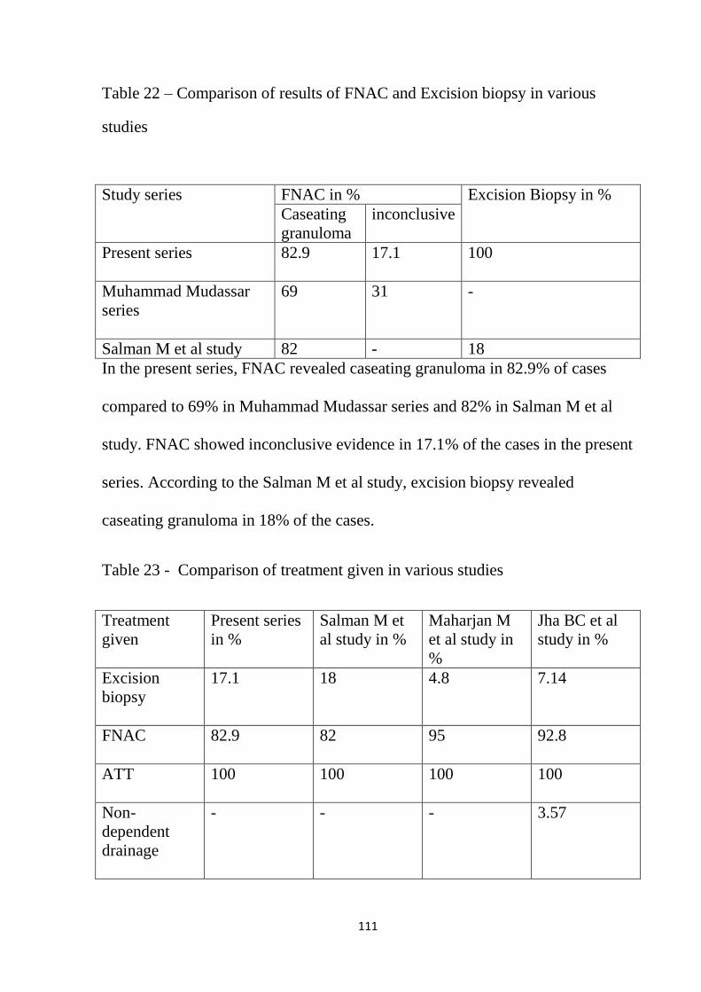
111
Table 22 – Comparison of results of FNAC and Excision biopsy in various
studies
Study series FNAC in % Excision Biopsy in %
Caseating
granuloma
inconclusive
Present series
82.9 17.1 100
Muhammad Mudassar
series
69 31 -
Salman M et al study 82 - 18
In the present series, FNAC revealed caseating granuloma in 82.9% of cases
compared to 69% in Muhammad Mudassar series and 82% in Salman M et al
study. FNAC showed inconclusive evidence in 17.1% of the cases in the present
series. According to the Salman M et al study, excision biopsy revealed
caseating granuloma in 18% of the cases.
Table 23 - Comparison of treatment given in various studies
Treatment
given
Present series
in %
Salman M et
al study in %
Maharjan M
et al study in
%
Jha BC et al
study in %
Excision
biopsy
17.1 18 4.8 7.14
FNAC
82.9 82 95 92.8
ATT
100 100 100 100
Non-
dependent
drainage
- - - 3.57
112
12 cases (17.1%) were subjected to excision biopsy of the affected lymph
nodes according to the present study. 58 cases (82.9%) were diagnosed by
FNAC alone. This result is similar to that of the Salman M et al study in which
82% of the cases were diagnosed by FNAC alone and 18% of the cases were
diagnosed by excision biopsy. In Jha BC et al study, FNAC proved effective in
92.8% of the cases and excision biopsy was conducted in 7.14% of the cases.
3.57% of the cases underwent non-dependent aspiration of the nodes according
to the Jha BC et al study. In the present study, none of the cases were subjected
to non-dependant aspiration.
After confirmation of diagnosis, patients were put on Anti-tubercular drugs and
were followed up. The cure rate was 100%. This result is also quite similar to
that of the Jha BC et al study and Maharjan M et al study.
Over the years, because of the advancement in medical science, chemotherapy
is more effective and has almost limited the role of surgery to drainage of the
cold abscess, excision of the residual lymph node mass or scars, excision biopsy
of the affected lymph nodes.

113
FOLLOW UP:
The cases which were put on anti-tubercular treatment were followed up at an
interval of about 1-2 months from the commencement of ATT to a period of 6
months. During this period, cases were assessed by clinical examination and
ESR examination. The decrease in the size of the swelling and the generalised
improvement in the health status of the patients were studied and majority of the
cases showed improvement within 4weeks of the start of the treatment.
114
CONCLUSION
Historical aspects on tubercular cervical lymphadenitis has been reviewed.
A brief description on the anatomy, physiology of the lymph nodes draining the
neck, along with the distribution of the lymph nodes, with the aetiology,
pathology, clinical aspects and the type of treatment are discussed in detail.
100 cases were taken, out of which 70 cases of cervical tuberculous
lymphadenitis were studied, analysed in detail and the following conclusions
were drawn.
The incidence of tuberculous cervical lymphadenopathy was maximum in the
age group of 21-30 years (35 cases-50%). The youngest in the study is 12years.
The disease showed an increasing incidence in females as compared to the male
patients with the female to male ratio being 1.5:1.
It is more common in the lower socio-economic status people (90%).
The disease shows increased incidence among the rural population which
accounts for about 70% of the cases.
All the 70 cases presented with the complaint of swelling in the neck. 14 cases
presented with fever, 7 cases presented with pain, weight loss and cough
respectively.
The lymph node involvement was discrete in 77.1% of the cases and matted in

115
22.9% of the cases. 70% patients had firm consistency of the lymph nodes.
The upper deep cervical nodes were involved in 60% which was the most
common lymph node involved in tuberculous cervical lymphadenopathy.
57.1% presented with raised ESR . 8.6% had features suggestive of pulmonary
tuberculosis on chest x-ray. 82.9% showed caseating granuloma on FNAC.
FNAC was inconclusive in 17.1% of the cases who were then subsequently
subjected to Excision Biopsy which proved positive for tuberculous cervical
lymphadenopathy. Tuberculin test was positive in 27.1% of the cases.
The patients were then categorised as Category I or Category II patients and
were put on Anti-tubercular treatment based on the histopathological
(FNAC/Excision Biopsy) reports and were followed up for a period of 6
months. 65 cases were symptom free at the end of the study.

116
SUMMARY
This is a study involving 100 cases out of which70 cases proved to be
tuberculous cervical lymphadenitis. These cases were studied at Govt. Kilpauk
Medical College and Hospital for a period of 1 year from Aug2013-Aug2014.
1. Tuberculosis is more common in the 21-30 years age group which is
about 35 cases (50%) according to the present study. The youngest in the
present series is 12 years of age.
2. The disease showed a preponderance in females as compared to the male
patients with the female to male ratio being 1.5:1. It is quite similar to
that of the Salman M et al study where the female to male ratio is 1.4:1.
3. It is more common in the lower socio-economic status people (90%).
4. The disease shows increased incidence among the rural population which
accounts for about 70% .
5. All the 70 cases presented with the complaint of swelling in the neck.
14cases presented with fever, 7cases presented with pain, weight loss and
cough respectively. The left side nodes are most commonly involved
(57.1%). The lymph node involvement was discrete in 77.1% and firm in
70% . 22.9% presented with matted lymph nodes according to the
present study. The upper deep cervical nodes were involved in 60%
which was the most common lymph node involved in tuberculous
cervical lymphadenopathy.
117
6. 57.1% presented with raised ESR according to the present study. 8.6%
had features suggestive of pulmonary tuberculosis on chest x-ray. 82.9%
showed caseating granuloma on FNAC.
7. FNAC was inconclusive in 17.1% which were then subsequently
subjected to Excision Biopsy which proved positive for tuberculous
cervical lymphadenopathy.
8. Tuberculin test was positive in 27.1% .
9. The patients were then categorised as Category I or Category II patients
and were put on Anti-tubercular treatment based on the histopathological
(FNAC/Excision Biopsy) reports. For the first 2 months , patients under
category I were given INH, Rifampicin, Ethambutol and pyrazinamide
and under category III were given INH, pyrazinamide, rifampicin. In the
continuation phase only INH and rifampicin were given to the patients for
a period of 4 months. They were followed up for a period of 6 months. 65
cases were symptom free at the end of the study.
118
BIBLIOGRAPHY
1. Jha BC, Dass A, Nagarkar NM et-al. Cervical tuberculous
lymphadenopathy: Changing clinical patterns and concepts in
management. Postgrad Med J 2001; 77 (905): 185-7.
2. Iseman MD. A clinician‟s guide to tuberculosis. India: Lippincott
Williams and Willkins: 2000. Chapter 1. Tuberculosis down through the
centuries; p.2,4,7,14-17,51-53.
3. Pesani Edward L. History of tuberculosis: In tuberculosis a clinical
handbook. Larry.I.Lutwic, 1 edition, London: Chapman and Hall, 1995:1-
5.
4. Sadler TW. Langman‟s Medical Embryology. 10th
edition. New Delhi:
Lippincott William and Wilkins; 2008. Chapter 12, Cardiovascular
system: p.191-92.
5. Oliver G. Lymphatic vasculature development. Nat Rev Immunol.
2004;4:35-45.(Pub Med)
6. Wigley C, Cecil C. Blood, lymphoid tissue and haemopoiesis. In:
Standring S, editor. Gray‟s Anatomy. 40th
ed. Spain: Elsevier publishers;
2008. Chapter 4.p.72-73.
7. Singh IB. Textbook of Human Histology. 5th
edition. New Delhi: Jaypee
publishers; 2006. Chapter 11. Lymphatics and lymphoid tissue: p.184-
189.
119
8. Ganong WF. Dynamics of Blood and Lymph flow. Review of Medical
Physiology. 22nd
edition; McGrawHill;2005:p.593.
9. David JH, Valerie JL. Pharynx, larynx and neck. In: Norman SW,
Christopher JK, Bulstrode, O‟Connell PR, editors. Bailey and Love‟s
short practice of surgery. 25th
ed, London : Edward Arnold Ltd; 2008.
Chapter 45.p.731-732.
10. Decker GAG. Lee Mcgregor‟s synopsis of Surgical Anatomy. 12th
ed.
Bombay: Varghese Publication; 1999. Chapter 16. Lymph tissues of the
head and neck, surgical anatomy; p.189-196.
11. Paniker CKJ. Textbook of microbiology. 7th
ed. Chennai: Orient
Longman publishers; 2005. Chapter 39, Mycobacterium tuberculosis;
p.351-64.
12. Brooks GF, Karen CC. Mycobacteria. In: Jawetz, Melnick, Adelberg,
editors. Medical microbiology. 24th ed. USA: McGraw Hill; 2007.
Chapter 24, p.320-27.
13. Kayser FH, Bienz KA, Eckert J, Kayser Medical Microbiology. 10th
ed.
Germany: 2005. Chapter 4, Bacteria as human pathogens; p.264.
14. Bloch H, Sorkin E. A toxic lipid component of tubercle bacillus. II:
Isolation from petroleum ether extracts of young bacterial cultures. Am
Rev Tuberc 1953;67:629-643.
15. Meltezon HC, Spyridis P, Kafetzis DA. Extrapulmonary tuberculosis in
children. Arch Dis Child 2000;83:342-6.

120
16. Butt T, Riffat AN, Syed KY, Raja AK, Mahmood A. An update on the
diagnosis of tuberculosis. JCPSP 2003;13 (12): 728-34.
17. Tuberculosis incidence (Internet) 2012 (cited 2010 october). Available
from:http://www.who.int/mediacentre/factsheets/fs104/en/index.html.
18. Park K. Park‟s textbook of preventive and social medicine. 20th ed.
Jabalpur: Bhenot;2009. Chapter 5, Epidemiology of communicable
disease; p.160-161, 163-73.
19. Wares F, Balasubramanian R. Mohan A, Sharma SK. Extrapulmonary
tuberculosis: Management and Control. In: Agarwal SV, Chauhan LS,
editors. Tuberculosis control in India. New Delhi: Directorate General of
Health Services. Ministry of Health and Family Welfare; 2005. P. 95-114.
20. Robbins, Kumar. Pathology basis of disease. 8th ed. W.B. Saunders,
Philadelphia;2008. Chapter 13. The lung and upper respiratory tract:
p.517-19.
21. Dass A, Nagarkar NM et- al. Cervical tuberculous lymphadenopathy:
Changing clinical pattern and concepts in management. Postgrad Med J.
2001; 77 (905):185-7.
22. Head and neck imaging. Ed. By Peter M. Som, Hugh D. Curtin. St Louis
(Mo): Mosby-Year Book,2003.
23. Ioachim‟s lymph node pathology. Editors. Herry L. Ioachim, L. Jeffrey
Mederios. Philadelphia, Pa ; Lippincott Williams and Wilkins, 2008.
121
24. Jones PG, Campbell PE. Tuberculous lymphadenitis in childhood: the
significance of anonymous mycobacteria. Br J Surg 1962;50:302-14.
25. Gupta AK, Nayar M, Chandra M, et al. Critical appraisal of fine needle
aspiration cytology in tuberculous lymphadenitis. Acta Cytol 1992; 36:
391-4.
26. Lakhey M, Bhatta CP, Mishra S. Diagnosis of tubercular
lymphadenopathy by fine needle aspiration cytology, acid-fast staining
and Mantoux test. JNMA J Nepal Med Assoc. 2009 Jul-Sep; 48 (175):
230-3.
27. Avery G. Mycobacterium tuberculosis and other non- tuberculous
mycobacteria. In: Connie RM, Donal CL, George M, editors. Text book
of diagnostic microbiology. 3rd
ed. New Delhi. Elsevier publishers; 2007.
Chapter 26, 693-699, 700-05.
28. Manitchotpisit B, Kunachek S, Kulapralithram B: Combined use of fine
needle aspiration cytology and polymerase chain reaction in the diagnosis
of cervical tubercular lymphadenitis. J Med Assoc Thai 1999; 82: 363-68.
29. Kwon KS, Oh CK, Jang HS, Lee CW: detection of mycobacterial DNA
in cervical granulomatous lymphadenopathy from formalin-fixed,
paraffin embedded tissue by PCR. J Dermatol 2000; 27: 355-360.
30. Mario CR, Richard JO. Tuberculosis. In : Fauci AS, Braunwald E, Kasper
DL, Hauser SL, editors. Harrison‟s principle of internal medicine. 17th ed.
McGraw Hill; 2008. Chapter 158, p.1006-1020.
122
31. Das S. A concise textbook of surgery. 6th
ed. Kolkata; 2010. Chapter 17.
Diseases of the lymphatic system; p.296-309.
32. Treatment of tuberculosis: American thoracic society/ Centre for disease
control/ Infectious disease Society of America- Recommendations and
Reports. MMWR Morb Mortal Wkly Rep. 2003;52: 1-77.
33. British Thoracic Society Research Committee. Six months versus nine
months chemotherapy for tuberculosis of lymph nodes: Final results.
Respir Med 1993; 87: 621-3.
34. Tripathi KD. Essentials of medical pharmacology 5th
ed. New Delhi:
Jaypee publishers; 2004. Chapter 53, Antitubercular drugs; p.698-708.
35. Joel GH, Lee EL, Gillman AG. Goodman and Gillman‟s the
Pharmacological basis of therapeutics. 10th ed. USA: McGraw Hill; 2001.
Chapter 48. Drugs used in chemotherapy of tuberculosis and leprosy;
p.1274-86.
36. Van Loenhout-Rooyackers JH, Laheij RJ, Richter C, Werbeek AL:
Shortening the duration of treatment for cervical tuberculous
lymphadenitis. Eur Respir J 2000; 15: 192-95.
37. Dalton, T. Cegielski, P. Akksilp, S. Asencios, L. Caoili, J. C. Cho,
S.N.Erokhin, V.V.Ershova, J.et al. (2012). “Prevalence and risk factors
for resistance to second line drugs in people with multidrug-resistant
tuberculosis in eight centuries: A prospective cohort study”. The Lancet.
Doi: 10.1016/S0140-6736(12)60734-X.

123
38. Goble M, Iseman MD, Madsen LA, Waite D, Ackerson L, Horsburgh CR
Jr(1993).” Treatment of 171 patients with pulmonary tuberculosis
resistant to isoniazid and rifampin”. N Engl J Med 328(8): 527-532.
39. Farquharson M, Moran B. Farquharson‟s text book of operative general
surgery. 9th
ed. Great Britain: Holder Arnold; 2005. Chapter 9, Surgery of
the neck; p.164-165.
40. Danielides V, Patrikakos G, Moerman M, Bonte K, Dhooge C,
Vermeersch H: Diagnosis, management and surgical treatment of non
tuberculous mycobacterial head and neck infection in children. ORL J
Otorhinolaryngol Relat Spec 2002;64;284-289.
41. Alessi DP, Dudley JP: Atypical mycobacteria induced cervical adenitis.
Treatment by needle aspiration. Arch Otolaryngol Head Neck
Surg.1999;1109-1113.
42. Panesar J, Higgins K, Daya H, Forte V, Allen U; Nontubercuolous
mycobacterial cervical adenitis: A ten-year retrospective review.
Laryngoscope 2003;113:149-154.
43. Hussain Md.W, Haque Md.A, Banu SA, Ekram ARMS, Rahman MF.
Extra pulmonary tuberculosis: Experience in Rajshahi chest disease clinic
and chest disease hospital. The journal of Teachers Association-June
2004; Volume 17(1): 16-19.

124
44. Biswas PK, Begum SMK. Tubercular cervical lymphadenopathy
Clinicopathological study of thirty cases. The journal of Teachers
Association- June 2007; Volume 20(1): 36-38.
45. Muhammad MM, Bukhari MH. Evaluation for granulomatous
inflammation on Fine Needle Aspiration Cytology using special stains.
SAGE-Hindawi Access to Research Pathology. Research International –
May 2011; Article ID 851524, 1-8.
46. Salman M, Zeba A, Mahira Y, Muhammad SM. Evaluation of
tuberculosis cervical lymphadenopathy. Pakistan Journal of Surgery –
2009; Volume 25(3):176-79.
47. Pamra, Mathur. Tuberculosis in children. The Indian Journal of
Tuberculosis- April 1987; Vol 34(2): 41-44. Table 4, character to glands
related to type of lymphadenitis; p.44.
48. Maharajan M, Hirachan S, Kafla PK, Bista M, Shrestha S, Taran KC.
Incidence of tuberculosis in enlarged neck nodes. Our experience.
Kathmandu University Medical Journal-2009; Vol.7(25);53-54.
49. Lai KK, Stottmaier KD, Sherman IH, et al. Mycobacterial cervical
adenopathy. JAMA 1984;251:1286-9.
50. Lincoln EM, Sewell EM, Tuberculosis in children. New York, NY: Mc
Graw – Hill Book Company Inc, 1963:207-15.
51. Campbell IA. The treatment of superficial tuberculous lymphadenitis.
Tubercle 1990;71:1-3.

125
52. Seth V, Kabra SK, Semwal OP, et al. Tubercular lymphadenitis: Clinical
manifestations. Indian J Pediatr 1995;62:565-70.
53. Belin RP, Richardson JD, Richardson DL, et al. Diagnosis and
management of scrofula in children. J Pediatr Surg 1974;9:103-7.
126
ANNEXURE-I
PROFORMA
1) PARTICULARS OF THE PATIENT:
Name:
Age:
Sex:
I.P/O.P no:
Religion:
Occupation:
Income:
2) CHIEF COMPLAINS:
3) HISTORY OF PRESENT ILLNES:
a) Duration of node enlargement/ swelling in the neck
b) Associated symptoms
Fever and cough
Loss of appetite
Throat pain
Oropharyngeal and scalp sepsis.

127
c) Pain
d) Sinus present or absent
If so, 1) duration
ii) How did it start
iii) Nature of discharge
4) PAST HISTORY
Pulmonary or Extrapulmonary
Lymphadenitis
5) FAMILY HISTORY
6) PERSONAL HISTORY
Contact with tuberculosis
Habit of taking unboiled milk
Living conditions and diet
7) MENSTRUAL / OBSTETRIC HISTORY
8) GENERAL PHYSICAL EXAMINATION
Build
Febrile
Anemia
Jaundice
Clubbing
Lymphadenopathy
Weight

128
Pulse
Blood pressure
SYSTEMIC EXAMINATION
CVS
Respiratory system
Abdomen
o Liver
o Spleen
o Lymph nodes
o Others
Local examination: swelling in the neck
o Unilateral- single groups or multiple groups
o Bilateral- single groups or multiple groups
o Skin over the swelling
Associated with any other group of lymph nodes
Axillary -pectoral
Subscapular
Lateral
Central
Apical
Inguinal- horizontal
129
Vertical
Mediastinal
Nature of the nodes
Situation
Size
Shape
Number
Inflammation
Consistency
Discrete or matted
Fixity ]
Skin involvement
Surface
Sinus if present
Number
Situation
Search for feeding node
Discharge
Other physical character
Examination of draining area
Oral cavity
130
Tonsil
9) INVESTIGATIONS
a. Blood
Hb%
TC
DC
ESR
b. Sputum if present for AFB
c. Radiological examination
X-ray neck
X-ray chest
d. Biopsy
Macroscopic picture
Biopsy report
e. Tuberculin test
10) TREATMENT
i. Anti tubercular
ii. Supportive
iii. Surgical
11) FOLLOW UP
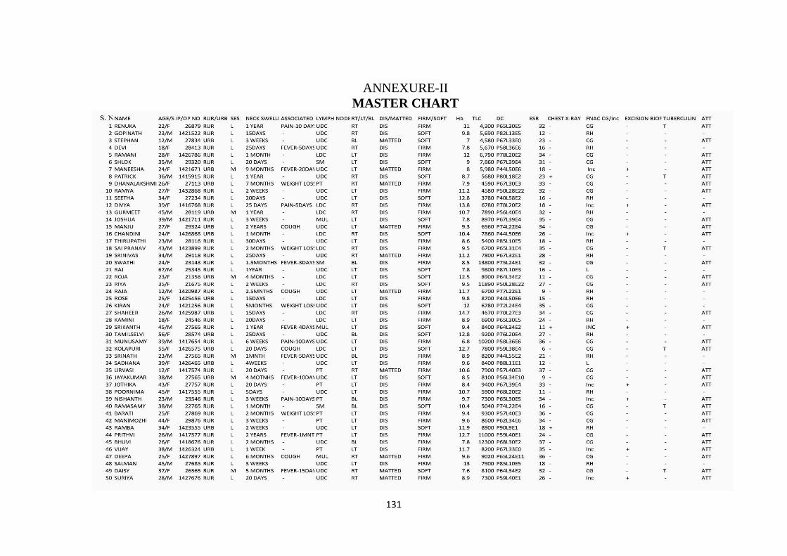
131
ANNEXURE-II
MASTER CHART
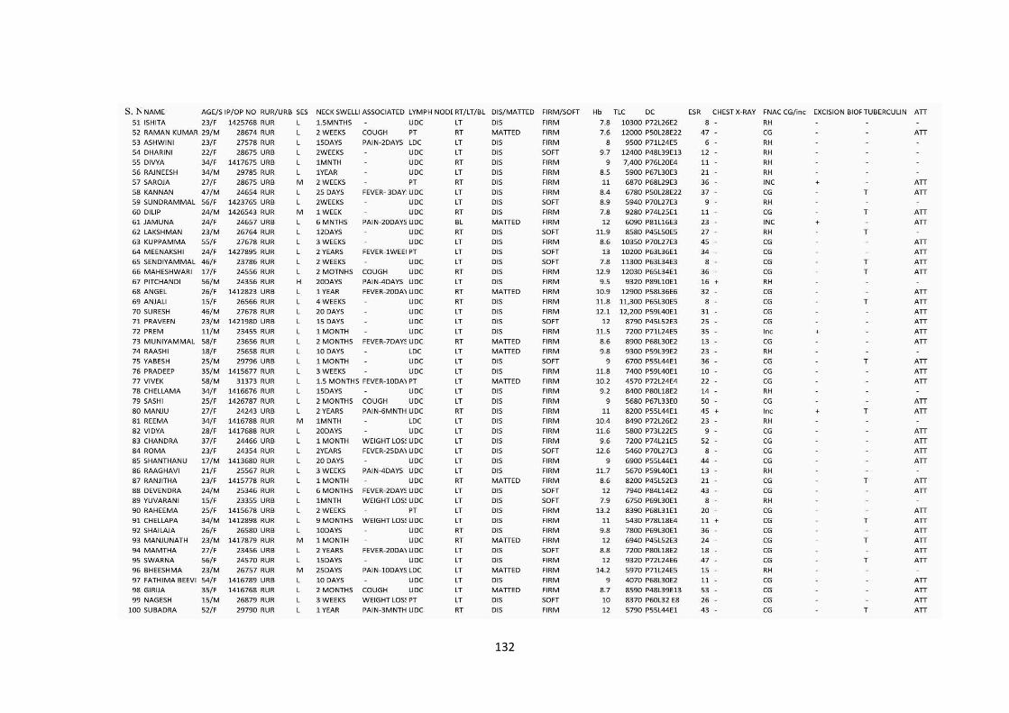
132
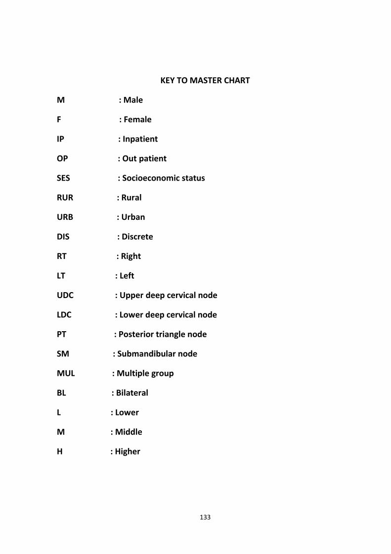
133
KEY TO MASTER CHART
M : Male
F : Female
IP : Inpatient
OP : Out patient
SES : Socioeconomic status
RUR : Rural
URB : Urban
DIS : Discrete
RT : Right
LT : Left
UDC : Upper deep cervical node
LDC : Lower deep cervical node
PT : Posterior triangle node
SM : Submandibular node
MUL : Multiple group
BL : Bilateral
L : Lower
M : Middle
H : Higher

134

135