The structure of alanyl-tRNA synthetase with editing … · The structure of alanyl-tRNA synthetase...
6
The structure of alanyl-tRNA synthetase with editing domain Masaaki Sokabe a,1 , Toyoyuki Ose a , Akiyoshi Nakamura a , Keita Tokunaga a , Osamu Nureki b,c , Min Yao a , and Isao Tanaka a,2 a Faculty of Advanced Life Sciences, Hokkaido University, Sapporo 060-0810, Japan; b Department of Biological Information, Graduate School of Bioscience and Biotechnology, Tokyo Institute of Technology, Kanagawa 226-8501, Japan; and c Department of Basic Medical Sciences, Institute of Medical Science, University of Tokyo, Tokyo 108-8639, Japan Edited by Dieter So ¨ ll, Yale University, New Haven, CT, and approved May 12, 2009 (received for review April 29, 2009) Alanyl-tRNA synthetase (AlaRS) catalyzes synthesis of Ala-tRNA Ala and hydrolysis of mis-acylated Ser- and Gly-tRNA Ala at 2 different catalytic sites. Here, we describe the monomer structures of C- terminal truncated archaeal AlaRS, with both activation and edit- ing domains in the apo form, in complex with an Ala-AMP analog, and in a high-resolution lysine-methylated form. The structures show docking of the editing domain to the activation domain opposite from the predicted tRNA-binding surface. Thus, the ed- iting site is positioned >35 Å from the activation site, prompting us to model 2 different tRNA complexes: one binding tRNA at the activation site, and the other binding tRNA at the editing site. Interestingly, a gel-shift assay also implies the presence of 2 types of tRNA complex with different mobility. These results suggest that tRNA translocation via a canonical CCA flipping is unlikely to occur in AlaRS. The structure also demonstrated the binding of zinc in the editing site, in which the specific coordination of zinc would be facilitated by a conserved GGQ motif, implying that the editing mechanism may not be the same as in ThrRS. As Asn-194 in eubacterial AlaRS important for Ser misactivation is replaced by Thr-213 in archaeal AlaRS, a different Ser accommodation mecha- nism is proposed. amino acid recognition class II aminoacyl-tRNA synthetase crystal structure A lanyl-tRNA synthetase (AlaRS) belongs to the class II aminoacyl-tRNA synthetases (aaRSs) and specifically at- taches Ala to the 3-end of the cognate tRNA Ala (1). AlaRS has an additional catalytic site (termed an editing site), which specifically hydrolyzes a mischarged Ser- or Gly-tRNA Ala , be- cause the accuracy of discrimination of cognate Ala from noncognate Ser/Gly at the aminoacylation site is not sufficient (2). Because even a mild defect in the editing activity has been shown to cause neural degeneration in mice, such quality control in AlaRS is crucial, especially for higher eukaryotes (3). The protein is made up of 4 functional modules (Fig. S1), which are responsible for aminoacylation catalysis, tRNA recognition, editing, and oligomerization (2, 4). Eubacterial AlaRS form tetramers (5) whereas those in eukaryotes are monomers (4). It has been shown that the C-terminal half, the region responsible for editing and oligomerization, is not necessary for aminoacy- lation, even though the activity is significantly decreased in its absence (6). The anti-codon stem of tRNA Ala is also not necessary for aminoacylation, and thus, the acceptor stem provides sufficient information for specific recognition by AlaRS, which is particularly dependent on the G3:U70 base pair known as the second genetic code (7). A tRNA-binding model has been proposed by superposition of the AspRS-tRNA complex onto the structure of the N-terminal half of Aquifex aeolicus AlaRS (N453). The acceptor stem of the tRNA binds to a concave surface made by the activation domain and the C-terminal helical domains, which approach the G3:U70 base pair from the major groove side (8). In most aaRS harboring an editing domain, the editing site approaches the acceptor stem of the bound tRNA from the side opposite that for aminoacy- lation so that the 3-end of the tRNA can be translocated rapidly from 1 site to the other site by simply flipping its CCA tail without significantly moving its body (9). For example, in class II ThrRS, ProRS, and PheRS, the activation domain approaches from the major groove side whereas the editing domain is located at the minor groove side. The situation in AlaRS remains unclear because structures containing either the editing domain or the tRNA have not been reported. However, it has been shown recently that the C-terminal half fragment, which lacks the region essential for aminoacylation, still has considerable editing activity. Surprisingly, this fragment can specifically recognize Ser-tRNA Ala , at least in part, in a manner dependent on the G3:U70 base pair, suggesting that the editing domain has its own tRNA-binding capability mostly independent of the activation site (10). Misactivation of noncognate amino acids at the activation site in AlaRS has been studied with the structures of N453 in complex with Ala/Ser/Gly (11). The structures show the accom- modation of a Ser hydroxyl by an induced-fit involving the Asn-194 amide group (hereafter, prime refers to N453), which forms a hydrogen bond with the Ser hydroxyl. Discrimination of Ala- from Ser-tRNA Ala at the editing site has been studied in our group by determining the structure of an autonomous homolog of the editing domain (AlaXS) in a complex with Ser (12). The structure suggested that the conserved Thr-30 in AlaXS (con- served as Gln-633 in Pyrococcus horikoshii AlaRS) is important for discrimination. Indeed, T30V/Q633M mutants of AlaXS/ AlaRS show mis-editing of a cognate Ala-tRNA Ala (12). The catalytic mechanism of deacylation remains controversial. The structure of the editing domain in ThrRS, which has homology with that in AlaRS, in complex with SerA76 (a nonhydrolyzable analog of the 3-terminus of Ser-tRNA) suggests that the con- served 73 HXXXH 77 and 182 CXXXH 186 motifs do not bind zinc for deacylation, but rather are directly involved in both recog- nition and catalysis of the substrate (13, 14). In contrast, in the structure of AlaXS-Ser, the binding of zinc to the motif is apparently concomitant with the binding of Ser (12). Although AlaRS has also been suggested to bind zinc at the editing site (2), whether it is required for the deacylation activity is unclear. Author contributions: M.S., T.O., A.N., M.Y., and I.T. designed research; M.S., T.O., A.N., and K.T. performed research; M.S. and O.N. contributed new reagents/analytic tools; M.S., T.O., A.N., K.T., M.Y., and I.T. analyzed data; and M.S., T.O., M.Y., and I.T. wrote the paper. The authors declare no conflict of interest. This article is a PNAS Direct Submission. Data deposition footnote: The atomic coordinates have been deposited in the Protein Databank, www.pdb.org [PDB ID code 2ZZF (N752-Zn), 2ZZG (N752-AlaSA), and 2ZZE (N752m)]. 1 Present address: Department of Biochemistry and Molecular Medicine, School of Medi- cine, University of California, Davis, CA 95616. 2 To whom correspondence should be addressed. E-mail: [email protected]. This article contains supporting information online at www.pnas.org/cgi/content/full/ 0904645106/DCSupplemental. 11028 –11033 PNAS July 7, 2009 vol. 106 no. 27 www.pnas.orgcgidoi10.1073pnas.0904645106
-
Upload
vuongkhuong -
Category
Documents
-
view
233 -
download
0
Transcript of The structure of alanyl-tRNA synthetase with editing … · The structure of alanyl-tRNA synthetase...
The structure of alanyl-tRNA synthetase withediting domainMasaaki Sokabea,1, Toyoyuki Osea, Akiyoshi Nakamuraa, Keita Tokunagaa, Osamu Nurekib,c, Min Yaoa,and Isao Tanakaa,2
aFaculty of Advanced Life Sciences, Hokkaido University, Sapporo 060-0810, Japan; bDepartment of Biological Information, Graduate School of Bioscienceand Biotechnology, Tokyo Institute of Technology, Kanagawa 226-8501, Japan; and cDepartment of Basic Medical Sciences, Institute of Medical Science,University of Tokyo, Tokyo 108-8639, Japan
Edited by Dieter Soll, Yale University, New Haven, CT, and approved May 12, 2009 (received for review April 29, 2009)
Alanyl-tRNA synthetase (AlaRS) catalyzes synthesis of Ala-tRNAAla
and hydrolysis of mis-acylated Ser- and Gly-tRNAAla at 2 differentcatalytic sites. Here, we describe the monomer structures of C-terminal truncated archaeal AlaRS, with both activation and edit-ing domains in the apo form, in complex with an Ala-AMP analog,and in a high-resolution lysine-methylated form. The structuresshow docking of the editing domain to the activation domainopposite from the predicted tRNA-binding surface. Thus, the ed-iting site is positioned >35 Å from the activation site, promptingus to model 2 different tRNA complexes: one binding tRNA at theactivation site, and the other binding tRNA at the editing site.Interestingly, a gel-shift assay also implies the presence of 2 typesof tRNA complex with different mobility. These results suggestthat tRNA translocation via a canonical CCA flipping is unlikely tooccur in AlaRS. The structure also demonstrated the binding of zincin the editing site, in which the specific coordination of zinc wouldbe facilitated by a conserved GGQ motif, implying that the editingmechanism may not be the same as in ThrRS. As Asn-194 ineubacterial AlaRS important for Ser misactivation is replaced byThr-213 in archaeal AlaRS, a different Ser accommodation mecha-nism is proposed.
amino acid recognition � class II aminoacyl-tRNA synthetase �crystal structure
A lanyl-tRNA synthetase (AlaRS) belongs to the class IIaminoacyl-tRNA synthetases (aaRSs) and specifically at-
taches Ala to the 3�-end of the cognate tRNAAla (1). AlaRS hasan additional catalytic site (termed an editing site), whichspecifically hydrolyzes a mischarged Ser- or Gly-tRNAAla, be-cause the accuracy of discrimination of cognate Ala fromnoncognate Ser/Gly at the aminoacylation site is not sufficient(2). Because even a mild defect in the editing activity has beenshown to cause neural degeneration in mice, such quality controlin AlaRS is crucial, especially for higher eukaryotes (3). Theprotein is made up of 4 functional modules (Fig. S1), which areresponsible for aminoacylation catalysis, tRNA recognition,editing, and oligomerization (2, 4). Eubacterial AlaRS formtetramers (5) whereas those in eukaryotes are monomers (4). Ithas been shown that the C-terminal half, the region responsiblefor editing and oligomerization, is not necessary for aminoacy-lation, even though the activity is significantly decreased in itsabsence (6). The anti-codon stem of tRNAAla is also notnecessary for aminoacylation, and thus, the acceptor stemprovides sufficient information for specific recognition byAlaRS, which is particularly dependent on the G3:U70 base pairknown as the second genetic code (7).
A tRNA-binding model has been proposed by superposition ofthe AspRS-tRNA complex onto the structure of the N-terminalhalf of Aquifex aeolicus AlaRS (N453). The acceptor stem of thetRNA binds to a concave surface made by the activation domainand the C-terminal helical domains, which approach the G3:U70base pair from the major groove side (8). In most aaRS harboringan editing domain, the editing site approaches the acceptor stem
of the bound tRNA from the side opposite that for aminoacy-lation so that the 3�-end of the tRNA can be translocated rapidlyfrom 1 site to the other site by simply flipping its CCA tailwithout significantly moving its body (9). For example, in classII ThrRS, ProRS, and PheRS, the activation domain approachesfrom the major groove side whereas the editing domain is locatedat the minor groove side. The situation in AlaRS remains unclearbecause structures containing either the editing domain or thetRNA have not been reported. However, it has been shownrecently that the C-terminal half fragment, which lacks theregion essential for aminoacylation, still has considerable editingactivity. Surprisingly, this fragment can specifically recognizeSer-tRNAAla, at least in part, in a manner dependent on theG3:U70 base pair, suggesting that the editing domain has its owntRNA-binding capability mostly independent of the activationsite (10).
Misactivation of noncognate amino acids at the activation sitein AlaRS has been studied with the structures of N453 incomplex with Ala/Ser/Gly (11). The structures show the accom-modation of a Ser hydroxyl by an induced-fit involving theAsn-194� amide group (hereafter, prime refers to N453), whichforms a hydrogen bond with the Ser hydroxyl. Discrimination ofAla- from Ser-tRNAAla at the editing site has been studied in ourgroup by determining the structure of an autonomous homologof the editing domain (AlaXS) in a complex with Ser (12). Thestructure suggested that the conserved Thr-30 in AlaXS (con-served as Gln-633 in Pyrococcus horikoshii AlaRS) is importantfor discrimination. Indeed, T30V/Q633M mutants of AlaXS/AlaRS show mis-editing of a cognate Ala-tRNAAla (12). Thecatalytic mechanism of deacylation remains controversial. Thestructure of the editing domain in ThrRS, which has homologywith that in AlaRS, in complex with SerA76 (a nonhydrolyzableanalog of the 3�-terminus of Ser-tRNA) suggests that the con-served 73HXXXH77 and 182CXXXH186 motifs do not bind zincfor deacylation, but rather are directly involved in both recog-nition and catalysis of the substrate (13, 14). In contrast, in thestructure of AlaXS-Ser, the binding of zinc to the motif isapparently concomitant with the binding of Ser (12). AlthoughAlaRS has also been suggested to bind zinc at the editing site (2),whether it is required for the deacylation activity is unclear.
Author contributions: M.S., T.O., A.N., M.Y., and I.T. designed research; M.S., T.O., A.N., andK.T. performed research; M.S. and O.N. contributed new reagents/analytic tools; M.S., T.O.,A.N., K.T., M.Y., and I.T. analyzed data; and M.S., T.O., M.Y., and I.T. wrote the paper.
The authors declare no conflict of interest.
This article is a PNAS Direct Submission.
Data deposition footnote: The atomic coordinates have been deposited in the ProteinDatabank, www.pdb.org [PDB ID code 2ZZF (N752-Zn), 2ZZG (N752-AlaSA), and 2ZZE(N752m)].
1Present address: Department of Biochemistry and Molecular Medicine, School of Medi-cine, University of California, Davis, CA 95616.
2To whom correspondence should be addressed. E-mail: [email protected].
This article contains supporting information online at www.pnas.org/cgi/content/full/0904645106/DCSupplemental.
11028–11033 � PNAS � July 7, 2009 � vol. 106 � no. 27 www.pnas.org�cgi�doi�10.1073�pnas.0904645106
Here, we describe the crystal structures of a C-terminaltruncated AlaRS from archaea, which harbors the regionsessential for editing as well as for aminoacylation, in its apo form(2.70-Å resolution), in complex with an alanyl-adenylate analog(3.10 Å), and in a high-resolution lysine-methylated form (2.16Å), showing that the predicted tRNA-binding sites for amino-acylation and editing are far apart in the monomer structure.
ResultsOverall Structure. We have crystallized the N-terminal 752 (out of915) residues of AlaRS from the archaeon P. horikoshii (N752),which shows both aminoacylation and editing activities (12). Weanalyzed 3 different crystal forms of N752: the apo form at2.70-Å resolution (N752-Zn) with R and Rfree factors of 20.6 and26.3%; in complex with the alanyl-adenylate analog AlaSA(5�-O-[N- l-alanyl-sulfamoyl]-adenosine) at 3.10-Å resolution(N752-AlaSA) with R and Rfree factors of 20.0 and 27.2%; anda high-resolution lysine-methylated form at 2.16-Å resolution(N752m) with R and Rfree factors of 18.9 and 22.7%. Themethylated and nonmethylated forms showed essentially thesame structure, indicating that the methylation did not perturbthe structure significantly. However, in the methylated structure,the Cys-717 side chain, which is thought to bind zinc at theediting site, appears to be oxidized to cysteic acid, probably dueto the long incubation for the methylation reaction and thus doesnot bind zinc. Hereafter, we generally do not distinguish thestructures unless otherwise indicated.
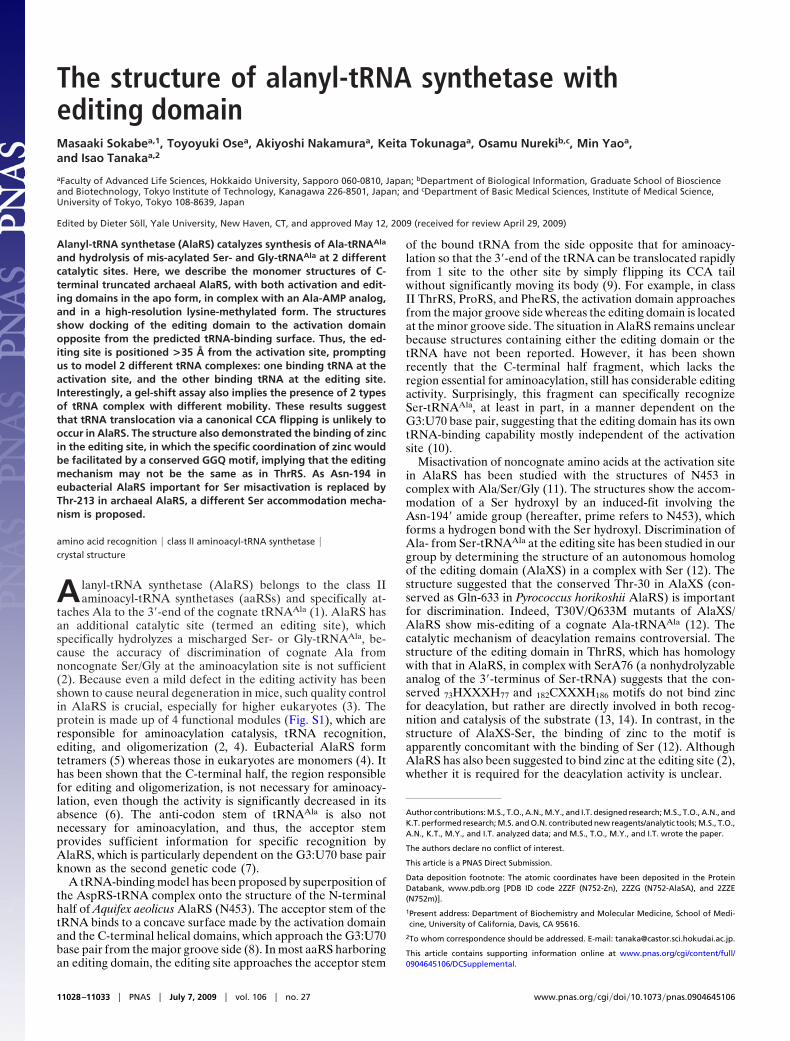
The structure consists of 2 regions (Fig. 1A). The N-terminalregion (almost corresponding to N453 fragment) is comprised of3 consecutive domains: the activation domain (AD, residues1–263), the helical domain N-terminal half (HN, 264–423), andthe helical domain C-terminal half (HC, 424–496). HN and HCare separated by Tyr-423 in the middle of the �15 helix, whichis kinked as in the N453 structure (8). The C-terminal part iscomprised of the �-barrel domain (BD, 504–602) and the editingdomain (ED, 603–752). An interdomain loop between these N-and C-terminal regions (497–503) is disordered and could not bemodeled, although SDS/PAGE analysis of the N752-Zn crystalsclearly showed that the protein molecules in the crystals are notdegraded at all. Considering the distance between both ends ofthe disordered region (�20 Å for 7 residues), it is also notfeasible that the BD-ED region of the adjacent molecules islocated at this position. In each crystal form, each molecule doesnot make significant contact with another molecule, and thus
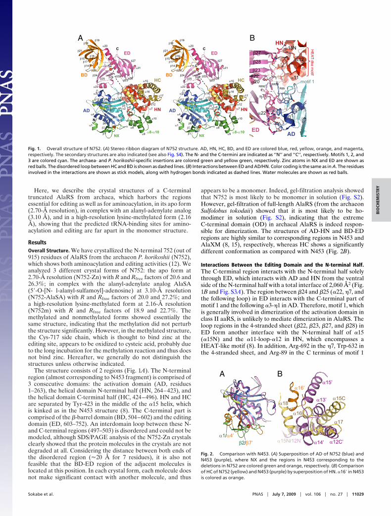
appears to be a monomer. Indeed, gel-filtration analysis showedthat N752 is most likely to be monomer in solution (Fig. S2).However, gel-filtration of full-length AlaRS (from the archaeonSulfolobus tokodaii) showed that it is most likely to be ho-modimer in solution (Fig. S2), indicating that the extremeC-terminal domain (OD) in archaeal AlaRS is indeed respon-sible for dimerization. The structures of AD-HN and BD-EDregions are highly similar to corresponding regions in N453 andAlaXM (8, 15), respectively, whereas HC shows a significantlydifferent conformation as compared with N453 (Fig. 2B).
Interactions Between the Editing Domain and the N-terminal Half.The C-terminal region interacts with the N-terminal half solelythrough ED, which interacts with AD and HN from the ventralside of the N-terminal half with a total interface of 2,060 Å2 (Fig.1B and Fig. S3A). The region between �24 and �25 (�22, �7, andthe following loop) in ED interacts with the C-terminal part ofmotif 1 and the following �3-�1 in AD. Therefore, motif 1, whichis generally involved in dimerization of the activation domain inclass II aaRS, is unlikely to mediate dimerization in AlaRS. Theloop regions in the 4-stranded sheet (�22, �23, �27, and �28) inED form another interface with the N-terminal half of �15(�15N) and the �11-loop-�12 in HN, which encompasses aHEAT-like motif (8). In addition, Arg-692 in the �7, Trp-632 inthe 4-stranded sheet, and Arg-89 in the C terminus of motif 1
ED
α15N
α22
β22β23
β28
β27
α20
η7
η1α3
α11
α12
motif 1R89
W632R692
AD
HN
HE
AT-like m
otif
α1
α21
α20
α18α17
α16
α15
α14α13
α12α11
α10
α9
α8
α7α6
α5α4
α3
α2
α22
N
C
β1
β28
β27
β25β24
β23β22
β21
β20
β19β18
β16β15
β13
β12
β11
β10β9
β8
β7
β6
β5
β4
β2
β26
β14β17
α1
α21
α20
α18α17
α16
α15
α14α13
α12α11
α10
α9
α8
α7α6
α5α4
α3
α2
α22
N
C
β1
β28
β27
β25β24
β23β22
β21
β20
β19β18
β16β15
β13
β12
β11
β10β9
β8
β7
β6
β5
β4
β2
β26
β14β17
AD HN
HCBD
ED
AD HN
HCBD
ED
NXα8-α10
NXα8-α10
A B
Fig. 1. Overall structure of N752. (A) Stereo ribbon diagram of N752 structure. AD, HN, HC, BD, and ED are colored blue, red, yellow, orange, and magenta,respectively. The secondary structures are also indicated (see also Fig. S4). The N- and the C-termini are indicated as ‘‘N’’ and ‘‘C’’, respectively. Motifs 1, 2, and3 are colored cyan. The archaea- and P. horikoshii-specific insertions are colored green and yellow green, respectively. Zinc atoms in NX and ED are shown asred balls. The disordered loop between HC and BD is shown as dashed lines. (B) Interactions between ED and AD/HN. Color coding is the same as in A. The residuesinvolved in the interactions are shown as stick models, along with hydrogen bonds indicated as dashed lines. Water molecules are shown as red balls.
BA
α16’
α1/α4’β2/β7’
α15C
α16
α17
α18
α12C’
α13’
α14’
α15’
α15N/12N’
Fig. 2. Comparison with N453. (A) Superposition of AD of N752 (blue) andN453 (purple), where NX and the regions in N453 corresponding to thedeletions in N752 are colored green and orange, respectively. (B) Comparisonof HC of N752 (yellow) and N453 (purple) by superposition of HN. �16� in N453is colored as orange.
Sokabe et al. PNAS � July 7, 2009 � vol. 106 � no. 27 � 11029
BIO
CHEM
ISTR
Y

form iminoaromatic stacking at the center of these 2 interfaces,and make overall contact to form a continuous interface (Fig.1B). The domain interface involves an extensive hydrophobiccore and a dense hydrogen bond network (some are mediated bywater molecules) through residues especially conserved amongarchaea (Fig. 1B and Fig. S3A), indicating that this interface isfairly stable. Similar conservation is also discernible in a corre-sponding region in the eubacterial N453 structure (Fig. S3B),suggesting that similar interdomain contact is also likely foreubacterial AlaRS. It has been shown that mutations in Esche-richia coli AlaRS in the region corresponding to �3 show defectin the oligomerization state (5). Because these mutations arelikely to affect the interaction between ED and AD, the properorientation of these domains would be important for AlaRSoligomer formation.
Comparison with the Eubacterial AlaRS. Although the overall struc-tures of AD and HN are highly similar to those of eubacterialN453, N752 has 2 large insertions (the N-terminal extension(NX: residues 1–54), and the �8-�10 insertion between �7 and�11) and 2 large deletions (35 residues between �6 and �7, and19 residues between �9 and �10 as compared with E. coli AlaRS)within the N-terminal half, all of which are characteristic ofarchaeal AlaRS (Fig. 1 A and Fig. S4). NX is comprised of the�1 helix, 2 anti-parallel strands (�1 and �2) that are part of thecentral anti-parallel � sheet in AD, and the following 25-residue-long loop (Fig. 2A). Further, NX contains a conserved Cys4 zincbinding motif (20CXXCG24 between �1 and �2 and 37CG-DXPC42 in the long loop), which physically connects the �1-�2turn and the long loop and thus would be important for thestructural stability of the NX region. The binding of a zinc ionwas confirmed by X-ray absorption spectroscopy and anomalousdifference Fourier map analysis (Fig. S5A). The long loop makesconservative hydrophobic interactions, including Tyr-45, Phe-47,Ile-48, Pro-51, and Ile-53, with a cavity formed by �8 and a loopbetween �9 and �10, both of which are also characteristic partsof archaeal AlaRS (one is the insertion, and the other is thedeletion). Interestingly, the region in N453 corresponding to thedeletion between �6 and �7 in N752 consists of �4� and �7�,which appear to mimic �1 and �2 in NX, although �7� is in theopposite orientation (Fig. 2 A). The �8-�10 insertion is com-prised of the 3-helix bundle lying at the concave surface made byAD, HN, and HC, and interacts with the N terminus of �18 inHC, the interdomain loop between AD and HN, the loopbetween �4 and �5 in motif 2, and the long loop in NX. Thisinsertion shows a relatively low level of sequence conservation(no strictly conserved residues within this region), although thelengths are almost same among species. Further, temperaturefactors of the �8–�9 region, which interact mainly with motif 2and �18, are relatively high (�42 Å2, whereas the total averagevalue is 33 Å2). Together, these 2 archaea-specific insertionswrap around 1 side of the N-terminal half and form extensivebridges across HC to AD (Fig. 1 A).
HC in N752 shows a significantly different conformation fromthat of N453 (Fig. 2B), although it still consists of an �15C-�173-helix bundle and an additional �18 helix as in N453 (�12�-�14�and �15�). Compared with N453, i) the axis of �15C is rotatedby �30° away from the concave; ii) �15C is rotated by �30°around its helix axis; iii) the position of �16–�17 relative to �15Cis rotated by �20° toward the concave surface around the axisof �15C; and iv) �18 shows a different orientation. Conse-quently, �17 and the following loop face the concave surface,which is in contrast to N453, in which �12� and �14� (�15 and �17in N752) face the concave surface instead. In fact, HC showsrelatively high temperature factors as observed in N453, suggestingthat HC has intrinsic flexibility, which was already suggested basedon the susceptibility to proteolysis at the kink in �15.
The Activation Site. The activation site is located at the center ofthe antiparallel � sheet in AD, and consists of motif 2 (�4 and�5), motif 3 (�13), �7, and �10. AlaSA binds to the pocket in amanner similar to Ala and the AMP moiety of ATP in theN453-Ala/ATP structures, although both moieties in AlaSAcome slightly closer to make a covalent bond with each other(Fig. 3A). The residues interacting with AlaSA are spatially wellconserved compared with those interacting with Ala and ATP inN453, except Trp-192 (Trp-161� in N453) that interacts with acarboxyamino ester of AlaSA. In N453-Ala, Trp-161� is locatedfarther from Ala and interacts indirectly with the amino groupby a water-mediated hydrogen bond. In fact, the Trp192s in theabsence of AlaSA (N752-Zn and N752m) are less well orderedand show conformational variability among each molecule,suggesting a propensity to fluctuate.
Interestingly, Asn-194� in N453, which is strictly conservedamong bacteria and eukaryote, is replaced by Thr-213 in mostarchaeal AlaRS (49/51 species investigated, otherwise it isconserved as Asn). In the N453-Ser complex structure, theAsn-194� side chain exhibits an induced-fit upon Ser binding toform a hydrogen bond with the Ser �-hydroxyl group, providingthe structural basis of Ser misactivation in eubacterial AlaRS. InN752-AlaSA, the �- and �-carbons of Thr-213 face the aminoacid binding pocket and thus offer an aliphatic environmentsuitable for an Ala side chain. However, in 1 molecule in N752m,the side chain of Thr-213 is f lipped �120°, and thus its hydroxylgroup faces the binding pocket (Fig. 3A). Moreover, this Thr-213hydroxyl group forms a hydrogen bond with a water moleculepositioned near the �-hydroxyl of the bound Ser in N453-Ser.Therefore, this situation is likely to mimic Ser accommodationin N752, and thus suggests a different mechanism of Ser mis-activation in archaeal AlaRS that involves the intrinsic f lexibilityof the Thr-213 side chain.
The Editing Site. The editing site harbors the 613HXXXH617 and717CXXXH721 zinc binding motif, which is conserved throughAlaRS, AlaX and even ThrRS, whereas the opposite side of theediting site consists of conservative hydrophilic side chains, suchas Thr-616, Gln-633, Ser-636, Gln-695, and Gln-715 (Fig. 3B).
R128
D131
F142
F145
R255
E210
T213M147
V215
D248
W192
G250
W
A B
H613
H617C717
H721
Q555
Q633S636
WN
SerA76
Q695
Q715
T616
Zn
Fig. 3. The catalytic sites. (A) The activation site of N752-AlaSA. AlaSA andinteracting residues are shown as gray and yellow stick models, respectively.Thr-213 and an interacting water molecule in N752m are superposed as greenstick and red ball models (marked as ‘‘W’’), respectively. Hydrogen bonds inN752-AlaSA and N752m are indicated as black and red dashed lines, respec-tively. The omit map of AlaSA (at 3.1 Å resolution, contoured at 3 �) is alsoshown. (B) Comparison of the editing sites of N752-Zn (yellow) and ThrRS-SerA76 complex (light blue). Only the zinc-binding motifs and SerA76 areshown for ThrRS. The water molecule coordinating to zinc in N752-Zn, and thenucleophile in ThrRS-SerA76 are marked as ‘‘W’’ and ‘‘N’’, respectively. TheGGQ loop is colored green. Interactions in N752-Zn and ThrRS-SerA76 areshown as black and blue dashed lines, respectively.
11030 � www.pnas.org�cgi�doi�10.1073�pnas.0904645106 Sokabe et al.

Again, anomalous difference Fourier map analysis clearly dem-onstrates the presence of a zinc ion (Fig. S5B), which is tetra-coordinated by His-613, His-617, Cys-717, and 1 water moleculein a tetrahedral geometry. Interestingly, His-721 in the motifdoes not bind zinc, but forms a hydrogen bond with Cys-717 S�instead. A similar coordination was observed in the AlaXMstructure (15), although the fourth ligand, a water molecule, wasabsent in AlaXM probably due to data quality. However, thesituation is different in AlaXS, which binds zinc by all 4 residuesin the motif in a tetrahedral manner (12). In ThrRS, it has beenwell established that the motif should not bind zinc but rather isinvolved directly in the catalysis of deacylation (13, 14), eventhough a possible zinc-binding capability in the motif has beensuggested by atomic absorption spectroscopy and mass spec-trometry (13, 16). In N752, the side chain of Gln-555 in thestrictly conserved 553GGQ555 loop in BD, which is absent inThrRS and AlaXS, approaches the editing site and forms ahydrogen bond with His-613 N� in the motif, causing theimidazolium ring to adopt a specific conformation. Again, asimilar interaction between the GGQ loop and the zinc bindingmotif is observed in AlaXM (15), suggesting that this interactionwould be important for the specific zinc coordination in AlaRS/AlaXM.
DiscussionBinding of tRNA to the Activation Site. According to the previoustRNA docking model with N453 (8), the tRNA acceptor stembinds on its major groove side to the concave surface made byAD, HN, and HC, whereas the anticodon stem does not makesignificant contact with the protein. In the model, motif 2 and�14� in HC (�17 in N752), which have been suggested to beinvolved in tRNA binding, approach the major groove side of thethird and the fourth base pairs of the modeled tRNA. In archaealAlaRS, motif 2 is also highly conserved and shares residuesimportant for amino acid transfer [such as Arg-69, Asp-76, andPhe-90 in E. coli AlaRS (17), corresponding to Arg-128, Asp-131, and Phe-145 in N752, respectively (Fig. S4)]. Indeed, theactivation site and motif 2 in N752 comprise a continuousconserved surface resembling that in N453 (Fig. 4A and Fig.S3C), suggesting that motif 2 in archaeal AlaRS would also beimportant for tRNA recognition. However, the �8–�10 insertionin N752 covers motif 2, and thus severely interferes with theacceptor stem of the modeled tRNA (Fig. 4A). Because this
insertion has very low sequence conservation among archaea, itis difficult to envisage that the insertion alternatively comprisesan archaea-specific tRNA interface (compare the surfacearound the insertion and motif 2 in Fig. 4 A and B). Thus, it ismore likely that the insertion exhibits a conformational changeupon tRNA binding (the most plausible direction of the con-formational change is shown in Fig. 4A). In fact, the regioncovering motif 2 (�8–�9) shows relatively high temperaturefactors, implying its f lexibility.
In N752, the surface of HC seems to be compatible with themodeled tRNA (Fig. 4A). The �17 approaches the minor grooveof the T stem, and the following loop and the N terminus of �18lie along the backbone of the acceptor stem from the majorgroove side. In the model with N453, HC shows a differentstructure and thus �14� (�17 in N752) interacts with tRNA byinserting its hydrophilic side chains into the major groove withina contact distance to the third and the fourth base pairs (8).Although either model is consistent with the significance of HCin tRNA recognition, it is envisaged that HC undergoes con-formational change upon tRNA binding when considering itspossible flexibility. This possibility will be clarified directly by thestructure of the tRNA complex in future studies. Regarding HC,it should be mentioned here that the structure of HC in N453might involve partial artifacts probably due to the C-terminaltruncation at the middle of BD (Fig. S1). In N453, the C-terminal20 residues comprise �16� packed into the cleft between HC andHN (Fig. 2B) whereas residues between �15� and �16� aredisordered. In N752, the corresponding region (residues 509–523) comprises an integral part of BD N terminus (�14–�19),which is �50 Å far from where �16� is in N453 (compare Figs.1A and 2B). Such a dramatic conformational change seemsunfeasible, and thus the truncation in N453 might cause unnat-ural conformations of �16� region and hence the interacting HC.
Binding of tRNA to the Editing Site. Recently, it has been shown thatthe C-terminal half fragment of E. coli AlaRS is still highly activein deacylation of exogenous Ser-tRNAAla and is able to specif-ically recognize the cognate tRNAAla moiety at least in part ina manner dependent on the G3:U70 base pair (10). This studystrongly suggested that the C-terminal half has a distinct tRNAbinding capability mostly independent from the N-terminal half,which bind tRNA for aminoacylation. The interaction of tRNAwith ED has originally been proposed with the structure of
Editing site
~35 Å
tRNAAsp
tRNAThr
OD
K421R428R429
α8-α10β27-β28
Motif 2G3:U70
α17
α18
R745
Activation site
G3:U70A B
α8-α10tRNA
tRN
A
N75
2
N75
2+tR
NA
protein
C
TB stained CB stained
tRN
A
N75
2
N75
2+tR
NA
Fig. 4. The tRNA complexes. (A) tRNAAsp (orange) was modeled by superposition of AspRS-tRNA complex onto the AD-HN region. Surface representation ofN752 without the �8-�10 insertion is shown and was colored by the sequence conservation among archaeal AlaRS using the program ConSurf 3.0 (23): from lowto high, cyan, white, and purple. The �8-�10 insertion is shown as a ribbon diagram (green). The proposed direction of conformational change of the �8-�9 regionis indicated by a black dashed arrow. (B) tRNAThr (blue) is modeled by superposition of ThrRS-tRNA complex onto ED. Surface representation of the whole N752(including the �8-�10 insertion) is shown as in A. Possible orientation of the CCA tail is shown as blue dashed lines. Proposed orientation of OD is shown as graydashed circle connecting to the C terminus of N752 by dashed lines. (C) Gel-shift assay. N752 (50 �M) was mixed with tRNAAla (100 �M) and then analyzed. Thesame gel was stained with TB (Left) and then CB after destaining TB (Right). The major and the minor (blurry) complex bands are indicated by orange and bluearrows, respectively. The image was arranged from the single larger gel image.
Sokabe et al. PNAS � July 7, 2009 � vol. 106 � no. 27 � 11031
BIO
CHEM
ISTR
Y

AlaXS by its superposition onto the editing domain of theThrRS-tRNA complex, showing binding of the strand-loop-strand region (corresponding to �27–�28 in N752) around theG3:U70 base pair from the minor groove side (12). An R693Kmutation (corresponding to R745K in N752) in this region in theE. coli C-terminal fragment confers relaxed specificity for Ser-tRNAThr, suggesting that the previous model with AlaXS is alsolikely for AlaRS (10). In N752, the region around this strand-loop-strand shows a positively charged concave surface formedby �27, the loop between �23 and �21 in ED, and �15 in HN,which is generally suitable for binding negatively charged nucleicacids. These regions and the editing site also constitute a highlyconserved surface (Fig. 4B). A tRNA molecule could reasonablybe modeled to this concave surface without severe steric clash bysuperposition of ThrRS-tRNA (Fig. 4B). In this model, thestrand-loop-strand region approaches the first to the fourth basepairs from the minor groove side, as proposed (10, 12). Fur-thermore, Lys-421, Arg-428, and Arg-429 in the middle region of�15 are adjacent to the backbone phosphates of nucleotides63–65 in the T arm. A conservative hydrophilic patch, consistingof the N terminus of �12 and the C terminus of �16 in HC (Fig.S3A), is also located near the 5�-terminus of the modeled tRNA.A similar hydrophilic patch (consisting of a10� and a13�) is alsodiscernible in N453 (Fig. S3B), although it shows a slightlydifferent conformation due to the flexibility of HC. Althoughthese 2 regions in HC are candidates for the tRNA interface, theymay have a minor contribution when considering the dispens-ability of the N-terminal half for editing (10). Recently, elimi-nation of the C-terminal OD was shown to markedly reduce theediting activities of the C-terminal half fragment possibly due toreduced affinity to tRNA (10). It is still unclear whether OD hasa tRNA binding activity or the oligomerization per se promotestRNA binding. In our model, the anticodon stem is orientedtoward the solvent and could be within a distance suitable forinteraction with OD (Fig. 4B). This implies that the formerpossibility is conceivable, although it does not exclude the latterpossibility. The possible interaction between the anticodon stemand OD is consistent with the previous observation that AlaRSrequires a whole L-shaped tRNA for efficient deacylation, unlikeaminoacylation (18).
In class II aaRS, such as ThrRS, the editing site is generallyadjacent to the activation site so that the CCA tail of the boundtRNA can be flipped from 1 site to the other site without movingits body. However, at least as the N752 fragment, which lacks ODand thus is a monomer, the editing site in AlaRS is located on theopposite side as compared with that of ThrRS with a distance of�35 Å (Fig. 4A), and the proposed tRNA binding sites for the 2reactions are too far for such tRNA translocation (Fig. 4 A and B).Interestingly, in the gel-shift assay, the N752 fragment displayed 2types of complex band, which can be stained with both Toluidineblue (TB, for nucleic acids) and Coomassie blue (CB, for proteins),when mixed with the excess amount of tRNA (Fig. 4C, major andminor blurry bands). The different mobility of these 2 complexesimplies the structural difference among them, which is consistentwith the above 2 tRNA binding models. Although the minor bandis weak and blurry, it is conceivable that binding of tRNA to theediting site would be weak due to lacking of mischarged amino acidmoiety and OD. Considering the strong interaction between EDand the N-terminal half, a dramatic movement of ED also seems tobe infeasible. One possibility is that, in the dimer (or tetramer in thecase of E. coli AlaRS), the editing site in 1 subunit is closely locatedto the activation site in the other subunit. To assess this idea, a dimerstructure was modeled by the superposition of each AD and ED in2 N752 molecules onto those in the ThrRS-tRNA, by the structuralhomology of each domain (Fig. S6A). However, the resulting dimershowed a completely asymmetric structure, which is unusual for ahomodimer, even though it did not show severe clash amongsubunits. Indeed, the C-termini of subunits, which are followed by
OD in the full-length protein, are far apart, inconsistent with theOD-mediated dimerization in archaeal AlaRS (Fig. S2). Further,the tetramer model showed serious overlap among subunits (Fig.S6B), suggesting that these types of oligomer are unlikely. Indeed,eukaryotic AlaRS is a monomer in solution (4), implying thatoligomerization per se is not necessary for AlaRS function. In thecanonical CCA flipping, the body of the tRNA is thought to remainbound to the activation domain even after flipping. In this situation,editing would be significantly dependent on the activation domain,which is not the case in AlaRS (10). Therefore, it is suggested that,at least with regard to CCA flipping, either internal or intersubunittRNA translocations are unlikely to occur in AlaRS, and thus amisacylated tRNA would have to dissociate from the activation siteto rebind the editing site. This idea implies that editing in AlaRS isnot efficient as other aaRS, partly rationalizing the conservation ofautonomous trans-editing AlaX proteins in many organisms, whichwould compensate for the editing activity (19).
Editing Reaction. In ThrRS, it has been well established that the73HXXXH77 and 182CXXXH186 motifs in the editing site do notbind zinc but are rather involved directly in the catalysis ofdeacylation (13, 14). In fact, a presence of zinc inhibited thebinding of Ser to the editing site (13). His-73 in the motif(His-613 in N752, which interacts with the GGQ loop) has beenshown to serve as a general base for a nucleophilic watermolecule whereas Cys-182 (Cys-717 in N752) recognizes the2�-OH of the ribose (13). Although a similar mechanism isconceivable for AlaRS when considering the functional andsequence homologies, little information has been reported todate regarding the AlaRS editing mechanism. Our structure,along with anomalous difference Fourier analysis, clearly dem-onstrated the binding of zinc to the editing site in AlaRS (Fig.3B and Fig. S5B), in which the zinc ion is coordinated by the 3residues in the zinc-binding motif (His-613, His-617, and Cys-717) and 1 water molecule. This specific coordination seems tobe facilitated by the interaction between the coordinating His-613 and Gln-555 in the GGQ loop, which is conserved amongAlaRS and AlaXM but not in ThrRS. The presence of thecoordinating water molecule suggests that this zinc has a func-tional rather than a structural role, in which zinc coordination isgenerally completed by 4 protein residues. However, when wecompared the deacylation activities against Ser-tRNAAla in thepresence and absence of zinc, the results were ambiguous.Because the protein was purified in buffer containing zinc, itlikely already contained zinc. Indeed, the addition of zinc (10�M) did not affect the deacylation rate significantly as comparedwith that in its absence (90 � 5.8%). However, elimination ofzinc by 10 mM EDTA resulted in only a slight reduction of thedeacylation rate to 73 � 15%. Similar ambiguity of zinc require-ment has also been observed for AlaXS (12), although it bindszinc by all 4 residues in the zinc-binding motif. However, inclearly contrast to ThrRS, zinc binding did not interfere withdeacylation at all in AlaRS, implying that the mechanism may bedifferent.
Although the zinc requirement is ambiguous, it would beinteresting to discuss the possible roles of zinc in AlaRS editing.One possibility might be that zinc mediates hydrolysis, in whichan activated water molecule coordinating to zinc serves as anucleophile. However, we could not identify any candidate as aproton acceptor around the coordinated water molecule, whichis generally required for activation of the water molecule inzinc-mediated hydrolases. Another possibility is recognition ofthe substrate directly by the zinc ion. In the ThrRS activation site,a zinc ion, which is tetra-coordinated by 3 protein residues and1 water molecule in the absence of a substrate, is penta-coordinated by a side chain hydroxyl and an amino groups of abound Thr (or Ser) moiety in addition to the 3 residues alreadycoordinating to zinc (20). This specific recognition of �-hydroxyl
11032 � www.pnas.org�cgi�doi�10.1073�pnas.0904645106 Sokabe et al.

and �-amino groups by zinc logically exclude �-methyl of isos-teric Val from the activation site. The situation in the AlaRSediting site is reminiscent of this situation; zinc is coordinated by3 protein residues and 1 water molecule in the absence of thesubstrate, and the editing site is thought to recognize Ser but notAla. Although the side chain of Ala is shorter than that of Val,the �-methyl of Ala would still be within the distance conflictingwith zinc when its amino group is coordinated. In this context,Gly would not be rejected because of the absence of the sidechain. Interestingly, mutation of Gln-633 to a hydrophobicresidue (Q633M) resulted in relaxed specificity to the cognateAla-tRNAAla (12). Because the Gln-633 side chain is locatednear the coordinating water molecule with a distance of 4.5 Å(Fig. 3B), this residue, and possibly the surrounding hydrophilicresidues, may provide an unfavorable environment for the Ala�-methyl. These 2 possibilities of the role of zinc will beconfirmed by the structural analysis of SerA76 complex in futurestudies.
Materials and MethodsFull Details of the Methods Used Are Provided in SI Text. The gene encodingN752 was expressed in E. coli, and purified by heat treatment followed bycation-exchange and size-exclusion chromatography. The C717A mutant wasprepared in a manner similar to the wild type. The C-terminal His-taggedfull-length AlaRS from S. tokodaii was expressed in E. coli, and purified by heattreatment followed by Ni-NTA affinity, size-exclusion, and heparin-affinitychromatography. Gel-filtration analysis was performed with a Superdex20010/300 GL column (GE Healthcare).
For N752m crystals, the purified N752 was subjected to a reductive-methylation procedure targeting its lysine residues (21). Both methylated andnonmethylated N752 proteins were crystallized by the hanging drop vapordiffusion method at 20 °C. The N752-Zn crystals were grown in buffer con-taining 0.1 M Mes-Na, pH 5.6, 6% PEG6000, 108 mM succinic acid, and 6%
glucose using 9 mg/mL protein. The N752-AlaSA crystals were grown underthe same condition with buffer containing 1 mM AlaSA. The N752m crystalswere grown with 0.1 M tri-sodium citrate dihydrate, pH 5.6, 0.2 M ammoniumacetate, and 30% PEG4000, using 5 mg/mL protein.
The X-ray diffraction data were collected at beamlines BL-5A of PhotonFactory and BL41XU of SPring-8 at 100 K. The N752-Zn crystals belong to thespace group C2 with 1 molecule in an asymmetric unit. The N752-AlaSA crystalsbelong to the space group P1 with 2 molecules in an asymmetric unit. TheN752m crystals belong to the space group P21 with 2 molecules in an asym-metric unit. The structure was solved by molecular replacement with thesequential search of N453 and AlaXS as models, yielding 2 sets of uniquesolutions for the N752m dataset. The structure was refined with programLAFIRE (22) and subsequent cycles of manual fitting and restrained refinementwith TLS parameters. The N752-Zn and N752-AlaSA crystals showed highWilson B-factors (85 Å2 and 80 Å2, respectively), which would be a majorreason for the relatively high Rfree values in these structures. The statistics aresummarized in Table S1.
In gel-shift assay, N752 (50 �M) was mixed with tRNAAla (100 �M) in 50 mMTris-borate, pH 7.0, and 2 mM MgOAc2, and incubated at 4 °C for 1 h. Then,10% glycerol was added, and aliquots of 20-�L samples were analyzed by 5%polyacrylamide gel electrophoresis in the same buffer at 4 °C. The gel wasstained with TB, completely destained, and then stained again with CB.
Deacylation assay was performed essentially as described in ref. 12. 3HLabeled Ser-tRNAAla (2.5 �M) was incubated with 200 nM N752 at 55 °C. Toeliminate zinc, the purified protein was diluted extensively with buffer con-taining 10 mM EDTA, and then concentrated. Under the conditions withaddition of zinc, the reaction mixture was brought to 10 �M ZnOAc2. Thevalues were calculated from the averages of 2 independent experiments.
ACKNOWLEDGMENTS. We thank Prof. J.W.B. Hershey for discussions andcomments on the manuscript and the staff of SPring-8 BL41XU and the PhotonFactory BL5A beamlines for help during the X-ray diffraction experiments. Thiswork was supported by Targeted Proteins Research Program from the Ministryof Education, Culture, Sports, Science, and Technology, Japan, partly by theHuman Frontier Science Program fellowship LT00575/2007-L (to M.S.)
1. Eriani G, Delarue M, Poch O, Gangloff J, Moras D (1990) Partition of tRNA synthetasesinto two classes based on mutually exclusive sets of sequence motifs. Nature 347:203–206.
2. Beebe K, Ribas De Pouplana L, Schimmel P (2003) Elucidation of tRNA-dependentediting by a class II tRNA synthetase and significance for cell viability. EMBO J 22:668–675.
3. Lee JW, et al. (2006) Editing-defective tRNA synthetase causes protein misfolding andneurodegeneration. Nature 443:50–55.
4. Schimmel P, Ripmaster T (1995) Modular design of components of the operational RNAcode for alanine in evolution. Trends Biochem Sci 20:333–334.
5. Ribas de Pouplana L, Schimmel P (1997) Reconstruction of quaternary structures of classII tRNA synthetases by rational mutagenensis of a conserved domain. Biochemistry36:15041–15048.
6. Buechter DD, Schimmel P (1993) Dissection of a class II tRNA synthetase: Determinantsfor minihelix recognition are tightly associated with domain for amino acid activation.Biochemistry 32:5267–5272.
7. Hou YM, Schimmel P (1988) A simple structural feature is a major determinant of theidentity of a transfer RNA. Nature 333:140–145.
8. Swairjo MA, et al. (2004) Alanyl-tRNA synthetase crystal structure and design foracceptor-stem recognition. Mol Cell 13:829–841.
9. Sankaranarayanan R, Moras D (2001) The fidelity of the translation of the genetic code.Acta Biochim Pol 48:323–335.
10. Beebe K, Mock M, Merriman E, Schimmel P (2008) Distinct domains of tRNA synthetaserecognize the same base pair. Nature 451:90–93.
11. Swairjo MA, Schimmel PR (2005) Breaking sieve for steric exclusion of a noncognateamino acid from active site of a tRNA synthetase. Proc Natl Acad Sci USA 102:988–993.
12. Sokabe M, Okada A, Yao M, Nakashima T, Tanaka I (2005) Molecular basis of alaninediscrimination in editing site. Proc Natl Acad Sci USA 102:11669–11674.
13. Dock-Bregeon AC, et al. (2004) Achieving error-free translation: The mechanism ofproofreading of threonyl-tRNA synthetase at atomic resolution. Mol Cell 16:375–386.
14. Waas WF, Schimmel P (2007) Evidence that tRNA synthetase-directed proton transferstops mistranslation. Biochemistry 46:12062–12070.
15. Fukunaga R, Yokoyama S (2007) Structure of the AlaX-M trans-editing enzyme fromPyrococcus horikoshii. Acta Crystallogr D Biol Crystallogr 63:390–400.
16. Dock-Bregeon A, et al. (2000) Transfer RNA-mediated editing in threonyl-tRNA syn-thetase. The class II solution to the double discrimination problem. Cell 103:877–884.
17. Davis MW, Buechter DD, Schimmel P (1994) Functional dissection of a predictedclass-defining motif in a class II tRNA synthetase of unknown structure. Biochemistry33:9904–9911.
18. Beebe K, Merriman E, Schimmel P (2003) Structure-specific tRNA determinants forediting a mischarged amino acid. J Biol Chem 278:45056–45061.
19. Chong YE, Yang XL, Schimmel P (2008) Natural homolog of tRNA synthetase editingdomain rescues conditional lethality caused by mistranslation. J Biol Chem 283:30073–30078.
20. Sankaranarayanan R, et al. (2000) Zinc ion mediated amino acid discrimination bythreonyl-tRNA synthetase. Nat Struct Biol 7:461–465.
21. Walter TS, et al. (2006) Lysine methylation as a routine rescue strategy for proteincrystallization. Structure 14:1617–1622.
22. Yao M, Zhou Y, Tanaka I (2006) LAFIRE: Software for automating the refinementprocess of protein-structure analysis. Acta Crystallogr D Biol Crystallogr 62:189–196.
23. Landau M, et al. (2005) ConSurf 2005: The projection of evolutionary conservationscores of residues on protein structures. Nucleic Acids Res 33:W299–302.
Sokabe et al. PNAS � July 7, 2009 � vol. 106 � no. 27 � 11033
BIO
CHEM
ISTR
Y