The Spinal Cord cranial nerves - 12 pr spinal nerves- 31 pr.
30
-
date post
19-Dec-2015 -
Category
Documents
-
view
241 -
download
5
Transcript of The Spinal Cord cranial nerves - 12 pr spinal nerves- 31 pr.
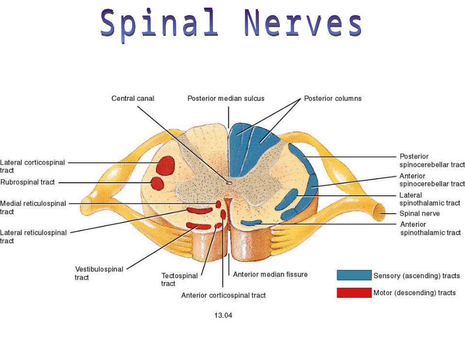
The Spinal Cord
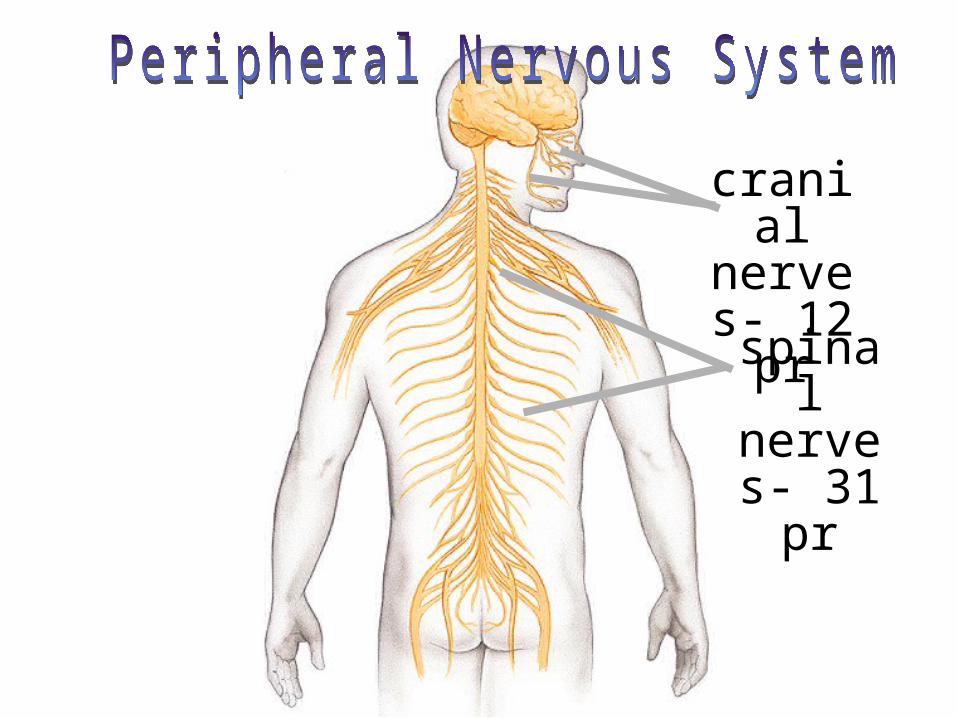
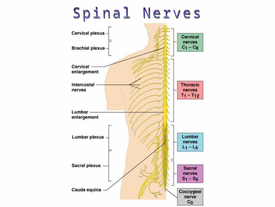
cranial nerves- 12 pr
spinal nerves- 31 pr
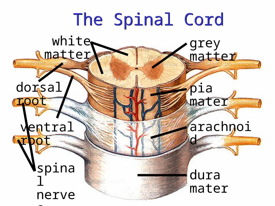
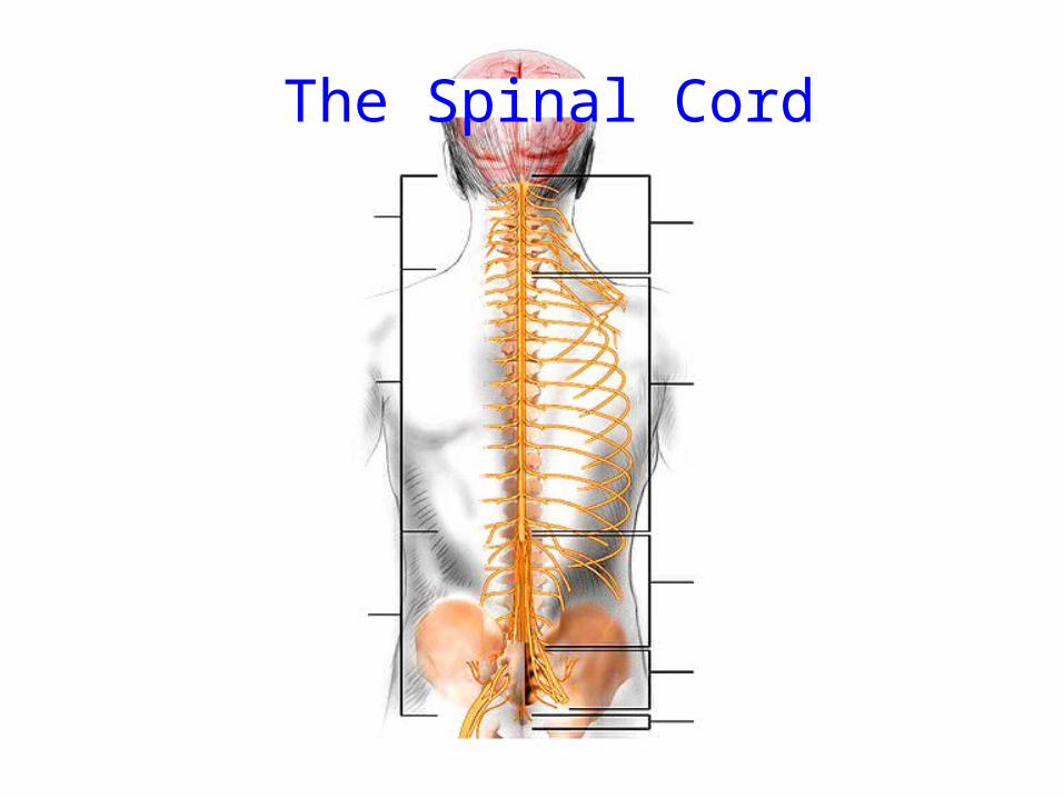
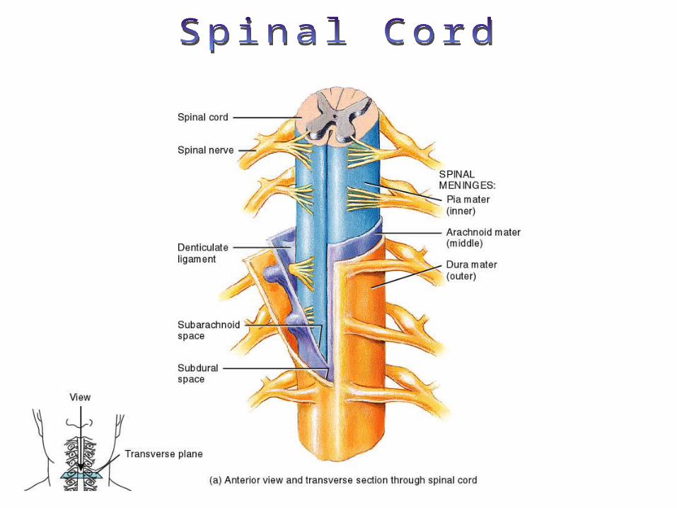
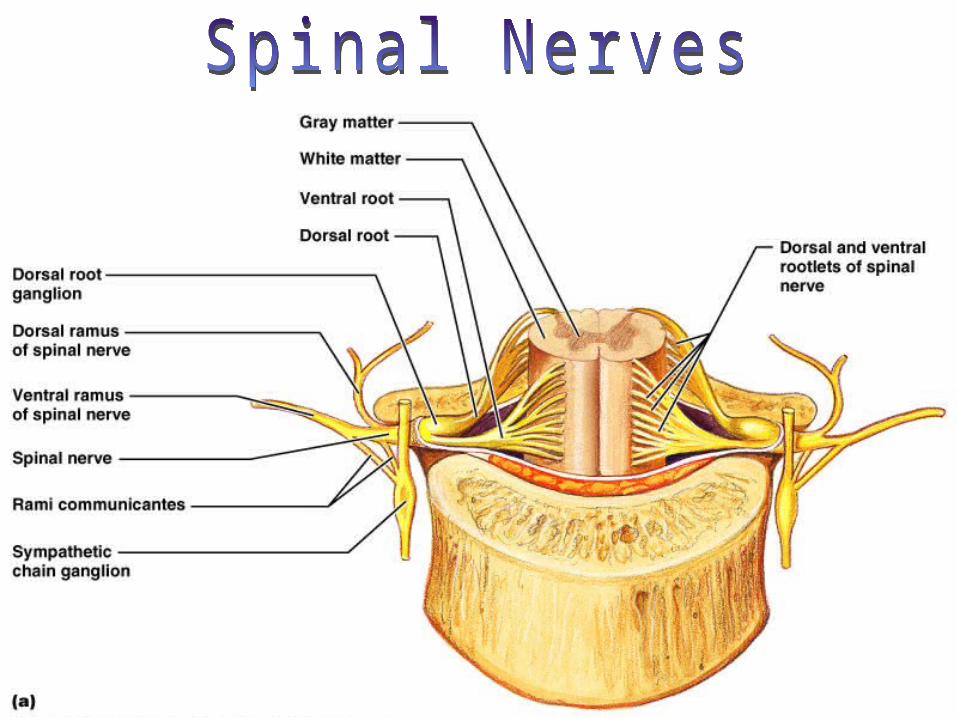
The Spinal CordThe Spinal Cord
ventral root
pia mater
dura mater
arachnoid
grey matter
dorsal root
white matter
spinal nerves
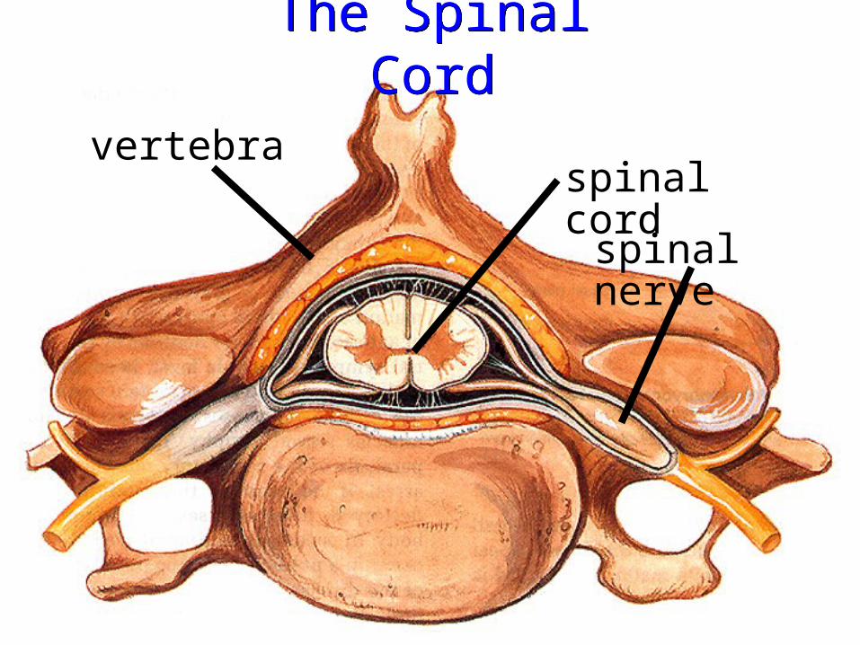
The Spinal CordThe Spinal Cord
spinal cord
spinal nerve
vertebra
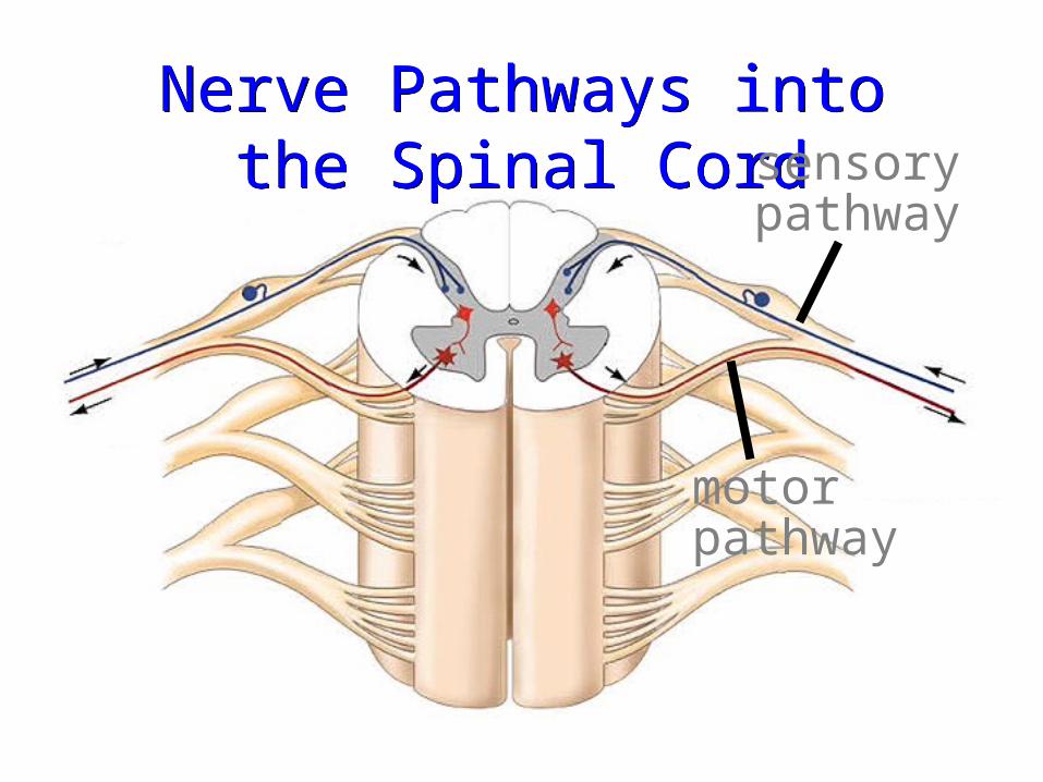
Nerve Pathways into the Spinal Cord
Nerve Pathways into the Spinal Cord sensory
pathway
motor pathway
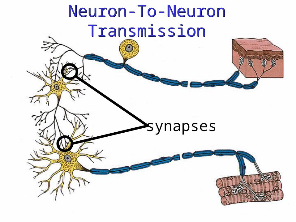
Neuron-To-Neuron TransmissionNeuron-To-Neuron Transmission
synapses
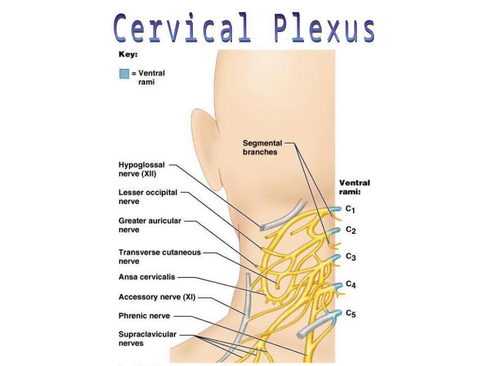
Cervical Plexus
• Formed by ventral rami of C1–C4
• Innervates skin and muscles of the neck, ear, back of head, and shoulders
• Phrenic nerve– Major motor and sensory nerve of the
diaphragm (receives fibers from C3–C5)
Brachial Plexus
• Formed by ventral rami of C5–C8 and T1 (and often C4 and T2)
• It gives rise to the nerves that innervate the upper limb
• Major branches of this plexus: – Roots—five ventral rami (C5–T1)– Trunks—upper, middle, and lower– Divisions—anterior and posterior – Cords—lateral, medial, and posterior
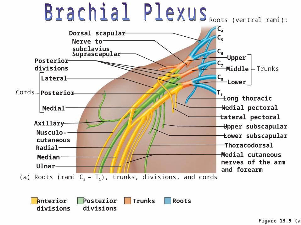
Figure 13.9 (a)
Upper
Middle Trunks
Lower
Roots (ventral rami):
Upper subscapular
Lower subscapular
Thoracodorsal
Medial cutaneousnerves of the armand forearm
Long thoracic
Medial pectoral
Lateral pectoral
Nerve tosubclaviusSuprascapular
Dorsal scapular
Posteriordivisions
Anteriordivisions
Lateral
PosteriorCords
Medial
Axillary
Musculo-cutaneousRadial
Median
Ulnar
Posteriordivisions
Trunks Roots
C4
C5
C6
C7
C8
T1
(a) Roots (rami C5 – T1), trunks, divisions, and cords
Brachial Plexus: Nerves• Axillary—innervates the deltoid, teres minor, and skin
and joint capsule of the shoulder• Musculocutaneous—innervates the biceps brachii and
brachialis and skin of lateral forearm• Median—innervates the skin, most flexors and
pronators in the forearm, and some intrinsic muscles of the hand
• Ulnar—supplies the flexor carpi ulnaris, part of the flexor digitorum profundus, most intrinsic muscles of the hand, and skin of medial aspect of hand
• Radial—innervates essentially all extensor muscles, supinators, and posterior skin of limb
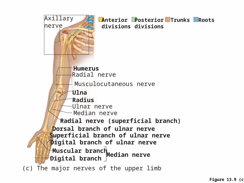
Figure 13.9 (c)
Median nerve
Musculocutaneous nerve
Radial nerveHumerus
Ulna
Ulnar nerveMedian nerve
Radius
Radial nerve (superficial branch)
Superficial branch of ulnar nerveDorsal branch of ulnar nerve
Digital branch of ulnar nerveMuscular branchDigital branch
(c) The major nerves of the upper limb
Axillarynerve
Anteriordivisions
Posteriordivisions
Trunks Roots
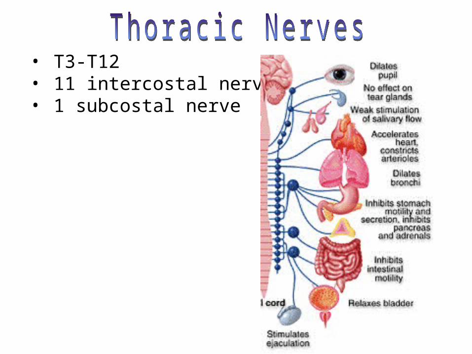
• T3-T12• 11 intercostal nerve• 1 subcostal nerve
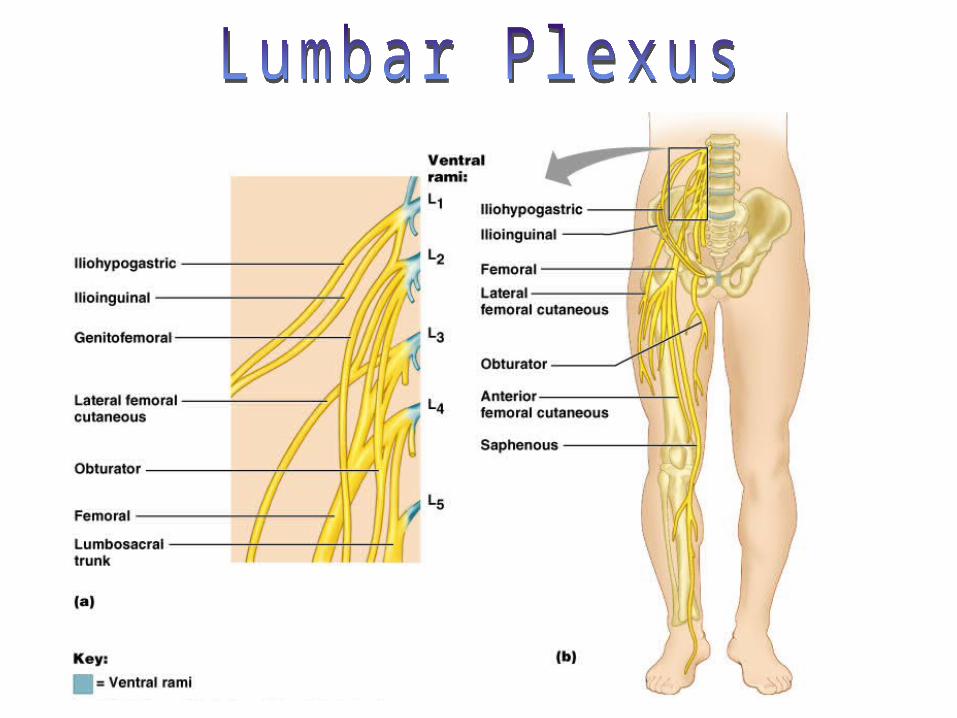
Lumbar Plexus
• Arises from L1–L4
• Innervates the thigh, abdominal wall, and psoas muscle
• Femoral nerve—innervates quadriceps and skin of anterior thigh and medial surface of leg
• Obturator nerve—passes through obturator foramen to innervate adductor muscles
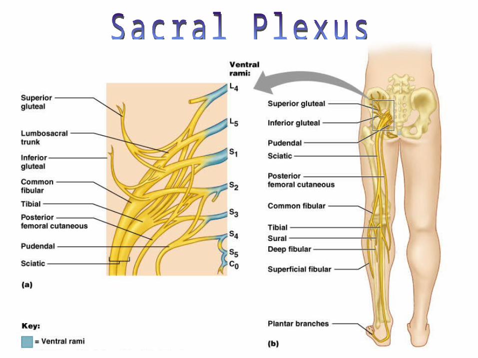
Sacral Plexus
• Arises from L4–S4
• Serves the buttock, lower limb, pelvic structures, and perineum
• Sciatic nerve– Longest and thickest nerve of the body– Innervates the hamstring muscles, adductor
magnus, and most muscles in the leg and foot– Composed of two nerves: tibial and common
fibular
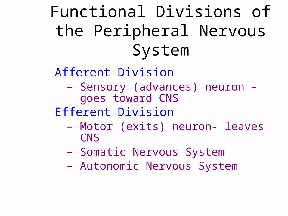
Functional Divisions of the Peripheral Nervous System
Afferent Division– Sensory (advances) neuron –
goes toward CNSEfferent Division
– Motor (exits) neuron- leaves CNS– Somatic Nervous System– Autonomic Nervous System
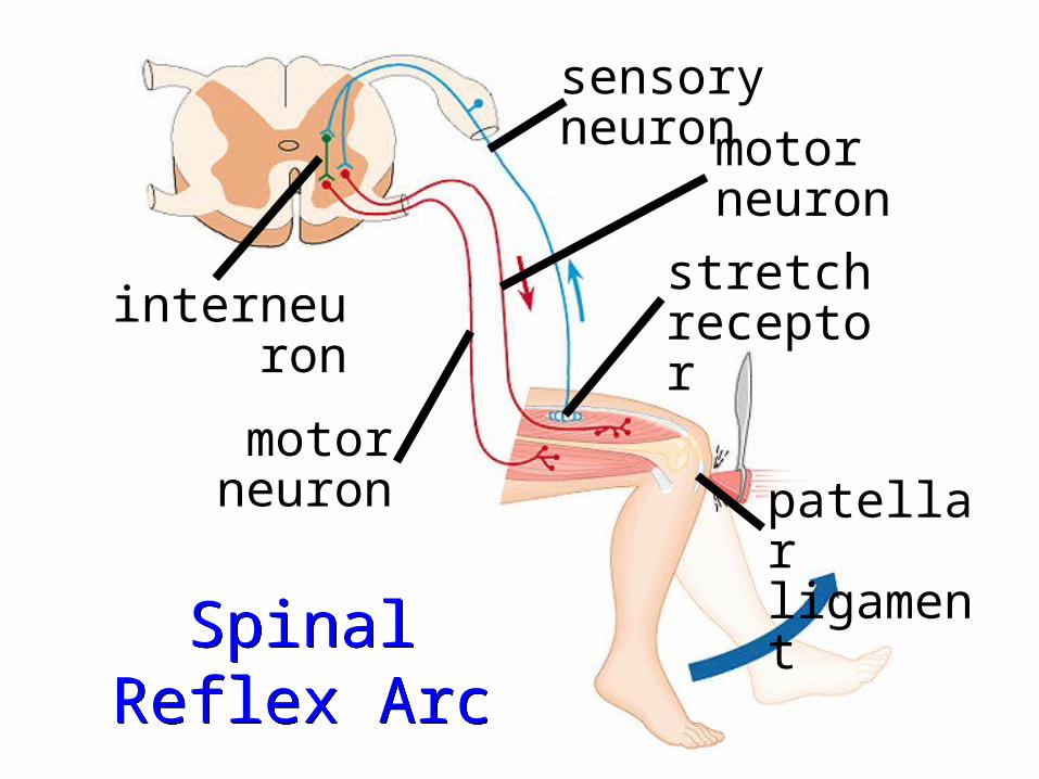
Spinal Reflex Arc
Spinal Reflex Arc
patellar ligament
stretch receptor
motor neuron
sensory neuron
motor neuron
interneuron
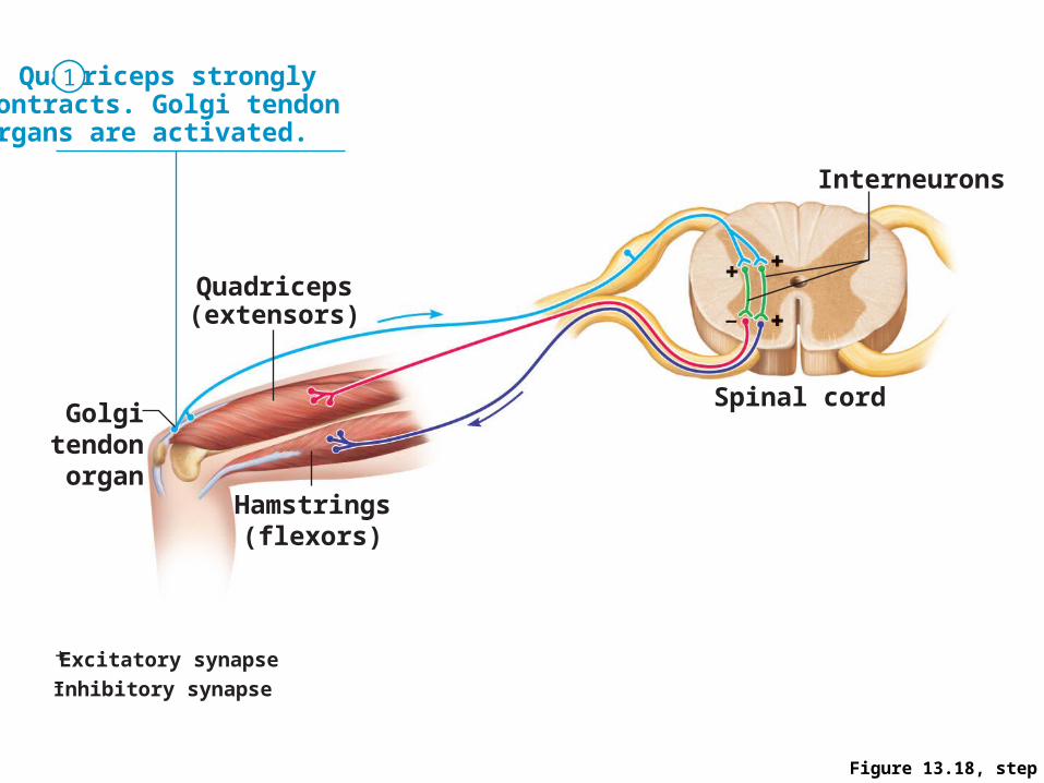
Figure 13.18, step 1
+ Excitatory synapse– Inhibitory synapse
Quadriceps strongly contracts. Golgi tendon organs are activated.
Interneurons
Spinal cord
Quadriceps(extensors)
Golgitendon
organHamstrings
(flexors)
1
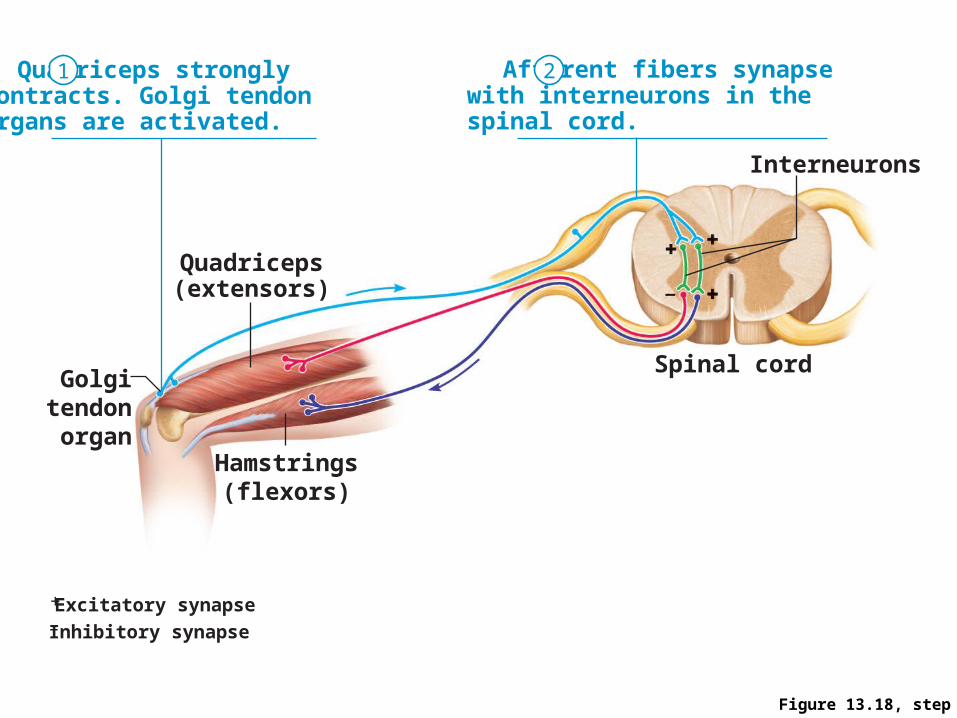
Figure 13.18, step 2
+ Excitatory synapse– Inhibitory synapse
Quadriceps strongly contracts. Golgi tendon organs are activated.
Afferent fibers synapse with interneurons in the spinal cord.
Interneurons
Spinal cord
Quadriceps(extensors)
Golgitendon
organHamstrings
(flexors)
1 2
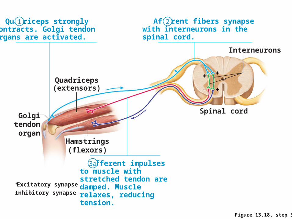
Figure 13.18, step 3a
+ Excitatory synapse– Inhibitory synapse
Quadriceps strongly contracts. Golgi tendon organs are activated.
Afferent fibers synapse with interneurons in the spinal cord.
Efferent impulses to muscle with stretched tendon are damped. Muscle relaxes, reducing tension.
Interneurons
Spinal cord
Quadriceps(extensors)
Golgitendon
organHamstrings
(flexors)
1 2
3a
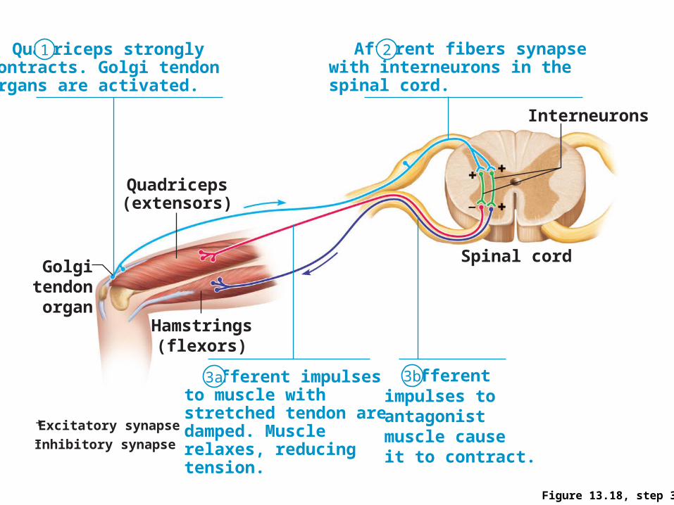
Figure 13.18, step 3b
+ Excitatory synapse– Inhibitory synapse
Quadriceps strongly contracts. Golgi tendon organs are activated.
Afferent fibers synapse with interneurons in the spinal cord.
Efferent impulses to muscle with stretched tendon are damped. Muscle relaxes, reducing tension.
Efferent impulses to antagonist muscle cause it to contract.
Interneurons
Spinal cord
Quadriceps(extensors)
Golgitendon
organHamstrings
(flexors)
1 2
3a 3b
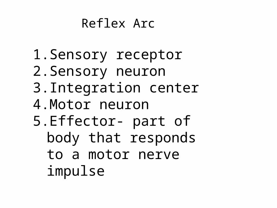
Reflex Arc
1. Sensory receptor2. Sensory neuron3. Integration center4. Motor neuron5. Effector- part of body that
responds to a motor nerve impulse
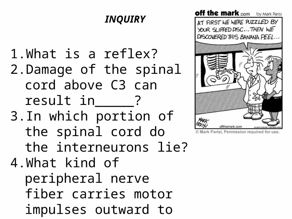
INQUIRY
1. What is a reflex?2. Damage of the spinal cord
above C3 can result in_____?
3. In which portion of the spinal cord do the interneurons lie?
4. What kind of peripheral nerve fiber carries motor impulses outward to smooth muscles and glands of internal organs?