The Spectrum of Benign Esophageal Lesions: Imaging Findings · The Spectrum of Benign Esophageal...
12
Korean J Radiol 3(3), September 2002 199 The Spectrum of Benign Esophageal Lesions: Imaging Findings Benign esophageal lesions occur in various diseases. Barium studies are use- ful for the evaluation of mucosal surface lesions but provide little information about the extramucosal extent of disease. Computed tomography and magnetic resonance imaging, on the other hand, permit the assessment of wall thickness, mediastinal involvement, adjacent lymphadenopathy, and distant spread. In dis- eases such as fibrovascular polyps, duplication cysts, scleroderma, trauma, caustic esophagitis, hiatal hernia, esophageal diverticulum, achalasia, and parae- sophageal varices, the findings of imaging studies are specific, obviating the need for further invasive diagnostic work-up. The advent of helical computed tomography and its volume data set allows the acquisition of multiplanar images, and magnetic resonance imaging is useful both for this and for tissue characteri- zation. Thus, multiplanar cross-sectional imaging further extends the role of imag- ing modalities to the evaluation of benign esophageal lesions. Through an aware- ness of the multiplanar cross-sectional appearances of various benign esophageal lesions, the radiologist can play an important role in the detection, diagnosis, further diagnostic planning, and treatment of the diseases in which they occur. arium contrast studies and endoscopy are useful for the evaluation of mu- cosal surface lesions of the esophagus but provide little information about the extramucosal extent of disease. Computed tomography (CT) and magnetic resonance imaging (MRI), on the other hand, permit the evaluation of wall thickness, mediastinal involvement, adjacent lymphadenopathy, and distant spread. With the advent of the helical technique, CT allows the rapid acquisition of both multi- planar and transaxial images, while MRI permits multiplanar imaging with high con- trast resolution. In this article, the authors describe the wide range of imaging and pathologic findings of various benign esophageal lesions. The Anatomy and Imaging Findings of the Normal Esophagus The esophagus is a muscular tube, 20 to 24 cm in length, that connects the pharynx to the stomach. It is composed of five layers: two muscle layers (outer longitudinal and inner circular), submucosa, muscularis mucosae, and stratified squamous epithelium which changes abruptly at the cardia of the stomach into simple columnar epithelium. There is no serosal layer. The esophagus has three segments: cervical, thoracic, and ab- dominal, the first of which lies anterior to the vertebrae and posterior to the trachea. Throughout its course in the posterior mediastinum, the thoracic esophagus is situated adjacent to vital structures including the trachea, vertebrae, lungs, heart, blood vessels, and lymphatics. Its upper third has an intimate relationship with the trachea, while its Kyung Mi Jang, MD 1 Kyung Soo Lee, MD 1 Soon Jin Lee, MD 1 Eun A Kim, MD 1 Tae Sung Kim, MD 1 Daehee Han, MD 1 Young Mog Shim, MD 2 Index terms : Esophagus, abnormalities Esophagus, diseases Esophagus, CT Esophagus, MR Esophagus, neoplasms Korean J Radiol 2002; 3: 199-210 Received April 23, 2002; accepted after revision July 3, 2002. Departments of 1 Radiology and 2 Thoracic Surgery, Samsung Medical Center, Sungkyunkwan University School of Medicine Presented as an Educational Exhibit at the 2001 RSNA Scientific Assembly Address reprint requests to : Kyung Soo Lee, MD, Department of Radiology, Samsung Medical Center, Sungkyunkwan University School of Medicine, 50 Ilwon-dong, Kangnam-gu, Seoul 135-710, Korea. Telephone: (822) 3410-2511 Fax: (822) 3410-2559 e-mail: [email protected] B
Transcript of The Spectrum of Benign Esophageal Lesions: Imaging Findings · The Spectrum of Benign Esophageal...
Korean J Radiol 3(3), September 2002 199
The Spectrum of Benign EsophagealLesions: Imaging Findings
Benign esophageal lesions occur in various diseases. Barium studies are use-ful for the evaluation of mucosal surface lesions but provide little informationabout the extramucosal extent of disease. Computed tomography and magneticresonance imaging, on the other hand, permit the assessment of wall thickness,mediastinal involvement, adjacent lymphadenopathy, and distant spread. In dis-eases such as fibrovascular polyps, duplication cysts, scleroderma, trauma,caustic esophagitis, hiatal hernia, esophageal diverticulum, achalasia, and parae-sophageal varices, the findings of imaging studies are specific, obviating theneed for further invasive diagnostic work-up. The advent of helical computedtomography and its volume data set allows the acquisition of multiplanar images,and magnetic resonance imaging is useful both for this and for tissue characteri-zation. Thus, multiplanar cross-sectional imaging further extends the role of imag-ing modalities to the evaluation of benign esophageal lesions. Through an aware-ness of the multiplanar cross-sectional appearances of various benignesophageal lesions, the radiologist can play an important role in the detection,diagnosis, further diagnostic planning, and treatment of the diseases in whichthey occur.
arium contrast studies and endoscopy are useful for the evaluation of mu-cosal surface lesions of the esophagus but provide little information aboutthe extramucosal extent of disease. Computed tomography (CT) and
magnetic resonance imaging (MRI), on the other hand, permit the evaluation of wallthickness, mediastinal involvement, adjacent lymphadenopathy, and distant spread.With the advent of the helical technique, CT allows the rapid acquisition of both multi-planar and transaxial images, while MRI permits multiplanar imaging with high con-trast resolution. In this article, the authors describe the wide range of imaging andpathologic findings of various benign esophageal lesions.
The Anatomy and Imaging Findings of the Normal Esophagus
The esophagus is a muscular tube, 20 to 24 cm in length, that connects the pharynxto the stomach. It is composed of five layers: two muscle layers (outer longitudinal andinner circular), submucosa, muscularis mucosae, and stratified squamous epitheliumwhich changes abruptly at the cardia of the stomach into simple columnar epithelium.There is no serosal layer. The esophagus has three segments: cervical, thoracic, and ab-dominal, the first of which lies anterior to the vertebrae and posterior to the trachea.Throughout its course in the posterior mediastinum, the thoracic esophagus is situatedadjacent to vital structures including the trachea, vertebrae, lungs, heart, blood vessels,and lymphatics. Its upper third has an intimate relationship with the trachea, while its
Kyung Mi Jang, MD1
Kyung Soo Lee, MD1
Soon Jin Lee, MD1
Eun A Kim, MD1
Tae Sung Kim, MD1
Daehee Han, MD1
Young Mog Shim, MD2
Index terms:Esophagus, abnormalitiesEsophagus, diseasesEsophagus, CT Esophagus, MR Esophagus, neoplasms
Korean J Radiol 2002;3:199-210Received April 23, 2002; accepted after revision July 3, 2002.
Departments of 1Radiology and 2ThoracicSurgery, Samsung Medical Center,Sungkyunkwan University School ofMedicine
Presented as an Educational Exhibit atthe 2001 RSNA Scientific Assembly
Address reprint requests to:Kyung Soo Lee, MD, Department ofRadiology, Samsung Medical Center,Sungkyunkwan University School ofMedicine, 50 Ilwon-dong, Kangnam-gu,Seoul 135-710, Korea.Telephone: (822) 3410-2511Fax: (822) 3410-2559e-mail: [email protected]
B
lower third lies close to the aorta. The abdominal esopha-gus is usually 3 cm or less in length and connects with thegastroesophageal junction.
In CT imaging of the esophagus, approximately 100120 mL (30 gm) of intravenous contrast material is usuallyinjected at a rate of 2 3 mL/sec prior to scanning, and foropacification of the esophageal lumen, an esophageal paste(Esopho-CAT; E-Z-Em, Westburg, NT, U.S.A.) may alsobe ingested. The apparent thickness of the esophageal wallat CT varies according to the degree of distention, and athickness of 3 mm or more has been considered abnormal(1). According to one study (2), however, in only 4% ofnormal subjects is the esophageal wall more than 5 mmthick, whereas in 42% its thickness is more than 3 mm pri-or to dilatation of the esophageal lumen by an effervescentagent. It seems, therefore, that 5 mm, rather than 3 mm, isan appropriate maximum normal value for esophageal wallthickness. At T1-weighted imaging, the esophagus appearsas a structure of low signal intensity, contrasted by fat ofhigh signal intensity, while T2-weighted imaging indicatesthat its muscular wall, which shows moderate enhance-ment after the injection of intravenous Gd-DTPA, is of lowsignal intensity.
Benign Esophageal Tumors
LeiomyomaEsophageal leiomyoma is the most common benign
esophageal tumor, accounting for more than 50% of allsuch tumors. Histopathologically, it consists of intersectingbands of muscle and fibrous tissue in a well-defined cap-sule. About 60% of these tumors are located in the distal
third of the esophagus, 30% in the middle third, and 10%in the proximal third (3). Growing slowly, a tumor usuallypresents grossly as a discrete submucosal mass, rangingfrom 2 to 8 cm in diameter. Patients with these tumors areusually asymptomatic; even where a large mass significant-ly indents the lumen, this may be so. Where symptoms areapparent, the most common symptom of these is dyspha-gia. In contrast to gastric leiomyomas, esophageal leiomy-omas rarely ulcerate, and hematemesis is thus extremelyrare. Esophageal leiomyomatosis is sometimes associatedwith Alport syndrome (4).
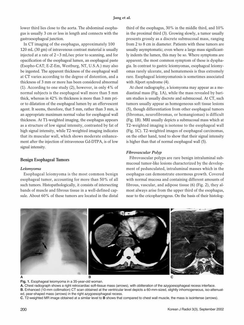
At chest radiography, a leiomyoma may appear as a me-diastinal mass (Fig. 1A), while the mass revealed by bari-um studies is usually discrete and submucosal. At CT, suchtumors usually appear as homogeneous soft tissue lesions(5), though differentiation from other esophageal tumors(fibromas, neurofibromas, or hemangiomas) is difficult(Fig. 1B). MRI usually depicts a submucosal mass which atT2-weighted imaging is isotense to the esophageal wall(Fig. 1C). T2-weighted images of esophageal carcinomas,on the other hand, tend to show that their signal intensityis higher than that of normal esophageal wall (5).
Fibrovascular PolypFibrovascular polyps are rare benign intraluminal sub-
mucosal tumor-like lesions characterized by the develop-ment of pedunculated, intraluminal masses which in theesophagus can demonstrate enormous growth. Coveredwith normal mucosa and containing different amounts offibrous, vascular, and adipose tissue (6) (Fig. 2), they al-most always arise from the upper third of the esophagus,near to the cricopharyngeus. On the basis of their histolog-
Jang et al.
200 Korean J Radiol 3(3), September 2002
Fig. 1. Esophageal leiomyoma in a 35-year-old woman.A. Chest radiograph shows a right retrocardiac soft-tissue mass (arrows), with obliteration of the azygoesophageal recess interface.B. Enhanced (10-mm collimation) CT scan obtained at the ventricular level depicts a 60-mm-sized, slightly inhomogeneous, iso-attenuat-ed, pear-shaped mass (arrows) in the right azygoesophageal recess.C. T2-weighted MR image obtained at a similar level to B shows that compared to chest wall muscle, the mass is isointense (arrows).
A B C
ic composition, these lesions have been termed lipomas, fi-bromas, fibrolipomas, or fibroepithelial polyps. More re-cently, they have been classified as fibrovascular polyp, a
term used by the World Heath Organization in their in-ternational histologic classification system. Dysphagia,vomiting, weight loss, and respiratory symptoms are themost frequent complaints of patients with fibrovascularpolyps; these with long stalks, however, can regurgitate in-to the pharynx or mouth and cause death from asphyxia-tion if the larynx is occluded (7).
Imaging Findings of Benign Esophageal Lesions
Korean J Radiol 3(3), September 2002 201
A B C
D E F
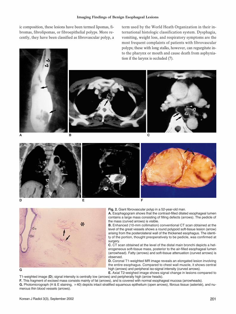
Fig. 2. Giant fibrovascular polyp in a 52-year-old man.A. Esophagogram shows that the contrast-filled dilated esophageal lumencontains a large mass consisting of filling defects (arrows). The pedicle ofthe mass (curved arrows) is visible.B. Enhanced (10-mm collimation) conventional CT scan obtained at thelevel of the great vessels shows a round polypoid soft-tissue lesion (arrow)arising from the posterolateral wall of the thickened esophagus. The identi-ty of the portion, thought preoperatively to be pedicle, was confirmed atsurgery.C. CT scan obtained at the level of the distal main bronchi depicts a het-erogeneous soft-tissue mass, posterior to the air-filled esophageal lumen(arrowhead). Fatty (arrows) and soft-tissue attenuation (curved arrows) isobserved.D. Coronal T1-weighted MR image reveals an elongated lesion involvingthe entire esophagus. Compared to chest wall muscle, it shows centralhigh (arrows) and peripheral iso-signal intensity (curved arrows).E. Axial T2-weighed image shows signal change in lesions compared to
T1-weighted image (D); signal intensity is centrally low (arrows) and peripherally high (arrow heads).F. This fragment of excised mass consists mainly of fat (arrows), and is covered with normal esophageal mucosa (arrowheads).G. Photomicrograph (H & E staining, 40) depicts intact stratified squamous epithelium (open arrows), fibrous tissue (asterisk), and nu-merous thin blood vessels (arrows).
G
Fibrovascular polyps can sometimes be identified atchest radiography by the presence of a right-sided superiormediastinal mass, anterior tracheal bowing, or both (6).The lesions usually appear at esophagography as smooth,expansile, intraluminal masses that arise in the cervicalesophagus and extend into the thoracic esophagus (Fig.2A). Although most fibrovascular polyps have a site of at-tachment in the cervical esophagus, barium studies oftenfail to demonstrate a proximal pedicle. Fibrovascularpolyps containing abundant adipose tissue may appear atCT as fat-attenuated lesions that expand the lumen of theesophagus (6) (Figs. 2B, C). Polyps containing equalamounts of adipose and fibrovascular tissue may appear as
heterogeneous lesions, with focal areas of fat attenuationjuxtaposed with areas of soft-tissue attenuation, and thosecontaining abundant fibrovascular tissue may appear assoft-tissue-attenuated lesions with a paucity of fat. At T1 -weighted MR imaging, fibrovascular polyps containingabundant adipose tissue are characterized by high signalintensity (8) (Fig. 2D).
Duplication Cyst Esophageal duplication cysts account for 0.5-2.5% of all
tumors or tumor-like lesions of the esophagus, and about20% of all gastrointestinal tract duplications (9). They re-sult from abnormal embryologic development in which
Jang et al.
202 Korean J Radiol 3(3), September 2002
A B
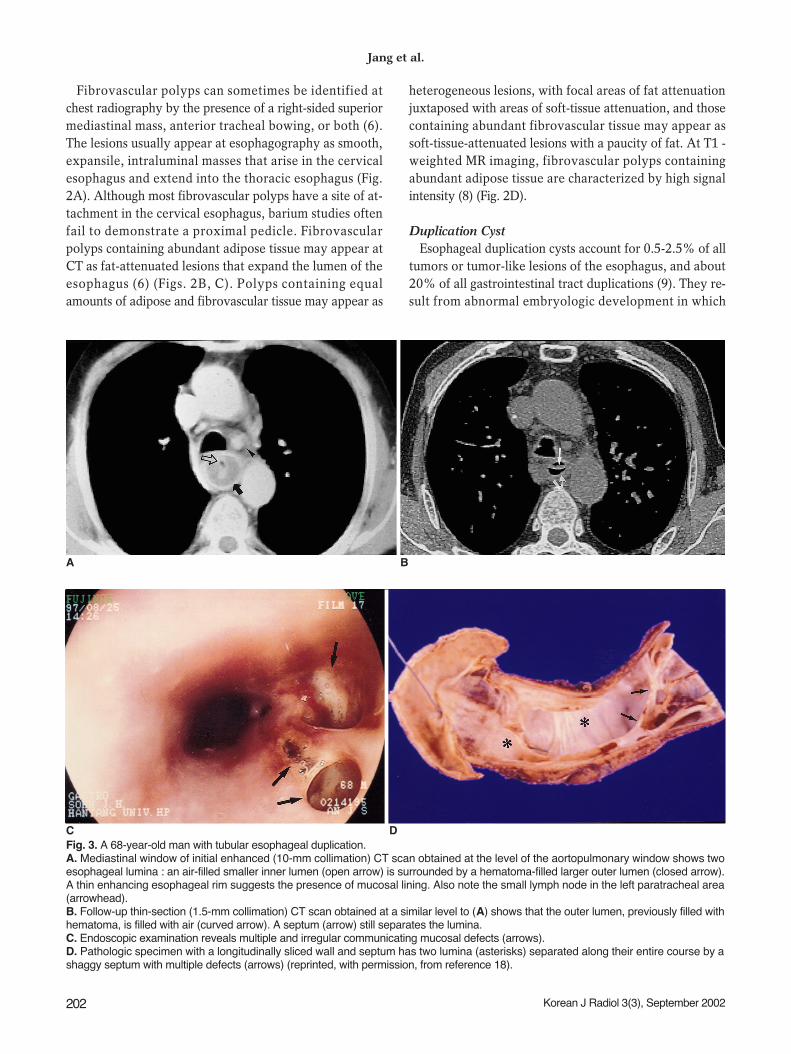
Fig. 3. A 68-year-old man with tubular esophageal duplication.A. Mediastinal window of initial enhanced (10-mm collimation) CT scan obtained at the level of the aortopulmonary window shows twoesophageal lumina : an air-filled smaller inner lumen (open arrow) is surrounded by a hematoma-filled larger outer lumen (closed arrow).A thin enhancing esophageal rim suggests the presence of mucosal lining. Also note the small lymph node in the left paratracheal area(arrowhead).B. Follow-up thin-section (1.5-mm collimation) CT scan obtained at a similar level to (A) shows that the outer lumen, previously filled withhematoma, is filled with air (curved arrow). A septum (arrow) still separates the lumina.C. Endoscopic examination reveals multiple and irregular communicating mucosal defects (arrows).D. Pathologic specimen with a longitudinally sliced wall and septum has two lumina (asterisks) separated along their entire course by ashaggy septum with multiple defects (arrows) (reprinted, with permission, from reference 18).
C D
nests of cells are sequestered from the primitive foregut,and may be classified as either cystic or, less commonly,tubular duplications. Although most duplication cysts arenoncommunicating, tubular duplications may occasionallycommunicate directly with the esophageal lumen (10) (Fig.3). Most adults with esophageal duplication cysts areasymptomatic, though symptoms caused by obstruction,bleeding, or an infected cyst may occasionally arise.
CT scans of cystic esophageal duplication depict a homo-geneous mass with low attenuation and a smooth border(11) (Fig. 4). Kuhlman et al. (11) found that CT can pro-vide information about the cystic nature, size and locationof a lesion and allow the use of transesophageal needle as-piration rather than the traditional approach of surgical ex-cision, for definitive diagnosis. CT scans of tubular duplica-tion cysts may show two esophageal lumina, and in theesophageal wall, a thin enhancing rim, representing mucos-al linings, may be apparent (10) (Fig. 3A). At T2-weightedMR imaging, duplication cysts may show bright signal in-tensity and a well-defined margin.
SchwannomaEsophageal schwannoma is extremely rare; only 15 cases
have been reported in the literature (12). A review ofthese previous reports indicates that benign schwannomasare usually located in the upper esophagus and occur more
frequently in middle-aged women. A barium esopha-gogram reveals a large, smooth, polypoid-filled defect, andCT scans depict a mediastinal mass (Fig. 5). No distinctivecharacteristics differentiate these from other submucosaltumors, and at preoperative examination it is thus very dif-ficult to distinguish between a schwannoma and other sub-mucosal masses. Correct diagnosis requires histologic ex-amination: positive immunohistochemical staining for S-100 protein and negative staining for smooth muscle mark-ers such as actin and desmin confirm that the tumor origi-nates from a nerve sheath (13).
Infectious and Inflammatory Diseases
TuberculosisTuberculosis involving the esophagus usually occurs in
the late stages of the disease secondary to pulmonary, me-diastinal, or disseminated diseases (14). It has been suggest-ed that CT of the mediastinum provides the most completedepiction of the tuberculous mediastinal lymphadenopathysurrounding and displacing the esophagus and the fistuloustract extending from the esophagus into the nodal mass(Fig. 6). Amorphous gas collection at enlarged mediastinallymph nodes in patients with tuberculous lymphadenitissuggests the presence of an esophagonodal fistula (14).
Imaging Findings of Benign Esophageal Lesions
Korean J Radiol 3(3), September 2002 203
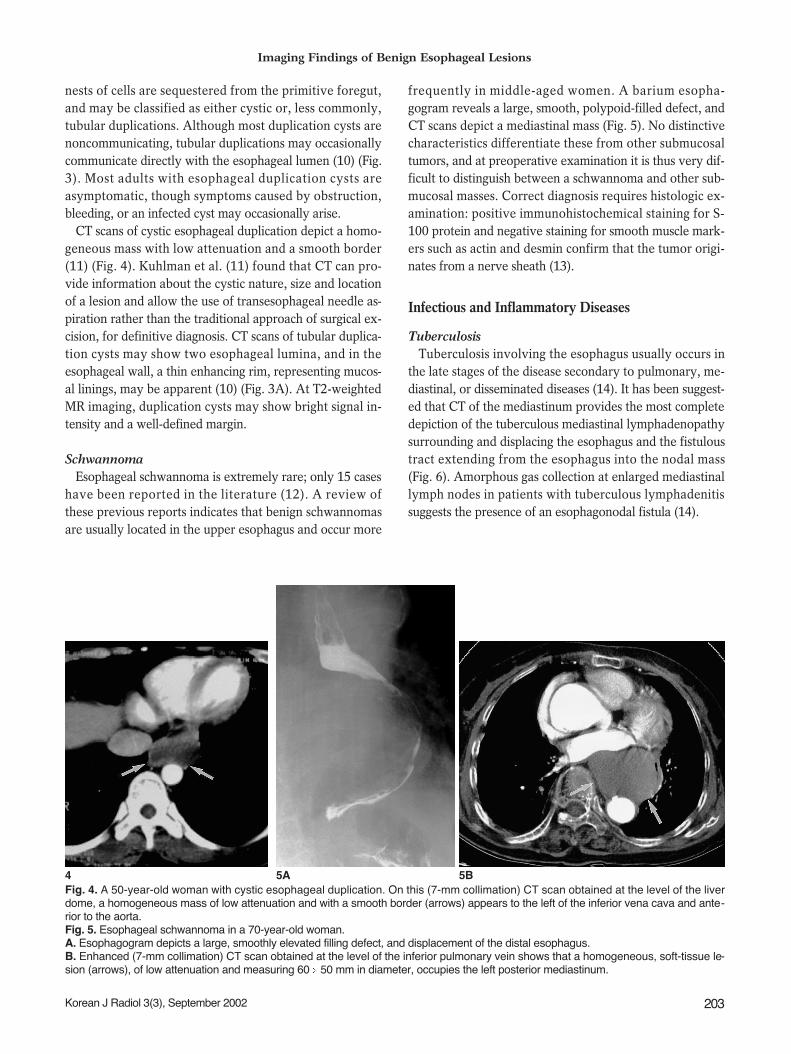
4 5AFig. 4. A 50-year-old woman with cystic esophageal duplication. On this (7-mm collimation) CT scan obtained at the level of the liverdome, a homogeneous mass of low attenuation and with a smooth border (arrows) appears to the left of the inferior vena cava and ante-rior to the aorta. Fig. 5. Esophageal schwannoma in a 70-year-old woman.A. Esophagogram depicts a large, smoothly elevated filling defect, and displacement of the distal esophagus.B. Enhanced (7-mm collimation) CT scan obtained at the level of the inferior pulmonary vein shows that a homogeneous, soft-tissue le-sion (arrows), of low attenuation and measuring 60 50 mm in diameter, occupies the left posterior mediastinum.
5B
Candida EsophagitisCandida esophagitis is the most common infection of the
esophagus, usually occurring opportunistically in patientsimmunocompromised as a result of underlying malignancy;other debilitating illnesses; treatment with radiation,steroids, or cytotoxic agents; or, most recently, AIDS (15,16). In the diagnosis of Candida esophagitis, double con-trast esophagography shows a sensitivity of about 90%
(15), demonstrating discrete plaque-like filling defectswhich have a finely nodular and granular, distinctive cob-blestone or snakeskin-like appearance and correspond tothe distinctive white plaques seen at endoscopy. Theseplaques consist of heaped-up areas of necrotic epithelialdebris or actual colonies of C. albicans on the esophagealmucosa; the esophagus per se has an irregular or shaggyappearance (15, 16) (Fig. 7). The CT findings of Candidaesophagitis, though nonspecific and commonly seen in var-ious kinds of esophagitis, are circumferential esophagealwall thickening of more than 5 mm, with relatively longsegmental involvement. Enhanced scans may also depictthe target sign (circumferential wall thickening and enhanc-ing internal mucosa) (2).
Reflux Esophagitis and Barrett’s EsophagusIn the diagnosis of reflux esophagitis, the use of double -
contrast esophagography has increased radiographic sensi-tivity to almost 90% (17). Double - contrast imaging re-veals the finely nodular or granular appearance of the mu-cosa in the distal third or half of the thoracic esophagus,occurring due to the presence of mucosal edema and in-flammation during the early stages of reflux esophagitis.As the disease progresses, double-contrast esophagographymay demonstrate shallow ulcers and erosions, thickenedlongitudinal folds, inflammatory polyps, scarring, and stric-tures (18) (Fig. 8).
Barrett’s esophagus involves the conversion of squamousepithelium in the distal esophagus into columnar epitheli-
Jang et al.
204 Korean J Radiol 3(3), September 2002
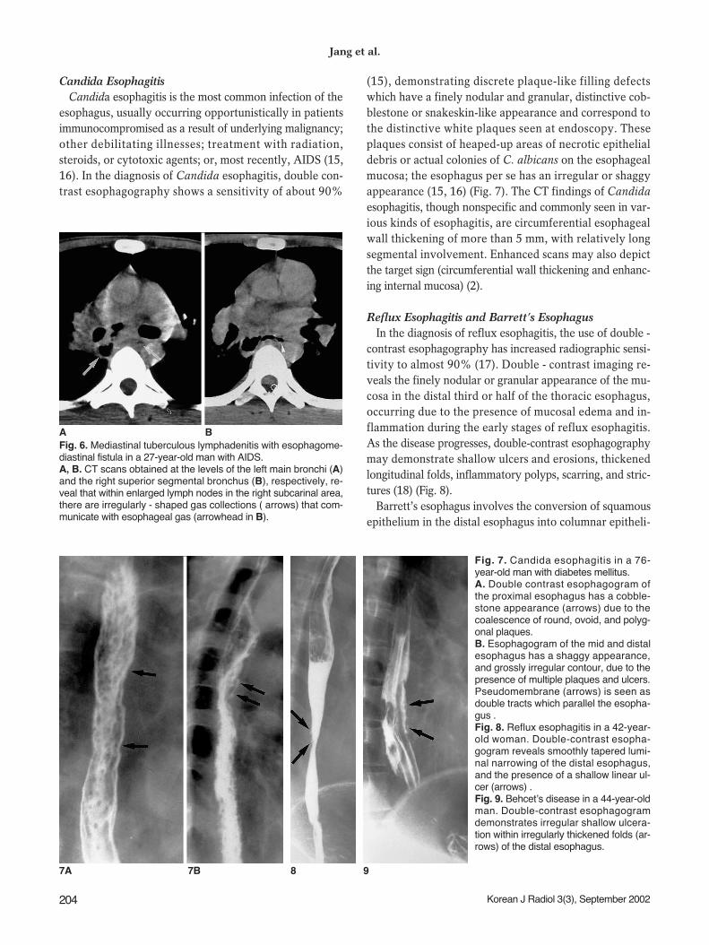
Fig. 7. Candida esophagitis in a 76-year-old man with diabetes mellitus.A. Double contrast esophagogram ofthe proximal esophagus has a cobble-stone appearance (arrows) due to thecoalescence of round, ovoid, and polyg-onal plaques.B. Esophagogram of the mid and distalesophagus has a shaggy appearance,and grossly irregular contour, due to thepresence of multiple plaques and ulcers.Pseudomembrane (arrows) is seen asdouble tracts which parallel the esopha-gus .Fig. 8. Reflux esophagitis in a 42-year-old woman. Double-contrast esopha-gogram reveals smoothly tapered lumi-nal narrowing of the distal esophagus,and the presence of a shallow linear ul-cer (arrows) .Fig. 9. Behcet’s disease in a 44-year-oldman. Double-contrast esophagogramdemonstrates irregular shallow ulcera-tion within irregularly thickened folds (ar-rows) of the distal esophagus.
7A 7B 8 9
Fig. 6. Mediastinal tuberculous lymphadenitis with esophagome-diastinal fistula in a 27-year-old man with AIDS. A, B. CT scans obtained at the levels of the left main bronchi (A)and the right superior segmental bronchus (B), respectively, re-veal that within enlarged lymph nodes in the right subcarinal area,there are irregularly - shaped gas collections ( arrows) that com-municate with esophageal gas (arrowhead in B).
A B
um. The most likely cause of the condition, which occurs inapproximately 10% of patients with reflux disease and ispremalignant with increased risk of adenocarcinoma (19),is chronic reflux esophagitis. In patients with long-segmentBarrett’s esophagus, the risk of developing adenocarcino-ma is thought to be 30 40 times greater than among thegeneral population (20). The characteristic radiologic fea-ture of Barrett’s esophagus is a midesophageal stricture orulcer, often associated with a sliding hiatal hernia or gas-troesophageal reflux (18, 19).
Behcet’s DiseaseBehcet’s disease is a multi-system disorder with a clinical
triad of oral and genital ulceration and ocular inflamma-tion. The mid or distal esophagus is occasionally involved,and double contrast esophagography may reveal the pres-ence there of discrete, superficial ulcers (21) (Fig. 9).
Scleroderma Patients with scleroderma often develop diffuse interstitial
lung disease as well as gastrointestinal symptoms such asdysphagia, regurgitation, and malabsorption. As well as re-vealing pulmonary parenchymal abnormalities, CT in suchpatients may demonstrate symptomatic esophageal dilata-tion and mediastinal lymph node enlargement (22) (Fig. 10).
Trauma and Caustic Esophagitis
PerforationPerforation is the most serious and life-threatening event
to affect the esophagus : in patients with untreated thoracicesophageal perforations, the fulminant mediastinitis whichoccurs leads to a mortality rate of almost 100%.Endoscopic procedures are responsible for up to 75% of allesophageal perforations (23), which may also be caused byforeign bodies or penetrating trauma. Most perforation in-volves the cervical esophagus ; spontaneous esophagealperforation (Boerhaave syndrome) due to a sudden, rapidincrease in intraluminal esophageal pressure causes fullthickness perforation of normal underlying esophageal tis-sue, and is frequently a result of violent retching or vomit-ing, usually after an alcoholic binge. Patients with negativeesophagograms in whom esophageal perforation is still sus-pected on clinical grounds may undergo CT ; in such cases,
Imaging Findings of Benign Esophageal Lesions
Korean J Radiol 3(3), September 2002 205
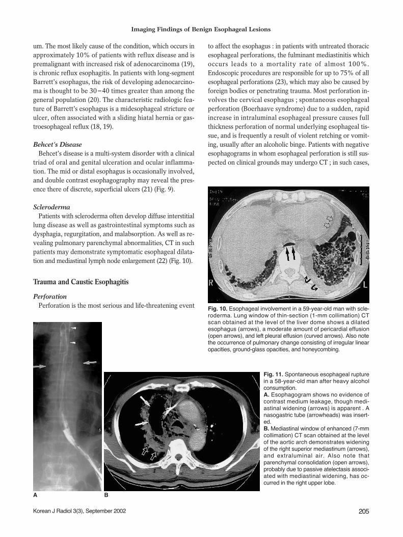
Fig. 10. Esophageal involvement in a 59-year-old man with scle-roderma. Lung window of thin-section (1-mm collimation) CTscan obtained at the level of the liver dome shows a dilatedesophagus (arrows), a moderate amount of pericardial effusion(open arrows), and left pleural effusion (curved arrows). Also notethe occurrence of pulmonary change consisting of irregular linearopacities, ground-glass opacities, and honeycombing.
A B
Fig. 11. Spontaneous esophageal rupturein a 58-year-old man after heavy alcoholconsumption.A. Esophagogram shows no evidence ofcontrast medium leakage, though medi-astinal widening (arrows) is apparent . Anasogastric tube (arrowheads) was insert-ed.B. Mediastinal window of enhanced (7-mmcollimation) CT scan obtained at the levelof the aortic arch demonstrates wideningof the right superior mediastinum (arrows),and extraluminal air. Also note thatparenchymal consolidation (open arrows),probably due to passive atelectasis associ-ated with mediastinal widening, has oc-curred in the right upper lobe.
findings of extraluminal gas in the mediastinum are highlysuggestive of esophageal perforation (Fig. 11). Where thishas occurred, CT is also useful for determining the extentof extraluminal gas and fluid in the mediastinum and formonitoring patients who are treated nonsurgically (24).
Fistulae
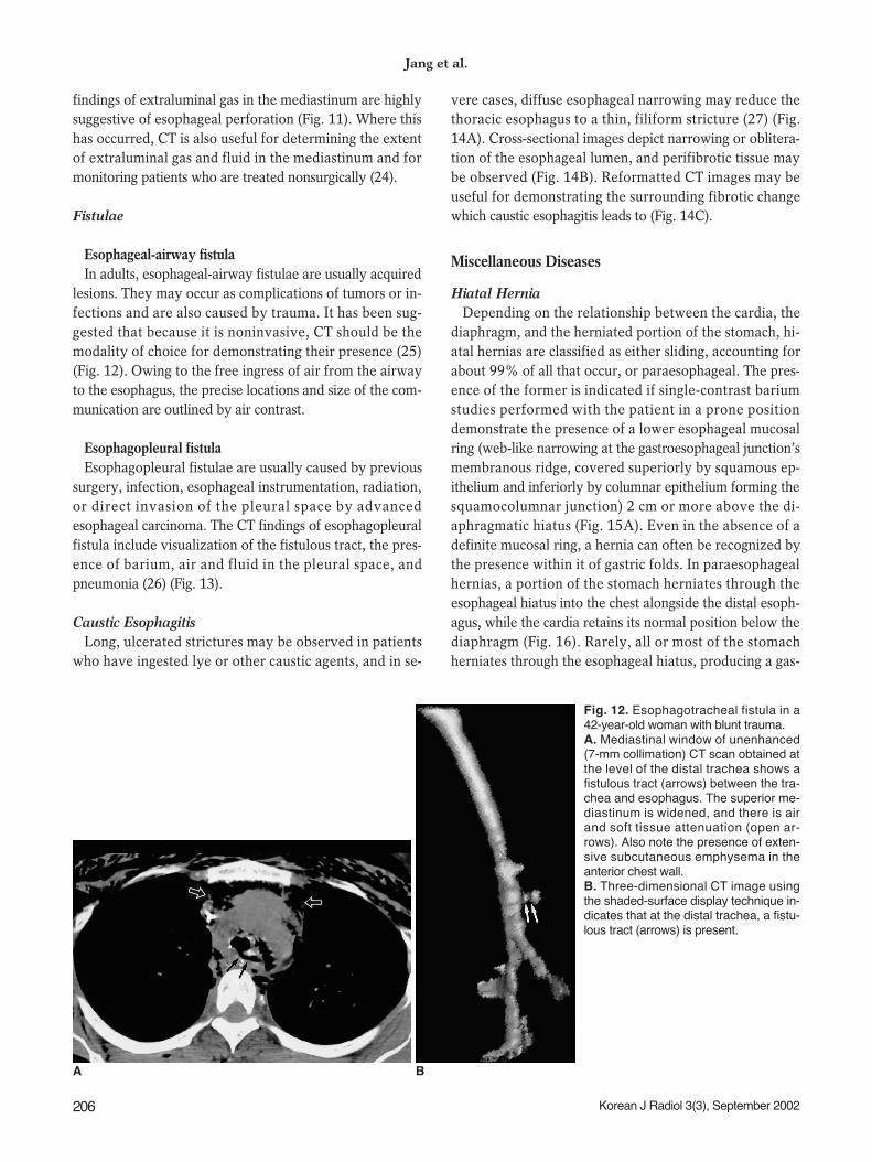
Esophageal-airway fistulaIn adults, esophageal-airway fistulae are usually acquired
lesions. They may occur as complications of tumors or in-fections and are also caused by trauma. It has been sug-gested that because it is noninvasive, CT should be themodality of choice for demonstrating their presence (25)(Fig. 12). Owing to the free ingress of air from the airwayto the esophagus, the precise locations and size of the com-munication are outlined by air contrast.
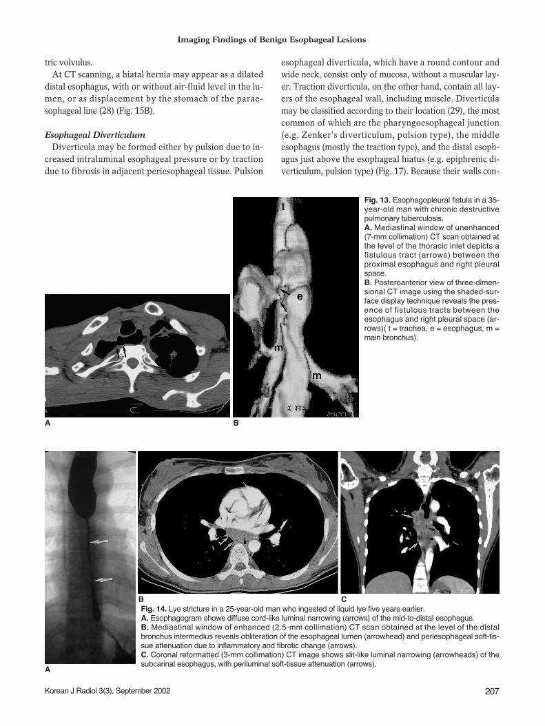
Esophagopleural fistulaEsophagopleural fistulae are usually caused by previous
surgery, infection, esophageal instrumentation, radiation,or direct invasion of the pleural space by advancedesophageal carcinoma. The CT findings of esophagopleuralfistula include visualization of the fistulous tract, the pres-ence of barium, air and fluid in the pleural space, andpneumonia (26) (Fig. 13).
Caustic EsophagitisLong, ulcerated strictures may be observed in patients
who have ingested lye or other caustic agents, and in se-
vere cases, diffuse esophageal narrowing may reduce thethoracic esophagus to a thin, filiform stricture (27) (Fig.14A). Cross-sectional images depict narrowing or oblitera-tion of the esophageal lumen, and perifibrotic tissue maybe observed (Fig. 14B). Reformatted CT images may beuseful for demonstrating the surrounding fibrotic changewhich caustic esophagitis leads to (Fig. 14C).
Miscellaneous Diseases
Hiatal HerniaDepending on the relationship between the cardia, the
diaphragm, and the herniated portion of the stomach, hi-atal hernias are classified as either sliding, accounting forabout 99% of all that occur, or paraesophageal. The pres-ence of the former is indicated if single-contrast bariumstudies performed with the patient in a prone positiondemonstrate the presence of a lower esophageal mucosalring (web-like narrowing at the gastroesophageal junction’smembranous ridge, covered superiorly by squamous ep-ithelium and inferiorly by columnar epithelium forming thesquamocolumnar junction) 2 cm or more above the di-aphragmatic hiatus (Fig. 15A). Even in the absence of adefinite mucosal ring, a hernia can often be recognized bythe presence within it of gastric folds. In paraesophagealhernias, a portion of the stomach herniates through theesophageal hiatus into the chest alongside the distal esoph-agus, while the cardia retains its normal position below thediaphragm (Fig. 16). Rarely, all or most of the stomachherniates through the esophageal hiatus, producing a gas-
Jang et al.
206 Korean J Radiol 3(3), September 2002
A B
Fig. 12. Esophagotracheal fistula in a42-year-old woman with blunt trauma. A. Mediastinal window of unenhanced(7-mm collimation) CT scan obtained atthe level of the distal trachea shows afistulous tract (arrows) between the tra-chea and esophagus. The superior me-diastinum is widened, and there is airand soft tissue attenuation (open ar-rows). Also note the presence of exten-sive subcutaneous emphysema in theanterior chest wall.B. Three-dimensional CT image usingthe shaded-surface display technique in-dicates that at the distal trachea, a fistu-lous tract (arrows) is present.
tric volvulus.At CT scanning, a hiatal hernia may appear as a dilated
distal esophagus, with or without air-fluid level in the lu-men, or as displacement by the stomach of the parae-sophageal line (28) (Fig. 15B).
Esophageal DiverticulumDiverticula may be formed either by pulsion due to in-
creased intraluminal esophageal pressure or by tractiondue to fibrosis in adjacent periesophageal tissue. Pulsion
esophageal diverticula, which have a round contour andwide neck, consist only of mucosa, without a muscular lay-er. Traction diverticula, on the other hand, contain all lay-ers of the esophageal wall, including muscle. Diverticulamay be classified according to their location (29), the mostcommon of which are the pharyngoesophageal junction(e.g. Zenker’s diverticulum, pulsion type), the middleesophagus (mostly the traction type), and the distal esoph-agus just above the esophageal hiatus (e.g. epiphrenic di-verticulum, pulsion type) (Fig. 17). Because their walls con-
Imaging Findings of Benign Esophageal Lesions
Korean J Radiol 3(3), September 2002 207
A B
Fig. 13. Esophagopleural fistula in a 35-year-old man with chronic destructivepulmonary tuberculosis.A. Mediastinal window of unenhanced(7-mm collimation) CT scan obtained atthe level of the thoracic inlet depicts afistulous tract (arrows) between theproximal esophagus and right pleuralspace. B. Posteroanterior view of three-dimen-sional CT image using the shaded-sur-face display technique reveals the pres-ence of fistulous tracts between theesophagus and right pleural space (ar-rows)( t = trachea, e = esophagus, m =main bronchus).
A
B CFig. 14. Lye stricture in a 25-year-old man who ingested of liquid lye five years earlier.A. Esophagogram shows diffuse cord-like luminal narrowing (arrows) of the mid-to-distal esophagus.B. Mediastinal window of enhanced (2.5-mm collimation) CT scan obtained at the level of the distalbronchus intermedius reveals obliteration of the esophageal lumen (arrowhead) and periesophageal soft-tis-sue attenuation due to inflammatory and fibrotic change (arrows).C. Coronal reformatted (3-mm collimation) CT image shows slit-like luminal narrowing (arrowheads) of thesubcarinal esophagus, with periluminal soft-tissue attenuation (arrows).
tain no muscle, pulsion diverticula tend to remain filled af-ter the esophagus has emptied of barium. When the esoph-agus collapses, traction diverticula tend to empty.
Esophageal AchalasiaAchalasia (megaesophagus) arises, in part, because relax-
ation at the lower esophageal sphincter level has failed tooccur, and is partly due to the failure of organized peristal-
sis. Among the various causes of dilatation of the esopha-gus, including achalasia, inflammatory stenosis, progressivesystemic sclerosis (PSS) and carcinoma, achalasia causesthe most severe generalized dilatation. Its symptoms in-clude dysphasia, pain on swallowing, and chronic coughand recurrent pneumonia due to aspiration. Esophagogra-phy reveals, typically, that the lower end of the esophagushas a smooth, tapered, beak-like appearance at the level of
Jang et al.
208 Korean J Radiol 3(3), September 2002
15A
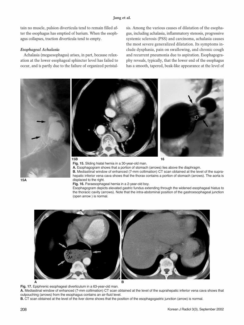
15B 16Fig. 15. Sliding hiatal hernia in a 30-year-old man.A. Esophagogram shows that a portion of stomach (arrows) lies above the diaphragm.B. Mediastinal window of enhanced (7-mm collimation) CT scan obtained at the level of the supra-hepatic inferior vena cava shows that the thorax contains a portion of stomach (arrows). The aorta isdisplaced to the right.Fig. 16. Paraesophageal hernia in a 2-year-old boy. Esophagogram depicts elevated gastric fundus extending through the widened esophageal hiatus tothe thoracic cavity (arrows). Note that the intra-abdominal position of the gastroesophageal junction(open arrow ) is normal.
Fig. 17. Epiphrenic esophageal diverticulum in a 63-year-old man.A. Mediastinal window of enhanced (7-mm collimation) CT scan obtained at the level of the suprahepatic inferior vena cava shows thatoutpouching (arrows) from the esophagus contains an air-fluid level. B. CT scan obtained at the level of the liver dome shows that the position of the esophagogastric junction (arrow) is normal.
A B
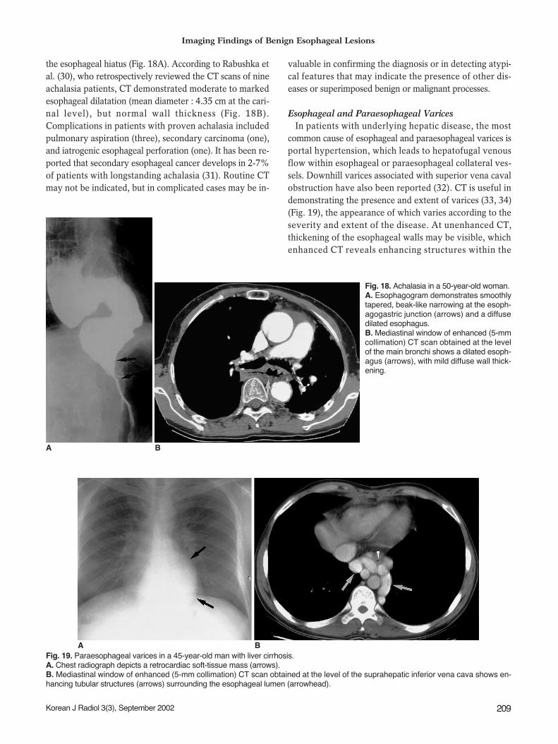
the esophageal hiatus (Fig. 18A). According to Rabushka etal. (30), who retrospectively reviewed the CT scans of nineachalasia patients, CT demonstrated moderate to markedesophageal dilatation (mean diameter : 4.35 cm at the cari-nal level), but normal wall thickness (Fig. 18B).Complications in patients with proven achalasia includedpulmonary aspiration (three), secondary carcinoma (one),and iatrogenic esophageal perforation (one). It has been re-ported that secondary esophageal cancer develops in 2-7%of patients with longstanding achalasia (31). Routine CTmay not be indicated, but in complicated cases may be in-
valuable in confirming the diagnosis or in detecting atypi-cal features that may indicate the presence of other dis-eases or superimposed benign or malignant processes.
Esophageal and Paraesophageal VaricesIn patients with underlying hepatic disease, the most
common cause of esophageal and paraesophageal varices isportal hypertension, which leads to hepatofugal venousflow within esophageal or paraesophageal collateral ves-sels. Downhill varices associated with superior vena cavalobstruction have also been reported (32). CT is useful indemonstrating the presence and extent of varices (33, 34)(Fig. 19), the appearance of which varies according to theseverity and extent of the disease. At unenhanced CT,thickening of the esophageal walls may be visible, whichenhanced CT reveals enhancing structures within the
Imaging Findings of Benign Esophageal Lesions
Korean J Radiol 3(3), September 2002 209
A B
Fig. 19. Paraesophageal varices in a 45-year-old man with liver cirrhosis.A. Chest radiograph depicts a retrocardiac soft-tissue mass (arrows). B. Mediastinal window of enhanced (5-mm collimation) CT scan obtained at the level of the suprahepatic inferior vena cava shows en-hancing tubular structures (arrows) surrounding the esophageal lumen (arrowhead).
A B
Fig. 18. Achalasia in a 50-year-old woman.A. Esophagogram demonstrates smoothlytapered, beak-like narrowing at the esoph-agogastric junction (arrows) and a diffusedilated esophagus. B. Mediastinal window of enhanced (5-mmcollimation) CT scan obtained at the levelof the main bronchi shows a dilated esoph-agus (arrows), with mild diffuse wall thick-ening.
esophageal wall or periesophageal region (33, 34).
CONCLUSION
The role of cross-sectional imaging in the evaluation ofbenign esophageal lesions continues to evolve. The adventof helical computed tomography and its volume data setfacilitates the acquisition of multiplanar images; magneticresonance imaging also helps acquire such images, as wellas permitting tissue characterization. Through an aware-ness of the detailed morphology, location and extent ofvarious benign esophageal lesions, as demonstrated bymultiplanar cross - sectional imaging, the radiologist canplay a more important role in the detection, diagnosis, andtreatment of the diseases described in this paper.
References1. Halber MD, Daffner RH, Thompson WM. CT of the esophagus:
normal appearance. AJR 1979;133:1047-10502. Berkovich GY, Levine MS, Miller WT. CT findings in patients
with esophagitis. AJR 2000;175:1431-14343. Seremetis MG, Lyons WS, DeGuzman VC, et al. Leiomyomata
of the esophagus: an analysis of 838 cases. Cancer1976;38:2166-2177
4. Cochat P, Guibaud P, Garcia Torres R, Roussel B, Guarner V,Larbre F. Diffuse leiomyomatosis in Alport syndrome. J Pediatr1988; 113:339-343
5. Yang PS, Lee KS, Lee SJ, et al. Esophageal leiomyoma: radio-logic findings in 12 patients. Korean J Radiol 2001; 2:132-137
6. Levine MS, Buck JL, Pantongrag-Brown L, Buetow PC,Hallman JR, Sobin LH. Fibrovascular polyps of the esophagus:clinical, radiographic, and pathologic findings in 16 patients.AJR 1996; 166:781-787
7. Cochet B, Hohl P, Sans M, et al. Asphyxia caused by laryngealimpaction of an esophageal polyp. Arch Otoloaryngol 1980;106:176-178
8. Stoane JM, Torrisi JM, Haller JO, David M. Fibrovascularpolyps of the esophagus: MRI findings. J Comput Assist Tomogr1995; 19:157-159
9. Macpherson RI. Gastrointestinal tract duplications: clinical,pathologic, etiologic, and radiologic considerations.RadioGraphics 1993; 13:1063-1080
10. Kim YS, Park CK, Choi YW, Jeon SC, Seo HS, Hahm CK.Esophageal tubular duplication complicated by intraluminalhematoma: a case report. J Korean Med Sci 2000; 15:463-466
11. Kuhlman JE, Fishman EK, Wang KP, Siegelman SS. Esophagealduplication cyst: CT and transesophageal needle aspiration. AJR1985; 145:531-532
12. Saito R, Kitamura M, Suzuki H, Ogawa J, Sageshima M.Esophageal schwannoma. Ann Thorac Surg 2000; 69:1947-1949
13. Ueyama T, Guo KJ, Hashimoto H, Daimaru Y, Enjoji M. Benignschwannoma of the gastrointestinal tract: a clinicopathologic and
immunohistochemical study. Hum Pathol 1988; 19:257-26414. Im J-G, Kim JH, Han MC, Kim CW. Computed tomography of
esophagomediastinal fistula in tuberculous mediastinal lym-phadenitis. J Comput Assist Tomogr 1990; 14:89-92
15. Levine MS, Macones AJ Jr, Laufer I. Candida esophagitis: accu-racy of radiographic diagnosis. Radiology 1985; 154:581-587
16. Vahey TN, Maglinte DD, Chernish SM. State-of-the-art bariumexamination in opportunistic esophagitis. Dig Dis Sci 1986 ;31:1192-1195
17. Graziani L, De Nigris E, Pesaresi A, Baldelli S, Dini L, MontesiA. Reflux esophagitis: radiologic-endoscopic correlation in 39symptomatic cases. Gastrointest Radiol 1983; 8:1-6
18. Laufer I. Radiology of esophagitis. Radiol Clin North Am 1982 ;20:687-699
19. Levine MS. Barrett’s esophagus: a radiologic diagnosis? AJR1988 ;151:433-438
20. Reid BJ. Barrett’s esophagus and esophageal adenocarcinoma.Gastroenterol Clin North Am 1991; 20:817-834
21. Mori S, Yoshihira A, Kawamura H, Takeuchi A, Hashimoto T,Inaba G. Esophageal involvement in Behcet’s disease. Am JGastroenterol 1983; 78:548-553
22. Bhalla M, Silver RM, Shepard JO, McLoud TC. Chest CT in pa-tients with scleroderma. AJR 1993; 161:269-272
23. Pasricha PJ, Fleischer DE, Kalloo AN. Endoscopic perforationsof the upper digestive tract: a review of their pathogenesis, pre-vention, and management. Gastroenterology 1994; 106:787-802
24. Backer CL, LoCicero J , Hartz RS, Donaldson JS, Shields T.Computed tomography in patients with esophageal perforation.Chest 1990; 98:1078-1080
25. Berkman YM, Auh YH. CT diagnosis of acquired tracheo-esophageal fistula in adults. J Comput Assist Tomogr 1985;9:302-304
26. Wechsler RJ. CT of esophageal-pleural fistulae. AJR 1986;147:907-909
27. Appelqvist P, Salmo M. Lye corrosion carcinoma of the esopha-gus: a review of 63 cases. Cancer 1980; 45:2655-2685
28. Lindell MM Jr, Bernardino ME. Diagnosis of hiatus hernia bycomputed tomography. J Comput Assist Tomogr 1981; 5:16-19
29. Kim KW, Berkmen YM, Auh YH, Kazam E. Diagnosis ofepiphrenic esophageal diverticulum by computed tomography. JComput Assist Tomogr 1988; 12:25-28
30. Rabushka LS, Fishman EK, Kuhlman JE. CT evaluation of acha-lasia. J Comput Assist Tomogr 1991; 15:434-439
31. Tishler JM, Shin MS, Stanley RJ, Koehler RE. CT of the thoraxin patients with achalasia. Dig Dis Sci 1983; 28:692-697
32. Hirose J, Takashima T, Suzuki M, Matsui O. “Downhill”esophageal varices demonstrated by dynamic computed tomog-raphy. J Comput Assist Tomogr 1984; 8:1007-1009
33 Balthazar EJ, Naidich DP, Megibow AJ, Lefleur RS. CT evalua-tion of esophageal varices. AJR 1987; 148:131-135
34. Lee SJ, Lee KS, Kim SA, Kim TS, Hwang JH, Lim JH.Computed radiography of the chest in patients with parae-sophageal varices: diagnostic accuracy and characteristic find-ings. AJR 1998; 170:1527-1531
Jang et al.
210 Korean J Radiol 3(3), September 2002