Exploiting the Electrothermal Timescale in PrMnO3 RRAM for ...
Upload
duongkhanhCategory
view
217download
0
The Spatial Structure of Stimuli Shapes the Timescale ofCorrelations in Population Spiking ActivityAshok Litwin-Kumar1,2*, Maurice J. Chacron3,4, Brent Doiron5,2*
1 Program for Neural Computation, Carnegie Mellon University and University of Pittsburgh, Pittsburgh, Pennsylvania, United States of America, 2 Center for the Neural
Basis of Cognition, Pittsburgh, Pennsylvania, United States of America, 3 Department of Physiology, McGill University, Montreal, Quebec, Canada, 4 Center for Applied
Mathematics in Biology and Medicine, McGill University, Montreal, Quebec, Canada, 5 Department of Mathematics, University of Pittsburgh, Pittsburgh, Pennsylvania,
United States of America
Abstract
Throughout the central nervous system, the timescale over which pairs of neural spike trains are correlated is shaped bystimulus structure and behavioral context. Such shaping is thought to underlie important changes in the neural code, butthe neural circuitry responsible is largely unknown. In this study, we investigate a stimulus-induced shaping of pairwisespike train correlations in the electrosensory system of weakly electric fish. Simultaneous single unit recordings of principalelectrosensory cells show that an increase in the spatial extent of stimuli increases correlations at short (*10 ms) timescaleswhile simultaneously reducing correlations at long (*100 ms) timescales. A spiking network model of the first two stages ofelectrosensory processing replicates this correlation shaping, under the assumptions that spatially broad stimuli bothsaturate feedforward afferent input and recruit an open-loop inhibitory feedback pathway. Our model predictions areexperimentally verified using both the natural heterogeneity of the electrosensory system and pharmacological blockade ofdescending feedback projections. For weak stimuli, linear response analysis of the spiking network shows that the reductionof long timescale correlation for spatially broad stimuli is similar to correlation cancellation mechanisms previouslysuggested to be operative in mammalian cortex. The mechanism for correlation shaping supports population-level filteringof irrelevant distractor stimuli, thereby enhancing the population response to relevant prey and conspecific communicationinputs.
Citation: Litwin-Kumar A, Chacron MJ, Doiron B (2012) The Spatial Structure of Stimuli Shapes the Timescale of Correlations in Population Spiking Activity. PLoSComput Biol 8(9): e1002667. doi:10.1371/journal.pcbi.1002667
Editor: Abigail Morrison, Research Center Julich, Germany
Received March 20, 2012; Accepted July 12, 2012; Published September 13, 2012
Copyright: � 2012 Litwin-Kumar et al. This is an open-access article distributed under the terms of the Creative Commons Attribution License, which permitsunrestricted use, distribution, and reproduction in any medium, provided the original author and source are credited.
Funding: ALK is supported by the Department of Defense NDSEG Program. MJC is supported by Canadian Institute for Health Research, Canada Foundation forInnovation, and Canada Research Chairs. BD is supported by National Science Foundation grant DMS-0817141 and is a Sloan Research Fellow. The funders had norole in study design, data collection and analysis, decision to publish, or preparation of the manuscript.
Competing Interests: The authors have declared that no competing interests exist.
* E-mail: [email protected] (ALK); [email protected] (BD)
Introduction
There is a clear link between the combined activity of neurons
and specific neural computations [1,2]. A common observation
from population recordings is that the correlation between the
activities of pairs of neurons can be modulated – for instance, by
the spatiotemporal structure of stimuli [3,4], the perceptual state of
the subject [5,6], or the spatial focus of attention [7–9].
Theoretical work has focused on the cellular and circuit
mechanisms that both determine and modulate correlation [10–
20]. However, the general applicability of these theories is unclear
[21], and how neural populations modulate the correlation
between their spiking activity remains an open question.
One complication is that spike train correlations reflect
common activity that may be measured at different timescales,
ranging from a few (synchrony) to hundreds of milliseconds (co-
variation of firing rates). For example, pairs of neurons in visual
cortex [22,23], olfactory bulb [24], and attention responsive
cortical areas [7–9] show increases in spike time synchrony which
accompany simultaneous decreases of rate co-variation. To
indicate the complex temporal aspects of this modulation, we
label a differential change in correlation over distinct timescales
correlation shaping [19,24]. In this study, we use a combination of in
vivo recordings and computational modeling of electrosensory
neurons to study how the spatial structure of a stimulus shapes the
correlation of primary sensory neurons.
Weakly electric fish detect perturbations of their self-generated
electric field through an array of electroreceptor neurons scattered
on their skin surface which synapse onto pyramidal neurons within
the electrosensory lateral line lobe (ELL) [25]. Relevant stimuli can
be broadly categorized as either local, stimulating only a small
fraction of the skin, or global, projecting to a broad area of the
animal’s body. Local inputs are a reasonable approximation to the
spatial scale of prey inputs, while global inputs mimic communica-
tion calls from conspecifics [26]. We recorded simultaneously from
pairs of ELL pyramidal neurons and found that global inputs
increased spike train correlations at short timescales while simulta-
neously decreasing correlations at long timescales, when compared
to the spike train correlation induced by local inputs. While there is a
general understanding about how local and global stimuli control
single neuron responses [26–30], the cellular and circuit mechanisms
that allow the spatial extent of stimuli to shape correlated population
activity in the electrosensory system are a new area of study.
Based on the well-characterized anatomy and physiology of
electrosensory circuits [25], we developed a spiking network model
PLOS Computational Biology | www.ploscompbiol.org 1 September 2012 | Volume 8 | Issue 9 | e1002667
of ELL pyramidal neurons that captured the experimentally
observed correlation shaping. Diffuse inhibitory feedback was
activated preferentially by global stimuli and provided a decorr-
elating signal that reduced correlations at long timescales. Further,
global stimuli recruited feedforward circuitry that increased
correlations at short timescales which were immune to feedback
decorrelation. For sufficiently weak stimuli, we use a linear
response framework [28,31] to show how correlation shaping is
consistent with a shaping of the single neuron stimulus-response
gain function. We tested our model predictions experimentally by
selectively blocking feedback input, causing spike train correlations
at long timescales to increase, rather than decrease. This directly
demonstrates how inhibition can be a source of decorrelation to
pyramidal neurons, rather than a source of synchrony as described
in many previous studies [10,11,32–35]. Finally, we used our
understanding of the population’s response properties to study
how feedback selectively attenuates responses to distractor stimuli,
improving the system’s ability to represent relevant signals. In
total, our results reveal novel principles by which feedforward and
feedback neural circuits are differentially activated by stimuli to
shape population spike train correlations.
Methods
Ethics StatementAnimals were obtained from local importers and were
acclimated to the laboratory as per published guidelines [36]. All
experimental procedures were approved by the McGill University
Animal Care Committee and have been described in detail
elsewhere [37].
ElectrophysiologyBriefly, dual extracellular recordings from the lateral and
centrolateral ELL segments of Apteronotus leptorhynchus were made
using metal-filled micropipettes [37]. Pyramidal cells within these
segments can be distinguished from cells within the centromedial
segment based on recording depth, the medio-lateral and rostro-
caudal positions of the recording electrode with respect to surface
landmarks such as the ‘‘T0’’ vein and its afferent veins [38], and
their responses to sensory input as previously described [39].
Superficial pyramidal cells were identified based on their low
(v25 Hz) whereas deep cells were identified based on their high
(w25 Hz) mean firing rates in the absence of EOD modulations
[26,30,40]. All data was sampled at 10 kHz.
Random amplitude modulations of the animal’s electric organ
discharge (EOD) consisting of white noise low-pass filtered with a
cutoff of 120 Hz (8th order Butterworth filter) were presented
either globally via two electrodes positioned on either side of the
animal or through a dipole located close to the skin surface [37].
The stimulus lasted L~120 s and consisted of 6 concatenated
segments of the same frozen noise epoch that lasted 20 s [37].
Pharmacological blockade of the indirect feedback from EGp
was performed by either applying the non-NMDA glutamate
receptor antagonist CNQX within the ELL molecular layer [30]
or by applying a 2% lidocaine solution to the praeminential-
cerebellar tract (PECB) as done previously [41]. Since both
manipulations gave rise to similar effects, the data was pooled.
Data AnalysisSpike train cross-covariance functions. The recorded
signals from a pair of neurons in response to the stimulus s(t)were thresholded in order to obtain the spike times
fti1,ti2, . . . ,tiNig, where Ni is the number of spikes from neuron
i (i~1,2). The spike train from neuron i is then given by:
yi(t)~X
k
dD(t{tik): ð1Þ
Here dD(t) is the discrete approximation of the Dirac delta
function with dD(t)~1=D if t[({D=2,D=2� and is zero otherwise;
throughout D~1 ms so that at most one spike was contained in
any time window. We note that this is equivalent to discretizing
time in bins of width 1 ms and setting the content of bin k to1
Dwhen there is a spike time til such that kDƒtilƒ(kz1)D and to 0otherwise, as was done previously [30].
The firing rate for neuron i is then estimated as:
ri~1
L
ðL
0
yi(s)ds
~Ni=L,
ð2Þ
where L is the duration of a recording (typically 120 s). The spike
train covariance at time lag t between neurons i and j is defined as:
qij(t)~1
L
ðL
0
yi(s)yj(s{t)ds{rirj
~1
M
XMk~1
yi(kD)yj(kD{t){rirj ,
ð3Þ
where the number of time bins in the discrete spike train is
M~L=D. We refer to qii(t) as the auto-covariance, while for i=j
qij(t) is called the cross-covariance.
Spike count correlations. We also considered the correla-
tions between the spike counts of pairs of neurons. The spike count
from neuron i is simply defined as the number of spikes occurring
in the time window (t,tzT). It is a random integer given by:
nTi (t)~
ðtzT
t
yi(s)ds: ð4Þ
Author Summary
The size of a stimulus that is sensed by the nervous systemcan control the activity of neurons in sensory areas. Howneural wiring supports this dependence remains an openquestion. We explore this general phenomenon usingweakly electric fish, which possess a sensory system thatdetects electric field modulations produced by thesurrounding environment. In particular, these animals’nervous systems are tuned to detect the differencebetween spatially compact prey inputs and spatially broadcommunication calls from other fish. In experiment, wediscover that these two classes of stimuli differentiallycontrol the synchrony between pairs of electrosensoryneurons. Using a computational model, we predict thatthis modulation is related to feedforward and feedbackneural pathways in the electrosensory system, and weverify this prediction with experiments. This architectureprevents low frequency distractor stimuli, such as theanimal’s own tail motion, from driving neural populationresponses. With our model, we demonstrate how acommon neural architecture enables a population-levelcode for behaviorally relevant stimuli.
Spatial Inputs Shape Temporal Correlations
PLOS Computational Biology | www.ploscompbiol.org 2 September 2012 | Volume 8 | Issue 9 | e1002667
For a given window size T , we computed a sequence of spike
counts from neuron i as Bi~fnTi (0),nT
i (T=2),nTi (T), . . . ,,
nTi (L{3T=2),nT
i (L{T)g, using overlapping windows to increase
the number of estimates. We have that SBiT~riT , where SBiTdenotes the mean value of the sequence Bi. We can also obtain
second order statistics from Bi including the spike count variance
and co-variance, which are defined by:
Var(nTi )~SB2
i T{SBiT2, ð5Þ
Cov(nTi ,nT
j )~SBiBjT{SBiTSBjT: ð6Þ
From these one can define the correlation coefficient between
the spike counts nTi and nT
j over a time window T :
rij(T)~Cov(nT
i ,nTj )ffiffiffiffiffiffiffiffiffiffiffiffiffiffiffiffiffiffiffiffiffiffiffiffiffiffiffiffiffiffiffiffiffi
Var(nTi )Var(nT
j )q : ð7Þ
We use r(T) to denote the average value of rij(T) across all pairs
i=j and similarly for other pairwise statistics. For small T , the
correlation coefficient r(T) measures the degree of synchrony
between the two trains, while, for large T , r(T) measures the co-
variation in the firing rates of a pair of neurons [12,13].
The variance and covariance functions of the spike count and
spike train are related by:
Var(nTi )~
ðT
{T
qii(t)(T{DtD)dt,
Cov(nTi ,nT
j )~
ðT
{T
qij(t)(T{DtD)dt:
ð8Þ
These equations are the well known relations between second
order spike count and spike train statistics [42], with T{DtDresulting from the convolution of the windowing function that
converts spike trains to spike counts.
Within-trial vs. across-trial covariance functions and
correlation coefficients. We note that both the spike train
covariance function qij(t) and correlation coefficient rij(T) are
within-trial measures of co-variability, since they incorporate both
signal induced as well as trial-to-trial variable (i.e noise) aspects of
common input fluctuations. Since we presented the same (i.e
frozen) realization of the signal six times in succession, we were
able to compute the spike train covariance and spike count
correlation that were due purely to the common signal by
computing joint statistics from neuron pairs recorded in different
trials (i.e. across-trial). Specifically, denote the spike train of neuron
i in response to the kth realization of the stimulus (k~1, . . . ,6) by
yki (t). The across-trial spike train covariance between neurons i
and j is then given by:
qsij(t)~
1
15
X6
k~1
X6
lwk
1
L
ðL
0
yki (s)yl
j(szt)dt{rirj : ð9Þ
In Eq. (9), L~20 s. Eq. (9) measures the joint spike statistics from
neuron pairs when the spike trains were not recorded simulta-
neously but were stimulated with the same signal. This is because
the summation runs over all possibly non-repeating combinations
(6_((6{1)=2~15) of the responses of each neuron to the six
presentations of the frozen stimulus.
Similarly, one can define the spike count sequence for neuron i
during stimulus realization k as Bki . The across-trial spike count
correlation coefficient between neurons i and j is then given by:
rsij(T)~
1
15
X6
k~1
X6
lwk
Cov(nTk,i,n
Tl,j)ffiffiffiffiffiffiffiffiffiffiffiffiffiffiffiffiffiffiffiffiffiffiffiffiffiffiffiffiffiffiffiffiffi
Var(nTi )Var(nT
j )q , ð10Þ
where Cov(nTk,i,n
Tl,j)~SBk,iBl,jT{SBk,iTSBl,jT with Bk,i the
sequence of spike counts from the kth realization of the stimulus.
Linear Response ApproximationWe use linear response theory in order to derive an expression
for the correlation coefficient rsij(T) in terms of the stimulus gain,
as done in past studies [12–14,19,28,31,43,44]. We consider the
Fourier transform of the spike train covariance function as the
length of the trial L becomes large and assuming the processes are
stationary:
Qij(f )~ limL??
ð?{?
qij(t)e{2pif tdt: ð11Þ
Throughout, we will refer to Qij(f ) with i=j as the cross spectrum
and Qii(f ) as the power spectrum. To relate spike count statistics
to spike train statistics, we use the Wiener-Khinchin theorem to
rewrite Eq. (8) (assuming L is large):
Cov(nTi ,nT
j )~
ðT
{T
ð?{?
Qij(f )e2pif t(T{DtD)dfdt ð12Þ
~
ð?{?
Qij(f )kT (f )df , ð13Þ
with kT (f ):1
p2f 2sin2 pfTð Þ. Note that kT (f )=T approaches a d-
function centered at 0 as T?? and a constant function on
({?,?) as T?0. Therefore, for large T , only the zero-
frequency components of the spectra contribute to the integral,
while for small T , all frequencies contribute. A similar relation
holds between Var(nTi ) and Qii(f ).
For a fixed stimulus s(t), we assume that [13,28,31,43]:
SYiDs(f )T&Gi(f )S(f ), ð14Þ
where YiDs(f ) is the Fourier transform of the mean-subtracted spike
train yi(t){ri given a particular realization of s(t), S(f ) is the
Fourier transform of the stimulus, and S:T denotes an expectation
over repeated presentations of the stimulus. Gi(f ) is the single
neuron stimulus-response gain of the neuron (which we refer to as
the stimulus gain for brevity). It relates the amplitude of the response
to that of a signal at a particular frequency. For both experimental
data and numerical simulations, we compute Gi(f ) as:
Gi(f )~Qis(f )
Qss(f ), ð15Þ
where Qis is the cross spectrum between yi(t) and s(t) and Qss is
the power spectrum of the signal.
Spatial Inputs Shape Temporal Correlations
PLOS Computational Biology | www.ploscompbiol.org 3 September 2012 | Volume 8 | Issue 9 | e1002667
Assuming that the spike trains are conditionally independent
given the stimulus, we can write Qij(f )~SSYiDs(f )YjDs(f )TTs&SSYiDs(f )TSYjDs(f )TTs, where S:Ts denotes an expectation over the
random stimulus. Substituting Eq. (15) into Eq. (14),
Qij(f )&Gi(f )Gj(f )Qss(f ) (i=j): ð16Þ
Finally, combining Eqs. (13) and (16) yields the following
approximation:
Cov(nTi ,nT
j )&ð?
{?Gi(f )Gj(f )Qss(f )kT (f )df : ð17Þ
Eq. (17) relates the joint spike count variability to the stimulus gain
Gi, and has been derived in several past studies [13,19]. We can
then approximate the predicted across-trial correlation as:
rsij(T)&
ð?{?
Gi(f )Gj(f )Qss(f )kT (f )df
ffiffiffiffiffiffiffiffiffiffiffiffiffiffiffiffiffiffiffiffiffiffiffiffiffiffiffiffiffiffiffiffiffiVar(nT
i )Var(nTj )
q : ð18Þ
ModelingELL anatomy. The neuroanatomy and physiology of the
electrosensory system have been extensively characterized [25].
Pyramidal neurons in the ELL are subdivided according to several
criteria. Roughly half of all pyramidal neurons have a basilar
dendritic tree (BP neurons) and receive direct electrosensory
afferent input. The other half lack a basal dendrite (nBP neurons)
and receive afferent input only indirectly via interneurons [45].
Both BP and nBP neurons have an apical dendritic arbor;
however, the extent of the arbor is variable across neurons.
Pyramidal neurons with small apical dendritic trees are called deep
neurons and do not receive much feedback input [30,45,46]. In
contrast, pyramidal neurons with large apical dendritic trees are
called superficial neurons and receive large amounts of feedback
[30,45,46]. It has been recently shown [45] that the spatial
projection of electroreceptor input to individual pyramidal
neurons establishes a putative column, composed of BP and nBP
deep and superficial pyramidal neurons.
The afferent and efferent projections between the ELL and
higher brain structures further distinguish ELL pyramidal
neurons. Indeed, only deep pyramidal neurons project to the
praeminentialis dorsalis (Pd) [46], a second order isthmic structure
that directly projects to the posterior eminentia granularis (EGp),
which in turn projects back to the ELL along the dorsal molecular
layer via parallel fibers [25] that make synaptic contact onto the
large apical dendritic trees of superficial pyramidal neurons. Thus,
the deep ELL?EGp?superficial ELL feedback pathway can be
characterized as open-loop [46]. Electrophysiological studies
suggests that EGp granule cells show temporal locking to
electrosensory input [46,47] and that the indirect feedback input
onto ELL pyramidal neurons is in the form of a negative image of
the stimulus that is activated by spatially diffuse but not by spatial
localized stimuli [30,46].
ELL model description. Our model of the deep pyramidal
neuron to superficial ELL feedback via the nP and EGp contained
three distinct neural populations: a deep (Dp) ELL population that
projected to a population of granule cells in the EGp, which in
turn provided feedback to a population of ELL superficial (Sf)
neurons. All cells were modeled with leaky integrate-and-fire (LIF)
dynamics [48]. Numerical values of model parameters can be
found in Table 1, and a detailed model summary [49] can be
found in Table S1. The membrane potential V (t) obeyed linear
subthreshold dynamics supplemented with a spike-reset rule so
that V (t)~Vth implied that V (tz)~VrevVth, and t was marked
as a spike time. The deep population consisted of NDp~800
neurons, and the membrane potential of the ith deep neuron
obeyed:
dVi,Dp
dt~
1
tDp
(mDp{Vi,Dp)zsffiffifficp
js(t)zffiffiffiffiffiffiffiffiffiffi1{cp
ji,n,Dp(t)� �
: ð19Þ
The first two terms of the right hand side of Eq. (19) model a static
rest state and an intrinsic leak process, respectively. The process
js(t) models Gaussian stimulus locked electroceptor activity, while
ji,n,x(t) models stimulus independent activity afferent to neuron i
in population x (x[fDp,EGp,Sf g). As in the experiments, we set
Sji,n,x(t)T~Sjs(t)T~0, but the temporal structure of the
processes was white with Sjs(t)js(t’)T~d(t{t’), Sji,n,x(t)
ji,n,x(t’)T~d(t{t’), and Sjs(t)ji,n,x(t’)T~Sji,n,x(t)jj,n,y(t’)T~0
for i=j or x=y. The electroreceptor input contrast was set by
s and the correlation of the stimulus locked component by c.
The EGp population consisted of NEGp~200 neurons, and the
membrane potential of the ith EGp granule cell followed:
dVi,EGp
dt~
1
tEGp
(mEGp{Vi,EGp)za1
XNDp
j~1
yj,Dp(t)zsji,n,EGp(t): ð20Þ
Here yi,Dp(t)~P
j d(t{tij,Dp) is the spike train from the ith deep
neuron, and a1w0 is the strength of excitation from the Deep
ELL?EGp. The time constant tEGp was chosen as 10 ms, based
on recent measurements of input resistance for these cells of
approximately 2 GV [47] and data from cerebellar granule cells
indicating typical capacitance values of 3–5 pF [50–52].
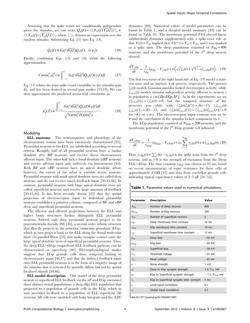
Table 1. Parameter values used in numerical simulations.
Parameter Description Value
NDp Number of deep neurons 800
NEGp Number of EGp neurons 200
NSf Number of superficial neurons 2
tDp Deep membrane time constant 10 ms
tEGp EGp membrane time constant 10 ms
tSf Superficial membrane time constant 15 ms
mDp Deep bias 256 mV
mEGp EGp bias 260 mV
mSf Superficial bias 256 mV
Vth Threshold voltage 255 mV
Vre Reset voltage 265 mV
s Noise strength 1 mV
a1 Deep to EGp synaptic strength 7:6=NDp mV
a2 EGp to Superficial synaptic strength {7:6=NEGp mV
ts EGp to Superficial synaptic time constant 5 ms
cl Local input correlation 0.1
cg Global input correlation 0.2
doi:10.1371/journal.pcbi.1002667.t001
Spatial Inputs Shape Temporal Correlations
PLOS Computational Biology | www.ploscompbiol.org 4 September 2012 | Volume 8 | Issue 9 | e1002667
Finally, since we are only interested in the pairwise correlation
between superficial neurons and because the feedback is open-
loop, it is only necessary to consider a pair of superficial pyramidal
neurons. As such, we set NSf ~2. The ith superficial pyramidal
cell’s membrane dynamics are given by:
dVi,Sf
dt~
1
tSf
(mSf {Vi,Sf )za2
XNEGp
j~1
w � yj,EGp(t)
zsffiffifficp
js(t)zffiffiffiffiffiffiffiffiffiffi1{cp
ji,n,Sf (t)� �
:
ð21Þ
Here w(t)~h(t)e{t=ts where h(t) is the Heaviside function. The
operation � denotes convolution. The inhibitory coupling from
EGp to the ELL was set by a2v0.
During local stimulation, a fraction g~0:05 of deep neurons
received coherent, stimulus-locked electroreceptor input (cl~0:1),
while all other deep neurons received uncorrelated input modeling
spontaneous afferent activity. During global stimulation, all deep
neurons (g~1) received stimulus-locked input (cg~0:2). The
increased value of cg reflects the fact that global stimuli will
spatially saturate the receptive field center and will thus more
effectively drive the afferent population [29,53].
In our model, a pair of neurons in a given layer could receive
correlated input from the previous layer in two ways. First, a
neuron in the previous layer could project to both downstream
neurons and thus correlate their input. Second, neurons in the
previous layer could become locked to the stimulus and their
pooled activity could correlate the downstream neurons, even if
their projections did not overlap anatomically. In the linear model,
we assumed that the first source of common input is negligible
relative to common input from stimulus locked, pooled activity, as
is often the case in feedforward networks [54]. Consequently,
correlations between model neurons were due only to external
signals that synchronously recruited electroreceptors. Therefore,
rij(T)~rsij(T) for the model.
To evaluate r(T) for our model using the linear response
approximation, we computed the superficial neuron stimulus gain
GSf (f ). For numerical simulations, we estimated GSf (f ) using Eq.
(15). However, following past work [28,31], we derived a
theoretical approach to compute GSf (f ). For global stimulation
and assuming that both the input correlations cg and the effective
coupling b1~a1NDp and b2~a2NEGp are sufficiently small, we
compute the feedback filter from the Deep
ELL?EGp?Superficial ELL using the serial computation
H(f )~b1b2W (f )AEGp(f )ADp(f ), ð22Þ
where W (f ) is the Fourier transform of the exponential synaptic
kernel w(t). This result follows simply from the linear convolution
of Deep ELL activity to EGp and then from EGp activity to
superficial ELL through w(t). Here we have introduced A(f ), the
single neuron cellular response function (which we refer to as the
cellular response for brevity) that measures a neuron’s response to an
applied current, independent of network feedback. A(f ) can be
computed using standard techniques from statistical mechanics
(see Text S1).
We note that H(f ) can be calculated for mixed excitatory and
inhibitory feedback to superficial neurons. In this case, the value of
b2 should be interpreted as the effective input strength from both
excitatory and inhibitory populations. For example, if the fraction
of excitatory synapses from EGp to superficial neurons is given by
f and the synaptic strength of excitation and inhibition are az2 w0
and a{2 v0, respectively, then we have b2~NEGp
(faz2 z(1{f )a{
2 ). Previous studies have established that the
stimulus-locked EGp feedback is net inhibitory [46], and we
therefore model the pathway as purely inhibitory for simplicity.
With H(f ), we calculate the stimulus gain of a superficial ELL
neuron GSf (f ) as given in Eq. (25). Further, these techniques also
permit a calculation for the power spectrum Qii(f ). With
theoretical expressions for G and Qii, and assuming the signal is
Gaussian white noise with unit variance, we use Eqs. (17) and (18)
to obtain a theoretical prediction for the spike count correlation
between the two superficial ELL neuron spike trains:
rSf (T)~
ð?{?
DGSf (f )D2kT (f )df
ð?{?
Q(f )kT (f )df
: ð23Þ
Here we have used the homogeneity of the spike trains to set
Gi(f )~Gj(f )~G(f ) and Qii(f )~Qjj(f )~Q(f ) for all superficial
neurons.
Results
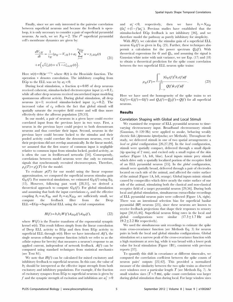
Correlation Shaping with Global and Local StimuliWe examined the response of ELL pyramidal neurons to time-
varying electrosensory input. Broadband electrosensory stimuli
(Gaussian, 0–120 Hz) were applied to awake, behaving weakly
electric fish (Apteronotus leptorhynchus; see Methods). Throughout the
study, we delivered stimuli in one of two spatial arrangements: a
local or global configuration [26,27,29]. In the local configuration,
stimuli were spatially compact, delivered through a small dipole
(tip spacing of 2 mm), and excited only a small region of the skin
surface (Figure 1A, left, blue). Local inputs mimic prey stimuli
which drive only a spatially localized portion of the receptive field
of an ELL pyramidal neuron [55]. In the global configuration,
stimuli were spatially broad, delivered through a pair of electrodes
located on each side of the animal, and affected the entire surface
of the animal (Figure 1A, left, orange). Global inputs mimic stimuli
caused by conspecifics which drive nearly the entire surface of one
side of the animal, stimulating both the classical and non-classical
receptive field of a target pyramidal neuron [29,56]. During both
local and global stimulation, simultaneous extracellular recordings
of ELL pyramidal neuron pairs were collected (Figure 1A, right).
There was an intentional selection bias for superficial basilar
pyramidal (BP) neurons [25], since these neurons are known to
receive feedback projections that shape their responses to sensory
input [30,45,46]. Superficial neuron firing rates in the local and
global configurations were similar (17:3+1:7 Hz and
19:2+2:2 Hz respectively).
We used the simultaneous unit recordings to estimate the spike
train cross-covariance function (see Methods Eq. 3) for neuron
pairs in both the local and global stimulus configurations. Global
stimulation set a narrow peak of the cross-covariance function with
a high maximum at zero lag, while it was broad with a lower peak
value for local stimulation (Figure 1B1), consistent with previous
reports [37].
To quantify this shift in covariance at different timescales, we
computed the correlation coeffcient between the spike counts of
neuron pairs’ outputs [22,42]. This provided a normalized
measure of the similarity between the two spike trains as observed
over windows over a particular length T (see Methods Eq. 7). At
small window sizes (T*5 ms), spike count correlation was larger
during global stimulation than during local. For large window sizes
Spatial Inputs Shape Temporal Correlations
PLOS Computational Biology | www.ploscompbiol.org 5 September 2012 | Volume 8 | Issue 9 | e1002667
(T*50 ms), this relationship was reversed (Figure 1C1). Corre-
lation r(T) is generally a rising function of window size [57], since
for small T few spikes will occur in the same window. However,
even small values of correlation (e.g. v0:1 in magnitude) have
substantial influence on the propagation of neural information
[54,58] and neural coding [59]. To provide a relative measure of
the shift in correlation between the two states, we considered the
ratio of global correlation to local correlation. This was a
decreasing function of window size which was substantially greater
than 1 for small window sizes and lower than 1 for large window
sizes (Figure 1D1).
We performed statistical tests to confirm that the trends
observed were significant. Nonparametric tests confirmed that
the distributions for the local and global conditions were different
(n~10, evaluated at T~95 ms, pv0:05, two-sample Kolmo-
gorov-Smirnov test). The trends with timescale were also
significant (n~10, T~2 ms compared with T~95 ms,
pv2|10{5 for local and pv:002 for global stimulation, two-
sample Kolmogorov-Smirnov tests). The means of the distribu-
tions were also different (n~10, evaluated at T~95 ms, pv:005,
paired t-test). In summary, the spatial extent of the electrosensory
signal shaped the timescales over which spike train pairs were
correlated.
Shifts in Single-Neuron Response Gain PredictCorrelation Shaping
In general, correlated neural activity can be decomposed into
stimulus induced and non-stimulus induced components [21,60].
Stimulus induced correlations reflect the two neurons locking to a
dynamic stimulus, while the non-stimulus induced correlations
reflect the neurons sharing a portion of their trial-variable noise,
presumably from a common pre-synaptic source. To uncover the
cellular and circuit mechanisms underlying correlation shaping,
we first determined whether the changes in correlation observed
were present across trials and therefore related to how neurons
responded to the repeated stimulus. Using spike trains across
different trials of identical stimulus presentations, we computed the
across-trial spike train cross-covariance functions and spike count
correlations (Figure 1B2,C2; see Methods Eqs. 9, 10). The
magnitude of these across-trial correlations was less than that of
the within-trial correlations, indicating the presence of some trial-
variable noise (compare Figure 1C1 and 1C2). Nevertheless, the
differential shaping of correlations at short and long timescales was
still present in the across-trial spike count correlation
(Figure 1C2,D2). This suggests that the way stimulus processing
shifts between local and global conditions is related to the
mechanisms responsible for correlation shaping.
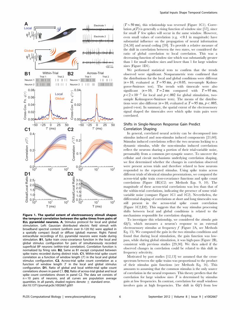
To investigate this relationship, we considered the stimulus gain
G(f ), which measures a neuron’s response to an external
electrosensory stimulus at frequency f (Figure 2A, see Methods
Eq. 15). We computed the gain in the two stimulus conditions and
found that during local stimulation, the gain function was low-
pass, while during global stimulation, it was high-pass (Figure 2B),
consistent with previous studies [29,30]. We then asked if the
observed changes in correlation could be related to this shift in
frequency selectivity.
Motivated by past studies [12,13] we assumed that the cross-
spectrum between the spike trains was proportional to the product
of their stimulus gain functions (see Methods Eq. 16). This
amounts to assuming that the common stimulus is the only source
of correlation in the neural responses. This theory predicts that the
correlation for large window sizes T is determined by stimulus
gain at low frequencies. In contrast, correlation for small windows
involves gain at high frequencies. The shift in G(f ) from low
Figure 1. The spatial extent of electrosensory stimuli shapesthe temporal correlation between the spike times from pairs ofELL pyramidal neurons. A, Stimulus protocol for local and globalstimulation. Left: Gaussian distributed electric field stimuli withbroadband spectral content (uniform over 0–120 Hz) were applied ina spatially compact (local) or diffuse (global) manner. Right: Pairedextracellular recordings of ELL pyramidal neurons were made duringstimulation. B1, Spike train cross-covariance function in the local andglobal stimulus configuration for pairs of simultaneously recordedsuperficial BP neurons (within-trial correlation). Correlation function isnormalized by firing rate. B2, Same as B1 except computed betweenspike trains recorded during distinct trials. C1, Within-trial spike countcorrelation as a function of window length (T ) in the local and globalstimulus configuration. C2, Across-trial spike count correlation as afunction of window length T in the local and global stimulusconfiguration. D1, Ratio of global and local within-trial spike countcorrelations shown in panel C1. D2, Ratio of across-trial global and localspike count correlations shown in panel C2. The data set consists ofn = 10 pairs of neurons, and all curves are population averagequantities. In all panels, shaded regions denote + standard error.doi:10.1371/journal.pcbi.1002667.g001
Spatial Inputs Shape Temporal Correlations
PLOS Computational Biology | www.ploscompbiol.org 6 September 2012 | Volume 8 | Issue 9 | e1002667
frequency transfer for local inputs to high frequency transfer for
global inputs therefore implies global stimulus correlation will be
enhanced for small T and attenuated for large T , with the inverse
true for local stimulation. We verified this hypothesis, obtaining a
prediction of the spike count correlation in the two states that
matched the experimental data (see Methods Eq. 18; Figure 2C,
solid versus dashed curves). Thus, the shift in the frequency-
selectivity of superficial neurons’ stimulus gain between the local
and global conditions indeed predicted the changes in correlation.
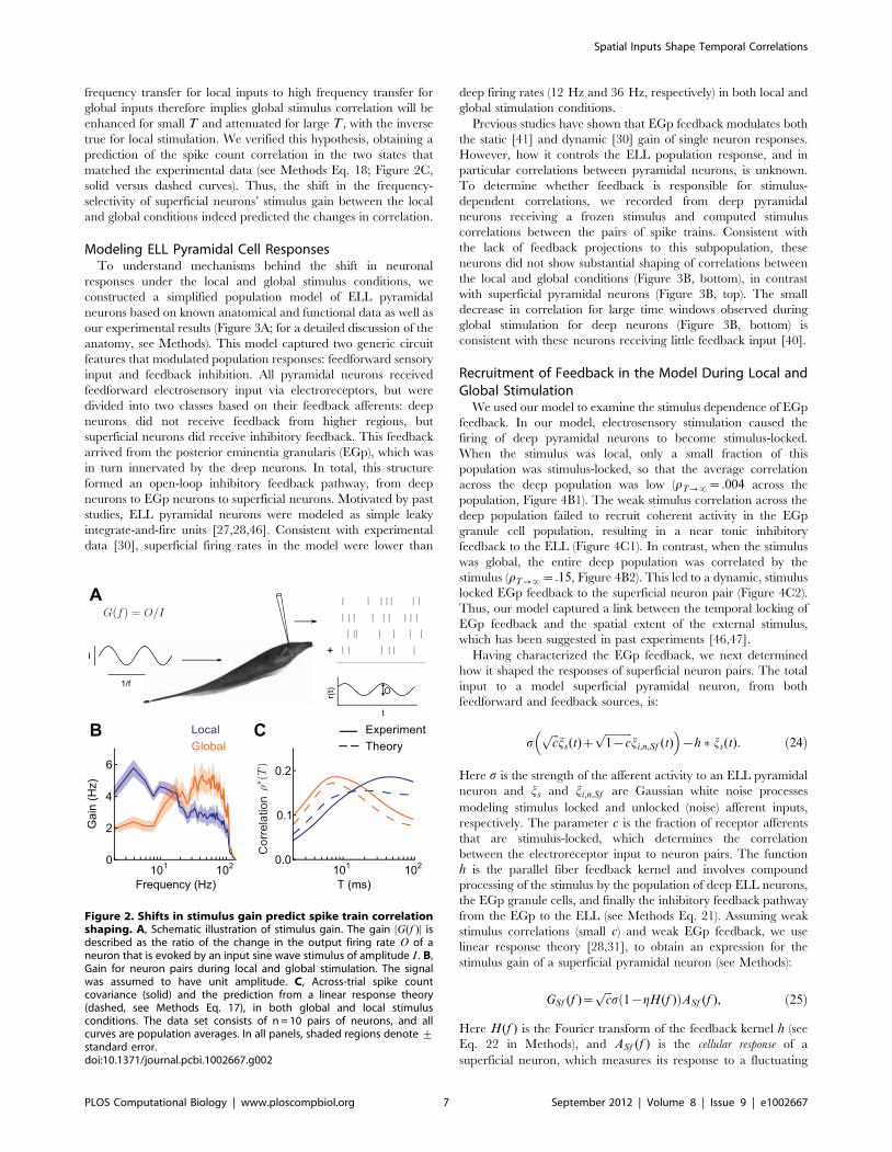
Modeling ELL Pyramidal Cell ResponsesTo understand mechanisms behind the shift in neuronal
responses under the local and global stimulus conditions, we
constructed a simplified population model of ELL pyramidal
neurons based on known anatomical and functional data as well as
our experimental results (Figure 3A; for a detailed discussion of the
anatomy, see Methods). This model captured two generic circuit
features that modulated population responses: feedforward sensory
input and feedback inhibition. All pyramidal neurons received
feedforward electrosensory input via electroreceptors, but were
divided into two classes based on their feedback afferents: deep
neurons did not receive feedback from higher regions, but
superficial neurons did receive inhibitory feedback. This feedback
arrived from the posterior eminentia granularis (EGp), which was
in turn innervated by the deep neurons. In total, this structure
formed an open-loop inhibitory feedback pathway, from deep
neurons to EGp neurons to superficial neurons. Motivated by past
studies, ELL pyramidal neurons were modeled as simple leaky
integrate-and-fire units [27,28,46]. Consistent with experimental
data [30], superficial firing rates in the model were lower than
deep firing rates (12 Hz and 36 Hz, respectively) in both local and
global stimulation conditions.
Previous studies have shown that EGp feedback modulates both
the static [41] and dynamic [30] gain of single neuron responses.
However, how it controls the ELL population response, and in
particular correlations between pyramidal neurons, is unknown.
To determine whether feedback is responsible for stimulus-
dependent correlations, we recorded from deep pyramidal
neurons receiving a frozen stimulus and computed stimulus
correlations between the pairs of spike trains. Consistent with
the lack of feedback projections to this subpopulation, these
neurons did not show substantial shaping of correlations between
the local and global conditions (Figure 3B, bottom), in contrast
with superficial pyramidal neurons (Figure 3B, top). The small
decrease in correlation for large time windows observed during
global stimulation for deep neurons (Figure 3B, bottom) is
consistent with these neurons receiving little feedback input [40].
Recruitment of Feedback in the Model During Local andGlobal Stimulation
We used our model to examine the stimulus dependence of EGp
feedback. In our model, electrosensory stimulation caused the
firing of deep pyramidal neurons to become stimulus-locked.
When the stimulus was local, only a small fraction of this
population was stimulus-locked, so that the average correlation
across the deep population was low (rT??~:004 across the
population, Figure 4B1). The weak stimulus correlation across the
deep population failed to recruit coherent activity in the EGp
granule cell population, resulting in a near tonic inhibitory
feedback to the ELL (Figure 4C1). In contrast, when the stimulus
was global, the entire deep population was correlated by the
stimulus (rT??~:15, Figure 4B2). This led to a dynamic, stimulus
locked EGp feedback to the superficial neuron pair (Figure 4C2).
Thus, our model captured a link between the temporal locking of
EGp feedback and the spatial extent of the external stimulus,
which has been suggested in past experiments [46,47].
Having characterized the EGp feedback, we next determined
how it shaped the responses of superficial neuron pairs. The total
input to a model superficial pyramidal neuron, from both
feedforward and feedback sources, is:
sffiffifficp
js(t)zffiffiffiffiffiffiffiffiffiffi1{cp
ji,n,Sf (t)� �
{h � js(t): ð24Þ
Here s is the strength of the afferent activity to an ELL pyramidal
neuron and js and ji,n,Sf are Gaussian white noise processes
modeling stimulus locked and unlocked (noise) afferent inputs,
respectively. The parameter c is the fraction of receptor afferents
that are stimulus-locked, which determines the correlation
between the electroreceptor input to neuron pairs. The function
h is the parallel fiber feedback kernel and involves compound
processing of the stimulus by the population of deep ELL neurons,
the EGp granule cells, and finally the inhibitory feedback pathway
from the EGp to the ELL (see Methods Eq. 21). Assuming weak
stimulus correlations (small c) and weak EGp feedback, we use
linear response theory [28,31], to obtain an expression for the
stimulus gain of a superficial pyramidal neuron (see Methods):
GSf (f )~ffiffifficp
s 1{gH(f )ð ÞASf (f ), ð25Þ
Here H(f ) is the Fourier transform of the feedback kernel h (see
Eq. 22 in Methods), and ASf (f ) is the cellular response of a
superficial neuron, which measures its response to a fluctuating
Figure 2. Shifts in stimulus gain predict spike train correlationshaping. A, Schematic illustration of stimulus gain. The gain DG(f )D isdescribed as the ratio of the change in the output firing rate O of aneuron that is evoked by an input sine wave stimulus of amplitude I . B,Gain for neuron pairs during local and global stimulation. The signalwas assumed to have unit amplitude. C, Across-trial spike countcovariance (solid) and the prediction from a linear response theory(dashed, see Methods Eq. 17), in both global and local stimulusconditions. The data set consists of n = 10 pairs of neurons, and allcurves are population averages. In all panels, shaded regions denote +standard error.doi:10.1371/journal.pcbi.1002667.g002
Spatial Inputs Shape Temporal Correlations
PLOS Computational Biology | www.ploscompbiol.org 7 September 2012 | Volume 8 | Issue 9 | e1002667
current applied directly to the neuron (see Eq. 8 in Text S1). In
contrast to the stimulus gain, the cellular response does not
depend on network feedback. The parameter g is the spatial
extent of the stimulus (0ƒgƒ1), with g&0 modeling the lack of
stimulus-coherent EGp feedback for local stimulation, and g&1
the full recruitment of EGp feedback for global stimulation
(Figure 4). With this model of how G(f ) shifts between local and
global stimulus configurations, we next build a theory for the
correlation shaping within the superficial ELL pyramidal neuron
population.
Figure 3. Open loop feedback inhibition in electrosensory neural circuitry. A, Detailed schematic of peripheral neural circuitry in theelectrosensory system. Basilar (BP) and non-basilar (nBP) pyramidal neurons in the electrosensory lateral line lobe (ELL) have their somata located inthe Pyramidal cell layer (PCL). Deep pyramidal neurons (green) have small apical dendritic arbors, projecting only to the Ventral Molecular Layer(VML). In contrast, superficial pyramidal neurons (red) have large apical dendritic arbors, projecting to the Dorsal Molecular Layer (DML). Pyramidalneurons receive direct and/or indirect input from feedforward electroreceptor afferent input to the Deep Fiber Layer (DFL). Deep pyramidal neuronsexcite neurons in the praminentialis dorsalis (Pd), which in turn excite granule cells in the posterior eminentia granularis (EGp). The EGp projectsparallel fiber feedback along the DML exclusively targeting ELL superficial pyramidal neurons. In total the deep ELL?EGp?superficial ELL pathway isan open loop feedback structure. Pyramidal neuron graphics were from example neurolucida traced neurons [46]. B, Stimulus correlation for pairs ofexperimentally recorded deep pyramidal neurons (n = 45 pairs; 10 neurons were used) that were driven by the stimulus in local and global (bottom).Little correlation shaping is present. For comparison purposes we show the stimulus correlation for pairs of superficial neurons (top, Figure 1C2). C,Simplified model of the ELL-EGp circuit. Individual neurons in the deep ELL, EGp, and superficial ELL were modeled with leaky integrate-and-fireneuron dynamics (example realizations on right). Electroreceptor input was modeled as white noise, with 5% of deep pyramidal neurons receiving astimulus-locked component in local and 100% in global. We studied the spike responses the pair of superficial pyramidal neurons (labeled 1 and 2)that receive both afferent and EGp feedback inputs.doi:10.1371/journal.pcbi.1002667.g003
Spatial Inputs Shape Temporal Correlations
PLOS Computational Biology | www.ploscompbiol.org 8 September 2012 | Volume 8 | Issue 9 | e1002667

Correlation Shaping in the ELL-EGp Network ModelWe used our ELL-EGp network model to relate the spatial
extent of an electrosensory stimulus and the timescale of the
pairwise correlation between spike trains from superficial BP
neurons. During local stimulation, pairs of nearby superficial
neurons received correlated electroreceptor input (Figure 3C). The
degree of correlation between the afferent input to the superficial
pair was c~cl . The EGp feedback did not exhibit a substantial
stimulus-locked component (g&0) during local stimulation, and
hence did not contribute to common fluctuations (Figure 4C1).
Thus, the stimulus gain in the local condition, denoted Gl(f ),reduced to:
Gl(f )~ffiffiffifficl
psASf (f ): ð26Þ
Our theoretical Gl(f ) (see Methods) quantitatively matched
estimates from simulations of the ELL-EGp network of leaky
integrate-and-fire neurons (Figure 5a, blue curve and blue dots)
and qualitatively matched the low-pass nature of G(f ) obtained
from experiments (Figure 2B, blue). The calculation demonstrates
that the gain to local stimuli of superficial pyramidal neurons is
primarily determined by the cellular response A(f ), suggesting that
feedback network dynamics can be ignored.
The lack of network activity for local stimulation (g&0), was
contrasted with the recruitment of EGp feedback for global
stimulation (g~1). During global stimulation, we also assumed
that the receptive fields of neurons were fully saturated, rather
than being partially driven due to the limited extent of the
stimulus, as suggested by experimental estimates [53]. We
therefore increased the correlation of electroreceptor afferents in
the global state, so that cgwcl . Combining these two model
assumptions, we expressed the gain in the global configuration,
Gg(f ), as:
Gg(f )~ffiffiffiffifficgp
s 1{H(f )ð ÞASf (f ): ð27Þ
If H(f )~1 – that is, if the negative feedback were a perfect replica
of the feedforward signal – the stimulus gain Gg(f ) would be zero,
indicating complete stimulus cancellation by the feedback path-
way. However, since the negative feedback was low-pass due to
neuronal integration and synaptic filtering along the feedback
pathway, only the low frequency components of the gain were
strongly attenuated. Consequently, Gg(f )vGl(f ) for sufficiently
low frequencies (Figure 5A, compare orange and blue curves for
f v15 Hz). However, Gg(f )wGl(f ) for high frequencies
(Figure 5A, compare orange and blue curves for f w15 Hz),
because of the increase in receptive field saturation (cgwcl ). Our
theoretical Gg(f ) matched simulations of the ELL-EGp network
(Figure 5A, orange curve and orange dots). Thus, the combination
of feedback recruitment and feedforward saturation during global
stimulation captured the experimentally determined shift in
stimulus gain known to occur between local and global stimulation
(Figure 2B and see [29,30]).
Next, we examined how this gain shift controlled correlations
across the population of superficial pyramidal neurons. Using the
linear response theory we used to predict signal correlations in the
experimental data (Figure 2, see Methods Eq. 23), we calculated
theoretically the correlations between model pyramidal neurons.
Global stimulation simultaneously increased short T correlation
and decreased long T correlation compared to local stimulation
(Figure 5B). These findings matched the experimental results
(compare Figures 1C and 5B) and are the primary theoretical
result of this study.
Our model provides clear intuition for how the combination of
receptive field saturation and the recruitment of EGp feedback
during global stimulation shapes the correlation of ELL pyramidal
neuron activity (Figure 5C). During local stimulation, EGp
feedback was not recruited and the feedback did not cancel the
feedforward signal from the electroreceptors (Figure 5C, left). This
case is contrasted with global stimulation, in which a broad
stimulus-induced synchronization of all of the deep ELL neurons
recruited a stimulus-locked EGp feedback. This feedback was low-
pass, and therefore canceled the low frequency components of the
signal (Figure 5C, middle), but not the high frequency components
(Figure 5C, right). Thus, correlations due to global stimulation
were canceled only for sufficiently long timescales T (Figure 5B,
Tw15 ms). Furthermore, the saturation of the receptive field
input (cgwcl ) enhanced the correlation r(T) for small T
(Figure 5B, Tv15 ms). In total, feedforward and feedback
circuitry shaped r(T) depending on the spatial profile of the
electrosensory signal.
Our ELL-EGp network model distills correlation shaping into
two hypotheses that link the spatial properties of an electrosensory
stimulus and the timescale of pairwise correlation between the spike
responses of ELL superficial pyramidal neurons:
1. Receptive field saturation for spatially broad signals increases
the short timescale correlation between the spike trains from
superficial pyramidal neurons.
Figure 4. Model EGp feedback is stimulus locked for global,but not local, stimulation. A Low-pass (0–60 Hz) filtered version ofthe electrosensory stimulus. Filtering was done as a visual aid in relatingthe stimulus to the feedback in (C2). B1, Raster plot of the deep neuronpopulation during local stimulation. The signal weakly correlated only asmall fraction of the population. B2, Same as (b1), but during globalstimulation. The spatially broad stimulus correlated the entire deeppopulation. C1, EGp feedback current during local stimulation, showinglittle stimulus locking. C2, EGp feedback was stimulus-modulated bythe global signal, due to recruitment of the deep population by thestimulus. The inhibitory feedback is a negative image of the stimulus(A2).doi:10.1371/journal.pcbi.1002667.g004
Spatial Inputs Shape Temporal Correlations
PLOS Computational Biology | www.ploscompbiol.org 9 September 2012 | Volume 8 | Issue 9 | e1002667
2. Recruitment of EGp feedback by spatially broad signals
decreases the long timescale correlation between the spike
trains from superficial pyramidal neurons.
To study these two components of correlation shaping in
isolation from one another, we used a combination of analysis on a
subclass of ELL pyramidal neurons and pharmacological blockade
of EGp feedback.
Correlation Shaping of nBP Neuron ResponsesWe first tested how short timescale correlation was affected by
receptive field saturation (Hypothesis 1). The ELL has two classes
of pyramidal neuron: non-basilar pyramidal (nBP) and basilar
pyramidal (BP) neurons, distinguished by the extent of their basilar
dendritic arbor (Figure 3A). While BP neurons respond to positive
deflections of the electric field, nBP neurons are oppositely tuned,
due to their afferent inputs arriving solely via an inhibitory
interneuron population [25]. This difference in the feedforward
afferent architecture to nBP neurons compared to BP neurons
produces nBP neuron classical receptive fields that are smaller
than those of BP neurons [26]. Despite the difference in
feedforward afferent input for BP and nBP neurons, both
superficial BP and nBP neurons receive near equivalent feedback
from EGp parallel fibers (Figure 3A). Thus, a comparison between
BP and nBP neurons is sensitive to a difference in feedforward
afferent drive, and not to EGp feedback. We hypothesized that
global inputs would not drive nBP neurons as strongly as BP
neurons because of their smaller classical receptive fields. Hence,
short timescale correlation during global stimulation for nBP
neurons should be less than for BP neurons.
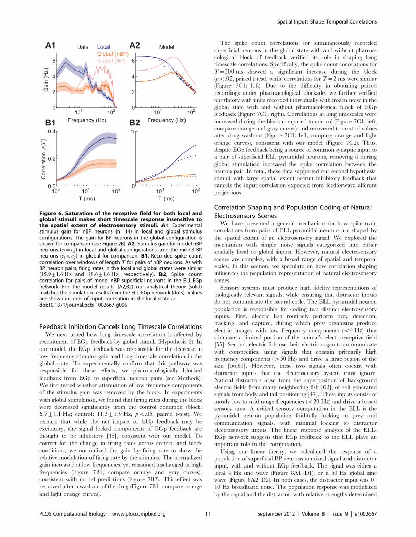
We first calculated the stimulus gain for nBP neurons. The
difference in gain between local and global stimuli for nBP
neurons was different than that for BP neurons (Figure 6A1; [30]).
In particular, while nBP and BP neurons both exhibited a
reduction in low frequency gain during global stimulation, nBP
neurons exhibited little enhancement of high frequency response.
Our model network replicated this difference (Figure 6A2) when
we assumed that the nBP neurons integrate stimuli over smaller
regions of space, such that local inputs saturate the receptive field
(cg~cl ), in contrast to the BP neuron case (cgwcl ). The lack of
high frequency shaping of gain for nBP neurons across local and
global configurations predicts that the small T correlations do not
substantially increase in the global state, while EGp feedback still
attenuates low frequency gain and hence large T correlations.
Measurements of r(T) for nBP neurons in both the ELL-EGp
model (Figure 6B2), as well as nBP neurons recorded in vivo
(Figure 6B1) supported this prediction. Thus, the known
differences between the receptive field sizes of nBP and BP
neurons, provide evidence for the link between the spatial extent of
electrosensory stimuli and short timescale correlation observed for
superficial BP neurons.
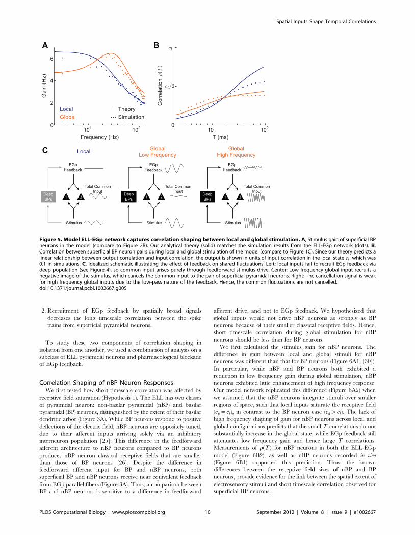
Figure 5. Model ELL-EGp network captures correlation shaping between local and global stimulation. A, Stimulus gain of superficial BPneurons in the model (compare to Figure 2B). Our analytical theory (solid) matches the simulation results from the ELL-EGp network (dots). B,Correlation between superficial BP neuron pairs during local and global stimulation of the model (compare to Figure 1C). Since our theory predicts alinear relationship between output correlation and input correlation, the output is shown in units of input correlation in the local state cl , which was0.1 in simulations. C, Idealized schematic illustrating the effect of feedback on shared fluctuations. Left: local inputs fail to recruit EGp feedback viadeep population (see Figure 4), so common input arises purely through feedforward stimulus drive. Center: Low frequency global input recruits anegative image of the stimulus, which cancels the common input to the pair of superficial pyramidal neurons. Right: The cancellation signal is weakfor high frequency global inputs due to the low-pass nature of the feedback. Hence, the common fluctuations are not cancelled.doi:10.1371/journal.pcbi.1002667.g005
Spatial Inputs Shape Temporal Correlations
PLOS Computational Biology | www.ploscompbiol.org 10 September 2012 | Volume 8 | Issue 9 | e1002667
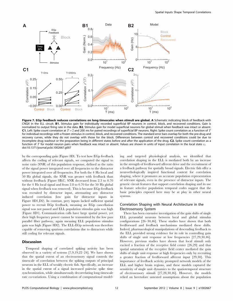
Feedback Inhibition Cancels Long Timescale CorrelationsWe next tested how long timescale correlation is affected by
recruitment of EGp feedback by global stimuli (Hypothesis 2). In
our model, the EGp feedback was responsible for the decrease in
low frequency stimulus gain and long timescale correlation in the
global state. To experimentally confirm that this pathway was
responsible for these effects, we pharmacologically blocked
feedback from EGp to superficial neuron pairs (see Methods).
We first tested whether attenuation of low frequency components
of the stimulus gain was removed by the block. In experiments
with global stimulation, we found that firing rates during the block
were decreased significantly from the control condition (block:
6:7+1:1 Hz; control: 11:3+1:9 Hz, pv:05, paired t-test). We
remark that while the net impact of EGp feedback may be
excitatory, the signal locked components of EGp feedback are
thought to be inhibitory [46], consistent with our model. To
correct for the change in firing rates across control and block
conditions, we normalized the gain by firing rate to show the
relative modulation of firing rate by the stimulus. The normalized
gain increased at low frequencies, yet remained unchanged at high
frequencies (Figure 7B1, compare orange and gray curves),
consistent with model predictions (Figure 7B2). This effect was
removed after a washout of the drug (Figure 7B1, compare orange
and light orange curves).
The spike count correlations for simultaneously recorded
superficial neurons in the global state with and without pharma-
cological block of feedback verified its role in shaping long
timescale correlations. Specifically, the spike count correlations for
T~200 ms showed a significant increase during the block
(pv:02, paired t-test), while correlations for T~2 ms were similar
(Figure 7C1; left). Due to the difficulty in obtaining paired
recordings under pharmacological blockade, we further verified
our theory with units recorded individually with frozen noise in the
global state with and without pharmacological block of EGp
feedback (Figure 7C1; right). Correlations at long timescales were
increased during the block compared to control (Figure 7C1; left,
compare orange and gray curves) and recovered to control values
after drug washout (Figure 7C1; left, compare orange and light
orange curves), consistent with our model (Figure 7C2). Thus,
despite EGp feedback being a source of common synaptic input to
a pair of superficial ELL pyramidal neurons, removing it during
global stimulation increased the spike correlation between the
neuron pair. In total, these data supported our second hypothesis:
stimuli with large spatial extent recruit inhibitory feedback that
cancels the input correlation expected from feedforward afferent
projections.
Correlation Shaping and Population Coding of NaturalElectrosensory Scenes
We have presented a general mechanism for how spike train
correlations from pairs of ELL pyramidal neurons are shaped by
the spatial extent of an electrosensory signal. We explored the
mechanism with simple noise signals categorized into either
spatially local or global inputs. However, natural electrosensory
scenes are complex, with a broad range of spatial and temporal
scales. In this section, we speculate on how correlation shaping
influences the population representation of natural electrosensory
scenes.
Sensory systems must produce high fidelity representations of
biologically relevant signals, while ensuring that distractor inputs
do not contaminate the neural code. The ELL pyramidal neuron
population is responsible for coding two distinct electrosensory
inputs. First, electric fish routinely perform prey detection,
tracking, and capture, during which prey organisms produce
electric images with low frequency components (v4 Hz) that
stimulate a limited portion of the animal’s electroreceptive field
[55]. Second, electric fish use their electric organ to communicate
with conspecifics, using signals that contain primarily high
frequency components (w50 Hz) and drive a large region of the
skin [56,61]. However, these two signals often coexist with
distractor inputs that the electrosensory system must ignore.
Natural distractors arise from the superposition of background
electric fields from many neighboring fish [62], or self generated
signals from body and tail positioning [47]. These inputs consist of
mostly low to mid range frequencies (v20 Hz) and drive a broad
sensory area. A critical sensory computation in the ELL is the
pyramidal neuron population faithfully locking to prey and
communication signals, with minimal locking to distractor
electrosensory inputs. The linear response analysis of the ELL-
EGp network suggests that EGp feedback to the ELL plays an
important role in this computation.
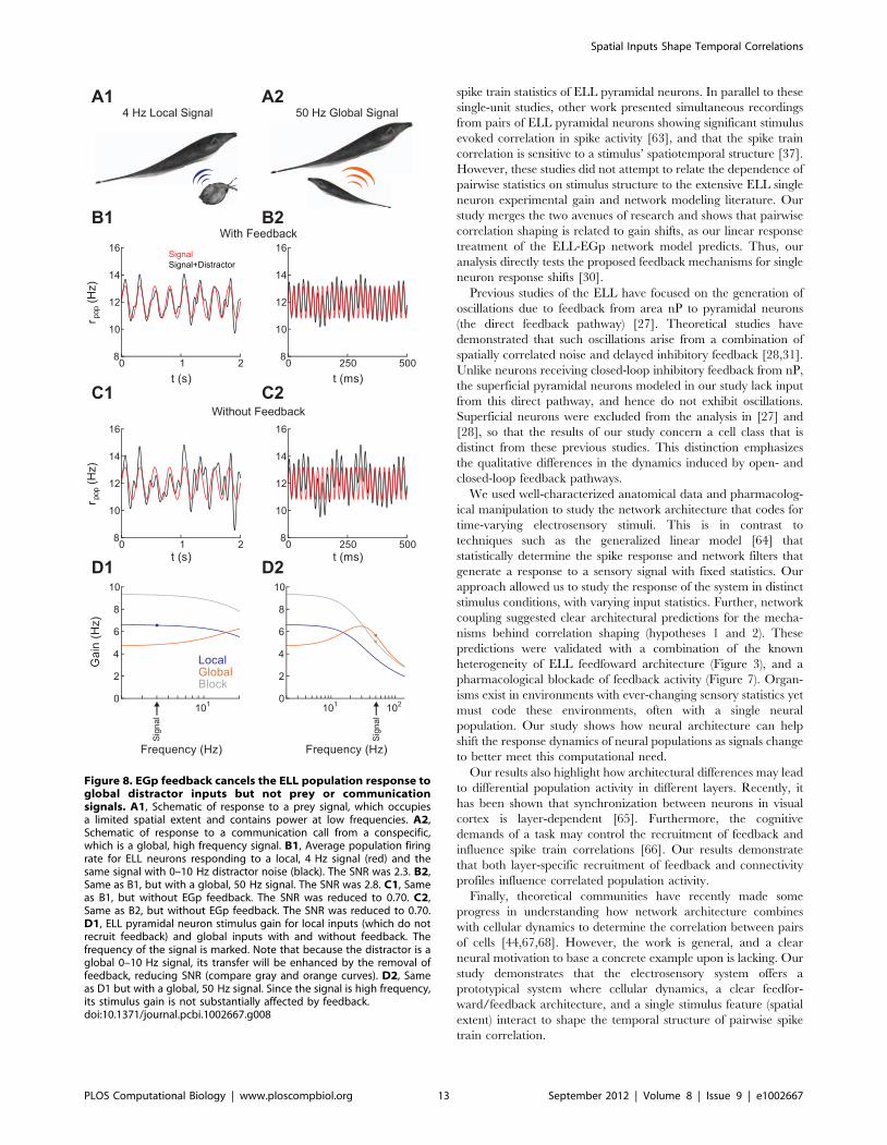
Using our linear theory, we calculated the response of a
population of superficial BP neurons to mixed signal and distractor
input, with and without EGp feedback. The signal was either a
local 4 Hz sine wave (Figure 8A1–D1), or a 50 Hz global sine
wave (Figure 8A2–D2). In both cases, the distractor input was 0–
10 Hz broadband noise. The population response was modulated
by the signal and the distractor, with relative strengths determined
Figure 6. Saturation of the receptive field for both local andglobal stimuli makes short timescale response insensitive tothe spatial extent of electrosensory stimuli. A1, Experimentalstimulus gain for nBP neurons (n = 14) in local and global stimulusconfigurations. The gain for BP neurons in the global configuration isshown for comparison (see Figure 2B). A2, Stimulus gain for model nBPneurons (cl~cg) in local and global configurations, and the model BPneurons (clvcg) in global for comparison. B1, Recorded spike countcorrelation over windows of length T for pairs of nBP neurons. As withBP neuron pairs, firing rates in the local and global states were similar(15:9+1:6 Hz and 18:6+1:6 Hz, respectively). B2, Spike countcorrelation for pairs of model nBP superficial neurons in the ELL-EGpnetwork. For the model results (A2,B2) our analytical theory (solid)matches the simulation results from the ELL-EGp network (dots). Valuesare shown in units of input correlation in the local state cl .doi:10.1371/journal.pcbi.1002667.g006
Spatial Inputs Shape Temporal Correlations
PLOS Computational Biology | www.ploscompbiol.org 11 September 2012 | Volume 8 | Issue 9 | e1002667
by the corresponding gain (Figure 8D). To test how EGp feedback
affects the coding of relevant signals, we computed the signal to
noise ratio (SNR) of this population response, defined as the ratio
of the signal power integrated over all frequencies to the distractor
power integrated over all frequencies. For both the 4 Hz local and
50 Hz global signals, the SNR was greater with feedback than
without feedback (Figure 8B,C. SNR decreased from 2.3 to 0.70
for the 4 Hz local signal and from 2.8 to 0.70 for the 50 Hz global
signal when feedback was removed). This is because EGp feedback
was recruited by distractor input, attenuating any distractor
induced correlation (low gain for distractor inputs in
Figure 8D1,D2). In contrast, prey inputs lacked sufficient spatial
power to recruit EGp feedback, meaning an EGp cancellation
signal was not passed and ELL population stimulus gain was high
(Figure 8D1). Communication calls have large spatial power, yet
their high frequency power cannot be transmitted by the low pass
parallel fiber pathway, again meaning ELL population stimulus
gain was high (Figure 8D2). The ELL-EGp network was therefore
capable of removing spurious correlations due to distractors while
still coding for relevant signals.
Discussion
Temporal shaping of correlated spiking activity has been
observed in a variety of systems [7,9,19,22–24]. We have shown
that the spatial extent of an electrosensory signal controls the
timescale of correlation between the spiking outputs of principal
neurons in the ELL of weakly electric fish. Specifically, an increase
in the spatial extent of a signal increased pairwise spike time
synchronization, while simultaneously decorrelating long timescale
rate co-variations. Using a combination of computational model-
ing and targeted physiological analysis, we identified that
correlation shaping in the ELL is mediated both by an increase
in the strength of feedforward afferent drive and the recruitment of
a feedback pathway for spatially broad signals. Electric fish offer a
neuroethologically inspired functional context for correlation
shaping, where it promotes an accurate population representation
of relevant signals, even in the presence of distractor inputs. The
generic circuit features that support correlation shaping and its use
in feature selective population temporal codes suggest that the
basic principles exposed here may be at play in other neural
systems.
Correlation Shaping with Neural Architecture in theElectrosensory System
There has been extensive investigation of the gain shifts of single
ELL pyramidal neurons between local and global stimulus
configurations [26–30,46]. These studies have shown that both
feedforward and feedback mechanisms mediated these shifts.
Indeed, pharmacological manipulations of descending feedback to
the ELL provided strong evidence for its role in controlling gain
shifts of single unit response at low frequencies [27,29,30,46].
However, previous studies have shown that local stimuli only
excited a fraction of the receptive field center [26,29] and that
spatial saturation of the receptive field center mediated the gain
shifts of single unit response at high frequencies only by recruiting
a greater fraction of feedforward afferent input [29,30]. This
importance of feedback activity prompted network models of the
ELL and higher brain regions, and these models captured the
sensitivity of single unit dynamics to the spatiotemporal structure
of electrosensory stimuli [27,28,30,46]. However, the models
relied on heretofore untested assumptions about the population
Figure 7. EGp feedback reduces correlations on long timescales when stimuli are global. A Schematic indicating block of feedback withCNQX in the ELL circuit. B1, Stimulus gain for individually recorded superficial BP neurons in control, block, and recovered conditions. Gain isnormalized to output firing rate in the data. B2, Stimulus gain for model superficial neurons for global stimuli when feedback was intact or absent.C1, Left: Spike count correlation at T~2 and 200 ms for paired recordings of superficial BP neurons. Right: Spike count correlation as a function of Tfor individual recordings with a frozen stimulus in control, block, and recovered conditions. The standard error bars overlap for both the pre-drug andrecovery curves, while they do not overlap with those for the block. Differences between control and recovered conditions could be due toincomplete drug washout or the preparation being in different states before and after the application of the drug. C2, Spike count correlation as afunction of T for model neuron pairs when feedback was intact or absent. Values are shown in units of input correlation in the local state cl .doi:10.1371/journal.pcbi.1002667.g007
Spatial Inputs Shape Temporal Correlations
PLOS Computational Biology | www.ploscompbiol.org 12 September 2012 | Volume 8 | Issue 9 | e1002667
spike train statistics of ELL pyramidal neurons. In parallel to these
single-unit studies, other work presented simultaneous recordings
from pairs of ELL pyramidal neurons showing significant stimulus
evoked correlation in spike activity [63], and that the spike train
correlation is sensitive to a stimulus’ spatiotemporal structure [37].
However, these studies did not attempt to relate the dependence of
pairwise statistics on stimulus structure to the extensive ELL single
neuron experimental gain and network modeling literature. Our
study merges the two avenues of research and shows that pairwise
correlation shaping is related to gain shifts, as our linear response
treatment of the ELL-EGp network model predicts. Thus, our
analysis directly tests the proposed feedback mechanisms for single
neuron response shifts [30].
Previous studies of the ELL have focused on the generation of
oscillations due to feedback from area nP to pyramidal neurons
(the direct feedback pathway) [27]. Theoretical studies have
demonstrated that such oscillations arise from a combination of
spatially correlated noise and delayed inhibitory feedback [28,31].
Unlike neurons receiving closed-loop inhibitory feedback from nP,
the superficial pyramidal neurons modeled in our study lack input
from this direct pathway, and hence do not exhibit oscillations.
Superficial neurons were excluded from the analysis in [27] and
[28], so that the results of our study concern a cell class that is
distinct from these previous studies. This distinction emphasizes
the qualitative differences in the dynamics induced by open- and
closed-loop feedback pathways.
We used well-characterized anatomical data and pharmacolog-
ical manipulation to study the network architecture that codes for
time-varying electrosensory stimuli. This is in contrast to
techniques such as the generalized linear model [64] that
statistically determine the spike response and network filters that
generate a response to a sensory signal with fixed statistics. Our
approach allowed us to study the response of the system in distinct
stimulus conditions, with varying input statistics. Further, network
coupling suggested clear architectural predictions for the mecha-
nisms behind correlation shaping (hypotheses 1 and 2). These
predictions were validated with a combination of the known
heterogeneity of ELL feedfoward architecture (Figure 3), and a
pharmacological blockade of feedback activity (Figure 7). Organ-
isms exist in environments with ever-changing sensory statistics yet
must code these environments, often with a single neural
population. Our study shows how neural architecture can help
shift the response dynamics of neural populations as signals change
to better meet this computational need.
Our results also highlight how architectural differences may lead
to differential population activity in different layers. Recently, it
has been shown that synchronization between neurons in visual
cortex is layer-dependent [65]. Furthermore, the cognitive
demands of a task may control the recruitment of feedback and
influence spike train correlations [66]. Our results demonstrate
that both layer-specific recruitment of feedback and connectivity
profiles influence correlated population activity.
Finally, theoretical communities have recently made some
progress in understanding how network architecture combines
with cellular dynamics to determine the correlation between pairs
of cells [44,67,68]. However, the work is general, and a clear
neural motivation to base a concrete example upon is lacking. Our
study demonstrates that the electrosensory system offers a
prototypical system where cellular dynamics, a clear feedfor-
ward/feedback architecture, and a single stimulus feature (spatial
extent) interact to shape the temporal structure of pairwise spike
train correlation.
Figure 8. EGp feedback cancels the ELL population response toglobal distractor inputs but not prey or communicationsignals. A1, Schematic of response to a prey signal, which occupiesa limited spatial extent and contains power at low frequencies. A2,Schematic of response to a communication call from a conspecific,which is a global, high frequency signal. B1, Average population firingrate for ELL neurons responding to a local, 4 Hz signal (red) and thesame signal with 0–10 Hz distractor noise (black). The SNR was 2.3. B2,Same as B1, but with a global, 50 Hz signal. The SNR was 2.8. C1, Sameas B1, but without EGp feedback. The SNR was reduced to 0.70. C2,Same as B2, but without EGp feedback. The SNR was reduced to 0.70.D1, ELL pyramidal neuron stimulus gain for local inputs (which do notrecruit feedback) and global inputs with and without feedback. Thefrequency of the signal is marked. Note that because the distractor is aglobal 0–10 Hz signal, its transfer will be enhanced by the removal offeedback, reducing SNR (compare gray and orange curves). D2, Sameas D1 but with a global, 50 Hz signal. Since the signal is high frequency,its stimulus gain is not substantially affected by feedback.doi:10.1371/journal.pcbi.1002667.g008
Spatial Inputs Shape Temporal Correlations
PLOS Computational Biology | www.ploscompbiol.org 13 September 2012 | Volume 8 | Issue 9 | e1002667
Decorrelating with InhibitionThe role of inhibition in neural circuits is a complex topic of
study. Inhibition is linked to rhythmic, temporal locking between
pairs of pyramidal neurons [32]. On fast timescales, inhibition is
often thought to synchronize the activity of pairs of pyramidal
neurons in both recurrent [10,27,33–35] and feedforward
architectures [11,32]. However, on longer timescales, inhibition
mediates competitive dynamics between populations of pyramidal
neurons, and as such may be a source of anti-correlated activity
[24]. Recently, studies of densely coupled cortical networks with
balanced excitation and inhibition [17,18] and feedforward
inhibitory cortical circuits [20,69] have provided new insights into
the role of inhibitory dynamics. In these studies, fluctuations in
correlated excitation to a pair of pyramidal neurons are cancelled
by correlated inhibitory dynamics, yielding a roughly asynchro-
nous cortical state. This cancellation of correlation is similar to the
one explored in our study responsible for the reduction of
correlation for global stimuli. However, our study was motivated
by a primarily feedforward sensory architecture in which an
external signal can drive correlated activity.
The strengths of the electrosensory preparation allowed us to
extend the correlation cancellation mechanism along two impor-
tant directions. First, the ease in controlling the spatiotemporal
properties of external stimuli allowed an analysis of the limitations
of correlation cancellation. The diffuse ELL?EGp feedforward
path restricts correlation cancellation to signals with broad spatial
scale, while the slow filtering by the parallel fiber pathway can only
cancel correlations of low frequency stimuli. Second, the well
segregated parallel fibers that mediate EGp feedback to the ELL
permitted a pharmacological blockade of inhibition, directly
providing evidence for correlation cancellation. The parallel fibers
are a source of common input to pyramidal neurons, and a naive
analysis would predict that their removal would thus decrease
pyramidal neuron spike train correlation. Nevertheless, the
blockade of parallel fiber inputs increased the spike train correlation,
suggesting that the common inhibition interacts with the common
feedforward afferent input in a destructive, rather than coopera-
tive, manner.
Studies of neural codes often investigate the distinction between
signal evoked, across-trial correlations and additional ‘noise’
induced, within-trial correlations [60]. Across-trial correlations
are attributable to a dynamic locking of the spike train pairs to the
common signal. Within-trial correlations measure the trial-to-trial
co-variability of a pair of spike trains and may be increased relative
to across-trial correlations due to common synaptic input to the
neuron pair. These common fluctuations are often deleterious to
cortical population codes [60], acting as a source of variability that
cannot be removed through population averaging. The majority of
our study presented simultaneously recorded spike train data
which contains across-trial correlation as well as additional within-
trial correlation. However, the shaping of correlation by the spatial
profile of a stimulus was explained from knowledge of only of the
across-trial correlation (Figures 1 and 2), and thus our ELL-EGp
network model ignored other sources of correlation entirely. Our
analysis did study the effects of irrelevant distractor inputs which
can act as a source of noise, though originating from external
signals rather than internal circuit mechanisms. We found that low
frequency distractors that drive a substantial portion of the
network recruit a cancellation signal. We therefore predict that
within-trial correlations may be cancelled by a similar mechanism
if they drive a large number of neurons synchronously. This may
be the case when within-trial correlations are driven by the local
field potential, which is often low frequency and widespread to
populations of neurons [70,71].
In summary, we have identified the combination of feedforward
and feedback architecture that allows the spatial extent of a
stimulus to shape the temporal correlations between the spike
trains of pairs of electrosensory principal cells. Furthermore,
correlation shaping allows populations of neurons to respond to
stimuli that match a specific spatiotemporal profile and ignore
distractor inputs. The generic architectural features of our network
and the fact that sensory systems must filter irrelevant signals
suggest that our findings may generalize to other systems.
Supporting Information
Table S1 Model summary.
(PDF)
Text S1 Computation of cellular response function and power
spectrum.
(PDF)
Acknowledgments
We thank Kresimir Josic for comments on the manuscript.
Author Contributions
Conceived and designed the experiments: ALK MJC BD. Performed the
experiments: ALK MJC. Analyzed the data: ALK MJC BD. Wrote the
paper: ALK MJC BD.
References
1. Harris KD (2005) Neural signatures of cell assembly organization. Nat Rev
Neurosci 6: 399–407.
2. Vogels T, Rajan K, Abbott L (2005) Neural network dynamics. Annu Rev
Neurosci 28: 357–376.
3. deCharms RC, Merzenich MM (1996) Primary cortical representation of sounds
by the coordination of action-potential timing. Nature 381: 610–613.
4. Usrey W, Reid R (1999) Synchronous activity in the visual system. Annu Rev
Physiol 61: 435–456.
5. Romo R, Hernandez A, Zainos A, Salinas E (2003) Correlated neuronal
discharges that increase coding efficiency during perceptual discrimination.
Neuron 38: 649–657.
6. Vaadia E, Haalman I, Abeles M, Bergman H, Prut Y, et al. (1995) Dynamics of
neuronal interactions in monkey cortex in relation to behavioural events. Nature
373: 515–518.
7. Cohen MR, Maunsell JHR (2009) Attention improves performance primarily by
reducing interneuronal correlations. Nat Neurosci 12: 1594–1600.
8. Mitchell JF, Sundberg KA, Reynolds JH (2009) Spatial attention decorrelates
intrinsic activity uctuations in macaque area V4. Neuron 63: 879–888.
9. Womelsdorf T, Fries P, Mitra P, Desimone R (2006) Gamma-band
synchronization in visual cortex predicts speed of change detection. Nature
439: 733–736.
10. Tiesinga P, Fellous J, Salinas E, Jose J, Sejnowski T (2004) Inhibitory synchrony
as a mechanism for attentional gain modulation. J Physiol (Paris) 98: 296–
314.
11. Galan RF, Fourcaud-Trocme N, Ermentrout GB, Urban NN (2006)
Correlation-Induced synchronization of oscillations in olfactory bulb neurons.
J Neurosci 26: 3646–3655.
12. de la Rocha J, Doiron B, Shea-Brown E, Josic K, Reyes A (2007) Correlation
between neural spike trains increases with firing rate. Nature 448: 802–806.
13. Shea-Brown E, Josic K, de la Rocha J, Doiron B (2008) Correlation and
synchrony transfer in integrate-and-fire neurons: Basic properties and conse-
quences for coding. Phys Rev Lett 100: 108102.
14. Ostojic S, Brunel N, Hakim V (2009) How connectivity, background activity,
and synaptic properties shape the cross-correlation between spike trains.
J Neurosci 29: 10234–10253.
15. Rosenbaum R, Josic K (2011) Mechanisms that modulate the transfer of spiking
correlations. Neural Comput 23: 1–45.
16. Kumar A, Rotter S, Aertsen A (2010) Spiking activity propagation in neuronal
networks: reconciling different perspectives on neural coding. Nat Rev Neurosci
11: 615–627.
17. Renart A, de la Rocha J, Bartho P, Hollender L, Parga N, et al. (2010) The
asynchronous state in cortical circuits. Science 327: 587–590.
Spatial Inputs Shape Temporal Correlations
PLOS Computational Biology | www.ploscompbiol.org 14 September 2012 | Volume 8 | Issue 9 | e1002667
18. Hertz J (2010) Cross-correlations in high-conductance states of a model cortical
network. Neural Comput 22: 427–447.
19. Litwin-Kumar A, Oswald AM, Urban N, Doiron B (2011) Balanced synaptic
input shapes the correlation between neural spike trains. PLoS Comput Biol 7:
e1002305.
20. Middleton J, Omar C, Doiron B, Simons D (2012) Correlated activity is stimulus
modulated in a feedforward inhibitory circuit. J Neurosci 32: 506–518.
21. Cohen M, Kohn A (2011) Measuring and interpreting neuronal correlations.
Nat Neurosci 14: 811–819.
22. Kohn A, Smith MA (2005) Stimulus dependence of neuronal correlation in
primary visual cortex of the macaque. J Neurosci 25: 3661–3673.
23. Yu J, Ferster D (2010) Membrane potential synchrony in primary visual cortex
during sensory stimulation. Neuron 68: 1187–1201.
24. Giridhar S, Doiron B, Urban NN (2011) Timescale-dependent shaping of
correlation by olfactory bulb lateral inhibition. Proc Natl Acad Sci U S A 108:
5843–5848.
25. Berman N, Maler L (1999) Neural architecture of the electrosensory lateral line
lobe: adaptations for coincidence detection, a sensory searchlight and frequency-
dependent adaptive filtering. J Exp Biol 202: 1243–1253.
26. Bastian J, Chacron MJ, Maler L (2002) Receptive field organization determines
pyramidal cell stimulus-encoding capability and spatial stimulus selectivity.
J Neurosci 22: 4577–4590.
27. Doiron B, Chacron MJ, Maler L, Longtin A, Bastian J (2003) Inhibitory
feedback required for network oscillatory responses to communication but not
prey stimuli. Nature 421: 539–543.
28. Doiron B, Lindner B, Longtin A, Maler L, Bastian J (2004) Oscillatory activity in
electrosensory neurons increases with the spatial correlation of the stochastic
input stimulus. Phys Rev Lett 93: 048101.
29. Chacron MJ, Doiron B, Maler L, Longtin A, Bastian J (2003) Non-classical
receptive field mediates switch in a sensory neuron’s frequency tuning. Nature
423: 77–81.
30. Chacron MJ, Maler L, Bastian J (2005) Feedback and feedforward control of
frequency tuning to naturalistic stimuli. J Neurosci 25: 5521–5532.
31. Lindner B, Doiron B, Longtin A (2005) Theory of oscillatory firing induced by
spatially correlated noise and delayed inhibitory feedback. Phys Rev E 72:
061919.
32. Buzsaki G (2006) Rhythms of the Brain. New York: Oxford University Press.
464 pp.
33. Borgers C, Kopell N (2003) Synchronization in networks of excitatory and
inhibitory neurons with sparse, random connectivity. Neural Comput 15: 509–
538.
34. Brunel N (2000) Dynamics of sparsely connected networks of excitatory and
inhibitory spiking neurons. J Comput Neurosci 8: 183–208.
35. Brunel N, Hansel D (2006) How noise affects the synchronization properties of
recurrent networks of inhibitory neurons. Neural Comput 18: 1066–1110.
36. Hitschfeld E, Stamper S, Vonderschen K, Fortune E, Chacron MJ (2009) Effects
of restraint and immobilization on electrosensory behaviors of weakly electric
fish. ILAR J 50: 361–372.
37. Chacron MJ, Bastian J (2008) Population coding by electrosensory neurons.
J Neurophysiol 99: 1825–1835.
38. Maler L, Sas E, Johnston S, Ellis W (1991) An atlas of the brain of the electric
fish Apteronotus leptorhynchus. J Chem Neuroanat 4: 1–38.
39. Krahe R, Bastian J, Chacron MJ (2001) Temporal processing across multiple
topographic maps in the electrosensory system. J Neurophysiol 100: 852–867.
40. Bastian J, Nguyenkim J (2001) Dendritic modulation of burst-like firing in
sensory neurons. J Neurophysiol 85: 10–22.
41. Bastian J (1986) Gain control in the electrosensory system mediated by
descending inputs to the electrosensory lateral line lobe. J Neurosci 6: 553–562.
42. Cox DR, Isham V (1980) Point processes. London: Chapman and Hall. 206 pp.
43. Vilela R, Lindner B (2009) A comparative study of different integrate & fire
neurons: spontaneous activity, dynamical response, and stimulus-induced
correlation. Phys Rev E 80: 031909.
44. Trousdale J, Hu Y, Shea-Brown E, Josic K (2012) Impact of network structure
and cellular response on spike time correlations. PLoS Comput Biol 8:
e1002408.
45. Maler L (2009) Receptive field organization across multiple electrosensory maps.
I. Columnar organization and estimation of receptive field size. J Comp Neurol516: 376–393.
46. Bastian J, Chacron MJ, Maler L (2004) Plastic and nonplastic pyramidal cells
perform unique roles in a network capable of adaptive redundancy reduction.Neuron 41: 767–779.
47. Sawtell N (2010) Multimodal integration in granule cells as a basis for associativeplasticity and sensory prediction in a cerebellum-like circuit. Neuron 66: 573–
584.
48. Dayan P, Abbott LF (2001) Theoretical Neuroscience: Computational andMathematical Modeling of Neural Systems. Cambridge: MIT Press. 576 pp.
49. Nordlie E, Gewaltig MO, Plesser HE (2009) Towards reproducible descriptionsof neuronal network models. PLoS Comput Biol 5: e1000456.
50. Chadderton P, Margrie T, Hausser M (2004) Integration of quanta in cerebellargranule cells during sensory processing. Nature 428: 856–860.
51. Farrant M, Brickley S (2003) Properties of GABAA receptor-mediated
transmission at newly formed golgi-granule cell synapses in the cerebellum.Neuropharmacology 44: 181–189.
52. Brickley S, Revilla V, Cull-Candy S, Wisden W, Farrant M (2001) Adaptiveregulation of neuronal excitability by a voltage- independent potassium
conductance. Nature 409: 88–92.
53. Chacron MJ (2006) Nonlinear information processing in a model sensory system.J Neurophysiol 95: 2933–2946.
54. Rosenbaum R, Trousdale J, Josic K (2010) Pooling and correlated neuralactivity. Front Comput Neurosci 4: 9.
55. Nelson M, Maciver M (1999) Prey capture in the weakly electric fishApteronotus albifrons: sensory acquisition strategies and electrosensory conse-
quences. J Exp Biol 202: 1195–1203.
56. Metzner W (1999) Neural circuitry for communication and jamming avoidancein gymnotiform electric fish. J Exp Biol 202: 1365–1375.
57. Kass RE, Ventura V (2006) Spike count correlation increases with length of timeinterval in the presence of trial-to-trial variation. Neural Comput 18: 2583–
2591.
58. Salinas E, Sejnowski TJ (2001) Correlated neuronal activity and the ow of neuralinformation. Nat Rev Neurosci 2: 539–550.
59. Zohary E, Shadlen MN, Newsome WT (1994) Correlated neuronal dischargerate and its implications for psychophysical performance. Nature 370: 140–143.
60. Averbeck BB, Latham PE, Pouget A (2006) Neural correlations, populationcoding and computation. Nat Rev Neurosci 7: 358–366.
61. Hupe G, Lewis J (2008) Electrocommunication signals in free swimming brown
ghost knifefish, Apteronotus leptorhynchus. J Exp Biol 211: 1657–1667.62. Tan E, Nizar J, Carrera-G E, Fortune E (2005) Electrosensory interference in
naturally occurring aggregates of a species of weakly electric fish, Eigenmanniavirescens. Behav Brain Res 164: 83–92.
63. Krahe R, Kreiman G, Gabbiani F, Koch C, Metzner W (2002) Stimulus
encoding and feature extraction by multiple sensory neurons. J Neurosci 22:2374–2382.
64. Pillow J, Shlens J, Paninski L, Sher A, Litke A, et al. (2008) Spatio-temporalcorrelations and visual signalling in a complete neuronal population. Nature
454: 995–999.65. Hansen BJ, Dragoi V (2011) Adaptation-induced synchronization in laminar
cortical circuits. Proc Natl Acad Sci U S A 108: 10720–10725.
66. Takeuchi D, Hirabayashi T, Tamura K, Miyashita Y (2011) Reversal ofinterlaminar signal between sensory and memory processing in monkey
temporal cortex. Science 331: 1443–1447.67. Pernice V, Staude B, Cardanobile S, Rotter S (2011) How structure determines
correlations in neuronal networks. PLoS Comput Biol 7: e1002059.
68. Zhao L, Beverlin BI, Netoff T, Nykamp D (2011) Synchronization from secondorder network connectivity statistics. Front Comput Neurosci 5: 28.
69. Ly C, Middleton J, Doiron B (2012) Cellular and circuit mechanisms maintainlow spike covariability and enhance population coding in somatosensory cortex.
Front Comput Neurosci 6: 7.
70. Nauhaus I, Busse L, Carandini M, Ringach D (2008) Stimulus contrastmodulates functional connectivity in visual cortex. Nat Neurosci 12: 70–76.
71. Kelly R, Smith M, Kass R, Lee T (2010) Local field potentials indicate networkstate and account for neuronal response variability. J Comput Neurosci 29: 567–
579.
Spatial Inputs Shape Temporal Correlations
PLOS Computational Biology | www.ploscompbiol.org 15 September 2012 | Volume 8 | Issue 9 | e1002667