The Sonographic Evaluation of Diffuse Thyroid Disease and ... · •Medical conditions: pregnancy,...
56
The Sonographic Evaluation of Diffuse Thyroid Disease and Thyroiditis Jill E Langer, MD Associate Professor of Radiology And Endocrinology Co-Director of the Thyroid Nodule Clinic Hospital of the University of Pennsylvania
Transcript of The Sonographic Evaluation of Diffuse Thyroid Disease and ... · •Medical conditions: pregnancy,...
The Sonographic Evaluation of Diffuse Thyroid Disease and
Thyroiditis
Jill E Langer, MD
Associate Professor of Radiology
And Endocrinology
Co-Director of the Thyroid Nodule Clinic
Hospital of the University of Pennsylvania
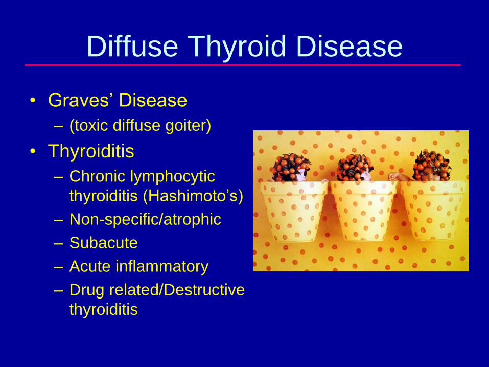
Diffuse Thyroid Disease
• Graves’ Disease
– (toxic diffuse goiter)
• Thyroiditis
– Chronic lymphocytic
thyroiditis (Hashimoto’s)
– Non-specific/atrophic
– Subacute
– Acute inflammatory
– Drug related/Destructive
thyroiditis
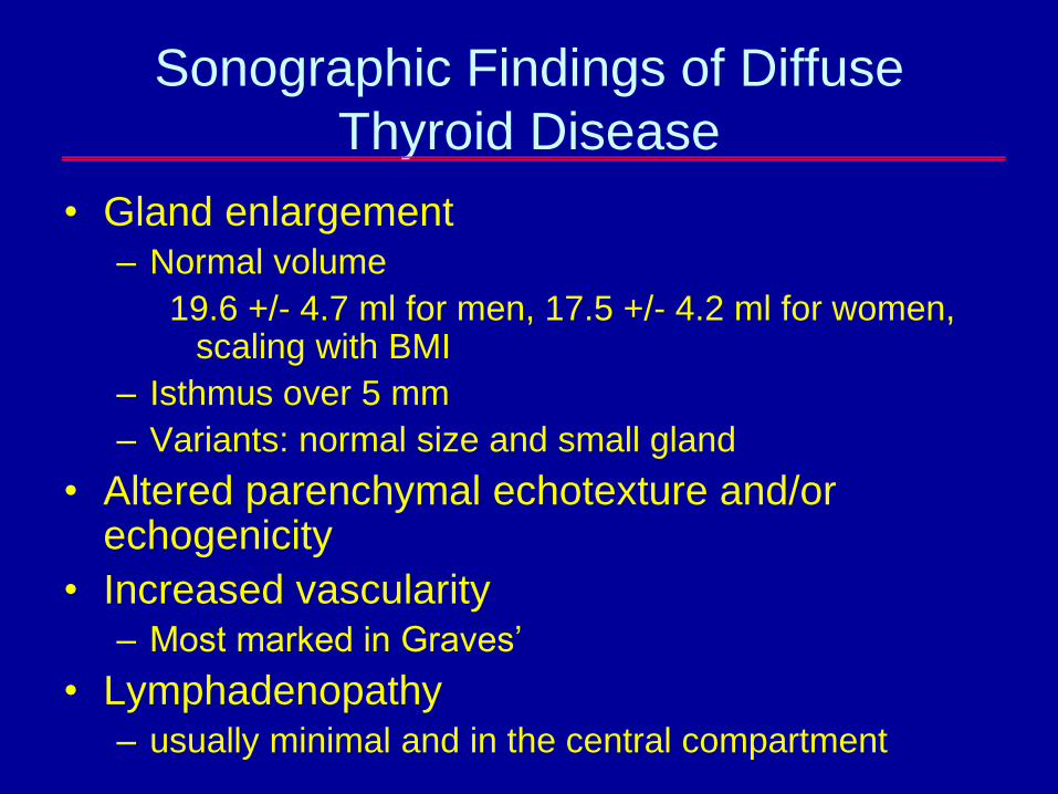
Sonographic Findings of Diffuse
Thyroid Disease
• Gland enlargement– Normal volume
19.6 +/- 4.7 ml for men, 17.5 +/- 4.2 ml for women, scaling with BMI
– Isthmus over 5 mm
– Variants: normal size and small gland
• Altered parenchymal echotexture and/or echogenicity
• Increased vascularity– Most marked in Graves’
• Lymphadenopathy– usually minimal and in the central compartment
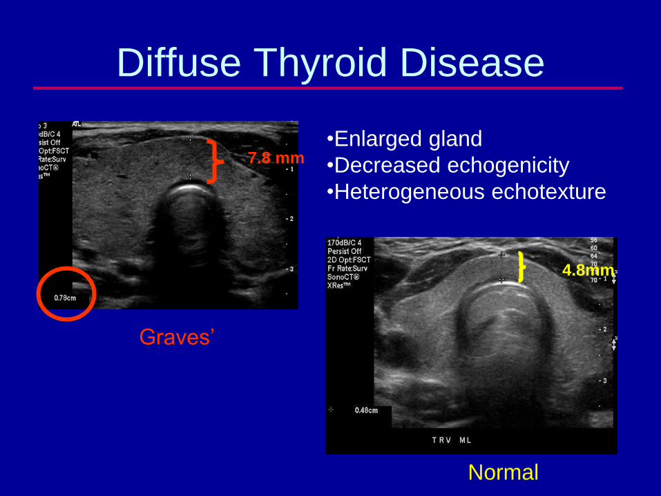
Diffuse Thyroid Disease
Normal
7.8 mm•Enlarged gland
•Decreased echogenicity
•Heterogeneous echotexture
4.8mm
Graves’
Enlarged Thyroid with Normal
Echogenicity and Echotexture
• Normal variation-Height, BMI, Gender,
Race, Age
• Mild iodine deficiency
• Medical conditions: pregnancy, renal
disease
• Subclinical autoimmune thyroid disease
• Check serum TSH
Thyroid Volume and Subclinical
Disease
• Retrospective analysis of 1,089
adolescents in Slovakia, mean age 17
years
• Correlated thyroid volume with TSH and
TPO Abs in 50% of the population studied
• Assessed whether enlarged thyroid
volume had a relationship with subclinical
or early thyroid dysfunction
Langer P. Endocrine Journal 2003;50(2):117-125.
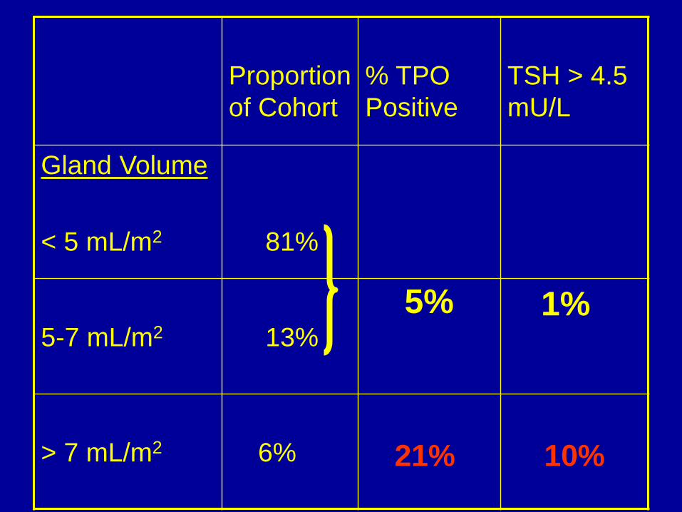
Proportion
of Cohort
% TPO
Positive
TSH > 4.5
mU/L
Gland Volume
< 5 mL/m2 81%
5-7 mL/m2 13%
> 7 mL/m2 6% 21% 10%
1%5%
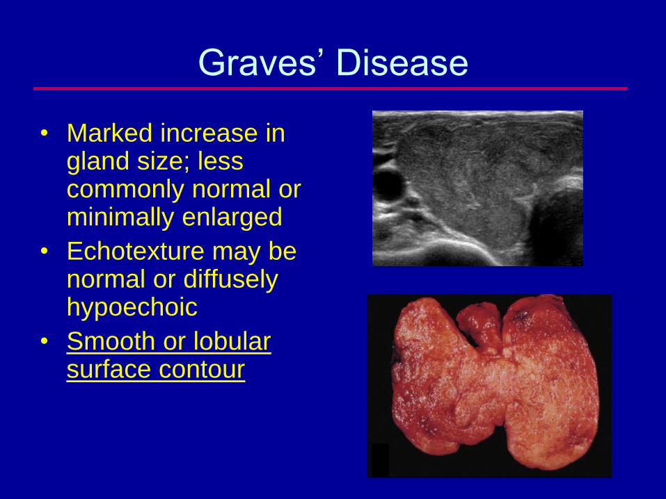
Graves’ Disease
• Marked increase in gland size; less commonly normal or minimally enlarged
• Echotexture may be normal or diffusely hypoechoic
• Smooth or lobular surface contour
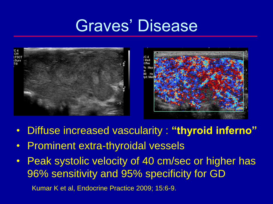
Graves’ Disease
• Diffuse increased vascularity : “thyroid inferno”
• Prominent extra-thyroidal vessels
• Peak systolic velocity of 40 cm/sec or higher has
96% sensitivity and 95% specificity for GD
Kumar K et al, Endocrine Practice 2009; 15:6-9.
Role of Sonography in
Graves’ Disease
• CDUS may be used to confirm diagnosis in lieu
of I-123 scan
– sensitivity of CDUS (95% vs. 97%) and specificity
(95% vs. 99%) for Dx of GD
• Screening for occult cancer
– Sonography identified 68/426 (16%) focal nodules vs.
9/462 (2.1%) on I-123 scan
– Thyroid cancer found in 30/68 (48%) of these patients
• All patients with GD should be screened by US-
management changed to surgical
Cappelli C et al, Eur J Rad 2008; 65;99-103
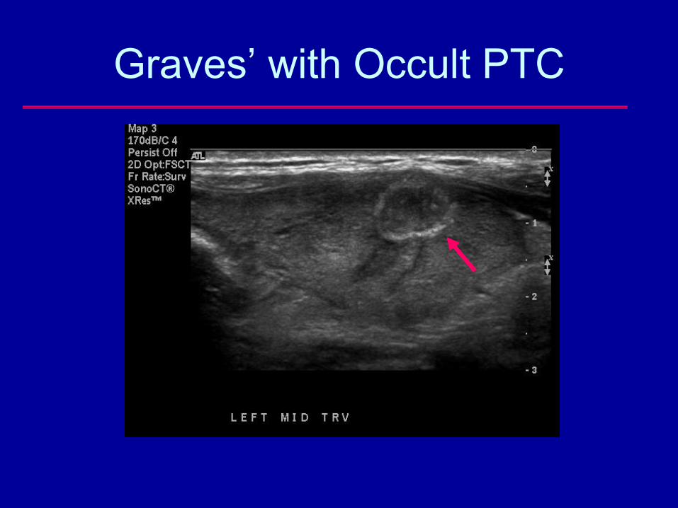
Graves’ with Occult PTC
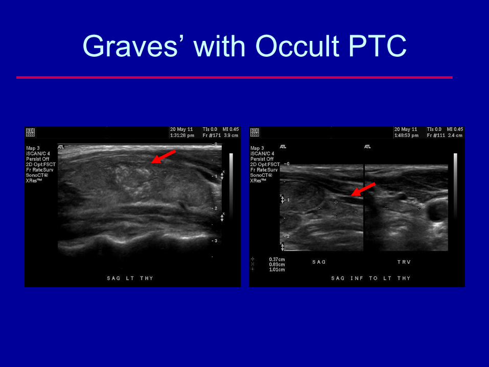
Graves’ with Occult PTC
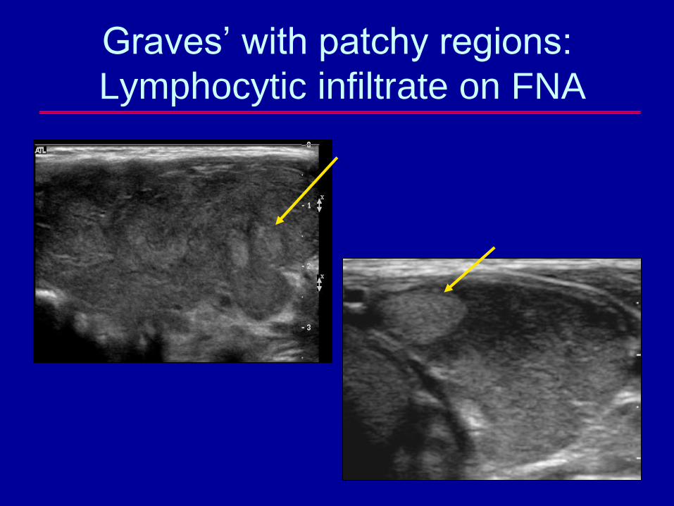
Graves’ with patchy regions:
Lymphocytic infiltrate on FNA
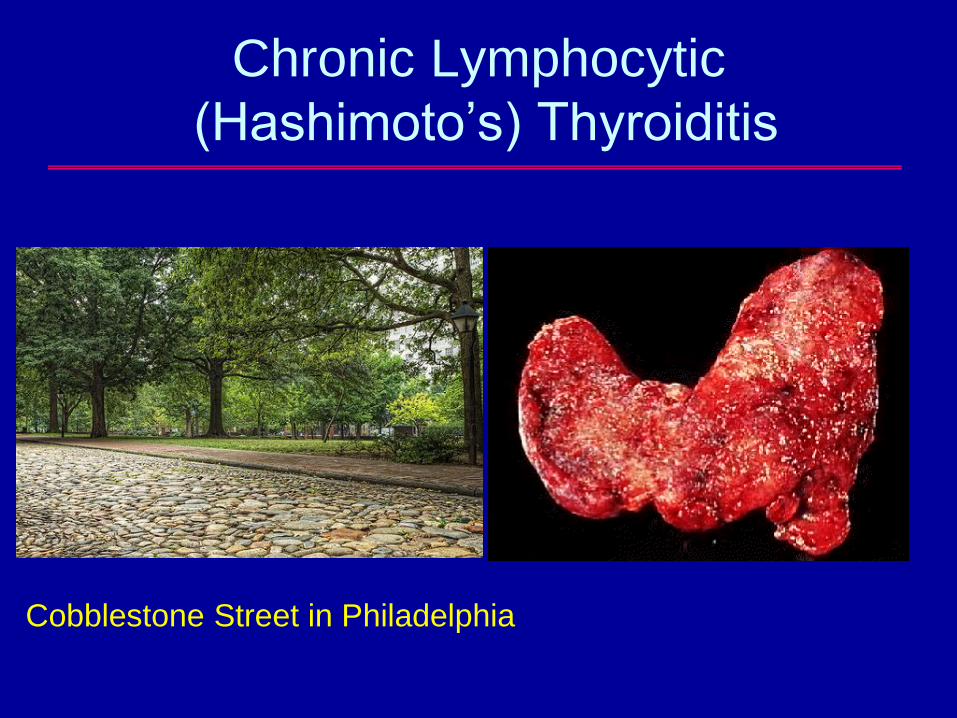
Chronic Lymphocytic
(Hashimoto’s) Thyroiditis
• Most common type of thyroiditis
– 5 to 10% of the adult population is affected
• Autoimmune disease occurring most
frequently in middle aged women, with strong
familial predisposition
• Patients may be eu-, hypo- or hyperthyroid
• 95% of patients have circulating anti-
thyroglobulin antibodies
Chronic Lymphocytic
(Hashimoto’s) Thyroiditis
Cobblestone Street in Philadelphia
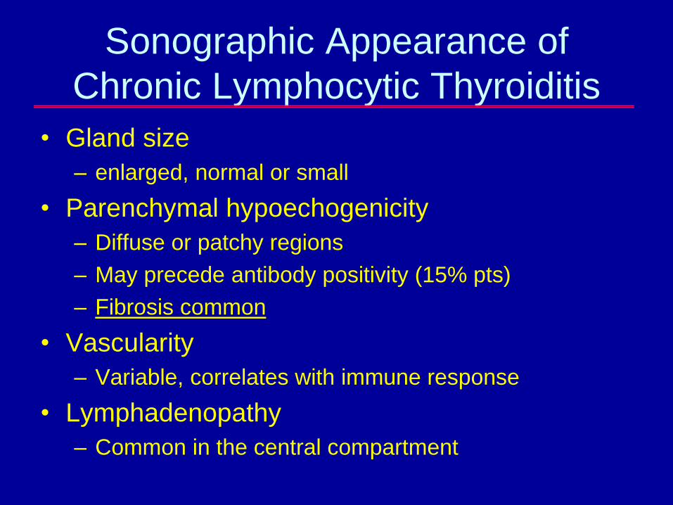
Sonographic Appearance of
Chronic Lymphocytic Thyroiditis
• Gland size
– enlarged, normal or small
• Parenchymal hypoechogenicity
– Diffuse or patchy regions
– May precede antibody positivity (15% pts)
– Fibrosis common
• Vascularity
– Variable, correlates with immune response
• Lymphadenopathy
– Common in the central compartment
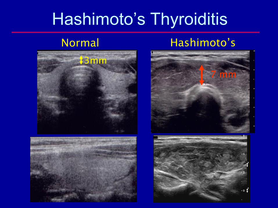
Hashimoto’s Thyroiditis
Normal Hashimoto’s
7 mm
3mm
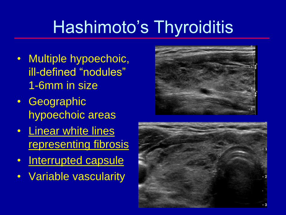
Hashimoto’s Thyroiditis
• Multiple hypoechoic,
ill-defined “nodules”
1-6mm in size
• Geographic
hypoechoic areas
• Linear white lines
representing fibrosis
• Interrupted capsule
• Variable vascularity
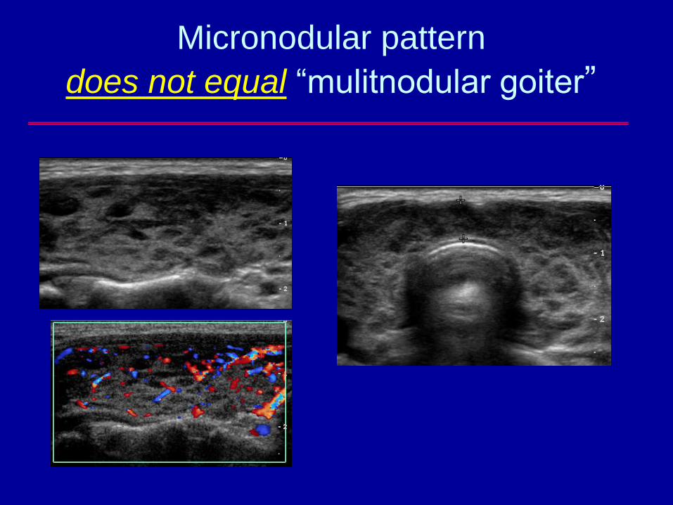
Micronodular pattern
does not equal “mulitnodular goiter”
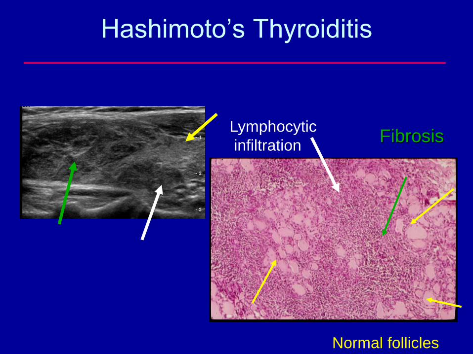
Hashimoto’s Thyroiditis
Normal follicles
Lymphocytic
infiltrationFibrosis
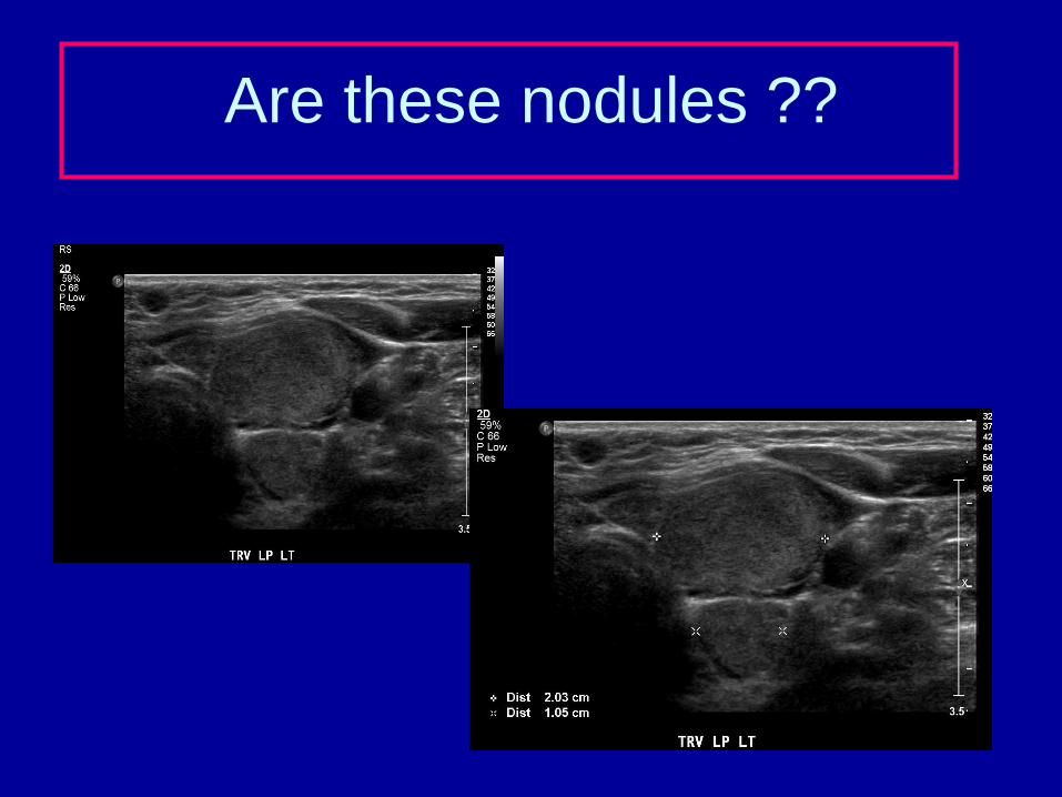
Are these nodules ??
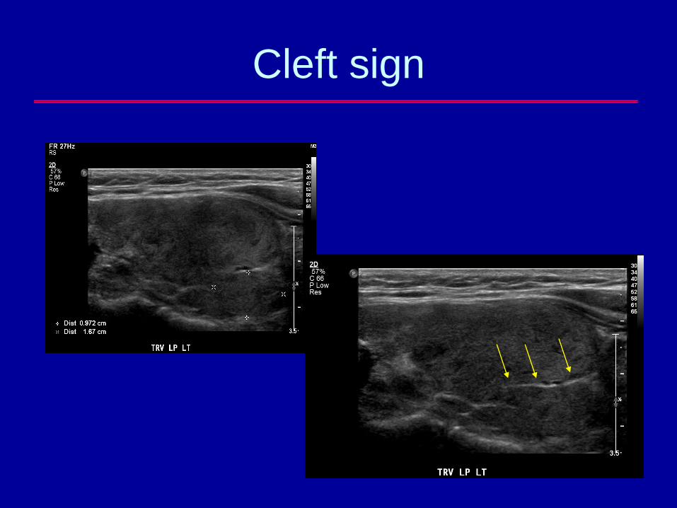
Cleft sign
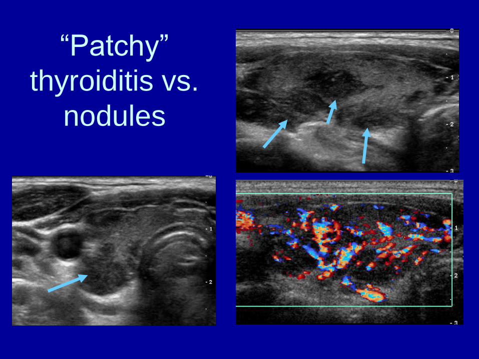
“Patchy”
thyroiditis vs.
nodules
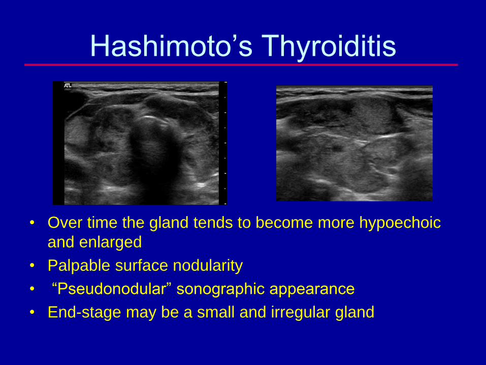
Hashimoto’s Thyroiditis
• Over time the gland tends to become more hypoechoic
and enlarged
• Palpable surface nodularity
• “Pseudonodular” sonographic appearance
• End-stage may be a small and irregular gland
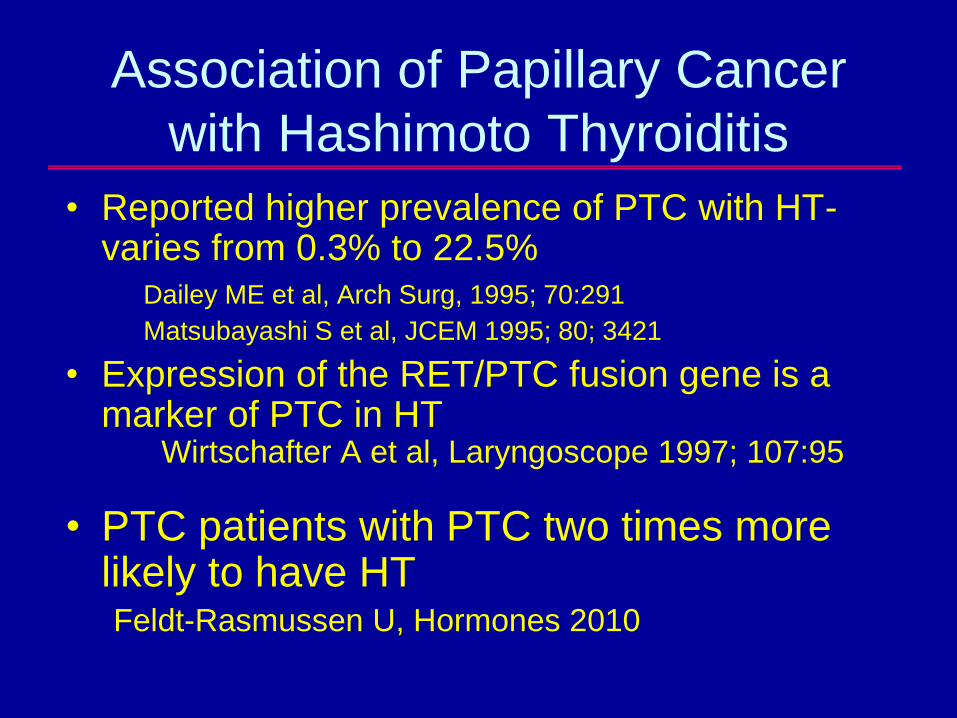
Association of Papillary Cancer
with Hashimoto Thyroiditis
• Reported higher prevalence of PTC with HT-varies from 0.3% to 22.5%
Dailey ME et al, Arch Surg, 1995; 70:291
Matsubayashi S et al, JCEM 1995; 80; 3421
• Expression of the RET/PTC fusion gene is a marker of PTC in HT
Wirtschafter A et al, Laryngoscope 1997; 107:95
• PTC patients with PTC two times more likely to have HTFeldt-Rasmussen U, Hormones 2010
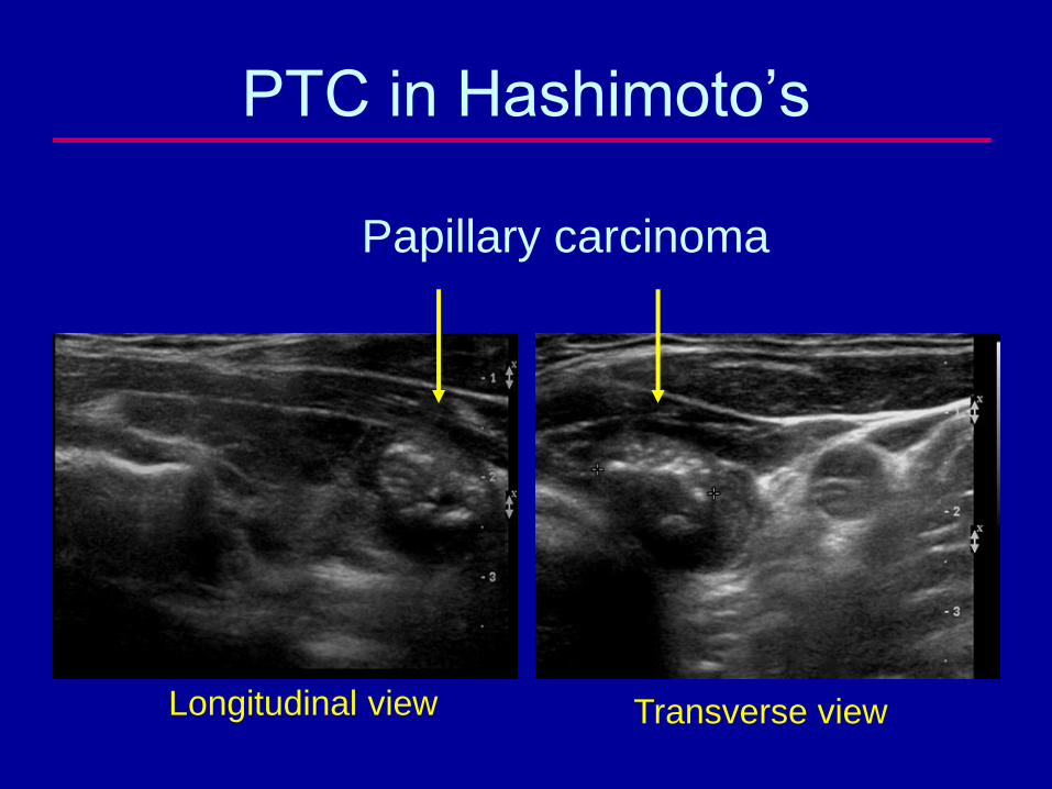
PTC in Hashimoto’s
Papillary carcinoma
Longitudinal view Transverse view
Appearance of PTC in HT glands
• Typical PTC features overlap with HT features– Hyopechogenicity, solid consistency,
irregular or infiltrating margins
• Key finding is pattern of calcifications– Clustered microcalcifications or dystrophic
calcifications
– Asymmetrical lobar involvement
Ohmori N et al, Internal Medicine (Japanese Society of Internal Medicine) 2007; 46; 547.
Liu F et al, J Clin Ultrasound 2009; 37:487-492.
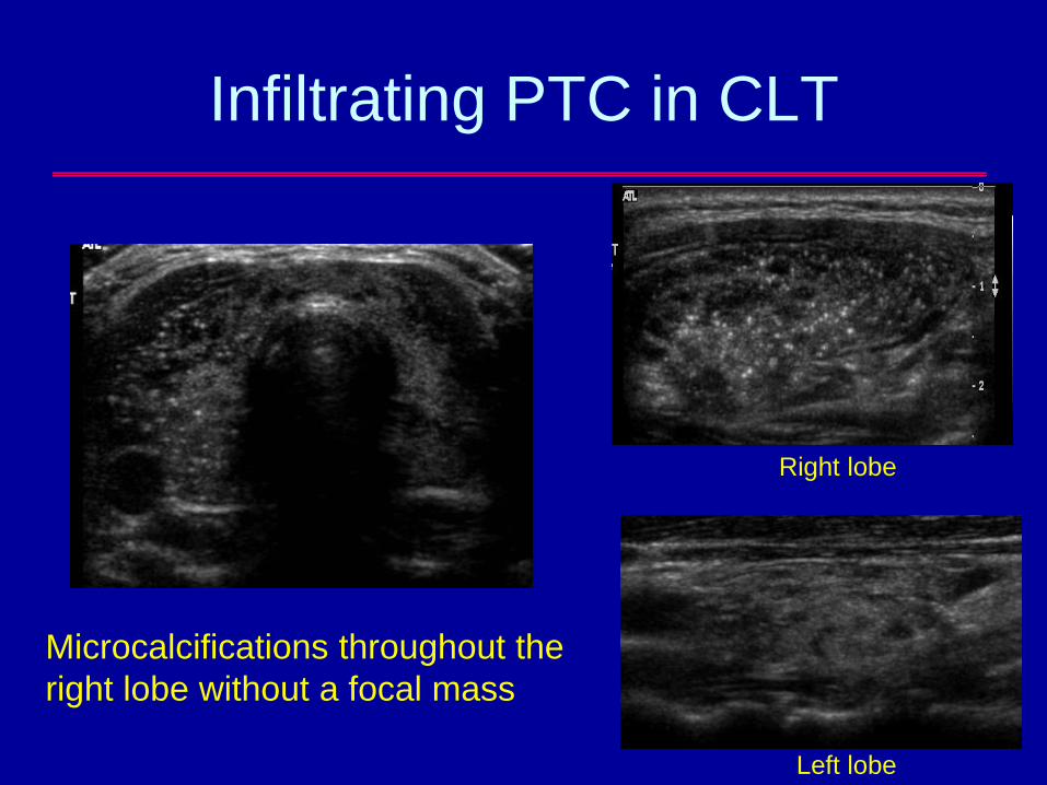
Infiltrating PTC in CLT
Microcalcifications throughout the
right lobe without a focal mass
Left lobe
Right lobe
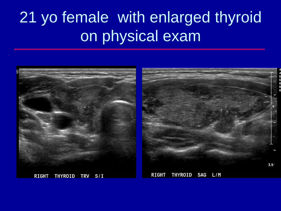
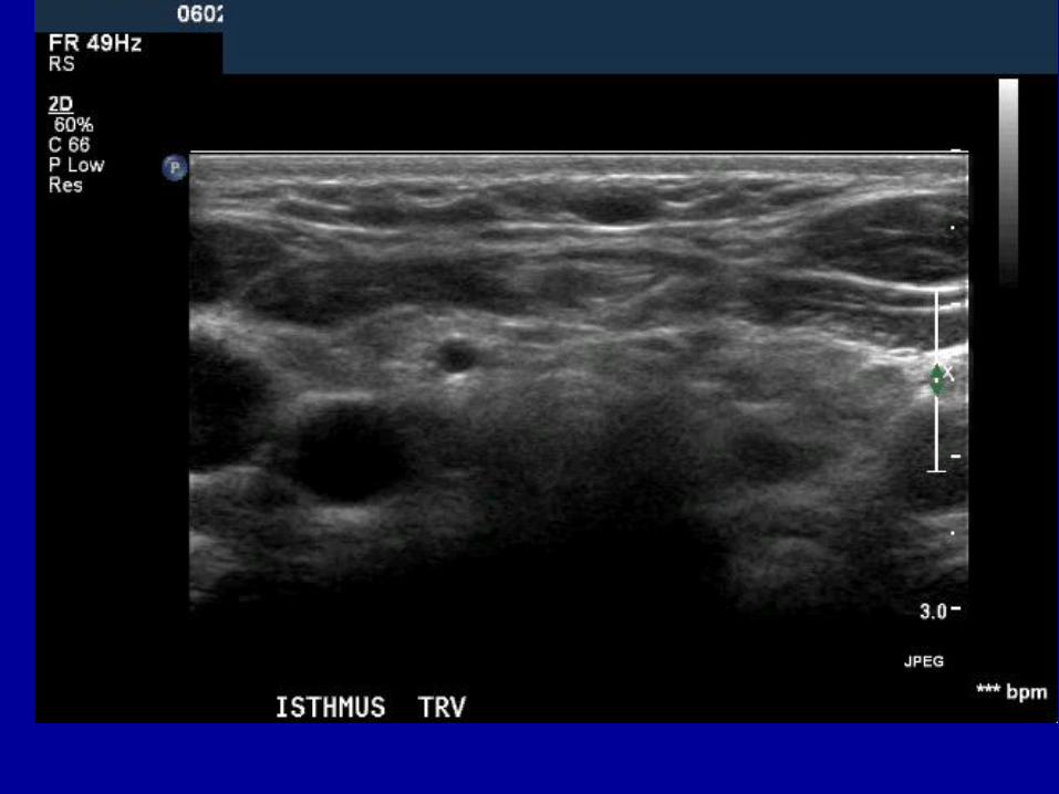
21 yo female with enlarged thyroid
on physical exam
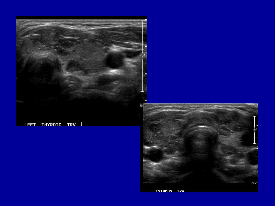
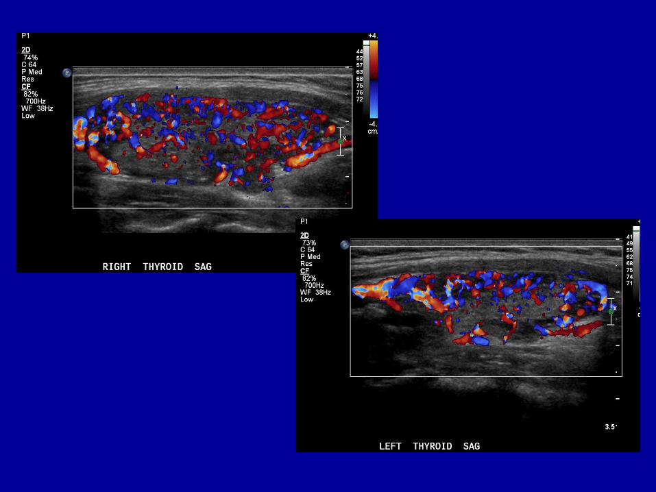
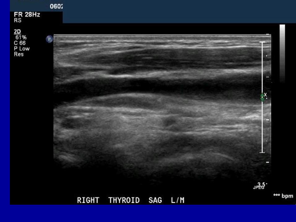
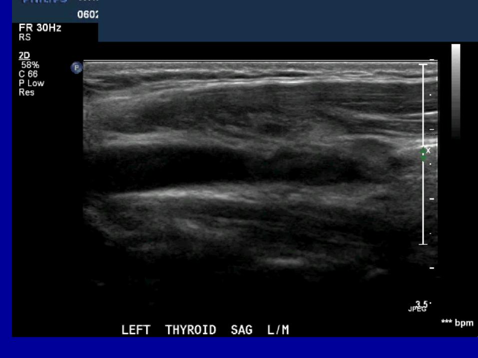
Diagnosis?
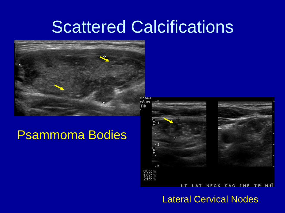
Scattered Calcifications
Lateral Cervical Nodes
Psammoma Bodies
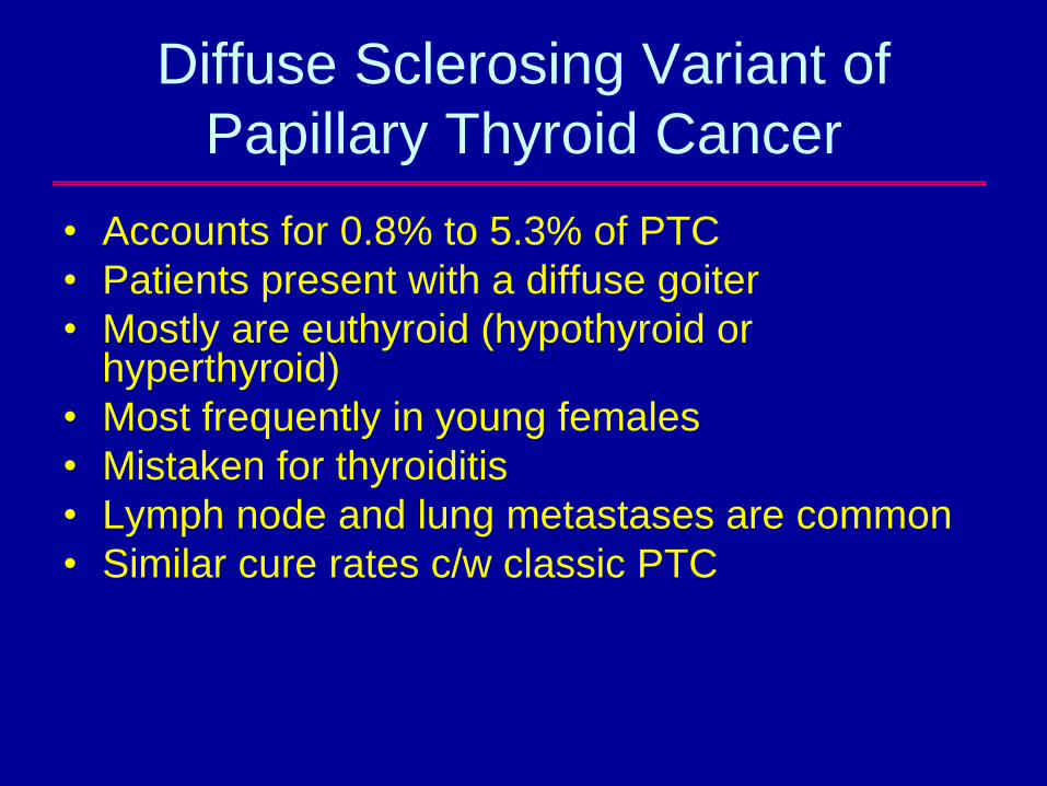
Diffuse Sclerosing Variant of
Papillary Thyroid Cancer
• Accounts for 0.8% to 5.3% of PTC
• Patients present with a diffuse goiter
• Mostly are euthyroid (hypothyroid or hyperthyroid)
• Most frequently in young females
• Mistaken for thyroiditis
• Lymph node and lung metastases are common
• Similar cure rates c/w classic PTC
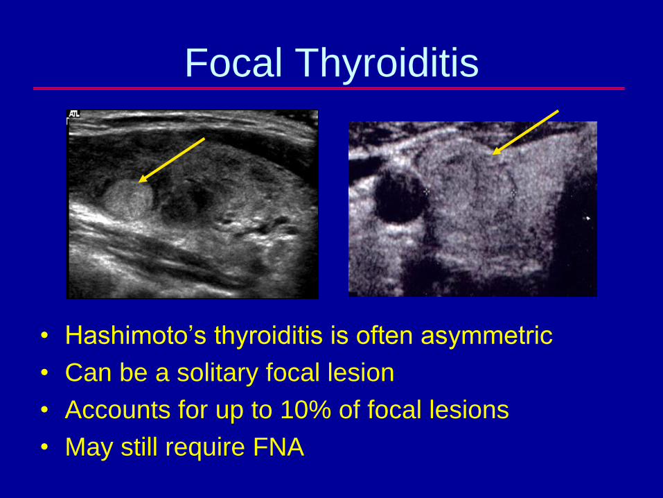
Focal Thyroiditis
• Hashimoto’s thyroiditis is often asymmetric
• Can be a solitary focal lesion
• Accounts for up to 10% of focal lesions
• May still require FNA

Dilemma: Nodules in patients with
Diffuse Thyroid Disease
• May have patchy irregular areas that are
pseudo-nodules
– Tend to be small (under 15 mm), hyperechoic
and non-calcified
– Larger lesions or those with irregular margins
raise concern for a neoplasm
• Focal calcifications and asymmetric
calcifications should be considered
suspect for papillary carcinoma
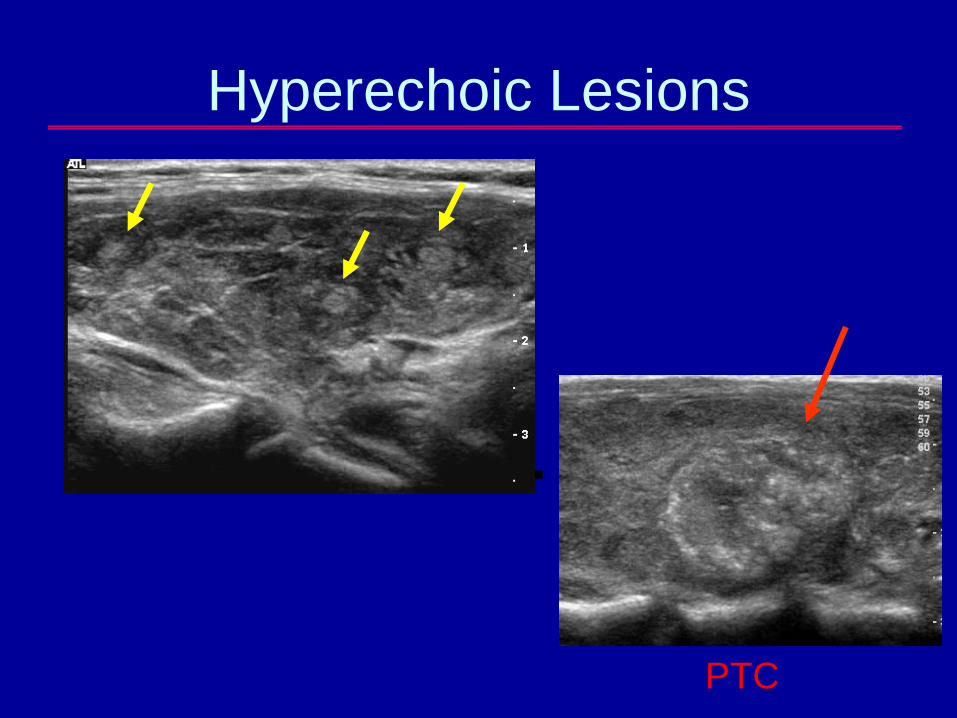
Hyperechoic Lesions
PTC
Malignant Lymphoma
• Usually occurs in a CLT gland
• 2 to 5% of all thyroid malignancies
• Nodular pattern
– Homogeneously hypoechoic with lobulated
but well defined border; enhanced though
transmission
• Diffuse disease-asymmetric enlargement
• Mixed pattern
Ito Y et al, World J Surg 2010; 34:1171-80,
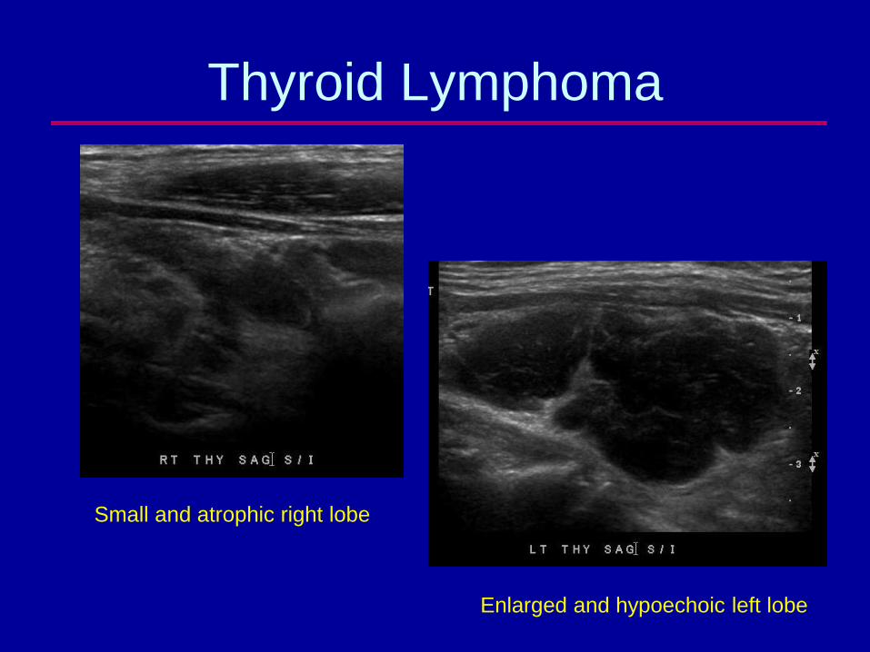
Thyroid Lymphoma
Small and atrophic right lobe
Enlarged and hypoechoic left lobe
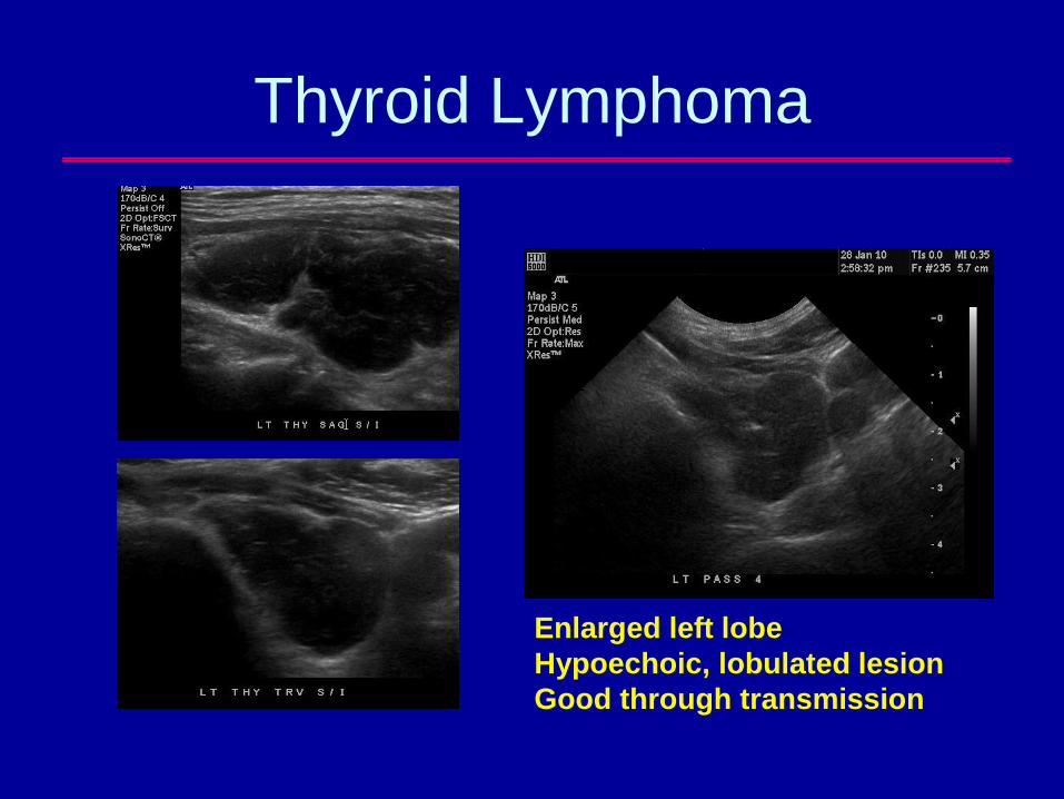
Thyroid Lymphoma
Enlarged left lobe
Hypoechoic, lobulated lesion
Good through transmission
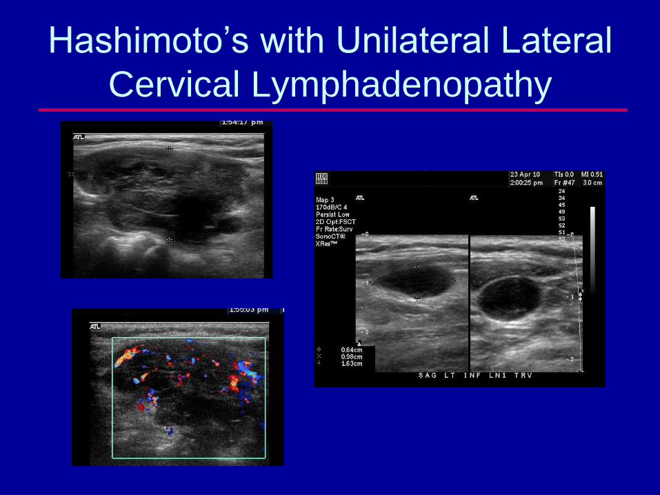
Hashimoto’s with Unilateral Lateral
Cervical Lymphadenopathy
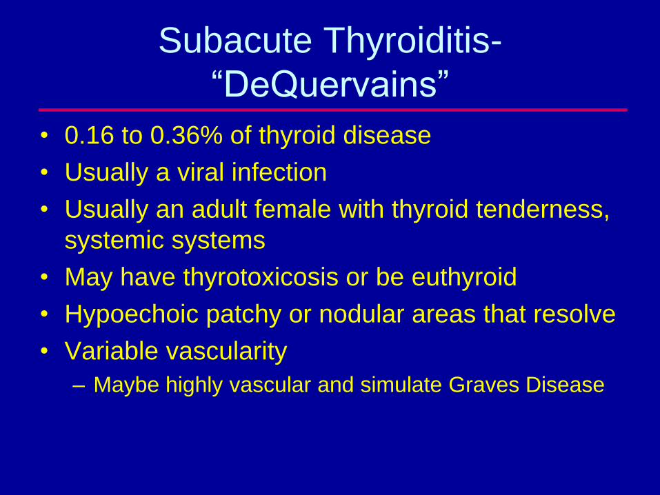
Subacute Thyroiditis-
“DeQuervains”
• 0.16 to 0.36% of thyroid disease
• Usually a viral infection
• Usually an adult female with thyroid tenderness,
systemic systems
• May have thyrotoxicosis or be euthyroid
• Hypoechoic patchy or nodular areas that resolve
• Variable vascularity
– Maybe highly vascular and simulate Graves Disease
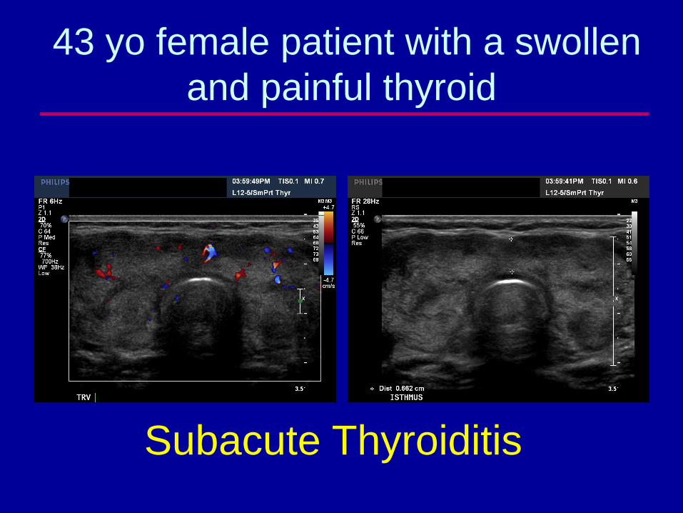
43 yo female patient with a swollen
and painful thyroid
Subacute Thyroiditis
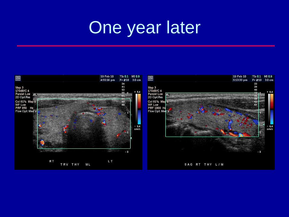
One year later
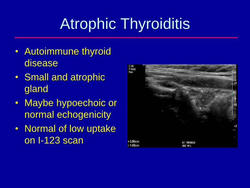
Atrophic Thyroiditis
• Autoimmune thyroid
disease
• Small and atrophic
gland
• Maybe hypoechoic or
normal echogenicity
• Normal of low uptake
on I-123 scan
Amiodarone-Induced
Thyrotoxicosis (AIT)
• More commonly patients develop
hypothyroidism due to iodine content
• The minority develop thyrotoxicosis
• Type 1 is an iodine load-induced
hyperthyroidism which occurs in abnormal
glands (MNG or Graves); increased vascularity
• Type 2 is a destructive thyroiditis; normal gland;
normal or decreased vascularity; low/absent
upatke on RAIU
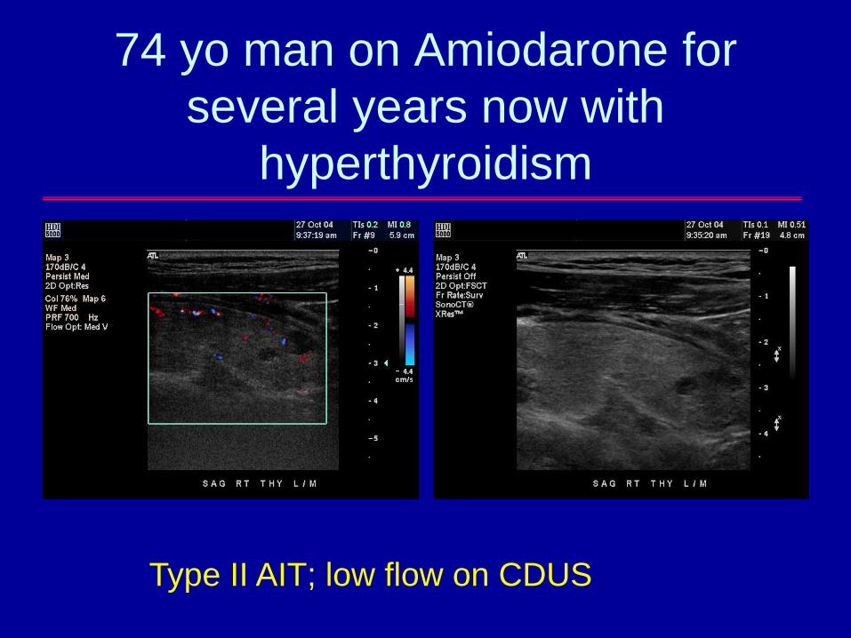
74 yo man on Amiodarone for
several years now with
hyperthyroidism
Type II AIT; low flow on CDUS
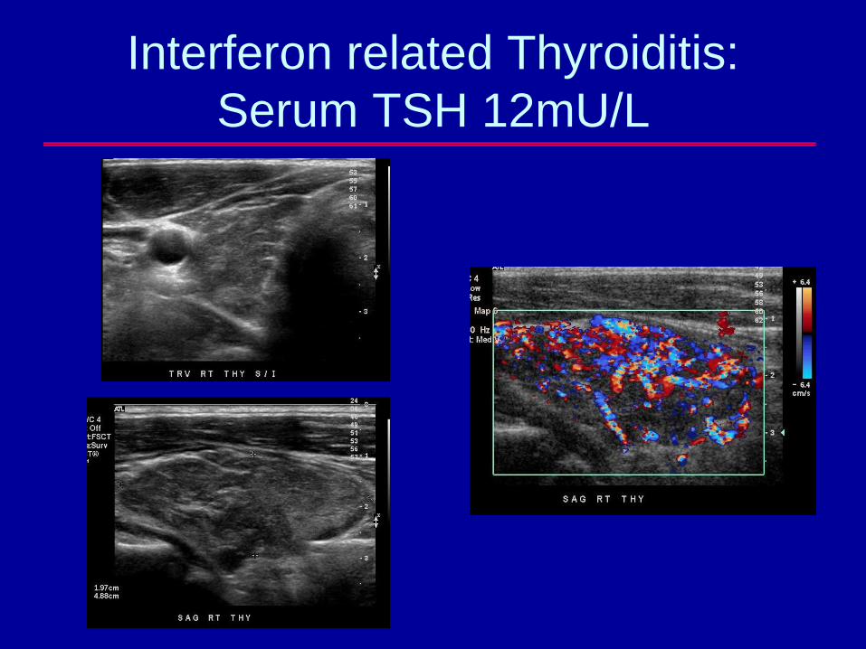
Interferon related Thyroiditis:
Serum TSH 12mU/L
Conclusions
• Sonographic markers of autoimmune thyroid disease include enlarged size, heterogeneous echotexture, increased vascularity, but are not specific
• Clinical information is key
• Differentiation of “pseudo-nodules” from true nodules and tumors may be challenging
– Asymmetric calcifications
– Unilateral large LNS
Thank you for your attention!
Nodular DiseaseDiffuse Disease