The Sliding Filament Theorymstaradavis.weebly.com/uploads/1/1/0/7/110763663/... · The Sliding...
7
Do VJot TI( t~ ~ih 'j<M The Sliding Filament Theory ''How do muscle cells contract?" Model 1: Muscle Histology Review K.._____-/ Organlutfon of Connective Tissues: s -e. i fV\. V. i V IVl s -e~ rv----1Sjvrt1 1 Yer- °M':'.1£, rll During muscia oonlnlciion the myosin headgroups attach lo actln and putt them toward the cenlet of tho sarcomcm. This causes tho sareomere lo shorton. The movement of myosln hoadgroul)$ Is fike • the motion of oara In a boal ft~ ~- } General structu,- "Tubes within Tubes•: 1 W~o\e WltASl. 1€ 2 pgs r, 1..e 3 MvSCI~ Fi1oeie. 4 M~oe,~r:i 1 From macroscopic to microscopic, the geneml !III\ICIUre of sk-ol muscle Is tike a sertetl of tubea 2 within lvbet. 3 ~ ,__,.- Eech actln (thin) ntemenl la . ' like a double-stranded chain 9 / of pearls (not Including the - 10 11 troponln/lropomyosln complox). n ,•--- Structure of a sarcomere: _,._.,_-4 8 N\'-\ b'5 j VI n The ~yosln headgroupa are arranged 9 M '1=~ j "1 In a manner slmltar to the taH fealhera 10 -f(C tjp ·21 11 lnanarrow. 11 2- Li~.e.. Use your knowledge of muscle tissue histology to fill in the blanks numbered 1-11 with the following terms: - Fasicle, Myofibril, Perimysium, Myosin heads, Actin (thin) filaments, Whole muscle, Skeletal myocyte, Epimysium, Endomysium, Myosin (thick filaments), Z line. \.
Transcript of The Sliding Filament Theorymstaradavis.weebly.com/uploads/1/1/0/7/110763663/... · The Sliding...

Do VJot
TI( t~
~ih 'j<M ~ The Sliding Filament Theory
''How do muscle cells contract?"
Model 1: Muscle Histology Review K.._____-/
Organlutfon of Connective Tissues: s -e. ~ i fV\. V. ~ i V IVl s -e~ rv----1Sjvrt1 1 Yer- °M':'.1£, rll
During muscia oonlnlciion the myosin headgroups attach lo actln and putt them toward the cenlet of tho sarcomcm. This causes tho sareomere lo shorton. The movement of myosln hoadgroul)$ Is fike
• the motion of oara In a boal ~-~
ft~ ~ ~ ~ ~~ ~-} ~
General structu,"Tubes within Tubes•:
1 W~o\e WltASl.1€ 2 pgs r, 1..e 3 MvSCI~ Fi1oeie. 4 M~oe,~r:i 1
From macroscopic to microscopic, the geneml !III\ICIUre of sk-ol muscle Is tike a sertetl of tubea
2 within lvbet.
3
~
,__,.-Eech actln (thin) ntemenl la
. ' like a double-stranded chain 9 / of pearls (not Including the
-10
11 troponln/lropomyosln complox).
n ,•--- Structure of a sarcomere: _,._.,_-4 8 N\'-\ b'5 j VI n ~
◊ The ~yosln headgroupa are arranged ~ 9 M '1=~ j "1 In a manner slmltar to the taH fealhera 10 -f(C tjp ·21 11 lnanarrow. 11 2- Li~.e..
Use your knowledge of muscle tissue histology to fill in the blanks numbered 1-11 with the following terms: -Fasicle, Myofibril, Perimysium, Myosin heads, Actin (thin) filaments, Whole muscle, Skeletal myocyte, Epimysium, Endomysium, Myosin (thick filaments), Z line.
\.

Model 2: Muscle contraction takes place at the level of the sarcomere. Z4irut I
+- -+
'9 :::: ., <iii :;: ., ~
+- ....... '" ....... ~-·--··- ....
Critical Thinking Questions
I Z~llne
Ac~n llhln)
+- nt / "'""
+-
Myosln ~(-) ~-·
. .
+-
98 aa ~
+-
-+
·•· ' ·,
::: ~ ---: -+
+---. ., +-
-+ -a=, ~ 4Jl!lo ....
1. What structural component of the sarcomere is associated with arrows in model 2?
Z -- Li vi--e_ 2. Based on this, which component of the sarcomere is actually being moved when the sarcomere contracts and relaxes?
3. Which component of the sarcomere is physically attached to the structure that gets moved
(i.e. the answer to question 2)?
Ac h ln 4. What component of the sarcomere is NOT directly attached to the Z-line?
5. Based on your answer to question 4, what component of the sarcomere is pulling on the thin filaments to bring the Z-lines closer together?
Application ~ M~oS:iK h~CA.ots 6. In the space below, write a short description as a group that explains the role of the think filaments, thick filaments, and Z-line in sarcomere contraction.
Thin filament:
S hGY- +e ~ 1vie Sa re o \fV'ere ~ (cdA SR Yl/1 fA Sc I e_s tc.1 C c5V1 iYz;l C f--
Thick Filament:
A11G\tcltl w 1~1 Vl f ,l ci ~Y'~Y7 1] ~ ?VI II 1vi.em Wlvcurdf Z-line: f0p (pVJ-{CJY" 0( f V,-e SJ v{o VJ,0,e rz
51;)/ o Y-lc ~; s tu~'.:,~ 1r111;; Sri rs r o:}v"' r f } f{l)J 1vifr1 vvv1Pn 1v1Pj re /(t.. 'f

Model 3: Molecular events of the contraction cycle
LOCATION .....
+-y---✓
Sarcomere
Tropomyosfn
7Tuiv1 --) fi\U.Wl.e%\-\-1n blndlng stte Q
Inge 1
e2
COTTON SWAB ANALOGY A myosln moiewle la llke a double-headed COiton swab whose end has been cut ol!
end is able to bend ffke a hinge In two places
@(~'iWi
~!)._~ a . (Now ATP molecule enters the cycle)
ROWER ANALOGY
During muscle contraction the myosln headgroups attach to eclln and pun lhem toward the center ot
the sarcomere. Thil causes the sarcomere to shorten. This m<Mlmenl
of the myosln headgl'Ollp6 la ralher Ilka the motion of oars In a boat.

Critical Thinking Questions
7. Find the thin filament in part 1 of model 3 (attachment) and draw a bracket and label it 'thin
filament'. ~e Wloul.e I 3 8. Which component of the thin filament (which isn't labeled in this model) makes the main
'string-of-pearls' portion of the filament? A c ti n 'J)'(c) --J.e i Vl
a. in the space below, list the other molecules that are found in/on the thin filament.
tY?JPonin +- tr-v Po (}1'-/0S.1n 9. The myosin head is bound to some other molecules, what are they?
ADP..}- P a. To which specific region of actin is the myosin head bound?
Wl'joSin bind ihq S1K 10. In your own words, describe the change that occurs in the myosin molecule between stage
1 (attachment) and stage 2 (pulling). r'VlljoSin Cvio.vi@R';: ~"1Cl(->--€_ 'f- lo.e,,hd.S -lo P1A11 tvi.e act,·Y1
a. Circle the two places where the myosin molecule bends in this stage on part 2 of the
model. ~e mod.e( 3 11. The myosin head doesn't release from the actin binding site until a new molecule enters the cycle. According to the model, what molecule allows myosin to release from actin?
A1P 12. In stage 4 of this cycle, the myosin molecule moves back into the cocked position. Where does it get the energy to re-cock itself?
ATP 13. Look at all the myosin binding sites on the actin filament. Does it appear that each site lines
up perfectly with a myosin head? '--{-e__ _$
a. Myosin can only bind to a binding site that is at the tip of a helix in the actin filament (like the one in phase 1 of the model). After the power stroke (pulling phase) and detachment is the myosin head lined up with a binding site at the tip of the filament?
tJo b. Can this particular myosin head bind to the actin filament
forthe next stroke? N O
c. Look at the feet of these children. Are they all able to pull at exactly the same time?
'4-~.S d. As a group, explain how myosin pulling on actin works kind of like kids playing tug-of-war.
~ can do -fh;s rP1.Q__ Q
4.

Model 4: Effects of calcium on the thin filament
14. Look at the left half of the model. Is calcium present here? N Q
a. Is the myosin binding site of actin exposed so that myosin can actually bind to it?
N◊ b. What is blocking myosin's access to the binding site on actin?
1 ro Po l'Ylvf ~ 5 i V1 15. Now look at the right side ot'the model. Is calcium present here?
'i.e$ a. To what molecule did it bind?
--rr=-c ponin b. What changed about the troponin molecule after it bound calcium? (hint look at the
little orange calcium binding site on troponin) It Y-6 fu-}-ed d(JWYl ~ io 1he .. r,jh f-c. What effect did this change in troponin have on tropomyosin?
P(;1.SV1ed mf<:Jr1--jDSin out of IV'.e w-4 d. Is the myosin bindin~ site of actin exposed when calcium is present?
~~s. ~ 16. In the space below, write a grammatically correct sentence (in English) that explains the 'ff'ul·,, role of troponin, tropomyosin, and calcium in muscle cell contraction. l( . (\
W½iV' (tA.+t- bfndS to mfonivt, f"roPom~oSiS ~c: 15 Vno\ie. IAWOAj
Application (A ( f ClW i Yl <J fy1JVY1 1vtR ~tJSin Joihd.in:f Si+-f <:D JIVttjoSin fo 0dVtd to a ck h.
17. What would happen to a muscle cell in ·either of the following situations? a. The cell runs out of ATP
Yn~-as-,~ Canvzof- c\.e ~ch ~ a. chY")
b. There is an excess amount of calcium in the cell.
l'vlvtgc,~r Cov,~c+- rap,o/f~ + Ca.CA~ YJ!tu ~ vle f':,Pot ~ rn .
5.

Model 5: Neuromuscular junction ·3, Ath binds to receptors on muscle cell and
released from ~reticulum
So..ao Pl(ilSrn i c...
B
18. Can muscles contract if there isn't any free calcium in the cell?
NO a. According to model 5, where is calcium stored inside a muscle cell?
s~Y{o f(v1.Smic, rRffc<A,IUvn b. What causes the calcium to be released from where it is stored?
.etectv,ca I s rgr1vt I i y1~,'d e Vvt VI s C ( -e C --e I I 19. What chemical messenger transmits the electrical impulse between the nerve cell and the muscle cell? ft c e+-111 cv,o Ii n~ Application
Av-A e,tAo\ i vieS~n7t ~ 20. Acetylcholine esterase is an enzyme that degrades Achin the neuromuscular junction. The
nerve gas Sarin blocks this enzyme causing Ach to remain available for binding to the muscle cell receptors.
What effect would Sarin gas have on the muscles of your body?
ConvvtlSiv-e. vviv1sc r.e 5P'1SW7S ~ S e.rz,v<,ve S
As a group, explain why Sarin gas is a deadly poison. [hint: your diaphragm is a muscle]
(o.
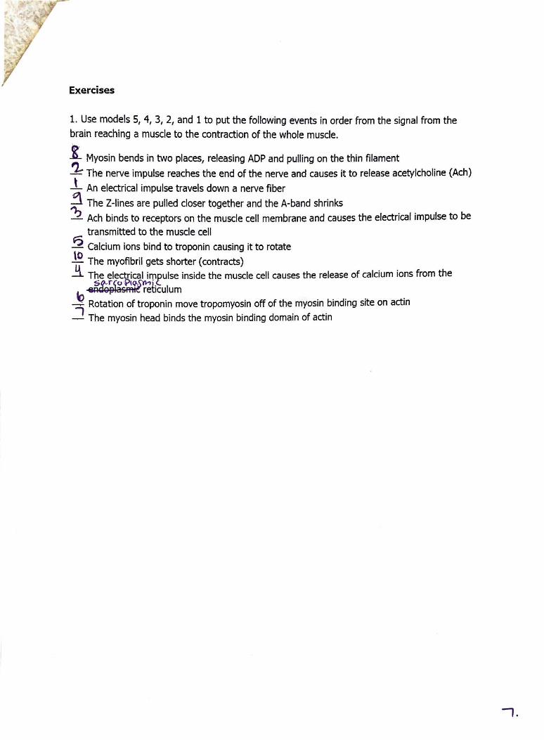
Exercises
1. Use models 5, 4, 3, 2, and 1 to put the following events in order from the signal from the
brain reaching a muscle to the contraction of the whole muscle.
1 Myosin bends in two places, releasing ADP and pulling on the thin filament
'2- The nerve impulse reaches the end of the nerve and causes it to release acetylcholine (Ach)
.l_ An electrical impulse travels down a nerve fiber
..::\ The 2-lines are pulled closer together and the A-band shrinks
? Ach binds to receptors on the muscle cell membrane and causes the electrical impulse to be transmitted to the muscle cell
? Calcium ions bind to troponin causing it to rotate
~ The myofibril gets shorter (contracts) ~ The electrical impulse inside the muscle cell causes the release of calcium ions from the sg;,rio~j.<.
~ R ~ as ret1culum ~ Rotation of troponin move tropomyosin off of the myosin binding site on actin 7 The myosin head binds the myosin binding domain of actin
7 .