THE SKELETAL SYSTEM - fuenscience.weebly.com · Types of Joints (articulations) Synarthrotic (not...
76
THE SKELETAL SYSTEM
Transcript of THE SKELETAL SYSTEM - fuenscience.weebly.com · Types of Joints (articulations) Synarthrotic (not...
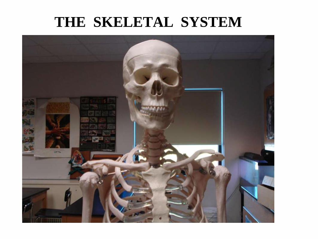
THE SKELETAL SYSTEM
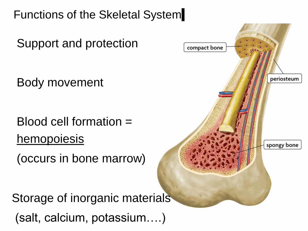
Functions of the Skeletal System
Support and protection
Body movement
Blood cell formation =
hemopoiesis
(occurs in bone marrow)
Storage of inorganic materials
(salt, calcium, potassium….)
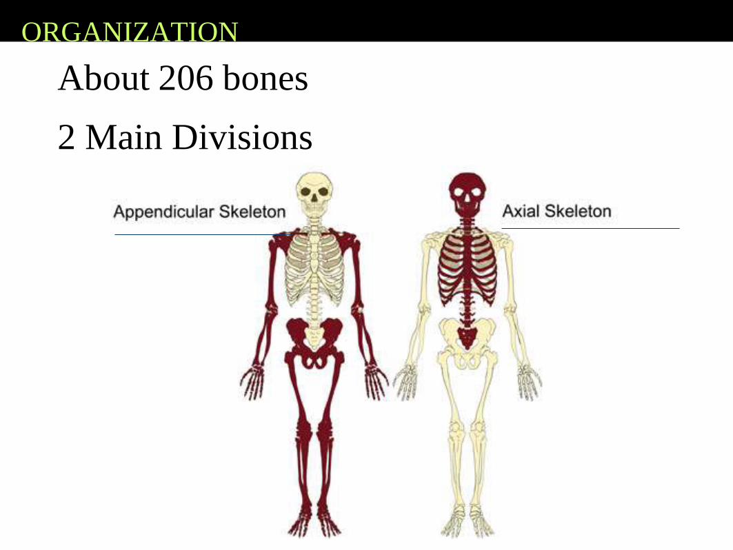
ORGANIZATION
About 206 bones
2 Main Divisions
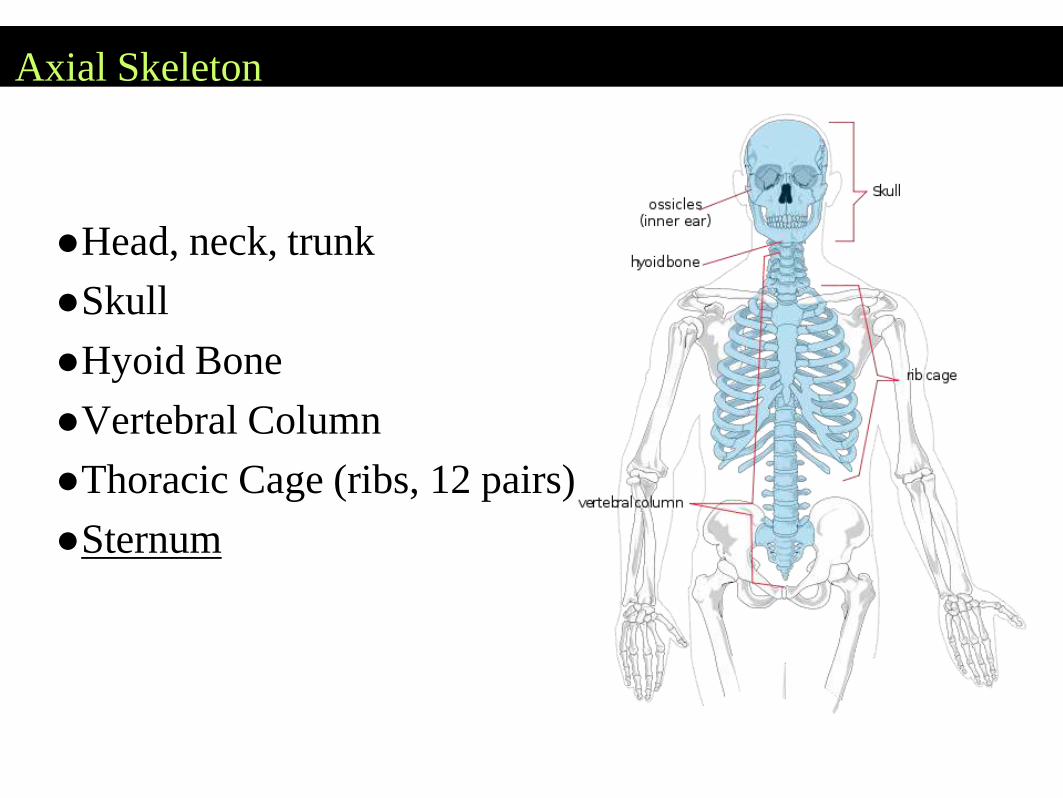
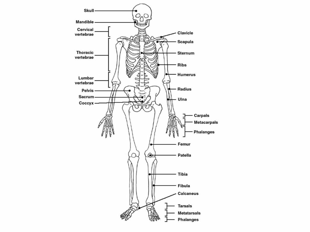
Axial Skeleton
●Head, neck, trunk
●Skull
●Hyoid Bone
●Vertebral Column
●Thoracic Cage (ribs, 12 pairs)
●Sternum
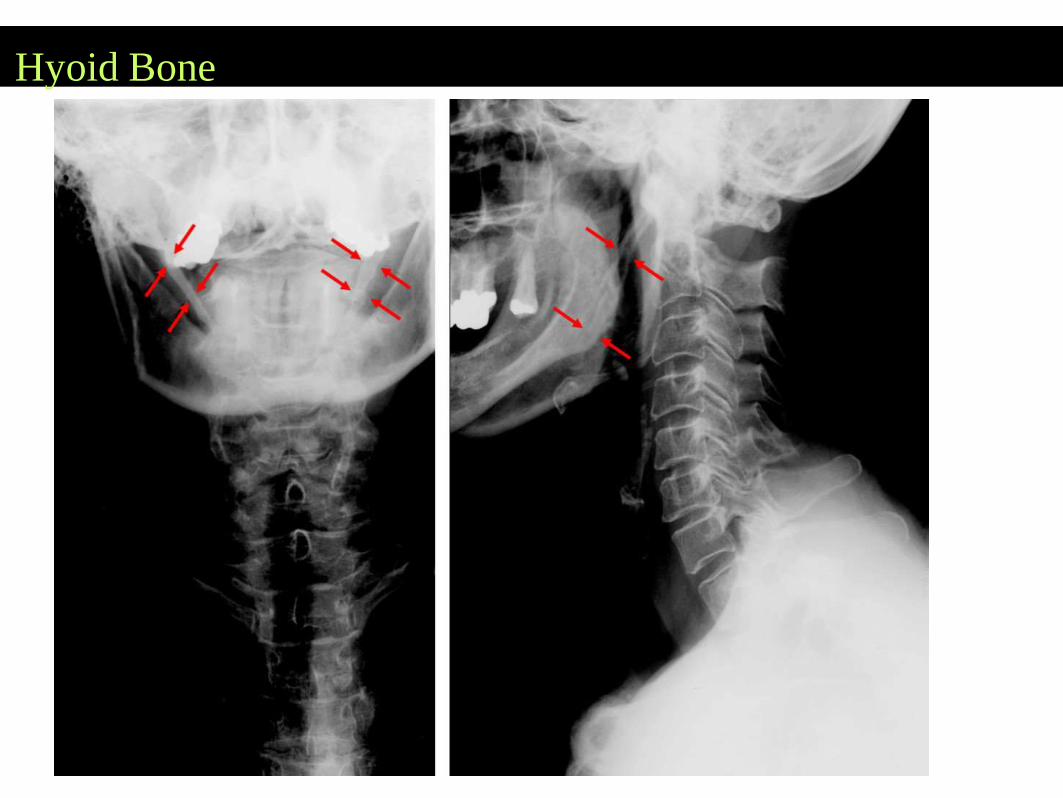
Hyoid Bone
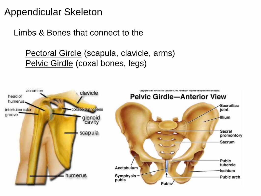
Appendicular Skeleton
Limbs & Bones that connect to the
Pectoral Girdle (scapula, clavicle, arms)
Pelvic Girdle (coxal bones, legs)
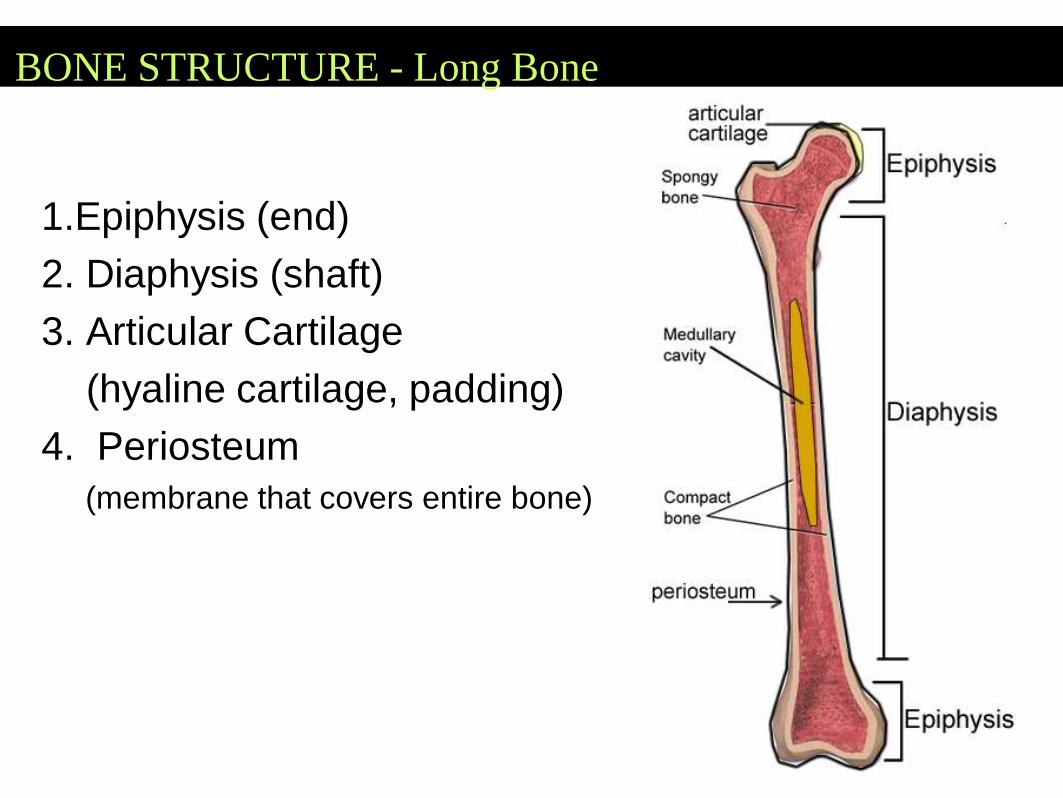
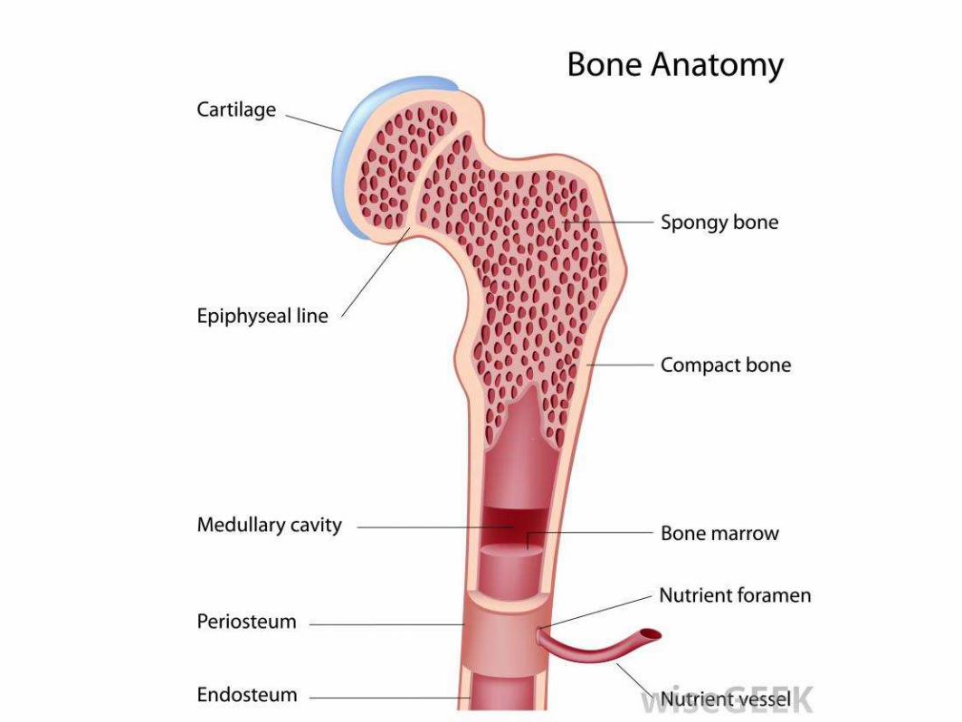
BONE STRUCTURE - Long Bone
1.Epiphysis (end)
2. Diaphysis (shaft)
3. Articular Cartilage
(hyaline cartilage, padding)
4. Periosteum
(membrane that covers entire bone)
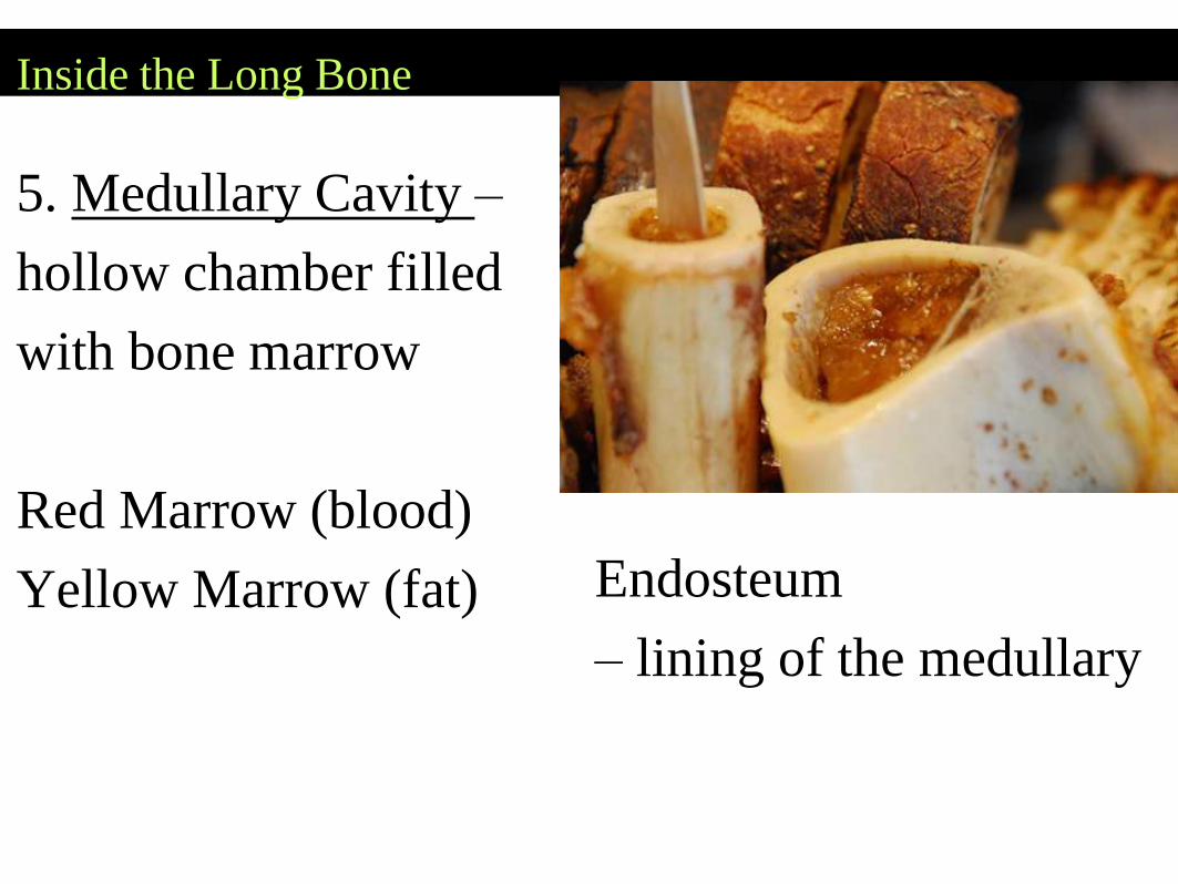
Inside the Long Bone
5. Medullary Cavity –
hollow chamber filled
with bone marrow
Red Marrow (blood)
Yellow Marrow (fat) Endosteum
– lining of the medullary
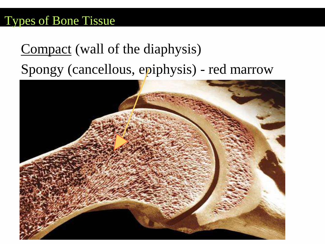
Types of Bone Tissue
Compact (wall of the diaphysis)
Spongy (cancellous, epiphysis) - red marrow
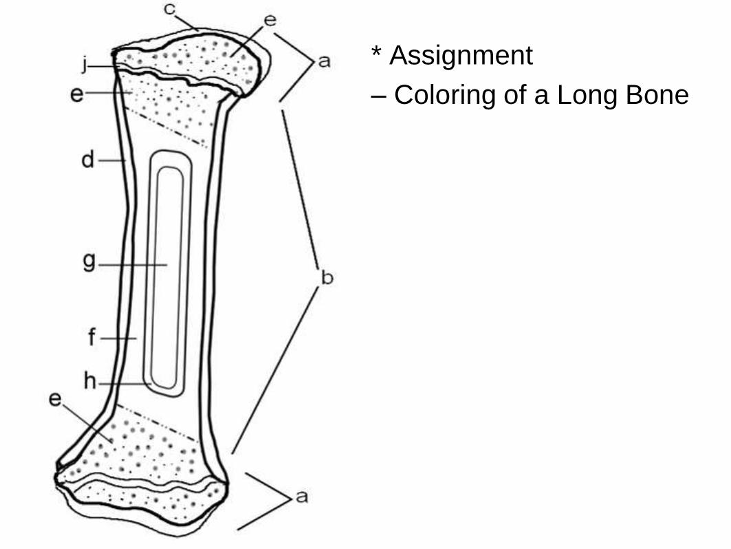
* Assignment
– Coloring of a Long Bone
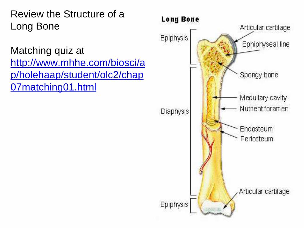
Review the Structure of a
Long Bone
Matching quiz at
http://www.mhhe.com/biosci/a
p/holehaap/student/olc2/chap
07matching01.html
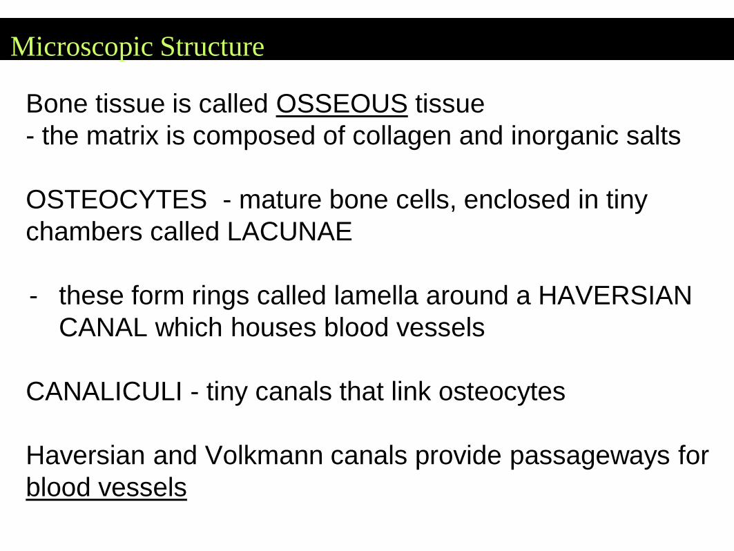
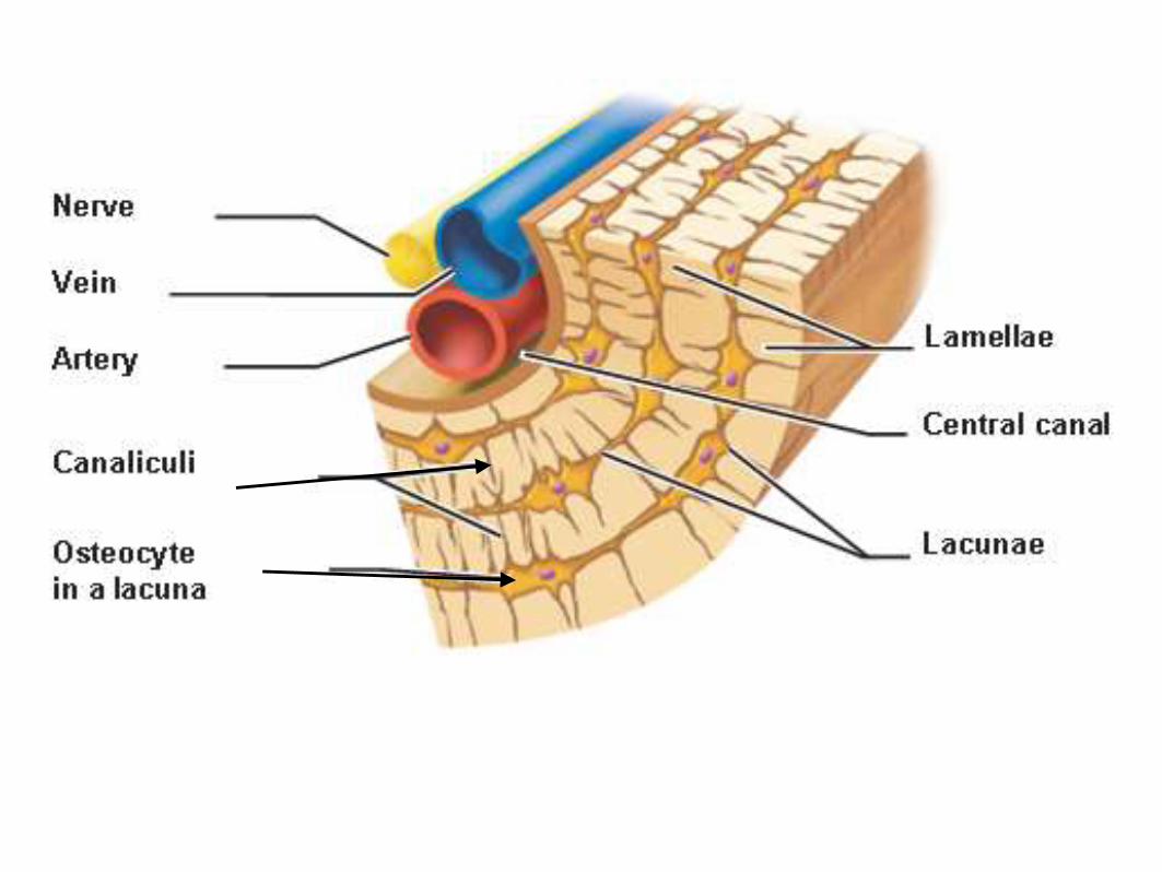
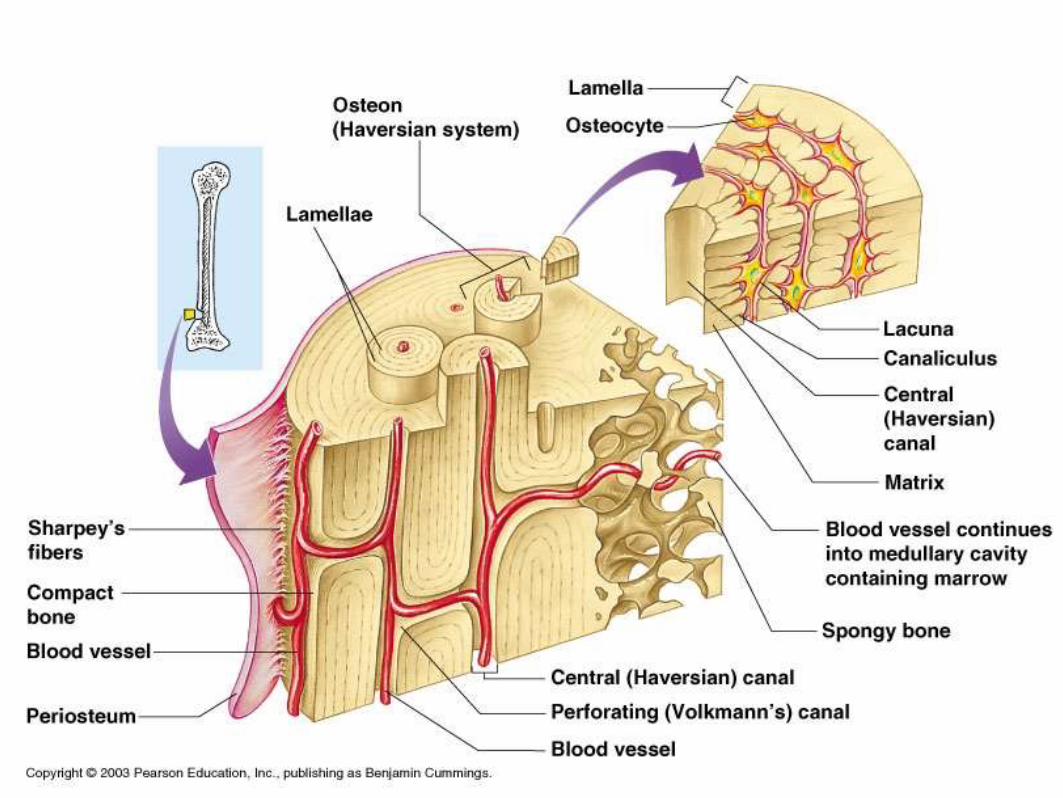
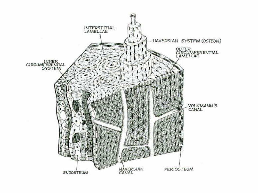
Microscopic Structure
Bone tissue is called OSSEOUS tissue
- the matrix is composed of collagen and inorganic salts
OSTEOCYTES - mature bone cells, enclosed in tiny
chambers called LACUNAE
- these form rings called lamella around a HAVERSIAN
CANAL which houses blood vessels
CANALICULI - tiny canals that link osteocytes
Haversian and Volkmann canals provide passageways for
blood vessels
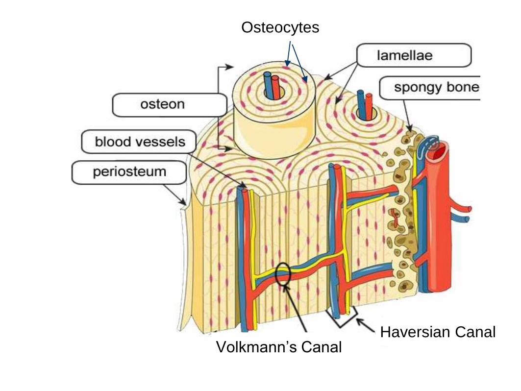
Osteocytes
Haversian CanalVolkmann’s Canal
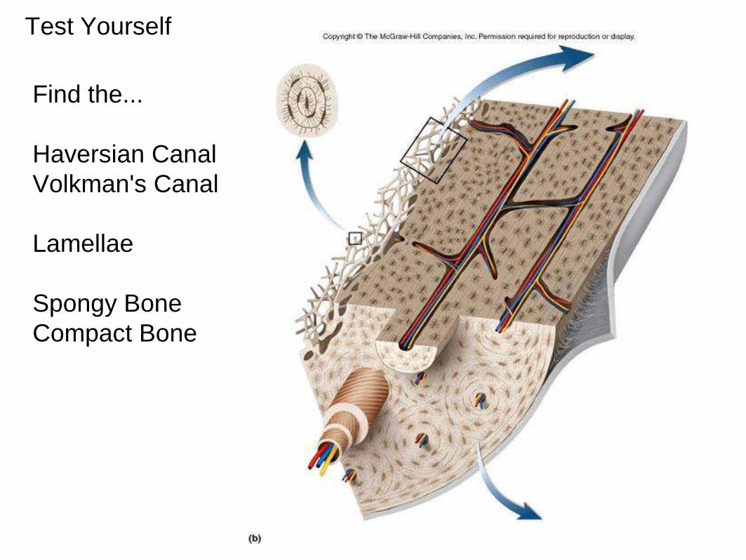
Test Yourself
Find the...
Haversian Canal
Volkman's Canal
Lamellae
Spongy Bone
Compact Bone
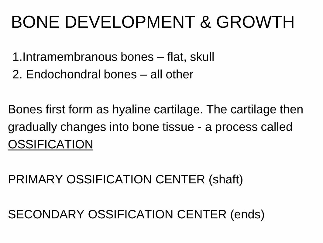
BONE DEVELOPMENT & GROWTH
1.Intramembranous bones – flat, skull
2. Endochondral bones – all other
Bones first form as hyaline cartilage. The cartilage then
gradually changes into bone tissue - a process called
OSSIFICATION
PRIMARY OSSIFICATION CENTER (shaft)
SECONDARY OSSIFICATION CENTER (ends)
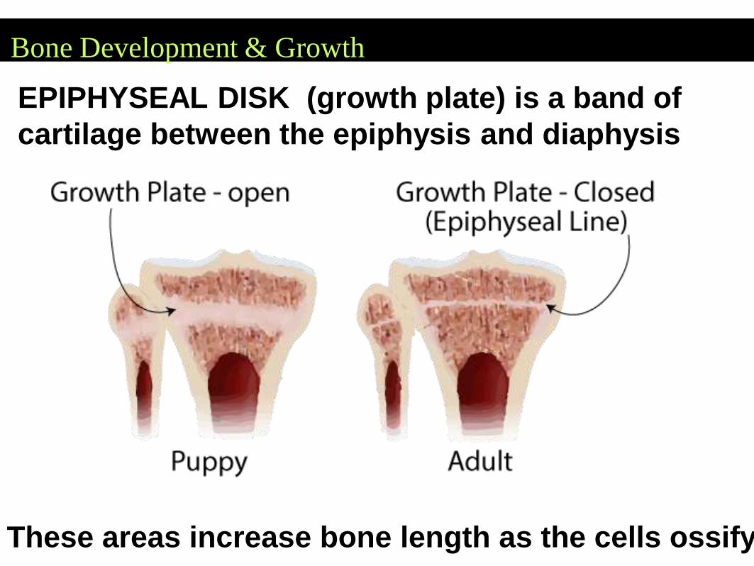
Bone Development & Growth
EPIPHYSEAL DISK (growth plate) is a band of
cartilage between the epiphysis and diaphysis
These areas increase bone length as the cells ossify
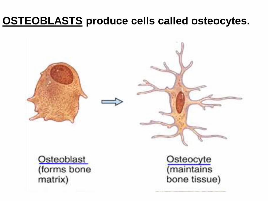
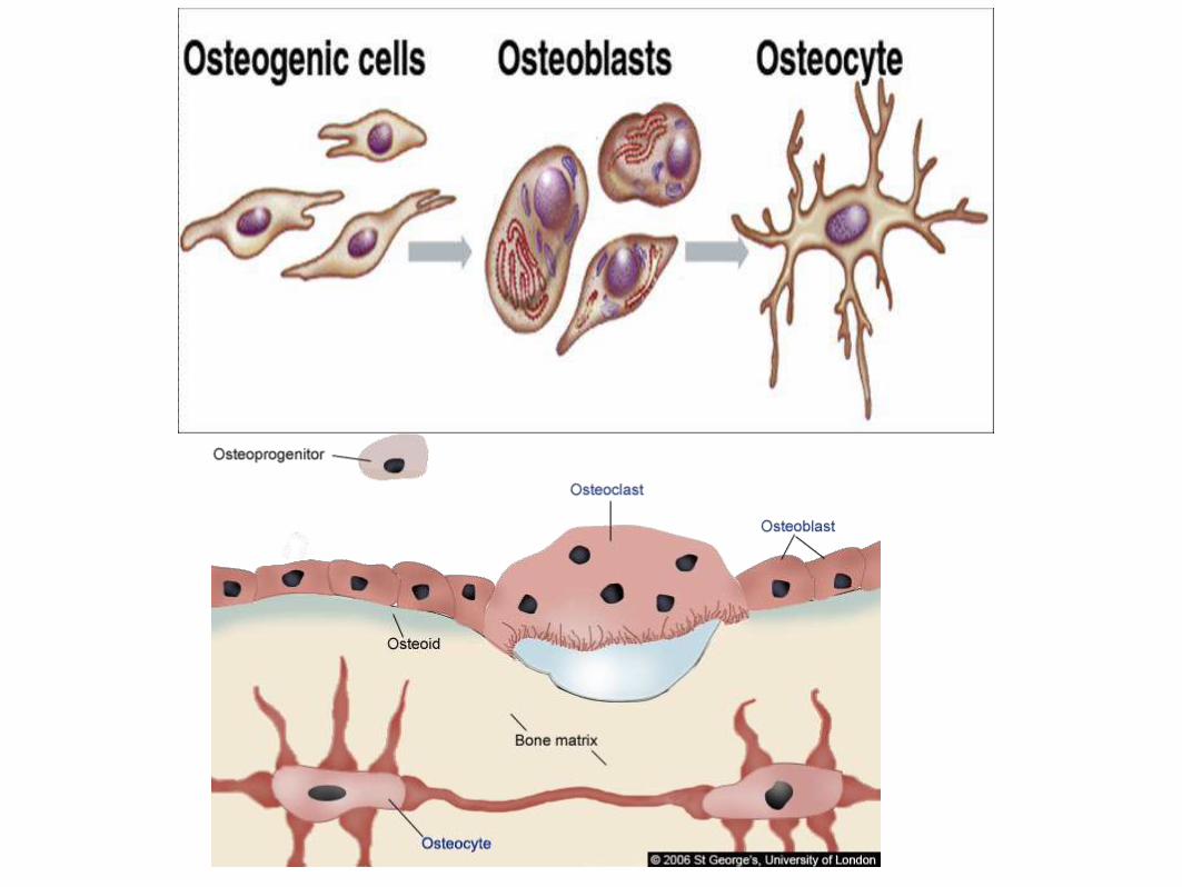
OSTEOBLASTS produce cells called osteocytes.
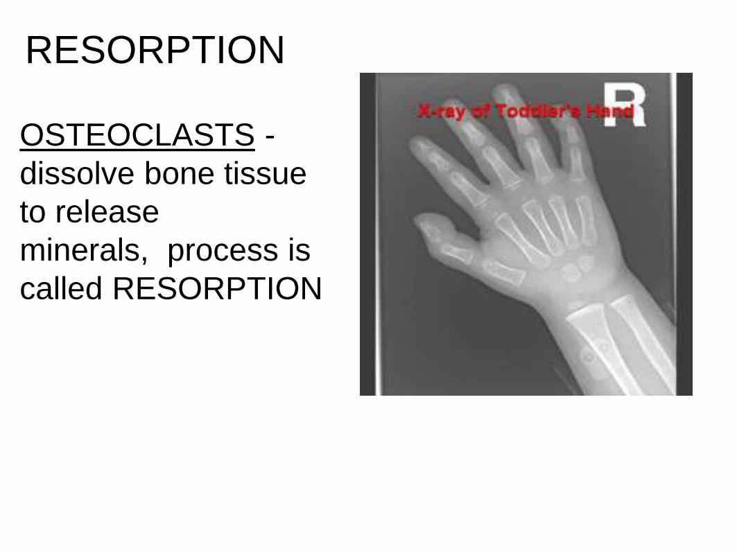
RESORPTION
OSTEOCLASTS -
dissolve bone tissue
to release
minerals, process is
called RESORPTION
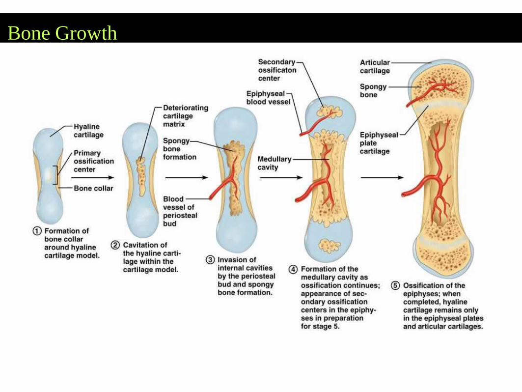
Bone Growth
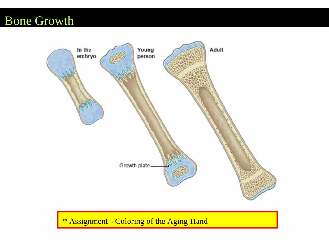
Bone Growth
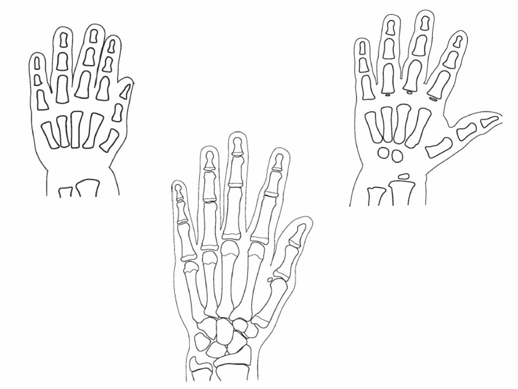
* Assignment - Coloring of the Aging Hand
Types of Joints (articulations)
Synarthrotic (not moveable, aka sutures)
*skull
Amphiarthrotic (slightly movable)
*vertebrae
Diarthrotic (moveable joint )
*knees, elbows, wrist, shoulder..etc
*synovial fluid for lubrication
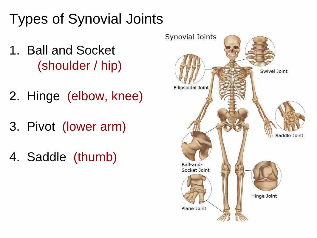
Types of Synovial Joints
1. Ball and Socket
(shoulder / hip)
2. Hinge (elbow, knee)
3. Pivot (lower arm)
4. Saddle (thumb)
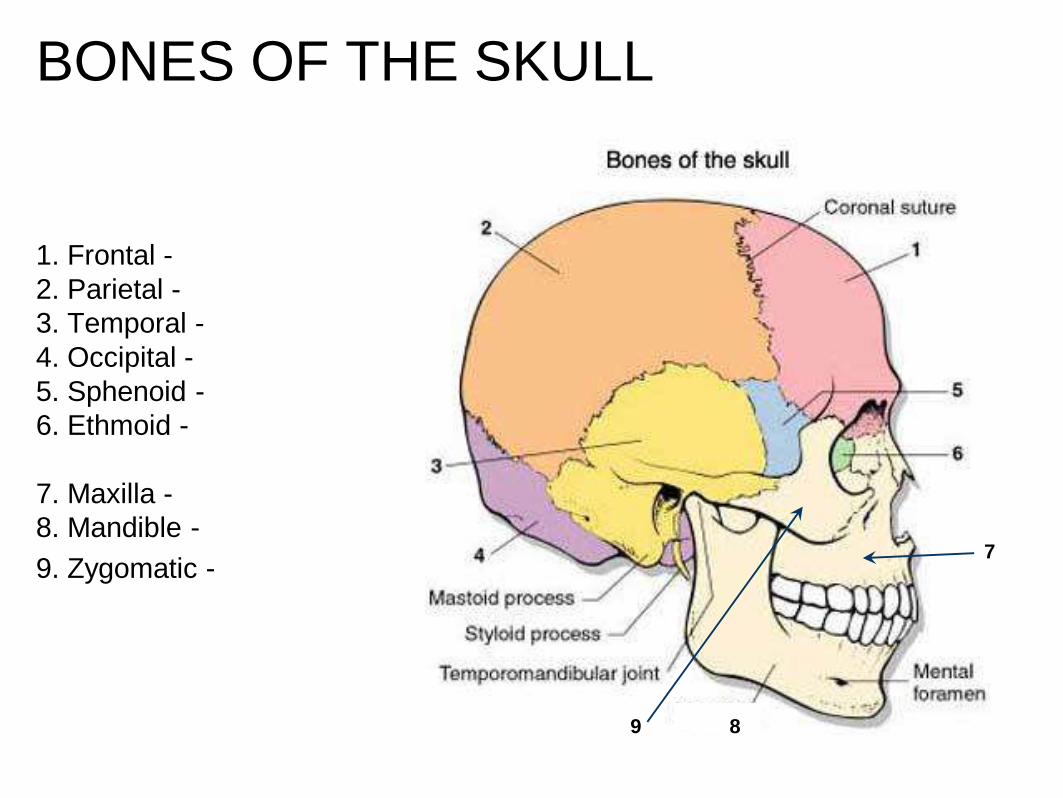
BONES OF THE SKULL
1. Frontal -
2. Parietal -
3. Temporal -
4. Occipital -
5. Sphenoid -
6. Ethmoid -
7. Maxilla -
8. Mandible -
9. Zygomatic -7
9 8
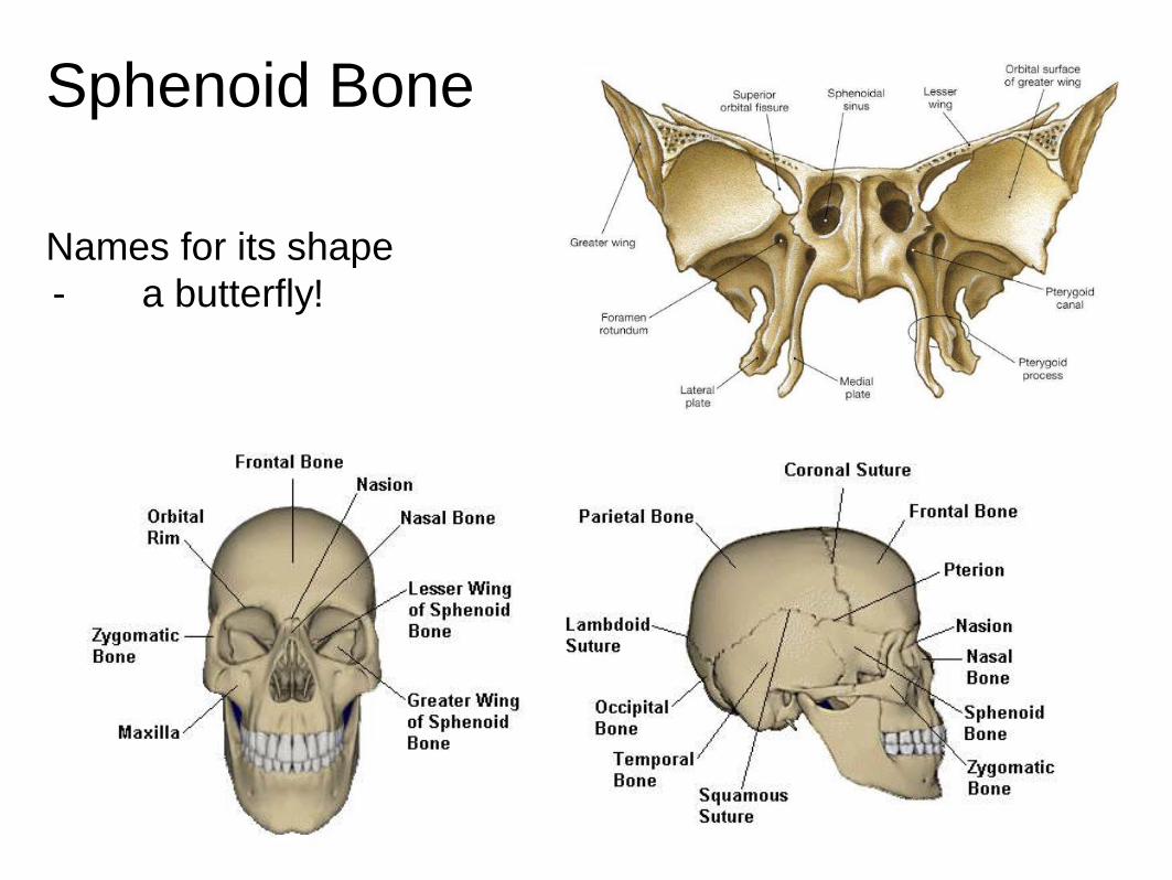
Sphenoid Bone
Names for its shape
- a butterfly!
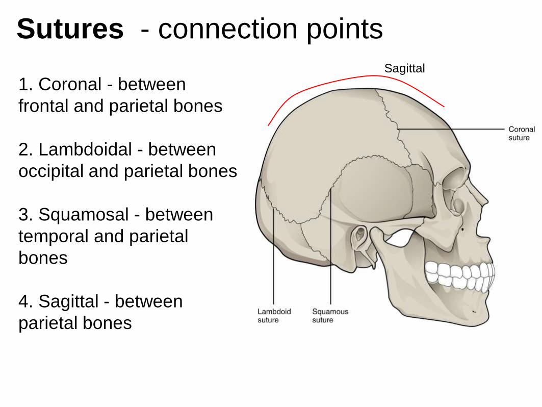
Sutures - connection points
1. Coronal - between
frontal and parietal bones
2. Lambdoidal - between
occipital and parietal bones
3. Squamosal - between
temporal and parietal
bones
4. Sagittal - between
parietal bones
Sagittal

Suture - refers to any connection between large
bones (in fetal skulls, these are called fontanels)
Fissure - any wide gap between bones

Fontanels are “soft spots” on an infant’s skull
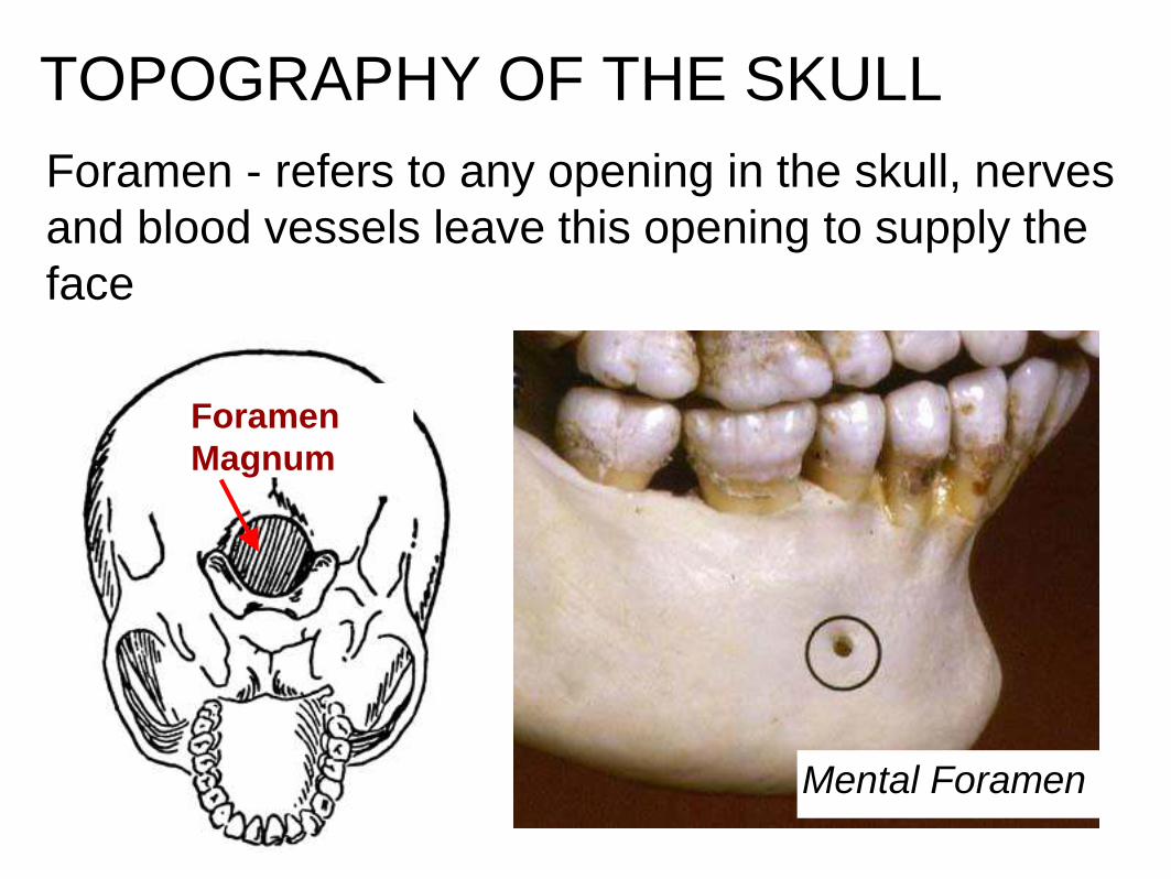
TOPOGRAPHY OF THE SKULL
Foramen - refers to any opening in the skull, nerves
and blood vessels leave this opening to supply the
face
Mental Foramen
Foramen
Magnum

Foramen Magnum
* Assignment: Skull Labeling
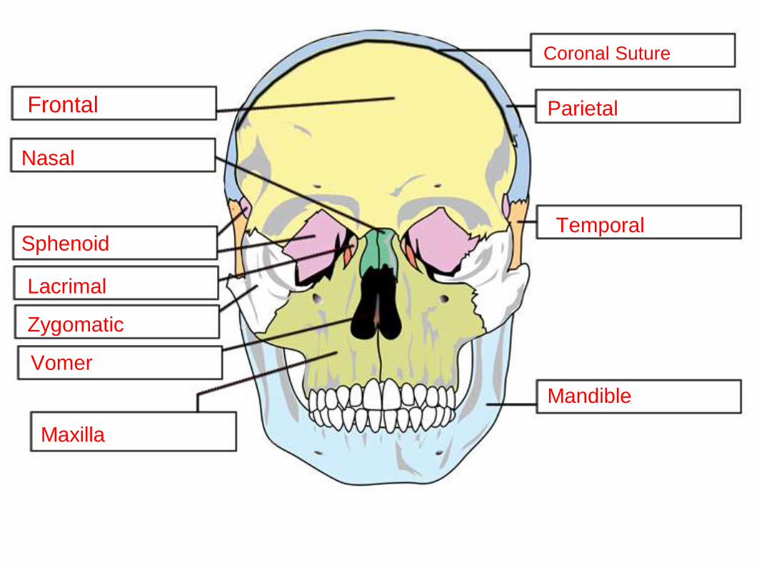
Coronal Suture
Parietal Frontal
Nasal
SphenoidTemporal
Lacrimal
Zygomatic
Vomer
Maxilla
Mandible
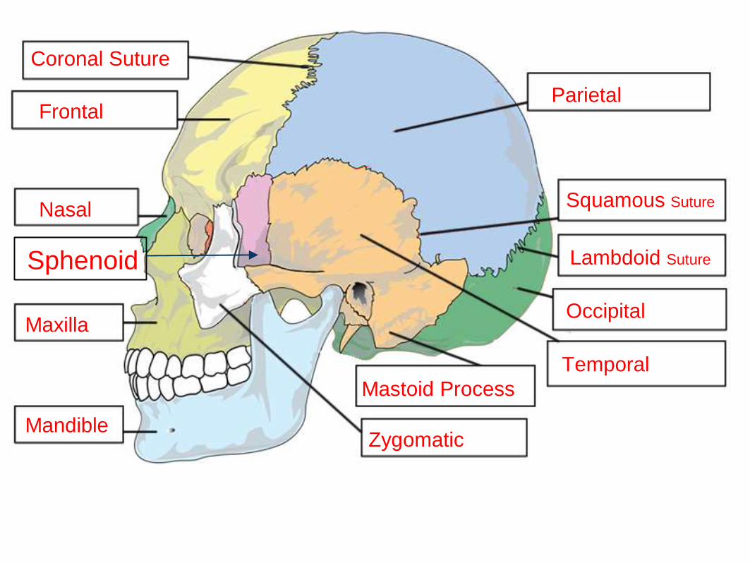
Coronal Suture
Frontal
Nasal
Maxilla
Mandible
Parietal
Squamous Suture
Lambdoid Suture
Occipital
Temporal
Mastoid Process
Zygomatic
Sphenoid
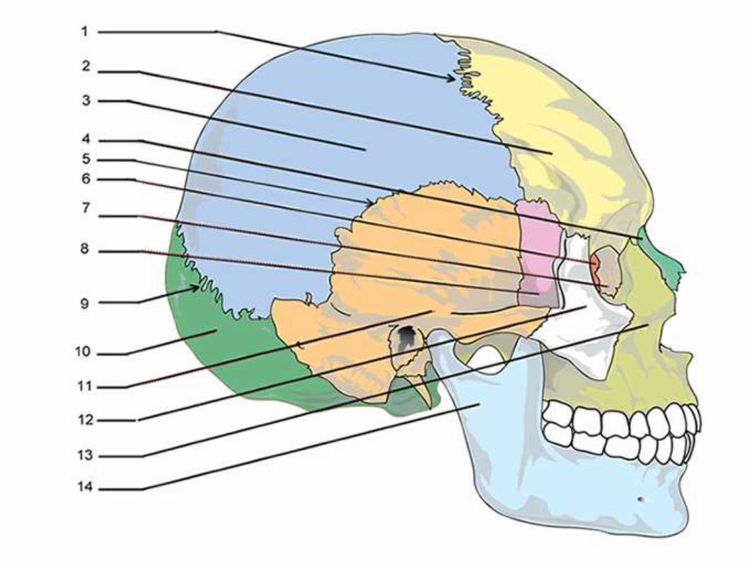
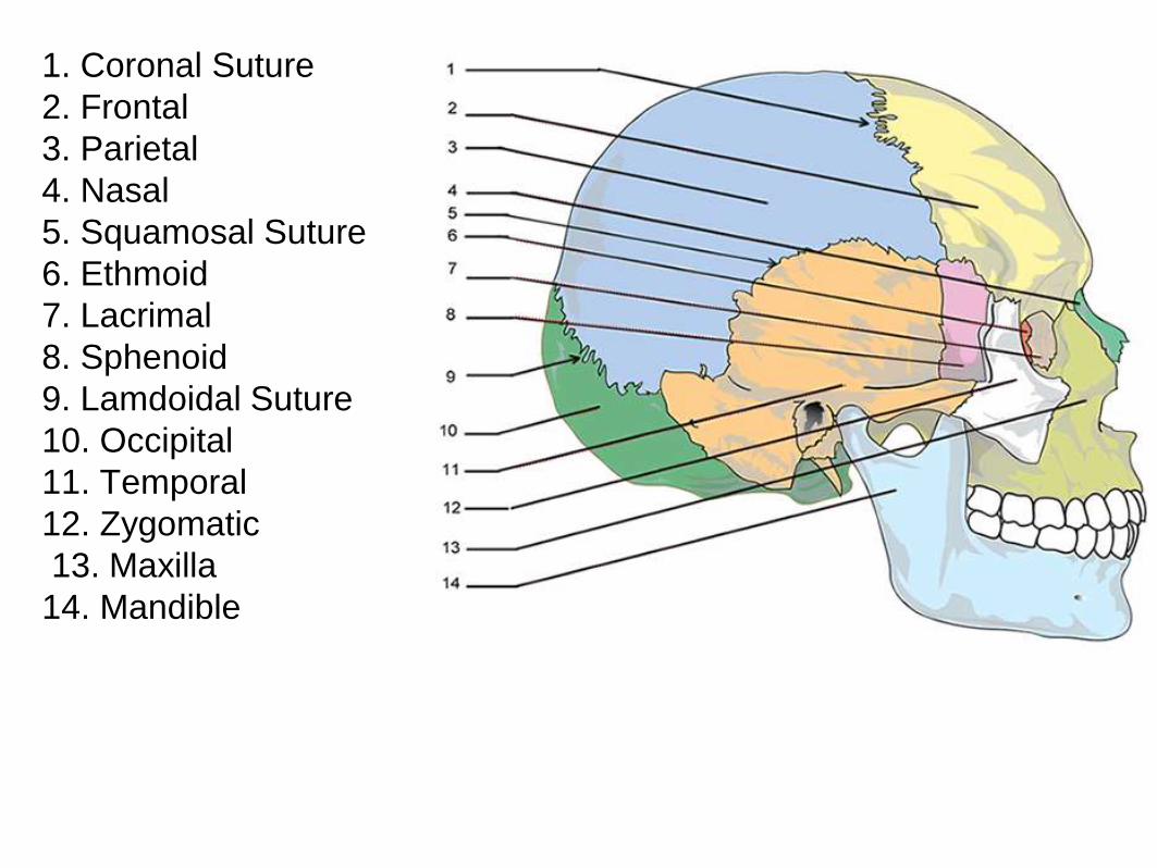
1. Coronal Suture
2. Frontal
3. Parietal
4. Nasal
5. Squamosal Suture
6. Ethmoid
7. Lacrimal
8. Sphenoid
9. Lamdoidal Suture
10. Occipital
11. Temporal
12. Zygomatic
13. Maxilla
14. Mandible
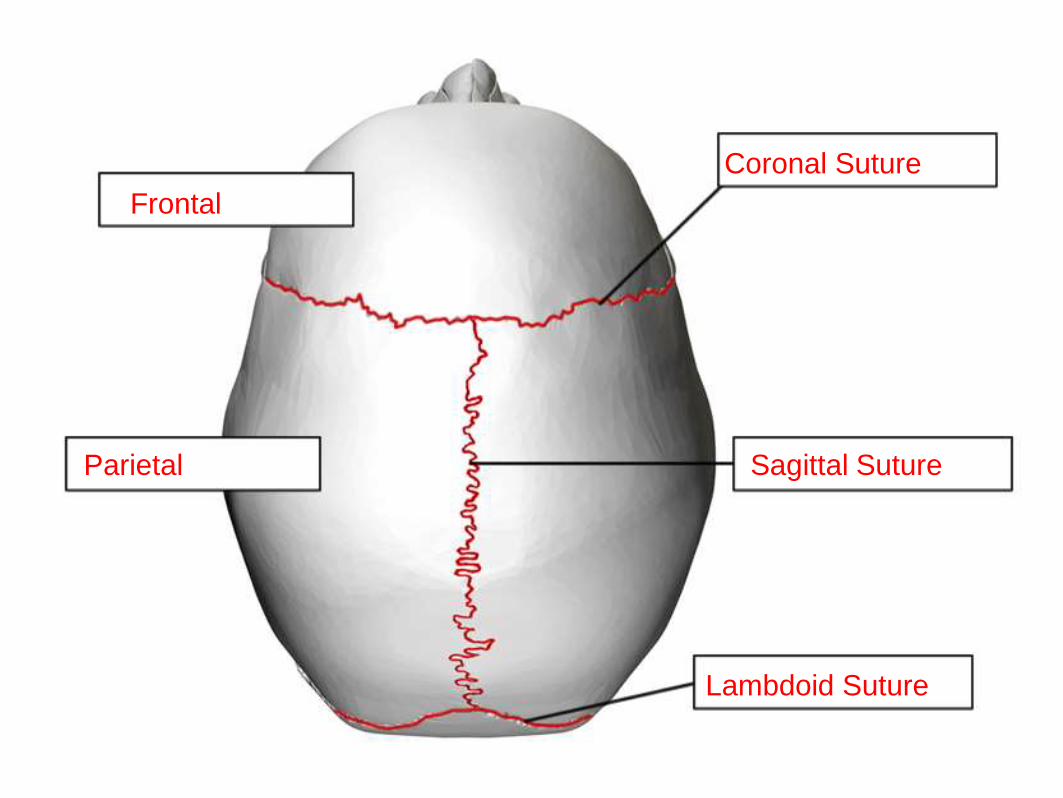
Frontal
Coronal Suture
Parietal Sagittal Suture
Lambdoid Suture
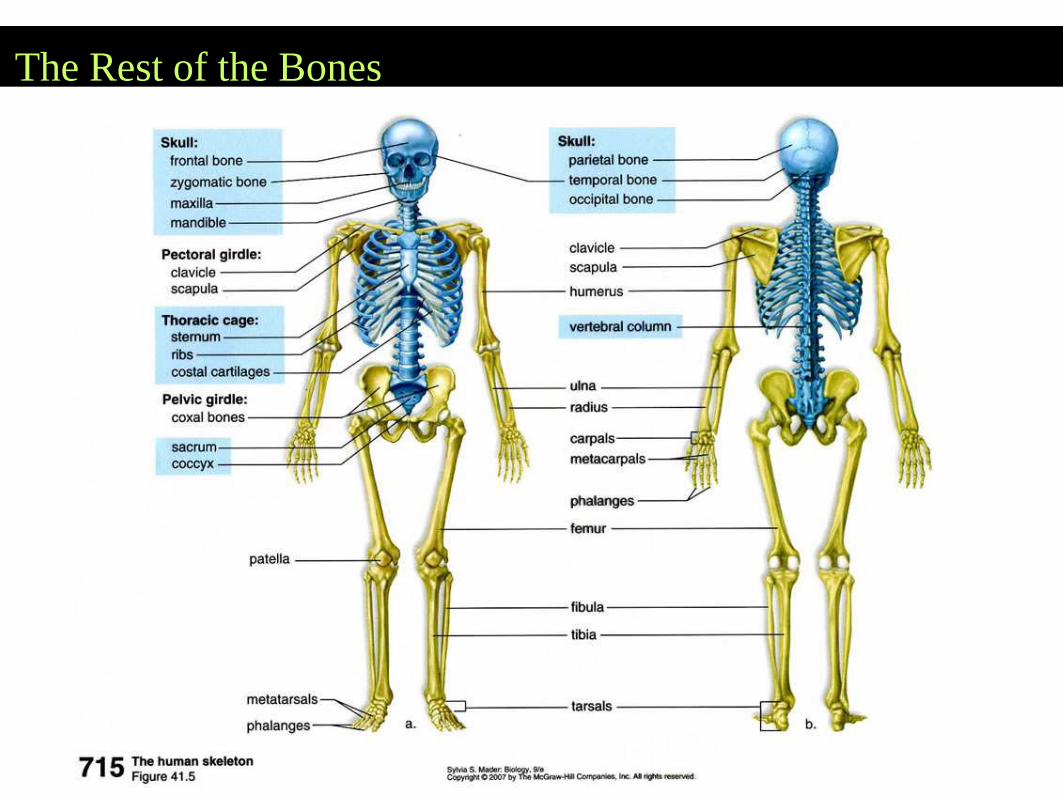
The Rest of the Bones
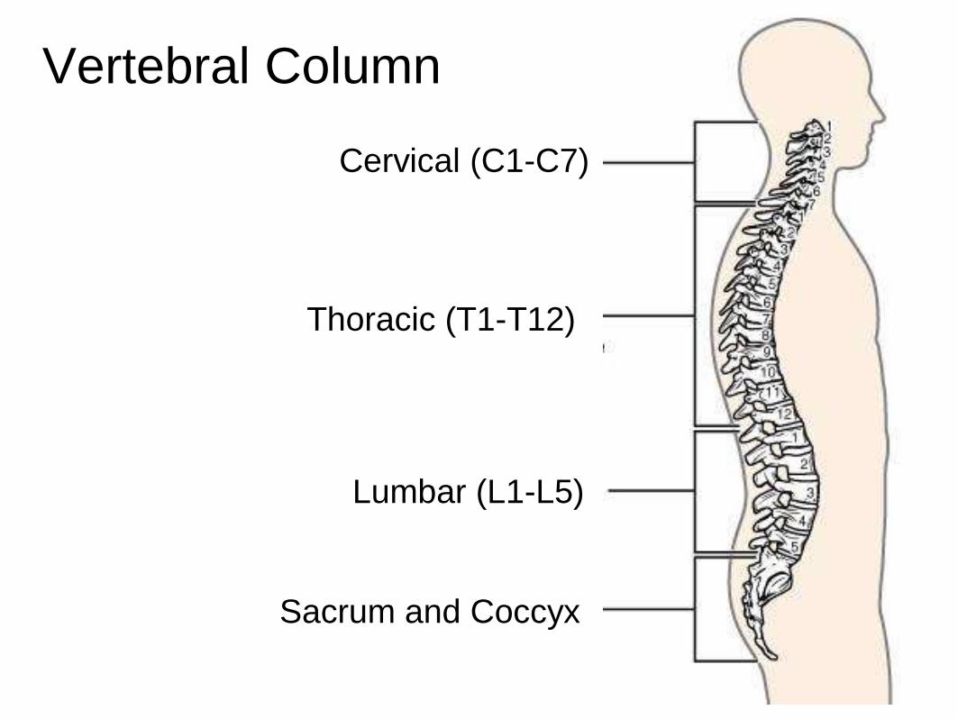
Vertebral Column
Lumbar (L1-L5)
Sacrum and Coccyx
Cervical (C1-C7)
Thoracic (T1-T12)

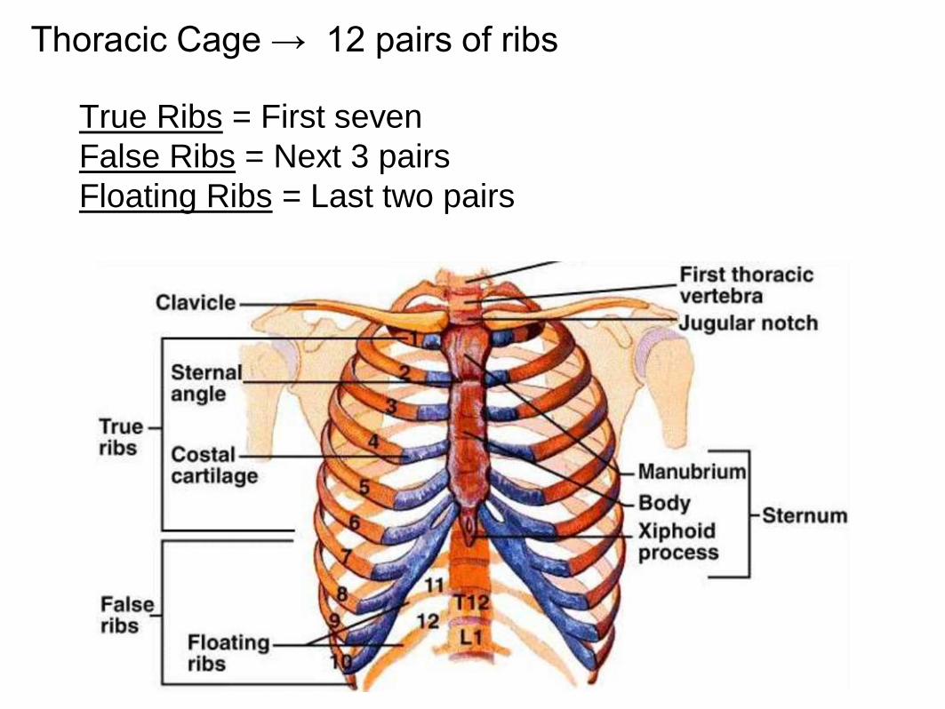
Thoracic Cage → 12 pairs of ribs
True Ribs = First seven
False Ribs = Next 3 pairs
Floating Ribs = Last two pairs
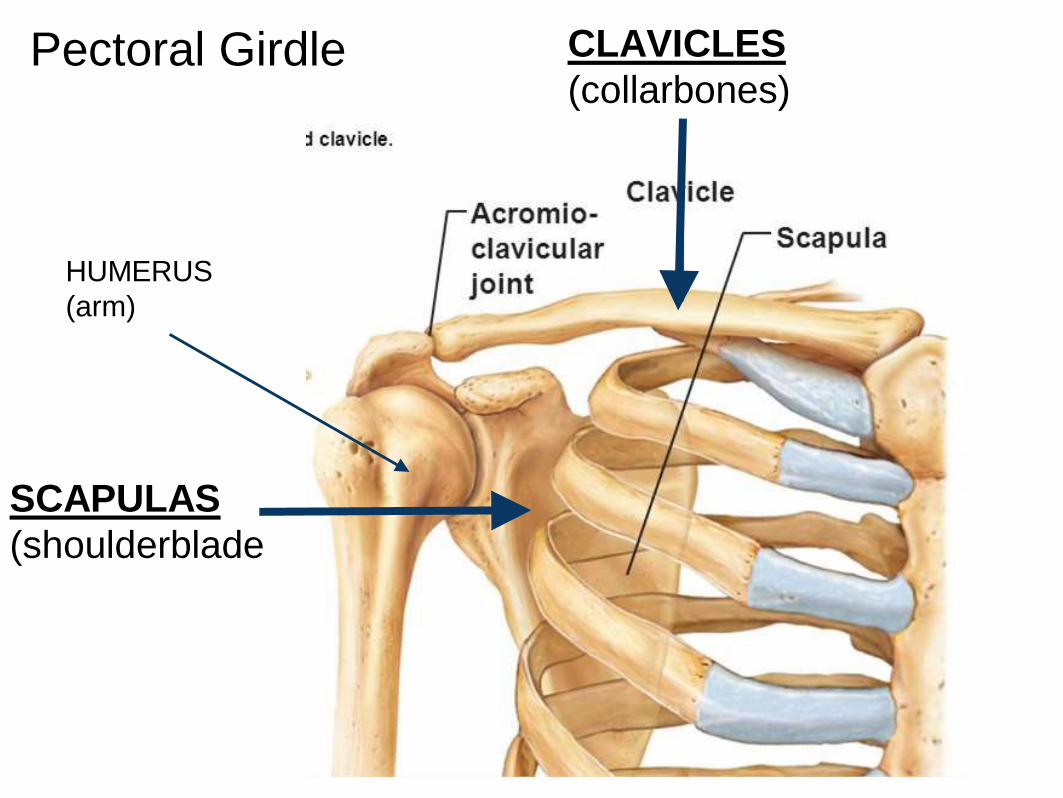
Pectoral Girdle
SCAPULAS
(shoulderblade
CLAVICLES
(collarbones)
HUMERUS
(arm)
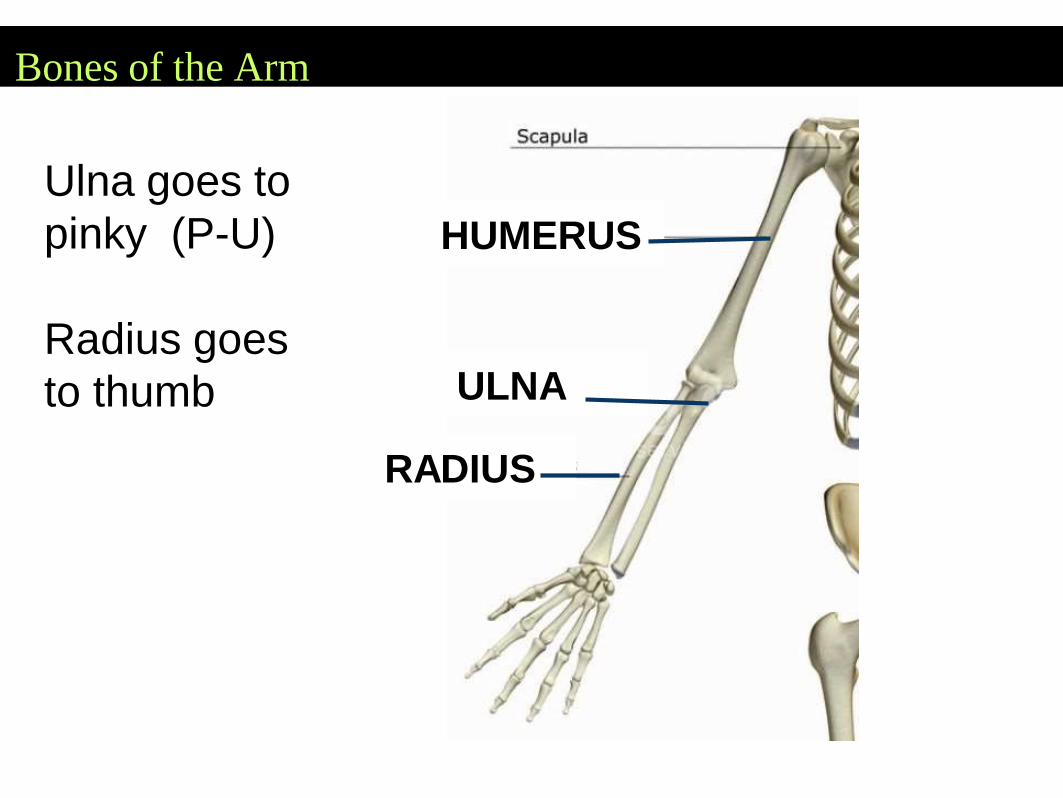
Bones of the Arm
Ulna goes to
pinky (P-U)
Radius goes
to thumb ULNA
RADIUS
HUMERUS
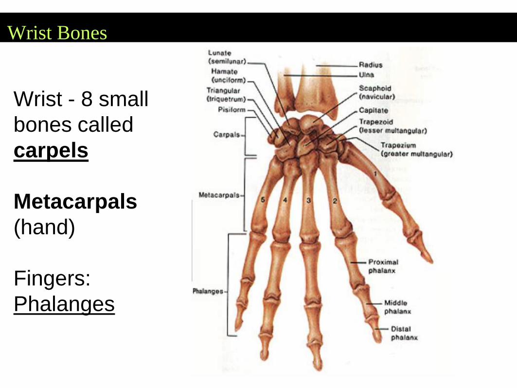
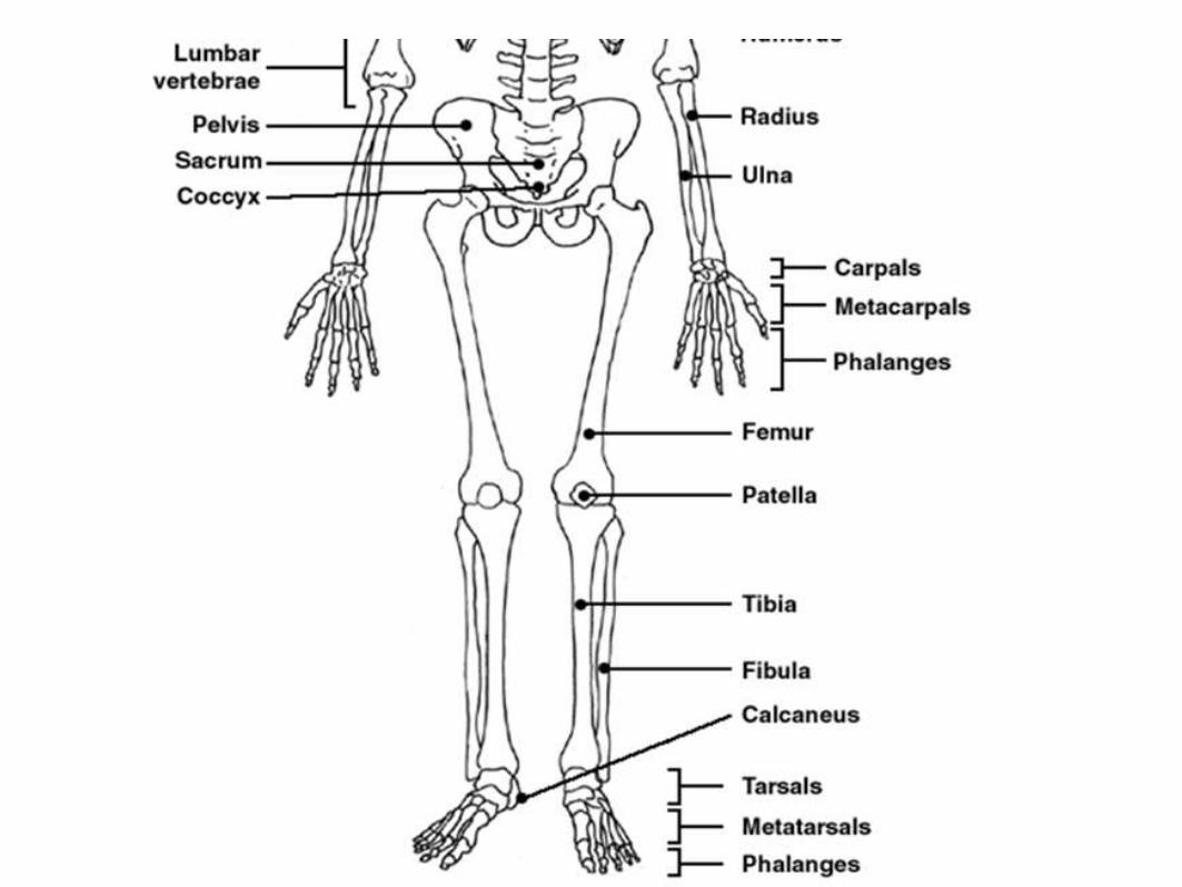
Wrist Bones
Wrist - 8 small
bones called
carpels
Metacarpals
(hand)
Fingers:
Phalanges
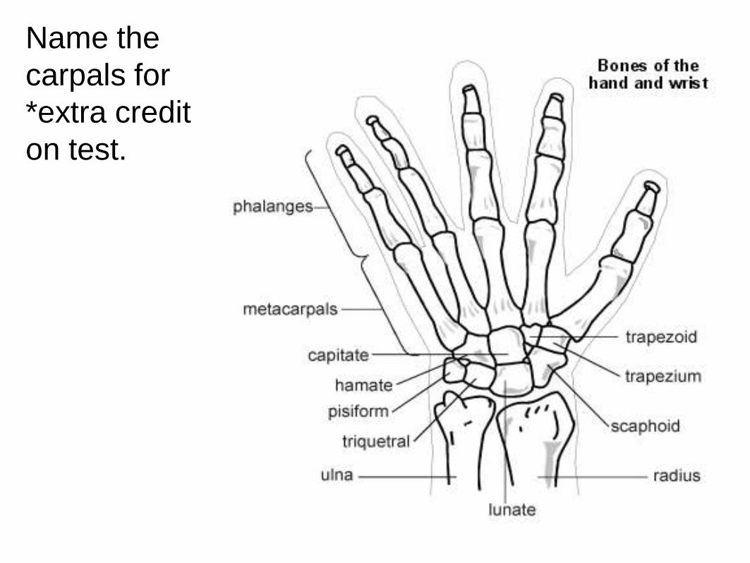
Name the
carpals for
*extra credit
on test.
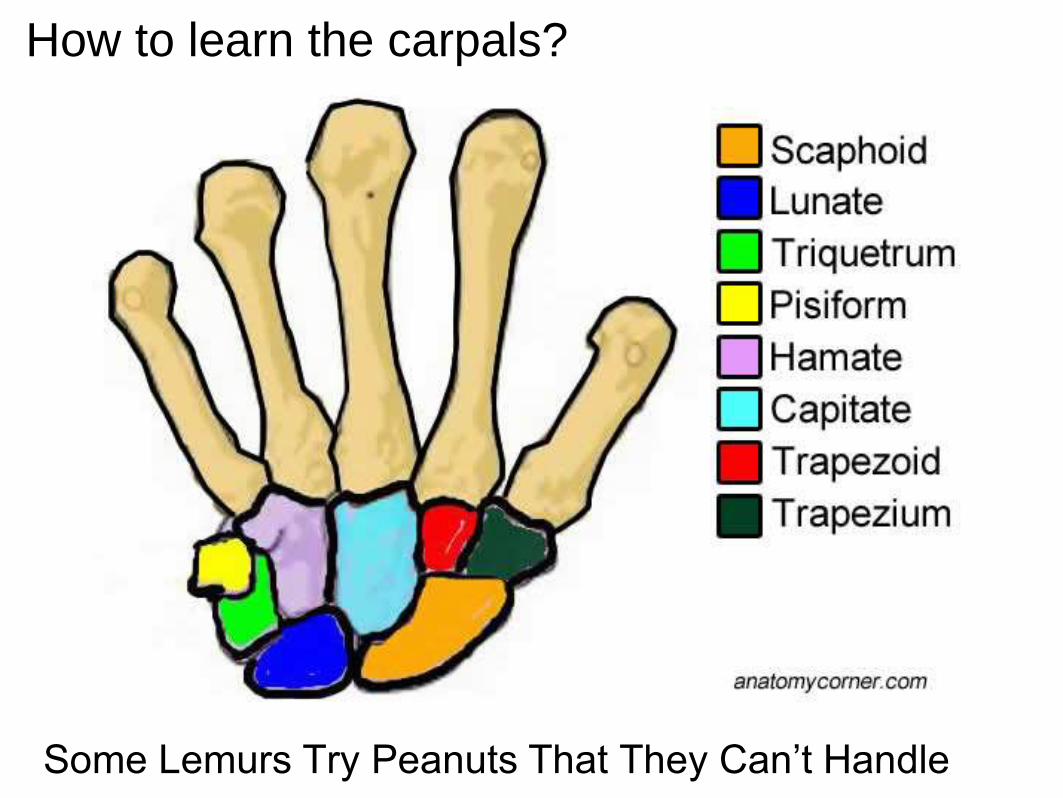
How to learn the carpals?
Some Lemurs Try Peanuts That They Can’t Handle
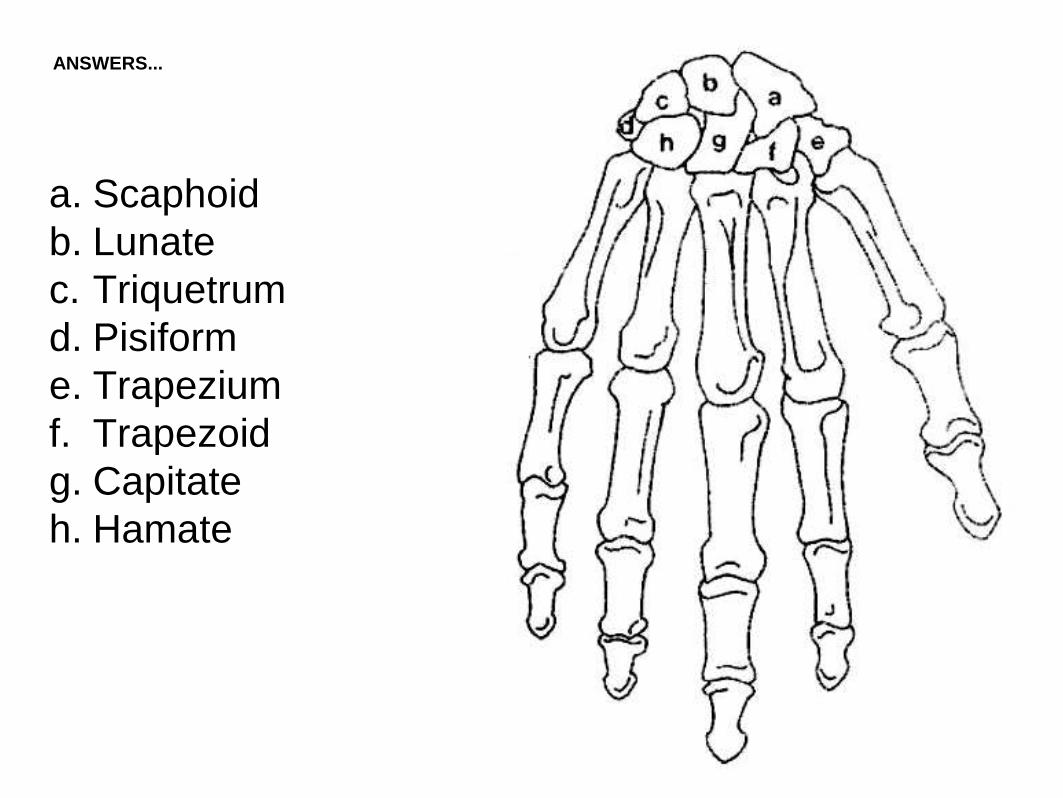
a. Scaphoid
b. Lunate
c. Triquetrum
d. Pisiform
e. Trapezium
f. Trapezoid
g. Capitate
h. Hamate
ANSWERS...
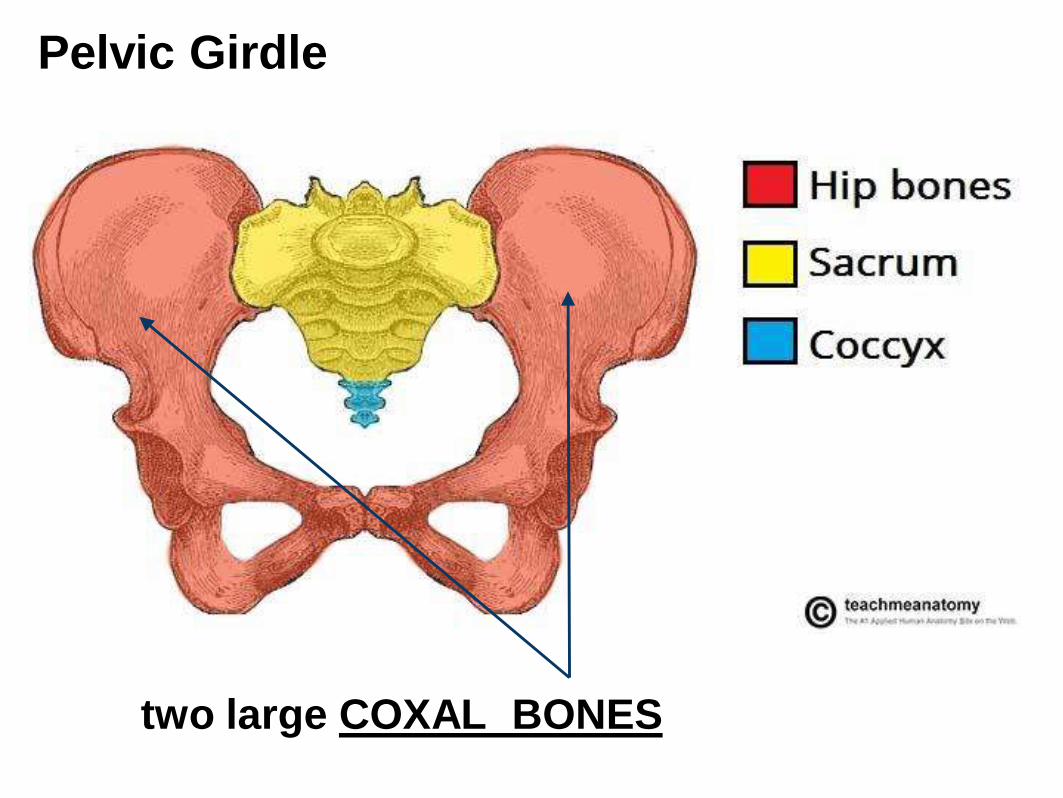
two large COXAL BONES
Pelvic Girdle
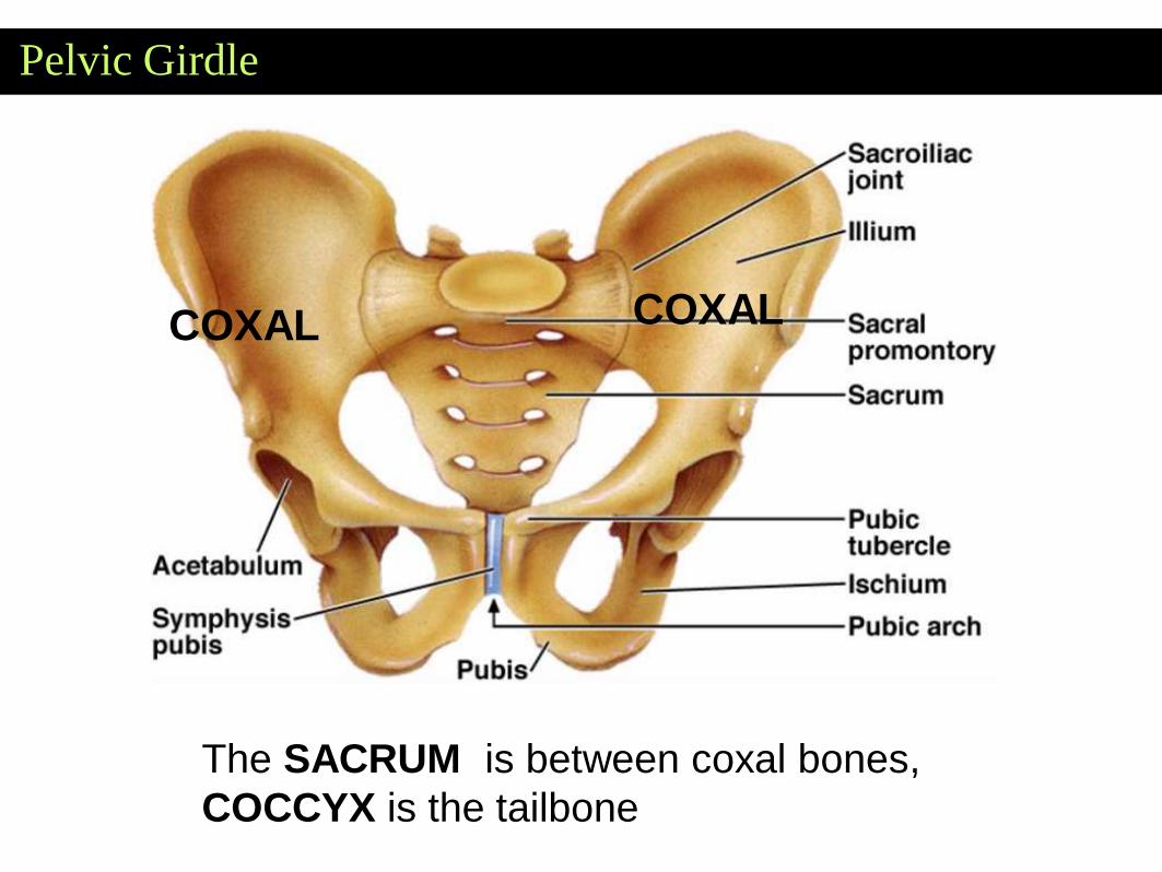
Pelvic Girdle
COXALCOXAL
The SACRUM is between coxal bones,
COCCYX is the tailbone
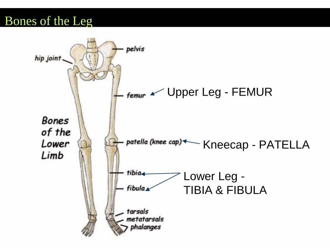
Bones of the Leg
Upper Leg - FEMUR
Lower Leg -
TIBIA & FIBULA
Kneecap - PATELLA
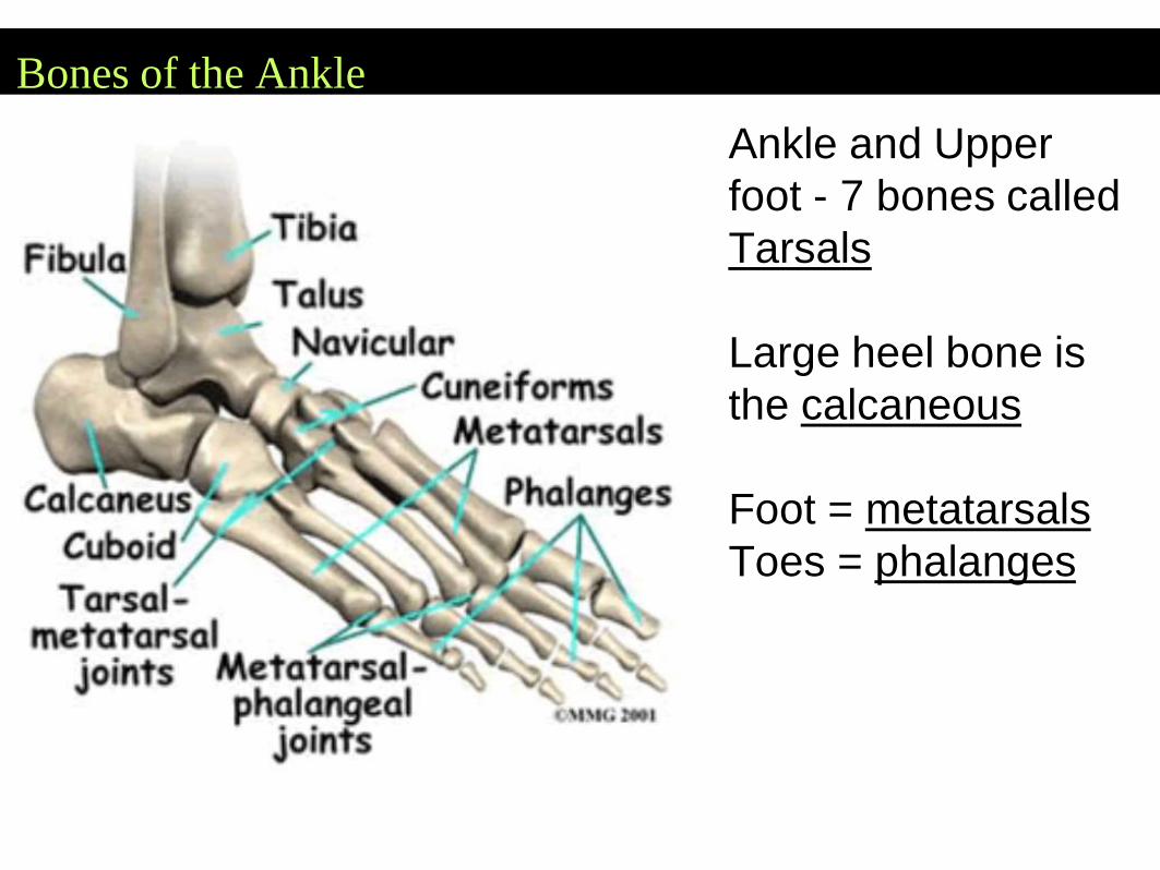
Bones of the Ankle
Ankle and Upper
foot - 7 bones called
Tarsals
Large heel bone is
the calcaneous
Foot = metatarsals
Toes = phalanges
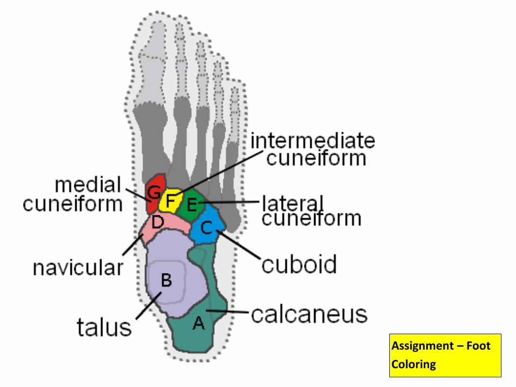
Assignment – Foot
Coloring
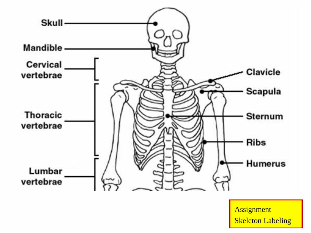
Assignment –
Skeleton Labeling
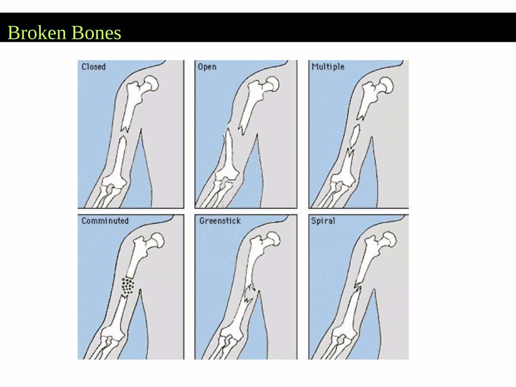
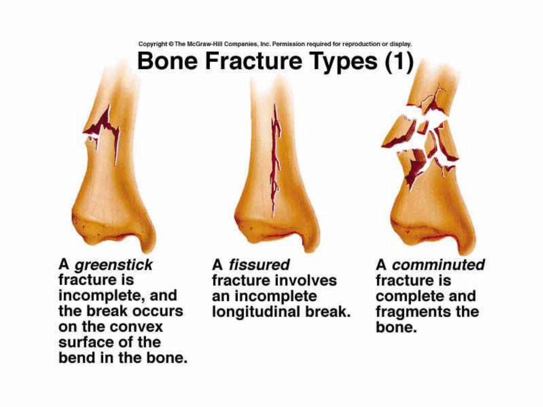
Broken Bones
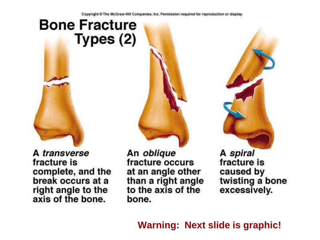
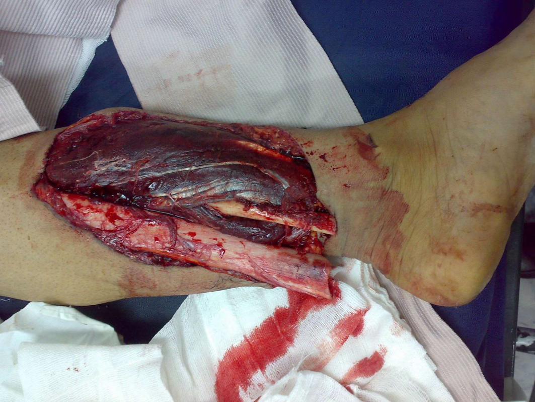
Warning: Next slide is graphic!
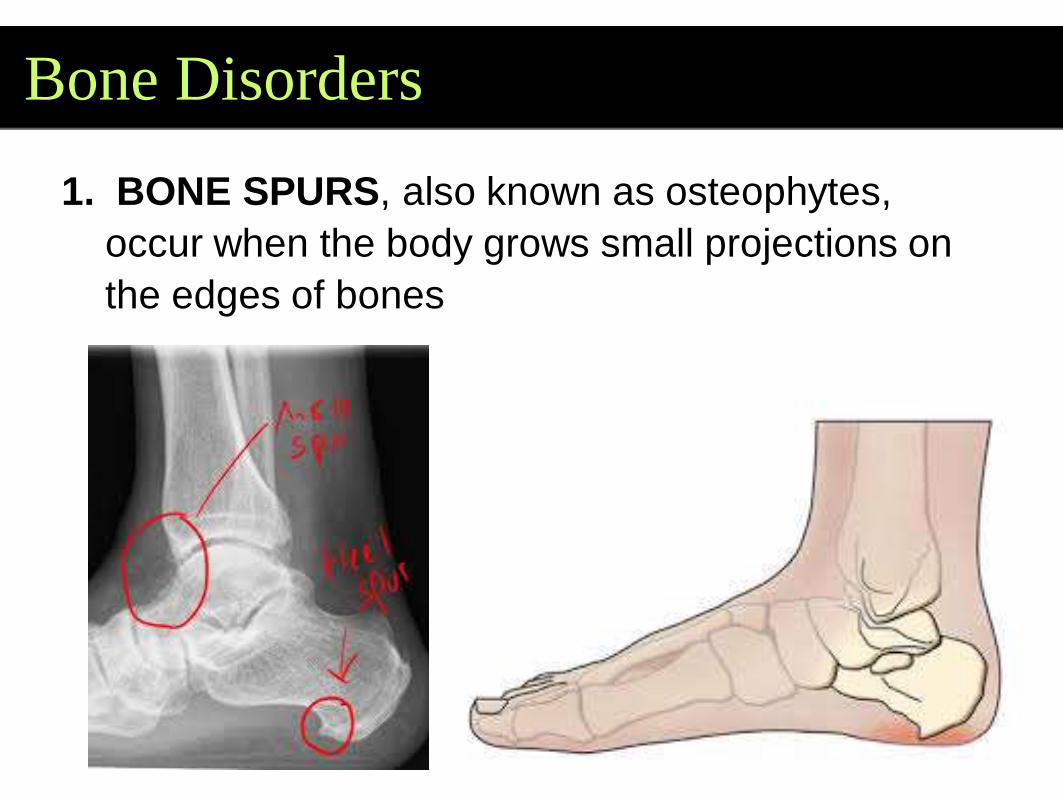
Bone Disorders
1. BONE SPURS, also known as osteophytes,
occur when the body grows small projections on
the edges of bones
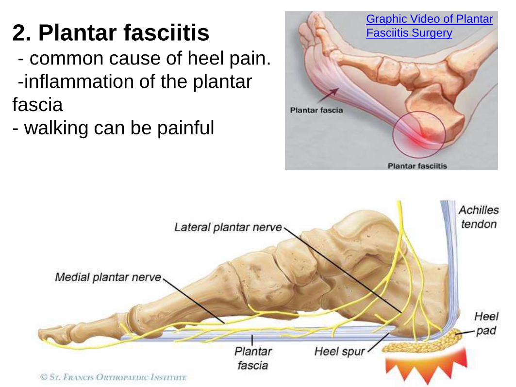
2. Plantar fasciitis - common cause of heel pain.
-inflammation of the plantar
fascia
- walking can be painful
Graphic Video of Plantar
Fasciitis Surgery
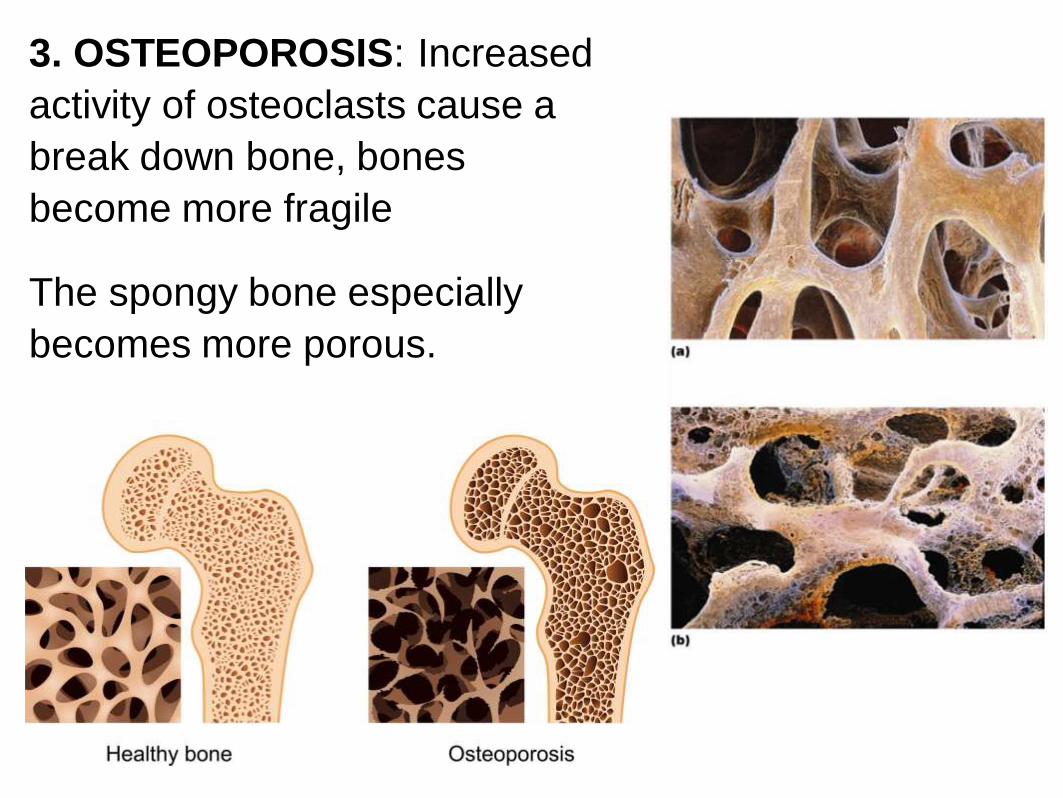
3. OSTEOPOROSIS: Increased
activity of osteoclasts cause a
break down bone, bones
become more fragile
The spongy bone especially
becomes more porous.
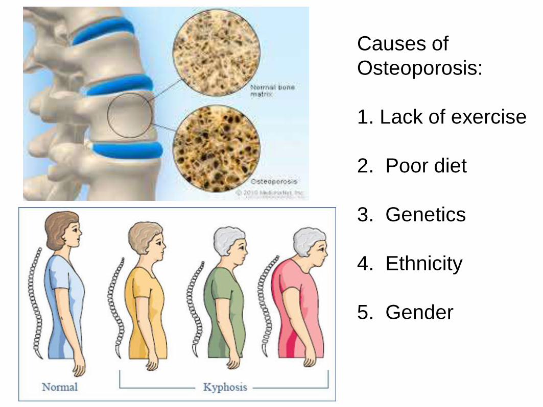
Causes of
Osteoporosis:
1. Lack of exercise
2. Poor diet
3. Genetics
4. Ethnicity
5. Gender
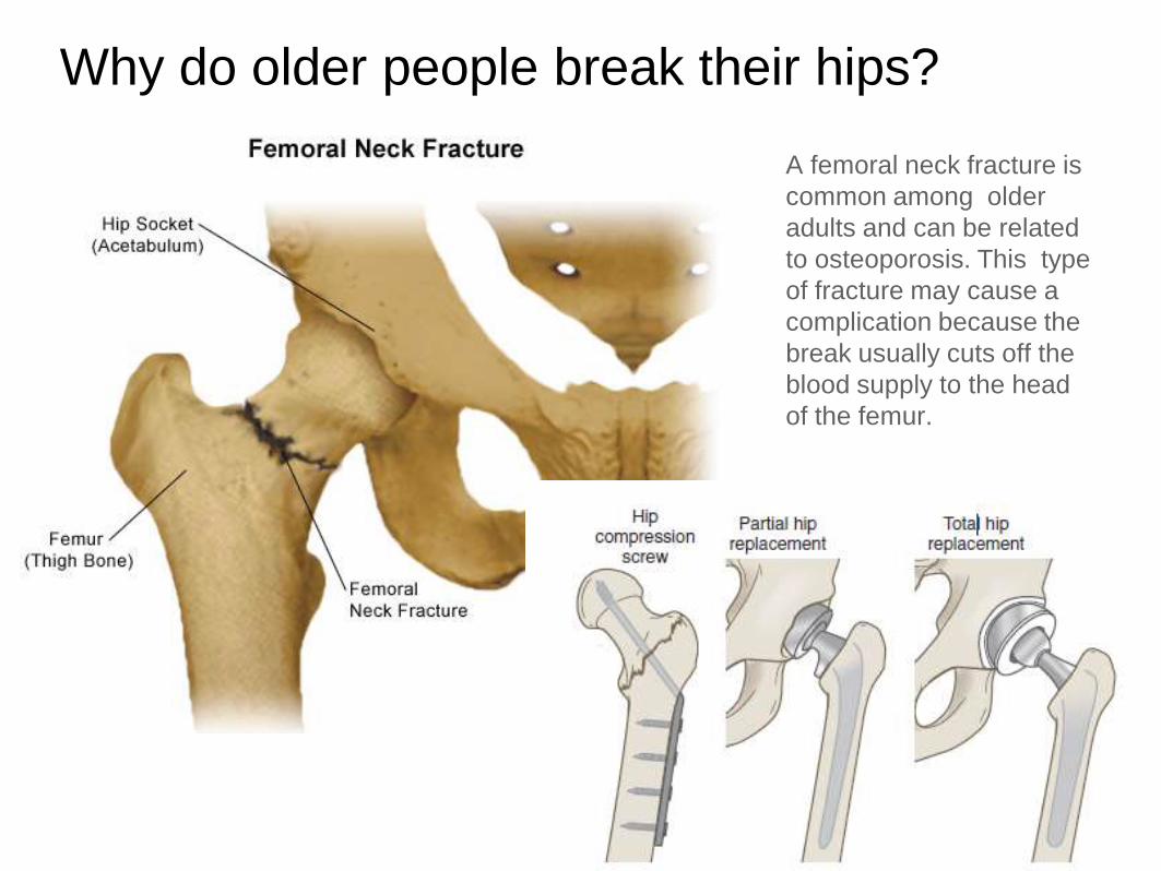
Why do older people break their hips?
A femoral neck fracture is
common among older
adults and can be related
to osteoporosis. This type
of fracture may cause a
complication because the
break usually cuts off the
blood supply to the head
of the femur.
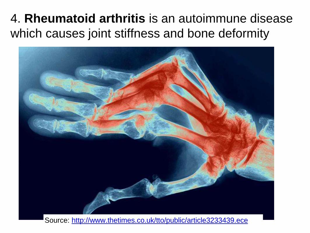
4. Rheumatoid arthritis is an autoimmune disease
which causes joint stiffness and bone deformity
Source: http://www.thetimes.co.uk/tto/public/article3233439.ece
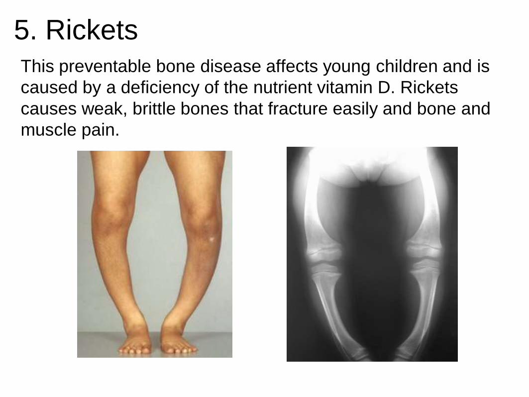
5. RicketsThis preventable bone disease affects young children and is
caused by a deficiency of the nutrient vitamin D. Rickets
causes weak, brittle bones that fracture easily and bone and
muscle pain.
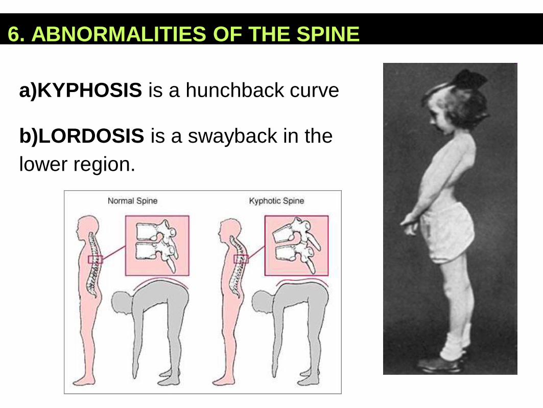
6. ABNORMALITIES OF THE SPINE
a)KYPHOSIS is a hunchback curve
b)LORDOSIS is a swayback in the
lower region.
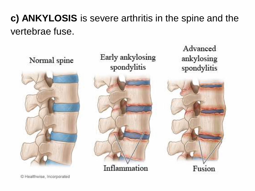
c) ANKYLOSIS is severe arthritis in the spine and the
vertebrae fuse.
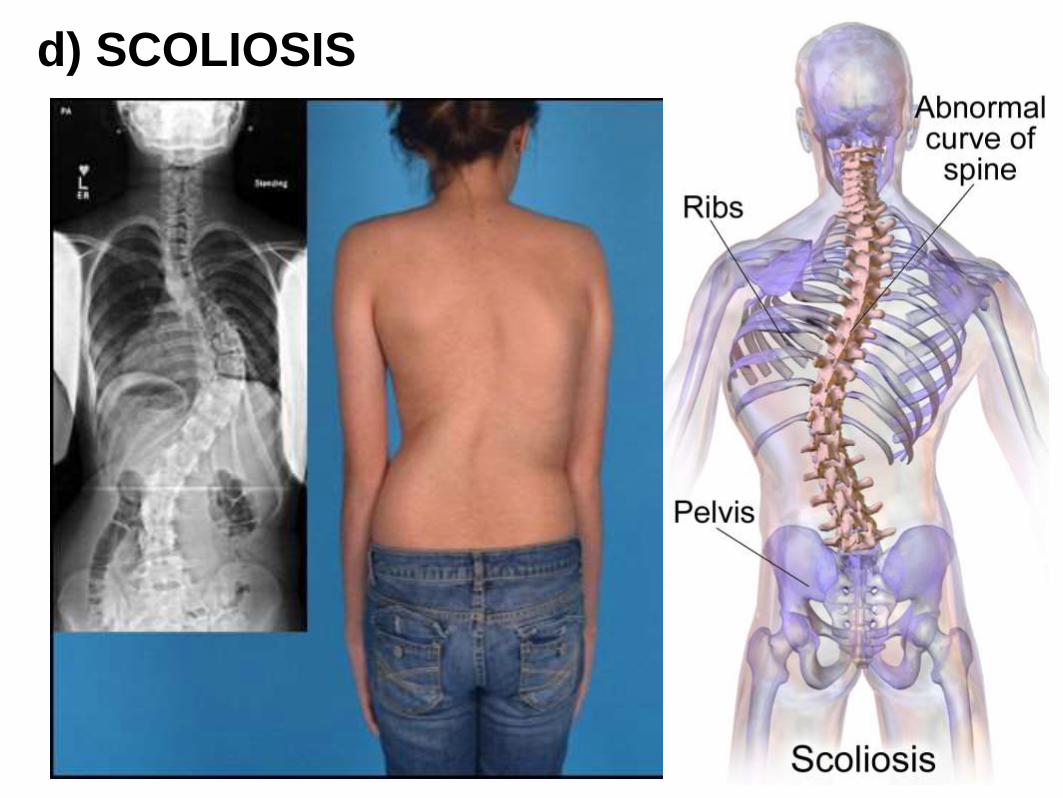
d) SCOLIOSIS

7. Fibrodysplasia ossificans progressiva (FOP) →
soft tissue regrows as bone. Sufferers are slowly imprisoned by
their own skeletons.
Munchmeyer disease" or "stone man syndrome"
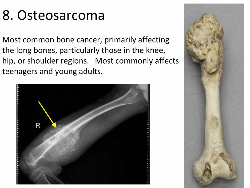
8. Osteosarcoma
Most common bone cancer, primarily affecting the long bones, particularly those in the knee, hip, or shoulder regions. Most commonly affects teenagers and young adults.
FUN FACTS ABOUT BONES
Bone is made of the same type of minerals as
limestone.
●Babies are born with 300 bones, but by
adulthood we have only 206 in our bodies.
●The giraffe has the same number of bones
in its neck as a human: seven in total.
●The long horned ram can take a head butt
at 25 mph. The human skull will fracture at
5 mph.