The Skeletal System: Structure, Function. The Skeletal System Copyright © 2003 Pearson Education,...
45
The Skeletal System: Structure, Function
-
Upload
raymond-beverly-horn -
Category
Documents
-
view
231 -
download
2
Transcript of The Skeletal System: Structure, Function. The Skeletal System Copyright © 2003 Pearson Education,...
The Skeletal System:
Structure, Function

Is this the correct anatomical position?

The Skeletal System
Copyright © 2003 Pearson Education, Inc. publishing as Benjamin Cummings
· Parts of the skeletal system· Bones (skeleton)
· Joints
· Cartilages
· Ligaments (bone to bone)(tendon=bone to muscle)
· Divided into two divisions· Axial skeleton- skull, spinal column
· Appendicular skeleton – limbs and girdle

Functions of Bones
Copyright © 2003 Pearson Education, Inc. publishing as Benjamin Cummings
· Support of the body
· Protection of soft organs
· Movement due to attached skeletal muscles
· Storage of minerals and fats
· Blood cell formation

Bones of the Human Body
Copyright © 2003 Pearson Education, Inc. publishing as Benjamin Cummings
· The skeleton has 206 bones
· Two basic types of bone tissue· Compact bone
· Homogeneous
· Spongy bone· Small needle-like
pieces of bone
· Many open spaces Figure 5.2b

Bones are classified by their shape:
1.Long- bones are longer than they are wide (arms, legs)
2.Short- usually square in shape, cube like (wrist, ankle)
3.Flat- flat , curved (skull, Sternum)
4.Irregular- odd shapes (vertebrae, pelvis)

Classification of Bones on the Basis of Shape
Copyright © 2003 Pearson Education, Inc. publishing as Benjamin Cummings
Figure 5.1

Types of Bone Cells
Copyright © 2003 Pearson Education, Inc. publishing as Benjamin Cummings
· Osteocytes· Mature bone cells
· Osteoblasts· Bone-forming cells
· Osteoclasts· Bone-destroying cells· Break down bone matrix for remodeling and
release of calcium· Bone remodeling is a process by both
osteoblasts and osteoclasts

Changes in the Human Skeleton
Copyright © 2003 Pearson Education, Inc. publishing as Benjamin Cummings
· In embryos, the skeleton is primarily hyaline cartilage
· During development, much of this cartilage is replaced by bone
· Cartilage remains in isolated areas· Bridge of the nose
· Parts of ribs
· Joints

The formation of blood cells, (hematopoiesis), takes place mainly in the red marrow of the bones.
In infants, red marrow is found in the bone cavities. With age, it is largely replaced by yellow marrow for fat storage.
In adults, red marrow is limited to the spongy bone in the skull, ribs, sternum, clavicles, vertebrae and pelvis. Red marrow functions in the formation of red blood cells, white blood cells and blood platelets.
Red and Yellow Bone Marrow

The Axial Skeleton
Slide 5.20a
Copyright © 2003 Pearson Education, Inc. publishing as Benjamin Cummings
· Forms the longitudinal part of the body
· Divided into three parts· Skull
· Vertebral Column
· Rib Cage

A joint, or articulation, is the place where two bones come together.
Fibrous- Immovable:connect bones, no movement. (skull and pelvis).
Cartilaginous- slightly movable, bones are attached by cartilage, a little movement (spine or ribs).
Synovial- freely movable, much more movement than cartilaginous joints. Cavities between bones are filled with synovial fluid. This fluid helps lubricate and protect the bones.
Joints

Where bone meets bone Function: They hold the bones together. They give mobility to the rigid skeleton.
Joints

The Synovial Joint
Slide 5.51Copyright © 2003 Pearson Education, Inc. publishing as Benjamin Cummings
Figure 5.28

Types of Synovial Joints Based on Shape
Slide 5.52a
Copyright © 2003 Pearson Education, Inc. publishing as Benjamin Cummings
Figure 5.29a–c

Types of Synovial Joints Based on Shape
Slide 5.52b
Copyright © 2003 Pearson Education, Inc. publishing as Benjamin Cummings
Figure 5.29d–f

Hinge- A hinge joint allows extension and retraction of an appendage. (Elbow, Knee)
Types of Joints

Ball and Socket- A ball and socket joint allows for radial movement in almost any direction. They are found in the hips and shoulders. (Hip, Shoulder)

Gliding- In a gliding or plane joint bones slide past each other. Mid-carpal and mid-tarsal joints are gliding joints. (Hands, Feet)

Saddle- This type of joint occurs when the touching surfaces of two bones have both concave and convex regions with the shapes of the two bones complementing one other and allowing a wide range of movement. (Thumb)
Ligamentsstrong cords of fibrous tissue connect
bones

Cartilage
Cartilage covers ends of movable bones Reduces friction
Lubricated by fluid from capillaries

Body movement (Locomotion) Maintenance of posture Respiration
◦ Diaphragm and intercostal contractions Communication (Verbal and Facial) Constriction of organs and vessels
◦ Peristalsis of intestinal tract◦ Vasoconstriction of b.v. and other structures
(pupils) Heart beat Production of body heat (Thermogenesis)
Muscular System Functions

Made up of bundles of muscle fibers Provide the force to move bones Assist in maintaining posture Assist with heat production
CHARACTERISTICS OF MUSCLES

Organs of Muscular System◦ Muscles
Muscular System

Excitability: capacity of muscle to respond to a stimulus
Contractility: ability of a muscle to shorten and generate pulling force
Extensibility: muscle can be stretched back to its original length
Elasticity: ability of muscle to recoil to original resting length after stretched
Properties of Muscle

Medical Terminology: A Living Language, Fourth EditionBonnie F. Fremgen and Suzanne S. Frucht
Copyright ©2009 by Pearson Education, Inc.Upper Saddle River, New Jersey 07458
All rights reserved.
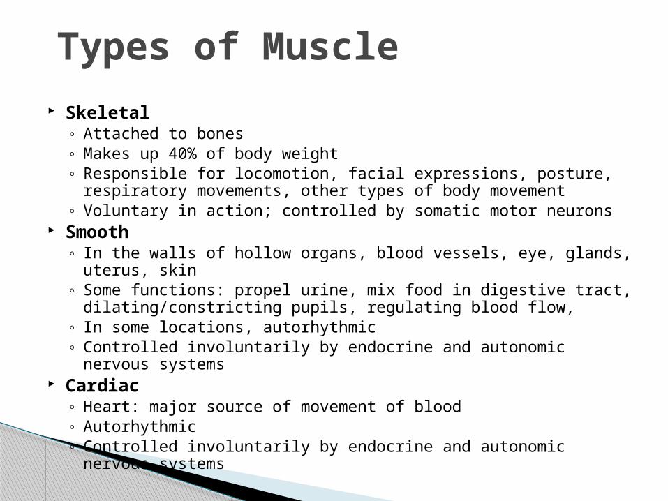
Skeletal◦ Attached to bones◦ Makes up 40% of body weight◦ Responsible for locomotion, facial expressions, posture,
respiratory movements, other types of body movement◦ Voluntary in action; controlled by somatic motor neurons
Smooth◦ In the walls of hollow organs, blood vessels, eye, glands, uterus,
skin◦ Some functions: propel urine, mix food in digestive tract,
dilating/constricting pupils, regulating blood flow, ◦ In some locations, autorhythmic◦ Controlled involuntarily by endocrine and autonomic nervous
systems Cardiac
◦ Heart: major source of movement of blood◦ Autorhythmic◦ Controlled involuntarily by endocrine and autonomic nervous
systems
Types of Muscle

Motor neurons◦ stimulate muscle fibers to contract◦ Neuron axons branch so that each muscle fiber (muscle
cell) is innervated◦ Form a neuromuscular junction (= myoneural junction)
Capillary beds surround muscle fibers◦ Muscles require large amts of energy◦ Extensive vascular network delivers necessary
oxygen and nutrients and carries away metabolic waste produced by muscle fibers
Nerve and Blood Vessel Supply

Medical Terminology: A Living Language, Fourth EditionBonnie F. Fremgen and Suzanne S. Frucht
Copyright ©2009 by Pearson Education, Inc.Upper Saddle River, New Jersey 07458
All rights reserved.
Figure 4.21 – The three types of muscles: skeletal, smooth, and cardiac.

Attached to bones Produce voluntary movement of skeleton Also referred to as striated muscle
◦ Looks striped under microscope
Skeletal Muscles
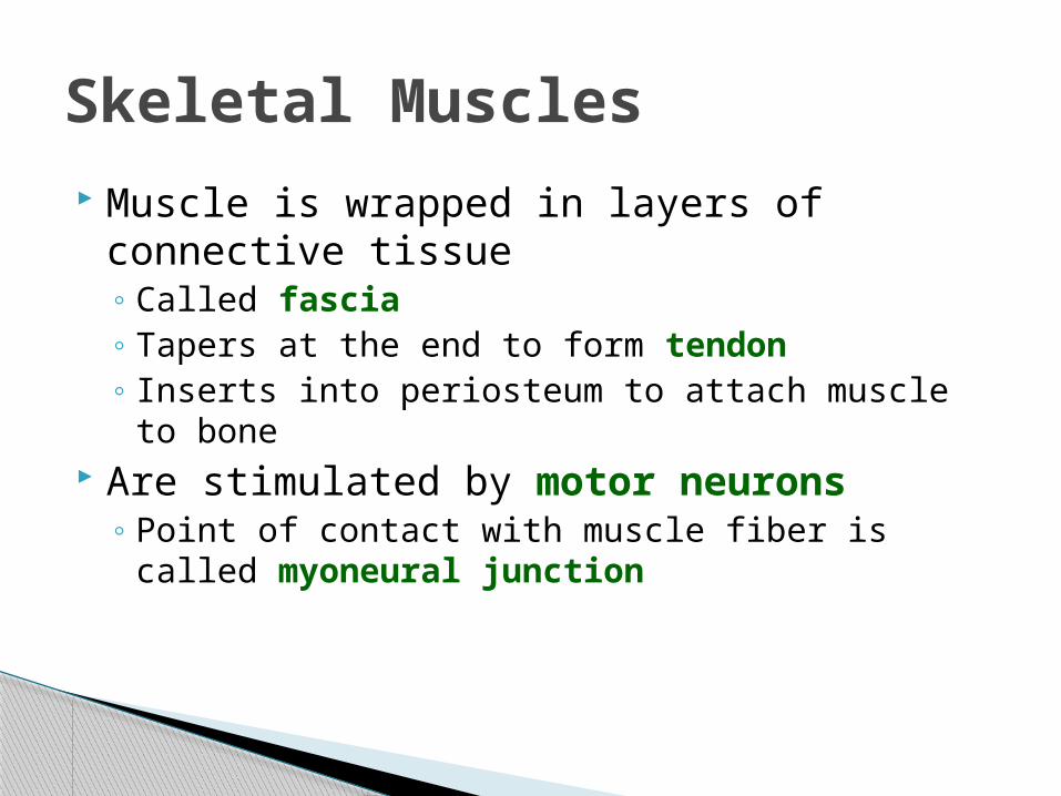
Muscle is wrapped in layers of connective tissue◦ Called fascia◦ Tapers at the end to form tendon◦ Inserts into periosteum to attach muscle to bone
Are stimulated by motor neurons ◦ Point of contact with muscle fiber is called
myoneural junction
Skeletal Muscles

Medical Terminology: A Living Language, Fourth EditionBonnie F. Fremgen and Suzanne S. Frucht
Copyright ©2009 by Pearson Education, Inc.Upper Saddle River, New Jersey 07458
All rights reserved.
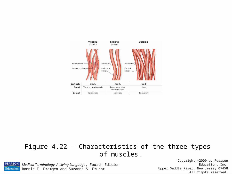
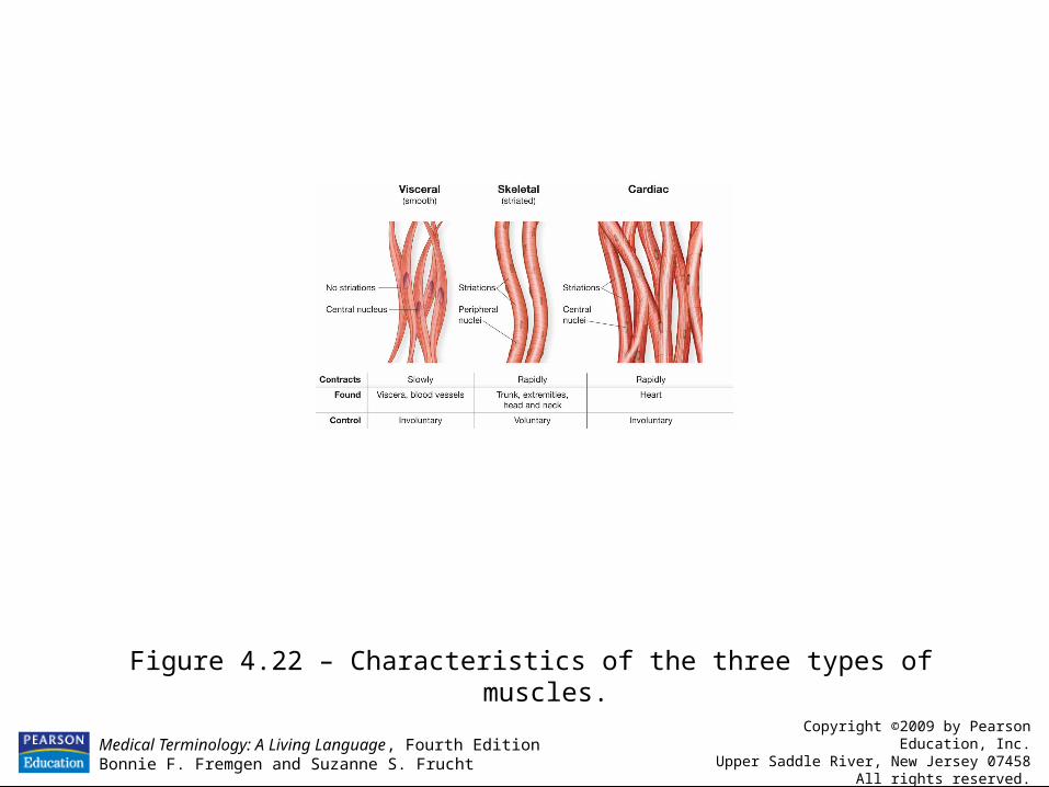
Figure 4.22 – Characteristics of the three types of muscles.

Associated with internal organs◦ Also called visceral muscle ◦ Stomach◦ Respiratory airways◦ Blood vessels
Called smooth because has no microscopic stripes
Produces involuntary movement of these organs
Smooth Muscles
Medical Terminology: A Living Language, Fourth EditionBonnie F. Fremgen and Suzanne S. Frucht
Copyright ©2009 by Pearson Education, Inc.Upper Saddle River, New Jersey 07458
All rights reserved.
Figure 4.22 – Characteristics of the three types of muscles.

Also called myocardium Makes up walls of heart Involuntary contraction of heart to pump
blood
Cardiac Muscle
Medical Terminology: A Living Language, Fourth EditionBonnie F. Fremgen and Suzanne S. Frucht
Copyright ©2009 by Pearson Education, Inc.Upper Saddle River, New Jersey 07458
All rights reserved.
Figure 4.22 – Characteristics of the three types of muscles.

Comparison of Muscle Types
Muscle Type Cardiac
FunctionMovement of bone
Walls of internal organs + in skin
LocationAttached to bone
Heart
SmoothSkeletal
Striated- light and dark bandsMany nuclei
StriatedOne or two nuclei
Characteristics
Non-striatedOne nucleus(visceral)
Long + slender BranchingShape Spindle shape
Control Mode
Beating of heart
Involuntary Involuntary
Movement of internal organs
Voluntary

Excitation Coupling Contraction Relaxation
4 Steps of Muscle Contraction

Mechanics of a Muscle Contraction
Where does stimulation occur?◦ Neuromuscular junction
How do motor neurons communicate with muscle cells?◦ Neurotransmitters (typically
acetylcholine) carryimpulse signal across the gap
What happens when a muscle cell is stimulated?◦ Calcium ions are released into the muscle
cellMyofibrils are surrounded by calcium-containing sarcoplasmic reticulum.
Neurotransmitters

Mechanics of a Muscle Contraction
What do calcium ions do?◦ Cause interaction between actin and myosin
How do actin and myosin interact?◦ Actin filaments slide over the myosin
filaments. What model explains this?
◦ Sliding Filament Model
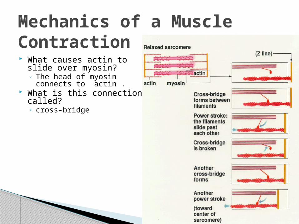
Mechanics of a Muscle Contraction What causes actin to slide
over myosin?◦ The head of myosin
connects to actin . What is this connection
called?◦ cross-bridge

Mechanics of a Muscle Contraction
What provides the energy to swivel the head of myosin? _____
How exactly does the sliding filament model work?◦ In the sliding filament model of muscle contraction,
the (thin) actin filaments[red] (that are attachedto the Z-line) slide (areactually pulled) inward along the (thick)myosin filaments [blue], and the sarcomere (measuredfrom one Z line to thenext) is shortened.
ATP

Mechanics of a Muscle Contraction
When each sarcomere becomes shorter it causes each myofibril to become shorter.
When each myofibril becomesshorter it causes the muscle fibers to become shorter
When each muscle fiber shortens the overall muscle contracts.
Sarcomere

Control of a Muscle Contraction
How long does a muscle cell remain contracted?◦ Until the release of acetylcholine
stops.